Auditory processing in visual brain areas of the early blind: evidence from event-related potentials
10
418 Electroencephalography and clinicalNeurophysiology, 86 (1993) 418-427 © 1993 Elsevier Scientific Publishers Ireland, Ltd. 0013-4649/93/$06.00 EEG 92578 Auditory processing in visual brain areas of the early blind: evidence from event-related potentials * K. Alho, T. Kujala, P. Paavilainen, H. Summala and R. N i it inen Department of Psychology, University of Helsinki, SF-O0014 Helsinki (Finland) (Accepted for publication: 11 January 1993) Summary Auditory event-related potentials (ERPs) were recorded in early blind subjects and sighted controls when they attended to stimuli delivered to a designated ear under dichotic conditions. The scalp distribution of the processing negativity (PN), the endogenous negativity elicited by attended stimuli, was in the blind posterior to that in the sighted. This suggests that posterior brain areas normally involved in vision participate in auditory selective attention in the early blind. Furthermore, occasional higher-frequency tones in the to-be-ignored ear elicited a negativity (presumably the mismatch negativity; MMN) that had a posterior scalp distribution in the blind as compared to controls. This suggests that the posterior brain areas of the blind also participate in processing of auditory stimulus changes occurring outside the focus of attention. Key words: Blind; Visual deprivation; Audition; Attention; Event-related potentials Blindness beginning at an early developmental stage modifies processing in the non-deprived sensory modalities. Early binocular deprivation increases the proportion of neurons responsive to tactile stimuli dur- ing a somatosensory task in the occipital (Brodmann's area 19) and parietal (area 7) cortices of the monkey (Hyv~irinen et al. 1981a,b; Carlson et al. 1987). Consis- tent with this, Uhl et al. (1991) found that negative DC potential shifts associated with a tactile task were larger over the occipital scalp in early blind humans than in sighted controls. Early blindness also affects auditory processing. The blind may develop an ability to use echoes in order to perceive spatial positions of objects (e.g., Strelow and Brabyn 1982). Development of such auditory abilities in the blind might be related to changes in the auditory system. For example, Ryugo et al. (1975) found that early visual or somatosensory deafferentation increases the density of dendritic spines in the auditory cortex of the rat. Furthermore, Merzenich et al. (1984) suggested that even in normal development of the auditory sys- tem, the cortical mechanisms for sound localization are Correspondence to: Kimmo Alho, Department of Psychology, P.O. Box 11, University of Helsinki, SF-00014 Helsinki (Finland). Tel.: (3580) 191 3407; Fax: (3580) 191 3443. * This research was supported by the University of Helsinki and the Academy of Finland. The authors wish to thank the anonymous referees for their constructive comments and criticism. dynamically maintained and altered throughout life. This plasticity is necessary because of changes in bin- aural localization cues which occur as a result of fac- tors such as the growth of the head and external ears during development and with hearing loss related to aging. Intracranial recording of evoked potentials in the cat has shown that early auditory deprivation leads to enhanced responsiveness of primary auditory cortex to visual stimuli (Rebillard et al. 1977). Furthermore, Neville et al. (1983) found larger visual event-related potentials (ERPs) over temporal and frontal scalp ar- eas in congenitally deaf human adults than in hearing controls. This suggests that congenital deafness causes enhanced visual processing in brain areas normally participating in audition (see also Neville and Lawson 1987). In early blind humans, the negative N1 deflection (peak at about 100 msec from stimulus onset) and the subsequent positive P2 and P3 deflections of auditory ERPs show shortened latencies and enhanced ampli- tudes (Niemeyer and Starlinger 1981; Woods et al. 1985). However, there has been no indication that cortical areas normally involved in vision participate in auditory processing in the blind. Although Wanet-De- falque et al. (1988) found higher occipital glucose metabolism (measured with positron emission tomogra- phy; PET) in early blind subjects than in blindfolded controls during auditory and tactile discrimination tasks, a similar difference was also found in a resting
-
Upload
independent -
Category
Documents
-
view
0 -
download
0
Transcript of Auditory processing in visual brain areas of the early blind: evidence from event-related potentials

418 Electroencephalography and clinical Neurophysiology, 86 (1993) 418-427 © 1993 Elsevier Scientific Publishers Ireland, Ltd. 0013-4649/93/$06.00
EEG 92578
Auditory processing in visual brain areas of the early blind: evidence from event-related potentials *
K. Alho, T. Kujala, P. Paavilainen, H. Summala and R. N i it inen Department of Psychology, University of Helsinki, SF-O0014 Helsinki (Finland)
(Accepted for publication: 11 January 1993)
Summary Auditory event-related potentials (ERPs) were recorded in early blind subjects and sighted controls when they attended to stimuli delivered to a designated ear under dichotic conditions. The scalp distribution of the processing negativity (PN), the endogenous negativity elicited by attended stimuli, was in the blind posterior to that in the sighted. This suggests that posterior brain areas normally involved in vision participate in auditory selective attention in the early blind. Furthermore, occasional higher-frequency tones in the to-be-ignored ear elicited a negativity (presumably the mismatch negativity; MMN) that had a posterior scalp distribution in the blind as compared to controls. This suggests that the posterior brain areas of the blind also participate in processing of auditory stimulus changes occurring outside the focus of attention.
Key words: Blind; Visual deprivation; Audition; Attention; Event-related potentials
Blindness beginning at an early developmental stage modifies processing in the non-deprived sensory modalities. Early binocular deprivation increases the proportion of neurons responsive to tactile stimuli dur- ing a somatosensory task in the occipital (Brodmann's area 19) and parietal (area 7) cortices of the monkey (Hyv~irinen et al. 1981a,b; Carlson et al. 1987). Consis- tent with this, Uhl et al. (1991) found that negative DC potential shifts associated with a tactile task were larger over the occipital scalp in early blind humans than in sighted controls.
Early blindness also affects auditory processing. The blind may develop an ability to use echoes in order to perceive spatial positions of objects (e.g., Strelow and Brabyn 1982). Development of such auditory abilities in the blind might be related to changes in the auditory system. For example, Ryugo et al. (1975) found that early visual or somatosensory deafferentation increases the density of dendritic spines in the auditory cortex of the rat. Furthermore, Merzenich et al. (1984) suggested that even in normal development of the auditory sys- tem, the cortical mechanisms for sound localization are
Correspondence to: Kimmo Alho, Department of Psychology, P.O. Box 11, University of Helsinki, SF-00014 Helsinki (Finland). Tel.: (3580) 191 3407; Fax: (3580) 191 3443.
* This research was supported by the University of Helsinki and the Academy of Finland. The authors wish to thank the anonymous referees for their constructive comments and criticism.
dynamically maintained and altered throughout life. This plasticity is necessary because of changes in bin- aural localization cues which occur as a result of fac- tors such as the growth of the head and external ears during development and with hearing loss related to aging.
Intracranial recording of evoked potentials in the cat has shown that early auditory deprivation leads to enhanced responsiveness of primary auditory cortex to visual stimuli (Rebillard et al. 1977). Furthermore, Neville et al. (1983) found larger visual event-related potentials (ERPs) over temporal and frontal scalp ar- eas in congenitally deaf human adults than in hearing controls. This suggests that congenital deafness causes enhanced visual processing in brain areas normally participating in audition (see also Neville and Lawson 1987).
In early blind humans, the negative N1 deflection (peak at about 100 msec from stimulus onset) and the subsequent positive P2 and P3 deflections of auditory ERPs show shortened latencies and enhanced ampli- tudes (Niemeyer and Starlinger 1981; Woods et al. 1985). However, there has been no indication that cortical areas normally involved in vision participate in auditory processing in the blind. Although Wanet-De- falque et al. (1988) found higher occipital glucose metabolism (measured with positron emission tomogra- phy; PET) in early blind subjects than in blindfolded controls during auditory and tactile discrimination tasks, a similar difference was also found in a resting
AUDITORY ERPs IN BLIND 419
condition. Therefore, the differences in occipital metabolism observed during the discrimination tasks between the blind and sighted appear to have been caused by a general enhancement of occipital glucose metabolism associated with blindness.
The present purpose was to determine whether scalp distributions of auditory ERPs suggest participation of visual cortex in auditory attention in early blind sub- jects. To this end, the processing negativity (PN), the principal ERP index of auditory selective attention (N~i~it~inen 1982, 1990, 1992), was studied in early blind subjects and in sighted controls. Subjects attended to tone pips delivered to a designated ear, in order to detect occasional deviant (higher) tones occurring among these tones, and ignored tones delivered to the opposite ear. PN causes a negative displacement of the ERP to attended stimuli in relation to that elicited by unattended stimuli. PN consists of at least two consec- utive, partially overlapping, components. The earlier PN component commences at the time zone of the N1 deflection and reaches its maximum amplitude at fronto-central scalp sites. This PN component is gener- ated in the auditory cortex (see also Hari et al. 1989), presumably by a neural matching process selecting attended stimuli for further processing on the basis of their physical features (N~i~it~inen 1982, 1990, 1992; Alho et al. 1986b, 1987a,b, 1989). The later PN compo- nent peaks at around 300-400 msec from stimulus onset and might be generated by a frontal source (Giard et al. 1988) presumably associated with control of attention (N~i~it~inen 1982, 1990, 1992; N~i~it~inen and Picton 1987).
ERPs to the higher tones infrequently occurring among attended and unattended tones were also exam- ined. Such deviant stimuli elicit the mismatch negativ- ity (MMN; N~i~it~inen et al. 1978; for reviews, see N~iat~inen 1990, 1992), a change-specific ERP compo- nent, which overlaps with the N1 and subsequent P2 components. The change-detection process generating MMN appears to be automatic since MMN is elicited even by deviant stimuli occurring among unattended stimuli (N~i~it~inen et al. 1978; Sams et al. 1985; Alho et al. 1989, 1992; Woods et al. 1992; see, however, Woldorff et al. 1991, and for a reply, Na~it~inen 1991). MMN consists of at least two temporally overlapping components, one generated in the supratemporal audi- tory cortex (e.g., Hari et al. 1984; Scherg et al. 1989; Javitt et al. 1991; Paavilainen et al. 1991) and the other in the frontal cortex (Giard et al. 1990; Na~it~inen and Michie 1979). While the supratemporal MMN compo- nent is presumably generated by a neural change-de- tection process, the process generating the frontal MMN component might underlie involuntary orienting of attention towards this change (N~i~it~inen 1990, 1992; Alho 1992). The fronto-centrally maximal positive P3a component often following MMN to deviant stimuli of
unattended auditory input is probably associated with such involuntary orienting of attention (e.g., Naat~inen et al. 1982; N~i~it~inen and Gaillard 1983; Sams et al. 1985). When a deviant stimulus occurs in an attended input, MMN is partially overlapped by a negative N2b component and by the early phase of a parietal P3b component (N~i~it~inen et al. 1982, 1986).
In sum, while PN is generated by brain mechanisms of selective attention, ERP components elicited by stimulus change in an unattended auditory input, espe- cially MMN and P3a, may reflect mechanisms of auto- matic auditory processing and involuntary attention. The purpose of the present study was to examine whether in the early blind these ERP components differ from those in the sighted. Possible group differ- ences might result from differential development of brain mechanisms of auditory processing.
Methods
Subjects There were 9 blind (aged 20-29 years) and 9 nor-
mally sighted (aged 19-25 years) subjects, 3 males and 6 females in each group. All subjects were students and participated voluntarily to the experiment. None of them had previously served as subjects in similar exper- iments. All blind subjects had a diagnosed peripheral deficit which caused early blindness beginning within the first two years of life. Four of them had a retinopa- thy of prematurity, one had an inherited retinal degen- eration, one's peripheral blindness was caused by the mother's rubella during pregnancy, one's eyes had been ablated because of retinal cancer, and one had suf- fered congenital atrophy of the optic tracts. For one subject, the cause of blindness, diagnosed as periph- eral, was unknown. Five blind subjects reported that they could still differentiate between dark and light. In addition, one subject had been capable of doing so up to the age of 5 years and another up to the age of 2 years. According to their own reports, none had any contour or pattern vision, and none could discriminate the direction of light. In addition, seven of the blind subjects were presented with flash stimuli in another study (Kujala et al. 1993) and no visual ERPs were found, while in sighted controls the flashes elicited sizeable ERPs.
Experimental procedure The experiment was carried out in an electrically
and acoustically shielded, dimly lit room. Monaural tone pips (sine-wave bursts, duration 60 msec including 10 msec rise and fall times, intensity 75 dB SPL) were presented through headphones in random order to the left and right ears at a constant rate of 1 tone/610 msec in blocks of 500 stimuli. There were two types of

420 K. ALHO ET AL.
tone in each ear: 600 Hz standard tones (probability for each ear 0.45) and 650 Hz deviant tones (probabil- ity for each ear 0.05). Subjects were instructed to attend to the tones presented to the designated ear (2 blocks in each condition) and to count silently the deviant tones occurring in that ear. The order of the conditions was counterbalanced within each group. Subjects were instructed to avoid eye movements and blinking during the attention tasks.
EEG recording and analysis The E E G (0.1-100 Hz, - 3 dB points)was recorded
with Ag/AgC1 electrodes at 19 scalp sites. In addition, two E OG electrodes were attached laterally to the outer canthi of the left and right eyes in order to monitor voltage changes caused by horizontal eye movements. Activity caused by vertical eye movements or blinks was recorded with a frontopolar E E G elec- trode (Fpz). All E E G and EOG electrodes were re- ferred to an electrode attached to the nose.
The E E G analysis epochs of 420 msec (sampling rate 250 Hz) began 50 msec before each stimulus onset. The first 10 epochs of each block, as well as epochs contaminated by blinks, eye movements, muscle activ- ity, or other extracerebral artifacts (voltage variation during an epoch exceeding 150 /xV at any electrode) were automatically rejected from averaging. Frequen- cies higher than 30 Hz were digitally filtered out (FFT filter) from the ERPs.
To study PN, attended-unattended difference waves were formed for each subject by subtracting, separately for each ear, ERPs to standard tones when they were unat tended from ERPs to the same tones when at- tended. In addition, to study ERP components elicited by deviant tones, deviant-standard difference waves were formed, separately for the at tended and unat- tended tones, by subtracting ERPs to standard tones from ERPs to deviant tones.
For statistical comparisons, ERP and difference wave amplitudes were measured for each subject, in reference to the 50 msec prestimulus baseline, as mean voltages over consecutive 50 msec periods within 350 msec from stimulus onset. In addition, N1 peak laten- cies were determined at Cz and N1 amplitudes at each electrode were measured at these peak latencies.
To compare scalp distributions of ERP and differ- ence wave amplitudes between the two groups, these amplitudes were normalized. This was done separately for each subject and each measurement period by dividing the amplitude at each electrode by the square root of the sum of the squared amplitudes at the electrodes included in the analysis (McCarthy and Wood 1985).
In the statistical analysis of ERP and performance data, analyses of variance included in the BMDP Sta- tistical Software (Dixon et al. 1988) were used. In
Results, the reported significance levels for the F val- ues are corrected with a Greenhouse-Geisser correc- tion where appropriate.
Results
Performance The reported number of deviant target tones dif-
fered from the correct number on average by 10.0% and 12.6% in the blind and sighted groups, respec- tively. According to an analysis of variance, the differ- ence between the two groups was insignificant.
ERPs to standard tones At the top of Fig. 1, grand-average ERPs to at-
tended and unattended standard tones are superim- posed separately for the two groups. The N1 deflection to the unattended standard tones tended to be larger and somewhat later in the sighted (mean peak ampli- tude at Cz -3 .9 /~V; mean peak latency 94 msec) than in the blind ( - 2 . 4 / z V , 90 msec), but these differences were statistically insignificant. Moreover, an analysis of variance (factors: Group, Ear, Electrode) for normal- ized (see Methods) unattended-tone N1 amplitudes (measured at the N1 peak latency determined at Cz) showed no differences between the two groups in the N1 midline distribution (electrodes Fpz, Fz, Cz, Pz, Oz) or in the N1 distribution on a tilted coronal line crossing the approximate locations of left and right auditory cortices (electrodes Mc, Tc, FCc, Fz, FCi, Ti, Mi; see Fig. 1, top).
In both groups, ERPs to at tended standard tones were negatively displaced in relation to those to unat- tended standard tones. These displacements, presum- ably caused by PN, are shown by at tended-unattended difference waves at the bottom of Fig. 1. In both groups, these displacements were largest at fronto- central scalp sites. Analyses of variance (factors: Group, Attention, Ear) for the standard-tone ERP amplitudes at Fz indicated significant effects of attention over consecutive 50 msec periods between 100 and 350 msec (F (1, 16) varied between 23.67 and 48.10, P < 0.001 in all cases).
At Fz no significant differences in the PN amplitude (measured from attended-unattended difference waves) between the two groups were found, whereas at the posterior scalp sites, this amplitude was larger for the blind than for the sighted (Fig. 1, bottom). In the blind, prominent PNs were observed even at occipital elec- trodes (at Oz, mean PN amplitudes measured over the consecutive 50 msec periods between 100 and 350 msec varied from - 0.5/zV to - 1.3 tzV), while in the sighted, the occipital PN amplitudes approximated 0 /zV. A comparison of PN amplitudes at occipital electrodes

AUDITORY ERPs IN BLIND 421
(TOc, Oc, Oz, Oi, TOi; see Fig. 1, bo t tom) indicated
significant differences be tween the two groups at 250- 300 msec ( F (1, 1 6 ) = 4.53, P < 0.05) and 300-350 msec F (1, 1 6 ) = 4.83, P < 0.05) and near ly significant differences at 150-200 msec ( F (1, 16) = 3.11, P < 0.10) and 200-250 msec ( F (1, 16) = 3.89, P < 0.07).
Thus, in the bl ind, PN was dis t r ibuted posteriorly to that in the sighted. This was conf i rmed by analyses of
var iance (factors: Group , Ear, Electrode) for normal-
ized PN ampl i tudes at the midl ine (Fpz, Fz, Cz, Pz, Oz). These analyses indicated significant G r oup x Elect rode in teract ions over all consecutive 50 msec periods be tween 100 and 350 msec from st imulus onset ( F (4, 64) varying from 4.20 to 5.66, P < 0.05 in all cases). In contrast , no significant G r oup × Electrode in teract ions were found in analogous analyses of vari-
M i
ERPs TO - - A T T E N D E D .... . . . . U N A T T E N D E D
F C i ~ _ - ~
' ..."....~ . .:.,..,......
c~ A ~ Mo ..........
~....- ..,
\ / \ .& p• Pz P c ~
A " ~ POi ~ ' " - " - " P O c ............ . ~ ............. ~ ..............
TOi Oi . - .,.... O z T O c . ~ ~ " " '-....-
• .,... ......., • ..... . . .
- 2 p V ] ~
0 300ms
STANDARDS
.,••_•GHTED • . . . . . . . . . . . . . . " . ~
, . . . .
"......
DIFFERENCE" ATT-UNATT B L I N D
. . . . . . . . S I G H T E D
\ /
. . . .. . . . . - - . . , . . . , . . . .
. . . . . .~ . . . . . . . , . . , . . . . . . . . , . . . . . . . , .
/
Fig. 1. Top: grand-average ERPs at different scalp sites to attended (solid lines) and unattended (dashed lines) standard tones in the blind (left; n = 9) and sighted (right; n = 9). The ERPs were averaged across the left and right ear standard tones. Before combining the data for the left and right ear stimuli, the ERPs to the right ear stimuli over the lateral scalp sites were mirrored to the opposite hemisphere. Thus, the ERPs at scalp sites contralateral to the stimulated ear are shown over the right hemisphere and the ERPs at scalp sites ipsilateral to the stimulated ear are shown over the left hemisphere (stimulus onset at 0 msec; c = contralateral; i = ipsilateral; HEOGc and HEOGi electrodes located at the outer canthi of the eyes, used for monitoring EOG changes caused by horizontal eye movements; Mc and Mi are electrodes at the mastoids; FCc/FCi and Tc/Ti were equidistantly placed between Fz-Mc or Fz-Mi; Pc/Pi are the sites P3/P4; Oc/Oi are the sites O1/O2; OPc/OPi located halfway between Oz and P3/P4; TOc/TOi located halfway between O1-T5 or O2-T6). Bottom: difference waves for the blind (solid lines) and sighted (dashed lines) formed by subtracting the grand-average ERPs to standard tones when unattended from the ERPs to the same tones when
attended.

422 K. ALHO ET AL.
ance for normalized PN amplitudes.on a tilted coronal line (electrodes Mc, Tc, FCc, Fz, FCi, Ti, Mi; see Fig. 1, bottom)•
At most scalp sites, the attended-tone ERPs, as well as the unattended-tone ERPs, were more positive in the blind than in the sighted (Fig. 1, top). At the midline electrodes (Fpz, Fz, Cz, Pz, Oz), this group difference was significant for the attended standard tones at 100-200 msec (100-150 msec: F (1, 16) = 7.01, P < 0.02; 150-200 msec: F (1, 16) = 6.17, P < 0•025) and for the unattended standard tones at 50-200 msec (50-100 msec: F (], 16) = 7.08, P < 0.02; 100-150 msec: F (1, 16) --- 7.72, P < 0.015; 150-200 msec: F (1, 16) = 4.75, P < 0.05). No significant Group × Electrode in-
teractions were found in analyses of variance (factors: Group, Ear, Electrode) for normalized midline ampli- tudes of the unattended-standard ERPs at consecutive 50 msec periods between 50-350 msec, whereas analo- gous analyses for attended-standard ERPs indicated a significant Group × Electrode interaction at 300-350 msec (F (4, 64)= 3.19, P < 0.02) and a nearly signifi- cant Group × Electrode interaction at 200-250 msec (F (4, 64) = 2.23, P < 0.08). These interactions suggest that the posterior distribution of the attended-un- attended difference waves in the blind was indeed due to an effect of blindness on ERPs to attended tones rather than on ERPs to unattended tones. Thus, these analyses also support the proposal that the PN elicited
ERPs TO UNATTENDED TONES ........ STANDARD
DEVIANT BLIND
• • ; : . . . . . . . . .
-2pvL_ o 360ms
~ . ~ . ~ . ~ , SIGHTED
: /
\ /
DIFFERENCE: DEV-STAND BLIND
........ SIGHTED
A ,
\ /
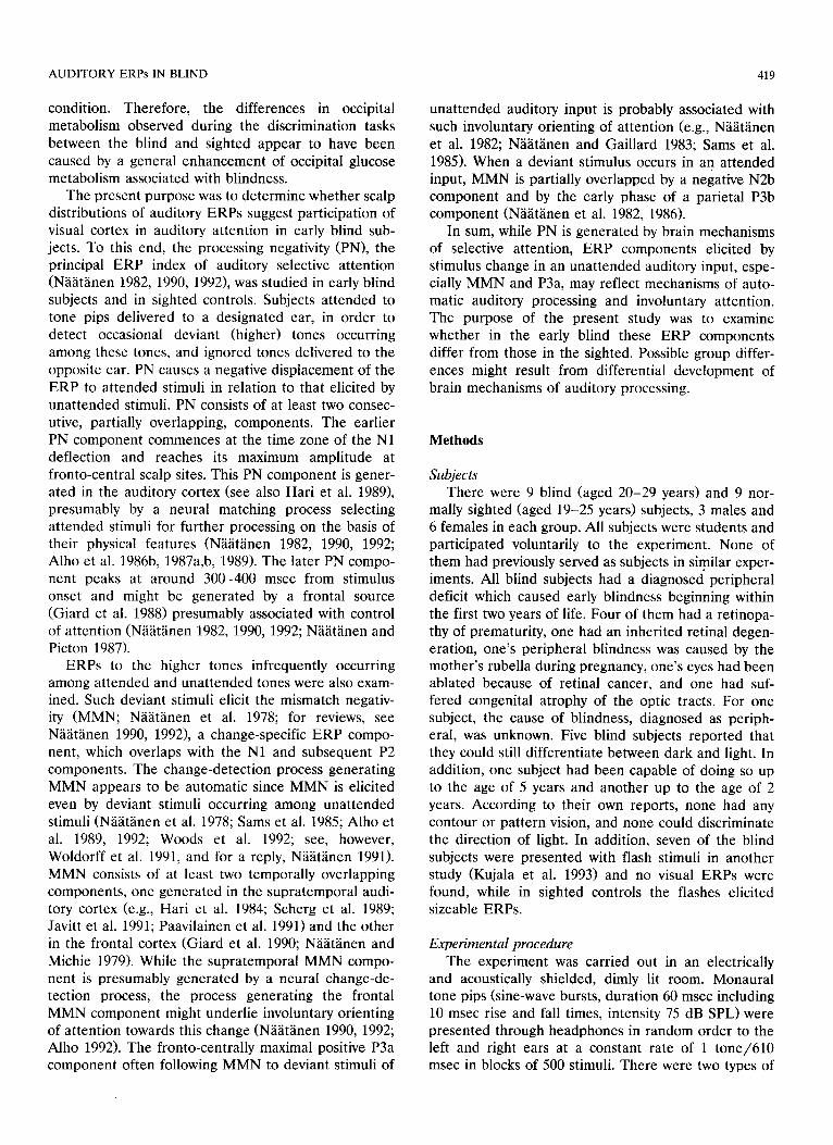
Fig. 2. Top: grand-average ERPs in the blind and sighted to standard (dashed lines) and deviant (solid lines) tones of unattended stimulus sequence. (For further details, see Fig. 1.) Bottom: difference waves for the blind (solid lines) and sighted (dashed lines) groups formed by
subtracting the grand-average ERPs to standard tones of unattended stimulus sequence from the ERPs to deviant tones of the same sequence.

AUDITORY ERPs IN BLIND 423
by the attended tones was posteriorly distributed in the blind.
ERPs to deviant tones For the unattended ear, deviant-tone ERPs were
negatively displaced in relation to standard-tone ERPs in both groups (Fig. 2, top). This negative displacement was largest at fronto-central scalp sites, as shown by the deviant-standard difference waves at the bottom of Fig. 2. Analyses of variance (factors: Group, Ear, Stim- ulus) for unattended-ear ERP amplitudes at Fz indi- cated that this negative displacement was significant at 100-250 msec (100-150 msec: F (1, 16)= 14.09, P <
0.02; 150-200 msec: F (1, 16) = 16.72, P < 0.001; 200- 250 msec (F (1, 16)= 17.82, P < 0.001) and, further, that there were no significant group differences in its amplitude. However, at posterior scalp sites, a promi- nent negativity was elicited by unattended-ear deviant tones in the blind but not in the sighted, as shown by the difference waves in Fig. 2. Analyses of variance for difference-wave amplitudes at occipital electrodes (TOc, Oc, Oz, Oi, TOi; see Fig. 2, bottom) indicated a significant group difference at 200-250 msec (F (1, 16) = 6.63, P < 0.025), where the mean amplitudes at Oz were - 1 . 3 / z V for the blind and + 0.6/xV for the sighted. The posterior scalp distribution of the
ERPs TO ....... STANDARD
DEVIANT
ATTENDED TONES
GHTED BLIND
HEOGi ~ HEOGc
Fz- "F \ FCi Cc."
Ti " Tc .. _ ~ .
0 300ms
\ /
DIFFERENCE: DEV-STAND - - BLIND ....... S IGHTED
\
Fig. 3. Top: grand-average ERPs in the blind and sighted to standard tones (dashed lines) and deviant tones (targets; solid lines) of attended stimulus sequence. (For further details, see Fig. 1.) Bottom: difference waves for the blind (solid lines) and sighted (dashed lines) groups formed by subtracting the grand-average ERPs to standard tones of attended stimulus sequence from the ERPs to deviant tones of the same sequence.

424 K. ALHO ET AL.
deviant-tone negativity in the blind was confirmed by a significant Group × Electrode interaction at 200-250 msec (F (4, 64)= 3.66, P < 0.04) in an analysis of variance (factors: Group, Ear, Electrode) for normal- ized difference-wave amplitudes at the midline (Fpz, Fz, Cz, Pz, Oz).
Furthermore, the negativity to unattended-ear de- viant tones tended to be larger in the blind than sighted at lateral scalp sites over the auditory cortex (Fig. 2, bottom), although this effect, tested at elec- trodes FCc, TCc, Mc, FCi, Ti, and Mi, did not quite reach statistical significance (200-250 msec: F (1, 16) = 3.95, P < 0.065). This group difference was larger over the hemisphere contralateral (electrodes FCc, Tc, Mc) than ipsilateral (FCi, Ti, Mi) to the stimulated ear, as indicated by a significant Group x Hemisphere in- teraction (200-250 msec: F (1, 16) = 5.05, P < 0.04; 250-300 msec: F (1, 16) = 7.08, P < 0.02). However, the hemispheric asymmetry may have been caused by small, unrejected (see Methods) eye movement arti- facts in the blind seen in Fig. 2 especially in H E O G channels used for monitoring horizontal eye move- ments.
Interestingly, at latencies shorter than 200 msec the negativity to unat tended-ear deviant tones tended to reverse its polarity at the mastoid electrodes (Mc and Mi), as indicated by a positivity in deviant-standard difference waves at these electrode sites (Fig. 2, bot- tom). At longer latencies, a negativity was observed at the mastoids in the blind but not in the sighted.
As seen in Fig. 2, the negativity to unat tended-ear deviant tones was followed by the onset of a P3 positiv- ity. This P3 was larger in the blind than in the sighted over frontal and central, but not over posterior, scalp sites. This difference in the P3 scalp distribution was confirmed by an analysis of variance for normalized difference-wave amplitudes at the midline (Fpz, Fz, Cz, Pz, Oz) at 300-350 msec (F (4, 64)= 3.11, P < 0.05). Unfortunately, a more detailed examination of P3 am- plitude and peak latency was not possible because in many subjects, P3 did not peak during the post-stimu- lus analysis period of 370 msec.
Fig. 3 presents ERPs to standard and deviant (target) tones and deviant-standard difference waves for the attended ear. These difference waves show a small deviant-tone negativity followed by a large P3 deflec- tion. In the sighted, P3 was largest at posterior scalp sites, whereas in the blind, P3 was widely distributed and had large amplitudes even at anterior scalp sites. This group difference in the P3 distribution was con- firmed by an analysis of variance for normalized differ- ence wave amplitudes at the midline (Fpz, Fz, Cz, Pz, Oz) at 300-350 msec (F (4, 64) = 3.09, P < 0.05). Again, P3 could not be studied in more detail, because of the 370 msec analysis period which was too short for this purpose.
The negativity preceding the P3 to deviant tones was small (Fig. 3, bottom) and did not reach statistical significance in either group at any electrode. Further- more, no significant group difference in the amplitude of this negativity was found, although it tended to be larger in the blind than sighted (Cz, 150-200 msec: blind - 1 . 6 pN; sighted +0.9 ~V; F (1, 16)=2.53, P < 0.14). As revealed by Fig. 3, the small amplitude of this negativity may have been caused in both groups by an overlap of an early P3 onset.
Discussion
The present results show that in the early blind subjects, the processing negativity (PN) elicited by at- tended tones had a scalp distribution posterior to that in the sighted controls. In the blind subjects, a large PN was even observed at occipital scalp sites (Fig. 1, bottom), suggesting that their PN might get a contribu- tion, in addition to auditory and possibly frontal cor- tices (N~i~it~inen and Michie 1979; N~it~inen t982, 1990; Giard et al. 1988), from occipital or parietal cortices which normally participate in visual processing. Fur- thermore, this group difference in the PN scalp distri- bution was observed over a wide latency range (100-350 msec from stimulus). Thus, in the blind, both the earlier and later auditory PN components may be in part generated in the posterior brain areas. Also a recent dichotic selective-attention study by Simpson and his colleagues (G.V. Simpson, Albert Einstein Col- lege of Medicine, personal communication, March 1992) suggests that in congenitally blind subjects, PN has a scalp distribution posterior to that in sighted controls.
In the present study, blindness had also other ef- fects on the ERPs to standard tones. Both for the attended and unattended standard tones, the ERPs in the blind were significantly more positive than those in the sighted (Fig. 1, top) at 100-200 msec from stimulus onset (for unattended standard tones, this effect was seen even at 50-100 msec). An enhancement of the exogenous P2 component in the blind might contribute to this positivity, especially at 150-200 msec (cf., Niemeyer and Starlinger 1981; Woods et al. 1985), but cannot solely account for it because it was significant also at shorter latencies. Because this positivity was elicited both by the attended and unattended standard tones and because no significant effects of blindness on the midline distributions of attended-standard or unat- tended-standard ERPs were observed at the latencies where this positivity was significant (50-200 msec from stimulus onset), this positivity cannot explain the poste- rior distribution of the at tended-unattended difference waves in the blind (Fig. 1, bottom). However, at longer

AUDITORY ERPs IN BLIND 425
latencies there was a significant effect of blindness on the midline distribution of ERPs to the attended stan- dard tones but not on the distribution of ERPs to the unattended standard tones. Also, these findings sup- port the present proposal that, in the blind, the poste- rior distribution of the negativity indicated by the at- tended-unattended difference waves was indeed caused by a posteriorly distributed PN elicited by the attended stimuli.
Deviant tones occurring in the unattended stimulus sequence elicited negatively displaced ERPs in relation to standard-tone ERPs (Fig. 2). In the sighted, this effect was largest at frontal scalp sites and was presum- ably caused by the mismatch negativity (MMN) elicited by deviant tones (N~i~it~inen et al. 1978). In the blind, the negativity to unattended-ear deviant tones was of considerable size also over posterior scalp sites (Fig. 2, bottom). This suggests that in them, posterior brain areas normally involved in vision might participate in detecting changes occurring in an unattended auditory input.
In the blind, the negativity to unattended-ear de- viant tones tended to be enhanced also at lateral scalp sites (Fig. 2, bottom). This suggests that early blindness might enhance processing of auditory stimulus changes also in auditory cortex. Although artifacts caused by small (unrejected; see Methods) horizontal eye move- ments in the blind may have contributed to this group difference they cannot account for the whole differ- ence, because the group difference was present even over the hemisphere ipsilateral to the stimulated ear, where the eye movement artifact of the blind caused a positivity (Fig. 2, bottom). Furthermore, potentials caused by eye movements are strongly attenuated at long distances from the eyes (e.g., Hillyard and Galam- bos 1970) and cannot therefore explain the enhanced negativity to unattended-ear deviant tones in the poste- rior scalp areas of the blind.
In the blind, the posterior negativity to the unat- tended-ear deviant tones peaked later than the frontally recorded MMN to these tones (Fig. 2, bottom). This posterior negativity coincided with a negativity which was observed in the blind at the temporal scalp sites and which did not invert in polarity at the mastoids. The polarity reversal of MMN observed in both groups at shorter latencies is consistent with the previous findings suggesting that the MMN partly originates from supratemporal auditory cortex (e.g., Hari et al. 1984; Alho et al. 1986a; Scherg et al. 1989; Javitt et al. 1991; Paavilainen et al. 1991). However, MMN proba- bly also has a frontal subcomponent (Giard et al. 1990) and might even have a subcomponent generated in the auditory association areas on the lateral surface of the temporal lobe (Paavilainen et al. 1991). The deviant- tone negativity observed in the blind over the temporal scalp might be the temporal MMN subcomponent of
Paavilainen et al. (1991). Judging from its large ampli- tudes at posterior scalp sites, parietal or occipital brain areas might also contribute to this MMN subcompo- nent in the blind.
Another possibility is that the posterior distribution of the deviant-tone negativity in the blind was caused by a posteriorly distributed N2b component overlap- ping with the MMN. In normal subjects, MMN is usually followed and partly overlapped by the N2b component in ERPs to deviant stimuli occurring in an attended stimulus sequence (NS~it~nen et al. 1982; Sams et al. 1985, 1990; Alho et al. 1990; N~i~itSnen 1990; Novak et al. 1990). In a parallel experiment (Kujala et al. 1992), the N2b component elicited in early blind subjects by deviant stimuli in an auditory discrimina- tion task appeared to be distributed posteriorly to that of sighted controls. Simpson and his colleagues (G.V. Simpson, Albert Einstein College of Medicine, per- sonal communication, March 1992) have recently ob- tained similar results. If the present posterior distribu- tion of the negativity elicited by unattended-ear de- viant tones in the blind was indeed caused by a posteri- orly distributed N2b component overlapping with the MMN, this might indicate that changes in unattended auditory stimuli cause an attention shift, and therefore elicit the N2b more easily in the blind than in the sighted. Low thresholds for involuntary orienting of attention may have developed in the blind because of their increased dependence on auditory stimuli.
Unfortunately, in the present study, the effects of early blindness on MMN and N2b to target stimuli in the attended input were difficult to estimate because P3 components apparently overlapped with these nega- tive components in the target ERPs (Fig. 3). Interest- ingly, in these ERPs, as well as in the ERPs to unat- tended-ear deviant tones (Fig. 2), P3 was more frontally distributed in the blind than in the sighted. This sug- gests a larger fronto-centrally dominant P3a compo- nent (Squires et al. 1975; N~i~it~inen et al. 1982; Sams et al. 1985) in the blind. Also this finding might be related to enhanced orienting to auditory stimulus changes in the blind.
Magnetic resonance imaging (MRI) and positron emission tomography (PET) have indicated no macroanatomical or functional degeneration of occipi- tal cortex in the early blind (Phelps et al. 1981; Wanet- Defalque et al. 1988). These findings contradict the possibility that degeneration of occipital cortex in the blind had changed the orientation of ERP sources in auditory cortex so that this would have resulted in large PN and MMN (or N2b) amplitudes at posterior scalp areas. This possibility is also contradicted by the present result that the N1 deflection (mainly originat- ing from auditory cortex at a high stimulation rate; N~i~it~inen and Picton 1987) was similarly distributed over the scalp in the blind and sighted.

426 K. ALHO ET AL.
In conclusion, the present data, together with previ- ous ERP studies of early sensory deprivation in hu- mans (Niemeyer and Starlinger 1981; Neville et al. 1983; Woods et al. 1985; Neville and Lawson 1987; Uhl et al. 1991; Kujala et al. 1992), show that plastic changes occurring in non-deprived modalities may be studied with scalp-recorded ERPs. The present ERP data sug- gest that cortical areas normally involved in vision might participate in auditory selective and involuntary attention in the early blind. Further research with more elaborated methods for localizing cortical sources of ERPs associated with auditory processing (cf., Hari et al. 1984, 1989; Giard et al. 1988, 1990; Scherg et al. 1989) is needed to confirm that the posterior scalp distributions of PN and the deviant-tone negativity (MMN or N2b) observed in the early blind are indeed caused by an involvement of occipital or parietal corti- cal areas in auditory processing.
References
Alho, K. Selective attention in auditory processing as reflected by event-related brain potentials. Psychophysiology, 1992, 29: 247- 263.
Alho, K., Paavilainen, P., Reinikainen, K., Sams, M. and Nhhthnen, R. Separability of different negative components of the event-re- lated brain potential associated with auditory stimulus processing. Psychophysiology, 1986a, 23: 613-623.
Alho, K., Sams, M., Paavilainen, P. and Niiiit~inen, R. Small pitch separation and the selective-attention effect on the ERP. Psy- chophysiology, 1986b, 23: 186-197.
Alho, K., Donauer, N., Paavilainen, P., Reinikainen, K., Sams, M. and Nhhthnen, R. Stimulus selection during auditory spatial attention as expressed by event-related potentials. Biol. Psychol., 1987a, 24: 153-162.
Alho, K., T6tt61ii, K., Reinikainen, K., Sams, M. and N~iiit~inen, R. Brain mechanisms of selective listening reflected by event-related potentials. Electroenceph. clin. Neurophysiol., 1987b, 68: 458- 470.
Alho, K., Sams, M., Paavilainen, P., Reinikainen, K. and Nhhthnen, R. Event-related brain potentials reflecting processing of relevant and irrelevant stimuli during selective listening. Psychophysiology, 1989, 26: 514-528.
Alho, K., Lavikainen, J., Reinikainen, K., Sams, M. and Niiiithnen, R. Event-related brain potentials in selective listening to frequent and rare stimuli. Psychophysiology, 1990, 27: 73-86.
Alho, K., Woods, D.L., Algazi, A. and Nhhthnen, R. lntermodal selective attention. II. Effects of attentional load on processing of auditory and visual stimuli in central space. Electroenceph. olin. Neurophysiol., 1992, 82: 356-368.
Carlson, S., Pertovaara, A. and Tanila, H. Late effects of early binocular visual deprivation on the function of Brodmann's area 7 of monkeys (Macaca arctoides). Dev. Brain Res., 1987, 33: 101-111.
Dehay, C., Kennedy, H. and Bullier, J. Characterization of transient cortical projections from auditory, somatosensory, and motor cortices to visual areas 17, 18, and 19 in the kitten. J. Comp. Neurol., 1988, 272: 68-89.
Dixon, W.J., Brown, M.B., Engelman, L., Hill, M.A. and Jennrich, R.I. (Eds.). BMDP Statistical Software Manual. University of California Press, Berkeley, CA, 1988.
Giard, M.H., Perrin, F., Pernier, J. and Peronnet, F. Several atten- tion-related wave forms in auditory areas: a topographic study. Electroenceph. din. Neurophysiol., 1988, 69: 371-384.
Giard, M.H., Perrin, F., Pernier, J. and Bouchet, P. Brain generators implicated in the processing of auditory stimulus deviance: a topographic ERP study. Psychophysiology, 1990, 27: 627-640.
Hari, R., H~im~iliiinen, M., Ilmoniemi, R., Kaukoranta, E., Reinikainen, K., Salminen, J., Alho, K., Niiiitiinen, R. and Sams, M. Responses of the primary auditory cortex to pitch changes in a sequence of tone pips: neuromagnetic recordings in man. Neu- rosci. Lett., 1984, 50: 127-132.
Hari, R., Hiimiil~iinen, M., Kaukoranta, E., Miikelii, J., Joutsiniemi, S.L. and Tiihonen, J. Selective listening modifies activity of the human auditory cortex. Exp. Brain Res., 1989, 74: 463-470.
Hillyard, S.A. and Galambos, R. Eye movement artifact in the CNV. Electroenceph. clin. Neurophysiol., 1970, 28: 173-182.
Hyv~irinen, J., Carlson, S. and Hyv§rinen, L. Early visual deprivation alters modality of neuronal responses in area 19 of monkey cortex. Neurosci. Lett., 1981a, 26: 239-243.
Hyvhrinen, J., Hyv~irinen, L. and Linnankoski, I. Modification of parietal association cortex and functional blindness after binocu- lar deprivation in young monkeYs. Exp. Brain Res., 1981b, 42: 1-8.
Javitt, D.C., Schroeder, C.E., Arezzo, J. and Vaughan, Jr., H.G. Selective inhibition of "processing-contingent" auditory event-re- lated potentials by the phencyclidine (PCP)-like agent MK-801. Electroenceph. clin. Neuropbysiol., 1989, 79: 65P.
Kujala, T., Alho, K., Paavilainen, P., Summala, H. and Niihtiinen, R. Neural plasticity in processing sound location by the early blind: an event-related potential study. Electroenceph. clin. Neurophys- iol., 1992, 84: 469-472.
Kujala, T., Kekoni, J., Reinikainen, K., Hhmiihiinen, H., N'ahthnen, R. and Alho, K. Auditory and somatosensory processing in the early blind. 1993, in preparation.
McCarthy, G. and Wood, C.C. Scalp distributions of event-related potentials: an ambiguity associated with analysis of variance mod- els. Electroenceph. clin. Neurophysiol., 1985, 62: 203-208.
Merzenich, M.M., Jenkins, W.M. and Middlebrooks, J.C. Observa- tions and hypotheses on special organizational features of the central auditory nervous system. In: G.M. Edelman, W.E. Gall and W.M. Cowan (Eds.), Dynamic Aspects of Neocortical Func- tion. Wiley, New York, 1984: 397-424.
Nh~thnen, R. Processing negativity: an ew~ked-potential reflection of selective attention. Psychol. Bull., 1982, 92: 605-640.
Nhhthnen, R. The role of attention in auditory information process- ing as revealed by event-related brain potentials. Behav. Brain Sci., 1990, 13: 201-288.
Nh~ithnen, R. Mismatch negativity (MMN) outside strong attentional focus: a commentary on Woldorff et al. (1991). Psychophysiology, 1991, 28: 478-484.
Nii~ithnen, R. Attention and Brain Function. Erlbaum, New York, 1992.
N~i~thnen, R. and Gaillard, A.W.K. The orienting reflex and the N2 deflection of the ERP. In: A.W.K. Gaillard and W. Ritter (Eds.), Tutorials in Event-Related Potential Research: Endogenous Components. North-Holland Publ, Amsterdam, 1983: 119-114.
N~i~ithnen, R. and Michie, P.T. Early selective-attention effect on the evoked potential. A critical review and reinterpretation. Biol. Psychol., 1979, 8: 81-136.
Nhhthnen, R. and Picton, T.W. The N1 wave of the human electric and magnetic response to sound: a review and an analysis of component structure. Psychophysiology, 1987, 24: 375-425.
Nhhthnen, R., Gaillard, A.W.K. and M~intysalo, S. Early selective-at- tention effect on evoked potential reinterpreted. Acta Psychol. (Amst.), 1978, 42: 313-329.
Nh~ithnen, R., Simpson, M. and Loveless, N.E. Stimulus deviance and evoked potentials. Biol. Psychol., 1982, 14: 53-98.

AUDITORY ERPs IN BLIND 427
N~i~it~inen, R., Sams, M. and Alho, K. The mismatch negativity: the ERP sign of a cerebral mismatch process. In: W.C. McCallum, R. Zappoli and F. Denoth (Eds.), Cerebral Psychophysiology. Elec- troenceph, clin. Neurophysiol, Suppl. 38. Elsevier, Amsterdam, 1986: 172-178.
Neville, H.J. and Lawson, D. Attention to central and peripheral visual space in a movement detection task: an event-related potential and behavioral study. II. Congenitally deaf adults. Brain Res., 1987, 405: 268-283.
Neville, H.J., Schmidt, A. and Kutas, M. Altered visual-evoked potentials in congenitally deaf adults. Brain Res., 1983, 226: 127-132.
Niemeyer, W. and Starlinger, I. Do the blind hear better? Investiga- tions on auditory processing in congenital or early acquired blindness. II. Central functions. Audiology, 1981, 20: 510-515.
Novak, G.P., Ritter, W., Vaughan, Jr., H.G. and Wiznitzer, M.L. Differentiation of negative event-related potentials in an auditory discrimination task. Electroenceph. clin. Neurophysiol., 1990, 75: 255-275.
Paavilainen, P., Alho, K., Reinikainen, K., Sams, M. and Nii~it~inen, R. Right-hemisphere dominance of different mismatch negativi- ties. Electroenceph. clin. Neurophysiol., 1990, 78: 466-479.
Parasuraman, R. Effects of information processing demands on slow negative shift latencies and N100 amplitude in selective and divided attention. Biol. Psychol., 1980, 11: 217-233.
Phelps, M.E., Mazziota, J.C., Kuhl, D.E., Nuwer, M., Packwood, J., Metter, J. and Engel, J. Tomographic mapping of human cerebral metabolism: visual stimulation and deprivation. Neurology, 1981, 31: 512-529.
Rebillard, G., Carlier, E., Rebillard, M. and Pujol, R. Enhancement of visual responses in the primary auditory cortex of the cat after an early destruction of cochlear receptors. Brain Res., 1977, 129: 162-164.
Ryugo, D.K., Ryugo, R., Globus, A. and Killackey, H.P. Increased
spine density in auditory cortex following visual or somatic deaf- ferentation. Brain Res., 1975, 90: 143-146.
Sams, M., Paavilainen, P., Alho, K. and N~i~it~inen, R. Auditory frequency discrimination and event-related potentials. Electroen- ceph. clin. Neurophysiol., 1985, 62: 437-448.
Sams, M., Aulanko, R., Aaltonen, O. and N~it~inen, R. Event-re- lated potentials to infrequent changes in synthesized phonetic stimuli. J. Cogn. Neurosci., 1990, 2: 344-357.
Scherg, M., Vajsar, J. and Picton, T. A source analysis of the human auditory evoked potentials. J. Cogn. Neurosci., 1989, 1: 336-355.
Squires, N.K., Squires, K.C. and Hillyard. S.A. Two varieties of long-latency positive waves evoked by unpredictable auditory stimuli in man. Electroenceph. clin. Neurophysiol., 1975, 38: 387-401.
Strelow, E.R. and Brabyn, J.A. Locomotion of the blind controlled by natural sound cues. Perception, 1982, 11: 635-640.
Uhl, F., Franzen, P., Lindinger, W., Lang, W. and Deecke, L. On the functionality of the visually deprived occipital cortex in early blind persons. Neurosci. Lett., 1991, 124: 256-259.
Wanet-Defalque, M.C., Veraart, C., De Voider, A., Metz, R., Michel, C., Dooms, G. and Goffinet, A. High metabolic activity in the visual cortex of early blind human subjects. Brain Res., 1988, 446: 369-373.
Woldorff, M., Hackley, S.A. and Hillyard, S.A. The effects of chan- nel-selective attention on the mismatch negativity wave elicited by deviant tones. Psychophysiology, 199l. 28: 30-42.
Woods, D.L., Clayworth, C.C. and Bach-y-Rita, P. Early blindness reorganizes auditory processing in humans. In: Abstracts, Society for Neuroscience, 15th Annual Meeting, Dallas TX, Oct. 20-25, 1985: 449.
Woods, D.L., Alho, K. and Algazi, A. Intermodal selective attention. I. Effects on event-related potentials to lateralized auditory and visual stimuli. Electroenceph. clin. Neurophysiol., 1992, 82: 341- 355.