Age-related changes in centripetal ciliary body movement relative to centripetal lens movement in...
20
Age-Related Changes in Centripetal Ciliary Body Movement Relative to Centripetal Lens Movement in Monkeys Mary Ann Croft 1 , Jared P. McDonald 1 , Nivedita V. Nadkarni 2 , Ting-Li Lin 2 , and Paul L. Kaufman 1,3 1 Department of Ophthalmology and Visual Sciences, University of Wisconsin, Madison, WI 53792 2 Biostatistics and Medical Informatics, University of Wisconsin, Madison, WI 53792 3 Wisconsin National Primate Research Center, University of Wisconsin, Madison, WI 53792 Abstract The goal was to determine the age-related changes in accommodative movements of the lens and ciliary body in rhesus monkeys. Varying levels of accommodation were stimulated via the Edinger- Westphal (E-W) nucleus in 26 rhesus monkeys, aged 6-27 years, and the refractive changes were measured by coincidence refractometry. Centripetal ciliary process (CP) and lens movements were measured by computerized image analysis of goniovideographic images. Ultrasound biomicroscopy (UBM) at 50 MHz was used to visualize and measure accommodative forward movements of the ciliary body in relation to age, accommodative amplitude, and centripetal CP and lens movements. At ∼3 diopters of accommodation, the amount of centripetal lens movement required did not significantly change with age (p=0.10; n=18 monkeys); however, the amount of centripetal CP movement required significantly increased with age (p=0.01; n=18 monkeys), while the amount of forward ciliary body movement significantly decreased with age (p=0.007; n=11 monkeys). In the middle-aged animals (12-16.5 years), a greater amount of centripetal CP movement was required to induce a given level of lens movement and thereby a given level of accommodation (p=0.01), compared to the young animals (6-10 yrs). Collectively, the data suggests that, with age, the accommodative system may be attempting to compensate for the loss of forward ciliary body movement by increasing the amount of centripetal CP movement. This, in turn, would allow enough zonular relaxation to achieve the magnitude of centripetal lens movement necessary for a given amplitude of accommodation. Keywords accommodation; presbyopia; ciliary muscle; ciliary body; lens; monkey; rhesus; ultrasound biomicroscopy To whom correspondence should be addressed: Mary Ann Croft, MS, Department of Ophthalmology and Visual Sciences, University of Wisconsin Clinical Science Center, 600 Highland Avenue, F4/342 CSC 3220, Madison, WI 53792-3284, Telephone: +1 608 263 7738, Fax: +1 608 263 0543, [email protected]. Portions of these results were presented at the Association for Research in Vision and Ophthalmology annual meeting, Fort Lauderdale, Florida, May 1, 2005 and May 3, 2006. Publisher's Disclaimer: This is a PDF file of an unedited manuscript that has been accepted for publication. As a service to our customers we are providing this early version of the manuscript. The manuscript will undergo copyediting, typesetting, and review of the resulting proof before it is published in its final citable form. Please note that during the production process errors may be discovered which could affect the content, and all legal disclaimers that apply to the journal pertain. NIH Public Access Author Manuscript Exp Eye Res. Author manuscript; available in PMC 2010 December 1. Published in final edited form as: Exp Eye Res. 2009 December ; 89(6): 824–832. doi:10.1016/j.exer.2009.07.009. NIH-PA Author Manuscript NIH-PA Author Manuscript NIH-PA Author Manuscript
-
Upload
independent -
Category
Documents
-
view
0 -
download
0
Transcript of Age-related changes in centripetal ciliary body movement relative to centripetal lens movement in...

Age-Related Changes in Centripetal Ciliary Body MovementRelative to Centripetal Lens Movement in Monkeys
Mary Ann Croft1, Jared P. McDonald1, Nivedita V. Nadkarni2, Ting-Li Lin2, and Paul L.Kaufman1,31Department of Ophthalmology and Visual Sciences, University of Wisconsin, Madison, WI 537922Biostatistics and Medical Informatics, University of Wisconsin, Madison, WI 537923Wisconsin National Primate Research Center, University of Wisconsin, Madison, WI 53792
AbstractThe goal was to determine the age-related changes in accommodative movements of the lens andciliary body in rhesus monkeys. Varying levels of accommodation were stimulated via the Edinger-Westphal (E-W) nucleus in 26 rhesus monkeys, aged 6-27 years, and the refractive changes weremeasured by coincidence refractometry. Centripetal ciliary process (CP) and lens movements weremeasured by computerized image analysis of goniovideographic images. Ultrasound biomicroscopy(UBM) at 50 MHz was used to visualize and measure accommodative forward movements of theciliary body in relation to age, accommodative amplitude, and centripetal CP and lens movements.At ∼3 diopters of accommodation, the amount of centripetal lens movement required did notsignificantly change with age (p=0.10; n=18 monkeys); however, the amount of centripetal CPmovement required significantly increased with age (p=0.01; n=18 monkeys), while the amount offorward ciliary body movement significantly decreased with age (p=0.007; n=11 monkeys). In themiddle-aged animals (12-16.5 years), a greater amount of centripetal CP movement was required toinduce a given level of lens movement and thereby a given level of accommodation (p=0.01),compared to the young animals (6-10 yrs). Collectively, the data suggests that, with age, theaccommodative system may be attempting to compensate for the loss of forward ciliary bodymovement by increasing the amount of centripetal CP movement. This, in turn, would allow enoughzonular relaxation to achieve the magnitude of centripetal lens movement necessary for a givenamplitude of accommodation.
Keywordsaccommodation; presbyopia; ciliary muscle; ciliary body; lens; monkey; rhesus; ultrasoundbiomicroscopy
To whom correspondence should be addressed: Mary Ann Croft, MS, Department of Ophthalmology and Visual Sciences, Universityof Wisconsin Clinical Science Center, 600 Highland Avenue, F4/342 CSC 3220, Madison, WI 53792-3284, Telephone: +1 608 263 7738,Fax: +1 608 263 0543, [email protected] of these results were presented at the Association for Research in Vision and Ophthalmology annual meeting, Fort Lauderdale,Florida, May 1, 2005 and May 3, 2006.Publisher's Disclaimer: This is a PDF file of an unedited manuscript that has been accepted for publication. As a service to our customerswe are providing this early version of the manuscript. The manuscript will undergo copyediting, typesetting, and review of the resultingproof before it is published in its final citable form. Please note that during the production process errors may be discovered which couldaffect the content, and all legal disclaimers that apply to the journal pertain.
NIH Public AccessAuthor ManuscriptExp Eye Res. Author manuscript; available in PMC 2010 December 1.
Published in final edited form as:Exp Eye Res. 2009 December ; 89(6): 824–832. doi:10.1016/j.exer.2009.07.009.
NIH
-PA Author Manuscript
NIH
-PA Author Manuscript
NIH
-PA Author Manuscript

IntroductionAccommodation in the human eye occurs with the forward and centripetal movement of theciliary muscle during its contraction, releasing tension on the zonula that are attached to thelens and allowing the lens to thicken and increase in curvature. Presbyopia is the loss of theeye's ability to accommodate as it ages and has been attributed to increased hardening of thelens with age (Fisher, 1971, 1977; Pau and Krantz, 1991; Glasser and Campbell, 1998,1999), or to the inability of the ciliary muscle to undergo configurational changes with age(Tamm et al., 1991, 1992a).
Existing evidence supports the theory that the lens plays a role in presbyopia (Fisher, 1969,1971, 1977; Bito and Miranda, 1989; Koretz et al., 1989; Pau and Krantz, 1991; Glasser andCampbell, 1998, 1999; Heys et al., 2004; Croft et al., 2006a). Indeed, age-related loss ofdeformability in the older excised human lens (i.e., above ∼40 years of age) can account entirelyfor presbyopia (Glasser and Campbell, 1998, 1999). However, lens hardening may occur as aresult of reduced accommodative effect on the lens due to reduced ciliary muscleconfigurational change during accommodation. Decreased centripetal lens movement couldbe consequent to decreased ciliary body forward movement, given that there is a significantcorrelation between them (Croft et al., 2006a).
The ciliary muscle does not lose the ability to contract with age, but it does lose the ability tomove forward and centripetally with age, perhaps due to an increasingly inelastic posteriorattachment (Tamm et al., 1992a, 1992b; Croft et al., 2006a). The loss of muscle movementwith age is sufficient to explain losses in centripetal lens movement and in accommodativeamplitude (Croft et al., 2006a) and may be involved in the pathophysiology of presbyopia.
The rhesus monkey provides an excellent model with which to study human accommodationand presbyopia. Although there are some differences between the species, the accommodativemechanism in the rhesus is virtually identical to that in humans, and both species developpresbyopia on the same relative timescale.
In rhesus monkeys, we studied accommodation and the magnitude of the movements made bythe components of the accommodative apparatus, to determine if any early differential age-related changes occurred between components that could provide clues to the presbyopiapuzzle.
Materials and MethodsDetails of all experimental preparations, equipment, iridectomy, goniovideography andultrasound biomicroscopic (UBM) imaging, electrode implantation, central stimulation,measurement of accommodation, image calibration, etc., have been thoroughly describedpreviously (Kaufman and Lütjen-Drecoll, 1975; Crawford et al., 1989; Vilupuru and Glasser,2002; Croft et al., 2006a, 2006b). Brief descriptions and illustrations are provided below.
MonkeysTwenty-six rhesus monkeys (Macaca mulatta), of either sex, aged 6 to 27 years and weighing5.5 to 15.1 kg, were used for this study. Prior to the start of the study, all animals included hadnormal ocular biomicroscopic slit-lamp examinations, with no signs of ocular pathology (otherthan age-related lenticular opacification).
Details of all animal handling procedures and anesthesia, surgical and experimentalpreparations, iridectomy, etc., have been described previously (Kaufman et al., 1975; Crawfordet al., 1989; Vilupuru et al., 2002; Croft et al., 2006a, 2006b). All procedures conformed to the
Croft et al. Page 2
Exp Eye Res. Author manuscript; available in PMC 2010 December 1.
NIH
-PA Author Manuscript
NIH
-PA Author Manuscript
NIH
-PA Author Manuscript

ARVO Statement for the Use of Animals in Research and were in accordance withinstitutionally approved animal protocols.
Measurement ProceduresEdinger-Westphal (E-W) Stimulation and Accommodation—Accommodation wasstimulated via the E-W nucleus (Crawford et al., 1989; Croft et al., 2006a). A Hartingercoincidence refractometer (aus Jena, Jena, Germany) was used to measure resting refractiveerror and accommodation in response to stimulation of the E-W nucleus.
Definitions–Accommodative Stimulus—Maximal Stimulus: the level of E-W stimuluscurrent necessary to induce maximum accommodative change. Supramaximal Stimulus: anylevel of E-W stimulus current above the maximal stimulus. Submaximal Stimulus: any levelof E-W stimulus current below the maximal stimulus.
Goniovideography—Various stimulus levels were given to induce accommodation fromzero diopters up to and beyond that necessary to induce maximum accommodation. Centripetallens movement and centripetal ciliary process (CP) movement (reflects centripetal ciliary bodymovement) were measured by computerized image analysis of goniovideographic images (Fig.1) (Croft et al., 2006a). The amount of accommodation was tabulated along with thecorresponding amount of CP and lens movement at each stimulus level (Croft et al., 2006a).
Ultrasound Biomicroscopy (UBM)—Ultrasound biomicroscopy (UBM) at 50 MHz wasused to visualize accommodative movements of the ciliary body (Fig. 2). Using these images,the angle between the anterior aspect of the ciliary body and the inner aspect of the cornea(CB-Cornea angle) was measured in the unaccommodated (resting) eye and duringsupramaximal stimulation to induce accommodation (Croft et al., 2006a). The extent that theCB-Cornea angle narrowed in the accommodated versus the unaccommodated state (definedas the accommodative CB-Cornea angle change) was used as a surrogate indicator of forwardciliary body movement (Fig. 2) (Croft et al., 2006a) and is referred to as such hereafter. Forwardciliary body movement was examined in relation to age, accommodative amplitude, andcentripetal lens and centripetal CP movement.
Statistical AnalysisSimple linear regression (i.e., centripetal lens or forward ciliary body movement versus age,and centripetal lens versus centripetal CP movement) and multiple regression analysis (i.e.,accommodation versus lens and centripetal CP movement) were undertaken.
Further, general linear regression models were used to assess the relationships ofaccommodation versus centripetal CP movement, accommodation versus lens movement, andaccommodation versus both centripetal CP movement and lens movement in all monkeys. Thecorrelation structure of the observations within each monkey (i.e., instances in which therewere two eyes from the same monkey, and instances in which there were varying stimuluslevels within a single eye) was modeled by generalized estimating equations (GEEs). Modelswith linear and quadratic terms were applied to all variables of interest to determine the bestfit, and such models have no associated correlation coefficient. In addition, log transformationof accommodation was undertaken in order to stabilize the residuals so that the proposedmodels were not unduly affected by variance fluctuations. There was no specific pattern in theresidual plots and therefore no concern as to the validity of the model.
For some analyses, the data were grouped according to monkey age: young (6-10 years); middl-aged (12-16.5 years); and older (above 20 years). Based on a life span of 35 years and 75 yearsfor monkeys and humans, respectively, equivalent age divisions in the human would be: young
Croft et al. Page 3
Exp Eye Res. Author manuscript; available in PMC 2010 December 1.
NIH
-PA Author Manuscript
NIH
-PA Author Manuscript
NIH
-PA Author Manuscript

(12-21 years); middle-aged (26-35 years); and older (above 43 years). Middle-age in the humanis generally considered to be older than 35; however, for the sake of comparison in thismanuscript the above age groupings were used and referred to as such hereafter. According toDuane's curve (Duane, 1922), the average accommodative amplitude (over each age rangespecified) in the young human eye is ∼13.5 diopters, which declines to ∼9.0 diopters by middleage (33% loss in accommodation), and further declines to ∼2.0 diopters in the older group(85% loss in accommodative amplitude). Duane's curve also indicates that ∼2/3 of humanaccommodative amplitude is lost by the single age point of 35 years. The ∼2.0 diopters thatremain in the older human eye (i.e., above age 50) could be attributed to depth of focus, opticalaberrations, etc.
ResultsAmplitudes of accommodation, forward ciliary body movement, centripetal CP movement,and centripetal lens movement for the young, middle-aged, and older monkey eyes in responseto supramaximal stimulation (∼25% above maximal stimulation) are summarized in Table 1.In the middle-aged eyes compared to the young eyes, all four variables declined significantly:forward ciliary body movement declined most dramatically (54.8%); followed by the declinein accommodative amplitude (46.7%); centripetal lens movement (30.4%); and centripetal CPmovement (19.4%). While the 55% loss in forward ciliary body movement occurred by middleage, a 55% loss in centripetal lens movement did not occur until older age.
Centripetal lens movement per diopter of accommodation (mm/D) was the same in the youngversus the middle-aged monkeys (Fig. 3).
UBM ImagingBy qualitative examination of dynamic UBM images of a 16-year-old rhesus monkey eyeduring accommodation, one can observe dampened forward ciliary body movement (comparedto the young eye in Fig. 2) with substantial muscle apex thickening and centripetal CPmovement still present (Video Clip #1).
GoniovideographyAccommodative Amplitude versus Centripetal Ciliary Process (CP) orCentripetal Lens Movement Measured GonioscopicallySubmaximal and Maximal Stimulus Levels (Figs. 3 A, C, E): In regard to CP movementversus accommodation, a statistically significant difference existed between the curves of allthree age groups (p<0.01). In regard to the lens movement versus accommodation, there wasweak evidence of a statistically significant difference between the curves of the middle-agedversus older eyes (p=0.064), and between the older versus the young eyes (p=0.071), but notbetween the middle-aged versus the young eyes (p=0.49). It is important to note that the amountof accommodation per mm of centripetal lens movement (D/mm) was the same in the youngand middle-aged monkeys (Fig. 3). The amount of accommodation per mm of centripetal CPmovement (D/mm) was less in the middle-aged compared to the young monkeys (Fig. 3). Thismeans that a greater amount of centripetal CP movement was required to induce a given levelof lens movement for a given level of accommodation in the middle-aged eyes compared tothe young eyes (Figs. 3 A, C; Fig. 4).
Submaximal, Maximal and Supramaximal Stimulus Levels (Figs. 3 B, D, F): The resultswere similar with the data from the supramaximal stimulus levels included in the analysis.Further, with supramaximal stimulation included (Figs. 3 B, D, F), greater centripetal CPmovement was induced but with no further centripetal lens movement or accommodation. Inthe middle-aged eyes, there was an upper limit (∼0.28 mm) to centripetal lens movement (Fig.
Croft et al. Page 4
Exp Eye Res. Author manuscript; available in PMC 2010 December 1.
NIH
-PA Author Manuscript
NIH
-PA Author Manuscript
NIH
-PA Author Manuscript

3 D), thus limiting accommodative amplitude. The loss in centripetal lens movement amplitudein the middle-aged compared to the young eyes could have been due to loss in forward ciliarybody movement (see section below reporting maximum forward ciliary body movementamplitudes).
Results were similar if the centripetal CP movement data above 0.3 mm were removed fromthe analysis (Supplemental Figure 1). In this case, the best fit for both the centripetal CP andcentripetal lens data remained quadratic.
Thus, the best-fit equation describing the relationship between accommodation and CP or lensmovement was log(accommodation) = b0 + b1*CP + b2*CP2 or log accommodation) = b0+b1*lens+b2*lens2, respectively. The coefficients of these more complex regression equationscannot be interpreted. To compare the results between age groups, one must plot the curves ofthe regressions equations (Fig. 3).
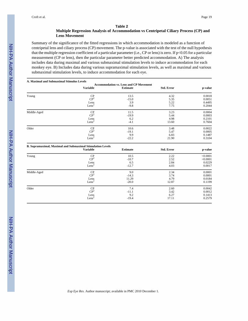
Multiple and Stepwise Regression AnalysisIn all three age groups, the multiple regression model (Table 2 A) explained accommodativeamplitude better than the model used in Figs. 3 A, C, and E above. This was not surprising,since both centripetal CP and centripetal lens movements are needed for accommodation. Withall of the parameters included, the best predictor of accommodative amplitude over the fullaccommodative range for all age groups was centripetal CP movement (Table 2).
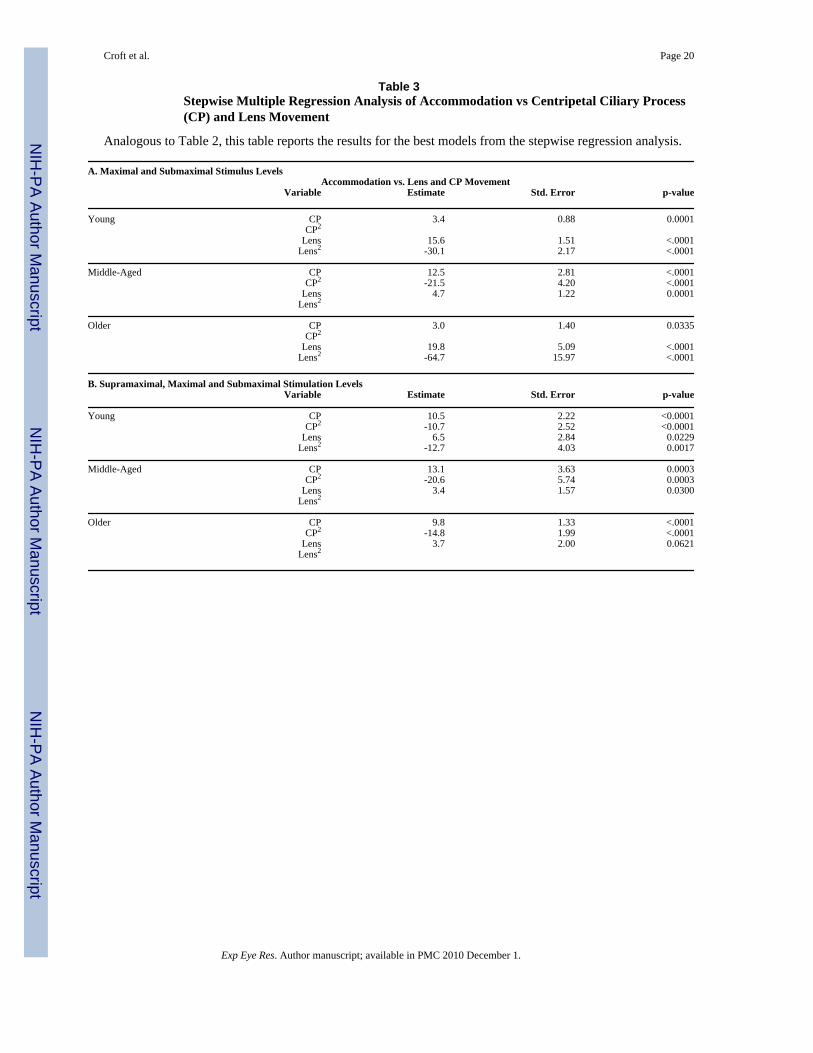
A stepwise regression was undertaken to choose the best model with which to predictaccommodation (Table 3). The results showed that both CP and lens movements wereimportant in predicting accommodation.
Forward Ciliary Body Movement, Centripetal Lens or Centripetal Ciliary Process(CP) Movement versus Age during ∼3 Diopters of Accommodation—Weexamined the age-related change in forward ciliary body movement, or centripetal lens orcentripetal CP movement amplitude, during ∼3 diopters of accommodation (Fig. 5). Theamount of centripetal lens movement required to induce ∼3 diopters of accommodation didnot change significantly with age (p=0.10). However, the amount of centripetal CP movementsignificantly increased (p=0.01), while the amount of forward ciliary body movementsignificantly decreased with age (p=0.007).
Forward Ciliary Body, Centripetal Lens, and Centripetal Ciliary Process (CP)Movement at Maximum Amplitudes (during supramaximal stimulus; 28 eyes, 23monkeys)—Maximum accommodative forward ciliary body movement was linearlycorrelated with maximum amplitude of both centripetal CP and centripetal lens movement(Fig. 6 A; p<0.02); however, these relationships clearly changed depending on the age group.Centripetal CP and centripetal lens movement versus accommodative forward ciliary bodymovement were plotted for the three age groups (Fig. 6 B). In the young eyes, as forward ciliarybody movement amplitude increased, there was a slight but statistically significant decline incentripetal CP but not centripetal lens movement (Fig. 6 B). In the middle-aged eyes, theaverage amount of forward ciliary body movement dropped dramatically (see above), and therewas a significant increase in CP movement per degree of forward ciliary body movement andan increase in centripetal lens movement per degree of forward ciliary body movement (Fig.6 B). In the older eyes, the increase in centripetal CP movement per degree of forward ciliarybody movement was even more dramatic, while the increase in lens movement per unit offorward ciliary body movement was less pronounced but similar to that found for the middle-aged eyes.
Croft et al. Page 5
Exp Eye Res. Author manuscript; available in PMC 2010 December 1.
NIH
-PA Author Manuscript
NIH
-PA Author Manuscript
NIH
-PA Author Manuscript

In the middle-aged eyes, there was a weak positive relationship between centripetal CP andforward ciliary body movement (0.0042 mm/°; p=0.108) and a significant relationship betweenthe lens centripetal movement and forward ciliary body movement (0.0058 mm/°; p=0.007;Fig. 6 B). The positive relationship between centripetal CP and forward ciliary body movementwas significant and most pronounced in the older eyes, in which the unit of centripetal CPmovement per unit of forward ciliary body movement (0.0146 mm/°; p=0.014) was the highestof all three age groups (Fig. 6 B). In the older monkey eyes, lens movement was not significantlycorrelated with forward ciliary body movement (p=0.279) but was significantly related to CPmovement (p=0.0001, Fig. 4).
DiscussionWe have documented the age-related functional changes of various components of theaccommodative apparatus of the rhesus monkey. These results demonstrate that study of theentire age range is required to determine which component of the accommodative apparatuschanges first with age.
Collectively, the data of the current study show that the loss in forward ciliary body movementwith age occurs sooner than the loss in centripetal lens equator movement. The loss in forwardmuscle movement is partially compensated for by the level of increased centripetal musclemovement required to achieve zonular relaxation and lens rounding. Overall, the amplitude ofthe centripetal ciliary body movement is somewhat reduced with age. It is unlikely that thelenses of the middle-aged monkeys changed substantially in internal refractive properties orhardness compared to those of the young animals since the centripetal lens movement perdiopter of accommodation was the same in both the young and middle-aged animals.
The higher amplitudes of centripetal lens movement were not achieved in the middle-agedeyes, possibly due to the 55% loss in forward ciliary body movement. Even duringsupramaximal stimulation, despite the compensating centripetal CP movement, no further lensmovement was induced in the middle-aged eyes. There are hundreds of zonular fibers extendingfrom the valleys of the ciliary processes to the anterior and posterior lens surfaces, in additionto those that extend between the ciliary processes and the lens equator (Glasser and Campbell,1998; Rohen, 1979). Some of these zonular attachments may be more dependent on forwardciliary body movement than centripetal CP movement to achieve relaxation and allow lensrounding.
Without the supramaximal stimulation data included, multiple regression analysis showed thatmost of the variability in accommodative amplitude was explained by centripetal CP movementin all three age groups, while the centripetal lens movement was also important to explain somepart of accommodative amplitude (based on the stepwise regression analysis; Table 3). Thiswas not surprising, since it is known that both parameters are needed for accommodation.Centripetal CP movement may reflect both the centripetal lens equatorial movement and thethickening of the lens, which may be why centripetal CP movement was so important in thestepwise regression models to predict accommodation.
Goniovideographically measured centripetal CP movements predominantly representcentripetal ciliary body movements, but these measurements do not really distinguishcentripetal from forward ciliary body movement. Thus, centripetal CP movements that wereport here may actually be a hybrid or composite, in contrast to movements measured by UBMthat can isolate measurement of forward ciliary body movement. Nonetheless, the techniquesof measuring forward ciliary body movement by UBM and centripetal CP movement bygoniovideography clearly provide separate and distinct information about ciliary body functionand its change with age (Croft et al., 2006a).
Croft et al. Page 6
Exp Eye Res. Author manuscript; available in PMC 2010 December 1.
NIH
-PA Author Manuscript
NIH
-PA Author Manuscript
NIH
-PA Author Manuscript
Presbyopia is by definition the loss in accommodative amplitude, due to the loss in the abilityof the lens to change shape. Given the CP versus lens relationships in Fig. 4, one might arguethat the CP movement has not dampened with age but the lens movement has. However, thesame amount of lens movement per diopter of accommodation exists in both the young andmiddle-aged eyes (Fig. 3).
Given the difference between young and middle-aged eyes in CP and forward ciliary bodymovement, one might consider the possibility of a Type I error (the possibility of a falseassumption of a significant difference between the groups, with no biological basis). Theprobability of such an error is low (typically p=0.05 or a 5% chance) and, based on an overallexamination of the data, we considered the existence of a Type I error an unlikely possibility.
The active movement of the ciliary body is possible only by ciliary muscle contraction. Dueto the posterior restriction of muscle movement in the aging eye, the longitudinal portion ofthe muscle may undergo more of an isometric contraction than the circular portion—thus themarked loss in forward movement. However, the circular portion of the muscle duringcontraction applies its force centripetally, a direction that is perpendicular to the restriction andthat is not in direct opposition to the restriction. In support of this idea, Tamm et al. (1992)reported that the area encompassing the circular portion of the ciliary muscle increases withage in excised human eyes, while the area of the longitudinal portion of the ciliary muscledecreases with age. This aging change may be adaptive to compensate for the muscle's posteriorrestriction or to further support the accommodative effort as the lens thickens with age. Theaging change mentioned above would likely not be to compensate for the decreaseddeformability of the lens (at least in the middle-aged monkey eyes), since, by extrapolationfrom human data, changes in lens deformability do not occur until after age 19 in monkeys(∼age 40 in humans; see second to last paragraph of the discussion, below).
By examination of dynamic UBM images of a 16-year-old rhesus monkey eye duringsupramaximal stimulation to induce accommodation, one can understand how there could bean age-related loss in forward ciliary body movement while substantial centripetal CPmovement remained, based on ocular geometry (Video Clip #1). Age-related stiffening of theposterior attachments (i.e., choroid, posterior muscle tendons (Tamm et al., 1992), and/orposterior vitreous zonule, which extend in a straight line from the ora serrata to the zonularplexus in the valleys of the ciliary processes (Lütjen-Drecoll et al., Unpublished results)) coulddampen forward ciliary body movement, with substantial centripetal CP movement remaining.
With the lens substance removed (leaving an empty capsular bag), the centripetal musclemovement was enhanced but forward muscle movement was unchanged compared to thenormal iridectomized eye. With the lens substance and capsule removed (thus severing theanterior zonular attachments between the ciliary muscle and the lens capsule), the loss offorward muscle movement was far more pronounced (50%) than the loss in centripetal musclemovement (10-15%) (Croft et al., 2008) (Wasilewski et al., 2008). This suggests that there maybe agonistic tractional forces, supplied by the attachment of the anterior zonula/lens complexto the ciliary muscle during accommodation, that enhance muscle movement and are far moreimportant to forward movement than to centripetal movement of the muscle. These forcesprovide anterior traction to the muscle and counterbalance those forces that pull the muscleback into the resting state (i.e., posterior elastic tendons, choroid). Alternatively, the anteriorattachments of the muscle to the anterior zonula/lens complex may simply be an anchor bywhich the muscle pulls itself forward and thereby contribute passively to forward musclemovement. Whether the attachment of the anterior zonula/lens complex to the ciliary muscleprovides traction or plays a “passive anchor” role in ciliary muscle contraction, it facilitatesforward muscle movement until zonular relaxation is achieved during the accommodativeresponse.
Croft et al. Page 7
Exp Eye Res. Author manuscript; available in PMC 2010 December 1.
NIH
-PA Author Manuscript
NIH
-PA Author Manuscript
NIH
-PA Author Manuscript
Formation of chemical bonds between lens fibers might also cause dampened lens equatormovement, but it was beyond the scope of this study to determine bonding between the lensfiber cells in the young and middle-aged rhesus eyes. In excised human eyes, age-relatedchanges in lens compliance (Weeber et al., 2005) and lens resistance to deformation (Glasserand Campbell, 1999) were minimal prior to age 40 (age in monkey years ∼19). The lensresistance to deformation increased dramatically after the age of 40 in excised human lenses(Glasser and Campbell, 1999). Age-related lens thickening by itself could be considered asignificant change and could play a role in the pathophysiology of presbyopia. However, Alióet al. (2005) reported that, while human lens thickness begins increasing before the age of 40,the density of the human lens nucleus only begins increasing after age 40, and does so linearlywith age. Alió also reported an increase in intraocular light scattering and aberrations after age40, which decreases the optical image quality (Alió et al., 2005). This suggests that significantcumulative lens changes are not apparent until after the age of 40 in humans, by which time,as mentioned previously, more than 2/3 of the accommodative ability has been lost (Duane,1922).
This study demonstrates that ciliary body function begins to change with age before lensfunction changes. Our data show that the lens accommodative response (i.e., centripetal lensmovement required to induce a given level of accommodation) was not significantly changedin the middle-aged monkeys (12-16.5 years) compared to the young monkeys; thus, it isunlikely that the resting lenses of the middle-aged monkeys changed in refractive powercompared to the young animals. The age-related loss in ciliary body function (i.e., loss offorward ciliary body movement) that we measured could be due to decreasing elasticity of thechoroid, the posterior ciliary muscle tendons, or the posterior vitreous zonule. Chemical orphysical lysis or other treatment of these inelastic attachments may sustain the ability of theciliary body to move forward during accommodative effort and thus prevent or delay secondaryage-related lenticular changes and perhaps facilitate the mobility/deformability ofaccommodating IOLs.
Supplementary MaterialRefer to Web version on PubMed Central for supplementary material.
AcknowledgmentsNEI (EY10213); Ocular Physiology Research & Education Foundation; Research to Prevent Blindness (UnrestrictedGrant and Physician-Scientist Award); Core Grant for Vision Research grant # P30 EY016665; and Retina ResearchFoundation (Walter H. Helmerich Chair). This publication was made possible in part by Grant Number P51 RR000167from the National Center for Research Resources (NCRR), a component of the National Institutes of Health (NIH),to the Wisconsin National Primate Research Center, University of Wisconsin-Madison. This research was conductedin part at a facility constructed with support from Research Facilities Improvement Program grant numbersRR15459-01 and RR020141-01. This publication's contents are solely the responsibility of the authors and do notnecessarily represent the official views of NCRR or NIH.
We also acknowledge James Reed, for technical expertise with image analysis systems.
Support: This work was funded by NEI grants EY10213 & R21EY018370 to PLK, by an unrestricted gift from theOcular Physiology Research & Education Foundation, and by the Walter H. Helmerich Chair of the Retina ResearchFoundation. We also acknowledge the Wisconsin National Primate Research Center, University of Wisconsin-Madison base grant # 5P51 RR 000167 and the Core Grant for Vision Research grant # P30 EY016665.
ReferencesAlió JL, Schimchak P, Negri HP, Montés-Micó R. Crystalline lens optical dysfunction through aging.
Ophthalmology 2005;112:2022–2029. [PubMed: 16183126]
Croft et al. Page 8
Exp Eye Res. Author manuscript; available in PMC 2010 December 1.
NIH
-PA Author Manuscript
NIH
-PA Author Manuscript
NIH
-PA Author Manuscript
Bito, LZ.; Miranda, OC. Accommodation and presbyopia. In: Reinecke, RD., editor. Ophthalmologyannual. New York: Raven Press; 1989. p. 103-128.
Crawford K, Terasawa E, Kaufman PL. Reproducible stimulation of ciliary muscle contraction in thecynomolgus monkey via a permanent indwelling midbrain electrode. Brain Res 1989;503:265–272.[PubMed: 2605519]
Croft MA, Glasser A, Heatley G, McDonald J, Ebbert T, Dahl DB, Nadkarni NV, Kaufman PL.Accommodative ciliary body and lens function in rhesus monkeys: I. Normal lens, zonule and ciliaryprocess configuration in the iridectomized eye. Invest Ophthalmol Vis Sci 2006a;47:076–1086.
Croft MA, Glasser A, Heatley G, McDonald J, Ebbert T, Nadkarni NV, Kaufman PL. The zonula, lens,and circumlental space in the normal iridectomized rhesus monkey eye. Invest Ophthalmol Vis Sci2006b;47:1087–1095. [PubMed: 16505045]
Croft MA, McDonald JP, James RJ, Heatley GA, Lin TL, Lütjen-Drecoll E, Kaufman PL. Surgicalintervention and accommodative responses, I: Centripetal ciliary body, capsule, and lens movementsin rhesus monkeys of various ages. Invest Ophthalmol Vis Sci 2008;49:5484–5494. [PubMed:18552393]
Duane A. Studies in monocular and binocular accommodation with their clinical applications. Am JOphthalmol 1922;5:867–877.
Fisher RF. Elastic constants of the human lens capsule. J Physiol (Lond) 1969;201:1–19. [PubMed:5773553]
Fisher RF. The elastic constants of the human lens. J Physiol (Lond) 1971;212:147–180. [PubMed:5101807]
Fisher RF. The force of contraction of the human ciliary muscle during accommodation. J Physiol (Lond)1977;270:51–74. [PubMed: 915798]
Glasser A, Campbell MCW. Presbyopia and the optical changes in the human crystalline lens with age.Vision Res 1998;38:209–229. [PubMed: 9536350]
Glasser A, Campbell MCW. Biometric, optical and physical changes in the isolated human crystallinelens with age in relation to presbyopia. Vision Res 1999;39:1991–2015. [PubMed: 10343784]
Heys KR, Cram SL, Truscott RJ. Massive increase in the stiffness of the human lens nucleus with age:the basis for presbyopia? Mol Vis 2004;10:956–963. [PubMed: 15616482]
Kaufman PL, Lütjen-Drecoll E. Total iridectomy in the primate in vivo: surgical technique andpostoperative anatomy. Invest Ophthalmol 1975;14:766–771. [PubMed: 810452]
Koretz JF, Kaufman PL, Neider NW, Goeckner PA. Accommodation and presbyopia in the human eye.II. Aging of the anterior segment. Vision Res 1989;29:1685–1692. [PubMed: 2631389]
Ludwig K, Wegscheider E, Hoops JP, Kampik A. In vivo imaging of the human zonular apparatus withhigh-resolution ultrasound biomicroscopy. Graefes Arch Clin Exp Ophthalmol 1999;237:361–371.[PubMed: 10333101]
Pau H, Krantz J. The increasing sclerosis of the human lens with age and its relevance to accommodationand presbyopia. Graefes Arch Clin Exp Ophthalmol 1991;229:294–296. [PubMed: 1869070]
Rohen JW. Scanning electron microscopic studies of the zonular apparatus in human and monkey eyes.Invest Ophthalmol Vis Sci 1979;18:133–144. [PubMed: 104933]
Tamm E, Croft MA, Jungkunz W, Lütjen-Drecoll E, Kaufman PL. Age-related loss of ciliary musclemobility in the rhesus monkey: role of the choroid. Arch Ophthalmol 1992a;110:871–876. [PubMed:1596237]
Tamm E, Lütjen-Drecoll E, Jungkunz W, Rohen JW. Posterior attachment of ciliary muscle in young,accommodating old, presbyopic monkeys. Invest Ophthalmol Vis Sci 1991;32:1678–1692.[PubMed: 2016145]
Tamm S, Tamm E, Rohen JW. Age-related changes of the human ciliary muscle. A quantitativemorphometric study. Mech Ageing Dev 1992b;62:209–221. [PubMed: 1569790]
Vilupuru AS, Glasser A. Dynamic accommodation in rhesus monkeys. Vision Res 2002;42:125–141.[PubMed: 11804637]
Wasilewski R, McDonald JP, Heatley G, Lütjen-Drecoll E, Kaufman PL, Croft MA. Surgical interventionand accommodative responses, II. Forward ciliary body accommodative movement is facilitated byzonular attachments to the lens capsule. Invest Ophthalmol Vis Sci 2008;49:5495–5502. [PubMed:18552391]
Croft et al. Page 9
Exp Eye Res. Author manuscript; available in PMC 2010 December 1.
NIH
-PA Author Manuscript
NIH
-PA Author Manuscript
NIH
-PA Author Manuscript
Weeber HA, Eckert G, Soergel F, Meyer CH, Pechhold W, van der Heijde RGL. Dynamic mechanicalproperties of human lenses. Exp Eye Res 2005;80:425–434. [PubMed: 15721624]
Croft et al. Page 10
Exp Eye Res. Author manuscript; available in PMC 2010 December 1.
NIH
-PA Author Manuscript
NIH
-PA Author Manuscript
NIH
-PA Author Manuscript

Figure 1.Goniovideography images of normal lens and ciliary process (CP) configuration in theaccommodated and unaccommodated states. To obtain quantitative measurements, a 9-0 nylonsuture placed at the corneoscleral limbus served as a reference point (left solid vertical line)from which to measure distances to the lens equator (right solid vertical line) and the CPs(cross-hairs) for each image during a 2.2-sec stimulus period. Reprinted with permission from:Croft et al. Accommodative Ciliary Body and Lens Function in Rhesus Monkeys I. Normallens, zonule and ciliary process configuration in the iridectomized eye. Invest Ophthalmol VisSci 2006;47:1076-1086.
Croft et al. Page 11
Exp Eye Res. Author manuscript; available in PMC 2010 December 1.
NIH
-PA Author Manuscript
NIH
-PA Author Manuscript
NIH
-PA Author Manuscript

Figure 2.Ultrasound biomicroscopy images of two normal monkey eyes, aged 6 years (A, C) and 23years (B, D), in the unaccommodated and accommodated states. The change in angle betweenthe anterior aspect of the ciliary body and the inner aspect of the cornea during supramaximalcentral stimulation was used as a surrogate indicator of forward ciliary body movement (FCB)(Croft et al., 2006a). Panels B and D adapted with permission from: Croft et al. AccommodativeCiliary Body and Lens Function in Rhesus Monkeys I. Normal lens, zonule and ciliary processconfiguration in the iridectomized eye. Invest Ophthalmol Vis Sci 2006;47:1076-1086.
Croft et al. Page 12
Exp Eye Res. Author manuscript; available in PMC 2010 December 1.
NIH
-PA Author Manuscript
NIH
-PA Author Manuscript
NIH
-PA Author Manuscript

Figure 3.Accommodation versus centripetal ciliary process (CP) or centripetal lens movement in young(ages 6-10 years; n=8 eyes; 5 monkeys); middle-aged (ages 12-16.5 years; n=5 eyes; 5monkeys); and older (ages 20 years and above; n=15 eyes; 10 monkeys) monkey eyes. A, C,E: Maximal and Submaximal Stimulus Levels. Various levels of accommodation, from zerodiopters up to maximum accommodation, were induced and plotted versus the correspondingcentripetal CP and centripetal lens movement for each monkey eye. Centripetal CP andcentripetal lens movements began to plateau at 0.3 mm of movement. The fitted regressioncurve of the amplitude of the centripetal CP (solid line) or centripetal lens (dashed line)movement versus accommodation is shown for each panel. The p-values are obtained from theLikelihood Ratio Test. B, D, F: Supramaximal, Maximal and Submaximal Stimulus Levels.Includes data collected during various supramaximal stimulation levels for each monkey eyeplus data contained in panels A, C, and E. The results were similar when data from thesupramaximal stimulus levels were included in the analysis.
Croft et al. Page 13
Exp Eye Res. Author manuscript; available in PMC 2010 December 1.
NIH
-PA Author Manuscript
NIH
-PA Author Manuscript
NIH
-PA Author Manuscript

Figure 4.Lens versus corresponding centripetal ciliary process (CP) movement. There was a significantincrease in units of centripetal CP movement per unit of centripetal lens movement betweenthe young and middle-aged eyes (p<0.01) and between the middle-aged and older eyes(p<0.01). A linear model was the best fit for the data. This model excluded data points at thehigh end of the response curve (above 0.3 mm CP movement) where the data begins to plateau.
Croft et al. Page 14
Exp Eye Res. Author manuscript; available in PMC 2010 December 1.
NIH
-PA Author Manuscript
NIH
-PA Author Manuscript
NIH
-PA Author Manuscript
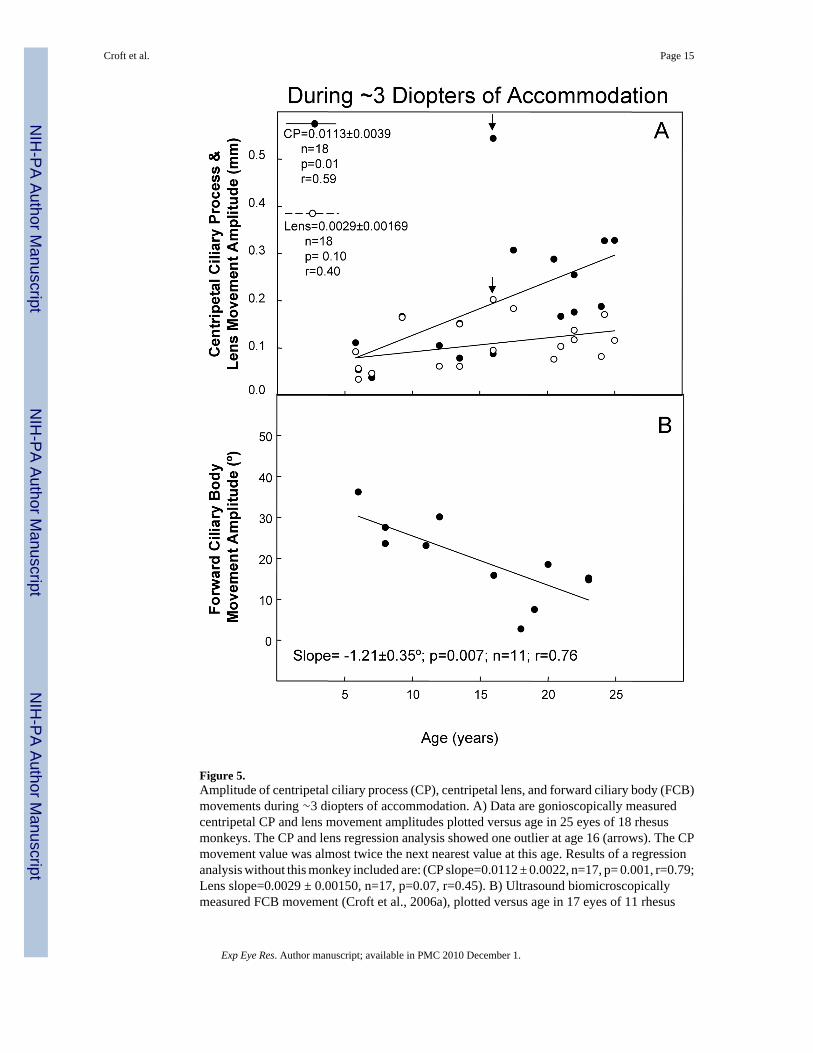
Figure 5.Amplitude of centripetal ciliary process (CP), centripetal lens, and forward ciliary body (FCB)movements during ∼3 diopters of accommodation. A) Data are gonioscopically measuredcentripetal CP and lens movement amplitudes plotted versus age in 25 eyes of 18 rhesusmonkeys. The CP and lens regression analysis showed one outlier at age 16 (arrows). The CPmovement value was almost twice the next nearest value at this age. Results of a regressionanalysis without this monkey included are: (CP slope=0.0112 ± 0.0022, n=17, p= 0.001, r=0.79;Lens slope=0.0029 ± 0.00150, n=17, p=0.07, r=0.45). B) Ultrasound biomicroscopicallymeasured FCB movement (Croft et al., 2006a), plotted versus age in 17 eyes of 11 rhesus
Croft et al. Page 15
Exp Eye Res. Author manuscript; available in PMC 2010 December 1.
NIH
-PA Author Manuscript
NIH
-PA Author Manuscript
NIH
-PA Author Manuscript

monkeys. The lines represent least squares linear regression of the CP, FCB, or lens responseamplitude versus age. Numbers represent slopes ± s.e.m.; P, probability that the slope=0.0;r=correlation coefficient. For both panels A and B, for instances in which there were two eyesfrom one monkey, the data were averaged to provide one data point.
Croft et al. Page 16
Exp Eye Res. Author manuscript; available in PMC 2010 December 1.
NIH
-PA Author Manuscript
NIH
-PA Author Manuscript
NIH
-PA Author Manuscript
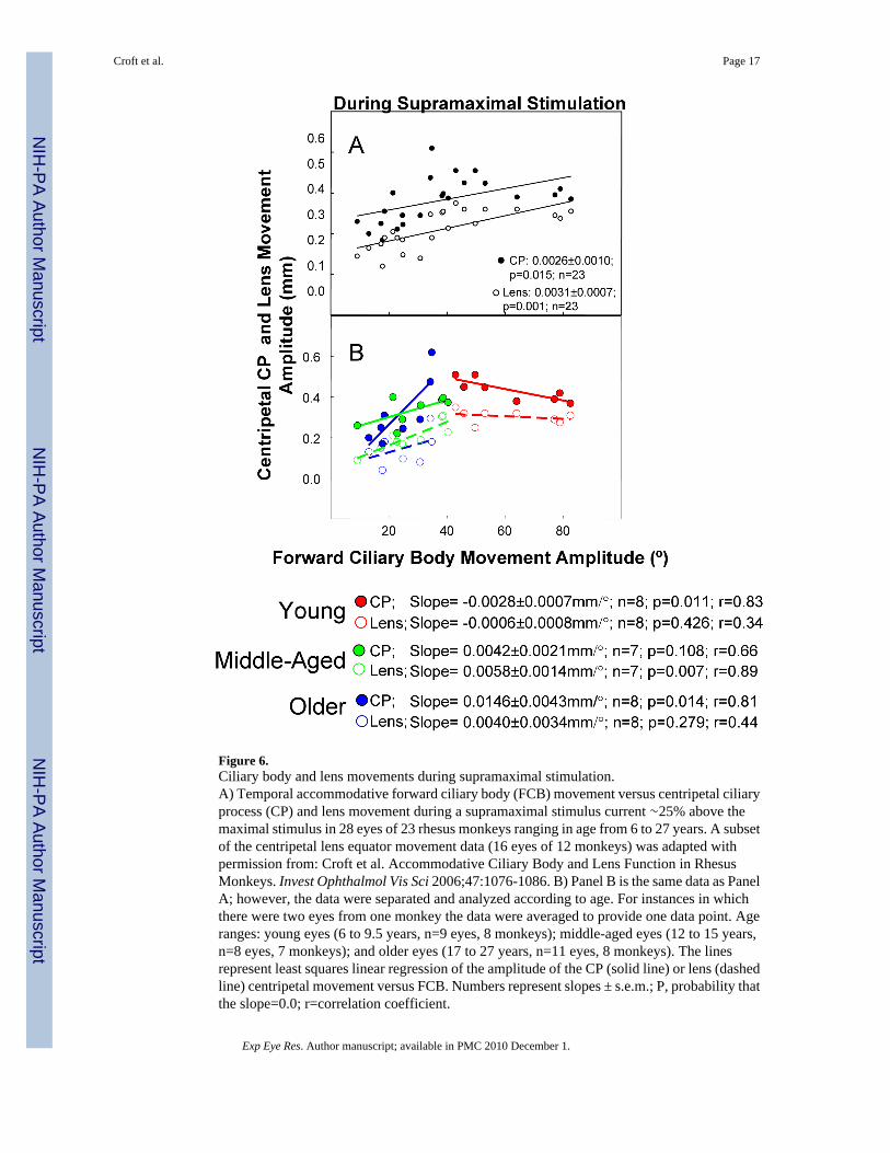
Figure 6.Ciliary body and lens movements during supramaximal stimulation.A) Temporal accommodative forward ciliary body (FCB) movement versus centripetal ciliaryprocess (CP) and lens movement during a supramaximal stimulus current ∼25% above themaximal stimulus in 28 eyes of 23 rhesus monkeys ranging in age from 6 to 27 years. A subsetof the centripetal lens equator movement data (16 eyes of 12 monkeys) was adapted withpermission from: Croft et al. Accommodative Ciliary Body and Lens Function in RhesusMonkeys. Invest Ophthalmol Vis Sci 2006;47:1076-1086. B) Panel B is the same data as PanelA; however, the data were separated and analyzed according to age. For instances in whichthere were two eyes from one monkey the data were averaged to provide one data point. Ageranges: young eyes (6 to 9.5 years, n=9 eyes, 8 monkeys); middle-aged eyes (12 to 15 years,n=8 eyes, 7 monkeys); and older eyes (17 to 27 years, n=11 eyes, 8 monkeys). The linesrepresent least squares linear regression of the amplitude of the CP (solid line) or lens (dashedline) centripetal movement versus FCB. Numbers represent slopes ± s.e.m.; P, probability thatthe slope=0.0; r=correlation coefficient.
Croft et al. Page 17
Exp Eye Res. Author manuscript; available in PMC 2010 December 1.
NIH
-PA Author Manuscript
NIH
-PA Author Manuscript
NIH
-PA Author Manuscript

NIH
-PA Author Manuscript
NIH
-PA Author Manuscript
NIH
-PA Author Manuscript
Croft et al. Page 18Ta
ble
1A
vera
ge M
axim
um A
mpl
itude
s of A
ccom
mod
atio
n, F
orw
ard
Cili
ary
Bod
y, C
iliar
y Pr
oces
s (C
P) a
nd L
ens M
ovem
ent i
n Y
oung
, Mid
dle-
Age
dan
d O
lder
Eye
s
A) D
ata
are
mea
n ±
s.e.m
. acc
omm
odat
ive
ampl
itude
(dio
pter
s; D
) at s
upra
max
imal
(∼25
% a
bove
that
nec
essa
ry to
indu
ce m
axim
um a
ccom
mod
atio
n)st
imul
us le
vels
in 2
8 ey
es o
f 23
rhes
us m
onke
ys. B
) Dat
a ar
e m
ean
± s.e
.m. f
orw
ard
cilia
ry b
ody
mov
emen
t (FC
B; i
n un
its o
f deg
rees
as p
revi
ousl
y de
fined
(Fig
. 2) (
Cro
ft et
al.,
200
6a))
; cen
tripe
tal c
iliar
y pr
oces
s mov
emen
t (C
P); a
nd le
ns m
ovem
ent a
mpl
itude
(mm
) at s
tand
ard
supr
amax
imal
stim
ulus
setti
ngs.
Age
rang
es: y
oung
eye
s (6
to 9
.5 y
ears
); m
iddl
e-ag
ed e
yes (
12 to
15
year
s); a
nd o
lder
eye
s (17
to 2
7 ye
ars)
. A p≤0
.05
repr
esen
ts a
sign
ifica
nt d
iffer
ence
betw
een
the
youn
g ag
e gr
oup
vers
us th
e ot
her a
ge g
roup
s by
two
sam
ple
t-tes
t. C
) Per
cent
dec
reas
e is
cal
cula
ted
as [(
mid
dle-
aged
/you
ng)-
1]*1
00 o
r ((o
lder
/yo
ung)
-1)*
100
for e
ach
varia
ble.
For
inst
ance
s in
whi
ch th
ere
wer
e tw
o ey
es fr
om o
ne m
onke
y, th
e da
ta w
ere
aver
aged
to p
rovi
de o
ne d
ata
poin
t. A
subs
etof
the C
P an
d ce
ntrip
etal
lens
equa
tor m
ovem
ent d
ata (
16 ey
es o
f 12
mon
keys
) was
adap
ted
with
per
mis
sion
from
: Cro
ft et
al. A
ccom
mod
ativ
e Cili
ary
Bod
yan
d Le
ns F
unct
ion
in R
hesu
s Mon
keys
. Inv
est O
phth
alm
ol V
is S
ci 2
006;
47:1
076-
1086
.
A.
B.
C.
Cen
trip
etal
Mov
emen
t (m
m)
% D
eclin
e
Cen
trip
etal
Acc
omm
odat
ion
(D)
FCB
(°)
nC
PL
ens
Acc
omm
odat
ion
(D)
FCB
CP
Len
s
You
ngM
ean
15.2
61.8
80.
440.
31--
----
----
----
--(6
-9.5
yrs
)s.e
.m.
1.0
5.7
0.02
0.01
Mid
dle-
Age
dMea
n8.
127
.97
0.35
0.22
46.7
54.8
19.4
30.4
(12-
15 y
rs)s
.e.m
.0.
54.
40.
030.
03
Mid
dle-
Age
d vs
You
ngp=
0.00
10.
001
0.01
20.
022
Old
erM
ean
2.4
23.9
80.
320.
1484
.061
.326
.754
.3(1
7-26
yrs
)s.e
.m.
0.6
3.0
0.05
0.03
Old
er v
s You
ngp=
0.00
10.
001
0.08
0.00
1
Mid
dle-
Age
d vs
Old
erp=
0.00
10.
258
0.58
40.
166
37.3
6.5
7.3
23.9
Exp Eye Res. Author manuscript; available in PMC 2010 December 1.
NIH
-PA Author Manuscript
NIH
-PA Author Manuscript
NIH
-PA Author Manuscript
Croft et al. Page 19
Table 2Multiple Regression Analysis of Accommodation vs Centripetal Ciliary Process (CP) andLens Movement
Summary of the significance of the fitted regressions in which accommodation is modeled as a function ofcentripetal lens and ciliary process (CP) movement. The p-value is associated with the test of the null hypothesisthat the multiple regression coefficient of a particular parameter (i.e., CP or lens) is zero. If p<0.05 for a particularmeasurement (CP or lens), then the particular parameter better predicted accommodation. A) The analysisincludes data during maximal and various submaximal stimulation levels to induce accommodation for eachmonkey eye. B) Includes data during various supramaximal stimulation levels, as well as maximal and varioussubmaximal stimulation levels, to induce accommodation for each eye.
A. Maximal and Submaximal Stimulus LevelsAccommodation vs. Lens and CP Movement
Variable Estimate Std. Error p-value
Young CP 13.5 4.32 0.0018CP2 -15.0 5.35 0.0051
Lens 3.9 5.22 0.4495Lens2 -9.8 7.75 0.2044
Middle-Aged CP 11.5 3.23 0.0004CP2 -19.9 5.44 0.0003
Lens 6.2 4.98 0.2101Lens2 -4.1 13.60 0.7604
Older CP 10.6 3.48 0.0022CP2 -19.1 5.47 0.0005
Lens 9.9 6.83 0.1487Lens2 -22.2 21.90 0.3104
B. Supramaximal, Maximal and Submaximal Stimulation LevelsVariable Estimate Std. Error p-value
Young CP 10.5 2.22 <0.0001CP2 -10.7 2.52 <0.0001
Lens 6.5 2.84 0.0229Lens2 -12.7 4.03 0.0017
Middle-Aged CP 9.0 2.34 0.0001CP2 -14.3 3.74 0.0001
Lens 11.29 4.79 0.0184Lens2 -20.0 12.87 0.1199
Older CP 7.4 2.60 0.0042CP2 -11.1 3.42 0.0012
Lens 9.2 6.27 0.1413Lens2 -19.4 17.11 0.2579
Exp Eye Res. Author manuscript; available in PMC 2010 December 1.
NIH
-PA Author Manuscript
NIH
-PA Author Manuscript
NIH
-PA Author Manuscript
Croft et al. Page 20
Table 3Stepwise Multiple Regression Analysis of Accommodation vs Centripetal Ciliary Process(CP) and Lens Movement
Analogous to Table 2, this table reports the results for the best models from the stepwise regression analysis.
A. Maximal and Submaximal Stimulus LevelsAccommodation vs. Lens and CP Movement
Variable Estimate Std. Error p-value
Young CP 3.4 0.88 0.0001CP2
Lens 15.6 1.51 <.0001Lens2 -30.1 2.17 <.0001
Middle-Aged CP 12.5 2.81 <.0001CP2 -21.5 4.20 <.0001
Lens 4.7 1.22 0.0001Lens2
Older CP 3.0 1.40 0.0335CP2
Lens 19.8 5.09 <.0001Lens2 -64.7 15.97 <.0001
B. Supramaximal, Maximal and Submaximal Stimulation LevelsVariable Estimate Std. Error p-value
Young CP 10.5 2.22 <0.0001CP2 -10.7 2.52 <0.0001
Lens 6.5 2.84 0.0229Lens2 -12.7 4.03 0.0017
Middle-Aged CP 13.1 3.63 0.0003CP2 -20.6 5.74 0.0003
Lens 3.4 1.57 0.0300Lens2
Older CP 9.8 1.33 <.0001CP2 -14.8 1.99 <.0001
Lens 3.7 2.00 0.0621Lens2
Exp Eye Res. Author manuscript; available in PMC 2010 December 1.