Age-dependent cerebrovascular dysfunction in a transgenic mouse model of cerebral amyloid angiopathy
10
Age-dependent cerebrovascular dysfunction in a transgenic mouse model of cerebral amyloid angiopathy Hwa Kyoung Shin, 1 Phillip B. Jones, 2 Monica Garcia-Alloza, 2 Laura Borrelli, 2 Steven M. Greenberg, 3 Brian J. Bacskai, 2 Matthew P. Frosch, 2 Bradley T. Hyman, 2 Michael A. Moskowitz 1 and Cenk Ayata 1,3 1 Stroke and Neurovascular Regulation Laboratory, Department of Radiology, 2 Alzheimer’s Disease Research Laboratory and 3 Stroke Service and Neuroscience Intensive Care Unit, Department of Neurology, Massachusetts General Hospital, Harvard Medical School, Charlestown, MA 02129, USA Correspondence to: Cenk Ayata, MD, Stroke and Neurovascular Regulation Laboratory, 149 13th Street, Room 6403, Charlestown, MA 02129, USA E-mail: [email protected] The Tg2576 transgenic mouse model of human cerebral amyloid angiopathy is characterized by age-dependent cerebrovascular deposition of amyloid-b (Ab ) starting from 9 months of age and progressively worsening to involve most pial arterioles by 18 months; soluble Ab levels are elevated long before vascular deposition takes place in this model. It has been suggested that elevated soluble Ab levels alone are sufficient to impair cerebral blood flow (CBF) regulation thereby contributing to the early progression of Alzheimer’s disease. Using laser speckle flowmetry through an intact skull, we studied the impact of elevated soluble Ab levels and vascular Ab deposition on a wide range of CBF responses to evaluate vasodilation and vasoconstriction in young or aged Tg2576 mice. Nineteen-month-old Tg2576 with severe vascular Ab deposits showed an attenuated hyper- aemic response during hypercapnia and whisker stimulation compared to wild-type littermates. The anticipated increase in CBF due to isoflurane anaesthesia was also suppressed, as were the typical hypoperfusion responses during cortical spreading depression and a-chloralose anaesthesia. The responses of 8 -month-old Tg2576 with elevated soluble Ab levels, but without vascular Ab deposition, did not differ from age-matched controls. In con- clusion, our data suggest that vascular Ab deposition is associated with impaired vasodilator as well as vasocon- strictor responses to a wide range of stimuli. These responses do not differ from controls when studied non-invasively prior to vascular Ab deposition, thus challenging the view that elevated soluble Ab levels are sufficient to cause cerebrovascular dysfunction. Keywords: cerebral blood flow; laser speckle flowmetry; hypercapnia; whisker stimulation; spreading depression Abbreviations: CAA ¼ cerebral amyloid angiopathy; CSD ¼ cortical spreading depression; LSF ¼ laser speckle flowmetry Received January 17 , 2007.Revised May 25, 2007 . Accepted June 13, 2007 . Advance Access publication July 16, 2007 Introduction Amyloid-b (Ab) peptides derived from proteolytic proces- sing of the b-amyloid precursor protein (APP) character- istically accumulate as cerebral parenchymal plaques in Alzheimer’s disease (AD). Deposition of Ab peptide in cerebral vessel walls, termed cerebral amyloid angiopathy (CAA), is found in the majority of AD brains. It is well established that CAA leads to ischaemic infarcts as well as lobar intracerebral haemorrhages, and thus may exacerbate the cognitive decline in AD (Vonsattel et al., 1991; Greenberg et al., 1995; Olichney et al., 1995). In support of this, recent studies in transgenic mouse models of AD and CAA demonstrated an association between elevated soluble Ab levels and impaired cerebrovascular function (Iadecola, 2004). Importantly, this association was apparent prior to vascular Ab deposition, raising the possibility that elevated soluble Ab levels in AD brain may adversely impact progression of the disease via hemody- namic mechanisms. However, there is as yet no clinical evidence to support cerebrovascular dysfunction in young asymptomatic individuals destined to develop AD or CAA. Furthermore, evidence is also lacking for a hemodynamic mechanism hastening the cognitive decline in AD, except when overt ischaemic or haemorrhagic strokes introduce doi:10.1093/brain/awm156 Brain (2007), 130, 2310 ^2319 ß 2007 The Author(s) This is an Open Access article distributed under the terms of the Creative Commons Attribution Non-Commercial License (http://creativecommons.org/licenses/by-nc/2.0/uk/) which permits unrestricted non-commercial use, distribution, and reproduction in any medium, provided the original work is properly cited.
-
Upload
independent -
Category
Documents
-
view
3 -
download
0
Transcript of Age-dependent cerebrovascular dysfunction in a transgenic mouse model of cerebral amyloid angiopathy
Age-dependent cerebrovascular dysfunctionin a transgenic mouse model of cerebralamyloid angiopathyHwa Kyoung Shin,1 Phillip B. Jones,2 Monica Garcia-Alloza,2 Laura Borrelli,2 Steven M.Greenberg,3
Brian J. Bacskai,2 Matthew P. Frosch,2 BradleyT. Hyman,2 Michael A. Moskowitz1 and Cenk Ayata1,3
1Stroke and Neurovascular Regulation Laboratory, Department of Radiology, 2Alzheimer’s Disease Research Laboratory and3Stroke Service and Neuroscience Intensive Care Unit, Department of Neurology, Massachusetts General Hospital, HarvardMedical School, Charlestown, MA 02129, USA
Correspondence to: Cenk Ayata, MD, Stroke and Neurovascular Regulation Laboratory, 149 13th Street, Room 6403,Charlestown, MA 02129, USAE-mail: [email protected]
TheTg2576 transgenic mouse model of human cerebral amyloid angiopathy is characterized by age-dependentcerebrovascular deposition of amyloid-b (Ab) starting from 9 months of age and progressively worsening toinvolve most pial arterioles by 18 months; soluble Ab levels are elevated long before vascular deposition takesplace in this model. It has been suggested that elevated soluble Ab levels alone are sufficient to impair cerebralblood flow (CBF) regulation thereby contributing to the early progression of Alzheimer’s disease.Using laserspeckle flowmetry through an intact skull, we studied the impact of elevated soluble Ab levels and vascularAb deposition on a wide range of CBF responses to evaluate vasodilation and vasoconstriction in young oraged Tg2576 mice. Nineteen-month-old Tg2576 with severe vascular Ab deposits showed an attenuated hyper-aemic response during hypercapnia and whisker stimulation compared to wild-type littermates.The anticipatedincrease in CBF due to isoflurane anaesthesia was also suppressed, as were the typical hypoperfusion responsesduring cortical spreading depression and a-chloralose anaesthesia. The responses of 8 -month-old Tg2576 withelevated soluble Ab levels, but without vascular Ab deposition, did not differ from age-matched controls. In con-clusion, our data suggest that vascular Ab deposition is associated with impaired vasodilator as well as vasocon-strictor responses to a wide range of stimuli. These responses do not differ from controls when studiednon-invasively prior to vascular Ab deposition, thus challenging the view that elevated soluble Ab levels aresufficient to cause cerebrovascular dysfunction.
Keywords: cerebral blood flow; laser speckle flowmetry; hypercapnia; whisker stimulation; spreading depression
Abbreviations: CAA¼ cerebral amyloid angiopathy; CSD¼ cortical spreading depression; LSF¼ laser speckle flowmetry
Received January17, 2007. Revised May 25, 2007. Accepted June 13, 2007. Advance Access publication July 16, 2007
IntroductionAmyloid-b (Ab) peptides derived from proteolytic proces-sing of the b-amyloid precursor protein (APP) character-istically accumulate as cerebral parenchymal plaques inAlzheimer’s disease (AD). Deposition of Ab peptide incerebral vessel walls, termed cerebral amyloid angiopathy(CAA), is found in the majority of AD brains. It is wellestablished that CAA leads to ischaemic infarcts as well aslobar intracerebral haemorrhages, and thus may exacerbatethe cognitive decline in AD (Vonsattel et al., 1991;Greenberg et al., 1995; Olichney et al., 1995). In supportof this, recent studies in transgenic mouse models of AD
and CAA demonstrated an association between elevatedsoluble Ab levels and impaired cerebrovascular function(Iadecola, 2004). Importantly, this association was apparentprior to vascular Ab deposition, raising the possibilitythat elevated soluble Ab levels in AD brain mayadversely impact progression of the disease via hemody-namic mechanisms. However, there is as yet no clinicalevidence to support cerebrovascular dysfunction in youngasymptomatic individuals destined to develop AD or CAA.Furthermore, evidence is also lacking for a hemodynamicmechanism hastening the cognitive decline in AD, exceptwhen overt ischaemic or haemorrhagic strokes introduce
doi:10.1093/brain/awm156 Brain (2007), 130, 2310^2319
� 2007 The Author(s)This is an Open Access article distributed under the terms of the Creative Commons Attribution Non-Commercial License (http://creativecommons.org/licenses/by-nc/2.0/uk/) whichpermits unrestricted non-commercial use, distribution, and reproduction in any medium, provided the original work is properly cited.

additional neuropathological lesions (Snowdon et al., 1997;
Pfeifer et al., 2002).We, therefore, tested whether the presence of vascular
amyloid deposits impact cerebrovascular responses intransgenic mice (Tg2576) overexpressing a mutant humanAPP associated with early onset familial AD and CAA(Hsiao et al., 1996). In these experiments, blood flow servedas a surrogate measure for the ability of vessels to dilate orconstrict under diverse stimuli. In Tg2576 mice, brain Ablevels are significantly elevated even at 2–3 months of ageand levels rapidly increase starting around 7 months;parenchymal and vascular Ab deposition appear after9 months (Kawarabayashi et al., 2001). With advanceddisease (i.e. 19 months) severe Ab deposition encasevascular smooth muscle cells and may cause cell loss(Christie et al., 2001). Using a novel non-invasive opticalimaging technique with high spatiotemporal resolution, wereport that 8-month-old Tg2576 mice with elevated solubleAb but no vascular Ab deposition show cerebrovascularresponses that do not differ from wild-type mice whenchallenged with 5% inhaled CO2, whisker stimulation orcortical spreading depression. In contrast, 19-month-oldTg2576 mice with widespread and severe vascular Abdeposits exhibit profound impairment in the typical bloodflow responses to the same experimental stimuli. The datawe present in this manuscript do not support a vascularrole for soluble Ab in early AD pathogenesis.
MethodsExperimental animalsHeterozygous Tg2576 expressing Swedish double mutant APP(APPK670/671L) driven by the hamster prion protein promoter(Hsiao et al., 1996) on a mixed C57BL/6J-SJL/J background, andtheir wild-type littermates, were studied at 8 (28–29 g) or19 months of age (33–39 g). In addition, 3-month-old wild-typemice were compared to 8- or 19-month-old wild-type mice tocontrol for the effects of ageing. Only male mice were used in allgroups. Eight-month-old Tg2576 mice show elevated Ab levels butno vascular deposits, whereas 19-month-old mice accumulatesevere parenchymal and vascular Ab deposition with loss ofsmooth muscle cells, and thus represent an advanced stage of ADand CAA (Christie et al., 2001; Domnitz et al., 2005; Robbinset al., 2006).
General surgical preparation andphysiological monitoringMice were initially anaesthetized with isoflurane (2% induction,
1% maintenance, in 70% N2O and 30% O2) and intubated via a
tracheostomy. Femoral artery was catheterized for blood pressure,
heart rate and blood gas monitoring (ETH 400 transducer
amplifier, CB Sciences, Milford, MA). Mice were paralysed
(pancuronium bromide, 0.4mg/kg/h, i.p.), mechanically ventilated
(CWE, SAR-830, Ardmore, PA), placed in a stereotaxic frame
(David Kopf, Tujunga, CA) and scalp and periosteum were
retracted. At the end of surgical preparation, anaesthesia was
switched from isoflurane to a-chloralose (50mg/kg/h, i.v.) to
preserve vascular responses (Ayata et al., 2004a). Anaesthetic
depth was adjusted to abolish cardiovascular reflexes to tail pinch.
Rectal temperature was kept at 36.8–37.1�C using a thermo-
statically controlled heating mat (FHC, Brunswick, ME). Arterial
blood gases and pH were measured immediately before a stimulus
and at least once every hour, in 30 ml blood samples (Corning
178 blood gas/pH analyzer, Ciba Corning Diagnostics, Medford,
MA). These data (Table 1) were within the previously reported
normal limits for mice and did not differ among groups (Dalkara
et al., 1995; Niwa et al., 2001; Park et al., 2004; Lacombe et al.,
2005). The data were continuously recorded using a data
acquisition and analysis system (PowerLab, AD Instruments,
Medford, MA).
Laser speckle flowmetryLaser speckle flowmetry (LSF) (Dunn et al., 2001) was used to
study the spatiotemporal characteristics of CBF changes in mice as
previously described in detail (Ayata et al., 2004a). Briefly, the
scalp was shaved, incised at midline and reflected laterally. A CCD
camera (Cohu, San Diego, CA) was positioned above the head to
image the entire right hemisphere (Fig. 1). A laser diode (780 nm)
was used to illuminate the intact skull surface in a diffuse manner.
The penetration depth of the laser is estimated to be �500mmfrom the pial surface. Raw speckle images were used to compute
speckle contrast, which is a measure of speckle visibility related to
the velocity of the scattering particles, and therefore CBF. The
speckle contrast is defined as the ratio of the standard deviation of
pixel intensities to the mean pixel intensity in a small region of
the image. Ten consecutive raw speckle images were acquired at
15Hz (an image set), processed by computing the speckle contrast
using a sliding grid of 7� 7 pixels and averaged to improve signal-
to-noise ratio. Relative CBF images (percentage of baseline) were
Table 1 Arterial pressure and blood gases inTg2576 mice
BP (n) pCO2Strain Age (mo) a-Chloralose Isoflurane pH pO2 Baseline 5% CO2
Wild type 3 78� 6 (9) 81�2 (6) 7.34� 0.04 131�25 39�5 62� 8Wild type 8 83� 9 (5) 71�9� (5) 7.32� 0.07 126�24 39� 6 64� 7Tg2576 8 90� 7 (7) 94� 6 (4) 7.30� 0.04 128�14 40� 6 63� 4Wild type 19 87�5 (16) 91�5 (8) 7.34� 0.03 139�18 37� 3 58� 6Tg2576 19 86� 4 (8) 93�13 (5) 7.31�0.04 131�21 38� 5 63�11
Note: Values are mean� SD. BP (mean arterial blood pressure), pH, pO2 and baseline pCO2 (mmHg) are the average from all experimentalparadigms, whereas pCO2 during 5% CO2 inhalation is the average during hypercapnia only. Numbers of animals (n) indicate the totalnumber used in all experimental paradigms. �P50.05 versus a-chloralose.
Vasomotor paralysis in CAA Brain (2007), 130, 2310^2319 2311
calculated by computing the ratio of a baseline image ofcorrelation time values (�c) to subsequent images.
Experimental paradigmsHypercapniaHypercapnia was induced by 5% CO2 inhalation for 5min.Arterial blood gas samples were obtained just before the onset andthe termination of hypercapnia. Arterial pCO2 increase duringhypercapnia was �20mmHg (Table 1). LSF images were obtainedevery 30 s, for a total of 20 images. The first two images weretaken as baseline, and the time course of CBF change duringhypercapnia was calculated by drawing the margins of region ofinterest (ROI) around the entire hemisphere visible on specklecontrast image (Fig. 1, dotted line). When measurements weremade using smaller ROIs (0.25 by 0.25mm) placed over capillarybed or pial arterioles, the hypercapnic CBF increases did not differfrom those recorded over the entire hemisphere, although the datashowed higher variability. Peak CBF increase was taken asresponse amplitude. The rate of rise of CBF during hypercapnia(i.e. rising slope,%/min) was calculated as the peak CBF increase(%) divided by its latency from the onset of hypercapnia (min).The CO2 reactivity index (%/mmHg) was calculated by dividingthe peak CBF increase by the increase in arterial pCO2 duringhypercapnia.
Whisker stimulationWhiskers were trimmed (5mm) and manually stimulatedvertically using a cotton tip applicator (5Hz, 30 s). Whiskerstimulation was started after the acquisition of the first image,which was taken as baseline. Laser speckle perfusion imageswere obtained every 7.5 s. The ROIs (0.25 by 0.25mm) wereplaced within the whisker barrel field at the centre of regionalhyperaemia (between 0 to 1mm posterior, and 3.5 to 4.5mm
lateral from bregma). Flow changes in capillary bed weremeasured by placing the ROI to avoid large pial vessels usingspeckle contrast images. In addition, arterial flow changeswere selectively measured by placing the ROI on the middlecerebral artery branch supplying the whisker barrel cortex. Themaximum increase in CBF during whisker stimulation was takenas response amplitude (% above baseline). Whisker stimulationand hypercapnia were typically tested once consecutively in thesame animal.
Cortical spreading depression(CSD) typically causes a characteristic triphasic CBF change inmice, with an initial transient vasoconstriction followed by aprolonged post-CSD oligemia (Ayata et al., 2004b). We studiedthe vasoconstrictive effect of the spontaneous CSD triggeredduring distal middle cerebral artery occlusion using a micro-vascular clip (Ohwa Tsusho, Tokyo, Japan) (Shin et al., 2006); thisCSD evokes CBF changes in non-ischaemic cortex that are similarto KCl-induced CSDs, while allowing the imaging to be performedthrough intact skull (Ayata et al., 2004b; Shin et al., 2006). Weplaced a ROI (0.25 by 0.25mm) within the non-ischaemic cortexavoiding large pial vessels (between 0.5 to 1.5mm anterior, and0.5 to 1.5mm lateral to lambda; Fig. 1, circle). Only the first CSDafter occlusion was studied in each experiment. We analysed CBFchanges (% of baseline) by defining the onset of hypoperfusion astime zero, and measured the latency to and the amplitude of thefollowing CBF deflection points: the onset and trough ofhypoperfusion, peak of transient normalization, and 3 and 5minafter the onset of hypoperfusion. The CBF changes during CSDdetermined this way were plotted against time, and comparedbetween strains, as well as between different age groups.
Resting CBFmeasurementsWe used the inverse correlation time values (1/�c) obtained fromlaser speckle analysis as a measure of resting CBF in arbitraryunits, and compared this between groups. We have previouslyshown the validity and reproducibility of this approach in mice(Ayata et al., 2004a). Although laser speckle flowmetry doesnot provide absolute values of CBF in ml/100 g/min, 1/�c values(in arbitrary units) can be used to compare resting CBF amonggroups of mice (Ayata et al., 2004a). Because age-related skullchanges alter optical properties and resting 1/�c values, compar-isons can only be made for absolute resting 1/�c values within thesame age group, or for relative CBF changes among different agegroups. Therefore, in this study we did not attempt to directlycompare absolute resting CBF (i.e. 1/�c values) among 3-, 8- and19-month-old mice.
Ab stainingAt the end of the experiments, all brains were removed, and freshfrozen. The imaged right hemisphere was topically stained for Abusing thioflavin S (0.001% in TBS, Sigma Aldrich, St Louis, MO).Briefly, brains were rinsed in 1�TBS for 10min, immersed inthioflavin S for 20min at 4�C and washed in 1�TBS for 10minto remove unbound thioflavin S (Domnitz et al., 2005).
Data analysisThe data were expressed as mean� standard deviation of mean.Statistical comparisons were made using paired or unpaired
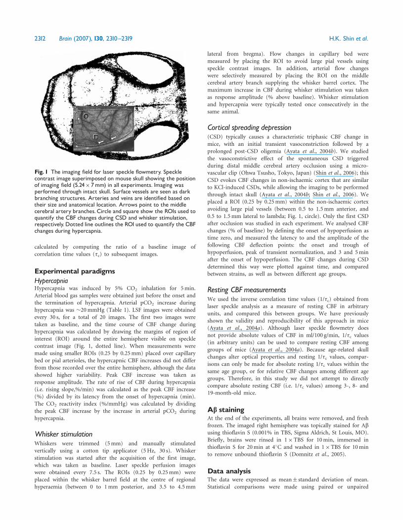
Fig. 1 The imaging field for laser speckle flowmetry. Specklecontrast image superimposed on mouse skull showing the positionof imaging field (5.24� 7mm) in all experiments. Imaging wasperformed through intact skull. Surface vessels are seen as darkbranching structures. Arteries and veins are identified based ontheir size and anatomical location. Arrows point to the middlecerebral artery branches.Circle and square show the ROIs used toquantify the CBF changes during CSD and whisker stimulation,respectively.Dotted line outlines the ROI used to quantify the CBFchanges during hypercapnia.
2312 Brain (2007), 130, 2310^2319 H.K. Shin et al.
Student’s t-test, one-way ANOVA, or two-way ANOVA forrepeated measures followed by Student–Newman–Keuls test.P50.05 was considered statistically significant.
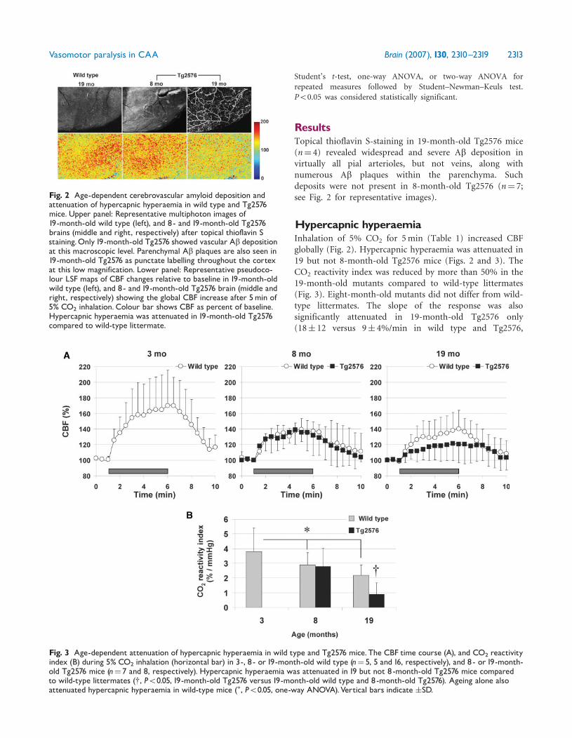
ResultsTopical thioflavin S-staining in 19-month-old Tg2576 mice(n¼ 4) revealed widespread and severe Ab deposition invirtually all pial arterioles, but not veins, along withnumerous Ab plaques within the parenchyma. Suchdeposits were not present in 8-month-old Tg2576 (n¼ 7;see Fig. 2 for representative images).
Hypercapnic hyperaemiaInhalation of 5% CO2 for 5min (Table 1) increased CBFglobally (Fig. 2). Hypercapnic hyperaemia was attenuated in19 but not 8-month-old Tg2576 mice (Figs. 2 and 3). TheCO2 reactivity index was reduced by more than 50% in the19-month-old mutants compared to wild-type littermates(Fig. 3). Eight-month-old mutants did not differ from wild-type littermates. The slope of the response was alsosignificantly attenuated in 19-month-old Tg2576 only(18� 12 versus 9� 4%/min in wild type and Tg2576,
Fig. 3 Age-dependent attenuation of hypercapnic hyperaemia in wild type and Tg2576 mice.The CBF time course (A), and CO2 reactivityindex (B) during 5% CO2 inhalation (horizontal bar) in 3-, 8 - or 19-month-old wild type (n¼ 5, 5 and 16, respectively), and 8- or 19-month-old Tg2576 mice (n¼ 7 and 8, respectively). Hypercapnic hyperaemia was attenuated in 19 but not 8-month-old Tg2576 mice comparedto wild-type littermates (y, P50.05, 19-month-old Tg2576 versus 19-month-old wild type and 8-month-old Tg2576). Ageing alone alsoattenuated hypercapnic hyperaemia in wild-type mice (�, P50.05, one-way ANOVA).Vertical bars indicate �SD.
Fig. 2 Age-dependent cerebrovascular amyloid deposition andattenuation of hypercapnic hyperaemia in wild type and Tg2576mice.Upper panel: Representative multiphoton images of19-month-old wild type (left), and 8- and 19-month-old Tg2576brains (middle and right, respectively) after topical thioflavin Sstaining.Only19-month-old Tg2576 showed vascular Ab depositionat this macroscopic level. Parenchymal Ab plaques are also seen in19-month-old Tg2576 as punctate labelling throughout the cortexat this low magnification. Lower panel: Representative pseudoco-lour LSF maps of CBF changes relative to baseline in 19-month-oldwild type (left), and 8- and19-month-old Tg2576 brain (middle andright, respectively) showing the global CBF increase after 5min of5% CO2 inhalation. Colour bar shows CBF as percent of baseline.Hypercapnic hyperaemia was attenuated in 19-month-old Tg2576compared to wild-type littermate.
Vasomotor paralysis in CAA Brain (2007), 130, 2310^2319 2313
respectively; P50.05). Interestingly, ageing alone attenuatedthe response to hypercapnia in wild-type mice (P50.05;one-way ANOVA).
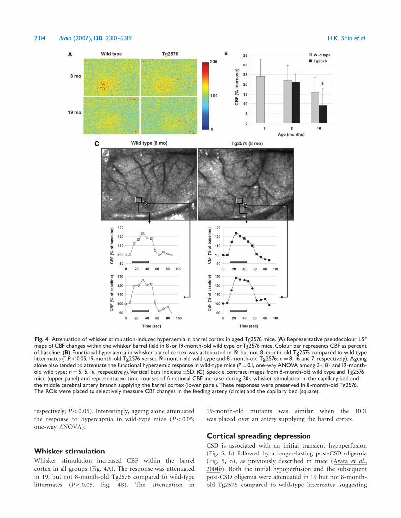
Whisker stimulationWhisker stimulation increased CBF within the barrelcortex in all groups (Fig. 4A). The response was attenuatedin 19, but not 8-month-old Tg2576 compared to wild-typelittermates (P50.05, Fig. 4B). The attenuation in
19-month-old mutants was similar when the ROIwas placed over an artery supplying the barrel cortex.
Cortical spreading depressionCSD is associated with an initial transient hypoperfusion(Fig. 5, h) followed by a longer-lasting post-CSD oligemia(Fig. 5, o), as previously described in mice (Ayata et al.,2004b). Both the initial hypoperfusion and the subsequentpost-CSD oligemia were attenuated in 19 but not 8-month-old Tg2576 compared to wild-type littermates, suggesting
Fig. 4 Attenuation of whisker stimulation-induced hyperaemia in barrel cortex in aged Tg2576 mice. (A) Representative pseudocolour LSFmaps of CBF changes within the whisker barrel field in 8-or19-month-old wild type orTg2576 mice.Colour bar represents CBF as percentof baseline. (B) Functional hyperaemia in whisker barrel cortex was attenuated in 19, but not 8-month-old Tg2576 compared to wild-typelittermates (�,P50.05, 19-month-old Tg2576 versus 19-month-old wild type and 8-month-old Tg2576; n¼ 8, 16 and 7, respectively). Ageingalone also tended to attenuate the functional hyperaemic response in wild-typemice (P¼ 0.1, one-way ANOVA among 3-, 8- and19-month-old wild type; n¼ 5, 5, 16, respectively).Vertical bars indicate �SD. (C) Speckle contrast images from 8-month-old wild type and Tg2576mice (upper panel) and representative time courses of functional CBF increase during 30 s whisker stimulation in the capillary bed andthe middle cerebral artery branch supplying the barrel cortex (lower panel). These responses were preserved in 8-month-old Tg2576.The ROIs were placed to selectively measure CBF changes in the feeding artery (circle) and the capillary bed (square).
2314 Brain (2007), 130, 2310^2319 H.K. Shin et al.
that the ability of cerebral vessels to constrict may also beimpaired after amyloid deposition (P50.05, two-wayANOVA for repeated measures; Fig. 5). In wild-type mice,age alone did not affect the CBF response to CSD (P¼ 0.45,two way ANOVA for repeated measures among 3-, 8- and19-month-old wild-type mice; data not shown), contrastingwith the impact of aging on hypercapnic hyperaemia.
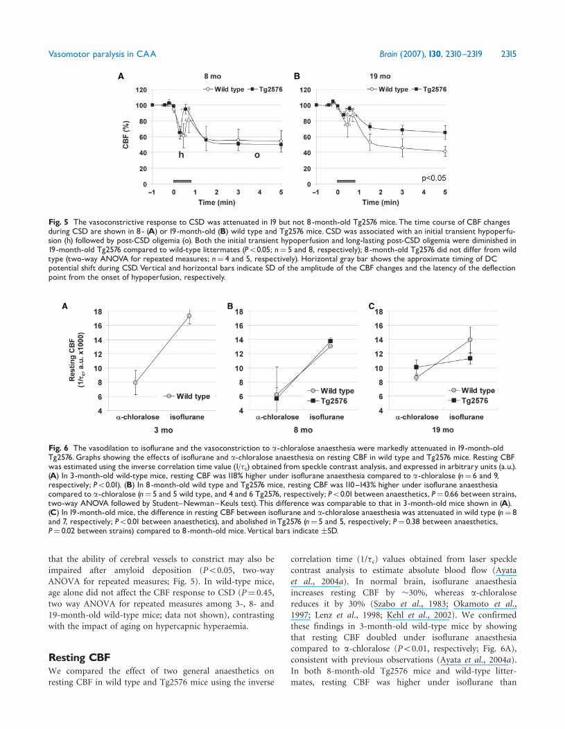
Resting CBFWe compared the effect of two general anaesthetics onresting CBF in wild type and Tg2576 mice using the inverse
correlation time (1/�c) values obtained from laser specklecontrast analysis to estimate absolute blood flow (Ayataet al., 2004a). In normal brain, isoflurane anaesthesiaincreases resting CBF by �30%, whereas a-chloralosereduces it by 30% (Szabo et al., 1983; Okamoto et al.,1997; Lenz et al., 1998; Kehl et al., 2002). We confirmedthese findings in 3-month-old wild-type mice by showingthat resting CBF doubled under isoflurane anaesthesiacompared to a-chloralose (P50.01, respectively; Fig. 6A),consistent with previous observations (Ayata et al., 2004a).In both 8-month-old Tg2576 mice and wild-type litter-mates, resting CBF was higher under isoflurane than
Fig. 6 The vasodilation to isoflurane and the vasoconstriction to a-chloralose anaesthesia were markedly attenuated in 19-month-oldTg2576.Graphs showing the effects of isoflurane and a-chloralose anaesthesia on resting CBF in wild type and Tg2576 mice. Resting CBFwas estimated using the inverse correlation time value (1/�c) obtained from speckle contrast analysis, and expressed in arbitrary units (a.u.).(A) In 3-month-old wild-type mice, resting CBF was 118% higher under isoflurane anaesthesia compared to a-chloralose (n¼ 6 and 9,respectively; P50.01). (B) In 8-month-old wild type and Tg2576 mice, resting CBF was 110^143% higher under isoflurane anaesthesiacompared to a-chloralose (n¼ 5 and 5 wild type, and 4 and 6 Tg2576, respectively; P50.01between anaesthetics, P¼ 0.66 between strains,two-way ANOVA followed by Student^Newman^Keuls test). This difference was comparable to that in 3-month-old mice shown in (A).(C) In 19-month-old mice, the difference in resting CBF between isoflurane and a-chloralose anaesthesia was attenuated in wild type (n¼ 8and 7, respectively; P50.01 between anaesthetics), and abolished inTg2576 (n¼ 5 and 5, respectively; P¼ 0.38 between anaesthetics,P¼ 0.02 between strains) compared to 8-month-old mice.Vertical bars indicate �SD.
Fig. 5 The vasoconstrictive response to CSD was attenuated in 19 but not 8-month-old Tg2576 mice. The time course of CBF changesduring CSD are shown in 8- (A) or 19-month-old (B) wild type and Tg2576 mice. CSD was associated with an initial transient hypoperfu-sion (h) followed by post-CSD oligemia (o). Both the initial transient hypoperfusion and long-lasting post-CSD oligemia were diminished in19-month-old Tg2576 compared to wild-type littermates (P50.05; n¼ 5 and 8, respectively); 8 -month-old Tg2576 did not differ from wildtype (two-way ANOVA for repeated measures; n¼ 4 and 5, respectively). Horizontal gray bar shows the approximate timing of DCpotential shift during CSD.Vertical and horizontal bars indicate SD of the amplitude of the CBF changes and the latency of the deflectionpoint from the onset of hypoperfusion, respectively.
Vasomotor paralysis in CAA Brain (2007), 130, 2310^2319 2315

a-chloralose anaesthesia, and did not differ between strains(P40.05, wild type versus Tg2576; Fig. 6B). In 19-month-old wild-type mice, the difference in resting CBF betweenisoflurane and a-chloralose anaesthesia was attenuatedcompared to 3- and 8-month-old wild-type mice(P50.05; Fig. 6C). More importantly, resting CBF in19-month-old mutants did not differ between the twoanaesthetics (P¼ 0.38); both isoflurane-induced hyperaemiaand a-chloralose-induced oligemia were diminished(Fig. 6C).
DiscussionHere, we confirm that vasomotor function is impaired in atransgenic mouse model of CAA, but show that this doesnot manifest itself before the overt deposition of amyloid incerebral blood vessels under the stated experimentalconditions. We tested the blood flow responses to diversephysiological and pharmacological stimuli that promotevasodilation (i.e. hypercapnia, whisker barrel field activa-tion and isoflurane anaesthesia), and vasoconstriction(i.e. cortical spreading depression and a-chloralose anaes-thesia), and showed that hyperaemic and oligemicresponses are both impaired in 19-month-old mutantmice. Whether this vasomotor paralysis is due to decreasedcompliance or other mechanical properties of amyloid-laden vessels, or due to reduced reactivity or number ofsmooth muscle cells in advanced stages of CAA remains tobe determined.
Vasodilatory dysfunctionImpaired cerebrovascular vasodilation to physiological(functional cortical activation, hypercapnia, hypotension)and pharmacological stimuli (acetylcholine, bradykinin,A23187) was previously shown in mouse models of CAA,developing as early as 2–3 months of age (Iadecola et al.,1999; Niwa et al., 2000a, b, 2002a; Park et al., 2004, 2005).Using three distinct vasodilator stimuli, we found thatcerebral hyperaemic responses were attenuated in19-month-old Tg2576 mice with severe vascular amyloiddeposition. In contrast, blood flow responses prior tovascular amyloid deposition (8 months) did not differ fromwild-type littermates. We showed that flow changes weresimilarly attenuated in large pial arteries in 19 but not8-month-old Tg2576, paralleling flow changes measured inbrain parenchyma. Therefore, our data challenges theprevailing notion that elevated soluble Ab is sufficient tocause cerebrovascular dysfunction.Differences in experimental methodology may partly
explain the discrepancy in the age of onset of impairedhypercapnic or functional hyperaemia in Tg2576 mice.Previous studies showing impaired vasodilator responses in2–3-month-old CAA mutant models employed an opencranial window preparation (Zhang et al., 1997; Iadecolaet al., 1999; Niwa et al., 2000b, 2002a), compared to
imaging through an intact skull in the present study.Imaging the brain through an intact skull eliminates thepossibility of injury during craniotomy and dural excision,exposure to air, cortical temperature fluctuations andaltered intracranial pressure. It is possible that craniotomyand open skull preparation revealed a difference thatbecame manifest perhaps via increased free radical produc-tion and endothelial dysfunction by Ab (Iadecola et al.,1999; Park et al., 2004, 2005). More studies will be neededto clarify this point.
Our data suggest that mild to moderate elevation of Ablevels in 8-month-old or younger Tg2576 mice areinsufficient to impair cerebrovascular dilation to thetested physiological and pharmacological stimuli, and thatthe deposition of vascular amyloid may be a critical factor.Consistent with this, cerebral arteriolar dilation to topicalpharmacological vasodilators acetylcholine- and sodiumnitroprusside did not differ between 6-month-old Tg2576mice and wild-type littermates, but was significantlyattenuated in 14-month-old Tg2576 mice, when testedusing a closed cranial window (Christie et al., 2001). Inanother mouse model expressing human APP with Swedishdouble mutation under neuron-specific Thy-1 promoter(APP23) (Calhoun et al., 1999; Burgermeister et al., 2000;Winkler et al., 2001), magnetic resonance angiographyshowed flow voids corresponding to slow or microturbulentflow, and corrosion casts demonstrated focal constriction,vessel elimination and rearrangement in major cerebralvessels in 60% of 20-month-old transgenic mice; none ofthe 11-month-old APP23 transgenic mice displayed any ofthese abnormalities (Beckmann et al., 2003). In the samemouse model, hyperaemia following bicuculline- or acet-azolamide was attenuated in 15-month-old or oldermice showing deposits, but not in 8-month-old micewithout amyloid deposition (Mueggler et al., 2002). TheAPP23 model develops vascular amyloid depositionthat is qualitatively and quantitatively comparable toTg2576 (Calhoun et al., 1999). Our data extends existingknowledge by non-invasively showing vasomotor paralysis(i.e. impaired dilation as well as constriction) to physiolog-ical stimuli, that does not become manifest before vascularamyloid deposition. However, not all mouse mutationscausing Ab deposition lead to vascular dysfunction. In micewith CAA overexpressing the London mutant of humanAPP (Moechars et al., 1999), resting CBF and hypercapnichyperaemia (7% CO2 for 2min) were similar to wild-typelittermates even at 20–24 months of age (Van Dorpe et al.,2000), suggesting that the relationship between vascular Abdeposition and vasomotor dysfunction may be complex.It should be noted that different transgenic models havedifferent kinetics of disease development, tissue Ab increaseand anatomic distribution of deposition, which makesdirect comparisons across mouse models difficult.
Functional activation evoking the whisker barrel bloodflow response tests the integrity of both vascular as well asparenchymal elements. It remains possible that the
2316 Brain (2007), 130, 2310^2319 H.K. Shin et al.

attenuated CBF response to whisker stimulation in19-month-old Tg2576 mice may be due to abnormalfunctional or metabolic activation of barrel cortex as aconsequence of elevated Ab levels and parenchymal plaques.Cerebral metabolism is not normal as evidencedby attenuated resting cerebral glucose utilization in2–3-month-old Tg2576 mice (Niwa et al., 2002b).Electrophysiologically, intrinsic membrane potential proper-ties and overall synaptic innervation did not differ,although there was increased synaptic jitter, when neuronalresponses from aged (414 months) Tg2576 and wild-typelittermates were compared (Stern et al., 2004). Whetherfunctional electrophysiological changes impact metabolic orvascular coupling mechanisms in Tg2576 remains to bedetermined. Regardless of its mechanisms, impaired vaso-dilation in advanced CAA may predispose to chronic orepisodic metabolic mismatch thereby contributing toparenchymal neuropathology in AD.
Vasoconstrictive dysfunctionOur data also suggest that arterial constriction is alsoimpaired during CSD or under a-chloralose anaesthesia in19 but not 8-month-old Tg2576. These two stimuliconstrict cerebral vessels via different mechanisms. TheCBF response to CSD in mice is characterized by a transientand profound vasoconstriction during the DC shift, that isbelieved to be directly mediated by elevated extracellularpotassium concentration (50mM or more) (Godfraindet al., 1986; Ayata et al., 2004b; Windmuller et al., 2005;Shin et al., 2006). This is followed by a longer lasting post-CSD oligemia (Fig. 5) which may involve vasoconstrictiveprostanoids (Shibata et al., 1992). a-Chloralose, on theother hand, is believed to reduce CBF via suppression ofcerebral metabolism (Sandor et al., 1977; Nakao et al.,2001). Hence, our data demonstrate cerebral vasoconstric-tive dysfunction, regardless of the mediators and mechan-isms involved, which develops only after vascular amyloiddeposition in aged Tg2576. This is clinically relevantbecause impaired vasoconstriction may reduce the abilityof cerebral arterioles to dampen sudden surges in micro-circulatory perfusion pressure during hypertensive episodes,and predispose to intracerebral haemorrhages.
Correlation of pathology andcerebrovascular dysfunctionWe chose to study Tg2576 mice at 8 and 19 months of agein order to test the impact of elevated Ab without CAA andwith severe CAA, respectively. Vessels from 8-month-oldTg2576 mice do not harbour amyloid deposits, whereas in19-month-old Tg2576 more than 90% of pial arteries andarterioles are heavily laden with amyloid deposits (Fig. 2),with loss of smooth muscle cells (Domnitz et al., 2005;Robbins et al., 2006). In contrast, parenchymal soluble Ablevels are already elevated at 2–3 months of age (Niwaet al., 2000b), and increase exponentially starting around
6–7 months (Kawarabayashi et al., 2001). Our data suggestthat elevated soluble Ab alone without deposition isinsufficient to cause vasomotor dysfunction as testedherein. Because soluble Ab40 and Ab42 levels continue torise between 8 and 19 months of age, and 19-month-oldTg2576 mice also harbour parenchymal plaques, they maycontribute to the vasomotor dysfunction in 19-month-oldTg2576 mice.
In humans, the incidence of CAA increases with age; halfof all individuals over age 70 years and 80–90% of ADpatients have varying degrees of CAA (Mandybur, 1975;Vinters, 1987; Ellis et al., 1996). Cerebral amyloid angio-pathy often becomes symptomatic in patients with severevascular Ab deposition, usually after age 70 years. Althoughrecurrent lobar haemorrhages are the typical presentingfeature, patients with CAA also have a higher risk ofdeveloping ischaemic stroke, and leukoencephalopathy(Olichney et al., 1995, 1997; Greenberg, 2002). Cerebralamyloid angiopathy has been proposed as an exacerbatingfactor for cognitive dysfunction in AD based on data frompopulation studies and familial CAA syndromes (Snowdonet al., 1997; Grabowski et al., 2001; Natte et al., 2001;Pfeifer et al., 2002). However, it is unclear whether thenegative impact of CAA on cognition in AD patients simplyreflects the cumulative effect of cerebral infarcts, or whetherCAA causes chronic cerebrovascular dysregulation in theabsence of overt infarction. The latter view is supported byexperimental data showing vascular impairment by Ab andin transgenic mouse models of AD and CAA (Thomaset al., 1996; Crawford et al., 1998; Iadecola et al., 1999;Niwa et al., 2000a, 2001; Paris et al., 2003). Indeed in somestudies vascular dysfunction manifested long before thedevelopment of CAA, suggesting that elevated Ab levelsmay be sufficient to disrupt vascular function (Zhang et al.,1997; Iadecola et al., 1999; Niwa et al., 2000b, 2002a, b).However, pathologically demonstrated cerebrovascularamyloid deposition is an important independent riskfactor for cognitive impairment (odds ratio 9.3) (2001;Greenberg et al., 2004; Greenberg, 2006) as well assubcortical white matter disease (Haglund and Englund,2002). The data shown in the Tg2576 are consistentwith this notion, and suggest that Ab impacts cerebrovas-cular function only after vascular deposition. Whethervascular deposition is also a prerequisite for cerebrovasculardysfunction in patients with AD remains to be tested.
Effects of ageing on cerebrovascular functionWe observed that aged wild-type mice also displayedattenuated hypercapnic hyperaemia, metabolic couplingand anaesthetic influences on resting CBF. These datashould be interpreted with caution because imaging wasperformed through intact skull, and age-dependent changesin skull properties might have influenced the relative CBFchanges recorded. However, hypercapnic hyperaemia wasattenuated in wild-type mice only between 3 and 8 months
Vasomotor paralysis in CAA Brain (2007), 130, 2310^2319 2317
of age, whereas functional hyperaemia was attenuated onlybetween 8 and 19 months of age; CSD-induced CBFchanges did not differ among the age groups. Hence, ourdata suggest that the age-dependent cerebrovascular dys-function in these experiments is not merely a non-specificand uniform attenuation of all ‘measured’ relative CBFresponses due to age-dependent changes in optical proper-ties of the skull. The impact of ageing on cerebrovascularfunction was previously studied in both cerebral andsystemic vessels. For example, endothelium-dependentcerebral vasodilation was significantly reduced in aged rats(22–24mo) compared to young adult rats (6–8mo) whenstudied using closed cranial windows (Mayhan et al., 1990).More recently, functional hyperaemia, and acetylcholine-and bradykinin-induced CBF increases were age-dependently attenuated in 12- and 24-month-old wild-typemice (Park et al., 2006). Our data confirm these reports,and underscore the importance of age when studying theimpact of slowly progressive cerebrovascular diseases ofageing on CBF regulation.
AcknowledgementsThis work was supported by the American HeartAssociation (0335519N, Ayata), National Institutes ofHealth (P50 NS10828 and PO1 NS35611, Moskowitz;Bacskai; AG08487, Hyman) and Fundacion Caja Madrid(Garcia-Alloza). Funding to pay the Open Access publica-tion charges for this article was provided by the AmericanHeart Association.
ReferencesPathological correlates of late-onset dementia in a multicentre, commu-
nity-based population in England and Wales. Neuropathology Group of
the Medical Research Council Cognitive Function and Ageing Study
(MRC CFAS). Lancet 2001; 357: 169–75.
Ayata C, Dunn AK, Gursoy OY, Huang Z, Boas DA, Moskowitz MA. Laser
speckle flowmetry for the study of cerebrovascular physiology in normal
and ischemic mouse cortex. J Cereb Blood Flow Metab 2004a; 24:
744–55.
Ayata C, Shin HK, Salomone S, Ozdemir-Gursoy Y, Boas DA, Dunn AK,
et al. Pronounced hypoperfusion during spreading depression in mouse
cortex. J Cereb Blood Flow Metab 2004b; 24: 1172–82.
Beckmann N, Schuler A, Mueggler T, Meyer EP, Wiederhold KH,
Staufenbiel M, et al. Age-dependent cerebrovascular abnormalities and
blood flow disturbances in APP23 mice modeling Alzheimer’s disease.
J Neurosci 2003; 23: 8453–9.
Burgermeister P, Calhoun ME, Winkler DT, Jucker M. Mechanisms of
cerebrovascular amyloid deposition. Lessons from mouse models. Ann
N Y Acad Sci 2000; 903: 307–16.
Calhoun ME, Burgermeister P, Phinney AL, Stalder M, Tolnay M,
Wiederhold KH, et al. Neuronal overexpression of mutant amyloid
precursor protein results in prominent deposition of cerebrovascular
amyloid. Proc Natl Acad Sci U S A 1999; 96: 14088–93.
Christie R, Yamada M, Moskowitz M, Hyman B. Structural and functional
disruption of vascular smooth muscle cells in a transgenic mouse model
of amyloid angiopathy. Am J Pathol 2001; 158: 1065–71.
Crawford F, Suo Z, Fang C, Mullan M. Characteristics of the in vitro
vasoactivity of beta-amyloid peptides. Exp Neurol 1998; 150: 159–68.
Dalkara T, Irikura K, Huang Z, Panahian N, Moskowitz MA.
Cerebrovascular responses under controlled and monitored
physiological conditions in the anesthetized mouse. J Cereb Blood
Flow Metab 1995; 15: 631–8.
Domnitz SB, Robbins EM, Hoang AW, Garcia-Alloza M, Hyman BT,
Rebeck GW, et al. Progression of cerebral amyloid angiopathy in
transgenic mouse models of Alzheimer disease. J Neuropathol Exp
Neurol 2005; 64: 588–94.
Dunn AK, Bolay H, Moskowitz MA, Boas DA. Dynamic imaging of
cerebral blood flow using laser speckle. J Cereb Blood Flow Metab 2001;
21: 195–201.
Ellis RJ, Olichney JM, Thal LJ, Mirra SS, Morris JC, Beekly D, et al.
Cerebral amyloid angiopathy in the brains of patients with Alzheimer’s
disease: the CERAD experience, Part XV. Neurology 1996; 46: 1592–6.
Godfraind T, Miller R, Wibo M. Calcium antagonism and calcium entry
blockade. Pharmacol Rev 1986; 38: 321–416.
Grabowski TJ, Cho HS, Vonsattel JP, Rebeck GW, Greenberg SM. Novel
amyloid precursor protein mutation in an Iowa family with dementia
and severe cerebral amyloid angiopathy. Ann Neurol 2001; 49: 697–705.
Greenberg SM. Cerebral amyloid angiopathy and vessel dysfunction.
Cerebrovasc Dis 2002; 13 (Suppl 2): 42–7.
Greenberg SM. Small vessels, big problems. N Engl J Med 2006; 354:
1451–3.
Greenberg SM, Gurol ME, Rosand J, Smith EE. Amyloid angiopathy-
related vascular cognitive impairment. Stroke 2004; 35: 2616–9.
Greenberg SM, Rebeck GW, Vonsattel JP, Gomez-Isla T, Hyman BT.
Apolipoprotein E epsilon 4 and cerebral hemorrhage associated with
amyloid angiopathy. Ann Neurol 1995; 38: 254–9.
Haglund M, Englund E. Cerebral amyloid angiopathy, white matter lesions
and Alzheimer encephalopathy - a histopathological assessment. Dement
Geriatr Cogn Disord 2002; 14: 161–6.
Hsiao K, Chapman P, Nilsen S, Eckman C, Harigaya Y, Younkin S, et al.
Correlative memory deficits, Abeta elevation, and amyloid plaques in
transgenic mice. Science 1996; 274: 99–102.
Iadecola C. Neurovascular regulation in the normal brain and in
Alzheimer’s disease. Nat Rev Neurosci 2004; 5: 347–60.
Iadecola C, Zhang F, Niwa K, Eckman C, Turner SK, Fischer E, et al.
SOD1 rescues cerebral endothelial dysfunction in mice overexpressing
amyloid precursor protein. Nat Neurosci 1999; 2: 157–61.
Kawarabayashi T, Younkin LH, Saido TC, Shoji M, Ashe KH, Younkin SG.
Age-dependent changes in brain, CSF, and plasma amyloid (beta)
protein in the Tg2576 transgenic mouse model of Alzheimer’s disease.
J Neurosci 2001; 21: 372–81.
Kehl F, Shen H, Moreno C, Farber NE, Roman RJ, Kampine JP, et al.
Isoflurane-induced cerebral hyperemia is partially mediated by nitric
oxide and epoxyeicosatrienoic acids in mice in vivo. Anesthesiology
2002; 97: 1528–33.
Lacombe P, Oligo C, Domenga V, Tournier-Lasserve E, Joutel A. Impaired
cerebral vasoreactivity in a transgenic mouse model of cerebral
autosomal dominant arteriopathy with subcortical infarcts and leu-
koencephalopathy arteriopathy. Stroke 2005; 36: 1053–8.
Lenz C, Rebel A, van Ackern K, Kuschinsky W, Waschke KF. Local
cerebral blood flow, local cerebral glucose utilization, and flow-
metabolism coupling during sevoflurane versus isoflurane anesthesia in
rats. Anesthesiology 1998; 89: 1480–8.
Mandybur TI. The incidence of cerebral amyloid angiopathy in
Alzheimer’s disease. Neurology 1975; 25: 120–6.
Mayhan WG, Faraci FM, Baumbach GL, Heistad DD. Effects of aging on
responses of cerebral arterioles. Am J Physiol 1990; 258: H1138–43.
Moechars D, Dewachter I, Lorent K, Reverse D, Baekelandt V, Naidu A,
et al. Early phenotypic changes in transgenic mice that overexpress
different mutants of amyloid precursor protein in brain. J Biol Chem
1999; 274: 6483–92.
Mueggler T, Sturchler-Pierrat C, Baumann D, Rausch M, Staufenbiel M,
Rudin M. Compromised hemodynamic response in amyloid precursor
protein transgenic mice. J Neurosci 2002; 22: 7218–24.
Nakao Y, Itoh Y, Kuang TY, Cook M, Jehle J, Sokoloff L. Effects of
anesthesia on functional activation of cerebral blood flow and
metabolism. Proc Natl Acad Sci U S A 2001; 98: 7593–8.
2318 Brain (2007), 130, 2310^2319 H.K. Shin et al.

Natte R, Maat-Schieman ML, Haan J, Bornebroek M, Roos RA, van
Duinen SG. Dementia in hereditary cerebral hemorrhage with
amyloidosis-Dutch type is associated with cerebral amyloid angiopathy
but is independent of plaques and neurofibrillary tangles. Ann Neurol
2001; 50: 765–72.
Niwa K, Carlson GA, Iadecola C. Exogenous A beta1-40 reproduces
cerebrovascular alterations resulting from amyloid precursor
protein overexpression in mice. J Cereb Blood Flow Metab 2000a; 20:
1659–68.
Niwa K, Kazama K, Younkin L, Younkin SG, Carlson GA, Iadecola C.
Cerebrovascular autoregulation is profoundly impaired in mice over-
expressing amyloid precursor protein. Am J Physiol Heart Circ Physiol
2002a; 283: H315–23.
Niwa K, Kazama K, Younkin SG, Carlson GA, Iadecola C. Alterations in
cerebral blood flow and glucose utilization in mice overexpressing the
amyloid precursor protein. Neurobiol Dis 2002b; 9: 61–8.
Niwa K, Porter VA, Kazama K, Cornfield D, Carlson GA, Iadecola C.
A beta-peptides enhance vasoconstriction in cerebral circulation. Am J
Physiol Heart Circ Physiol 2001; 281: H2417–24.
Niwa K, Younkin L, Ebeling C, Turner SK, Westaway D, Younkin S, et al.
Abeta 1-40-related reduction in functional hyperemia in mouse
neocortex during somatosensory activation. Proc Natl Acad Sci U S A
2000b; 97: 9735–40.
Okamoto H, Meng W, Ma J, Ayata C, Roman RJ, Bosnjak ZJ, et al.
Isoflurane-induced cerebral hyperemia in neuronal nitric oxide synthase
gene deficient mice. Anesthesiology 1997; 86: 875–84.
Olichney JM, Ellis RJ, Katzman R, Sabbagh MN, Hansen L. Types
of cerebrovascular lesions associated with severe cerebral
amyloid angiopathy in Alzheimer’s disease. Ann N Y Acad Sci 1997;
826: 493–7.
Olichney JM, Hansen LA, Hofstetter CR, Grundman M, Katzman R,
Thal LJ. Cerebral infarction in Alzheimer’s disease is associated with
severe amyloid angiopathy and hypertension. Arch Neurol 1995; 52:
702–8.
Paris D, Humphrey J, Quadros A, Patel N, Crescentini R, Crawford F,
et al. Vasoactive effects of A beta in isolated human cerebrovessels and
in a transgenic mouse model of Alzheimer’s disease: role of inflamma-
tion. Neurol Res 2003; 25: 642–51.
Park L, Anrather J, Forster C, Kazama K, Carlson GA, Iadecola C. Abeta-
induced vascular oxidative stress and attenuation of functional
hyperemia in mouse somatosensory cortex. J Cereb Blood Flow Metab
2004; 24: 334–42.
Park L, Anrather J, Zhou P, Frys K, Pitstick R, Younkin S, et al. NADPH
oxidase-derived reactive oxygen species mediate the cerebrovascular
dysfunction induced by the amyloid beta peptide. J Neurosci 2005; 25:
1769–77.
Park L, Zhou P, Anrather J, Iadecola C. Reactive oxygen species (ROS)
derived from a nox2-containing NADPH oxidase disrupt neurovascular
regulation in normal aging. Society for Neuroscience Annual
Meeting 2006.
Pfeifer LA, White LR, Ross GW, Petrovitch H, Launer LJ. Cerebral
amyloid angiopathy and cognitive function: the HAAS autopsy study.
Neurology 2002; 58: 1629–34.
Robbins EM, Betensky RA, Domnitz SB, Purcell SM, Garcia-Alloza M,
Greenberg C, et al. Kinetics of cerebral amyloid angiopathy progression
in a transgenic mouse model of Alzheimer disease. J Neurosci 2006; 26:
365–71.
Sandor P, Nyary I, Reivich M, Kovach AG. Comparative effects of
chloralose anesthesia and Sernylan analgesia on cerebral blood flow,
CO2 responsiveness, and brain metabolism in the baboon. Stroke 1977;
8: 432–6.
Shibata M, Leffler CW, Busija DW. Pial arteriolar constriction following
cortical spreading depression is mediated by prostanoids. Brain Res
1992; 572: 190–7.
Shin HK, Dunn AK, Jones PB, Boas DA, Moskowitz MA, Ayata C.
Vasoconstrictive neurovascular coupling during focal ischemic depolar-
izations. J Cereb Blood Flow Metab 2006; 26: 1018–30.
Snowdon DA, Greiner LH, Mortimer JA, Riley KP, Greiner PA,
Markesbery WR. Brain infarction and the clinical expression of
Alzheimer disease. The Nun Study. JAMA 1997; 277: 813–7.
Stern EA, Bacskai BJ, Hickey GA, Attenello FJ, Lombardo JA, Hyman BT.
Cortical synaptic integration in vivo is disrupted by amyloid-beta
plaques. J Neurosci 2004; 24: 4535–40.
Szabo L, Kovach AG, Babosa M. Local effect of anaesthesia on cerebral
blood flow in the rat. Acta Physiol Hung 1983; 62: 113–21.
Thomas T, Thomas G, McLendon C, Sutton T, Mullan M. beta-Amyloid-
mediated vasoactivity and vascular endothelial damage. Nature 1996;
380: 168–71.
Van Dorpe J, Smeijers L, Dewachter I, Nuyens D, Spittaels K, Van Den
Haute C, et al. Prominent cerebral amyloid angiopathy in transgenic
mice overexpressing the london mutant of human APP in neurons.
Am J Pathol 2000; 157: 1283–98.
Vinters HV. Cerebral amyloid angiopathy. A critical review. Stroke 1987;
18: 311–24.
Vonsattel JP, Myers RH, Hedley-Whyte ET, Ropper AH, Bird ED,
Richardson EP, Jr. Cerebral amyloid angiopathy without and with
cerebral hemorrhages: a comparative histological study. Ann Neurol
1991; 30: 637–49.
Windmuller O, Lindauer U, Foddis M, Einhaupl KM, Dirnagl U,
Heinemann U, et al. Ion changes in spreading ischaemia induce rat
middle cerebral artery constriction in the absence of NO. Brain 2005;
128: 2042–51.
Winkler DT, Bondolfi L, Herzig MC, Jann L, Calhoun ME,
Wiederhold KH, et al. Spontaneous hemorrhagic stroke in a
mouse model of cerebral amyloid angiopathy. J Neurosci 2001; 21:
1619–27.
Zhang F, Eckman C, Younkin S, Hsiao KK, Iadecola C. Increased
susceptibility to ischemic brain damage in transgenic mice over-
expressing the amyloid precursor protein. J Neurosci 1997; 17: 7655–61.
Vasomotor paralysis in CAA Brain (2007), 130, 2310^2319 2319