Combined triple treatment of brin amyloid - Research Square
38
Combined triple treatment of ヲbrin amyloid microclots and platelet pathology in individuals with Long COVID/ Post-Acute Sequelae of COVID-19 (PASC) can resolve their persistent symptoms Etheresia Pretorius ( [email protected] ) Stellenbosch University https://orcid.org/0000-0002-9108-2384 Chantelle Venter Stellenbosch University Gert Jacobus Laubscher Mediclinic Stellenbosch Maritha J. Kotze Stellenbosch University Kelebogile Moremi Stellenbosch University Sunday Oladejo Stellenbosch University Liam R. Watson Stellenbosch University Kanshu Rajaratnam Stellenbosch University Bruce W. Watson Stellenbosch University Douglas B. Kell University of Liverpool https://orcid.org/0000-0001-5838-7963 Article Keywords: Long COVID/PASC, Symptoms, Co-morbidities, Amyloid Fibrin(ogen), Hyperactivated platelets Posted Date: December 28th, 2021 DOI: https://doi.org/10.21203/rs.3.rs-1205453/v1 License: This work is licensed under a Creative Commons Attribution 4.0 International License. Read Full License
-
Upload
khangminh22 -
Category
Documents
-
view
1 -
download
0
Transcript of Combined triple treatment of brin amyloid - Research Square

Combined triple treatment of �brin amyloidmicroclots and platelet pathology in individuals withLong COVID/ Post-Acute Sequelae of COVID-19(PASC) can resolve their persistent symptomsEtheresia Pretorius ( [email protected] )
Stellenbosch University https://orcid.org/0000-0002-9108-2384Chantelle Venter
Stellenbosch UniversityGert Jacobus Laubscher
Mediclinic StellenboschMaritha J. Kotze
Stellenbosch UniversityKelebogile Moremi
Stellenbosch UniversitySunday Oladejo
Stellenbosch UniversityLiam R. Watson
Stellenbosch UniversityKanshu Rajaratnam
Stellenbosch UniversityBruce W. Watson
Stellenbosch UniversityDouglas B. Kell
University of Liverpool https://orcid.org/0000-0001-5838-7963
Article
Keywords: Long COVID/PASC, Symptoms, Co-morbidities, Amyloid Fibrin(ogen), Hyperactivated platelets
Posted Date: December 28th, 2021
DOI: https://doi.org/10.21203/rs.3.rs-1205453/v1
License: This work is licensed under a Creative Commons Attribution 4.0 International License. Read Full License


1
Combined triple treatment of fibrin amyloid microclots and platelet
pathology in individuals with Long COVID/ Post-Acute Sequelae of
COVID-19 (PASC) can resolve their persistent symptoms
Etheresia Pretorius1*, Chantelle Venter1, Gert Jacobus Laubscher2, Maritha J Kotze3,4
Kelebogile Moremi4, Sunday Oladejo5, Liam R. Watson5, Kanshu Rajaratnam5, Bruce W.
Watson5, Douglas B. Kell1,6,7*
1Department of Physiological Sciences, Faculty of Science, Stellenbosch University, Stellenbosch, Private Bag X1 Matieland, 7602, South Africa; 2Mediclinic Stellenbosch, Stellenbosch 7600, South Africa 3 Division of Chemical Pathology, Department of Pathology, National Health Laboratory Service, Tygerberg Hospital, Cape Town 8000, South Africa. 4 Division of Chemical Pathology, Department of Pathology, Faculty of Medicine and Health Sciences, Stellenbosch University, Cape Town 8000, South Africa. 5Centre for AI Research, School for Data-Science & Computational Thinking, Stellenbosch University, Stellenbosch 7600, South Africa 6Department of Biochemistry and Systems Biology, Institute of Systems, Molecular and Integrative Biology, Faculty of Health and Life Sciences, University of Liverpool, L69 7ZB, UK. 7The Novo Nordisk Foundation Centre for Biosustainability, Technical University of Denmark, Kemitorvet 200, 2800 Kgs Lyngby, Denmark. *Corresponding authors:
*Etheresia Pretorius
Department of Physiological Sciences, Stellenbosch University, Private Bag X1 Matieland, 7602, SOUTH AFRICA [email protected] http://www.resiapretorius.net/ ORCID: 0000-0002-9108-2384
*Douglas B. Kell
Department of Biochemistry and Systems Biology, Institute of Systems, Molecular and Integrative Biology, Faculty of Health and Life Sciences, University of Liverpool, L69 7ZB, UK. [email protected] The Novo Nordisk Foundation Centre for Biosustainability, Technical University of Denmark, Kemitorvet 200, 2800 Kgs Lyngby, Denmark. ORCID: 0000-0001-5838-7963

2
ABSTRACT
We recognise that fibrin(ogen) amyloid microclots and platelet hyperactivation, that we have
previously observed in COVID-19 and Long COVID/Post-Acute Sequelae of COVID-19 (PASC)
patients, might form a suitable set of foci for the clinical treatment of the symptoms of long
COVID/PASC. We first report on the comorbidities and symptoms found in a cohort of 845
South African Long COVID/PASC patients who filled in the South African Long COVID/PASC
registry, of which hypertension and high cholesterol levels (dyslipidaemia) were the most
important comorbidities. The gender balance (70% female) and the most commonly reported
Long COVID/PASC symptoms (fatigue, brain fog, loss of concentration and forgetfulness,
shortness of breath, as well as joint and muscle pains) were comparable to those reported
elsewhere. This suggests that our sample was not at all atypical. Using a previously published
scoring system for fibrin amyloid microclots and platelet pathology, we analysed blood
samples from 70 patients, and report the presence of significant fibrin amyloid microclots and
platelet pathology in all cases; these were associated with Long COVID/PASC symptoms that
persisted after the recovery from acute COVID-19. A subset of 24 patients was treated with
one month of dual antiplatelet therapy (DAPT) (Clopidogrel 75mg/Aspirin 75mg) once a day,
as well as a direct oral anticoagulant (DOAC) (Apixiban) 5 mg twice a day. A proton pump
inhibitor (PPI) pantoprazole 40 mg/day was also prescribed for gastric protection. Such a
regime must only be followed under strict and qualified medical guidance to obviate any
dangers, especially haemorrhagic bleeding, and of the therapy as a whole.
Thromboelastography (TEG®) was used to assist in determining their clotting status. Each of
the 24 treated cases reported that their main symptoms were resolved and fatigue as the main
symptom was relieved, and this was also reflected in a decrease of both the fibrin amyloid
microclots and platelet pathology scores. Nine patients were genotyped for genetic variation
in homocysteine metabolism implicated in hypertension, a common COVID-19 co-morbidity
reported in both patients found to be homozygous for the risk-associated MTHFR 677 T-allele.
Fibrin amyloid microclots that block capillaries and inhibit the transport of O2 to tissues,
accompanied by platelet hyperactivation, provide a ready explanation for the symptoms of
Long COVID/PASC. The removal and reversal of these underlying epitheliopathies underlying
this provide an important treatment option that seems to be highly efficacious, and warrants
controlled clinical studies.
KEYWORDS
Long COVID/PASC; Symptoms, Co-morbidities, Amyloid Fibrin(ogen); Hyperactivated platelets

3
INTRODUCTION
As many of 30% of COVID-19 patients infected with the severe acute respiratory syndrome
coronavirus 2 (SARS-Cov-2) continue, and in some cases begin, to suffer a variety of
debilitating symptoms weeks or months after the acute phase of infection. The precise
definition of this Long COVID/ Post-Acute Sequelae of COVID-19 (PASC) (here referred to as
Long COVID/PASC) is rather unclear and in some instances even vague. This is because
most pathophysiological mechanisms have not yet been fully identified, and many different
symptoms have been reported. The most frequently reported symptoms persist for as much
as 6 months or longer after acute infection 1. COVID-19 survivors complain of recurring fatigue
or muscle weakness, being out of breath, sleep difficulties, and suffer from anxiety or
depression 2. Symptoms noted in Long COVID/PASC patients show numerous similarities to
those seen in chronic illnesses, including Myalgic Encephalomyelitis/Chronic Fatigue
Syndrome (ME/CFS) 3, 4, 5, 6, 7, 8, postural orthostatic tachycardia syndrome 9 and Mast Cell
Activation Syndrome 1, 10. In a large global study, a survey of 3,762 Long COVID/PASC
patients from 56 countries found that nearly half still could not work full-time six months post-
infection, due mainly to fatigue, post-exertional malaise, and cognitive dysfunction 11.
An important component of severe COVID-19 disease is virus-induced endothelialitis. This
leads to disruption of normal endothelial function, initiating a state of failing normal clotting
physiology. Massively increased levels of von Willebrand Factor (VWF) lead to overwhelming
platelet activation, as well as activation of the enzymatic (intrinsic) clotting pathway. We have
previously found persistent circulating fibrin amyloid microclots, that are resistant to
fibrinolysis, in samples from acute COVID-19 patients 12, 13. Endothelial, microclot and platelet
pathologies are also present in Long COVID/PASC patients 14. In a recent study we identified
numerous dysregulated molecules in circulation that might cause or reflect the lingering
symptoms for those individuals with Long COVID/PASC 14. We used proteomics to study the
proteins present in both digested supernatant and trapped persistent pellet deposits (after
protein digestion via trypsin). Dysregulated molecules include the acute phase inflammatory
molecule Serum Amyloid A (SAA) and α(2)-antiplasmin (α2AP). We had previously discovered
that in many chronic diseases fibrinogen can clot into an amyloid form that is resistant to
fibrinolysis, and that these fibrin amyloid (micro)clots could be detected with a fluorogenic
amyloid stain 12, 15, 16, 17, 18, 19, 20, 21, 22. Thus, we also used fluorescence microscopy to report
large amyloid microclots and hyperactivated platelets present in blood samples from Long
COVID/PASC; we also showed that these deposits are highly resistant to fibrinolysis 14, 23. The
plasmin-antiplasmin system plays a key role in blood coagulation and fibrinolysis 24. Plasmin
and α2AP are primarily responsible for a controlled and regulated dissolution of the fibrin
polymers into soluble fragments such as D-dimer 24, 25. We also developed a platelet and

4
microclot grading system to classify platelet and microclot pathology 26. The grading system
should ideally be applied as part of a multi-pronged approach, which in addition to appropriate
anticoagulation, may also include antiviral treatment to limit cell entry of SARS-CoV2.
Differentiation of platelet and microclot pathology due to Long COVID/PASC from
cardiovascular disease (CVD), hypertension, hypercholesterolemia or diabetes as the main
co-morbidities associated with SARS CoV2 infection, is important in our search for ways to
influence the underlying pathogenesis prophylactically. We recently used a pathology-
supported genetic testing approach (Kotze et al., 2013), for interpretative commenting on the
potential clinical relevance of the cholesterol-raising apolipoprotein (APOE) e4 allele
associated with COVID-19 infection and severity 27. Other pathophysiological mechanisms
involving at least 20 metabolites may be involved, including a possible genetic link between
the development of Long COVID/PASC and homocysteine metabolism 28. However, genetic
variation may be difficult to interpret, where the presence of several other genetic
abnormalities may be needed to have phenotypic expression. In the next paragraph, we
consider genetic variation in the 5,10-methylenetetrahydrofolic acid reductase (MTHFR) gene
in the one-carbon-homocysteine pathway as a diagnostic, and conceivably an actionable
target, in the life-threatening course of COVID-19 29.
Homozygosity for the MTHFR 677C>T (rs1801133) variant or compound heterozygosity with
1298A>C (rs1801131) is the most common genetic cause of high homocysteine previously
studied in COVID-19 patients 30. The cut-off value of homocysteine to predict progression of
pathological findings in chest CT-imaging of COVID-19 patients was 10.58 µmol/L, compared
to the usual definition of high values above 15 µmol/L 31. This is in line with the effect on
platelet activation and the enzymatic clotting pathway 32. MTHFR enzyme function remains
largely preserved when dietary folate intake is sufficient and therefore supplementation with
B vitamins is suggested here. In patients with an unhealthy lifestyle, e.g. patients with high
body mass index (BMI) 33, reduced enzyme activity leads to accumulation of homocysteine
34). 35 highlighted the need for prospective intervention studies involving homocysteine as a
marker of inflammation that could be targeted to prevent endothelial damage and vascular
comorbidities. The value of a personalized approach based on randomized trials as previously
conducted in hypertensive patients pre-screened for genetic variation in the MTHFR gene 36
warrants further studies to elucidate the potential role of homocysteine accumulation in Long
COVID/PASC.
Many of the Long COVID/PASC symptoms that have been reported are related to symptoms
that are cardio-pulmonary in nature. In this work we present results from a cohort of 845

5
patients who completed an online South African Long COVID/PASC registry. In parallel, blood
samples of 70 patients who visited the clinical practice of our clinical co-author were collected
to report on the presence of microclots and platelet pathology associated with persistent
symptoms after recovery from acute COVID-19. Before contracting acute COVID-19, these
patients did not suffer from fatigue and other symptoms that they subsequently reported, and
which are typically associated with Long COVID/PASC. Thus, they were diagnosed as having
Long COVID/PASC by means of eliminating all other common diseases, including heart
failure. After diagnosis, 24 patients were treated according to the presence of microclots and
platelet pathology, and patient feedback obtained before and after treatment. These patients
experienced significant improvement, and even a return to their health at levels similar to those
before acute COVID-19. We also report genetic results in nine patients based on the
knowledge that oxidative stress and platelet activation may partly be caused by the deleterious
effect of folate deficiency on MTHFR activity and homocysteine levels 32. We conclude that
treatments focused on the elimination of these fibrin amyloid microclots and of platelet
hyperactivation, and – where genetics or lifestyle dictates – the additional use of folate/B-
vitamin supplements, can provide a solution to the symptoms of long COVID, and that in
consequence the amyloid microclots should be seen as largely responsible for them.
MATERIALS AND METHODS
Ethical clearance
Ethical clearance for the study was obtained from the Health Research Ethics Committee
(HREC) of Stellenbosch University (South Africa) (references: B21/03/001_COVID-19, project
ID: 21911 and N19/03/043, project ID 9521). Informed consent for generic studies and for
collecting data from patients who completed the South Africa Long COVID/PASC registry
online. For the volunteers who provided blood samples, the experimental objectives, risks,
and details were explained to volunteers and informed consent were obtained prior to blood
collection. Strict compliance to ethical guidelines and principles of the Declaration of Helsinki,
South African Guidelines for Good Clinical Practice, and Medical Research Council Ethical
Guidelines for Research were kept for the duration of the study and for all research protocols.
The layout the study design is shown in Figure 1.
Data collection and analysis of patients who filled in the South African Long
COVID/PASC registry
The South African Long COVID/PASC registry is an online platform where patients can report
long COVID/PASC symptoms and previous comorbidities. Data were analysed for risk factors
associated with developing Long COVID/PASC. All data were anonymised. The statistical
analysis of the South African Long COVID registry data was carried out in a Jupyter notebook

6
environment 37 and the Pandas library 38 was employed for data manipulation and statistical
analysis. With the aid of an interactive python data library, Plotly (https://plot.ly), visualisation
of the statistical analysis was effected using Sankey plots.
We have also used lattices (a technique based in knowledge representation and artificial
intelligence) to visually represent the data. Lattices for exploratory data-science and artificial
intelligence are less common than other techniques, but often yield different insights and paths
for further exploration 39. An introduction to lattices in exploratory data-science is given in 40
among others. The “conexp” software package (freely available from conexp.sourceforge.net)
was used for preparing the lattices in this paper. The data of the 845 participants in the cohort
were condensed into a single matrix mapping comorbidities to symptoms, in preparation for
drawing the lattices. The input was a comma-separated values (CSV) file containing one
patient per row, with entries of 0 or 1 (absence or presence) in columns, where the
comorbidities and symptoms appear as individual columns. First, the percentage prevalence
for each symptom was calculated by traversing all rows; this was subsequently used as a
threshold vector. Next, for each comorbidity, the comorbidity-implied percentage prevalence
was calculated for each symptom, giving a matrix of comorbidity (rows) versus symptoms
(columns) and percentage entries. Finally, in order to draw easily visualised lattices with 0 and
1 entries, this last matrix was normalised based on the initially calculated threshold vector.
Blood sample collection from the cohort of 70 patients
Blood was drawn from 70 patients (33 females and 37 males; (mean/SD age 51±17). Either a
qualified phlebotomist or medical practitioner drew citrated blood into sample tubes (BD
Vacutainer®, 369714), via venepuncture, adhering to standard sterile protocol. Whole blood
(WB) was centrifuged at 3000xg for 15 minutes at room temperature and the supernatant
platelet poor plasma (PPP) samples were collected and stored in 1.5mL Eppendorf tubes at -
80ºC, until the analysis was performed. Haematocrit samples were analysed on the day of
collection.
Long COVID/PASC diagnosis
Patients gave consent to study their blood samples, following clinical examination and/or after
filling in the South African Long COVID/PASC registry. Symptoms must have been new and
persistent symptoms noted after acute COVID-19. Initial patient diagnosis was the end result
of exclusions, only after all other pathologies had been excluded. This was done by taking a
history of previous symptoms (before and after acute COVID-19 infection), clinical
examinations, and investigations including: full blood counts; N-terminal pro b-type natriuretic
peptide (NTproBNP) levels (if raised it suggests cardiac damage); thyroid-stimulating

7
hormone (TSH); C-reactive protein levels; the ratio between the concentrations of the
enzymes aspartate transaminase and alanine transaminase (AST/ALT ratio) and
electrocardiogram (ECG) +/- stress testing. If the mentioned tests were in the normal ranges,
the lingering symptoms that can be ascribed to Long COVID/PASC were then assessed and
included shortness of breath; recurring chest pain; lingering low oxygen levels; heart rate
dysfunction (heart palpitations); constant fatigue (more than usual); joint and muscle pain;
brain fog; lack of concentration; forgetfulness; sleep disturbances and digestive and kidney
problems. These symptoms should have been persistent and new symptoms that were not
present before acute COVID-19 infection and persistent for at least two months after recovery
from acute (infective) COVID-19.
Patient treatment of a sub-group
We also studied samples from a subgroup of 24 Long COVID/PASC patients before and after
treatment. Clinical assessment identified Long COVID/PASC patients (as described above)
that might also have coagulation and platelet pathologies. Because of the very wide variety of
symptoms that may be present in Long COVID/PASC, a thorough clinical history and
examination is required, supplemented by the appropriate specialized investigations/blood
work. Caution should be used in deciding on the most cost-effective way to go about doing
these tests seeing that most are virtually always normal in Long COVID/PASC patients. This
may create the false impression that the patient has no organic disease process present.
Platelet and microclot analysis were also performed on these samples. “Best clinical practice
methods” were followed and hypercoagulation pathologies were treated using anticoagulation
medication. Patients were informed that this is not yet standard treatment for Long
COVID/PASC. Blood samples from patients were analysed before and after treatment, and
thromboelastography (TEG®) was used to assist in determining the clotting status of the
patients, to ensure that hypocoagulation or a likelihood of bleeding was not being induced 26,
41. Once a diagnosis of Long COVID/PASC was confirmed, and microclots and platelet
pathology were noted using fluorescence microscopy (see below), the patients were treated
with one month of dual antiplatelet therapy (DAPT) (Clopidogrel 75mg/Aspirin 75mg) once a
day before breakfast, as well as direct oral anticoagulants (DOAC) (Apixiban 5 mg twice a
day (bd) (therapeutic dose). A proton pump inhibitor (PPI) e.g. pantoprazole 40 mg/day taken
orally half an hour before main meal was added to this for gastric protection. After one month
the blood analysis was repeated and together with symptomatology will indicate if further
treatment is needed or not. Patients completed a checklist of symptoms, before and after
treatment regimes. Anticoagulation is not needed on a long-term basis (unless there is an
underlying alternative indication). Once platelet activation and microclot formation returns to

8
normal, indicating endothelial recovery, anticoagulation is stopped. The body is then able to
return to managing normal clotting/physiology.
Platelet pathology
Haematocrit samples of all 70 patients in the cohort were exposed to the two fluorescent
markers, CD62P (PE-conjugated) (platelet surface P-selectin) (IM1759U, Beckman Coulter,
Brea, CA, USA) and PAC-1 (FITC-conjugated) (340507, BD Biosciences, San Jose, CA,
USA). CD62P is a marker for P-selectin that is either on the membrane of platelets or found
inside them 13, 42. PAC-1 identifies platelets through marking the glycoprotein IIb/IIIa (gpIIb/IIIa)
on the platelet membrane. To study platelet pathology, 4µL CD62P and 4µL PAC-1 was added
to 20µL haematocrit, followed by incubation for 30 minutes (protected from light) at room
temperature. The excitation wavelength band for PAC-1 was set at 450 to 488nm and the
emission at 499 to 529nm and for the CD62P marker it was 540nm to 570nm and the emission
577nm to 607nm. Samples were viewed using a Zeiss Axio Observer 7 fluorescent
microscope with a Plan-Apochromat 63x/1.4 Oil DIC M27 objective (Carl Zeiss Microscopy,
Munich, Germany).
Platelet poor plasma (PPP) and the detection of amyloid (fibrin(ogen) protein and anomalous
micro-clotting
Microclot formation in PPP samples from all 70 treatment-naïve Long COVID/PASC patients
and the 24 patients in the treatment sub-group, after treatment were analysed. PPP were
exposed to the fluorescent amyloid dye, Thioflavin T (ThT) (final concentration: 0,005mM)
(Sigma-Aldrich, St. Louis, MO, USA) for 30 minutes (protected from light) at room temperature
18, 19, 20, 21. After incubation, 3uL PPP was placed on a glass slide and covered with a coverslip.
The excitation wavelength band for ThT was set at 450nm to 488nm and the emission at
499nm to 529nm and processed samples were viewed using a Zeiss Axio Observer 7
fluorescent microscope with a Plan-Apochromat 63x/1.4 Oil DIC M27 objective (Carl Zeiss
Microscopy, Munich, Germany) 12, 13, 23.
Genetic studies
Pathology-supported genetic testing as previously conceptualized for APOE genotyping 27
was used for assessment of clinically relevant non-communicable disease (NCD) pathways
coincident with the SARS-CoV-2 viral disease. Genomic DNA was extracted from anti-
coagulated whole blood samples using the Omega Bio-Tek E.Z.N.A® Blood DNA Mini Kit (Spin
Protocol). Genotyping of two functional single nucleotide polymorphisms (SNPs) in the
MTHFR (rs1801133, 677C>T and rs1801131, 1298A>C) gene was performed by high
throughput real-time polymerase chain reaction (RT-PCR) on the Roche LightCycler® 480II

9
instrument, using the TaqMan® SNP Genotyping Assays (ThermoFisher Scientific). The
accuracy of the high throughput genotyping was verified against direct DNA sequencing as
the gold standard 33. For the purpose of this study, MTHFR genotyping results of nine patients
with known APOE 2/3/4 genotype were interpreted according to the matrix shown in Table 1
43, which includes a questionnaire-based assessment as previously described by 33.
Table 1: Matrix for data integration and semi-automated report generation using pathology-supported genetic testing of the coagulation pathway analysis in Long COVID patients.
Disease pathway analysis Family medical history and genetic susceptibility
Environmental factors and treatment response
Clinical risk profile
• Deep vein thrombosis (DVT)
• Pulmonary embolism
• Recurrent pregnancy loss
DNA screening for genetic variation in the MTHFR gene to determine clinical relevance based on the personal and family medical conditions documented
Questionnaire-based assessment of dietary folate status and other lifestyle factors known to interact with the genetic variants selected for this study
Biochemical test results*
• Lipid profile
• Homocysteine*
• Vitamin B12*
Assessment of pathological indicators that may reflect gene-environment interactions as biological intermediates
Monitoring of relevant pathology/biochemical test results in relation to treatment response and potential side-effect profile
Available in one case only
RESULTS
South African Long COVID/PASC registry
In Figures 2 to 6, the distribution of the South African Long COVID/PASC registry participant
data (845 participants) was analysed according to the patients’ gender, comorbidities, age
group, initial COVID-19 symptoms, and Long COVID/PASC symptoms, using Sankey plots.
The same participant versus comorbidity versus symptom data were further manipulated to
produce a mapping between comorbidities and symptoms, represented as a matrix with
comorbidities as rows and symptoms as columns. This was used to draw a lattice, giving
insight into the implications (simple binary implications for visualisation) from comorbidities to
symptoms. The corresponding lattices with different components highlighted, correspond to
the most prominent comorbidities emerging from the Sankey diagrams: high blood pressure,
high cholesterol, Type 2 diabetes, auto-immune disease, and previous blood clots. In the
following figures (Figures 2 to 6) , we rehearse the implications in more detail.
Figure 2 gives a general overview of the South African Long COVID/PASC registry. About
10% (i.e. 87) of the participants were not initially tested for SARS-CoV2 using a PCR test,
whereas in 90% (i.e. 758) of the patients, a COVID-19 positive test was reported. Moreover,
patients were also categorised according to gender. Thus, 70% and 30% (i.e. 593 and 252)
of the study cohort identified as female and male, respectively, in line with common

10
observations 44, 45. The majority (i.e. 76%) of the participants were between ages 31-40, 41-
50, and 51-60. We observed that participants with comorbidities such as high blood pressure,
high cholesterol, type-2 diabetes, auto-immune disease, and previous blood clots were in the
majority.
Figure 3 shows the gender distribution for the participants with the Long COVID/PASC
symptoms in more detail. In a similar trend with Figure 2, the common Long COVID/PASC
symptoms were noted as constant fatigue; brain fog, loss of concentration, and forgetfulness;
shortness of breath, as well as joint and muscle pains. Interestingly, Long COVID/PASC
symptoms such as kidney problems, digestive problems, and low oxygen levels were less
commonly reported by the patients. Figure 4 shows the age versus Long COVID/PASC
symptoms distribution of the participants. We note that the majority of participants were within
the age ranges of 31-40, 41-50, and 51-60.
Figure 5 shows a Sankey plot that illustrates the population distribution of participants’
comorbidities versus Long COVID/PASC while Figures 6A and B shows representative lattice
plots of high blood pressure and high cholesterol levels, confirming in more detail the
correlations already shown in Figures 4 and 7. Reading upwards in the lattice, for example in
8A, the “high blood pressure” node connects upwards through a highlighted network to a
variety of symptoms, ranging from “digestive problems” (on the left) to “shortness of breath”
on the right. The complexity/density of the blue highlighted network represents prevalence of
the comorbidity amongst the patients, as well as implications of a variety of symptoms.
Blood analysis
We studied blood samples from 70 diagnosed Long COVID/PASC patients (age median/SD
51±17) (33 females and 37 males). Microclot and platelet analysis showed presence of
microclots and platelet pathologies in all 70 patients. We used a platelet grading system to
identify platelet pathologies, that we have developed and described previously 26 see Figure
7 and 8 and Table 2.
11
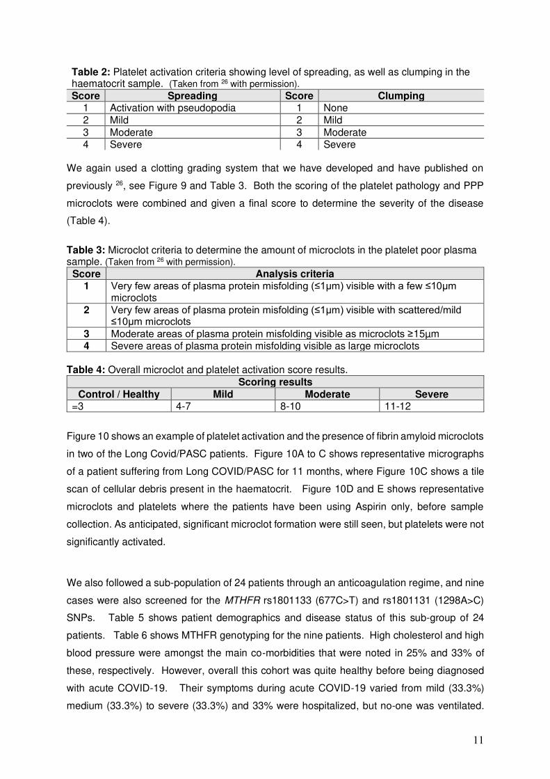
Table 2: Platelet activation criteria showing level of spreading, as well as clumping in the haematocrit sample. (Taken from 26 with permission).
Score Spreading Score Clumping 1 Activation with pseudopodia 1 None
2 Mild 2 Mild
3 Moderate 3 Moderate 4 Severe 4 Severe
We again used a clotting grading system that we have developed and have published on
previously 26, see Figure 9 and Table 3. Both the scoring of the platelet pathology and PPP
microclots were combined and given a final score to determine the severity of the disease
(Table 4).
Table 3: Microclot criteria to determine the amount of microclots in the platelet poor plasma sample. (Taken from 26 with permission).
Score Analysis criteria 1 Very few areas of plasma protein misfolding (≤1µm) visible with a few ≤10µm
microclots
2 Very few areas of plasma protein misfolding (≤1µm) visible with scattered/mild ≤10µm microclots
3 Moderate areas of plasma protein misfolding visible as microclots ≥15µm 4 Severe areas of plasma protein misfolding visible as large microclots
Table 4: Overall microclot and platelet activation score results.
Scoring results
Control / Healthy Mild Moderate Severe =3 4-7 8-10 11-12
Figure 10 shows an example of platelet activation and the presence of fibrin amyloid microclots
in two of the Long Covid/PASC patients. Figure 10A to C shows representative micrographs
of a patient suffering from Long COVID/PASC for 11 months, where Figure 10C shows a tile
scan of cellular debris present in the haematocrit. Figure 10D and E shows representative
microclots and platelets where the patients have been using Aspirin only, before sample
collection. As anticipated, significant microclot formation were still seen, but platelets were not
significantly activated.
We also followed a sub-population of 24 patients through an anticoagulation regime, and nine
cases were also screened for the MTHFR rs1801133 (677C>T) and rs1801131 (1298A>C)
SNPs. Table 5 shows patient demographics and disease status of this sub-group of 24
patients. Table 6 shows MTHFR genotyping for the nine patients. High cholesterol and high
blood pressure were amongst the main co-morbidities that were noted in 25% and 33% of
these, respectively. However, overall this cohort was quite healthy before being diagnosed
with acute COVID-19. Their symptoms during acute COVID-19 varied from mild (33.3%)
medium (33.3%) to severe (33.3%) and 33% were hospitalized, but no-one was ventilated.

12
Shortness of breath (70.8%), constant fatigue (87.5%) and brain fog, concentration and
forgetfulness (83.3%), were the main long COVID/PASC symptoms that were reported. After
treatment, every one of the 24 patients reported that their main symptoms were resolved and
fatigue as main symptom, relieved. Their platelets were also calmed and microclotting scores
were within healthy levels (see clotting and grading system).
Using the platelet mapping and plasma criteria 26 the patients displayed moderate platelet
spreading and mild platelet clumping (white arrows) before treatment (Figure 11 A, C, E, G, I)
with a reduction in platelet activity after treatment with mild spreading and no clumping (Figure
11 B, D, F, H, J). In the plasma samples a difference could be seen before and after treatment,
with moderate plasma microclots (white arrows) found in the before treatment patients (Figure
12 A, C, E, G, I) and a small number of microclots present in the plasma after treatment (Figure
12 B, D, F, H, J). Similar platelet hyperactivation and plasma clotting were seen in acute
COVID-19 and Long COVID/PASC patients in previous studies done on naïve COVID-19 and
Long COVID/PASC samples 12, 13.
Table 5: Patient demographics and disease status of 24 Long COVID/PASC patients before and after treatment.
Demographics of sub-group of 24 Long COVID/PASC patients Age 51±15.5
Gender Females 12 Males 12 Comorbidities
Comorbidities Yes (%) No (%) Type 1 diabetes 0% 100% Type 2 diabetes 4% 96% Cardiovascular disease 8% 92% High blood pressure 33% 67% High cholesterol 41.7% 58.3% Previous blood clots 17% 83% Previous stroke 4% 96% Previous heart attack 0% 100% Psoriasis 8% 92% Rheumatoid arthritis 13% 88% Lupus 0% 100% Auto-immune disease not listed 0% 100% Gingivitis and/or Periodontitis 0% 100%
Rosacea 0% 100% Acute COVID-19 data of cohort of 24 treated Long COVID/PASC patients
Acute phase disease severity Light Medium Severe
Severity of acute COVID-19 or Long COVID 33.3% 33.3% 33.3% Hospitalization during acute COVID-19 Yes (%) No (%)
Were you hospitalized? 33% 67% Did you receive oxygen? 33% 67%
Were you ventilated 0% 100% Long COVID Symptoms
Questioner questions Yes (%) No (%)
Shortness of breath 70.8% 29.2%
Recurring chest pain 37.5% 62.5%

13
Low oxygen levels 54.2% 45.8%
Heart rate dysfunction (heart palpitations) 41.7% 58.3%
Constant fatigue (more than usual) 87.5% 12.5%
Joint and muscle pain 62.5% 37.5%
Brain fog, concentration, forgetfulness 83.3% 16.7%
Sleep disturbances (more than before COVID-19) 58.3% 41.7%
Depression, anxiety (more than before COVID-19) 29.2% 70.8%
Digestive problems 16.7% 83.3%
Kidney problems 8.3% 91.7%
Did your main symptoms listed above resolve? 100% 0%
Fluorescence microscopy
Scoring Before treatment
After treatment
Platelet activation and clumping score 4.4 ± 0.9 3.6 ± 1.2 Microclotting score 2.7 ± 0.8 1.7 ± 0.6 Overall score for platelets and plasma micrographs based on platelet and microclot scoring system
7.0 ± 1.3 5.2 ± 1.3
Table 6 shows the clinical characteristics and MTHFR genotypes of nine Long COVID/PASC
patients, of whom eight (89%) tested positive for at least one risk-associated allele.
Homozygosity for the MTHFR 677 T-allele associated with the most severe effect on enzyme
function was detected in two of the three patients with hypertension reported as a co-morbidity,
prior to the diagnosis of Long COVID/PASC. In the majority of patients folate intake was
inadequate, considered to be low when the score is less than 11, moderate between 11 and
13, and high above 13 (see Table 6). The negative effect of a low folate score was evaluated
in relation to body mass index (BMI), as an indication of being overweight (>24.9 kg/m2) or
obese (>30 kg/m2). Detection of the APOE polymorphism (data not shown) and/or a high BMI
are known risk factors for development of dyslipidaemia frequently detected in our study
cohort. A family history of DVTs was reported by both patients with this condition prior to their
diagnosis of Long COVID/PASC, which warrants extended genetic testing to confirm or
exclude a more severe genetic cause of inherited thrombophilia.
Table 6: Clinical characteristics and MTHFR genotyping results of nine Long COVID/PASC patients. Folate score should be evaluated in relation to body mass index. Ages were not added to assist with de-identification: age range was from 40 – 60.
Gender BMI (kg.m-2) MTHFR Folate score Comorbidities*
Female 23.7 High -14 1298 A > C (++) Deep vein thrombosis Dyslipidaemia
Male 27.1 Very Low - 3 677 C > T (+-) Dyslipidaemia
Female 19.7 Low - 9 677 C > T (+-) Not reported Male 33.4 High -15 1298 A>C (+-) Dyslipidaemia
Female 24.7 Low - 9 677 C > T (+-) 1298 A > C (+-)
Not reported
Female 36.3 Low - 8 677 C > T (++) Dyslipidaemia Hypertension
Female 34.9 Low - 10
677 C > T (+-) Not reported
Male 28.1 Very Low - 4
677 C > T (++) Deep vein thrombosis Hypertension
*Prior to the diagnosis of Long COVID/PASC +- heterozygous and ++ homozygous

14
DISCUSSION
Here we report on the comorbidities and symptoms identified in a cohort of 845 South African
Long COVID/PASC patients who filled in the South African Long COVID/PASC registry. We
show that hypertension and high cholesterol levels (dyslipidemia) are important comorbidities
that may play a significant role in the development of Long COVID/PASC in this cohort. (We
also recognise that other comorbidities, such as previous viral infections, are also very
important, but may only manifest only in a larger cohort.) It is well-documented that impaired
endothelial function is associated with increased cholesterol and hypertension (which may be
underpinned by generic variation) due to increased vascular oxidative stress and inflammation
46, 47. Here we also showed the presence of fibrin amyloid microclotting and platelet
pathologies in another cohort of 70 patients that visited a clinical practice complaining of
persistent symptoms, where these patients were diagnosed with Long COVID/PASC. We
found that in this cohort, all of the patients did indeed have both increased amyloid
microclotting, as well as platelet pathologies, as assessed by a platelet and clotting grading
system that we have developed previously 26. In addition, we followed 24 of these patients
through their anticoagulation treatment regimens and demonstrated sufficient endothelium
recovery over a three to four week period to lower their microclot and platelet scores by almost
two full units (7.1 5.2). While this approach may not yet be widely accepted, it is based on
best clinical practice and GJL’s experience in managing affected patients. We included a minor
genetic component to determine the prevalence of two extensively studied MTHFR SNPs in 9
of the 24 patients. Detection of genetic variation in this MTHFR gene in the presence of
clinically relevant biochemical findings may therefore also facilitate risk stratification for
improved personalized patient management 48.
Normal blood clotting goes through a variety of established mechanisms 42, a major step being
the cleavage by thrombin of the complex fibrinogen molecule (roughly cylindrical, with a 5 x
45nm size). This releases two fibrinopeptides, and causes the thermodynamically favourable
formation of fibrin macrofibres, typically 50-100nm in diameter. They may be crosslinked by
Factor XIII. The clots are usually removed by fibrinolysis, leading to the residual formation of
D-dimer; its normally low background levels reflect this background activity. It was always
assumed that the normal conformation of a protein is that of its lowest free energy, as per
Christian Anfinsen’s famous protein refolding experiments. However, this is not the case.
Many proteins can fold into a form of lower free energy but retain the identical sequence. Some
of these forms, containing ordered beta-sheet structures, are generically referred to as
amyloids, and many are well known to be associated with certain diseases. Ab in Alzheimer’s
disease and synuclein in Parkinson’s disease are examples. Over 50 are recognised 18. Note
however that almost any proteins can form amyloid structures (e.g. recombinant insulin will do

15
it over time, as will lysozyme held at an acid pH). Another well-known example of a class of
proteins that can exist in two conformations of identical sequence is represented by prion
proteins. The normal form with alpha-helices is called PrPc and the amyloid one PrPSc, the
latter being of lower free energy i.e. more thermodynamically stable. It is also highly resistant
to proteolysis. This transition between the two forms can itself be catalysed by the PrPSc. The
key point of importance in microclot formation in Long COVID/PASC is that fibrin(ogen) too
can, in the presence of various trigger substances, fold into an amyloid form that has a very
different macrostructure characterised by different fibre diameters and pore sizes 49. We noted
that e.g. in type 2 Diabetes Mellitus (T2DM) clots have a netlike appearance 50, 51, 52, 53 while
in Alzheimer’s disease 16, 54, 55, 56 and Parkinson’s disease the fibres may be larger in size 15,
22; they are also much more resistant to proteolysis 17, and so are much more prevalent in the
steady state.
We have been observing fibrin(ogen) changes generally for many years, initially via electron
microscopy (e.g. 57, 58, 59, 60, 61, 62, and many others). Since 2011 we have also studied the effect
of fibrin(ogen) folding in the presence of various inflammatory molecules, such as iron ions 63,
that can stimulate the anomalous fibrin form 64. These anomalous structures could also be
found in a variety of disease states 17, such as T2DM 51 and Alzheimer’s disease 65. In this
earlier literature we often referred to these anomalous clots as ‘dense matted deposits’. In
2016, we showed that this anomalous resistance to fibrinolysis was because the anomalous
structures were in fact amyloid in nature 49. Such structures are easily observed under the
optical microscope, and in particular may be stained with the fluorogenic dye thioflavin T and
by the more recently developed oligothiophene dyes marketed by Ebba Biotech as
Amytrackers 15, 19, 21.
Most recently, we have demonstrated this explicitly in COVID-19 patients 12, 13. Here, these
microclots were observed without the addition of clotting agents, i.e. thrombin, and therefore,
these amyloid microclots were there in the plasma of the individuals at the time of sampling.
In particular, it was found not only that the clots contained fibrin (fibrinogen after all being one
of the most concentrated proteins in plasma) but that these microclots had entrapped many
other proteins such as alpha-2-antiplasmin 14 and a variety of other proteins and even
antibodies, which therefore were not observed in plasma from which the microclots had been
removed (so did not appear as biomarkers, even though they were there). Because these
clots are insoluble and effectively inert they do not contribute to plasma viscosity as
determined via TEG®, whose values can thus appear normal in Long COVID/PASC
(unpublished data). Another characteristic of COVID-19 is the extremely high levels of
activation of platelets 13. Together with platelet pathology and the presence of microclots in

16
the circulation, endothelial damage may be key drivers of persistent Long COVID/PASC
symptoms 14. See Figure 13 for a snapshot of the interactions that platelets have with
circulating blood cells and the various complexes they form (for a detailed review, see 66.
The intention of the treatment regime used in this study was to use best clinical practice
methods to return the patient’s clotting pathology to that of a normal clotting physiology and
not to make them hypo-coagulable. It is well-known that DOACs act upon various parts of the
clotting pathways 67, while platelet hyperactivation is prevented with DAPT 68, 69 This ‘triple
therapy’ treatment therefore prevents platelet hyperactivation and also prevents new
microclots from forming, while it allows the body’s own fibrinolysis pathways to clear existing
microclots. Figure 14 shows the various routes of action of the medication targeting the
enzymatic pathway and platelet hyperactivation 67, 68, 69. See Figure 15 for a concluding visual
on our understanding of the most relevant physiological processes.
CONCLUSION
In the current study, it was noted that each of 70 patients diagnosed with Long COVID/PASC
and who provided blood samples showed platelet hyperactivation and microclot formation.
We used a triple therapy to treat both the microclots and the platelet hyperactivation. During
this medication regime, where 24 patients were followed, the endothelium layers recover
sufficiently over a three-to-four week period, and this allows for a normal clotting physiology
and endothelium function to return. In the treated cohort, significant microclots were removed
and platelet hyperactivation returned to more normal levels. All patients in this cohort, reported
a significant reduction in their Long COVID/PASC symptoms. We suggest that a platelet and
clotting grading system should be used as a simple and cost-effective diagnostic method for
the early identification of long COVID/PASC. Diagnosis of Long COVID/PASC requires a
diagnosis that excludes other pathologies, including using the duration of symptoms (>two
months after acute infection). If a bleeding tendency (not seen commonly) is a concern, a
TEG® can be used to manage that, its role being as a safety-net to not overtreat the patient.
The exact combination of treatment and duration for mild cases probably needs further
investigation and refinement. Patients need to be informed that the above approach and
treatment is not yet widely accepted but is based on best clinical practice and experience in
managing these patients.
Our literature study on the effect of homocysteine on platelet and clotting pathway activation
underpinned by variation in the MTHFR gene is relevant to the COVID-19 era. Ponti and co-
workers in 2021 70, correlated infection patterns with the MTHFR 677 T-allele frequency which
could relate to studies we performed previously in different ethnic groups in South Africa 71.

17
We also provided convincing evidence that homocysteine levels are mediated by the effects
of MTHFR 677 C > T and the diet on BMI, which increases with a low folate score 33, 72. There
a low folate score correlates with a high BMI. Detection of this low-penetrance variation
therefore reinforces the importance of high intake of folate and other B-vitamins in the diet to
prevent or restore dysfunction of the methylation pathway. There are no controlled trials done
yet; however, this will be the important next steps to urgently consider in relation to clinical
monitoring using a multi-modal pathology-supported genetic testing approach 27.
FIGURES
Figure 1: Study layout of the current paper.

18
Figure 2: General Overview of the population distribution of the South African Long COVID/PASC registry data as presented in a Sankey plot.
Figure 3: Gender versus Long COVID symptoms population distribution of the South African Long COVID registry data.

19
Figure 4: Age - Long COVID/PASC symptoms participant distribution of the South African Long COVID/PASC registry data.

20
FIGURE 5: A Sankey plot showing participants comorbidities versus Long COVID/PASC symptoms population distribution of the South African Long COVID registry data.

21
Figure 6A and B: Lattice plots, showing participants comorbidities versus Long COVID/PASC symptoms lattice, highlighting the high blood pressure and high cholesterol comorbidities.
A
B

22
Figure 7: Fluorescence microscopy examples of the different stages of platelet activation and spreading, that was used to score the platelet activation in the Long COVID patients, with Stage 1, with minimally activated platelets, seen as small round platelets with a few pseudopodia, seen as healthy/control platelets that progresses to Stage 4, with egg-shaped platelets, indicative of spreading and the beginning of clumping. (Taken from 26 with permission).

23
Figure 8: Fluorescence microscopy examples of the different stages of platelet clumping. With no clumping occurring in the healthy/control samples in Stage 1 (no figures shown), progressing to severe clumping of platelets as seen in Stage 4. (Taken from 26 with permission).

24
Figure 9: Fluorescence microscopy showing microclots in platelet poor plasma (PPP) with representative examples of the different stages of different stages of microclot formation. Stage 1shows minimal microclot formation in healthy/control PPP which progresses to the presence of the severe microclotting Stage 4. Bottom row represents examples of stage 4 microclots using (A) bright-field microscopy, (B) fluorescence microscopy, and (C) an overlay of fluorescence and bright-field microscopy. (Taken from 26 with permission).

25
Figure 10: Microclots with platelets in 2 patients with Long COVID/PASC. (A): Representative micrographs of microclots in an untreated patient diagnosed with Long COVID/PASC, suffering from the condition for 11 months. The plasma was stained with thioflavin T (ThT); (B) Platelet hyperactivation of the same patient, where PAC-1 and CD62PE were used to mark platelets. (C) In this patient, cellular debris in the haematocrit was noted – here such cellular debris is shown in a tile scan. (D and E): Microclot presence and platelets from a patient who was on anti-platelet therapy before blood collection, where significant microclots were noted, but the platelets were not significantly hyperactivated, due to the use of anti-platelet therapy.

26
Figure 11: Representative fluorescence micrographs of platelet pathology before and after treatment. Moderate platelet spreading and mild platelet clumping (white arrows) was seen in the naïve patient’s samples (Fig. A, C, E, G, I) that improved after treatment, with mild platelet spreading and no clumps (Fig. B, D, F, H, J).

27
Figure 12: Representative PPP fluorescence micrographs with moderate areas of plasma protein misfolding forming microclots (some larger than 15µm; white arrows) before treatment (Fig. A, C, E, G, I), with a few microclots visible in the samples after treatment (Fig. B, D, F, H, J).

28
Figure13: (1) After activation, platelets express P-selectin on their membranes, followed by platelet-T cell complex formation (2); P-selectin on platelet membranes are also recognized by macrophages, possibly by the Fcγ-receptor; clearance may result due to either receptor binding or phagocytosis (3). CD40L is released from platelets and can migrate to membranes or shed as soluble (s)CD40L (4). sCD40L can bind to both the αIIbβ3 or CD40 receptors (5). The P-selectin on the membranes of sCD40L-activated platelets can also form complexes with monocytes (6). Platelet-neutrophils also form complexes (7). Diagram created with BioRender (https://biorender.com/) and adapted from 66.

29
Figure 14: Effects of selected direct oral anticoagulants (DOAC) and dual antiplatelet therapy (DAPT) medication on clotting and platelet function. Diagram created with BioRender (https://biorender.com/)

30
Figure 15: Understanding the pathological pathways of Long COVID/PASC. A) The rollercoaster vascular pathology in acute respiratory syndrome coronavirus 2 (COVID-19) [adapted from 42]. B) Long COVID/PASC pathology resulting in persistent microclot formation and platelet hypercoagulation. C) Best clinical practice regime that reduces platelet hyperactivation and prevent microclot formation. D) Treatment should result in endothelium healing and reduction of tissue hypoxia and reduction of long COVID/PASC symptoms Image created with BioRender (https://biorender.com/).

31
ACKNOWLEDGMENTS AND DECLARATIONS
Acknowledgements
We wish to thank all the long COVID/PASC patients who participated in this study. Also, Lezette Briedenhann, Ilse Geldenhuys and Anneke De Villiers who curated the patient consents and blood collections. Funding
DBK thanks the Novo Nordisk Foundation (grant NNF20CC0035580) for financial support. MJK acknowledges the South African Medical Research Council for support of the genetic studies. The funders had no role in study design, data collection and analysis, decision to publish, or preparation of the manuscript.
Consent for publication
All authors approved submission of the paper.
Availability of data and materials
The raw data supporting the conclusions of this article will be made available by the authors, without undue reservation. For data see: https://1drv.ms/u/s!AgoCOmY3bkKHi7Zrl0KftaiA7H35dg?e=sW8Q9L Competing interests
MK is a non-executive director and shareholder of Gknowmix (Pty) Ltd. EP is the managing director of BioCODE Technologies. The other authors have no competing interests to declare. Authors' contributions
EP: Sample analysis, Long COVID/PASC registry curation, writing of the paper, editing the paper, co-corresponding author Study-leader; CV: Platelet analysis and data curation; GJL: sample collection, diagnoses and treatment of patients; MJK and KM: genetic analysis and editing of paper; LPW, KR, BWW, SO: Long COVID/PASC registry data analysis; DBK: Editing and writing of paper, co-corresponding author.

32
REFERENCES
1. Huang C, et al. 6-month consequences of COVID-19 in patients discharged from hospital: a cohort study. Lancet 397, 220-232 (2021).
2. Lopez-Leon S, et al. More Than 50 Long-Term Effects of COVID-19: A Systematic
Review and Meta-Analysis. Res Sq, (2021). 3. Carfì A, Bernabei R, Landi F. Persistent Symptoms in Patients After Acute COVID-19.
Jama 324, 603-605 (2020). 4. Rubin R. As Their Numbers Grow, COVID-19 "Long Haulers" Stump Experts. Jama,
(2020). 5. Baig AM. Chronic COVID syndrome: Need for an appropriate medical terminology for
long-COVID and COVID long-haulers. J Med Virol, (2020). 6. Proal AD, VanElzakker MB. Long COVID or Post-acute sequelae of COVID-19
(PASC): An overview of 1 biological factors that may contribute to persistent symptoms. Frontiers in Microbiology In Press, (2021).
7. Wostyn P. COVID-19 and chronic fatigue syndrome: Is the worst yet to come? Med
Hypotheses 146, 110469 (2021). 8. Mohabbat AB, Mohabbat NML, Wight EC. Fibromyalgia and Chronic Fatigue
Syndrome in the Age of COVID-19. Mayo Clin Proc Innov Qual Outcomes 4, 764-766 (2020).
9. Goldstein DS. The possible association between COVID-19 and postural tachycardia
syndrome. Heart Rhythm 18, 508-509 (2021). 10. Afrin LB, Weinstock LB, Molderings GJ. Covid-19 hyperinflammation and post-Covid-
19 illness may be rooted in mast cell activation syndrome. Int J Infect Dis 100, 327-332 (2020).
11. Komaroff AL, Bateman L. Will COVID-19 Lead to Myalgic Encephalomyelitis/Chronic
Fatigue Syndrome? Front Med (Lausanne) 7, 606824 (2020). 12. Pretorius E, Venter C, Laubscher GJ, Lourens PJ, Steenkamp J, Kell DB. Prevalence
of readily detected amyloid blood clots in 'unclotted' Type 2 Diabetes Mellitus and COVID-19 plasma: a preliminary report. Cardiovasc Diabetol 19, 193 (2020).
13. Venter C, et al. Erythrocyte, Platelet, Serum Ferritin, and P-Selectin Pathophysiology
Implicated in Severe Hypercoagulation and Vascular Complications in COVID-19. Int J Mol Sci 21, (2020).
14. Pretorius E, et al. Persistent clotting protein pathology in Long COVID/ Post-Acute
Sequelae of COVID-19 (PASC) is accompanied by increased levels of antiplasmin. Cardiovasc Diabetol 20, 172 (2021).
15. Adams B, et al. Parkinson’s disease: a systemic inflammatory disease accompanied
by bacterial inflammagens. Front Ag Neurosci 11, 210 (2019). 16. de Waal GM, Engelbrecht L, Davis T, de Villiers WJS, Kell DB, Pretorius E. Correlative
Light-Electron Microscopy detects lipopolysaccharide and its association with fibrin

33
fibres in Parkinson's Disease, Alzheimer's Disease and Type 2 Diabetes Mellitus. Sci Rep 8, 16798 (2018).
17. Kell DB, Pretorius E. The simultaneous occurrence of both hypercoagulability and
hypofibrinolysis in blood and serum during systemic inflammation, and the roles of iron and fibrin(ogen). Integr Biol 7, 24-52 (2015).
18. Kell DB, Pretorius E. Proteins behaving badly. Substoichiometric molecular control and
amplification of the initiation and nature of amyloid fibril formation: lessons from and for blood clotting. Prog Biophys Mol Biol 123, 16-41 (2017).
19. Page MJ, et al. Serum amyloid A binds to fibrin(ogen), promoting fibrin amyloid
formation. Sci Rep 9, 3102 (2019). 20. Pretorius E, Mbotwe S, Kell DB. Lipopolysaccharide-binding protein (LBP) reverses
the amyloid state of fibrin seen in plasma of type 2 diabetics with cardiovascular co-morbidities. Sci Rep 7, 9680 (2017).
21. Pretorius E, Page MJ, Engelbrecht L, Ellis GC, Kell DB. Substantial fibrin
amyloidogenesis in type 2 diabetes assessed using amyloid-selective fluorescent stains. Cardiovasc Diabetol 16, 141 (2017).
22. Pretorius E, Page MJ, Mbotwe S, Kell DB. Lipopolysaccharide-binding protein (LBP)
can reverse the amyloid state of fibrin seen or induced in Parkinson’s disease. PlosOne 13, e0192121 (2018).
23. Grobbelaar LM, et al. SARS-CoV-2 spike protein S1 induces fibrin(ogen) resistant to
fibrinolysis: Implications for microclot formation in COVID-19. medRxiv, 2021.2003.2005.21252960 (2021).
24. Schaller J, Gerber SS. The plasmin-antiplasmin system: structural and functional
aspects. Cell Mol Life Sci 68, 785-801 (2011). 25. Miszta A, Huskens D, Donkervoort D, Roberts MJM, Wolberg AS, de Laat B. Assessing
Plasmin Generation in Health and Disease. Int J Mol Sci 22, (2021). 26. Laubscher GJ, Lourens PJ, Venter C, Kell DB, Pretorius E. TEG®, Microclot and
Platelet Mapping for Guiding Early Management of Severe COVID-19 Coagulopathy. J Clin Med 10, 5381 (2021).
27. Daramola O, et al. Towards AI-Enabled Multimodal Diagnostics and Management of
COVID-19 and Comorbidities in Resource-Limited Settings. Informatics 8, 63 (2021). 28. Roberts I, et al. Untargeted metabolomics of COVID-19 patient serum reveals potential
prognostic markers of both severity and outcome. Metabolomics 18, 6 (2021). 29. Karst M, Hollenhorst J, Achenbach J. Life-threatening course in coronavirus disease
2019 (COVID-19): Is there a link to methylenetetrahydrofolic acid reductase (MTHFR) polymorphism and hyperhomocysteinemia? Med Hypotheses 144, 110234 (2020).
30. Abu-Farha M, et al. Prognostic Genetic Markers for Thrombosis in COVID-19 Patients:
A Focused Analysis on D-Dimer, Homocysteine and Thromboembolism. Front Pharmacol 11, 587451 (2020).

34
31. Yang Z, et al. Predictors for imaging progression on chest CT from coronavirus disease 2019 (COVID-19) patients. Aging (Albany NY) 12, 6037-6048 (2020).
32. Dragani A, et al. Oxidative stress and platelet activation in subjects with moderate
hyperhomocysteinaemia due to MTHFR 677 C→T polymorphism. Thromb Haemost 108, 533-542 (2012).
33. Delport D, et al. Significance of dietary folate intake, homocysteine levels and MTHFR
677 C>T genotyping in South African patients diagnosed with depression: test development for clinical application. Metab Brain Dis 29, 377-384 (2014).
34. Kim J, Kim H, Roh H, Kwon Y. Causes of hyperhomocysteinemia and its pathological
significance. Arch Pharm Res 41, 372-383 (2018). 35. Moretti R. Homocysteine: New Aspects of an Ancient Enigma. Cardiology 144, 36-39
(2019). 36. McNulty H, Strain JJ, Hughes CF, Ward M. Riboflavin, MTHFR genotype and blood
pressure: A personalized approach to prevention and treatment of hypertension. Mol Aspects Med 53, 2-9 (2017).
37. Kluyver T, et al. Jupyter Notebooks - a publishing format for reproducible
computational workflows. In: ELPUB) (2016). 38. McKinney W. Data Structures for Statistical Computing in Python.) (2010). 39. Kuznetsov S, Watson BW. Algorithmic Support For Knowledge Discovery Techiques
Based On Formal Concept Analysis. . In: CEUR Workshop Series) (2015). 40. Valente CC, Bauer FF, Venter F, Watson B, Nieuwoudt HH. Modelling the sensory
space of varietal wines: Mining of large, unstructured text data and visualisation of style patterns. Scientific Reports 8, 4987 (2018).
41. Brown DL, et al. Benefits and Risks of Dual Versus Single Antiplatelet Therapy for
Secondary Stroke Prevention: A Systematic Review for the 2021 Guideline for the Prevention of Stroke in Patients With Stroke and Transient Ischemic Attack. Stroke 52, e468-e479 (2021).
42. Grobler C, et al. Covid-19: The Rollercoaster of Fibrin(Ogen), D-Dimer, Von Willebrand
Factor, P-Selectin and Their Interactions with Endothelial Cells, Platelets and Erythrocytes. Int J Mol Sci 21, (2020).
43. Kotze M. Application of advanced molecular technology in the diagnosis and
management of genetic disorders in South Africa. S Afr Med J 106, S114-118 (2016). 44. Stewart S, Newson L, Briggs TA, Grammatopoulos D, Young L, Gill P. Long COVID
risk - a signal to address sex hormones and women's health. Lancet Reg Health Eur 11, 100242 (2021).
45. Sigfrid L, et al. Long Covid in adults discharged from UK hospitals after Covid-19: A
prospective, multicentre cohort study using the ISARIC WHO Clinical Characterisation Protocol. Lancet Reg Health Eur 8, 100186 (2021).
46. Sugiura T, et al. Impact of lipid profile and high blood pressure on endothelial damage.
J Clin Lipidol 5, 460-466 (2011).

35
47. Hurtubise J, McLellan K, Durr K, Onasanya O, Nwabuko D, Ndisang JF. The Different
Facets of Dyslipidemia and Hypertension in Atherosclerosis. Curr Atheroscler Rep 18, 82 (2016).
48. Tan CW, et al. Cohort study to evaluate the effect of vitamin D, magnesium, and
vitamin B(12) in combination on progression to severe outcomes in older patients with coronavirus (COVID-19). Nutrition 79-80, 111017 (2020).
49. Pretorius E, Mbotwe S, Bester J, Robinson CJ, Kell DB. Acute induction of anomalous
and amyloidogenic blood clotting by molecular amplification of highly substoichiometric levels of bacterial lipopolysaccharide. J R Soc Interface 123, 20160539 (2016).
50. Pretorius E, Bester J. Viscoelasticity as a measurement of clot structure in poorly
controlled type 2 diabetes patients: towards a precision and personalized medicine approach. Oncotarget 7, 50895-50907 (2016).
51. Pretorius E, et al. Poorly controlled type 2 diabetes is accompanied by significant
morphological and ultrastructural changes in both erythrocytes and in thrombin-generated fibrin: implications for diagnostics. Cardiovasc Diabetol 14, 30 (2015).
52. Pretorius E, Lipinski B, Bester J, Vermeulen N, Soma P. Albumin stabilizes fibrin fiber
ultrastructure in low serum albumin type 2 diabetes. Ultrastruct Pathol 37, 254-257 (2013).
53. Pretorius E, Oberholzer HM, van der Spuy WJ, Swanepoel AC, Soma P. Qualitative
scanning electron microscopy analysis of fibrin networks and platelet abnormalities in diabetes. Blood Coagul Fibrinolysis 22, 463-467 (2011).
54. Pretorius E, Bester J, Page MJ, Kell DB. The potential of LPS-binding protein to
reverse amyloid formation in plasma fibrin of individuals with Alzheimer-type dementia. Frontiers Aging Neurosci 10, 257 (2018).
55. Bester J, Soma P, Kell DB, Pretorius E. Viscoelastic and ultrastructural characteristics
of whole blood and plasma in Alzheimer-type dementia, and the possible role of bacterial lipopolysaccharides (LPS). Oncotarget 6, 35284-35303 (2015).
56. Nielsen VG, Pretorius E, Bester J, Jacobsen WK, Boyle PK, Reinhard JP. Carbon
monoxide and iron modulate plasmatic coagulation in Alzheimer's disease. Curr Neurovasc Res 12, 31-39 (2015).
57. Pretorius E, Humphries P. Ultrastructural changes to rabbit fibrin and platelets due to
aspartame. Ultrastruct Pathol 31, 77-83 (2007). 58. Pretorius E, Oberholzer HM. Ultrastructural changes of platelets and fibrin networks in
human asthma: a qualitative case study. Blood Coag Fibrinol 20, 146-149 (2009). 59. Pretorius E, Bronkhorst P, Briedenhann S, Smit E, Franz RC. Comparisons of the fibrin
networks during pregnancy, nonpregnancy and pregnancy during dysfibrinogenaemia using the scanning electron microscope. Blood Coag Fibrinol 20, 12-16 (2009).
60. Pretorius E, Windberger UB, Oberholzer HM, Auer RE. Comparative ultrastructure of
fibrin networks of a dog after thrombotic ischaemic stroke. Onderstepoort J Vet Res 77, E1-4 (2010).

36
61. Pretorius E, Oberholzer HM, van der Spuy WJ, Meiring JH. The changed ultrastructure of fibrin networks during use of oral contraception and hormone replacement. J Thromb Thrombolysis 30, 502-506 (2010).
62. Pretorius E. The use of a desktop scanning electron microscope as a diagnostic tool
in studying fibrin networks of thrombo-embolic ischemic stroke. Ultrastruct Pathol 35, 245-250 (2011).
63. Pretorius E, Vermeulen N, Bester J, Lipinski B, Kell DB. A novel method for assessing
the role of iron and its functional chelation in fibrin fibril formation: the use of scanning electron microscopy. Toxicol Mech Methods 23, 352-359 (2013).
64. Pretorius E, Kell DB. Diagnostic morphology: biophysical indicators for iron-driven
inflammatory diseases. Integrative Biol 6, 486-510 (2014). 65. Pretorius E, Bester J, Kell DB. A bacterial component to Alzheimer-type dementia seen
via a systems biology approach that links iron dysregulation and inflammagen shedding to disease J Alzheimers Dis 53, 1237-1256 (2016).
66. Pretorius E. Platelets in HIV: A Guardian of Host Defence or Transient Reservoir of
the Virus? Frontiers in Immunology 12, (2021). 67. Eller T, et al. Dabigatran, rivaroxaban, apixaban, argatroban and fondaparinux and
their effects on coagulation POC and platelet function tests. Clin Chem Lab Med 52, 835-844 (2014).
68. Cattaneo M. P2Y12 receptors: structure and function. J Thromb Haemost 13 Suppl 1,
S10-16 (2015). 69. Scharf RE. Drugs that affect platelet function. Semin Thromb Hemost 38, 865-883
(2012). 70. Ponti G, et al. COVID-19 spreading across world correlates with C677T allele of the
methylenetetrahydrofolate reductase (MTHFR) gene prevalence. J Clin Lab Anal 35, e23798 (2021).
71. Scholtz CL, et al. Analysis of two mutations in the MTHFR gene associated with mild
hyperhomocysteinaemia--heterogeneous distribution in the South African population. S Afr Med J 92, 464-467 (2002).
72. Davis W, et al. The fat mass and obesity-associated FTO rs9939609 polymorphism is
associated with elevated homocysteine levels in patients with multiple sclerosis screened for vascular risk factors. Metab Brain Dis 29, 409-419 (2014).