Adsorption of DNA on biomimetic apatites: Toward the understanding of the role of bone and tooth...
9
Applied Surface Science 292 (2014) 867–875 Contents lists available at ScienceDirect Applied Surface Science j ourna l ho me page: www.elsevier.com/locate/apsusc Adsorption of DNA on biomimetic apatites: Toward the understanding of the role of bone and tooth mineral on the preservation of ancient DNA A. Grunenwald a,b , C. Keyser b , A.M. Sautereau a , E. Crubézy c , B. Ludes b , C. Drouet a,∗ a CIRIMAT Carnot Institute – Phosphates, Pharmacotechnics, Biomaterials, University of Toulouse, CNRS/INPT/UPS, ENSIACET, 4 allée Emile Monso, 31030 Toulouse Cedex 4, France b Institute of Legal Medicine, AMIS Laboratory, CNRS UMR 5288, University of Strasbourg, 11 rue Humann, 67085 Strasbourg Cedex, France c Molecular Anthropology and Image Synthesis Laboratory (AMIS), CNRS UMR 5288, University of Toulouse, 37 allée Jules Guesde, 31000 Toulouse, France a r t i c l e i n f o Article history: Received 11 October 2013 Received in revised form 10 December 2013 Accepted 11 December 2013 Available online 19 December 2013 Keywords: Nanocrystalline apatite Hydroxyapatite Ancient DNA Polyelectrolyte adsorption Temkin isotherm Elovich a b s t r a c t In order to shed some light on DNA preservation over time in skeletal remains from a physicochemical viewpoint, adsorption and desorption of DNA on a well characterized synthetic apatite mimicking bone and dentin biominerals were studied. Batch adsorption experiments have been carried out to determine the effect of contact time (kinetics), DNA concentration (isotherms) and environmentally relevant factors such as temperature, ionic strength and pH on the adsorption behavior. The analogy of the nanocrystalline carbonated apatite used in this work with biological apatite was first demonstrated by XRD, FTIR, and chemical analyses. Then, DNA adsorption kinetics was fitted with the pseudo-first order, pseudo-second order, Elovich, Ritchie and double exponential models. The best results were achieved with the Elovich kinetic model. The adsorption isotherms of partially sheared calf thymus DNA conformed satisfacto- rily to Temkin’s equation which is often used to describe heterogeneous adsorption behavior involving polyelectrolytes. For the first time, the irreversibility of DNA adsorption toward dilution and significant phosphate-promoted DNA desorption were evidenced, suggesting that a concomitant ion exchange pro- cess between phosphate anionic groups of DNA backbone and labile non-apatitic hydrogenphosphate ions potentially released from the hydrated layer of apatite crystals. This work should prove helpful for a better understanding of diagenetic processes related to DNA preservation in calcified tissues. © 2013 Elsevier B.V. All rights reserved. 1. Introduction Bone and tooth remains often represent the only – but also the best – biological materials available for deoxyribonucleic acids (DNA) typing in anthropology and may additionally be exploited in forensic sciences [1,2]. In ancient samples, DNA arising from degraded cells is generally strongly fragmented, with a mean size around 200 base pairs, and its amplification may be limited by the presence of substances persisting after purification, and inhib- iting PCR [3]. Although hard tissue samples are commonly used as a source of ancient DNA sequences, the processes of ancient DNA preservation in these mineralized tissues have received little systematic attention, both regarding qualitative and quantitative aspects. ∗ Corresponding author at: CIRIMAT Carnot Institute, ENSIACET, 4 allée Emile Monso, 31432 Toulouse Cedex 4, France. Tel.: +33 034 32 34 11; fax: +33 034 32 34 99. E-mail address: [email protected] (C. Drouet). Calcium phosphate apatites are the main components of human bone and teeth. The physico-chemical features of enamel highly resemble those of stoichiometric hydroxyapatite Ca 10 (PO 4 ) 6 (OH) 2 , both in terms of crystallographic structure and composition. In contrast, bone and dentin minerals are composed of non- stoichiometric apatite nanocrystals. Several studies have shown the possibility to prepare, under close-to-physiological conditions (temperature, pH), synthetic analogs to biological apatites that mimic their physico-chemical characteristics [4–6]. For both bio- logical specimens and biomimetic analogs, the surface of the nanocrystals was found to exhibit a structured but metastable non-apatitic hydrated layer [7–9], containing labile ions (e.g. Ca 2+ , HPO 4 2− , CO 3 2− . . .) leading to an exceptional surface reactivity, either in terms of ionic exchanges or of molecular adsorption [10–13]. Furthermore, the possibility to control, for synthetic biomimetic apatites, the maturation state of the nanocrystals was evidenced by modifying experimental conditions such as temper- ature, pH or maturation time in solution [7,14,15]. These findings then enable the preparation of samples mimicking either newly formed or mature bone mineral. 0169-4332/$ – see front matter © 2013 Elsevier B.V. All rights reserved. http://dx.doi.org/10.1016/j.apsusc.2013.12.063
Transcript of Adsorption of DNA on biomimetic apatites: Toward the understanding of the role of bone and tooth...

Aup
Aa
Tb
c
a
ARR1AA
KNHAPTE
1
t(idatiaDsa
Mf
0h
Applied Surface Science 292 (2014) 867– 875
Contents lists available at ScienceDirect
Applied Surface Science
j ourna l ho me page: www.elsev ier .com/ locate /apsusc
dsorption of DNA on biomimetic apatites: Toward thenderstanding of the role of bone and tooth mineral on thereservation of ancient DNA
. Grunenwalda,b, C. Keyserb, A.M. Sautereaua, E. Crubézyc, B. Ludesb, C. Droueta,∗
CIRIMAT Carnot Institute – Phosphates, Pharmacotechnics, Biomaterials, University of Toulouse, CNRS/INPT/UPS, ENSIACET, 4 allée Emile Monso, 31030oulouse Cedex 4, FranceInstitute of Legal Medicine, AMIS Laboratory, CNRS UMR 5288, University of Strasbourg, 11 rue Humann, 67085 Strasbourg Cedex, FranceMolecular Anthropology and Image Synthesis Laboratory (AMIS), CNRS UMR 5288, University of Toulouse, 37 allée Jules Guesde, 31000 Toulouse, France
r t i c l e i n f o
rticle history:eceived 11 October 2013eceived in revised form0 December 2013ccepted 11 December 2013vailable online 19 December 2013
eywords:anocrystalline apatiteydroxyapatite
a b s t r a c t
In order to shed some light on DNA preservation over time in skeletal remains from a physicochemicalviewpoint, adsorption and desorption of DNA on a well characterized synthetic apatite mimicking boneand dentin biominerals were studied. Batch adsorption experiments have been carried out to determinethe effect of contact time (kinetics), DNA concentration (isotherms) and environmentally relevant factorssuch as temperature, ionic strength and pH on the adsorption behavior. The analogy of the nanocrystallinecarbonated apatite used in this work with biological apatite was first demonstrated by XRD, FTIR, andchemical analyses. Then, DNA adsorption kinetics was fitted with the pseudo-first order, pseudo-secondorder, Elovich, Ritchie and double exponential models. The best results were achieved with the Elovichkinetic model. The adsorption isotherms of partially sheared calf thymus DNA conformed satisfacto-
ncient DNAolyelectrolyte adsorptionemkin isothermlovich
rily to Temkin’s equation which is often used to describe heterogeneous adsorption behavior involvingpolyelectrolytes. For the first time, the irreversibility of DNA adsorption toward dilution and significantphosphate-promoted DNA desorption were evidenced, suggesting that a concomitant ion exchange pro-cess between phosphate anionic groups of DNA backbone and labile non-apatitic hydrogenphosphateions potentially released from the hydrated layer of apatite crystals. This work should prove helpful fora better understanding of diagenetic processes related to DNA preservation in calcified tissues.
© 2013 Elsevier B.V. All rights reserved.
. Introduction
Bone and tooth remains often represent the only – but alsohe best – biological materials available for deoxyribonucleic acidsDNA) typing in anthropology and may additionally be exploitedn forensic sciences [1,2]. In ancient samples, DNA arising fromegraded cells is generally strongly fragmented, with a mean sizeround 200 base pairs, and its amplification may be limited byhe presence of substances persisting after purification, and inhib-ting PCR [3]. Although hard tissue samples are commonly useds a source of ancient DNA sequences, the processes of ancient
NA preservation in these mineralized tissues have received littleystematic attention, both regarding qualitative and quantitativespects.
∗ Corresponding author at: CIRIMAT Carnot Institute, ENSIACET, 4 allée Emileonso, 31432 Toulouse Cedex 4, France. Tel.: +33 034 32 34 11;
ax: +33 034 32 34 99.E-mail address: [email protected] (C. Drouet).
169-4332/$ – see front matter © 2013 Elsevier B.V. All rights reserved.ttp://dx.doi.org/10.1016/j.apsusc.2013.12.063
Calcium phosphate apatites are the main components of humanbone and teeth. The physico-chemical features of enamel highlyresemble those of stoichiometric hydroxyapatite Ca10(PO4)6(OH)2,both in terms of crystallographic structure and composition.In contrast, bone and dentin minerals are composed of non-stoichiometric apatite nanocrystals. Several studies have shownthe possibility to prepare, under close-to-physiological conditions(temperature, pH), synthetic analogs to biological apatites thatmimic their physico-chemical characteristics [4–6]. For both bio-logical specimens and biomimetic analogs, the surface of thenanocrystals was found to exhibit a structured but metastablenon-apatitic hydrated layer [7–9], containing labile ions (e.g. Ca2+,HPO4
2−, CO32−. . .) leading to an exceptional surface reactivity,
either in terms of ionic exchanges or of molecular adsorption[10–13]. Furthermore, the possibility to control, for syntheticbiomimetic apatites, the maturation state of the nanocrystals was
evidenced by modifying experimental conditions such as temper-ature, pH or maturation time in solution [7,14,15]. These findingsthen enable the preparation of samples mimicking either newlyformed or mature bone mineral.
8 Surfac
a(bpaabDwac[
hp[ngaeupa
iwmr
tth[aassppc
fiatsiad
2
2
1pCpnNottp
68 A. Grunenwald et al. / Applied
The role of the mineral apatitic matrix, potentially actings a physical and a chemical barrier against DNA deteriorationthrough microbiological activity and environmental factors) haseen widely mentioned in the literature to explain the exceptionalreservation of DNA in hard tissues [16–18]. In fact, even in thebsence of bacterial attack, the decay of free DNA molecules inqueous solutions is completely achieved over few thousand years,ased on spontaneous hydrolysis [17]. The presence of ancientNA in samples aged over 600 000 years appears then consistentith the hypothesis of an adsorption of DNA fragments on bone
nd tooth apatitic matrix, thus limiting the possibility of chemi-al degradation and the accessibility of bacteria and their enzymes19].
In another context, the ability of hydroxyapatite to adsorb DNAas been shown since 1957 for chromatographic purposes, usinghosphate solutions with a concentration gradient as mobile phase20–22]. More recently, apatites and other calcium phosphatesanoparticles have been investigated as drug carriers to deliverenetic materials to eukaryotic cells [23,24]. Okasaki et al. [25] havelso examined the crystal growth of hydroxyapatite in the pres-nce of DNA and suggested an affinity binding phenomenon basedpon electrostatic interaction between negatively charged phos-hate groups of the backbone of DNA and calcium ions from thepatite surface.
However, in spite of these various applications, the chemicalnteraction between apatites and DNA is generally assumed a priori,
ithout an in-depth understanding of the nature of the bindingechanism involved; moreover very few quantitative data were
eported to-date on apatite/DNA interactions.The adsorption of several biological (macro)molecules other
han nucleic acids (e.g. bovine serum albumin and other pro-eins, antibiotics, amino acids. . .) on synthetic biomimetic apatitesas on the contrary already been the object of various works11,12,26–28]. Interestingly, adsorption processes on biomimeticpatites was found to involve in some cases more complex mech-nisms than solely based on the deposition of the molecule on theurface of the solid phase: in some instances indeed, the release ofurface ions was found to occur simultaneously to the adsorptionrocess [11,29,30]. Moreover, considerable variations of adsorptionarameters are bound to be observed depending on the physico-hemical characteristics of the apatite crystals.
Based on these considerations, we report in this contribution arst physico-chemical insight on the interaction between DNA and
biomimetic apatite mimicking bone and dentin biominerals, withhe goal to explain the diagenetic persistence of DNA extracted fromkeletal remains. This experimental model was aimed at determin-ng adsorption and desorption features (using calf thymus DNAs model DNA), and at comparing the obtained data with reportsealing with other biological molecules.
. Materials and methods
.1. Synthesis of apatite analogous to dentin and bone mineral
A biomimetic carbonated apatite sample, referred to as hac-w, was synthesized by double decomposition according to therocedure described by Rey et al. [8]. Briefly, a calcium nitratea(NO3)2·4H2O solution (52.2 g in 750 ml of deionized water) wasoured at room temperature (20 ◦C) into a solution of ammo-ium hydrogenophosphate (NH4)2HPO4 and sodium bicarbonateaHCO3 (90 g of each in 1500 ml of deionized water). The excess
f phosphate in the second solution was used to buffer the pH ofhe solution at a close-to-physiological value, namely 7.2. Matura-ion in solution has then been carried out for 7 days (1 week). TheH of the medium was found to remain constant during the wholee Science 292 (2014) 867– 875
maturation process. The suspension was then filtered on a Büch-ner funnel, thoroughly washed with deionized water, freeze-driedand stored in a freezer (−18 ◦C) so as to avoid any alteration of theapatite nanocrystals.
The apatite powder was then sieved, and the size fraction in therange of 100–200 �m was used for all further experiments (adsorp-tion/desorption).
2.2. DNA
Adsorption and desorption experiments were performed withDNA solutions prepared from sheared calf thymus DNA solutions(Trevigen). According to the manufacturer, the concentration of themother calf thymus DNA solution is provided at 10 mg/ml in TEbuffer (10 mM Tris, 1 mM EDTA, pH 8) with a fragment size rangeof 500–1000 bp.
2.3. Physico-chemical characterization
The chemical composition of the apatite compound synthesizedwas determined from titrations carried out by complexometry forthe determination of the calcium content, by spectrophotometryfor the total phosphate content (determination of the sum of PO4
3−
and HPO42− ions, using the phospho-vanado-molybdenic method
[31]) and by coulometric method (UIC, Inc. CM 5014 coulometerwith CM 5130 acidification unit) for the carbonate content.
The crystal structure of the samples was checked by powderX-ray diffraction using an Inel diffractometer CPS 120 and themonochromatic Co K� radiation (�Co = 1.78892 A).
The specific surface area, Sw, was determined using a five pointsBET method (nitrogen adsorption) on a Tristar II Micromeriticsapparatus.
Fourier transform infrared (FTIR) analyses were performed on aPerkin Elmer 1700 spectrometer with a resolution of 4 cm−1, usingthe KBr pellet method.
The amount of adsorbed DNA, at equilibrium, after adsorptionexperiments was drawn by comparison of the concentrations inDNA before and after adsorption, using the UV absorption of theband at ca. 260 nm followed by spectrophotometry (Thermo Scien-tific NanoDrop 2000c spectrophotometer).
The amounts of calcium and (inorganic) phosphate ions presentin the supernatants after adsorption or desorption were titratedrespectively by atomic absorption (ContrAA 300, Analytik Jena)and by spectrophotometry using the phospho-vanado-molybdenicmethod as mentioned above. Prior to these analyses, the resid-ual DNA molecules present in the supernatants were removed bypretreatment in acidic medium and centrifugation.
2.4. Adsorption and desorption experiments
For all experiments, the amount of apatite powder and volumeof DNA solution (in glass flasks) were kept constant at respectively10 mg and 7.5 ml. DNA solutions were prepared at increasing con-centrations ranging from 0 to 500 �g DNA/ml, in deionized water.This concentration range has been chosen to remain in the linearzone of Beer Lambert Law and avoid high solution viscosity, whichcould interfere with adsorption processes.
Adsorption kinetics was followed, at room temperature and pH7, over a period of 4 weeks with a DNA solution of intermediateconcentration (160–195 �g/ml).
For the establishment of adsorption isotherms, experimentswere carried out with various pH values (by adding either HCl
0.1 M or NaOH 0.1 M), ionic strengths (by adding KCl) and tem-peratures, under constant mild stirring. Then the mixture wascentrifuged (20 min at 1850 g and the supernatant was retrievedby filtration with Acrodisc® syringe filters with nylon membrane
A. Grunenwald et al. / Applied Surface Science 292 (2014) 867– 875 869
Fsm
(r
atbta
3
3
cF
tdmooeca(w1nt
aepo((6ooiivbe
Fig. 2. FTIR spectra for biomimetic apatite (hac-1w) and for reference stoichiometrichydroxyapatite (HA) showing localization of apatitic hydroxyl ions, phosphates ions
ig. 1. XRD patterns (2� between 20◦ and 70◦) of (a) biomimetic carbonated apatiteample, hac-1w (maturation of 1 week), as compared to bone specimens: (b) rat (12onths), (c) human (man 57 y.o.).
0.45 �m, 25 mm). Each point was done in triplicate to check theeproducibility of our experimental model.
When mentioned in the text, the eventual desorption of pre-dsorbed DNA molecules was examined – in the same conditions ofemperature and contact time as for the adsorption experiments –y investigating the effect of dilution by a factor 5, where 80 vol.% ofhe supernatant were replaced by deionized water, with or withoutddition of KH2PO4 at a final concentration of 18 mM.
. Results and discussion
.1. Physico-chemical characterization of apatite sample
The apatite sample hac-1w synthesized in this work washaracterized by way of X-ray diffraction (XRD) analysis andourier-transform infrared (FTIR) spectroscopy.
The XRD pattern obtained (Fig. 1) was found to be characteris-ic of single-phased apatite, as no secondary crystallized phase wasetected. A comparison with the patterns obtained on bone speci-ens, namely human femur (man, 57 y.o.) and rat bone (12 months
ld), as illustrated in Fig. 1, confirmed the biomimetic characterf the hac-1w sample. As can be noted in all cases, the patternsxhibit the features of apatite with a rather low crystallinity stateharacteristic of biological apatites (except for tooth enamel). Thepplication of Scherrer’s formula to diffraction lines (0 0 2) and3 1 0), leading respectively to an estimate of the mean length andidth of the apatite crystals, gave mean crystallite dimensions of
5.7 nm length and 4.9 nm width. These values are clearly in theanometer scale thus conferring an additional element in favor ofhe bone- or dentin-like character of this apatite compound.
The apatite nature of the sample was also confirmed by FTIRnalysis (Fig. 2). Several differences could however, as expected, bevidenced by comparison to stoichiometric hydroxyapatite (HA). Inarticular, the presence of carbonate absorption bands was pointedut in the case of the sample hac-1w in the ranges 1350–1550 cm−1
assignable to the �3(CO3) vibration mode) and 840–910 cm−1
attributable to �2(CO3)). In contrast, absorption bands at 3572 and32 cm−1 typical of apatitic OH− ions are not clearly detectablen the spectrum of hac-1w (see for example inlet of Fig. 2). Thisbservation indicates that this apatite sample is largely depletedn OH− ions. This type of nonstoichiometry is common for biolog-
cal apatites (e.g. [32]), and is typically accompanied by cationicacancies (lack of calcium ions) and the substitution of PO43− ionsy divalent HPO4
2− or CO32−. Beside the already assessed pres-
nce of carbonate species in the apatite lattice, the existence of
(�1–�3) and carbonates ions (�2, �3, hac-1w only). Inset: spectral decomposition ofthe �4(PO4) vibration domain for hac-1w in the range 400–800 cm−1, revealing inparticular the vibration of non-apatitic hydrogenphosphate labile ions.
HPO42− ions can indeed be established here on the basis of IR
absorption appearing as a shoulder to the �4(PO4) bands, around535–550 cm−1 (see inset, Fig. 2).
The chemical composition of this apatite sample was then inves-tigated. In particular, the Ca/(P + C) molar ratio of the solid wasdetermined from the chemical titration of calcium, carbonate andorthophosphate ions, leading to the value 1.36. This Ca/(P + C) ratiois noticeably lower than 1.67, which is characteristic of stoichio-metric HA, and this finding confirms the nonstoichiometry of thiscompound already suggested by FTIR analyses.
All the results reported above therefore assess the biomimeticcharacter of the apatite sample hac-1w, with bone- or dentin-likecharacteristics, thus making of it an adequate substrate for DNAadsorption analyses in our context.
Taking into account the importance of the surface of apatiteexposed to the DNA solution in adsorption experiments, the samplewas sieved (between 100 and 200 �m) and the specific surface areaof the resulting powder fraction was found to be of Sw = 180 m2/gas determined by nitrogen adsorption (BET measurement).
3.2. Adsorption kinetics
The kinetics of adsorption of calf thymus DNA on the hac-1wbiomimetic apatite sample prepared above was first followed, withthe objective to determine experimental conditions allowing usto reach the thermodynamic equilibrium. To this aim, adsorptionexperiments were carried out (at room temperature and pH 7–7.5)starting from DNA solutions with a concentration in the range160–195 �g/ml.
Fig. 3a reports the experimental data points by plotting theamount of DNA adsorbed, denoted Nads (given in mg DNA/g ofapatite), as a function of contact time and over a period of 4 weeks.This kinetic curve shows a steep increase of Nads during the firstday of contact between DNA molecules and the apatite substrate,followed by a progressive stabilization (up to about 110–120 mgDNA/g apatite for this DNA concentration range). The analysis of thefirst derivative (see inset on Fig. 3a) allows one to examine in moredetails the variation of Nads during the first 24 h of apatite/DNA con-
tact. As can be seen, the vast majority of DNA molecules appear to beadsorbed within the first 2–3 h of contact – reaching coverage of theorder of 70–80% (relatively to the maximum equilibrated adsorbedamount). These findings suggest that the adsorption process is not
870 A. Grunenwald et al. / Applied Surfac
Fig. 3. Kinetics of DNA adsorption followed over 3 weeks at room temperature,neutral pH, on biomimetic apatite hac-1w. Adsorption is complete within 3 days,wdc
phnprt
ritpm
N
w(ld
r
dgrt
ith a trend following Elovich’s equation (dotted line): (a) Nads = f(t), inset: firsterivative of first day data points showing that the adsorption process is almostomplete within 3 h, (b) linear regression using Elovich’s equation, Nads = f(Ln(t)).
articularly hindered, during this first stage of adsorption, by stericindrance of the DNA macromolecules at the surface of apatiteanocrystals. However, beyond this stage, such phenomena canrobably contribute to the modification of slope observed (up toeaching thermodynamic equilibrium) and the rather slow evolu-ion before final stabilization.
The shape of the adsorption kinetic curve Nads = f(t) appearsoughly with a logarithmic shape, and this type of variation wasndeed confirmed by the rather good linearity (R2 = 0.9463) ofhe Nads = f(Ln(t)) plot as shown in Fig. 3b. Such a behavior is inarticular representative of kinetic processes following Elovich’sathematical model [33]:
ads = 1b
Ln(t + t0) + 1b
Ln(ab) (1)
here “t0” (pre-Elovich period) is often found to be close to zerowhich may reasonably be considered here taking into account theinearity found in Fig. 3b). This equation derives in fact from theescription of the adsorption rate ra = dNads/dt:
a = dNads
dt= a · exp (−b · Nads(t)) (2)
Interestingly, the Elovich model was frequently considered to
escribe the kinetics of adsorption of (bio)molecules on hetero-eneous surfaces [34,35], especially when the final stabilization isather slow to occur (such as for example the case of the adsorp-ion of hydrogen on mixed oxides [36]). Based on our findings, thise Science 292 (2014) 867– 875
model thus appears to simulate rather well the adsorption of DNAon biomimetic apatite. A rather slow stabilization is indeed likely tohappen here taking into account the length of the macromoleculesof DNA and potential modifications of conformation upon adsorp-tion.
Other kinetic models have however also been tested in this workin view of evaluating their potential pertinence. The general modelproposed by Ritchie [37,38] was in particular tested, being basedon an equation of the type:
d�
dt= kn(1 − �)n (3)
where “�” represents the coverage of the surface at time “t”, and“n” is the order of the reaction. Out of this general equation, twomodels are indeed often encountered in literature studies related tokinetics of adsorption, namely the pseudo-first order and pseudo-second order kinetic models:
Nads = Ne · [1 − exp (−k1 · t)] (pseudo-first order) (4)
Nads = Ne ·[
1 −(
11 + k2 · t
)](pseudo-second order) (5)
The application of these two types of equation to our experi-mental data however did not allow us to reach a satisfactory fitover the whole time range. In all cases, the fits obtained predicteda stabilization at significantly shorter contact times as comparedto the experimental data. The use of a double-exponential model(DEM):
Nads = Ne − a1 · exp (−k1 · t) − a2 · exp (−k2 · t) (6)
proposed in the literature [39] for describing the rate of adsorp-tion of two variants of nucleotides on montmorillonite also failedto simultaneously describe adequately the different stages of thekinetic adsorption curve observed in the present case (both forshort and longer contact times).
The above findings thus suggest that the Elovich model remainsthe best model tested here, for describing satisfactorily the kineticsof adsorption of DNA macromolecules on biomimetic apatite.
3.3. Adsorption isotherm and desorption study
Taking into account the preceding kinetic data, the followingsub-sections will aim at establishing DNA adsorption isotherms.On the basis of the above kinetics study, an apatite/DNA contacttime of 3 days has been selected for all experiments. In a first stage,the isotherm relative to so-called “reference” conditions – namelyroom temperature, neutral pH and deionized water medium – wasdetermined. In subsequent steps, the potential effects of pH, ionicstrength and temperature were examined for deriving general ten-dencies on the adsorption process.
3.3.1. Reference adsorption isotherm (room temperature, neutralpH, deionized water medium)
A reference adsorption isotherm was built by measuring theamount Nads of DNA adsorbed (either given in �g/mg or in �g/m2)on the hac-1w carbonated nanocrystalline apatite sample (Fig. 4)as a function of the equilibrium concentration of DNA in the super-natant Ceq (�g/ml). These experiments were carried out at roomtemperature (∼22 ◦C), neutral pH (initial value close to 7.4) and indeionized water.
The profile of this Nads = f(Ceq) curve is typical of an adsorption
isotherm plot tending toward a monolayer-like coverage, with asteep increase of the adsorbed amount over a narrow range in Ceqfollowed by the obtainment of a plateau (around Nm ≈ 160 mg/g).It may be noted however that experimental datapoints show a
A. Grunenwald et al. / Applied Surfac
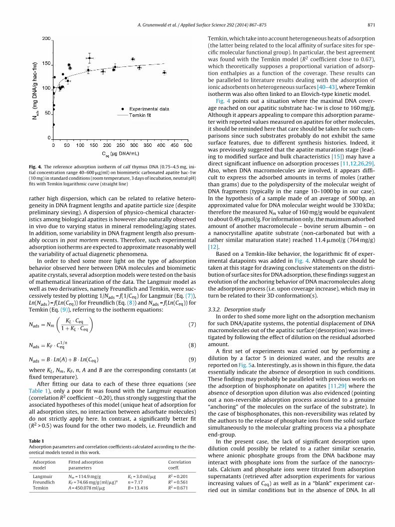
Fig. 4. The reference adsorption isotherm of calf thymus DNA (0.75–4.5 mg, ini-t(fi
rgpiiIaat
baowcLT
N
N
N
wfi
T(aad(
TAo
ial concentration range 40–600 �g/ml) on biomimetic carbonated apatite hac-1w10 mg) in standard conditions (room temperature, 3 days of incubation, neutral pH)ts with Temkin logarithmic curve (straight line)
ather high dispersion, which can be related to relative hetero-eneity in DNA fragment lengths and apatite particle size (despitereliminary sieving). A dispersion of physico-chemical character-
stics among biological apatites is however also naturally observedn vivo due to varying status in mineral remodeling/aging states.n addition, some variability in DNA fragment length also presum-bly occurs in post mortem events. Therefore, such experimentaldsorption isotherms are expected to approximate reasonably wellhe variability of actual diagenetic phenomena.
In order to shed some more light on the type of adsorptionehavior observed here between DNA molecules and biomimeticpatite crystals, several adsorption models were tested on the basisf mathematical linearization of the data. The Langmuir model asell as two derivatives, namely Freundlich and Temkin, were suc-
essively tested by plotting 1/Nads = f(1/Ceq) for Langmuir (Eq. (7)),n(Nads) = f(Ln(Ceq)) for Freundlich (Eq. (8)) and Nads = f(Ln(Ceq)) foremkin (Eq. (9)), referring to the isotherm equations:
ads = Nm
(KL · Ceq
1 + KL · Ceq
)(7)
ads = KF · C1/neq (8)
ads = B · Ln(A) + B · Ln(Ceq) (9)
here KL, Nm, KF, n, A and B are the corresponding constants (atxed temperature).
After fitting our data to each of these three equations (seeable 1), only a poor fit was found with the Langmuir equationcorrelation R2 coefficient ∼0.20), thus strongly suggesting that the
ssociated hypotheses of this model (unique heat of adsorption forll adsorption sites, no interaction between adsorbate molecules)o not strictly apply here. In contrast, a significantly better fitR2 > 0.5) was found for the other two models, i.e. Freundlich andable 1dsorption parameters and correlation coefficients calculated according to the the-retical models tested in this work.
Adsorptionmodel
Fitted adsorptionparameters
Correlationcoeff.
Langmuir Nm = 114.9 mg/g KL = 3.0 ml/�g R2 = 0.201Freundlich KF = 74.66 mg/g (ml/�g)n n = 7.17 R2 = 0.561Temkin A = 450.078 ml/�g B = 13.416 R2 = 0.671
e Science 292 (2014) 867– 875 871
Temkin, which take into account heterogeneous heats of adsorption(the latter being related to the local affinity of surface sites for spe-cific molecular functional group). In particular, the best agreementwas found with the Temkin model (R2 coefficient close to 0.67),which theoretically supposes a proportional variation of adsorp-tion enthalpies as a function of the coverage. These results canbe paralleled to literature results dealing with the adsorption ofionic adsorbents on heterogeneous surfaces [40–43], where Temkinisotherm was also often linked to an Elovich-type kinetic model.
Fig. 4 points out a situation where the maximal DNA cover-age reached on our apatitic substrate hac-1w is close to 160 mg/g.Although it appears appealing to compare this adsorption parame-ter with reported values measured on apatites for other molecules,it should be reminded here that care should be taken for such com-parisons since such substrates probably do not exhibit the samesurface features, due to different synthesis histories. Indeed, itwas previously suggested that the apatite maturation stage (lead-ing to modified surface and bulk characteristics [15]) may have adirect significant influence on adsorption processes [11,12,26,29].Also, when DNA macromolecules are involved, it appears diffi-cult to express the adsorbed amounts in terms of moles (ratherthan grams) due to the polydispersity of the molecular weight ofDNA fragments (typically in the range 10–1000 bp in our case).In the hypothesis of a sample made of an average of 500 bp, anapproximated value for DNA molecular weight would be 330 kDa;therefore the measured Nm value of 160 mg/g would be equivalentto about 0.49 �mol/g. For information only, the maximum adsorbedamount of another macromolecule – bovine serum albumin – ona nanocrystalline apatite substrate (non-carbonated but with arather similar maturation state) reached 11.4 �mol/g (764 mg/g)[12].
Based on a Temkin-like behavior, the logarithmic fit of exper-imental datapoints was added in Fig. 4. Although care should betaken at this stage for drawing conclusive statements on the distri-bution of surface sites for DNA adsorption, these findings suggest anevolution of the anchoring behavior of DNA macromolecules alongthe adsorption process (i.e. upon coverage increase), which may inturn be related to their 3D conformation(s).
3.3.2. Desorption studyIn order to shed some more light on the adsorption mechanism
for such DNA/apatite systems, the potential displacement of DNAmacromolecules out of the apatitic surface (desorption) was inves-tigated by following the effect of dilution on the residual adsorbedamount.
A first set of experiments was carried out by performing adilution by a factor 5 in deionized water, and the results arereported on Fig. 5a. Interestingly, as is shown in this figure, the dataessentially indicate the absence of desorption in such conditions.These findings may probably be paralleled with previous works onthe adsorption of bisphosphonate on apatites [11,29] where theabsence of desorption upon dilution was also evidenced (pointingout a non-reversible adsorption process associated to a genuine“anchoring” of the molecules on the surface of the substrate). Inthe case of bisphosphonates, this non-reversibility was related bythe authors to the release of phosphate ions from the solid surfacesimultaneously to the molecular grafting process via a phosphateend-group.
In the present case, the lack of significant desorption upondilution could possibly be related to a rather similar scenario,where anionic phosphate groups from the DNA backbone mayinteract with phosphate ions from the surface of the nanocrys-
tals. Calcium and phosphate ions were titrated from adsorptionsupernatants (retrieved after adsorption experiments for variousincreasing values of Ceq) as well as in a “blank” experiment car-ried out in similar conditions but in the absence of DNA. In all
872 A. Grunenwald et al. / Applied Surface Science 292 (2014) 867– 875
Fig. 5. Effect of dilution in (a) deionized water or (b) in the presence of phosphateions ([P] = 18 mM), pH 7, on the adsorption of DNA on biomimetic apatite. (a) After 3 dof adsorption, 80% of the solution was replaced with deionized water (dilution step)for 3 more days: the “desorption” data points do not follow the isotherm despitethe dilution of the medium, Nads remaining essentially unchanged for each point,indicating the quasi-absence of DNA desorption. (b) Same experiment with dilutionba
csai0istcpi(ac
esifvaia
surface of biomimetic apatites, namely either HPO4 or H2PO4 )
y neutral phosphate solution. Partial desorption of ca. 30–50% of DNA is observedfter 3 days.
ases, our results indicated the presence of both types of ions inolution, and in increasing amounts as a function of the adsorbedmount, with a concentration range of phosphorus and calciumn the supernatants, respectively between ca. 0.3–0.8 mmol/l and.4–1.3 mmol/l (data not shown). These findings could hypothet-
cally be attributed to the (partial) dissolution of the apatiticubstrate and/or to the release of ions from the surface due tohe adsorption process itself. The concomitant presence of bothalcium cations and phosphate anions, however, strongly sup-orts at least some extent of apatite dissolution. At this point, it
s difficult to differentiate between the two types of contributionsdissolution-related or adsorption-related); especially taking intoccount the non-congruence of dissolution processes for nonstoi-hiometric apatites [29,30].
Additional “desorption” tests were nonetheless run in the pres-nce of phosphate ions in the medium (by addition of KH2PO4,ee Section 2), while keeping the pH value neutral. Remarkably,n this case, the residual adsorbed amounts were systematicallyound to sharply decrease (Fig. 5b). This type of behavior was pre-iously observed for example for bisphosphonates [44] where the
dsorption process was accompanied by a release of phosphateons. Taking all these statements in consideration, and althoughdditional investigation dedicated to these aspects (e.g. to theFig. 6. Effect of pH on the adsorption process in standard conditions of DNA ontobiomimetic nanocrystalline carbonated apatite (6 < pH < 9.5).
incongruence of apatite dissolution) will be needed, our data sug-gest in the present case that phosphate ions compete with DNAmolecules for surface sites of apatite crystals.
3.3.3. Effect of pH on DNA adsorptionThe maximum adsorption amount Nm of DNA onto the poorly
crystalline biomimetic apatite sample hac-1w was then followed(still at 22 ◦C) as a function of the initial pH of the medium (in therange 6–9.5). Indeed, pH may presumably play a non-negligible rolein adsorption processes involving ionic crystals such as apatites.Also, pH may also vary upon post mortem conditions.
As shown in Fig. 6, the value of Nm was found to increase uponacidification (ca. +13% at pH 6) of the medium, and converselyto decrease upon alkalinization (ca. −23% at pH 9.5). Several fac-tors may potentially come into play for explaining this behavior.In particular, partial apatite (surface) dissolution is expected toincrease upon acidification [45], thus modifying exposed surfacecharacteristics. To a lesser extent, acidification could be seen as ameans to facilitate the release of HPO4
2− ions from the surface ofapatite nanocrystals, as these ions are known to be the predominantform of phosphate ions on such surfaces [46,47]. In turn, such anincreased mobility of HPO4
2− ions could then facilitate the anchor-ing of DNA phosphate groups onto the surface of the crystals (seediscussion in Section 3.3.2). Another potential explanation for thispH effect on Nm could be related to a change in DNA conforma-tion upon acidification/alkalinization, thus modifying the profile ofthe grafted molecules. In contrast it appears that these pH effectsmay not be related to the speciation of the phosphate groups (mainionization site of double-stranded DNA) from the DNA backbone.Indeed, their pKa value of ca. 1.5 [48] points to a situation wherethese phosphate groups are ionized for any pH > 1.5.
It is interesting to remark however that the experimental pHvalues of the media after adsorption (data not shown) showed thegeneral tendency to evolve toward neutrality, independently of theinitial pH value (whether acidic of alkaline). This effect may be bothdue (1) to a surface equilibration phenomenon of the apatite crystalsurface and (2) to the adsorption process itself leading to phos-phate ion release, as indicated in the previous sub-section. Indeed,the release of phosphate ions (which are mostly protonated on the
2− −
in a medium exhibiting a pH value between ca. 5 and 10 is expectedto evolve toward neutralization due to the buffering effect of theH2PO4
−/HPO42− acido-basic couple (pKa = 7.2) [49].
A. Grunenwald et al. / Applied Surface Science 292 (2014) 867– 875 873
F(
ttDsf
3
hmipmDm
icaDipsttt[iuToadDlobep
3
s
◦
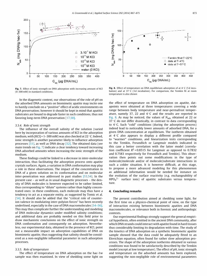
ig. 7. Effect of ionic strength on DNA adsorption with increasing amount of KCl0–300 mM) in standard conditions.In the diagenetic context, our observations of the role of pH onhe adsorbed DNA amounts on biomimetic apatite may incite oneo hastily conclude on a “positive” effect of acidic environments onNA preservation; however it should be kept in mind that apatitic
ubstrates are bound to degrade faster in such conditions, thus notavoring long-term DNA preservation [17,50].
.3.4. Role of ionic strengthThe influence of the overall salinity of the solution (varied
ere by incorporation of various amounts of KCl in the adsorptionedium, with [KCl] = 1–300 mM) was also checked at 22 ◦C. Indeed,
onic strength is another parameter likely to influence adsorptionrocesses [51], as well as DNA decay [52]. The obtained data (seeain trends on Fig. 7) indicate a clear tendency toward increasingNA adsorbed amounts when increasing the ionic strength of theedium.These findings could be linked to a decrease in inter-molecular
nteraction, thus facilitating the adsorption process onto apatiterystals surfaces. Again, a variation in DNA conformation may play
role in these observations. The impact of the concentration inNA of a given solution on its conformation and on molecular
nter-penetration was addressed in past studies [53,54]. In theresent case – as well as in usual diagenetic processes – the den-ity of DNA molecules is however expected to be rather limited,hus corresponding to “dilute” systems rather than highly concen-rated ones: in these conditions, each molecule may thus have aendency to act as a separate entity, as suggested by Tomic et al.54]. It should also be added that the “critical role of counter-on valence in modulating inter-polyion forces” has been recentlynderlined, especially in the case of DNA macromolecules [54–56].his may thus complicate further the understanding and modelingf DNA molecular dynamics under modified salinity conditions;nd additional data are probably needed on this field prior toraw mechanistic conclusions on the effect of ionic strength onNA conformation and consequently on adsorption. Neverthe-
ess, our experimental data, obtained in the presence of KCl, pointut a measurable impact on adsorption capabilities of DNA oniomimetic apatite, thus suggesting that salinity should be consid-red as a non-negligible influential parameter in such adsorptionrocesses.
.3.5. Role of temperatureThe effect of temperature on DNA adsorption on the hac-1w
ample was then examined. In view of shedding some light on
Fig. 8. Effect of temperature on DNA equilibrium adsorption of at 4 C (5 d incu-bation) and at 37 ◦C (3 d incubation). For comparison, the Temkin fit at roomtemperature is also shown
the effect of temperature on DNA adsorption on apatite, dat-apoints were obtained at three temperatures covering a widerange between body temperature and near-permafrost temper-ature, namely 37, 22 and 4 ◦C and the results are reported inFig. 8. As may be noticed, the values of Nads obtained at 22 or37 ◦C do not differ drastically, in contrast to data correspondingto 4 ◦C. Such “cold” conditions (during the adsorption process)indeed lead to noticeably lower amounts of adsorbed DNA, for agiven DNA concentration at equilibrium. The isotherm obtainedat 4 ◦C also appears to display a different profile comparedto “warmer” conditions, and linearization tests correspondingto the Temkin, Freundlich or Langmuir models indicated inthis case a better correlation with the latter model (correla-tion coefficient R2 = 0.8515 for Langmuir as opposed to 0.7832and 0.7583 respectively for Freundlich and Temkin). This obser-vation then points out some modifications in the type ofmolecule/molecule and/or of molecule/substrate interactions insuch a colder situation. It is however difficult, at this stage,to propose a more advanced modeling for this phenomenonas additional information would be needed for instance onthe evolution of the surface reactivity (e.g. exchangeability ofHPO4
2− surface ions) of apatitic substrates versus tempera-ture.
4. Concluding remarks
The present contribution aimed at shedding some light, forthe first time on a physico-chemical point of view, on the typeof interaction existing between biomimetic apatites and DNAmacromolecules, or relevance both in forensic and anthropologiccontexts.
Our experimental findings strongly support the general empiri-cal hypothesis, often emitted in the ancient DNA community, afterwhich DNA strands could interact with apatite found in hard tissuesthus considerably limiting its degradation with time. The study ofthe kinetics of DNA adsorption on a synthetic biomimetic apatitesample showed that the data could be adequately fitted to anElovichian equation, often found for rather slow adsorption pro-cesses. The shape of the adsorption isotherms obtained in various
conditions was found to be satisfactorily described by the Temkinmodel (except at low temperature). The effect of pH, ionic strengthand temperature on the adsorbed amounts has been explored,suggesting the non-negligible role of environmental parameters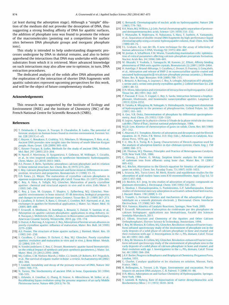
8 Surfac
(tstosi
casoe
taa
A
EF
R
[
[
[
[
[
[
[
[
[
[
[
[
[
[
[
[
[
[
[
[
[
[
[
[
[
[
[
[
[
[
[
[
[
[[
[
[
[
[
[
[
74 A. Grunenwald et al. / Applied
at least during the adsorption stage). Although a “simple” dilu-ion of the medium did not provoke the desorption of DNA, thusuggesting a strong binding affinity of DNA for apatitic surfaces,he addition of phosphate ions was found to promote the releasef the macromolecules (pointing out a competition for surfaceites between DNA phosphate groups and inorganic phosphateons).
This study is intended to help understanding diagenetic pro-esses undergone by DNA in skeletal remains, and also to betterpprehend the interactions that DNA may undertake with apatiticubstrates from which it is retrieved. More advanced knowledgen such interactions may also allow one to optimize, in turn, DNAxtraction procedures.
The dedicated analysis of the solids after DNA adsorption andhe exploration of the interaction of shorter DNA fragments withpatitic substrates represent upcoming perspectives for this work,nd will be the object of future complementary studies.
cknowledgements
This research was supported by the Institute of Ecology andnvironment (INEE) and the Institute of Chemistry (INC) of therench National Centre for Scientific Research (CNRS).
eferences
[1] T. Delabarde, C. Keyser, A. Tracqui, D. Charabidze, B. Ludes, The potential offorensic analysis on human bones found in riverine environment, Forensic Sci.Int. 228 (2013) e1–e5.
[2] C. Keyser, C. Bouakaze, E. Crubézy, V.G. Nikolaev, D. Montagnon, T. Reis, et al.,Ancient DNA provides new insights into the history of south Siberian Kurganpeople, Hum. Genet. 126 (2009) 395–410.
[3] C. Keyser-Tracqui, B. Ludes, Methods for the study of ancient DNA, MethodsMol. Biol. 297 (2005) 253–264.
[4] N. Nassif, F. Martineau, O. Syzgantseva, F. Gobeaux, M. Willinger, T. Coradin,et al., In vivo inspired conditions to synthesize biomimetic hydroxyapatite,Chem. Mater. 22 (2010) 3653–3663.
[5] A.S. Posner, F. Betts, Synthetic amorphous calcium phosphate and its relationto bone mineral structure, Acc. Chem. Res. 8 (1975) 273–281.
[6] C. Rey, Calcium phosphate biomaterials and bone mineral. Differences in com-position, structures and properties, Biomaterials 11 (1990) 13–15.
[7] E.D. Eanes, J.L. Meyer, The maturation of crystalline calcium phosphates inaqueous suspensions at physiologic pH, Calcif. Tissue Res. 23 (1977) 259–269.
[8] C. Rey, A. Hina, A. Tofighi, M.J. Glimcher, Maturation of poorly crystallineapatites: chemical and structural aspects in vivo and in vitro, Cells Mater. 5(1995) 345–356.
[9] C. Rey, J. Lian, M. Grynpas, F. Shapiro, L. Zylberberg, M.J. Glimcher, Non-apatitic environments in bone mineral: FT-IR detection, biological propertiesand changes in several disease states, Connect. Tissue Res. 21 (1989) 267–273.
10] S. Cazalbou, D. Eichert, X. Ranz, C. Drouet, C. Combes, M.F. Harmand, et al., Ionexchanges in apatites for biomedical application, J. Mater. Sci. Mater. Med. 16(2005) 405–409.
11] F. Errassifi, A. Menbaoui, H. Autefage, L. Benaziz, S. Ouizat, V. Santran, et al.,Adsorption on apatitic calcium phosphates: applications to drug delivery, in:R. Narayan, J. McKittrick (Eds.), Advances in Bioceramics and Biotechnologies,American Ceramic Society, Westerville, 2010, pp. 159–174.
12] S. Ouizat, A. Barroug, A. Legrouri, C. Rey, Adsorption of bovine serum albumin onpoorly crystalline apatite: influence of maturation, Mater. Res. Bull. 34 (1999)2279–2289.
13] A.S. Posner, The structure of bone apatite surfaces, J. Biomed. Mater. Res. 19(1985) 241–250.
14] S. Cazalbou, C. Combes, D. Eichert, C. Rey, M.J. Glimcher, Poorly crystallineapatites: evolution and maturation in vitro and in vivo, J. Bone Miner. Metab.22 (2004) 310–317.
15] N. Vandecandelaere, C. Rey, C. Drouet, Biomimetic apatite-based biomaterials:on the critical impact of synthesis and post-synthesis parameters, J. Mater. Sci.Mater. Med. 23 (2012) 2593–2606.
16] M.J. Collins, C.M. Nielsen-Marsh, J. Hiller, C.I. Smith, J.P. Roberts, R.V. Prigodich,et al., The survival of organic matter in bone: a review, Archaeometry 44 (2002)383–394.
17] T. Lindahl, Instability and decay of the primary structure of DNA, Nature 362(1993) 709–715.
18] N. Tuross, The biochemistry of ancient DNA in bone, Experientia 50 (1994)530–535.
19] L. Orlando, A. Ginolhac, G. Zhang, D. Froese, A. Albrechtsen, M. Stiller, et al.,Recalibrating Equus evolution using the genome sequence of an early MiddlePleistocene horse, Nature 499 (2013) 74–78.
[
[
e Science 292 (2014) 867– 875
20] G. Bernardi, Chromatography of nucleic acids on hydroxyapatite, Nature 206(1965) 779–783.
21] R.K. Main, M.J. Wilkins, L.J. Cole, Partial chromatographic separation of pentose-and deoxypentosenucleic acids, Science 129 (1959) 331–332.
22] T. Watanabe, K. Makitsuru, H. Nakazawa, S. Hara, T. Suehiro, A. Yamamoto,et al., Separation of double-strand DNA fragments by high-performance liquidchromatography using a ceramic hydroxyapatite column, Anal. Chim. Acta 386(1999) 69–75.
23] F.L. Graham, A.J. van der Eb, A new technique for the assay of infectivity ofhuman adenovirus 5 DNA, Virology 52 (1973) 456–467.
24] M. Jordan, A. Schallhorn, F.M. Wurm, Transfecting mammalian cells: optimiza-tion of critical parameters affecting calcium-phosphate precipitate formation,Nucleic Acids Res. 24 (1996) 596–601.
25] M. Okazaki, Y. Yoshida, S. Yamaguchi, M. Kaneno, J.C. Elliott, Affinity bindingphenomena of DNA onto apatite crystals, Biomaterials 22 (2001) 2459–2464.
26] H. Autefage, F. Briand-Mésange, S. Cazalbou, C. Drouet, D. Fourmy, S. Gonc alvès,et al., Adsorption and release of BMP-2 on nanocrystalline apatite-coated anduncoated hydroxyapatite/�-tricalcium phosphate porous ceramics, J. Biomed.Mater. Res. B: Appl. Biomater. 91B (2009) 706–715.
27] L. Benaziz, A. Barroug, A. Legrouri, C. Rey, A. Lebugle, Adsorption of O-phospho-l-serine and l-serine onto poorly crystalline apatite, J. Colloid Interface Sci. 238(2001) 48–53.
28] D.N. Misra, Adsorption and orientation of tetracycline on hydroxyapatite, Calcif.Tissue Int. 48 (1991) 362–367.
29] P. Pascaud, P. Gras, Y. Coppel, C. Rey, S. Sarda, Interaction between a bisphos-phonate, tiludronate, and biomimetic nanocrystalline apatites, Langmuir 29(2013) 2224–2232.
30] H. Tanaka, K. Miyajima, M. Nakagaki, S. Shimabayashi, Incongruent dissolutionof hydroxyapatite in the presence of phosphoserine, Colloid Polym. Sci. 269(1991) 161–165.
31] A. Gee, V.R. Deitz, Determination of phosphate by differential spectrophoto-metry, Anal. Chem. 25 (1953) 1320–1324.
32] R. Legros, Apport de la physico-chimie à l’étude de la phase minérale des tissuscalcifiés (Thèse d’État), Institut national polytechnique, 1984.
33] M.J.D. Low, Kinetics of chemisorption of gases on solids, Chem. Rev. 60 (1960)267–312.
34] C. Aharoni, F.C. Tompkins, Kinetics of adsorption and desorption and the Elovichequation, in: H. Pines, P.B. Weisz, D.D. Eley (Eds.), Advances in Catalysis, Aca-demic Press, 1970, pp. 1–49.
35] F.-C. Wu, R.-L. Tseng, R.-S. Juang, Characteristics of Elovich equation used forthe analysis of adsorption kinetics in dye–chitosan systems, Chem. Eng. J. 150(2009) 366–373.
36] J.M. Thomas, W.J. Thomas, Principles and Practice of Heterogeneous Catalysis,VCH, New York, USA, 1996.
37] C. Cheung, J. Porter, G. Mckay, Sorption kinetic analysis for the removalof cadmium ions from effluents using bone char, Water Res. 35 (2001)605–612.
38] A.G. Ritchie, Alternative to the Elovich equation for kinetics of adsorption ofgases on solids, J. Chem. Soc. Faraday Trans. 73 (1997) 1650–1653.
39] L. Sciascia, M.L. Turco Liveri, M. Merli, Kinetic and equilibrium studies for theadsorption of acid nucleic bases onto K10 montmorillonite, Appl. Clay Sci. 53(2011) 657–668.
40] J.O. Bockris, K.T. Jeng, In-situ studies of adsorption of organic compounds onplatinum electrodes, J. Electroanal. Chem. 330 (1992) 541–581.
41] G. Skodras, I. Diamantopoulou, G. Pantoleontos, G.P. Sakellaropoulos, Kineticstudies of elemental mercury adsorption in activated carbon fixed bed reactor,J. Hazard. Mater. 158 (2008) 1–13.
42] S. Trasatti, L. Formaro, Kinetics and mechanism of the adsorption of glyco-laldehyde on a smooth platinum electrode, J. Electroanal. Chem. InterfacialElectrochem. 17 (1968) 343–364.
43] M.A. Vannice, Kinetics of Catalytic Reactions, Springer, New York, 2005.44] F. Errassifi, Mécanismes d’adsorption du risédronate par des phosphates de
calcium biologiques: applications aux biomatériaux, Faculté des SciencesSemlalia-Marrakech, 2011.
45] J.C. Elliott, Structure and Chemistry of the Apatites and Other CalciumOrthophosphates, Elsevier Science & Technology, Amsterdam, 1994.
46] C. Rey, M. Shimizu, B. Collins, M.J. Glimcher, Resolution-enhanced Fourier trans-form infrared spectroscopy study of the environment of phosphate ion in theearly deposits of a solid phase of calcium phosphate in bone and enamel andtheir evolution with age: 2. Investigations in the �3 PO4 domain, Calcif. TissueInt. 49 (1991) 383–388.
47] C. Rey, M. Shimizu, B. Collins, M.J. Glimcher, Resolution-enhanced Fourier trans-form infrared spectroscopy study of the environment of phosphate ions in theearly deposits of a solid phase of calcium-phosphate in bone and enamel, andtheir evolution with age. I. Investigations in the �4 PO4 domain, Calcif. TissueInt. 46 (1990) 384–394.
48] J.A.V. Butler, Progress in Biophysics and Biophysical Chemistry, Pergamon Press,London, 1951.
49] G. Charlot, L’analyse qualitative et les réactions en solution, Masson, Paris,France, 1963.
50] R. Bollongino, A. Tresset, J.-D. Vigne, Environment and excavation. Pre-lab
impacts on ancient DNA analyses, C. R. Palevol. 7 (2008) 91–98.51] D.N. Misra, Adsorption on and Surface Chemistry of Hydroxyapatite, Springer,New York, 1984.
52] T. Lindahl, B. Nyberg, Rate of depurination of native deoxyribonucleic acid,Biochemistry (Mosc.) 11 (1972) 3610–3618.

Surfac
[
[
[
A. Grunenwald et al. / Applied
53] A.V. Dobrynin, M. Rubinstein, Theory of polyelectrolytes in solutions and at
surfaces, Prog. Polym. Sci. 30 (2005) 1049–1118.54] S. Tomic, D. Grgicin, T. Ivek, T. Vuletic, S. Dolanski Babic, R. Podgornik,Dynamics and structure of biopolyelectrolytes in repulsion regime char-acterized by dielectric spectroscopy, Phys. B: Condens. Matter 407 (2012)1958–1963.
[
e Science 292 (2014) 867– 875 875
55] A. Naji, M. Kanduc, R.R. Netz, R. Podgornik, Exotic electrostatics: unusual
features of electrostatic interactions between macroions, ArXiv Prepr, 2010arXiv:1008.0357.56] R.W. Wilson, V.A. Bloomfield, Counterion-induced condensation of deoxyri-bonucleic acid. A light-scattering study, Biochemistry (Mosc.) 18 (1979)2192–2196.