Adrenoleukodystrophy in a mother and son - Journal of ...
8
Journal of Neurology, Neurosurgery, and Psychiatry 1987;50:1165-1172 Adrenoleukodystrophy in a mother and son RODERICK H W SIMPSON,* JOHN RODDA,t CAROLUS J REINECKEt From the Postgraduate Medical School,* University of Exeter, UK, Department of Neurology,t Baragwanath Hospital, Johannesburg, and Department of Biochemistry,4 Potchesfstroom Universityfor CHO, Potchesfstroom, Transvaal, South Africa SUMMARY A 6 year old boy died from a degenerative brain disease which was clinically and pathologically typical of adrenoleukodystrophy. Shortly before his disease became manifest his 28 year old mother had presented with similar symptoms, and subsequently died. Her brain showed almost identical features including the presence of pathognomonic ultrastructural inclusions. The accumulation of very long chain fatty acids in cerebral white matter as well as high hexacosanoic to docosanoic acid (C26:22) ratios, substantiated the diagnosis in both cases. This is one of the few documented cases of adrenoleukodystrophy in an adult female, and is almost certainly an example of clinical manifestation of this X-linked inherited disease in a carrier. Adrenoleukodystrophy (ALD) is characterised clinically by progressive neurological deterioration involving mainly the pyramidal tracts together with cortical blindness. This is associated with adreno- cortical insufficiency which may be silent clinically, only appearing on adrenocorticotrophic hormone (ACTH) stimulation.1 Post mortem examination reveals a sudanophil leukodystrophy involving the cerebral hemispheric white matter of mainly the occipital, parietal and posterior temporal lobes, but sub-cortical arcuate fibres are generally spared. The cerebellar white matter is affected in some cases, but the brain stem and spinal cord show degeneration of only the descending fibre tracts. In the adrenal glands ballooned cortical cells are seen in the zonae fascicu- lata and reticularis.2 By electron microscopy, patho- gnomonic cytoplasmic inclusions are found in the brain, adrenal cortex3 and some other organs.4 Bio- chemical studies have shown an abnormal accumu- lation of long chain fatty acids in the brain' and cul- tured skin fibroblasts,6 and it has been suggested that ALD is a lipid storage disease, and that the basic defect may be a deficiency of one of the enzymes involved in peroxisomal oxidation of very long chain fatty acids.7 Address for reprint requests: Dr R H W Simpson, Postgraduate Medical School, University of Exeter, Barrack Road, Exeter EX2 5DW, UK. Received 1 July 1986 and in revised form 4 February 1987. Accepted 11 February 1987 Although most cases have been described in boys of 5 to 15 years of age, older and younger individuals may also be affected. In the older group, many fall into the category of adrenomyeloneuropathy (AMN), which is clinically8 and pathologically9 different in that the spinal cord is involved much more than the brain. However, there is adrenal insufficiency, and the ultrastructural and biochemical findings are identical to those in ALD. In addition, cases of ALD and AMN have occurred in the same families,8 10 leading to the suggestion that both are different varieties of the same disease."1 A neonatal form of ALD has also been recognised,'2 but although it shares some simi- larities with the other types, it is now considered to be a separate entity more closely related (but not identi- cal) to Zellweger's syndrome and infantile Refsum's disease.13- 15 With the exception of the neonatal form, which is autosomal recessive, ALD is inherited in a sex-linked recessive manner, and the gene locus itself has been accurately mapped on the X chromosome.'6 How- ever, a few cases of affected females other than neo- nates have been described61' 17-21 and we here report another individual studied clinically, neuro- pathologically and biochemically. Cliical findings Case I A previously well 28 year old black African woman presented to Baragwanath Hospital with a 4 month history of mental confusion. She had become socially withdrawn and was unable to care for herself or her family. She could 1165 guest. Protected by copyright. on January 21, 2022 by http://jnnp.bmj.com/ J Neurol Neurosurg Psychiatry: first published as 10.1136/jnnp.50.9.1165 on 1 September 1987. Downloaded from
-
Upload
khangminh22 -
Category
Documents
-
view
0 -
download
0
Transcript of Adrenoleukodystrophy in a mother and son - Journal of ...
Journal of Neurology, Neurosurgery, and Psychiatry 1987;50:1165-1172
Adrenoleukodystrophy in a mother and son
RODERICK H W SIMPSON,* JOHN RODDA,t CAROLUS J REINECKEtFrom the Postgraduate Medical School,* University ofExeter, UK, Department ofNeurology,t BaragwanathHospital, Johannesburg, and Department of Biochemistry,4 Potchesfstroom Universityfor CHO,Potchesfstroom, Transvaal, South Africa
SUMMARY A 6 year old boy died from a degenerative brain disease which was clinically andpathologically typical of adrenoleukodystrophy. Shortly before his disease became manifest his28 year old mother had presented with similar symptoms, and subsequently died. Her brain showedalmost identical features including the presence of pathognomonic ultrastructural inclusions. Theaccumulation of very long chain fatty acids in cerebral white matter as well as high hexacosanoic todocosanoic acid (C26:22) ratios, substantiated the diagnosis in both cases. This is one of the fewdocumented cases of adrenoleukodystrophy in an adult female, and is almost certainly an exampleof clinical manifestation of this X-linked inherited disease in a carrier.
Adrenoleukodystrophy (ALD) is characterisedclinically by progressive neurological deteriorationinvolving mainly the pyramidal tracts together withcortical blindness. This is associated with adreno-cortical insufficiency which may be silent clinically,only appearing on adrenocorticotrophic hormone(ACTH) stimulation.1 Post mortem examinationreveals a sudanophil leukodystrophy involving thecerebral hemispheric white matter of mainly theoccipital, parietal and posterior temporal lobes, butsub-cortical arcuate fibres are generally spared. Thecerebellar white matter is affected in some cases, butthe brain stem and spinal cord show degeneration ofonly the descending fibre tracts. In the adrenal glandsballooned cortical cells are seen in the zonae fascicu-lata and reticularis.2 By electron microscopy, patho-gnomonic cytoplasmic inclusions are found in thebrain, adrenal cortex3 and some other organs.4 Bio-chemical studies have shown an abnormal accumu-lation of long chain fatty acids in the brain' and cul-tured skin fibroblasts,6 and it has been suggested thatALD is a lipid storage disease, and that the basicdefect may be a deficiency of one of the enzymesinvolved in peroxisomal oxidation of very long chainfatty acids.7
Address for reprint requests: Dr R H W Simpson, PostgraduateMedical School, University of Exeter, Barrack Road, ExeterEX2 5DW, UK.
Received 1 July 1986 and in revised form 4 February 1987.Accepted 11 February 1987
Although most cases have been described in boysof 5 to 15 years of age, older and younger individualsmay also be affected. In the older group, many fallinto the category of adrenomyeloneuropathy (AMN),which is clinically8 and pathologically9 different inthat the spinal cord is involved much more than thebrain. However, there is adrenal insufficiency, and theultrastructural and biochemical findings are identicalto those in ALD. In addition, cases of ALD andAMN have occurred in the same families,8 10 leadingto the suggestion that both are different varieties ofthe same disease."1 A neonatal form ofALD has alsobeen recognised,'2 but although it shares some simi-larities with the other types, it is now considered to bea separate entity more closely related (but not identi-cal) to Zellweger's syndrome and infantile Refsum'sdisease.13- 15With the exception of the neonatal form, which is
autosomal recessive, ALD is inherited in a sex-linkedrecessive manner, and the gene locus itself has beenaccurately mapped on the X chromosome.'6 How-ever, a few cases of affected females other than neo-nates have been described61' 17-21 and we herereport another individual studied clinically, neuro-pathologically and biochemically.
Cliical findings
Case IA previously well 28 year old black African womanpresented to Baragwanath Hospital with a 4 month historyof mental confusion. She had become socially withdrawnand was unable to care for herself or her family. She could
1165
guest. Protected by copyright.
on January 21, 2022 byhttp://jnnp.bm
j.com/
J Neurol N
eurosurg Psychiatry: first published as 10.1136/jnnp.50.9.1165 on 1 S
eptember 1987. D
ownloaded from
1166not provide a full or accurate history, including that of herfamily, but she did not know of any other relations withcomparable symptoms. There was an apparently normal sis-ter, who had no issue, and the patient herself had three chil-dren: an 11 year old girl with well controlled epilepsy of longstanding, a 6 year old boy (Case 2) soon to present with thesame disease, and a 4 year old son who was clinically nor-mal. Her husband had recently abandoned the family andwas not traceable, but he was apparently healthy.Upon questioning, the patient knew her name, but was
disorientated for time and place, and had difficulty namingobjects. She exhibited concentration fatigue, as well as easilybecoming lost, but at this stage, her gait was normal. Exam-ination revealed mild generalised weakness, and increasedlimb tone with brisk reflexes, but flexor plantar responses.She had a left hemianopia with normal fundi and pupillaryreaction. Cranial nerves were otherwise intact. Coordinationwas poor, but there was no obvious loss of sensation orproprioception. Outwith the nervous system she was notedto be 30 weeks pregnant. She was normotensive (115/75),and there was no excess pigmentation.Among investigations CT scans, including one performed
shortly before death (fig 1), showed reduced densitythroughout the white matter with focal uptake of contrast.Lumbar puncture revealed a pressure of 3 cm of water, nor-mal chemistry and no cells. Peripheral blood haematologicaland biochemical investigations (including urea and electro-lytes) were within normal limits, and a large number of sero-logical studies were negative. At this stage a diagnosis of
Fig 1 Case 1: CTscan showing diffuse low density ofthewhite matter withfocal uptake ofcontrast.
Simpson, Rodda, ReineckeALD was not entertained and adrenal function tests werenot performed.The patient's clinical condition deteriorated rapidly. She
became more confused and was unable to feed herself, andeventually she could not even get out of bed. She wasdelivered of a macerated foetus, on whom no necropsy wasperformed. Shortly thereafter, she developed broncho-pneumonia and died 5 weeks after admission.
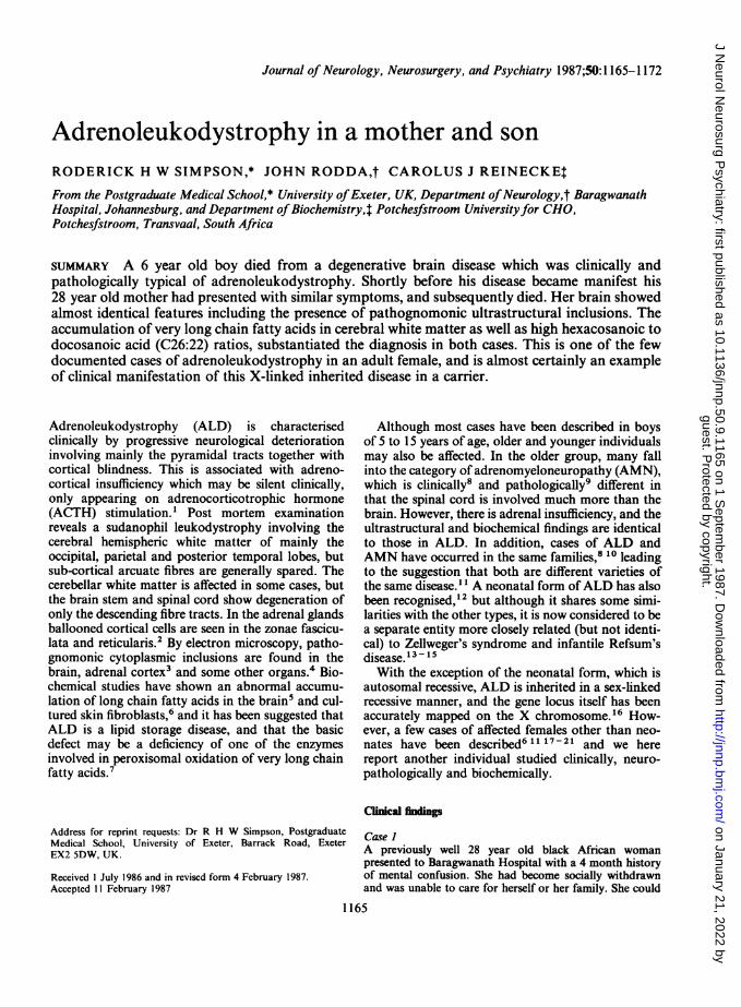
Case 2During the final stages of the illness of Case 1, her 6 year oldson was admitted to an outlying mission hospital whence hewas transferred to Baragwanath Hospital. Over a fewmonths he had begun to exhibit strange behaviour includinginappropriate outbursts of laughter, and he had also becomeblind. He was disorientated for time and place, but at thisstage still knew his name. He had a shuffling gait and oftencollided with furniture. All four limbs showed some increasein tone and briskness of reflexes, but Babinsky and Hoffmanreflexes were negative and sensation was normal. The blind-ness was cortical with normally reacting pupils and normalfundi. Other cranial nerves were intact. His growth parame-ters were also normal, and no obvious abnormality wasfound outwith the nervous system; blood pressure was100/60mm Hg and there were no features of adrenalinsufficiency.The CT scan revealed low density throughout the white
matter with uptake of contrast especially in the occipitallobes (fig 2). The EEG showed diffuse slowing in the poste-
-.-...Fig 2 Case 2: enhanced CTscan also showing decreaseddensity of the white matter with serpiginous uptake ofcontrast, especially in the occipital lobes corresponding to thegrey matter-white matter interface.
guest. Protected by copyright.
on January 21, 2022 byhttp://jnnp.bm
j.com/
J Neurol N
eurosurg Psychiatry: first published as 10.1136/jnnp.50.9.1165 on 1 S
eptember 1987. D
ownloaded from
Adrenoleukodystrophy in a mother and son
rior occipital areas, and visual evoked potentials confirmedthe presence of cortical blindness. On lumbar puncture, theCSF pressure was 5cm water; no cells were found andprotein was 0-39 g/l. Routine radiological and bloodinvestigations were non-contributory-plasma electrolyteswere within the normal range. However, an ACTH stimu-lation test using 25 units of intramuscular ACTH showed asub-normal response with plasma cortisol levels of411 nmol/l at the start, 483 after 30 minutes and 538 after60 minutes.The diagnosis of ALD was confirmed on brain biopsy,
and the patient's clinical condition progressively deterio-rated over the next 9 months. He became more confused andwithdrawn, developing focal seizures and generalised spas-ticity. He lapsed into a stuporose state, and died one yearafter his initial presentation.
1167
Pathological findings
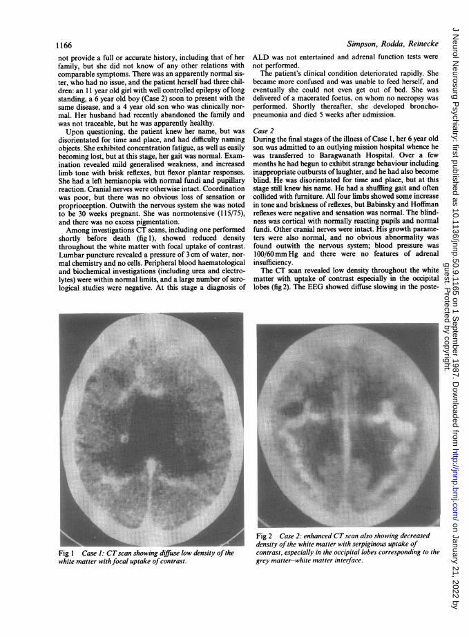
Case IA limited necropsy was performed 3 days after death, andonly the brain was available for study. After fixation in for-mol saline it weighed 1175g. The cerebral white mattershowed extensive areas of discolouration with a somewhatgranular appearance. This change involved the centrumsemiovale of the parietal and occipital lobes and the poste-rior part of the temporal lobes. It was generalised ratherthan patchy, although the subcortical arcuate fibres werelargely spared. The white matter of the frontal and anteriortemporal lobes was macroscopically unaffected. Similarchanges were evident in the posterior parts of the corpuscallosum and internal capsules. There was also involvementof the cerebellar white matter, as well as the cerebral pedun-
14 6'I
.4
*V 4 .t A,--
"'A44- ~ ~ 4 a
Cr ...-~~4*~&4*'~~~~~~~~~~~~~~~~~~~,, .\~~~~~~~~~~~~~~~ 1' ~ ~ ~ ~ ~ ~
4W~~ ~ ~ ~ ~ 4
f.44
Fig 3 Case 1: micrograph ofthe affected brain showing perivascular accumulation ofinflammatory cells, as well as astrocyticgliosis in the surrounding demyelinated white matter. (Haematoxylin and eosin, x 50).
guest. Protected by copyright.
on January 21, 2022 byhttp://jnnp.bm
j.com/
J Neurol N
eurosurg Psychiatry: first published as 10.1136/jnnp.50.9.1165 on 1 S
eptember 1987. D
ownloaded from

Simpson, Rodda, Reinecke
Fig 4 Case 1: electron micrograph ofthe affected white matter showing characteristic trilaminar inclusion bodies ofALD.
cles, medullary pyramids and long tracts of the pons. Histo-logical examination of the abnormal areas revealed almosttotal loss of myelin together with astrocytic proliferationand accumulation of sudanophil material both within andwithout phagocytic cells. There was also a perivascularinflammatory cell infiltrate which was heavier in the parietalthan in the occipital lobes (fig 3). Neurons were generallypreserved, but any loss was considered slight in comparisonto the devastation of the white matter. Electron microscopicexamination of gluteraldehyde fixed white matter confirmedthe loss of myelin and revealed numerous intracytoplasmictrilaminar bodies (fig 4).
Case 2Biopsy A wedge of brain tissue was received in which themain changes were in the white matter. These comprised lossof myelin, astrocytic proliferation and perivascular aggre-gation of macrophages, some of which had vacuolated cyto-plasm. Ultrastructural examination showed trilaminar inclu-sions characteristic of ALD.Necropsy The post mortem interval was 2 days. The brainweighed 820 g, and after representative areas had been takenfor biochemical study it was fixed in formol saline for 2
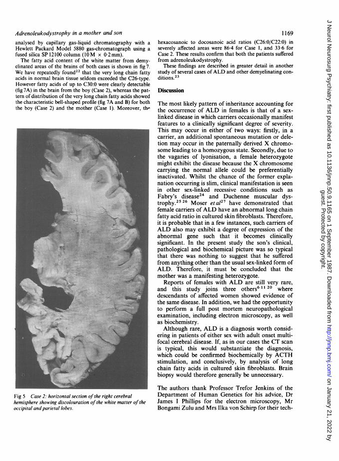
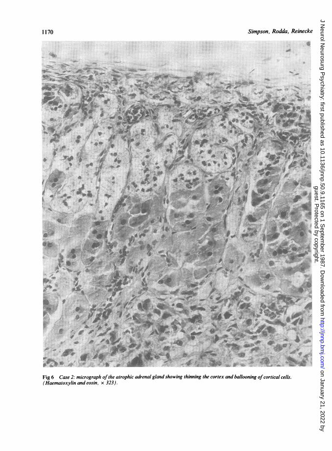
months. The cerebral white matter showed generalised dis-colouration (fig 5) with apparent sparing of the frontal andanterior temporal lobes. The overall distribution was similarto that in Case I except that the cerebellum was uninvolved.In the spinal cord, only the corticospinal and pyramidaltracts were affected. The histological picture was also thesame as in Case 1 with demyelination, astrocytic prolif-eration, accumulation of sudanophil material and a peri-vascular inflammatory cell infiltrate most marked in theparietal lobes. Trilaminar inclusions were identified onultrastructural examination.Both adrenal glands were atrophic. In the zonae fascicu-
lata and reticularis of the cortex swollen cells were seen, thecytoplasm ofwhich was either vacuolated or granular (fig 6).Plentiful trilaminar bodies were present by electron micro-scopy. In the rest of the body the only abnormality was afibrinous pleurisy. An extensive histological and ultra-structural examination was conducted, including the testesand skin, but no other abnormality or further trilaminarbodies were found.Biochemistry Lipids were extracted from the formalin fixedbrain tissue and fractionated as previously described.22Methylesters of the fatty acids, esterified to cholesterol, were
1168
guest. Protected by copyright.
on January 21, 2022 byhttp://jnnp.bm
j.com/
J Neurol N
eurosurg Psychiatry: first published as 10.1136/jnnp.50.9.1165 on 1 S
eptember 1987. D
ownloaded from
Adrenoleukodystrophy in a mother and son
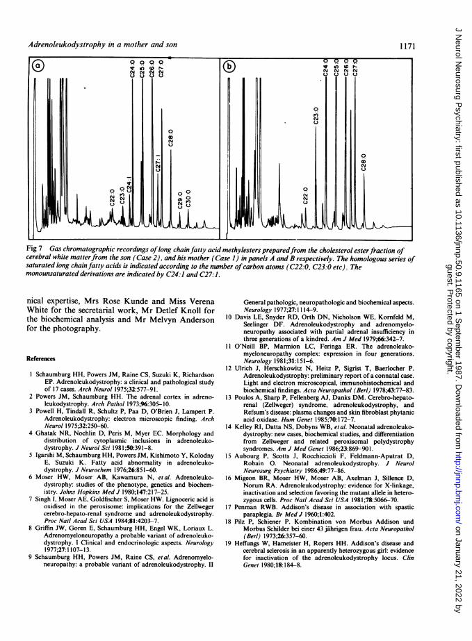
analysed by capillary gas-liquid chromatography with aHewlett Packard Model 5880 gas-chromatograph using afused silica SP 12100 column (IOM x 0-2 mm).The fatty acid content of the white matter from demy-
elinated areas of the brains of both cases is shown in fig 7.We have repeatedly found22 that the very long chain fattyacids in normal brain tissue seldom exceeded the C26-type.However fatty acids of up to C30:0 were clearly detectable(fig 7A) in the brain from the boy (Case 2), whereas the pat-tern of distribution of the very long chain fatty acids showedthe characteristic bell-shaped profile (fig 7A and B) for boththe boy (Case 2) and the mother (Case 1). Moreover, th-
Fig 5 Case 2: horizontal section of the right cerebralhemisphere showing discolouration of the white matter of theoccipital and parietal lobes.
1169hexacosanoic to docosanoic acid ratios (C26:0/C22:0) inseverely affected areas were 86-4 for Case 1, and 33-6 forCase 2. These results confirm that both the patients sufferedfrom adrenoleukodystrophy.
These findings are described in greater detail in anotherstudy of several cases ofALD and other demyelinating con-ditions.23
Discussion
The most likely pattern of inheritance accounting forthe occurrence of ALD in females is that of a sex-linked disease in which carriers occasionally manifestfeatures to a clinically significant degree of severity.This may occur in either of two ways: firstly, in acarrier, an additional spontaneous mutation or dele-tion may occur in the paternally derived X chromo-some leading to a homozygous state. Secondly, due tothe vagaries of lyonisation, a female heterozygotemight exhibit the disease because the X chromosomecarrying the normal allele could be preferentiallyinactivated. Whilst the chance of the former expla-nation occurring is slim, clinical manifestation is seenin other sex-linked recessive conditions such asFabry's disease24 and Duchenne muscular dys-trophy.25 26 Moser et a127 have demonstrated thatfemale carriers ofALD have an abnormal long chainfatty acid ratio in cultured skin fibroblasts. Therefore,it is probable that in a few instances, such carriers ofALD also may exhibit a degree of expression of theabnormal gene such that it becomes clinicallysignificant. In the present study the son's clinical,pathological and biochemical picture was so typicalthat there was nothing to suggest that he sufferedfrom anything other than the usual sex-linked form ofALD. Therefore, it must be concluded that themother was a manifesting heterozygote.
Reports of females with ALD are still very rare,and this study joins three others6 11 20 wheredescendants of affected women showed evidence ofthe same disease. In addition, we had the opportunityto perform a full post mortem neuropathologicalexamination, including electron microscopy, as wellas biochemistry.Although rare, ALD is a diagnosis worth consid-
ering in patients of either sex with adult onset multi-focal cerebral disease. If, as in our cases the CT scanis typical, this would substantiate the diagnosis,which could be confirmed biochemically by ACTHstimulation, and conclusively, by analysis of longchain fatty acids in cultured skin fibroblasts. Brainbiopsy would therefore generally be unnecessary.
The authors thank Professor Trefor Jenkins of theDepartment of Human Genetics for his advice, DrJames I Phillips for the electron microscopy, MrBongami Zulu and Mrs Ilka von Schirp for their tech-
guest. Protected by copyright.
on January 21, 2022 byhttp://jnnp.bm
j.com/
J Neurol N
eurosurg Psychiatry: first published as 10.1136/jnnp.50.9.1165 on 1 S
eptember 1987. D
ownloaded from
Simpson, Rodda, Reinecke
I........... .:2*
.~~ !4*'+.F w # .r.Tv">r-.w w^., oe ai *q d * * *1 '2I 4* s / z
b .s S k- +it* , . ''
e..~~~~~~~~~~~~~~,V~~~~~~~4.
a~ ~*+11 beU,-
I,~~~~~~~~~~~~~~~~~~~&
3, AL
t~~~~~~~~{'4 A
At. r~~~~~~~~~A V....Kr....
t,.3t4 6012g ;' *t * o 'f';t, g49*rei* r j e *} " $ t ,,,t ,,d,i ,, iC: w > , 4
4.~~~~ a~~~~~~~~. ..- 'P%
PS .........
'V~~~~~~~~~~~~~~~~~~~~~~~~~~~~~~~~~~~~O
fa4a'uvi~~~~~~~~~~~~~~~~~~~- 0~~~~~~~~-Vl
A ' *a
Fig 6 Case 2: micrograph of the atrophic adrenal gland showing thinning the cortex and ballooning ofcortical cells.(Haematoxylin and eosin, x 323).
1170
guest. Protected by copyright.
on January 21, 2022 byhttp://jnnp.bm
j.com/
J Neurol N
eurosurg Psychiatry: first published as 10.1136/jnnp.50.9.1165 on 1 S
eptember 1987. D
ownloaded from
Adrenoleukodystrophy in a mother and son 1171
Fig 7 Gas chromatographic recordings oflong chainfatty acid methylesters preparedfrom the cholesterol esterfraction ofcerebral white matterfrom the son (Case 2), and his mother (Case 1) in panels A and B respectively. The homologous series ofsaturated long chainfatty acids is indicated according to the number ofcarbon atoms (C22:0, C23:0 etc). Themonounsaturated derivations are indicated by C24:1 and C27:1.
nical expertise, Mrs Rose Kunde and Miss VerenaWhite for the secretarial work, Mr Detlef Knoll forthe biochemical analysis and Mr Melvyn Andersonfor the photography.
References
I Schaumburg HH, Powers JM, Raine CS, Suzuki K, RichardsonEP. Adrenoleukodystrophy: a clinical and pathological studyof 17 cases. Arch Neurol 1975;32:577-91.
2 Powers JM, Schaumburg HH. The adrenal cortex in adreno-leukodystrophy. Arch Pathol 1973;96:305-10.
3 Powell H, Tindall R, Schultz P, Paa D, O'Brien J, Lampert P.Adrenoleukodystrophy: electron microscopic finding. ArchNeurol 1975;32:250-60.
4 Ghatak NR, Nochlin D, Peris M, Myer EC. Morphology anddistribution of cytoplasmic inclusions in adrenoleuko-dystrophy. J Neurol Sci 198 1;50:391-8.
5 Igarshi M, Schaumburg HH, Powers JM, Kishimoto Y, KolodnyE, Suzuki K. Fatty acid abnormality in adrenoleuko-dystrophy. J Neurochem 1976;26:851-60.
6 Moser HW, Moser AB, Kawamura N, etal. Adrenoleuko-dystrophy: studies of the phenotype, genetics and biochem-istry. Johns Hopkins Med J 1980;147:217-25.
7 Singh I, Moser AE, Goldfischer S, Moser HW. Lignoceric acid isoxidised in the peroxisome: implications for the Zellwegercerebro-hepato-renal syndrome and adrenoleukodystrophy.Proc Nail Acad Sci USA 1984;81:4203-7.
8 Griffin JW, Goren E, Schaumburg HH, Engel WK, Loriaux L.Adrenomyeloneuropathy a probable variant of adrenoleuko-dystrophy. I Clinical and endocrinologic aspects. Neurology1977;27: 1107-13.
9 Schaumburg HH, Powers JM, Raine CS, etal. Adrenomyelo-neuropathy: a probable variant of adrenoleukodystrophy. II
General pathologic, neuropathologic and biochemical aspects.Neurology 1977;27:1 114-9.
10 Davis LE, Snyder RD, Orth DN, Nicholson WE, Kornfeld M,Seelinger DF. Adrenoleukodystrophy and adrenomyelo-neuropathy associated with partial adrenal insufficiency inthree generations of a kindred. Am J Med 1979;66:342-7.
11 O'Neill BP, Marmion LC, Feringa ER. The adrenoleuko-myeloneuropathy complex: expression in four generations.Neurology 1981;31:151-6.
12 Ulrich J, Herschkowitz N, Heitz P, Sigrist T, Baerlocher P.Adrenoleukodystrophy: preliminary report of a connatal case.Light and electron microscopical, immunohistochemical andbiochemical findings. Acda Neuropathol (Berl) 1978;43:77-83.
13 Poulos A, Sharp P, Fellenberg AJ, Danks DM. Cerebro-hepato-renal (Zellweger) syndrome, adrenoleukodystrophy, andRefsum's disease: plasma changes and skin fibroblast phytanicacid oxidase. Hum Genet 1985;70:172-7.
14 Kelley RI, Datta NS, Dobyns WB, et al. Neonatal adrenoleuko-dystrophy: new cases, biochemical studies, and differentiationfrom Zellweger and related peroxisomal polydystrophysyndromes. Am J Med Genet 1986;23:869-901.
15 Aubourg P, Scotts J, Rocchiccioli F, Feldmann-Aputrat D,Robain 0. Neonatal adrenoleukodystrophy. J NeurolNeurosurg Psychiatry 1986;49:77-86.
16 Migeon BR, Moser HW, Moser AB, Axelman J, Sillence D,Norum RA. Adrenoleukodystrophy: evidence for X-linkage,inactivation and selection favoring the mutant allele in hetero-zygous cells. Proc Nail Acad Sci USA 1981;78:5066-70.
17 Penman RWB. Addison's disease in association with spasticparaplegia. Br Med J 1960;1:402.
18 Pilz P, Schiener P. Kombination von Morbus Addison undMorbus Schilder bei einer 43 jahrigen frau. Acta Neuropathol(Berl) 1973;26:357-60.
19 Heffungs W, Hameister H, Ropers HH. Addison's disease andcerebral sclerosis in an apparently heterozygous girl: evidencefor inactivation of the adrenoleukodystrophy locus. ClinGenet 1980;18:184-8.
guest. Protected by copyright.
on January 21, 2022 byhttp://jnnp.bm
j.com/
J Neurol N
eurosurg Psychiatry: first published as 10.1136/jnnp.50.9.1165 on 1 S
eptember 1987. D
ownloaded from

117220 Morariu MA, Chason JL, Norum RA, Moser HW, Migeon B.
Adrenoleukodystrophy variant in a heterozygous female.Neurology 1982;32:A81.
21 Molzer B, Bernheimer H, Budka H, Pilz P, Toifl K. Accumula-tion of very long chain fatty acids is common to 3 variants ofadrenoleukodystrophy: classical ALD, atypical ALD (femalepatient) and adrenomyeloneuropathy. J Neurol Sci 1981;51:301-10.
22 Pretorius PJ, Reinecke CJ, Op't Hof J. Biochemical evidence forclinically diagnosed adrenoleukodystrophy in two brothers.S Afr Med J 1984;65:860-2.
23 Reinecke CJ, Knoll DP, Pretorius PJ, Steyn HS, Simpson RHW.The correlation between biochemical and histopathological
Simpson, Rodda, Reineckefindings in adrenoleukodystrophy. J Neurol Sci 1985;70:21-38.
24 Wise D, Wallace HJ, Jellinek EH. Angiokeratoma corporisdiffusum: a clinical study of eight affected families. Q J Med1962;31: 177-206.
25 Moser H, Emery AEH. The manifesting carrier in Duchennemuscular dystrophy. Clin Genet 1974;5:271-84.
26 Yoshioka M. Clinically manifesting carriers in Duchenne mus-cular dystrophy. Clin Genet 1981;20:6-12.
27 Moser HW, Moser AE, Trojak JE, Supplee SW. Identification offemale carriers of adrenoleukodystrophy. J Pediair 1983;103:54-59.
guest. Protected by copyright.
on January 21, 2022 byhttp://jnnp.bm
j.com/
J Neurol N
eurosurg Psychiatry: first published as 10.1136/jnnp.50.9.1165 on 1 S
eptember 1987. D
ownloaded from