Added value of 18F-florbetaben amyloid PET in the diagnostic ...
14
HAL Id: hal-03266901 https://hal.univ-lorraine.fr/hal-03266901 Submitted on 22 Jun 2021 HAL is a multi-disciplinary open access archive for the deposit and dissemination of sci- entific research documents, whether they are pub- lished or not. The documents may come from teaching and research institutions in France or abroad, or from public or private research centers. L’archive ouverte pluridisciplinaire HAL, est destinée au dépôt et à la diffusion de documents scientifiques de niveau recherche, publiés ou non, émanant des établissements d’enseignement et de recherche français ou étrangers, des laboratoires publics ou privés. Distributed under a Creative Commons Attribution - NonCommercial - NoDerivatives| 4.0 International License Added value of 18F-florbetaben amyloid PET in the diagnostic workup of most complex patients with dementia in France: A naturalistic study Mathieu Ceccaldi, Thérèse Jonveaux, Antoine Verger, Pierre Krolak-Salmon, Claire Houzard, Olivier Godefroy, Trevor Shields, Audrey Perrotin, Rossella Gismondi, Santiago Bullich, et al. To cite this version: Mathieu Ceccaldi, Thérèse Jonveaux, Antoine Verger, Pierre Krolak-Salmon, Claire Houzard, et al.. Added value of 18F-florbetaben amyloid PET in the diagnostic workup of most complex patients with dementia in France: A naturalistic study. Alzheimer’s & Dementia: Diagnosis, Assessment & Disease Monitoring, Wiley, 2018, 14 (3), pp.293-305. 10.1016/j.jalz.2017.09.009. hal-03266901
-
Upload
khangminh22 -
Category
Documents
-
view
3 -
download
0
Transcript of Added value of 18F-florbetaben amyloid PET in the diagnostic ...

HAL Id: hal-03266901https://hal.univ-lorraine.fr/hal-03266901
Submitted on 22 Jun 2021
HAL is a multi-disciplinary open accessarchive for the deposit and dissemination of sci-entific research documents, whether they are pub-lished or not. The documents may come fromteaching and research institutions in France orabroad, or from public or private research centers.
L’archive ouverte pluridisciplinaire HAL, estdestinée au dépôt et à la diffusion de documentsscientifiques de niveau recherche, publiés ou non,émanant des établissements d’enseignement et derecherche français ou étrangers, des laboratoirespublics ou privés.
Distributed under a Creative Commons Attribution - NonCommercial - NoDerivatives| 4.0International License
Added value of 18F-florbetaben amyloid PET in thediagnostic workup of most complex patients with
dementia in France: A naturalistic studyMathieu Ceccaldi, Thérèse Jonveaux, Antoine Verger, Pierre Krolak-Salmon,Claire Houzard, Olivier Godefroy, Trevor Shields, Audrey Perrotin, Rossella
Gismondi, Santiago Bullich, et al.
To cite this version:Mathieu Ceccaldi, Thérèse Jonveaux, Antoine Verger, Pierre Krolak-Salmon, Claire Houzard, et al..Added value of 18F-florbetaben amyloid PET in the diagnostic workup of most complex patients withdementia in France: A naturalistic study. Alzheimer’s & Dementia: Diagnosis, Assessment & DiseaseMonitoring, Wiley, 2018, 14 (3), pp.293-305. �10.1016/j.jalz.2017.09.009�. �hal-03266901�

Alzheimer’s & Dementia 14 (2018) 293-305
Featured Article
Added value of 18F-florbetaben amyloid PET in the diagnostic workup ofmost complex patients with dementia in France: A naturalistic study
Mathieu Ceccaldia,*, Th�er�ese Jonveauxb, Antoine Vergerc, Pierre Krolak-Salmond,Claire Houzarde, Olivier Godefroyf, Trevor Shieldsg, Audrey Perrotinh, Rossella Gismondih,Santiago Bullichi, Aleksandar Jovalekici, Nicola Raffaj, Florence Pasquierk, Franck Semahl,Bruno Duboism, Marie-Odile Habertn, David Wallono, Mathieu Chastanp, Pierre Payouxq,
NEUUS in AD study group, Andrew Stephensi, Eric Guedjr
aAP-HM–Hopital de la Timone, Neurology and Neuropsychology Department, and Aix Marseille University, Inserm, INS, Institut de Neurosciences des
Syst�emes, Marseille, FrancebGeriatric Department, CHRU de Nancy–Hopital Brabois, Vandoeuvre-les-Nancy, France
cINSERM U947, Unit�e d’Imagerie Adaptative Diagnostique et Interventionnelle, Nancy, FrancedClinical and Research Memory Center of Lyon, Hospices civils de Lyon, Universit�e Claude Bernard Lyon 1, Inserm 1028, Lyon, France
eNuclear Medicine Department, CHU Lyon, Lyon, FrancefNeurology Department, CHU Amiens Picardie–Hopital Sud, Amiens, France
gNuclear Medicine Department, CHU Amiens Picardie–Hopital Sud, Amiens, FrancehPiramal Imaging, Medical Affairs, Berlin, Germany
iPiramal Imaging, Clinical Research and Development, Berlin, GermanyjPiramal Imaging, Market Access and HEOR, Berlin, GermanykInserm 1171, Universit�e de Lille, CHU, DistAlz, Lille, France
lNuclear Medicine Department, Univ. Lille, U1171, CHU Lille, Lille, FrancemAP-HP–Hopital Piti�e Salp�etri�ere, Memory and Alzheimer Disease Institute IM2A, Paris, France
nLaboratoire d’Imagerie Biom�edicale, Sorbonne Universit�es, UPMC Univ Paris 06, Inserm U 1146, CNRS UMR 7371, Paris, FranceoNeurology Department, CHU de Rouen–Hopital Charles Nicolle, Rouen, France
pNuclear Medicine Department, Centre Henri Becquerel, Rouen, FranceqToNIC, Toulouse NeuroImaging Center, Universit�e de Toulouse, Inserm, UPS, Toulouse, France
rAP-HM–Hopital de la Timone, Nuclear Medicine Department, and Aix-Marseille University, CERIMED, CNRS, INT, Institut de Neurosciences de la Timone,
Marseille, France
Mathieu Ceccaldi received expertise honoraria from Lilly, GE Health-
care, and Piramal Imaging GmbH. Th�er�ese Jonveaux has served as PI in
clinical trials with Piramal Imaging GmbH, MSD, AXOVANT Lilly, and
has been invited as a congress speaker by Novartis. Antoine Verger has
nothing to disclose. Pierre Krolak-Salmon has nothing to disclose. Claire
Houzard has nothing to disclose. Olivier Godefroy has served at scientific
advisory boards (Novartis, Pharnext), received funding for travel speaking
honoraria and speaker bureau (Novartis, Lilly, Genzyme, Astrazeneca, Bio-
gen, TEVA, Pfizer, Boeringer, ISPEN, Covidien, BMSquibb), and has
received unrestricted research support from Lundbeck, ESAI and Novartis.
Trevor Shields has nothing to disclose. Audrey Perrotin is an employee of
Piramal Imaging. Rossella Gismondi is an employee of Piramal Imaging.
Santiago Bullich is an employee of Piramal Imaging. Aleksandar Jovalekic
is an employee of Piramal Imaging. Nicola Raffa is an employee of Piramal
Imaging. Florence Pasquier participated in several pharmaceutical and diag-
nostic trials sponsored by pharmaceutical compagnies, including Eli Lilly,
Axovant, Biogen, Roche, Julius Prodrug, Ramen, and selectively served
in advisory and scientific boards for Julius Prodrug, Sanofi, Novartis, Lilly.
Franck Semah has nothing to disclose. Bruno Dubois received consulting
fees from Boehringer Ingelheim, Eli Lilly, and grants for his institution
from Roche and Pfizer. Marie-Odile Habert received speaker honoraria
from Lilly, GE Healthcare, and Piramal Imaging GmbH. David Wallon
has nothing to disclose. Mathieu Chastan has nothing to disclose. Andrew
Stephens is an employee of Piramal Imaging. Eric Guedj received expertise
honoraria from Lilly, GE Healthcare, and Piramal Imaging GmbH.
*Corresponding author. Tel.: 133-04-91-38-59-28.
E-mail address: [email protected]
https://doi.org/10.1016/j.jalz.2017.09.009
1552-5260/� 2017 The Authors. Published by Elsevier Inc. on behalf of the Alzheimer’s Association. This is an open access article under the CC BY-NC-ND
license (http://creativecommons.org/licenses/by-nc-nd/4.0/).
M. Ceccaldi et al. / Alzheimer’s & Dementia 14 (2018) 293-305294
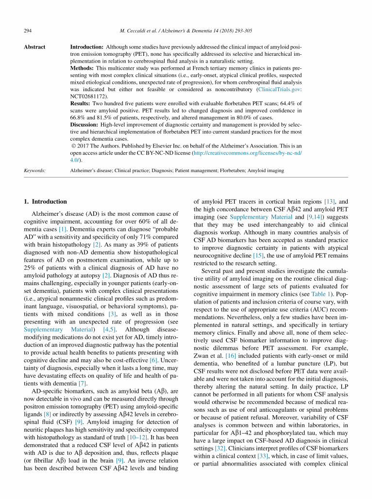
Abstract Introduction: Although some studies have previously addressed the clinical impact of amyloid posi-
tron emission tomography (PET), none has specifically addressed its selective and hierarchical im-plementation in relation to cerebrospinal fluid analysis in a naturalistic setting.Methods: This multicenter study was performed at French tertiary memory clinics in patients pre-senting with most complex clinical situations (i.e., early-onset, atypical clinical profiles, suspectedmixed etiological conditions, unexpected rate of progression), for whom cerebrospinal fluid analysiswas indicated but either not feasible or considered as noncontributory (ClinicalTrials.gov:NCT02681172).Results: Two hundred five patients were enrolled with evaluable florbetaben PET scans; 64.4% ofscans were amyloid positive. PET results led to changed diagnosis and improved confidence in66.8% and 81.5% of patients, respectively, and altered management in 80.0% of cases.Discussion: High-level improvement of diagnostic certainty and management is provided by selec-tive and hierarchical implementation of florbetaben PET into current standard practices for the mostcomplex dementia cases.� 2017 The Authors. Published by Elsevier Inc. on behalf of the Alzheimer’s Association. This is anopen access article under the CC BY-NC-ND license (http://creativecommons.org/licenses/by-nc-nd/4.0/).Keywords: Alzheimer’s disease; Clinical practice; Diagnosis; Patient management; Florbetaben; Amyloid imaging
1. Introduction
Alzheimer’s disease (AD) is the most common cause ofcognitive impairment, accounting for over 60% of all de-mentia cases [1]. Dementia experts can diagnose “probableAD” with a sensitivity and specificity of only 71% comparedwith brain histopathology [2]. As many as 39% of patientsdiagnosed with non-AD dementia show histopathologicalfeatures of AD on postmortem examination, while up to25% of patients with a clinical diagnosis of AD have noamyloid pathology at autopsy [2]. Diagnosis of AD thus re-mains challenging, especially in younger patients (early-on-set dementia), patients with complex clinical presentations(i.e., atypical nonamnestic clinical profiles such as predom-inant language, visuospatial, or behavioral symptoms), pa-tients with mixed conditions [3], as well as in thosepresenting with an unexpected rate of progression (seeSupplementary Material) [4,5]. Although disease-modifying medications do not exist yet for AD, timely intro-duction of an improved diagnostic pathway has the potentialto provide actual health benefits to patients presenting withcognitive decline and may also be cost-effective [6]. Uncer-tainty of diagnosis, especially when it lasts a long time, mayhave devastating effects on quality of life and health of pa-tients with dementia [7].
AD-specific biomarkers, such as amyloid beta (Ab), arenow detectable in vivo and can be measured directly throughpositron emission tomography (PET) using amyloid-specificligands [8] or indirectly by assessing Ab42 levels in cerebro-spinal fluid (CSF) [9]. Amyloid imaging for detection ofneuritic plaques has high sensitivity and specificity comparedwith histopathology as standard of truth [10–12]. It has beendemonstrated that a reduced CSF level of Ab42 in patientswith AD is due to Ab deposition and, thus, reflects plaque(or fibrillar Ab) load in the brain [9]. An inverse relationhas been described between CSF Ab42 levels and binding
of amyloid PET tracers in cortical brain regions [13], andthe high concordance between CSF Ab42 and amyloid PETimaging (see Supplementary Material and [9,14]) suggeststhat they may be used interchangeably to aid clinicaldiagnosis workup. Although in many countries analysis ofCSF AD biomarkers has been accepted as standard practiceto improve diagnostic certainty in patients with atypicalneurocognitive decline [15], the use of amyloid PET remainsrestricted to the research setting.
Several past and present studies investigate the cumula-tive utility of amyloid imaging on the routine clinical diag-nostic assessment of large sets of patients evaluated forcognitive impairment in memory clinics (see Table 1). Pop-ulation of patients and inclusion criteria of course vary, withrespect to the use of appropriate use criteria (AUC) recom-mendations. Nevertheless, only a few studies have been im-plemented in natural settings, and specifically in tertiarymemory clinics. Finally and above all, none of them selec-tively used CSF biomarker information to improve diag-nostic dilemmas before PET assessment. For example,Zwan et al. [16] included patients with early-onset or milddementia, who benefited of a lumbar puncture (LP), butCSF results were not disclosed before PET data were avail-able and were not taken into account for the initial diagnosis,thereby altering the natural setting. In daily practice, LPcannot be performed in all patients for whom CSF analysiswould otherwise be recommended because of medical rea-sons such as use of oral anticoagulants or spinal problemsor because of patient refusal. Moreover, variability of CSFanalyses is common between and within laboratories, inparticular for Ab1–42 and phosphorylated tau, which mayhave a large impact on CSF-based AD diagnosis in clinicalsettings [32]. Clinicians interpret profiles of CSF biomarkerswithin a clinical context [33], which, in case of limit values,or partial abnormalities associated with complex clinical

Table 1
Studies on clinical utility of amyloid PET imaging
#ID Paper
Ab-PETtracer
Subjects
(n) Study design Clinical centers
Natural
setting
Population (a.restrictive
selection, b.uncertainty
as selection criterion,
c.MCI)
In line
with AUC
recommendations
Full
diagnostic
workup
Ab biomarker
sequence
(CSF vs. PET)
Literature
1 Ceccaldi et al., (in prep) FBB 205 Prospective 18 tertiary
memory clinics
Yes a) Restrictive
b) High uncertainty
c) MCI excluded
Yes* Yes 1. CSF
2. Amyloid PET
2 Zwan et al., 2017 [16] FMM 211 Prospective Tertiary memory clinic Yes a) Restrictive
b) Early-onset and mild
dementia, uncertainty
c) MCI excluded
No Yes 1. Amyloid PET
3 Weidman et al., 2017 [17] FBP 16 Case series NA No a) Restrictive
b) Cognitive decline and
uncertain diagnosis
c) MCI included
Yesy Partially 1. Amyloid PET
4 Apostolova et al., 2016 [18] FBP 53 Retrospective NA No a) Nonrestrictive
b) Early-onset/Late-onset AD
c) MCI included
Yesy No 1. Amyloid PET
5 Boccardi et al., 2016 [19] FBP 228 Prospective 18 tertiary memory clinics Yes a) Restrictive
b) Cognitive impairment,
uncertainty
c) MCI included
No Yes 1. Amyloid PET
6 Grundman et al., 2016 [20] FBP 229 Prospective NA No a) Restrictive
b) Cognitive decline and
uncertain diagnosis
c) MCI included
Yesy Partially 1. Amyloid PET
7 Bensaidane et al., 2016 [21] NAV 28 Prospective Tertiary memory clinic Yes a) Restrictive
b) Atypical AD/unclear
c) MCI excluded
Yesz Yes 1. Amyloid PET
8 Shea et al, 2016 [22] PIB 102 Retrospective,
case series
Memory clinic Yes a) Nonrestrictive
b) Clinical population
c) MCI included
No Yes 1. Amyloid PET
9 Sch€onecker et al., 2016 [23] FBB 33 Case series Memory clinic Yes a) Nonrestrictive
b) Uncertain clinical
diagnosis
c) MCI included
No Yes 1. Amyloid PET
10 Sanchez-Juan et al., 2014 [24] PIB 137 Retrospective NA No a) Nonrestrictive
b) Cognitive impairment
and uncertainty
c) MCI included
No No 1. Amyloid PET
11 Mitsis et al., 2014 [25] (Grundman
cohort)
FBP 30 Case series Tertiary
memory clinic
Yes a) Nonrestrictive
b) Cognitive decline
c) MCI included
No No 1. Amyloid PET
(Continued )
M.Cecca
ldiet
al./Alzh
eimer’s
&Dem
entia
14(2018)293-305
295
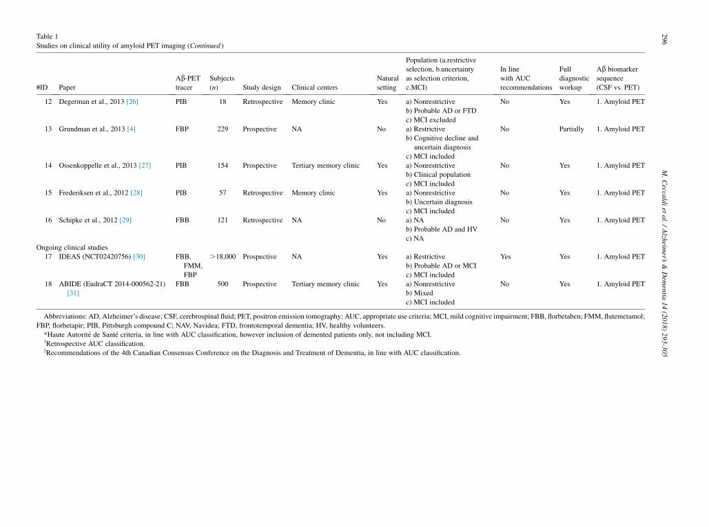
Table 1
Studies on clinical utility of amyloid PET imaging (Continued )
#ID Paper
Ab-PETtracer
Subjects
(n) Study design Clinical centers
Natural
setting
Population (a.restrictive
selection, b.uncertainty
as selection criterion,
c.MCI)
In line
with AUC
recommendations
Full
diagnostic
workup
Ab biomarker
sequence
(CSF vs. PET)
12 Degerman et al., 2013 [26] PIB 18 Retrospective Memory clinic Yes a) Nonrestrictive
b) Probable AD or FTD
c) MCI excluded
No Yes 1. Amyloid PET
13 Grundman et al., 2013 [4] FBP 229 Prospective NA No a) Restrictive
b) Cognitive decline and
uncertain diagnosis
c) MCI included
No Partially 1. Amyloid PET
14 Ossenkoppelle et al., 2013 [27] PIB 154 Prospective Tertiary memory clinic Yes a) Nonrestrictive
b) Clinical population
c) MCI included
No Yes 1. Amyloid PET
15 Frederiksen et al., 2012 [28] PIB 57 Retrospective Memory clinic Yes a) Nonrestrictive
b) Uncertain diagnosis
c) MCI included
No Yes 1. Amyloid PET
16 Schipke et al., 2012 [29] FBB 121 Retrospective NA No a) NA
b) Probable AD and HV
c) NA
No Yes 1. Amyloid PET
Ongoing clinical studies
17 IDEAS (NCT02420756) [30] FBB,
FMM,
FBP
.18,000 Prospective NA Yes a) Restrictive
b) Probable AD or MCI
c) MCI included
Yes Yes 1. Amyloid PET
18 ABIDE (EudraCT 2014-000562-21)
[31]
FBB 500 Prospective Tertiary memory clinic Yes a) Nonrestrictive
b) Mixed
c) MCI included
No Yes 1. Amyloid PET
Abbreviations: AD, Alzheimer’s disease; CSF, cerebrospinal fluid; PET, positron emission tomography; AUC, appropriate use criteria; MCI, mild cognitive impairment; FBB, florbetaben; FMM, flutemetamol;
FBP, florbetapir; PIB, Pittsburgh compound C; NAV, Navidea; FTD, frontotemporal dementia; HV, healthy volunteers.
*Haute Autorit�e de Sant�e criteria, in line with AUC classification, however inclusion of demented patients only, not including MCI.yRetrospective AUC classification.zRecommendations of the 4th Canadian Consensus Conference on the Diagnosis and Treatment of Dementia, in line with AUC classification.
M.Cecca
ldiet
al./Alzh
eimer’s
&Dem
entia
14(2018)293-305
296

M. Ceccaldi et al. / Alzheimer’s & Dementia 14 (2018) 293-305 297
situations, may prompt them to consider the CSF results as“noncontributory.” In this clinical context, it is unknownwhether amyloid imaging provides cumulative utility inroutine clinical diagnostic assessment including collectionof CSF. In the present study, we have addressed this issuewith a hierarchical approach and applied it to the Frenchclinical setting within a network of tertiary memory clinicsand with respect to national recommendations [34].
Florbetaben is a radiopharmaceutical approved in Europeand indicated, in conjunction with clinical evaluation, forPET imaging of Ab neuritic plaque density in the brains ofadults with cognitive impairment who are being evaluatedfor AD. A negative scan indicates sparse or no plaques,which is not consistent with a diagnosis of AD.
The present study is the first trial that exploreswhether florbetaben PET imaging provides added valuefor the assessment of complex dementia patients withoutinterfering with the current clinical setting, includinguse of LP wherever possible and in line with currentpractice in European expert centers [15]. Amyloid PETis only introduced as a “second-line” indication for pa-tients in whom the etiology of symptoms remained un-certain after a complete diagnostic workup and inabsence of conclusive CSF results. In detail, the trial in-vestigates whether amyloid PET imaging in patients un-der investigation for AD for whom LP was eithercontraindicated or refused, or did not provide conclusiveresults to the clinician, leads to changes of diagnosis andhigher levels of diagnostic confidence and alterations inpatient management.
2. Methods
2.1. Participants
Eligible patients were evaluated for AD in tertiary mem-ory centers and had a preliminary uncertain diagnosis aftera prior comprehensive workup, according to recommenda-tions from the French Health Authority (HAS) [34].Because of the diagnostic uncertainty, CSF examinationwas planned in these patients but was not obtained or notconsidered helpful by the expert clinician for one of thefollowing reasons: (1) LP was refused by the patient; (2)LP was not feasible for medical reasons; or (3) results ofCSF analysis were considered as noncontributory by theclinician (e.g., uninterpretable due to technical problems,values close to threshold, only one abnormal biomarkerout of three, only two abnormal biomarkers out of three,or result not consistent with clinical information). All pa-tients, who were included in the study, fulfilled at leastone of the AUC as defined by Johnson et al. in 2013 [8]and were in accordance with national recommendations[34]. Full eligibility criteria are provided in theSupplementary Material. All participants or their legalrepresentative provided written informed consent for studyparticipation, visits, and data source verification. The study
was conducted in accordance with the Declaration of Hel-sinki after approval of Ethics Committees.
2.2. Study design
This was a phase 4 multicenter open-label study(ClinicalTrials.gov: NCT02681172) conducted in the outpa-tient setting of 18 French tertiary memory clinics, withoutinterfering with the natural clinical setting. All participatingclinical sites belong to an established network of universityand regional expert centers, which have the mission tocontributing to diagnosis of patients with complex clinicalsituations and provide support for secondary-level struc-tures, that is, private specialists and memory centers in localhospitals. The participating investigators are either neurolo-gists or geriatricians, with a long-standing experience in de-mentia, sharing homogeneous practices, and with access to atechnical platform, including the possibility of performingCSF analyses and functional imaging examinations. Furtherdetails of the diagnostic workup in accordance with nationalguidelines [34] are summarized in Supplementary Fig. 1.
Each patient had three outpatient clinic visits: ascreening/baseline visit to record information on previousworkups and to establish an initial diagnosis (visit 1), a flor-betaben PET scan (visit 2), and a follow-up visit at which pa-tients were informed of the PET scan result and a finaldiagnosis was established (visit 3). Diagnostic certaintywas assessed using a five-point Likert scale. Clinicians couldreport up to three possible diagnoses for each patient andindicate the probability by rank order. Level 1 was definedas the most probable of up to three potential hypotheses(levels 1–3). Management changes were defined as initiationor withdrawal of any medication, additional diagnostic tests,referral to other specialists, or providing additional explana-tion or advice for patients and their families. Full details areprovided in the Supplementary Material.
2.3. Imaging protocol and visual assessment
Florbetaben PET images were acquired 90–110 minutesafter intravenous injection of 300 MBq (620%) florbetabenaccording to a standardized acquisition and image-processing protocol [10] established during a technical visitto each center. PET image interpretation was performedlocally in each center by nuclear physicians, who had under-gone training for appropriate interpretation of scans. Foreach individual site, florbetaben scans were assessed bythe associated nuclear physician. Results were interpretedas either “positive” or “negative” based on regional corticaltracer uptake assessment of the lateral temporal cortex, fron-tal cortex, posterior cingulate cortex/precuneus, and parietalcortex, as described previously [10].
2.4. Statistical methods
All patients from the full analysis set were included in theper-protocol set, as no major protocol deviations were

M. Ceccaldi et al. / Alzheimer’s & Dementia 14 (2018) 293-305298
reported. To test whether potential added value of florbeta-ben imaging is present for different patient categories classi-fied according to clinical status, PET categorization, orreason of eligibility, additional subgroups were analyzed(subgroup 1: patients who refused LP; subgroup 2: patientsin whom LP was not possible due to medical reasons; sub-group 3: patients with noncontributory results from CSFanalysis according to the clinician). These are presented inSupplementary Tables 2–6.
Mean 6 standard deviation values and frequency distri-butions are reported. Relative frequency and its two-sided95% confidence interval (Clopper-Pearson) are reported.Change of confidence level is reported by its frequency dis-tribution. Wilcoxon sign rank test is reported for comparisonof initial and final confidence levels. Kruskal-Wallis
Fig. 1. Patient disposition and subgroups. *All patients who received florbetaben, h
remained unexplained after a complete diagnostic workup. **Two subjects who re
viation: PET, positron emission tomography.
ANOVA with Dunn’s multiple comparisons and Mann-Whitney tests are performed to test variables of differentgroups. For comparison of frequency data sets, Fisher exactis applied. To test for proportion trends, the Cochran-Armitage test is applied.
3. Results
3.1. Diagnostic workup and diagnosis before PET scan
The full study cohort included 205 patients, of whom42.4% (n 5 87) underwent LP (Fig. 1). Baseline character-istics of patients are shown in Table 2. Based on the prescandiagnostic workup, the most frequent diagnosis hypothesis(probability level 1) was AD (72.2%; n 5 148); more than
ad a PET/computed tomography image, and in whom etiology of symptoms
fused lumbar puncture were also contraindicated for the procedure. Abbre-

Table 2
Baseline demographics and clinical characteristics
Full dataset N 5 205
Refused LP*,
N 5 75
Contraindicated
CSF*, N 5 45
Noncontributory**
CSF, N 5 87 P
Age (years), mean 6 SD (median) 70.9 6 9.7 (71.0) 74.01 6 8.70 (75.19) 72.91 6 9.47 (74.07) 66.75 6 9.09 (67.39) ,.0001x
Gender, n (%)
Male 103 (50.2) 35 27 43
Female 102 (49.8) 40 18 44
MMSE score***, mean 6 SD (median) 22.1 6 5.1 (23.0) 21.96 6 4.86 (22) 22.87 6 4.72 (24) 21.83 6 5.52 (23) ..05z
Educational level
None 2 1 1 0
Primary school 34 13 10 11
High school up to 15 years old 53 16 10 27
High school up to 18 years old 49 20 10 20
College 67 25 14 29
Study intervals
Time between visit 1 and visit 3 in days,
mean 6 SD (median)
80.84 6 39.30 (72) 85.17 6 40.96 (77) 78.24 6 34.02 (69) 79.54 6 40.31 (74) ..05z
Underwent lumbar puncture, n (%)y 87 (42.4)
Ambiguous CSF result 71 (34.6)
CSF result inconsistent with clinical
information
16 (7.8)
Uninterpretable CSF result for technical
reasons
4 (2.0)
Did not undergo lumbar puncture, n (%) 118 (57.6)
Patient refusal 75 (36.6)
Contraindicated or failed 45 (22.0)
Anticoagulant therapy 16 (7.8)
Thrombocytopenia 4 (2.0)
Lumbar puncture failed 8 (3.9)
Spinal problems 17 (8.3)
Abbreviations: CSF, cerebrospinal fluid; LP, lumbar puncture; MMSE, Mini–Mental State Examination; SD, standard deviation.
*Two subjects that refused LP were also contraindicated for the procedure.
**Uninterpretable CSF results due to technical problem, ambiguous CSF biomarker results (eg, Aß42, T-tau, P-Tau discordance or values of Aß close to the
threshold of significance) or divergent results with clinical information.
***Most recent score within the previous 12 months.yPatients may have had more than one reason for noncontributory CSF data.zKruskal-Wallis ANOVAwith Dunn’s multiple comparisons test.xMultiple comparison: Age was significantly lower in the noncontributory subgroup, when compared to the refused and contraindicated categories.
M. Ceccaldi et al. / Alzheimer’s & Dementia 14 (2018) 293-305 299
four-fifths of these cases were represented by a complexclinical situation (atypical form, early-onset AD, and rapidlyprogressive AD) (Table 3). A neurodegenerative (ND) non-AD dementia or mixed dementia was initially suspected in15.6% (n5 32) and 8.3% (n5 17) of subjects, respectively.A non-ND cause of dementia was reported in 4.9% (n5 10)of subjects. Overall, baseline confidence in the initial diag-nosis was moderate (52.3%).
3.2. Scan results
In total, 64.4% of scans (132/205) were evaluated as am-yloid positive on florbetaben PET, while 35.6% (73/205)were evaluated as amyloid negative.
3.3. Change in diagnosis, change in diagnosticconfidence, and change in management
After PET scan, 66.8% of diagnoses (137/205) werechanged (based on all prescan hypotheses). Moreover, thetotal amount of provided potential hypotheses (levels 1–3)
before and after florbetaben PET disclosure decreasedsignificantly (63.9% [393/615] potential hypotheses at initialdiagnosis vs. 43.6% [268/615] potential hypotheses at finaldiagnosis; P, .0001, Fisher exact test). Change of diagnosiswas significantly more common for amyloid-negative scansversus amyloid-positive scans (83.6% [61/73] diagnosischanges vs. 57.6% [76/132], respectively; P, .0001, Fisherexact test). After PET scan, 62.0% of subjects (127/205) hada dementia diagnosis due to AD. A ND non-AD dementia ormixed dementia was diagnosed in 17.6% (36/205) and 4.9%(10/205) of subjects after scan, respectively. Non-ND causesof dementia were reported in 17.1% of subjects (35/205).
Results were similar for patients grouped according toinitial clinical diagnosis (Supplementary Table 2), for pa-tients classified according to PET status (SupplementaryFig. 3), and in the three subsets of patients according toreason of eligibility (Supplementary Table 4). Demographiccharacteristics and global cognitive level, eligibility criteria,and prior diagnostic workup in the 12 months immediatelypreceding study entry were compared between patientswith changed and unchanged diagnosis and for patients

Table 3
Diagnosis (probability level 1*) before and after scan
Diagnosis (probability level 1),* n (%)
Before scan,
N 5 205yAfter scan,
N 5 205z
AD dementia 148 (72.2) 127 (62.0)
Sporadic AD, atypical form 67 (32.7) 43 (21.0)
Early-onset AD 50 (24.4) 40 (19.5)
Sporadic AD, typical form 27 (13.2) 41 (20.0)
Rapid progressive AD (CJD excluded) 4 (2.0) 3 (1.5)
Non-AD dementia (neurodegenerative) 32 (15.6) 36 (17.6)
Frontotemporal lobar dementia 14 (6.8) 17 (8.3)
Primary progressive aphasia 8 (3.9) 6 (2.9)
Lewy body disease 7 (3.4) 8 (3.9)
Corticobasal dementia 2 (1.0) 1 (0.5)
Semantic dementia 1 (0.5) 3 (1.5)
Parkinson’s disease — 1 (0.5)
Mixed dementia 17 (8.3) 10 (4.9)
Nonneurodegenerative dementia 10 (4.9) 35 (17.1)
Psychiatric disorders 3 (1.5) 11 (5.4)
Vascular dementia 2 (1.0) 8 (3.9)
Other 5 (2.4) 16 (7.8)
Abbreviations: AD, Alzheimer’s disease; CJD, Creutzfeldt-Jakob dis-
ease.
*Clinicians could report up to three possible diagnoses for each patient
and indicate the probability by rank order. Level 1 was defined as the
most probable of up to three potential hypotheses.yTwo patients had two level-1 diagnoses with equal probability.zThree patients had two level-1 diagnoses with equal probability (most
patients had more than one potential hypothesis, illustrating the level of un-
certainty and the complexity of the cases).
M. Ceccaldi et al. / Alzheimer’s & Dementia 14 (2018) 293-305300
with and without management changes (SupplementaryTables 5 and 6). The between-group comparison did notshow any significant differences on these variables.
Frequencies of the four possible underlying etiologies fordiagnosis based on probability level 1 for patients with achanged diagnosis are shown in Fig. 2. Of PET-negative sub-jects, 95.1% (58/61) fell into a category other than AD afterscan result disclosure. Before the scan, only 32.8% (20/61)of these subjects were categorized as other than AD. Simi-larly, 86.8% (66/76) of PET-positive subjects were diag-nosed with AD after PET result disclosure, whereas only63.2% (48/76) were considered to have some form of ADat initial diagnosis.
Fig. 2. Etiology of 137 patients with a change in diagnosis after positron emission t
initial diagnosis (gray) and final diagnosis (colored). The data set included 76 subje
based on a level-1 change. Abbreviations: AD, Alzheimer’s disease; ND, neurod
mixed dementia.
The baseline confidence was moderate (52.3%). Diag-nostic confidence improved significantly after PET (Fig. 3),with improved confidence reported for 167/205 cases(81.5%; P , .0001), and similar results found across sub-groups (Table 4, Supplementary Table 4). Weak and veryweak diagnostic confidence was reported in 46 patients(22.4%) at the initial assessment but only in 5 (2.4%) patientsafter PET result disclosure. Confidence was significantlyimproved for amyloid-positive scans compared to amyloid-negative scans (116/132 [87.9%] vs. 51/73 [69.9%], P 5.002, Fisher exact test, Table 4). Most of the changes includedan improvement by one category (n5 94 subjects, Fig. 3), andoverall diagnostic confidence improved by 30.2% leading to aconfidence of 82.6% after PET disclosure.
Changes in management were reported for 80.0% (164/205) of patients (Table 5); they were independent of amyloidstatus (amyloid positive 106/132 [80.3%], amyloid negative58/73 [79.5%]; P . .05, Fisher exact test). Initiation of newmedication was mainly reported after disclosure of amyloid-positive PET scans, whereas withdrawal of medication wasexclusively reported for amyloid-negative PET scans(Supplementary Fig. 4). Substantial management changes(i.e., any change in initiation or withdrawal of medication,additional diagnostic tests, or referral to a new specialist)were reported in 50.7% (104/205) of patients (Table 5). Re-sults were similar for different subgroups (SupplementaryTable 4). Two exemplary clinical cases are presented inSupplementary Fig. 2.
Increase in confidence, change in diagnosis, and changein management after PET were correlated with the initialdiagnostic confidence. All three parameters were changedmore often or showed a stronger increase for subjects withlower pre-PET diagnostic certainty (Supplementary Fig. 3).
4. Discussion
This is the first clinical prospective study that has soughtto define the impact of amyloid PET on diagnosis and man-agement in cases corresponding to the highest level of diag-nosis uncertainty—early-onset dementia and atypical
omography (PET) scan. Etiology was defined based on level-1 hypothesis of
cts with positive PETand 61 subjects with a negative PET; not all changes are
egenerative; non-AD, neurodegenerative non-Alzheimer’s disease; mixed,
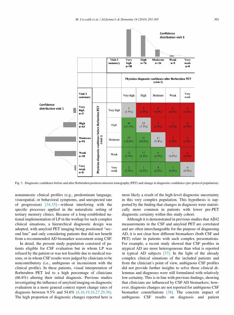
Fig. 3. Diagnostic confidence before and after florbetaben positron emission tomography (PET) and change in diagnostic confidence (per-protocol population).
M. Ceccaldi et al. / Alzheimer’s & Dementia 14 (2018) 293-305 301
nonamnestic clinical profiles (e.g., predominant language,visuospatial, or behavioral symptoms, and unexpected rateof progression) [34,35]—without interfering with thespecific processes applied in the naturalistic setting oftertiary memory clinics. Because of a long-established na-tional implementation of LP in the workup for such complexclinical situations, a hierarchical diagnostic design wasadopted, with amyloid PET imaging being positioned “sec-ond line” and only considering patients that did not benefitfrom a recommended AD biomarker assessment using CSF.
In detail, the present study population consisted of pa-tients eligible for CSF evaluation but in whom LP wasrefused by the patient or was not feasible due to medical rea-sons, or in whom CSF results were judged by clinicians to benoncontributory (i.e., ambiguous or inconsistent with theclinical profile). In these patients, visual interpretation offlorbetaben PET led to a high percentage of clinicians(66.8%) altering their initial diagnosis. Previous studiesinvestigating the influence of amyloid imaging on diagnosticevaluation in a more general context report change rates ofdiagnosis between 9.5% and 54.6% [4,16,19,24,27,29,36].The high proportion of diagnostic changes reported here is
most likely a result of the high-level diagnostic uncertaintyin this very complex population. This hypothesis is sup-ported by the finding that changes in diagnosis were statisti-cally more common in patients with lower pre-PETdiagnostic certainty within this study cohort.
Although it is demonstrated in previous studies that Ab42measurements in the CSF and amyloid PET are correlatedand are often interchangeable for the purpose of diagnosingAD, it is not clear how different biomarkers (both CSF andPET) relate in patients with such complex presentations.For example, a recent study showed that CSF profiles inatypical AD are more heterogeneous than what is reportedin typical AD subjects [37]. In the light of the alreadycomplex clinical situations of the included patients andfrom the clinician’s point of view, ambiguous CSF profilesdid not provide further insights to solve those clinical di-lemmas and diagnoses were still formulated with relativelylow certainty. This is in line with previous findings, showingthat clinicians are influenced by CSF AD biomarkers; how-ever, diagnosis changes are not reported for ambiguous CSFbiomarker constellations [38]. The concrete impact ofambiguous CSF results on diagnosis and patient

Table 4
Change in diagnosis and diagnostic confidence
Amyloid status on florbetaben PET
Independent of amyloid status N 5 205
Change in diagnosis, n (%) [95% CI] 137 (66.8) [59.9–73.2]
Improved confidence, n (%) [95% CI] 167 (81.5) [75.5–86.5]
Amyloid positive N 5 132
Change in diagnosis, n (%) [95% CI] 76 (57.6) [48.7–66.1]
Improved confidence, n (%) [95% CI] 116 (87.9) [81.1–92.9]
Amyloid negative N 5 73
Change in diagnosis, n (%) [95% CI] 61 (83.6) [73.0–91.2]
Improved confidence, n (%) [95% CI] 51 (69.9) [58.0–80.1]
Abbreviations: CI, confidence interval; PET, positron emission tomography.
M. Ceccaldi et al. / Alzheimer’s & Dementia 14 (2018) 293-305302
management changes was not evaluated in this study, andthus, the diagnostic impact of CSF cannot be derived.Further analysis on the relationship between CSF markers,amyloid imaging, and clinical findings in such a populationis important to understand the details of such complex pa-tients.
As reported in earlier studies [16,27], change in diagnosiswas correlated with confidence in the initial diagnosis.Change in diagnosis was particularly frequent when PETscan was negative (83.6% of cases), which is also in linewith previous findings [19]. Given the high negative predic-tive value of amyloid imaging in histopathology studies [10],this result is not surprising and indicates that a negative am-yloid scan is generally used by dementia experts as a vali-dated method to exclude a diagnosis of AD in mostcomplex cases. Of the 61 PET-negative subjects with achanged diagnosis, only 3 (4.9%) were considered to beAD after florbetaben assessment, while 41 (67.2%) were
Table 5
Change of management plan
Amyloid status on florbetaben PET
Independent of amyloid status N 5 205
Change, n (%) [95% CI] 164 (80.0) [73.9–85.2]
No change, n (%) 41 (20.0)
Changes of management, n (%)
New medication initiated 71 (34.6)
Medication withdrawn 8 (3.9)
Additional diagnostic tests 17 (8.3)
Referred to another specialist 16 (7.8)
Offered additional explanationy 105 (51.2)
Offered additional advicez 60 (29.3)
Other 57 (27.8)
Strong change of management plan as
listed*, n (%)
104 (50.7)
Amyloid positive
Change, n (%) [95% CI]
N 5 132
106 (80.3) [72.5–86.7]
Amyloid negative
Change, n (%) [95% CI]
N 5 73
58 (79.5) [68.4–88.0]
Abbreviations: CI, confidence interval; PET, positron emission tomogra-
phy.
*Defined as initiation or withdrawal of medication, additional diagnostic
tests, or referral to another specialist.yOffered additional explanation of diagnosis/testing results.zPatient and/or family offered additional advice on potential daily living/
safety concerns.
categorized as AD before scan. Similarly to what has beenpreviously acknowledged for CSF [39], our findings suggestthat clinicians do not view amyloid PET in a vacuum butinstead consider its utility in combination with other clinicaldetails and available assessments to establish decisions.
Diagnostic confidence increased for 81.5% of the subjectsafter PET result disclosure, and the strongest confidence in-creases were observed in subjects with relatively low certaintyin the initial diagnosis. Most of the changes included a confi-dence increase by one category leading to an overall diag-nostic increase of 30.2% after disclosure of PET results,which is higher than most previously reported values[4,19,21]. However, given the very low diagnosticconfidence at baseline, the particularly distinct impact is notsurprising and in general in alignment with previous studies.For example, change in diagnosis and increase in diagnosticconfidence were lower in studies with higher pre-PET diag-nostic confidence [4,27]. It is thus hypothesized thatamyloid PET scans have the greatest impact in situationswith significant diagnostic concerns after a standard workuphas been completed before PET. This is supported in at leastone trial [21]. In the present study, the general confidence in-crease was also reflected by a reduced number of provideddiagnosis hypotheses after PET disclosure.
After florbetaben PET, changes in management were re-ported for 80.0% of patients. Even when considering onlymanagement changes presumed to have a substantial impacton patients (i.e., new medication initiated, medication with-drawn, additional diagnostic tests, referred to anotherspecialist), changes were still reported for 50.7% of patients.These results are in line with an earlier study that usedsimilar criteria, with changes in management reported in87% of patients after amyloid PET [4]. In other studies ofamyloid PET, however, fewer management changes were re-ported (range, 37%–68%) [16,40]. Various factors, such asdifferences in methodological definitions, selectedvariables of patient management, and diagnosticuncertainty before PET assessment, may have contributedto the differences in findings. Interestingly, changes inmanagement were more common in patients with low pre-PET diagnostic confidence.
One concern of the study design is that patients who pre-sent medical contraindications that prevent LP, patients whorefuse LP, and patients in whom an LP has been achievedmight represent significantly different populations. Weanalyzed these subgroups and found no differences in thedistribution of level-1 pretest diagnosis; the percent positiveor negative scans; or percent change in diagnosis, diagnosticconfidence, or management.
Although the diagnostic accuracy of amyloid PET tracershas been thoroughly assessed in several pivotal histopathol-ogy studies [10–12], such information cannot be available inthe context of clinical utility studies. Clinical follow-up ofthe population may provide additional and long-term in-sights on the impact of amyloid PET on diagnostic and pa-tient management changes.

M. Ceccaldi et al. / Alzheimer’s & Dementia 14 (2018) 293-305 303
Another limitation of the present study was the binary vi-sual readout of amyloid PET scans. Although this approachreflects clinical routine usage, we cannot make further state-ments related to potentially ambiguous PET cases. Previouspublications have demonstrated high concordance betweenvisual assessment and quantification methods [41–43], andtherefore, semiquantitative approaches may provideadjunct information that might be useful, particularly, forambiguous cases close to standardized cutoffs. Finally,factors such as degree of experience with amyloid and thephysician’s beliefs on the clinical value of amyloidimaging may influence interpretation of results and werenot controlled for in the study.
The present study was designed with reference to a spe-cific health care model and was restricted to an establishednetwork of tertiary memory centers. It focused on a highlyselected patient population and included a hierarchical posi-tioning of AD biomarkers, with CSF collection in first line.Several other utility studies are currently being conductedand will further contribute to our knowledge on the impactof amyloid imaging in larger clinical scenarios such as theImaging Dementia–Evidence for Amyloid Scanning initia-tive which will enroll.18,000 patients, following AUC rec-ommendations put forward by Johnson et al. [8].
In conclusion, this naturalistic study provides evidencethat visual interpretation of florbetaben PET has a strongimpact on diagnostic outcome parameters and managementof patients in whom uncertainty is particularly common—that is, in early-onset, atypical, mixed, and rapidly pro-gressing presentations—even when they firstly benefitedfrom expert centers’ assessments. Given the general diag-nostic validity of amyloid PET and the previously reportedhigh concordance rates between imaging and CSF Ab42levels [9,13,36], it is obvious that PET can efficientlysubstitute LP in absence of valuable CSF results.Moreover, this study shows that outcome measures ofinitial assessment are significantly improved in mostcomplex cases. Most patients from this study share a verylow pre-PET confidence in the initial diagnosis. There isan ethical imperative to establish an early, validated diag-nosis wherever possible [44]. Moreover, it has been shownthat a clear diagnosis has a positive impact on caregivers ofdementia patients [21]. Providing a higher quality of diag-nosis, in terms of unambiguity, may directly contribute toincrease the positive impact of memory assessment ser-vices on patients’ health-related quality of life [45]. Fromthis point of view, our results highlight the immediate clin-ical utility of amyloid PET imaging for patients with com-plex dementia presentations in the context of the existinghealth care framework.
Acknowledgments
The trial was funded by Piramal Imaging. Medical writingsupport was provided by Daniel Booth (Bioscript Group,Macclesfield, UK) and funded by Piramal Imaging.
Authors’ contributions: The study was initiated by the prin-cipal investigators (Mathieu Ceccaldi and Eric Guedj)together with the sponsor, who supported the collection,analysis, and interpretation of the data, and the writing ofthe article. The principal investigators had full access tothe study data and had final responsibility for the decisionto submit for publication. Mathieu Ceccaldi, Rossella Gis-mondi, Nicola Raffa, Andrew Stephens, and Eric Guedjparticipated in the initiation, design, and concept of thestudy; performed data acquisition; were involved in dataanalysis; and drafted the article. Th�er�ese Jonveaux, AntoineVerger, Pierre Krolak-Salmon, Claire Houzard, Olivier God-efroy, Trevor Shields, Audrey Perrotin, Florence Pasquier,Franck Semah, Bruno Dubois, Marie-Odile Habert, DavidWallon, and Mathieu Chastan performed data acquisitionand were involved in article preparation and review. San-tiago Bullich participated in the design of the study, wasinvolved in data analysis, and drafted the article. AleksandarJovalekic was involved in data analysis and drafted thearticle.Investigators of the NEUUS in AD study group: Principal in-vestigators: Mathieu Ceccaldi and Eric Guedj (Hopital de laTimone, AP-HM, Marseille, France). Hopital de la Timone,AP-HM,Marseille, France: Mathieu Ceccaldi, Olivier Felic-ian, Mira Didic, Claude Gueriot, Lejla Koric, RadkaKletchkova-Gantchev, and Eric Guedj; CHU Amiens Picar-die, France: Olivier Godefroy, Daniela Andriuta, Agn�es De-vendeville, Diane Dupuis, Ingrid Binot, M�elanie Barbay,Marc-Etienne Meyer, and V�eronique Moullard; CHU de Be-sancon, France: Eloi Magnin, Ludivine Chamard, SophieHaffen, Olivier Morel, Cl�ement Drouet, and Hatem Boulah-dour; CHU de Brest, France: Philippe Goas and Sol�eneQuerellou-Lefranc; CHU de Caen, France: Vincent de laSayette, Julien Cogez, Pierre Branger, and Denis Agostini;Cyceron, Caen, France: Alain Manrique; CHU Dijon Bour-gogne, France: Olivier Rouaud, Yannick Bejot, and Agn�esJacquin-Piques; Centre GF Leclerc, Dijon, France: InnaDygai-Cochet and Alina Berriolo-Riedinger; CHU de Gre-noble, France: Olivier Moreaud, Mathilde Sauvee, andC�eline Gallazzani Cr�epin; CHU de Lille, France: FlorencePasquier, St�ephanie Bombois, Thibaud Lebouvier, Marie-Anne Mackowiak-Cordoliani, Vincent Deramecourt, Ade-line Rollin-Sillaire, Pascaline Cassagnaud-Thuillet, YaohuaChen, Franck Semah, and Gr�egory Petyt; Hopital des Charp-ennes, HCL, Lyon, France: Pierre Krolak-Salmon, DenisFederico, Keren Liora Danaila, Yves Guilhermet, Christo-phe Magnier, Zaza Makaroff, Isabelle Rouch, Jing Xie,Caroline Roubaud, Marie-H�el�ene Coste, Kenny David,Alain Sarciron, and Aziza Sediq Waissi; CERMEP, Lyon,France: Christian Scheiber; LUMEN, Lyon, France: ClaireHouzard; CHU de Montpellier, France: Audrey Gabelle-Deloustal, Karim Bennys, Cecilia Marelli, Lynda Touati,Denis Mariano-Goulart, and Delphine de Verbizier-Lonjon; CHRU de Nancy, France: Th�er�ese Jonveaux,Athanase Benetos, Anna Kearney-Schwartz, ChristinePerret-Guillaume, and Antoine Verger; CHU de Nantes,

M. Ceccaldi et al. / Alzheimer’s & Dementia 14 (2018) 293-305304
France: Martine Vercelletto, Claire Boutoleau-Bretonniere,H�el�ene Pouclet-Courtemanche, Nathalie Wagemann, andAmandine Pallardy; GH Lariboisi�ere Fernand-Widal Saint-Louis, AP-HP, Paris, France: Jacques Hugon, Claire Paquet,Julien Dumurgier, and Pascal Millet; Centre Cardiologiquedu Nord, Saint-Denis, France: Mathieu Queneau; Hopitalde la Piti�e-Salpetri�ere, AP-HP, Paris, France: Bruno Dubois,St�ephane Epelbaum, Marcel Levy, and Marie-Odile Habert;CHU de Reims, France: Jean-Luc Novella and Yacine Jaidi;Institut Jean Godinot, Reims, France: Dimitri Papathanas-siou and David Morland; CHU de Rennes, France: Dr SergeBelliard and Anne Salmon; Centre Eug�eneMarquis, Rennes,France: Florence Lejeune; CHU de Rouen, France: DidierHannequin, David Wallon, Olivier Martinaud, and AlineZarea; Centre Henri Becquerel, Rouen, France: MathieuChastan ; CHU Toulouse, France: J�er�emie Pariente, ClaireThalamas, Monique Galitzky-Gerber, Anne-Marie TricoireRicard, Fabienne Calvas, Emilie Rigal, Pierre Payoux, andAnne Hitzel; G�erontopole, CHU Toulouse, France: JulienDelrieu, Pierre-Jean Ousset, Francoise Lala, and NathalieSastre-Hengan.
Supplementary data
Supplementary data related to this article can be found athttps://doi.org/10.1016/j.jalz.2017.09.009.
RESEARCH IN CONTEXT
1. Systematic review: Although several studies investi-gated the clinical impact of amyloid positron emis-sion tomography (PET) imaging, none hasspecifically addressed its selective and hierarchicalimplementation in relation to cerebrospinal fluidanalysis in a naturalistic setting.
2. Interpretation: Whether amyloid PET provides cu-mulative utility in routine diagnostic assessment incomplex patients with an unresolved cerebrospinalfluid biomarker status is unknown. We have ad-dressed this issue with a hierarchical approach withina network of tertiary memory clinics in France andprovide robust evidence on the value of amyloid PET.
3. Future direction: Our results highlight the immediateclinical utility of amyloid PET imaging for patientswith complex dementia presentations and high levelsof diagnostic uncertainty in the context of the exist-ing health care framework. Additional health out-comes and economic data are required to assess thecost-effectiveness of amyloid PET in clinical routine.
References
[1] Alzheimer’s Association. 2016 Alzheimer’s disease facts and figures.
Alzheimers Dement 2016;12:459–509.
[2] Beach TG, Monsell SE, Phillips LE, Kukull W. Accuracy of the clin-
ical diagnosis of Alzheimer disease at National Institute on Aging Alz-
heimer Disease Centers, 2005-2010. J Neuropathol Exp Neurol 2012;
71:266–73.
[3] McKhann GM, Knopman DS, Chertkow H, Hyman BT, Jack CR Jr,
Kawas CH, et al. The diagnosis of dementia due to Alzheimer’s dis-
ease: recommendations from the National Institute on Aging-Alz-
heimer’s Association workgroups on diagnostic guidelines for
Alzheimer’s disease. Alzheimers Dement 2011;7:263–9.
[4] Grundman M, Pontecorvo MJ, Salloway SP, Doraiswamy PM,
Fleisher AS, Sadowsky CH, et al. Potential impact of amyloid imaging
on diagnosis and intended management in patients with progressive
cognitive decline. Alzheimer Dis Assoc Disord 2013;27:4–15.
[5] Wolk DA, Price JC, Madeira C, Saxton JA, Snitz BE, Lopez OL, et al.
Amyloid imaging in dementias with atypical presentation. Alzheimers
Dement 2012;8:389–98.
[6] Dubois B, Padovani A, Scheltens P, Rossi A, Dell’Agnello G. Timely
diagnosis for Alzheimer’s disease: a literature review on benefits and
challenges. J Alzheimers Dis 2016;49:617–31.
[7] Werner P, Karnieli-Miller O, Eidelman S. Current knowledge and future
directions about the disclosure of dementia: a systematic review of the
first decade of the 21st century. Alzheimers Dement 2013;9:e74–88.
[8] Johnson KA, Minoshima S, Bohnen NI, Donohoe KJ, Foster NL,
Herscovitch P, et al. Appropriate use criteria for amyloid PET: a report
of the Amyloid Imaging Task Force, the Society of Nuclear Medicine
and Molecular Imaging, and the Alzheimer’s Association. Alzheimers
Dement 2013;9:e-1-16.
[9] BlennowK,MattssonN, Sch€oll M, Hansson O, Zetterberg H. Amyloid
biomarkers in Alzheimer’s disease. Trends Pharmacol Sci 2015;
36:297–309.
[10] Sabri O, Sabbagh M N, Seibyl J, Barthel H, Akatsu H, Ouchi Y, et al.
Florbetaben PET imaging to detect amyloid plaques in Alzheimer dis-
ease: phase 3 study. Alzheimers Dement 2015;11:964–74.
[11] Clark CM, Pontecorvo MJ, Beach TG, Bedell BJ, Coleman RE,
Doraiswamy PM, et al. Cerebral PET with florbetapir compared
with neuropathology at autopsy for detection of neuritic amyloid-
beta plaques: a prospective cohort study. Lancet Neurol 2012;
11:669–78.
[12] Curtis C, Gamez JE, Singh U, Sadowsky CH, Villena T, Sabbagh MN,
et al. Phase 3 trial of flutemetamol labeled with radioactive fluorine 18
imaging and neuritic plaque density. JAMA Neurol 2015;72:287–94.
[13] Schipke CG, Koglin N, Bullich S, Joachim LK, Haas B, Seibyl J, et al.
Correlation of florbetaben PET imaging and the amyloid peptide
Ass42 in cerebrospinal fluid. Psychiatry Res 2017;265:98–101.
[14] Jansen WJ, Ossenkoppele R, Knol DL, Tijms BM, Scheltens P,
Verhey FR, et al. Prevalence of cerebral amyloid pathology in persons
without dementia: a meta-analysis. JAMA 2015;313:1924–38.
[15] Lehmann S, Teunissen CE. Editorial: biomarkers of Alzheimer’s dis-
ease: the present and the future. Front Neurol 2016;7:158.
[16] Zwan MD, Bouwman FH, Konijnenberg E, van der Flier WM,
Lammertsma AA, Verhey FR, et al. Diagnostic impact of [18F]flute-
metamol PET in early-onset dementia. Alzheimers Res Ther 2017;9:2.
[17] Weidman DA, Zamrini E, Sabbagh MN, Jacobson S, Burke A,
Belden C, et al. Added value and limitations of amyloid-PET imaging:
review and analysis of selected cases of mild cognitive impairment and
dementia. Neurocase 2017;23:41–51.
[18] Apostolova LG, Haider JM, Goukasian N, Rabinovici GD, Chetelat G,
Ringman JM, et al. Critical review of the Appropriate Use Criteria for
amyloid imaging: effect on diagnosis and patient care. Alzheimers De-
ment (Amst) 2016;5:15–22.

M. Ceccaldi et al. / Alzheimer’s & Dementia 14 (2018) 293-305 305
[19] Boccardi M, Altomare D, Ferrari C, Festari C, Guerra UP, Paghera B,
et al. Assessment of the incremental diagnostic value of florbetapir F
18 imaging in patients with cognitive impairment: the incremental
diagnostic value of amyloid PET with [18F]-Florbetapir (INDIA-
FBP) study. JAMA Neurol 2016;73:1417–24.
[20] Grundman M, Johnson KA, Lu M, Siderowf A, Dell’Agnello G,
Arora AK, et al. Effect of amyloid imaging on the diagnosis and man-
agement of patients with cognitive decline: impact of appropriate use
criteria. Dement Geriatr Cogn Disord 2016;41:80–92.
[21] Bensaidane MR, Beauregard JM, Poulin S, Buteau FA, Guimond J,
Bergeron D, et al. Clinical utility of amyloid PET imaging in the dif-
ferential diagnosis of atypical dementias and its impact on caregivers. J
Alzheimers Dis 2016;52:1251–62.
[22] Shea YF, Ha J, Lee SC, Chu LW. Impact of (18)FDG PET and (11)C-
PIB PET brain imaging on the diagnosis of Alzheimer’s disease and
other dementias in a regional memory clinic in Hong Kong. Hong
Kong Med J 2016;22:327–33.
[23] Schonecker S, Prix C, Raiser T, Ackl N,Wlasich E, Stenglein-Krapf G,
et al. [Amyloid positron-emission-tomography with [18 F]-florbetaben
in the diagnostic workup of dementia patients]. Nervenarzt 2017;
88:156–61.
[24] S�anchez-Juan P, Ghosh PM, Hagen J, Gesierich B, Henry M,
Grinberg LT, et al. Practical utility of amyloid and FDG-PET in an ac-
ademic dementia center. Neurology 2014;82:230–8.
[25] Mitsis EM, Bender HA, Kostakoglu L, Machac J, Martin J, Woehr JL,
et al. A consecutive case series experience with [18 F] florbetapir PET
imaging in an urban dementia center: impact on quality of life, deci-
sion making, and disposition. Mol Neurodegener 2014;9:10.
[26] Degerman Gunnarsson M, Lindau M, Santillo AF, Wall A, Engler H,
Lannfelt L, et al. Re-evaluation of clinical dementia diagnoses with
pittsburgh compound B positron emission tomography. Dement Ger-
iatr Cogn Dis Extra 2013;3:472–81.
[27] Ossenkoppele R, Prins ND, Pijnenburg YA, Lemstra AW, van der
Flier WM, Adriaanse SF, et al. Impact of molecular imaging on the
diagnostic process in a memory clinic. Alzheimers Dement 2013;
9:414–21.
[28] Frederiksen KS, Hasselbalch SG, Hejl AM, Law I, Hojgaard L,
Waldemar G. Added diagnostic value of (11)C-PiB-PET in memory
clinic patients with uncertain diagnosis. Dement Geriatr Cogn Dis Ex-
tra 2012;2:610–21.
[29] Schipke CG, Peters O, Heuser I, Grimmer T, Sabbagh MN, Sabri O,
et al. Impact of beta-amyloid-specific florbetaben PET imaging on
confidence in early diagnosis of Alzheimer’s disease. Dement Geriatr
Cogn Disord 2012;33:416–22.
[30] IDEAS, Imaging Dementia–Evidence for Amyloid Scanning (IDEAS)
Study (IDEAS) - NCT02420756.
[31] ABIDE, Amyloid-PET as a diagnostic marker in daily practice
(ABIDE)–EudraCT 2014-000562-21.
[32] Vos SJ, Visser PJ, Verhey F, Aalten P, Knol D, Ramakers I, et al. Vari-
ability of CSF Alzheimer’s disease biomarkers: implications for clin-
ical practice. PLoS One 2014;9:e100784.
[33] Spies PE, Claassen JA, Peer PG, Blankenstein MA, Teunissen CE,
Scheltens P, et al. A prediction model to calculate probability of Alz-
heimer’s disease using cerebrospinal fluid biomarkers. Alzheimers De-
ment 2013;9:262–8.
[34] Haute Autorit�e de Sante. Maladie d’Alzheimer et Maladies Apparentees;
2011. Available at: http://www.has-sante.fr/portail/upload/docs/applicat
ion/pdf/2011-12/recommandation_maladie_d_alzheimer_et_maladies_
apparentees_diagnostic_et_prsie_en_charge.pdf. Accessed November
4, 2016.
[35] Magnin E, Dumurgier J, Bouaziz-Amar E, Bombois S, Wallon D,
Gabelle A, et al. [Alzheimer’s disease cerebro-spinal fluid biomarkers:
A clinical research tool sometimes useful in daily clinical practice of
memory clinics for the diagnosis of complex cases]. Rev Med Interne
2016;38:250–5.
[36] Zwan MD, Rinne JO, Hasselbalch SG, Nordberg A, Lleo A,
Herukka SK, et al. Use of amyloid-PET to determine cutpoints for
CSF markers: A multicenter study. Neurology 2016;86:50–8.
[37] Paterson RW, Toombs J, Slattery CF, Nicholas JM, Andreasson U,
Magdalinou NK, et al. Dissecting IWG-2 typical and atypical Alz-
heimer’s disease: insights from cerebrospinal fluid analysis. J Neurol
2015;262:2722–30.
[38] Duits FH, Prins ND, Lemstra AW, Pijnenburg YA, Bouwman FH,
Teunissen CE, et al. Diagnostic impact of CSF biomarkers for Alz-
heimer’s disease in a tertiary memory clinic. Alzheimers Dement
2015;11:523–32.
[39] Gooblar J, Carpenter BD, Coats MA, Morris JC, Snider BJ. The influ-
ence of cerebrospinal fluid (CSF) biomarkers on clinical dementia
evaluations. Alzheimers Dement 2015;11:533–540.e2.
[40] Pontecorvo MJ, Arora AK, Devine M, Lu M, Galante N, Siderowf A,
et al. A randomized, controlled, multicenter, international study of the
impact of florbetapir (18F) PET amyloid imaging on patient manage-
ment and outcome. Alzheimers Dement 2015;11:P334.
[41] Seibyl J, Catafau AM, Barthel H, Ishii K, Rowe CC, Leverenz JB, et al.
Impact of trainingmethod on the robustness of the visual assessment of
18F-Florbetaben PET scans: results from a phase-3 study. J Nucl Med
2016;57:900–6.
[42] Bullich S, Seibyl J, Catafau AM, Jovalekic A, Koglin N, Barthel H,
et al. Optimized classification of 18F-Florbetaben PET scans as posi-
tive and negative using an SUVR quantitative approach and compari-
son to visual assessment. Neuroimage Clin 2017;15:325–32.
[43] Pontecorvo MJ, Arora AK, Devine M, Lu M, Galante N, Siderowf A,
et al. Quantitation of PET signal as an adjunct to visual interpretation
of florbetapir imaging. Eur J Nucl Med Mol Imaging 2017;44:
825–37.
[44] Molinuevo JL, Rami L. Applying the IWG research criteria in clinical
practice: feasibility and ethical issues. Med Clin North Am 2013;
97:477–84.
[45] Gomes M, Pennington M, Wittenberg R, Knapp M, Black N, Smith S.
Cost-effectiveness of Memory Assessment Services for the diagnosis
and early support of patients with dementia in England. J Health
Serv Res Policy 2017;22:1355819617714816.


![Usefulness of [18F]-DA and [18F]-DOPA for PET imaging in a mouse model of pheochromocytoma](https://static.fdokumen.com/doc/165x107/6325a7d9852a7313b70e9a7d/usefulness-of-18f-da-and-18f-dopa-for-pet-imaging-in-a-mouse-model-of-pheochromocytoma.jpg)


![18F]Fluoroazabenzoxazoles as potential amyloid plaque PET tracers: synthesis and in vivo evaluation in rhesus monkey](https://static.fdokumen.com/doc/165x107/631f7e5b3b43b66d3c0fcb6e/18ffluoroazabenzoxazoles-as-potential-amyloid-plaque-pet-tracers-synthesis-and.jpg)