Adaptive properties of human cementum and ... - eScholarship
14
UCSF UC San Francisco Previously Published Works Title Adaptive properties of human cementum and cementum dentin junction with age. Permalink https://escholarship.org/uc/item/3k89p301 Authors Jang, Andrew T Lin, Jeremy D Choi, Ryan M et al. Publication Date 2014-11-01 DOI 10.1016/j.jmbbm.2014.07.015 Peer reviewed eScholarship.org Powered by the California Digital Library University of California
-
Upload
khangminh22 -
Category
Documents
-
view
0 -
download
0
Transcript of Adaptive properties of human cementum and ... - eScholarship
UCSFUC San Francisco Previously Published Works
TitleAdaptive properties of human cementum and cementum dentin junction with age.
Permalinkhttps://escholarship.org/uc/item/3k89p301
AuthorsJang, Andrew TLin, Jeremy DChoi, Ryan Met al.
Publication Date2014-11-01
DOI10.1016/j.jmbbm.2014.07.015 Peer reviewed
eScholarship.org Powered by the California Digital LibraryUniversity of California
Available online at www.sciencedirect.com
www.elsevier.com/locate/jmbbm
j o u r n a l o f t h e m e c h a n i c a l b e h a v i o r o f b i o m e d i c a l m a t e r i a l s 3 9 ( 2 0 1 4 ) 1 8 4 – 1 9 6
http://dx.doi.org/10.1751-6161/& 2014 El
nCorresponding autE-mail address: s
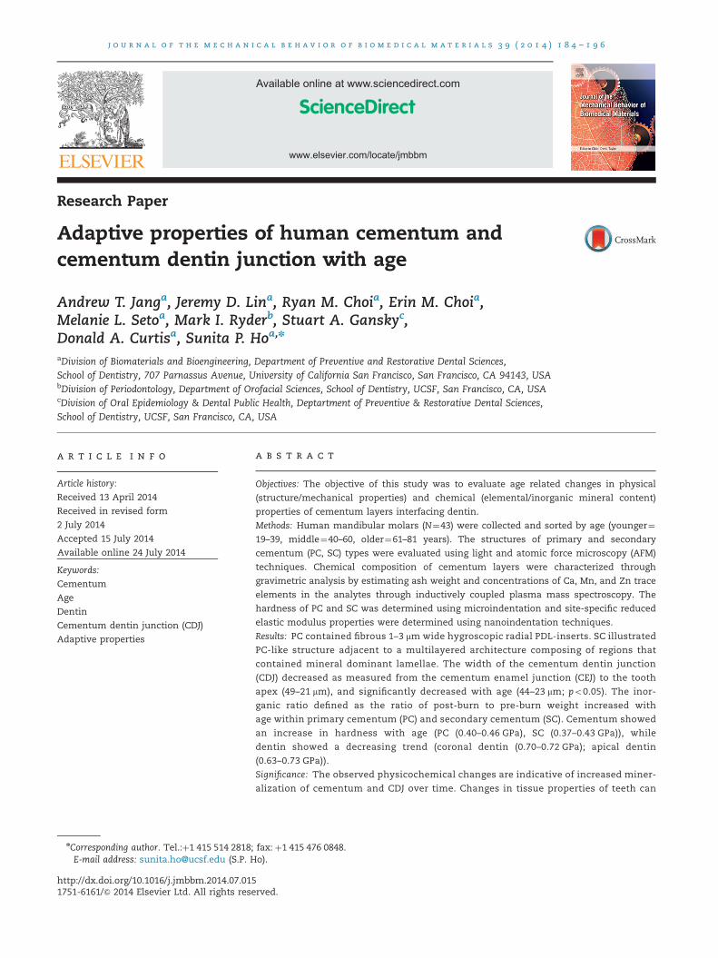
Research Paper
Adaptive properties of human cementum andcementum dentin junction with age
Andrew T. Janga, Jeremy D. Lina, Ryan M. Choia, Erin M. Choia,Melanie L. Setoa, Mark I. Ryderb, Stuart A. Ganskyc,Donald A. Curtisa, Sunita P. Hoa,n
aDivision of Biomaterials and Bioengineering, Department of Preventive and Restorative Dental Sciences,School of Dentistry, 707 Parnassus Avenue, University of California San Francisco, San Francisco, CA 94143, USAbDivision of Periodontology, Department of Orofacial Sciences, School of Dentistry, UCSF, San Francisco, CA, USAcDivision of Oral Epidemiology & Dental Public Health, Deptartment of Preventive & Restorative Dental Sciences,School of Dentistry, UCSF, San Francisco, CA, USA
a r t i c l e i n f o
Article history:
Received 13 April 2014
Received in revised form
2 July 2014
Accepted 15 July 2014
Available online 24 July 2014
Keywords:
Cementum
Age
Dentin
Cementum dentin junction (CDJ)
Adaptive properties
1016/j.jmbbm.2014.07.015sevier Ltd. All rights rese
hor. Tel.:þ1 415 514 2818;[email protected] (S.P. H
a b s t r a c t
Objectives: The objective of this study was to evaluate age related changes in physical
(structure/mechanical properties) and chemical (elemental/inorganic mineral content)
properties of cementum layers interfacing dentin.
Methods: Human mandibular molars (N¼43) were collected and sorted by age (younger¼19–39, middle¼40–60, older¼61–81 years). The structures of primary and secondary
cementum (PC, SC) types were evaluated using light and atomic force microscopy (AFM)
techniques. Chemical composition of cementum layers were characterized through
gravimetric analysis by estimating ash weight and concentrations of Ca, Mn, and Zn trace
elements in the analytes through inductively coupled plasma mass spectroscopy. The
hardness of PC and SC was determined using microindentation and site-specific reduced
elastic modulus properties were determined using nanoindentation techniques.
Results: PC contained fibrous 1–3 mm wide hygroscopic radial PDL-inserts. SC illustrated
PC-like structure adjacent to a multilayered architecture composing of regions that
contained mineral dominant lamellae. The width of the cementum dentin junction
(CDJ) decreased as measured from the cementum enamel junction (CEJ) to the tooth
apex (49–21 mm), and significantly decreased with age (44–23 mm; po0.05). The inor-
ganic ratio defined as the ratio of post-burn to pre-burn weight increased with
age within primary cementum (PC) and secondary cementum (SC). Cementum showed
an increase in hardness with age (PC (0.40–0.46 GPa), SC (0.37–0.43 GPa)), while
dentin showed a decreasing trend (coronal dentin (0.70–0.72 GPa); apical dentin
(0.63–0.73 GPa)).
Significance: The observed physicochemical changes are indicative of increased miner-
alization of cementum and CDJ over time. Changes in tissue properties of teeth can
rved.
fax: þ1 415 476 0848.o).
j o u r n a l o f t h e m e c h a n i c a l b e h a v i o r o f b i o m e d i c a l m a t e r i a l s 3 9 ( 2 0 1 4 ) 1 8 4 – 1 9 6 185
alter overall tooth biomechanics and in turn the entire bone–tooth complex including
the periodontal ligament. This study provides baseline information about the changes
in physicochemical properties of cementum with age, which can be identified as
adaptive in nature.
& 2014 Elsevier Ltd. All rights reserved.
1. Introduction
Adaptations to occlusal loads occur within the innervated andvascularized periodontal ligament (PDL), vascularized bone, andcementum (thought to be avascularized) that constitute a bone-PDL-tooth fibrous joint (Nanci, 2007). Overall, the softer PDL andharder cementum, dentin, and bone tissues, including theirinterfaces, remodel (turnover) in response to a shift in functionresulting in a change in matrix properties. When changes infunction occur within physiological limits, an adequate PDL-space of 150–380 μm is thought to provide an optimum biome-chanical function (Nanci, 2007; Bosshardt and Selvig, 2000).Optimum function is limited to joints where no significantchange in overall displacement of the tooth into the alveolarsocket in response to simulated loads is observed (Lin et al.,2013). However, inevitable adaptation due to innate age relatedphysiology can manifest as observable differences in tissueproperties and their interfaces that makeup the dentoalveolarcomplex (Ho et al., 2013). It is within this context that thephysicochemical properties of load bearing cementum tissuewill be investigated.
Cementum is a mineralized tissue that covers the outermostlayer of a tooth root (Yamamoto et al., 2010; Ren et al., 2010). Theprimary function of cementum is to confine tooth motion byway of the PDL, as well as to provide support and load absorption
during mastication (Ren et al., 2010; Rios et al., 2008). In general,cementum exists in two forms, primary and secondary. Primarycementum (PC), which covers the coronal two-thirds of the rootis the major contributor for attachment of dentition to alveolarbone (Nanci, 2007; Bosshardt and Selvig, 2000). Secondarycementum (SC), which is hypothesized to develop when thetooth assumes occlusion and function, covers the remaining
one-third of the root, and is thought to act predominantly as anocclusal load absorber during mastication (Bosshardt and Selvig,2000; Yamamoto et al., 1998). While these functional roles arerecognized, the time-related implications of changes related tomineralization for PC, SC, and their interfaces with softer PDL areless understood (Nanci 2007; Bosshardt and Selvig, 2000). Thephysical characteristics of cementum have been previouslyinvestigated by identifying its structure, chemical composition,
and mechanical properties (Ho et al., 2004, 2010). Fiber density,calcium-to-phosphate ratio, and hardness of PC have beenreported to be different than those of SC (Alvarez-Perez et al.,2005). However, studies have only determined the physicalproperties of cementum at a single point-in-time (Ho et al.,2004, 2010; Alvarez-Perez et al., 2005; Stamfelj et al., 2008), andthe interface that it forms with dentin is minimally investigated.
Cementum is not directly fused to dentin. It is attachedto dentin via a 100–200 mm thick interface within which a10–50 mm wide hygroscopic proteoglycan (PG)-rich layer known
as the cementum–dentin junction (CDJ) exists (Yamamoto et al.,2010; Ho et al., 2004; Hopewell-Smith, 1920; Yamamoto et al.,2000, 1999, 2000; Ho et al., 2005). Within the CDJ, the dominanceof collagen fibers that transverse radially to the mantle dentinare tied with proteoglycans and are thought to contribute to anincreased ratio of organic to inorganic content (Ho et al., 2004;Yamamoto et al., 2000). The water absorbing fibrous nature ofthe CDJ may be of importance to transfer loads betweenadjoining mineralizing tissues implying that physicochemicalchanges to the interface could play a key role in the overallbiomehanical response to function (Ho et al., 2004; Hopewell-Smith. 1920; Yamamoto et al., 2000, 1999). The collagenous andnoncollagenous proteins forming this region exhibit dominantwater retention characteristics which have been speculated tohelp dissipate accumulated function-related stresses (Ho et al.,2004). Therefore it is postulated that cementum and its gradedinterfaces act as biological and mechanical continua allowingfor optimum function (Ho et al., 2010).
Functional demands on a body/organ vary with age andthese shifts in mechanical signals can modulate physicochem-
ical properties of tissues (Parfitt, 1984). Modulation of physico-chemical properties can occur based on the classic tenet that
with an increase in age the magnitude of forces on mechan-osensitive tissues decrease due to an increase in muscle
atrophy (Jubrias et al., 1997; Lexell et al., 1988; Hamrick et al.,2006; Burr, 1997; Iinuma et al., 2012; Kiebzak, 1991; Singh, 2002;Dalzell et al., 2009). Studies specific to the bone–tooth organ
have shown an inverse relationship between age and mastica-tory force, citing decreasing strength of the main masticatory
muscles (temporalis, masseter, and medial pterygoid) andmuscular atrophy that accompany aging process (Iinuma
et al., 2012; Ikebe et al., 2005). It continues to be a challenge toseamlessly tie the cause and effect relationship within the
context of functional loads and resulting tissue properties. Thisis because of the innate feedback between several hierarchical
levels, that is, mechanical loads on organs are transmitted andabsorbed by tissues which in turn activate cellular mechanisms
(Kiebzak, 1991; Singh, 2002; Dalzell et al., 2009; Hernandez andKeaveny, 2006) that trigger production of extracellular matrix
proteins. Aging and related functional activity have beencorrelated by measuring changes in bone mineral density,
suggesting the lack of daily cyclic loads as a major determinantfor tissue atrophy (Havill et al., 2007). In fact, cyclic load
therapies have been introduced to combat and reverse thebone mineral density loss associated with inactivity indicating
that, load is a primary factor to regain and maintain “tissuequality” and overall functional integrity (Mohr et al., 1997).
Hence, in this study, it was hypothesized that the inherentphysical and chemical properties of PC and SC change within
and across age groups.
j o u r n a l o f t h e m e c h a n i c a l b e h a v i o r o f b i o m e d i c a l m a t e r i a l s 3 9 ( 2 0 1 4 ) 1 8 4 – 1 9 6186
The “temporal” and “spatial” responses of tissues wereinvestigated by mapping changes in physicochemical proper-ties with age from coronal to apical regions of the root,respectively. Therefore, the objective of the study was toestablish spatiotemporal age-related trends in the localstructure, chemical composition, and mechanical propertiesof cementum, dentin, and the cementum–dentin interface.
2. Materials and methods
Extracted healthy human mandibular molars (N¼43) werecollected from adult male subjects according to an approvedprotocol by the UCSF Committee on Human Research andwere sorted into three groups based on age of the humansubject and time of extraction (younger¼19–39, middle
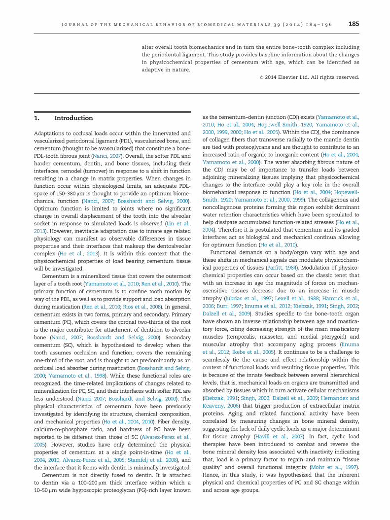
Fig. 1 – (a) Age ranges of younger, middle, and older humans frrow; mean þ/� standard deviation), microindentation, nanoinde(ICP-MS) (third row; mean þ/� standard deviation). Sample size omicroscopy images of sectioned molars demonstrate the clear fi
graphs of cementum width vs. anatomical location show a two-the cementum enamel junction (CEJ) to the root apex, that is, alwidth across age groups for primary cementum (PC) and secondaand cementum thickness increased as a function of age. Statistasterisk (n). (e) The thickness of the collagenous band indicativeAdditionally, a decrease in apical CDJ width compared to coronalmiddle and older groups were not different.
age¼40–60, older¼61–81 years) (Masoro and Austad, 2001)(Fig. 1a). The exclusion criterion for tooth samples wasperiodontal disease which was determined either throughpatient records and/or through specimen examination postextraction. The teeth were sterilized using 0.31 mrad ofγ-radiation (White et al., 1994). The total sample pool wasdivided into specimens for chemical and elemental analyses(N¼29) (Fig. 1a), and microscopy and indentation (N¼14)(Fig. S1).
Single 750 mm thick ground sections were taken from midregion of each tooth (Fig. S1). Each tooth was sectioned byusing a diamond wafering blade and a low speed rotary cutter(ISOMET, Buehler Ltd., IL, USA) under wet conditions. Thesection was subsequently used for CDJ and cementum widthanalyses. One remaining half of the tooth was embedded inepoxy (Expoxicure, Buehler, Il, USA) for microindentation,
om whom teeth obtained were used for microscopy (secondntation, and inductively coupled plasma mass spectroscopyf each group is shown in parentheses. (b) Transmission lightbrous cementum–dentin junction (CDJ). (c) Representativestep increase in cementum thickness when measured fromong the length of the root. (d) Graph of average cementumry cementum (SC) regions. In general, SC was thicker than PCical significant (po0.05) relationships are indicated with anof CDJ within PC and SC regions decreased with age.CDJ in younger group was observed. However, CDJ widths for
j o u r n a l o f t h e m e c h a n i c a l b e h a v i o r o f b i o m e d i c a l m a t e r i a l s 3 9 ( 2 0 1 4 ) 1 8 4 – 1 9 6 187
while the other half was prepared for structural examinationusing an atomic force microscope (AFM) and AFM-basednanoindentation technique (Fig. S1).
2.1. Light microscopy for cementum and CDJ widths
The 750 mm thick sections were ground and polished to athickness of 150 μm (Ho et al., 2004). The ground specimenswith polished surfaces were evaluated for CDJ width andcementum thickness using transmission and reflectance lightmicroscopy techniques (BX 51, Olympus America Inc., SanDiego, CA) and Image Pro Plus v6.0 software (Media Cyber-netics Inc., Silver Spring, MD), respectively (Ho et al., 2004).From the acquired light micrographs, the cementum and CDJwidths were measured using Matlab (Mathworks, Natick,MA). Linear regression was used to determine relationshipsbetween normalized root location (NRL), cementumwidth, andCDJ width (Fig. S1). Statistical significance was determinedthrough analysis of variance (ANOVA) followed by post hoc
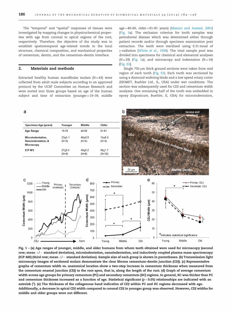
Fig. 2 – (a) Isolated cementum rings for ICP-MS analysis were sorcementum, SC – secondary cementum). Confirmation of dentinby using a light microscope for all specimens as shown at severatio (ash weight/preburn weight) of cementum as measured in thfor mineralized tissues enamel (E), dentin (D), and alveolar boneinorganic ratio and percentage of elemental composition (by wegroups in primary and secondary cementum (PC, SC) types. Thealso included.
multiple comparison adjusted t-tests (Holm–Šidak, unpaired,two-tails).
2.2. AFM and AFM-based nanoindentation for site-specifichardness values and gradients
The remaining longitudinal halves were cut into three or fourblocks from the CEJ to the apex (Fig. S1), such that all blockscontained dentin, cementum, and the CDJ (Fig. 3a). Theblocks were categorized into either coronal two-thirds orapical one-third to define sub-anatomical spatial locationsof a tooth. The blocks were then mounted on AFM steel stubs(Ted Pella Inc., Redding, CA) and ultrasectioned as describedpreviously, to generate a relatively flat surface to maintain anorthogonality between the nanoprobes of the AFM and thespecimen surface (Ho et al., 2004). Qualitative and quantita-tive analyses of the topography were performed using acontact mode AFM first under dry condition and subse-quently scanning under wet conditions (Nanoscope III, Multi-mode; DI-Veeco Instruments Inc., Santa Barbara, CA), and
ted by anatomical location and tooth specimen (PC – primaryremoval was performed by checking for any remnant dentinral magnification (a1 and a2). (b) Graph highlights inorganicis study vs. the age relative to reported values (dashed lines)(B), including cementum (C) (Nanci, 2007). (c) A table of
ight) for calcium, magnesium, and zinc is shown for all ageelemental standard (NIST 1400-bone ash) used for analysis is
j o u r n a l o f t h e m e c h a n i c a l b e h a v i o r o f b i o m e d i c a l m a t e r i a l s 3 9 ( 2 0 1 4 ) 1 8 4 – 1 9 6188
were analyzed using Nanoscope III version 4.43r8 software(Nanoscope III, Multimode; DI-Veeco Instruments Inc., SantaBarbara, CA) (Ho et al., 2004).
Wet nanoindentation (N¼3 per age group) was per-formed using a Hysitron Triboindenter (Hysitron Incor-porated, Minneapolis, MN). A top–down optics systemwas used for viewing the specimen surface and selectionof testing sites. Specimens were kept hydrated withdeionized water throughout testing to mimic conditionscloser to in vivo. A sharp diamond Berkovich indenterwith a radius of curvature less than 100 nm (Triboscope,Hysitron Incorporated, Minneapolis, MN) was fitted to thetransducer for elastic modulus and hardness measure-ments. For each specimen block, three rows of indentswith 6 mm between each indent were made in single linesfrom the cementum enthesis to dentin. Cementumenthesis is defined as the edge representative of the rootsurface. A maximum load of 1500 mN was used. Theloading curve for the nanoindentation cycle consisted ofthree 4.5 s segments of load, hold, and unload. Site-specific reduced elastic modulus (Er) and hardness (H)
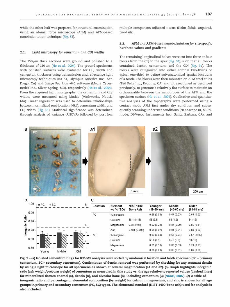
Fig. 3 – (a) Schematic of a tooth highlighting a region (rectangle) thforce microscopy techniques (b and c). The surface of the ultrasAFM (red squares with solid and dashed lines). AFM micrographssecondary cementum (c) under hydrated conditions. (c) The stru(arrows), and the CDJ (the zone between the dashed lines). Incremwith about a 10–40 μm spacing between each line. (For interpretareferred to web version of this article.)
values were evaluated using the Oliver–Pharr method (Beiet al., 2005).
2.3. Inductively coupled plasma mass spectrometry (ICP-MS) for elemental concentration
Another set of transverse sections 2–3 mm thick were cutalong the roots of mandibular molars (N¼29) using a dia-mond wafering blade on a low speed rotary cutter (ISOMET,Buehler Ltd., IL, USA) under wet conditions (Fig. 2a). Cemen-tum was isolated from these specimens by removing tubularand mantle dentin using a high speed dental hand-piece(Lares 757 workhorse high-speed hand-piece, Lares Research,Chico, CA) (Fig. 2a). Isolation of cementum was confirmedusing a light microscope (BX-51, Olympus America Inc., SanDiego, CA) (Fig. 2a).
Cementum specimens were desiccated for 6 months andweighed every 10 days to confirm weight stabilization withthe “pre-burn weight” representing the final dry weightbefore burning the specimens to ash. Inorganic ratio wasdetermined as the post-burn weight of the samples divided
at was ultrasectioned for imaging using light (a1) and atomicectioned block contains regions that were imaged using anillustrate fibrous CDJ region in primary cementum (b) and in
cture of secondary cementum includes lamellae, PDL-insertsental lines indicating growth are shown with an asterisk (n)tion of references to color in this figure legend, the reader is
j o u r n a l o f t h e m e c h a n i c a l b e h a v i o r o f b i o m e d i c a l m a t e r i a l s 3 9 ( 2 0 1 4 ) 1 8 4 – 1 9 6 189
by the pre-burn weight. The specimens were then transferredto aluminum oxide crucibles (CoorsTek Inc., Golden, CO) andplaced in a furnace (Ney, Bloomsfield, PA) for 24 h at 10001F(5401C) (Wolf et al., 2005). The ash was cooled to roomtemperature and a post-burn weight was immediatelyrecorded.
The ash was analyzed for elemental content using the ICP-MS (Agilent Technologies, Santa Clara, CA) with a nitric acidmatrix (Wolf et al., 2005). The technique was used to detectamounts of calcium (Ca), magnesium (Mg), and zinc (Zn).However, phosphorous (P) was not measured due to alimitation in the experimental configuration. Bone ash (NISTSRM-1400) and trace metals in water (NIST SRM-1643b) wereused as internal standards to verify detection and quantifica-tion of elements. Results were generated in parts per billion(ppb) and the percent weights were calculated using theinitial known weights. Statistical analysis was performedusing ANOVA in combination with post hoc t-tests.
2.4. Microindentation for hardness evaluation
The remaining sectioned halves were embedded in epoxyand polished as previously described (Ho et al., 2004). Micro-indentation was performed under dry conditions according tothe American Standard for Testing Materials (ASTM) standard(ASTM., 1999) on polished specimen blocks using a knoopmicroindenter (Buehler Ltd., Lake Bluff, IL). The spatial inter-val between indents was 35 mm in accordance with ASTMguidelines. Microindentation was performed at a maximumload of 10 g-force, and the long diagonal of each indent wasimmediately measured with a light microscope and Image-Pro data-acquisition software. Rows of microindents weremade from cementum through CDJ, to the tubular dentin.Each specimen contained 15 rows from the cementumenamel junction to the root apex. Each row contained aminimum of 10 indents. Knoop hardness values (HK) ofrespective regions were calculated as described previously(Ho et al., 2004). Linear mixed effects regression models wereused to fit the hardness data for each of the 3 anatomicallocations (cementum, CDJ, dentin) with random tooth speci-men effects (to account for within specimen correlation) andcementum, age, and cementum by age interaction (if needed)fixed effects. These results were compared against hardnessdata obtained via AFM-nanoindentation of similar regions.
3. Results
3.1. Cementum increased with age while CDJ widthdecreased with age
Light microscopy of ground sections illustrated a transitionregion between PC and SC. SC was marked by the presence ofidentifiable lacunae (Fig. 1b, Fig. S2). As a function of anato-mical location, cementum width gradually increased fromthe CEJ to the apex (Fig. 1c). The thickness of PC increased at alinear rate along the root length, while SC did not illustrate asimilar pattern. In general, the relationship between cemen-tum width and anatomical location was linear within PCcompared to the trend within SC, indicating differing growth/
resorption patterns compared with PC. Due to the anatomicaldependency of cementum width on root location, the respec-tive width ranges of PC (5–200 μm) and SC (400 μm–1 mm)were high (Fig. 1c, Fig. S2). By clustering measurements closerto the CEJ for PC and closer to the apex for SC, SC width(Fig. 1d, Fig. S2) was always greater than PC width (po0.05).The widths of cementum for both primary and secondarycementum types from the younger group were significantlylower than middle age group. Primary cementum widths inolder group were statistically higher than the middle group(po0.05), while SC widths were not statistically significantacross age groups (Fig. 1d).
CDJ widths as determined by polarized light microscopy inPC and SC regions were also reported (Fig. 1e). In both PC andSC, CDJ width was found to decrease as a function of age(po0.05). However, no statistical difference was foundbetween PC and SC regions except within the older group.In general, PC formed a wide CDJ with dentin compared withSC in specimens taken from younger individuals, wherecementum appeared to be fused with dentin with no appar-ent CDJ in specimens taken from older individuals (Fig. 1e).
3.2. Inorganic ratio of PC and SC increased with age andPC was significantly higher than SC within each age group
The inorganic ratio for PC and SC seemed to increase withage, although no statistical significance between age groups(p40.05) (Fig. 2b) was observed. When comparing PC to SC,the inorganic ratio of PC was significantly higher (p40.05)within each age group.
3.3. Elemental composition did not change with ageor anatomical location
Elemental weight concentration was determined by normal-izing elemental weight (using the ICP-MS measurements) withtotal sample weight (Fig. 2c). Calcium (Ca), magnesium (Mg),and zinc (Zn) were seen to have the highest contribution. Fromthe tested trace metals, the ranking always adhered to thefollowing trend; Ca4Mg4Zn. However, no statistically signifi-cant relationship between elemental composition of PC and SCwas found by age or anatomical location (Fig. 2c).
3.4. Architecture of primary and secondary cementumtypes
Ultrasectioned specimens (Fig. 3a) were imaged using alight microscope to provide a macroscale view before AFMwas performed. Fig. 3a illustrates the two types of cemen-tum based on the absence and presence of lacunae. Thesefeatures were used to identify types of cementum (Nanci,2007). Absence of lacunae is more representative of PC,while the presence of lacunae is representative of SC. PCunder AFM revealed a single layer containing a highernumber of hygroscopic principal PDL-fibers running per-pendicular to the root surface (Fig. 3b). In contrast, SC wascomposed of a multilayered lamellar structure. Adjacent tothe lamellar structure was also the extrinsic fiber-richcementum which contained hygroscopic principal fibersas revealed by an AFM (Fig. 3c).
j o u r n a l o f t h e m e c h a n i c a l b e h a v i o r o f b i o m e d i c a l m a t e r i a l s 3 9 ( 2 0 1 4 ) 1 8 4 – 1 9 6190
Scans of hydrated specimens in the PC region revealeda dense hygroscopic band in the CDJ region referred to asthe fibrous fringe by previous investigators (Bosshardt andSelvig, 2000). Under hydrated conditions, SC revealed thepresence of a lamellar structure formed by circumferentialfibers (Fig. 3c). Additionally, repeating bands within thesecondary cementum were observed. Hygroscopic princi-ple PDL fiber inserts were also found in the secondarycementum. The width of the hygroscopic CDJ region asmeasured using a light microscope decreased with anincrease in age. Results indicated that the hygroscopicCDJ band lost continuity towards the apical third withinolder age groups (not shown).
3.5. Knoop hardness of PC and SC increased with age
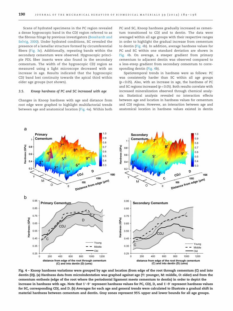
Changes in Knoop hardness with age and distance fromroot edge were graphed to highlight multifactorial trendsbetween age and anatomical location (Fig. 4a). Within both
Har
dnes
s (G
Pa)
1.0
0.9
0.8
0.7
0.6
0.5
0.4
0.3
Primary Cementum
0.25
0.35
0.45
0.55
0.65
0.75
0.85
0.95
Har
dnes
s (G
Pa)
distance from edge of the root through cementum(C) and into dentin (D) (ums)
YoungMiddle Old
C
D
CDJ
Primary Cementum
0 200 400 600 800 1000 1200
Fig. 4 – Knoop hardness variations were grouped by age and locdentin (D)). (a) Hardness data from microindentation was graphecementum enthesis (edge of the root where the periodontal ligaincrease in hardness with age. Note that 1″–9″ represent hardnefor SC, corresponding CDJ, and D. (b) Averages for each age andmaterial hardness between cementum and dentin. Gray zones
PC and SC, Knoop hardness gradually increased as cemen-tum transitioned to CDJ and to dentin. The data wereaveraged within all age groups with their respective rangesin order to highlight the gradual increase from cementumto dentin (Fig. 4b). In addition, average hardness values forPC and SC within one standard deviation are shown inFig. 4b. On average, a steeper gradient from primarycementum to adjacent dentin was observed compared toa less-steep gradient from secondary cementum to corre-sponding dentin (Fig. 4b).
Spatiotemporal trends in hardness were as follows: PCwas consistently harder than SC within all age groups(po0.05). Also, with an increase in age, the hardness of PCand SC regions increased (po0.05). Both results correlate withincreased mineralization observed through chemical analy-sis. Statistical analysis revealed no interaction effectsbetween age and location in hardness values for cementumand CDJ regions. However, an interaction between age andanatomical location in hardness values existed in dentin
Har
dnes
s (G
Pa)
1.0
0.9
0.8
0.7
0.6
0.5
0.4
0.3
Secondary Cementum
C
D CDJ
Secondary Cementum
0.25
0.35
0.45
0.55
0.65
0.75
0.85
0.95
0 200 400 600 800 1000 1200
Har
dnes
s (G
Pa)
distance from edge of the root through cementum(C) and into dentin (D) (ums)
YoungMiddle Old
ation (from edge of the root through cementum (C) and intod against age (Y: younger, M: middle, O: older) and from thement meets cementum to dentin) in order to depict thess values for PC, CDJ, D, and 10–90 represent hardness valuesgeneral trends were calculated to illustrate a gradual shift inrepresent 95% upper and lower bounds for all age groups.
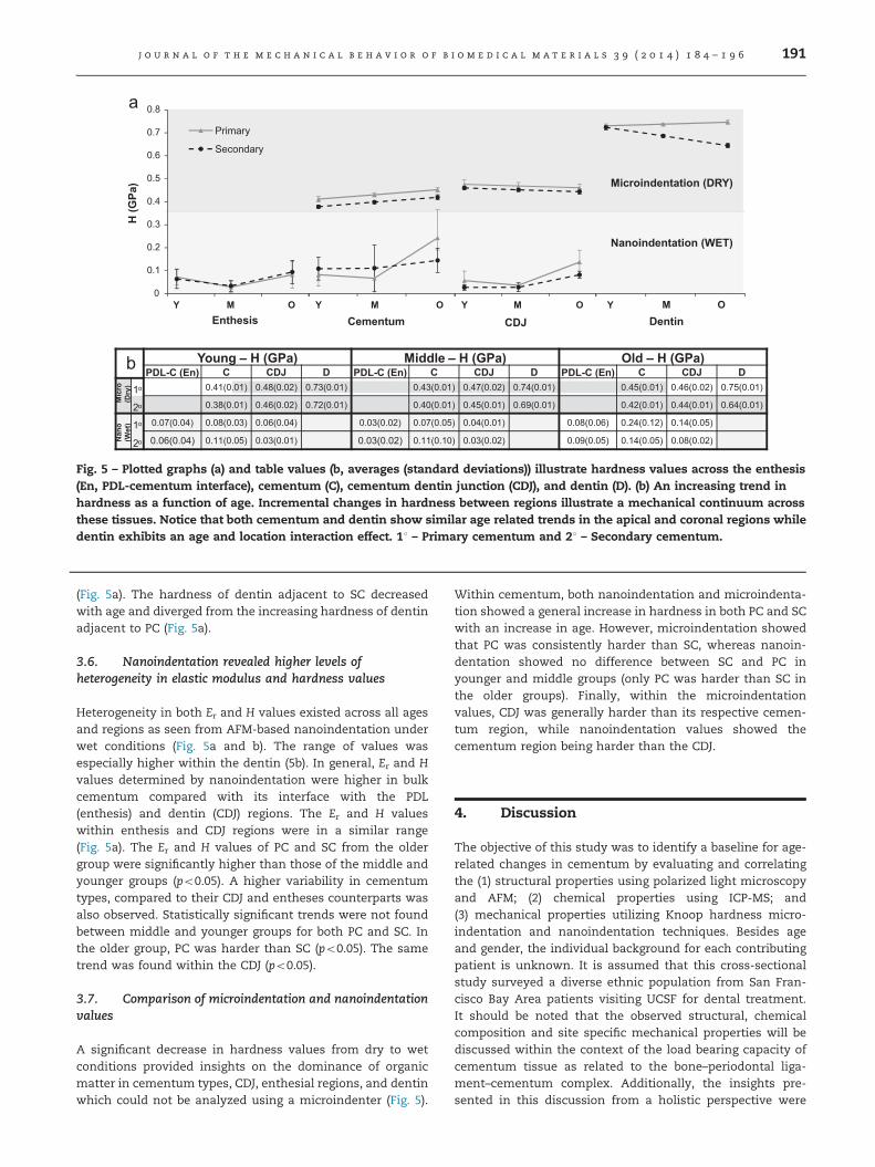
Young H (GPa) Middle H (GPa) Old H (GPa) PDL-C (En) C CDJ D PDL-C (En) C CDJ D PDL-C (En) C CDJ D
1o 0.41(0.01) 0.48(0.02) 0.73(0.01) 0.43(0.01) 0.47(0.02) 0.74(0.01) 0.45(0.01) 0.46(0.02) 0.75(0.01)
2o 0.38(0.01) 0.46(0.02) 0.72(0.01) 0.40(0.01) 0.45(0.01) 0.69(0.01) 0.42(0.01) 0.44(0.01) 0.64(0.01)
1o 0.07(0.04) 0.08(0.03) 0.06(0.04) 0.03(0.02) 0.07(0.05) 0.04(0.01) 0.08(0.06) 0.24(0.12) 0.14(0.05)
2o 0.06(0.04) 0.11(0.05) 0.03(0.01) 0.03(0.02) 0.11(0.10) 0.03(0.02) 0.09(0.05) 0.14(0.05) 0.08(0.02)
0
0.1
0.2
0.3
0.4
0.5
0.6
0.7
0.8
Primary
Secondary
Y M O Y M O Y M O Y M O
Microindentation (DRY)
Nanoindentation (WET)
H (G
Pa)
Enthesis DentinCementum CDJ
Nan
o (W
et)
Mic
ro
(Dry
)
Fig. 5 – Plotted graphs (a) and table values (b, averages (standard deviations)) illustrate hardness values across the enthesis(En, PDL-cementum interface), cementum (C), cementum dentin junction (CDJ), and dentin (D). (b) An increasing trend inhardness as a function of age. Incremental changes in hardness between regions illustrate a mechanical continuum acrossthese tissues. Notice that both cementum and dentin show similar age related trends in the apical and coronal regions whiledentin exhibits an age and location interaction effect. 11 – Primary cementum and 21 – Secondary cementum.
j o u r n a l o f t h e m e c h a n i c a l b e h a v i o r o f b i o m e d i c a l m a t e r i a l s 3 9 ( 2 0 1 4 ) 1 8 4 – 1 9 6 191
(Fig. 5a). The hardness of dentin adjacent to SC decreasedwith age and diverged from the increasing hardness of dentinadjacent to PC (Fig. 5a).
3.6. Nanoindentation revealed higher levels ofheterogeneity in elastic modulus and hardness values
Heterogeneity in both Er and H values existed across all agesand regions as seen from AFM-based nanoindentation underwet conditions (Fig. 5a and b). The range of values wasespecially higher within the dentin (5b). In general, Er and Hvalues determined by nanoindentation were higher in bulkcementum compared with its interface with the PDL(enthesis) and dentin (CDJ) regions. The Er and H valueswithin enthesis and CDJ regions were in a similar range(Fig. 5a). The Er and H values of PC and SC from the oldergroup were significantly higher than those of the middle andyounger groups (po0.05). A higher variability in cementumtypes, compared to their CDJ and entheses counterparts wasalso observed. Statistically significant trends were not foundbetween middle and younger groups for both PC and SC. Inthe older group, PC was harder than SC (po0.05). The sametrend was found within the CDJ (po0.05).
3.7. Comparison of microindentation and nanoindentationvalues
A significant decrease in hardness values from dry to wetconditions provided insights on the dominance of organicmatter in cementum types, CDJ, enthesial regions, and dentinwhich could not be analyzed using a microindenter (Fig. 5).
Within cementum, both nanoindentation and microindenta-tion showed a general increase in hardness in both PC and SCwith an increase in age. However, microindentation showedthat PC was consistently harder than SC, whereas nanoin-dentation showed no difference between SC and PC inyounger and middle groups (only PC was harder than SC inthe older groups). Finally, within the microindentationvalues, CDJ was generally harder than its respective cemen-tum region, while nanoindentation values showed thecementum region being harder than the CDJ.
4. Discussion
The objective of this study was to identify a baseline for age-related changes in cementum by evaluating and correlatingthe (1) structural properties using polarized light microscopyand AFM; (2) chemical properties using ICP-MS; and(3) mechanical properties utilizing Knoop hardness micro-indentation and nanoindentation techniques. Besides ageand gender, the individual background for each contributingpatient is unknown. It is assumed that this cross-sectionalstudy surveyed a diverse ethnic population from San Fran-cisco Bay Area patients visiting UCSF for dental treatment.It should be noted that the observed structural, chemicalcomposition and site specific mechanical properties will bediscussed within the context of the load bearing capacity ofcementum tissue as related to the bone–periodontal liga-ment–cementum complex. Additionally, the insights pre-sented in this discussion from a holistic perspective were
j o u r n a l o f t h e m e c h a n i c a l b e h a v i o r o f b i o m e d i c a l m a t e r i a l s 3 9 ( 2 0 1 4 ) 1 8 4 – 1 9 6192
possible by using currently available state-of-the-art equip-ment on cementum.
Cementum is one of the four mineralized load bearingtissues in most mammalian species. Cementum coversenamel in most herbivores, serving as a material to endureocclusal wear (Jones and Boyde, 1974). However, in humans,cementum primarily covers all of root dentin and overlapswith enamel forming another unique interface known as thecementum enamel junction (CEJ) (Ho et al., 2009; Listgarten,1972; Silness et al., 1976). Interestingly, it forms a weakinterface with enamel (Ho et al., 2009) but a relativelystronger and hygroscopic interface with root dentin. Despitethe proximity of cementum to the bone (only 150–300 mm),cementum was reported as a mineralized tissue that does notgo through the dynamic modeling/remodeling process asbone and PDL (Sodek and McKee 2000; Saygin et al., 2000).However, considering that many other ligament–bone inter-faces in the skeletal structure, including the opposing PDL–bone interface, are influenced by functional loads (Turner,1998; Lanyon, 1996; Melsen and Lang, 2001; Roberts et al.,1984), it was hypothesized that age-related changes occurover time as a result of an adaptation to the dynamic loadingthat occurs across the dento–alveolar complex. To test thishypothesis, cementum from mandibular molars was char-acterized using high resolution state-of-the-art equipment.Only molars from males were analyzed to minimize potentialeffects on cementum due to differences in muscle efficiencyand hormones compared with females (Bakke et al., 1990).Since functional loads were thought to play a role in age-related changes, molars which undergo the largest magni-tude of occlusal loads among all tooth types were used toaccurately identify spatiotemporal adaptation in cementumand its interface with root dentin. Furthermore, maxillarybone quality significantly differs from mandibular bonewhich could influence properties of maxillary teeth (Devlinet al., 1998; Norton and Gamble, 2001). Thus mandibular teethonly were selected. Finally, molars extracted due to period-ontal involvement were excluded since periodontal diseaseinfluences cementum growth and maintenance (Robertset al., 1984; Nyman et al., 1982; Hughes and Smales, 1986).
Normal physiologic dynamics for cementum is eitherappositional growth or resorption. Studies have hypothesizedthat the mechanism for this appositional growth is attributedto mineralization of the organic matter by the cementoblastsin response to mechanical strain along the PDL–cementuminterface (Rios et al., 2008; Toms et al., 2002; Smith andRoberts, 1980; Zajicek, 1974; Roberts and Jee, 1974; Nortonand Burstone, 1989). In line with previous studies, theincrease in cementum width over the age groups in both PCand SC (Fig. 1e) support the model that cementum continu-ously grows in a systematic fashion layer by layer throughoutthe life of an individual (Stamfelj et al., 2008). However, it wasidentified that cementum growth is controlled with occlusalloads and absence of load can lead to super eruption due toincreased apposition of secondary cementum (Luan et al.,2007). Hence, it is conceivable that the rate of secondarycementum apposition can increase as the mammal ages andloses its muscle efficiency due to a decrease in function.Following standardization of measurements, an increasedvariation in SC cementum width suggested that additional
factors unrelated to load could significantly influence SCgrowth (Fig. 1e). PC width also increased but not at the samerate as SC indicating that the mechanistic process for thegrowth of these layers as a function of anatomical locationcould be different. The individual growth rate differences inPC and SC, and mutual dependent growth rate could be dueto the dominance of variable mechanical strain as a result ofthe differences in forms (geometrical shapes) of the coronaland apical complexes. Based on the significant differencesbetween PC and SC, we speculate that these mechanismsafter the inception of function play a dominant role indefining the commonly known histological structures ofprimary and secondary cementum types. As a result, PCcontained an abundance of principal fibers which created anetwork throughout a single mineral layer. In contrast, AFMimaging revealed that SC's architecture contained a circum-ferential lamellar patterning with hygroscopic fibers runningboth radially and circumferentially.
The effect of an environment should also be consideredwhen studying tissues and their intrinsic properties asrelated to functional development. In an oral environment,changes in exogenous and local endogenous pH and alkali-nity are thought to play a role in functionally developingalveolar bone and cementum, despite the differential growthpatterns of cementum and bone. Although not categorized,the mineral content presented in this study could also befrom intra- and extra-fibrillar compartments within theorganic matrix. However, what is minimally understood isthe contribution of ratio of organic to inorganic constituentsin each of these compartments toward observed structuraland chemical heterogeneities, resulting in the mechanicalanisotropy as measured through nanoindentation. Observa-tions including increased or decreased mineral contents areconsistently made between the radial fibers, fibers within thelamellae, and interlamellae structures (Selvig, 1965). It ispossible that a change in the interspersed higher and lowermolecular weight proteoglycans due to a local change in pHcan aid in increased and decreased affinities for ions, andover time contribute toward observed mineral contents. Thusthe mineral changes identified by others using microprobe(Ho et al., 2005) and in this study (Fig. 2c) could be due tocommonly discussed factors that include cyclical effect offunctional loads, development, and available nutrition duringa season – all of which may affect the quality of cementumand the rate at which cementum grows (Lieberman, 1994).Results presented in this study corroborate with others(Yamamoto et al., 2010, 1998, 2000, 1999) in that the observedstructural bands (Fig. 3c) are manifested by variations incollagen orientation and mineral content, which can directlyinfluence both the elemental composition and mechanicalproperties.
The difference in inorganic content (Fig. 2b and c) seenbetween younger and middle groups compared to the differ-ences between middle and older groups is indicative of ageneral mineralization process throughout cementum, and islikely regulated through a network of mechanically sensitivecementocytes (Kagayama et al., 1997). The change in mineralcontent with age can be described as an adaptation todecreased load rates coupled with metabolic activity of cellssince older individuals present decreased muscle size and
j o u r n a l o f t h e m e c h a n i c a l b e h a v i o r o f b i o m e d i c a l m a t e r i a l s 3 9 ( 2 0 1 4 ) 1 8 4 – 1 9 6 193
strength compared with younger individuals (Iinuma et al.,2012; Hatch et al., 2001). Additionally, the effect of functionalloads is seen most between the younger and middle agegroups. Interestingly, similar effects are also observed inrodents, which are often used as animal models to investi-gate load and disease-mediated adaptations (Kuhr et al., 2004;Niver et al., 2011).
Generally accepted percentages of inorganic contentamong dental hard tissues vary from cementum (50%), bone(60%), dentin (70%), and enamel (95%) (Mohr et al., 1997)(Fig. 2b). The inorganic ratio results were above the expectedcementum range and within percent mineral of bone anddentin tissues. While these findings could suggest residualdentin in the cored-out cementum rings, as stated earlier,each specimen was inspected under a light microscope forany remnants of dentin. Using ICP-MS, the inorganic compo-nent of specimens contained higher amounts of Ca, Mg, andZn (Fig. 2c) elements. It is expected that calcium exists inhigher amounts as it is a major component of apatite(Alvarez-Perez et al., 2005; Masoro and Austad, 2001). Addi-tionally, our results (Fig. 2c) are in agreement with others, inwhich decrease in magnesium levels with an increase in agewere reported (White et al., 1994). Sources of zinc could havebeen derived from both exogenous and endogenous origins.As an endogenous source, zinc has been shown to exist as apart of the transcription factor osterix (OSX) which is shownto help with osteoblast differentiation (Nakashima et al.,2002). Other studies have cited that hydroxyapatite readilyuptakes exogenous forms of zinc and is maximized on theroot surface (i.e. cementum regions) with the majority of zincbeing deposited within dental tissues prior to eruption(Brudevold et al., 1963). Physiologically, zinc levels withinthe plasma have also been shown to decrease with age,allowing for less accumulation of zinc within dental miner-alized tissues in the elderly (Brudevold et al., 1963). Microbialagents such as zinc-citrate and zinc-chloride are exogenoussources of zinc from toothpastes (Gilbert and Ingram, 1988).In addition, elemental Zn is a temporal stamp of tissuemetabolic activity, and is currently hypothesized as a keytrace element for remodeling and modeling of matrices(Yamaguchi et al., 1988).
Effects of cementum mineralization over age groups canalso be seen across the CDJ. Both PC and SC containedhygroscopic fibers which could be traced through the miner-alized tissue with an increased density at the CDJ. Most often,the hygroscopic activity of the congregated fibrous bundlesare best visualized through wet atomic force microscopy(Figs. 1b and 3b). Compared with specimens from a youngergroup, specimens from middle and older groups had amarkedly decreased CDJ thickness (Fig. 1b). Thickness of theCDJ in the apical regions were especially affected in the apicalregions of molars as the CDJ could not be discerned usingboth light and atomic force microscopy techniques (data notshown). These structural changes could be a result of mineralformation across the CDJ over time which decreases thehygroscopic activity and could provide an appearance offusion of cementum with dentin (Ho et al., 2005).
Age-related fluctuations in reinforcement of organicmatrices with mineral can contribute to changes in hardnessvalues in cementum and the surrounding regions (Figs. 4 and 5)
(Bosshardt and Selvig, 2000). These findings correlate with thestructure and compositional results pointing in the direction ofactive biomineralization of cementum occurring throughoutthe life of an individual (Fig. 4b). The hardness values under dryconditions using a microindenter (which has a sampling sizeseveral times higher than that of a nanoindenter (Ho et al.,2004)) provided distinct differences between PC and SC. How-ever, by sampling with a smaller probe under wet conditions,the heterogeneous nature of the tissue under simulated in vivoconditions was identified. The heterogeneity could be due tothe probe sampling the predominantly organic Sharpey's fiberswithin the matrix of both PC and SC types. Despite the twohierarchical levels of measuring tissue mechanical properties,the results do not explain the effect of a water compartment asthe sampling size increased by 1000 times under dry conditionsby using a microindenter. However, it is likely that the effect ofthe individual constituents to their water binding capacitywithin the volume fraction of material under an indenter isbetter represented by the nanoindenter. Therefore, we mustemphasize the importance of scale factor, sampled volumefraction, and the testing conditions to produce the mostrepresentative and complementary results when characterizingmaterials. This effect is corroborated by the convergence ofhardness values under dry and wet conditions for the older agegroup with a correlation to increased ratio of inorganic toorganic contents. Hence, as mammals age, the increasedbiomineralization restricts and masks the hygroscopic activityof these fibers, thereby causing the inorganic component of thetissue to dominate, but with decreased elastic recoil even underhydrated conditions. These results (Figs. 4 and 5) are indicativeof decreased effect of the intrafibrillar water compartmentwithin the composite-like material.
While the aforementioned argument could explain theobserved physicochemical differences between PC and SCand with time, it does not provide a cause for increasedinorganic content and increased hardness of bulk cementum.In other words, it is likely that the cellular processes that leadto biomineralization at a younger age need not be the sameas those that prompt biomineralization at an older age.Hence, it is possible that the size of the crystal, its interactionwith the organic matrix, and the texture of the material canall be equal contributors to the measured heterogeneity inelastic modulus and hardness observed within PC and SC.Further age-related studies are necessary to identify thenature of biomineralization and the quality of crystal andits interaction with the organic matrix.
From a functional perspective, structural and mineraliza-tion effects have been coupled in finite element analysis (FEA)to understand the internal stresses, that occur within tissuesand periodontal space highlighting the apical portion of theroot as a prominent region resisting compressive loads (Renet al., 2010; Hohmann et al., 2009). Thus it is conceivable thatthe observed age-related changes in mineralization, struc-ture, and material properties related trends seen throughoutthis study can be load-mediated adaptations which accumu-late over an individual's lifespan. However, a fundamentalquestion that remains is “why is the structure of cementum,an outer layer of the root surface, lamellar in its architec-ture?” Cementum structure is analogous to that of growthrings of a tree and similar to that of osteonal architecture in
j o u r n a l o f t h e m e c h a n i c a l b e h a v i o r o f b i o m e d i c a l m a t e r i a l s 3 9 ( 2 0 1 4 ) 1 8 4 – 1 9 6194
the long bone and alveolar bone. Given these analogousstructures, the common denominators amongst these struc-tures are functional loads that prompt architecture conduciveto resist hoop strain. Hence, our reasoning to supportlamellar circumferential growth is limited to a postulate, inthat the outer layers of the organic tissue covering the rootsurface are subjected to hoop strain and prompts mineraliza-tion along the strained fibers similar to the formation ofosteons in bone. It is also possible that reduced functioncauses a decrease in hoop strain and a differential in turnoverrate of organic and inorganic matter that in turn can con-tribute to the measured heterogeneity within cementum.From a functional perspective, the observed age-relatedincrease in mineral concentration, decrease in organic com-ponent, and decrease in the width of the CDJ would representan increased concentration of stresses in both cementum anddentin and decreased tissue recovery in respective tissues.This study provides baseline information about the changesin physicochemical properties of cementum with age, andthat these properties can be identified as adaptive properties.
5. Conclusions
The physicochemical properties measured within this studywere taken to underline the baseline spatial-temporal beha-viors of cementum and those we postulate are a result ofboth age-related and environmental (epigenetic) influences.The growth of cementum width over age indicates a contin-uous appositional process throughout all sampled ages, butwith an affected quality. Cementum mineralization does notcease with initial formation, but rather continues througholder ages resulting in further cementation of the CDJ as wellas a gross increase in cementum hardness in older groups.As the age of the subject increases, the intrinsic differences inhardness and mineralization content between primary andsecondary cementum decrease due to a convergence inmineralization activity.
Cementum properties which show lower correlation andhigher variation within groups such as trace element com-position could be influenced by additional non-aging factorssuch as general health, diet, and nutrition. Other resultsdescribed in this study that show a higher relationship toage (inorganic content, cementum and CDJ width) couldimply a natural tendency which is relatively uninfluencedby these environmental variables. Similar studies could beexecuted in isolated homogenous populations in order todetect the effect of a significant variant as a function of age.
The current dogma suggests that the activation mechan-ism for mineralizing processes in cementum is a cyclicfunction due to mastication. Therefore, future studies shouldfocus on comparing teeth that undergo different masticationloading vectors (such as incisors or cuspids) to further under-stand the age-related relationship of mastication loads andcementum quality, but relative to the quality of the alveolarbone to which the tooth is attached to sustain function.Differences in cementum as a result of these functionalcharacteristics would also be expected due to the differentroot configurations and distribution of loads between teeth.Overall, the data collected elucidates the age-related changes
within cementum and opens up the future prospect for anaccurate age-related modeling of cementum and cementumdentin interfaces.
Acknowledgments
The authors acknowledge funding support NIH/NIDCRR00DE018212 (SPH), NIH/NIDCR R01DE022032 (SPH), NIH/NIDCR T32DE07306, and Departments of Preventive andRestorative Dental Sciences, UCSF. The authors also thankProf. Lisa Pruitt for the use of the Hysitron Triboindenter, UCBerkeley, Prof. Peter Sargent for the use of the ultramicro-tome, UCSF, and Joel Commisso of UC Davis (icpms.ucdavis.edu) for his help with ICP-MS.
Appendix A. Supporting information
Supplementary data associated with this article can be foundin the online version at http://dx.doi.org/10.1016/j.jmbbm.2014.07.015.
r e f e r e n c e s
ASTM., E., 1999. 384-99: Standard Test Method forMicroindentation Hardness of Materials. American Standardfor Testing Materials International, West Conshohocken, PA.
Alvarez-Perez, M.A., Alvarez-Fregoso, O., Ortiz-Lopez, J., Arzate, H.,2005. X-ray microanalysis of human cementum. Microsc.Microanal. 11, 313–318.
Bakke, M., Holm, B., Jensen, B.L., Michler, L., Moller, E., 1990.Unilateral, isometric bite force in 8–68-year-old women andmen related to occlusal factors. Scand. J. Dent. Res. 98,149–158.
Bei, H., George, E.P., Hay, J.L., Pharr, G.M., 2005. Influence ofindenter tip geometry on elastic deformation duringnanoindentation. Phys. Rev. Lett. 95, 045501.
Bosshardt, D.D., Selvig, K.A., 2000. Dental cementum: the dynamictissue covering of the root. Periodontology 1997 (13), 41–75.
Brudevold, F., Steadman, L.T., Spinelli, M.A., Amdur, B.H., Gron, P.,1963. A study of zinc in human teeth. Arch. Oral Biol. 8,135–144.
Burr, D.B., 1997. Muscle strength, bone mass, and age-relatedbone loss. J. Bone Miner. Res. 12, 1547–1551.
Dalzell, N., Kaptoge, S., Morris, N., Berthier, A., Koller, B., Braak, L.,et al., 2009. Bone micro-architecture and determinants ofstrength in the radius and tibia: age-related changes in apopulation-based study of normal adults measured with high-resolution pQCT. Osteoporos. Int. 20, 1683–1694.
Devlin, H., Horner, K., Ledgerton, D., 1998. A comparison ofmaxillary and mandibular bone mineral densities. J. Prosthet.Dent. 79, 323–327.
Gilbert, R.J., Ingram, G.S., 1988. The oral disposition of zincfollowing the use of an anticalculus toothpaste containing0.5-percent zinc citrate. J. Pharm. Pharmacol. 40, 399–402.
Hamrick, M.W., Ding, K.H., Pennington, C., Chao, Y.J., Wu, Y.D.,Howard, B., et al., 2006. Age-related loss of muscle mass andbone strength in mice is associated with a decline in physicalactivity and serum leptin. Bone 39, 845–853.
Hatch, J.P., Shinkai, R.S., Sakai, S., Rugh, J.D., Paunovich, E.D.,2001. Determinants of masticatory performance in dentateadults. Arch. Oral Biol. 46, 641–648.
j o u r n a l o f t h e m e c h a n i c a l b e h a v i o r o f b i o m e d i c a l m a t e r i a l s 3 9 ( 2 0 1 4 ) 1 8 4 – 1 9 6 195
Havill, L.M., Mahaney, M.C., Specker, T.L.B., 2007. BL. Effects ofgenes, sex, age, and activity on BMC, bone size, and areal andvolumetric BMD. J. Bone Miner. Res. 22, 737–746.
Hernandez, C.J., Keaveny, T.M., 2006. A biomechanical perspectiveon bone quality. Bone 39, 1173–1181.
Ho, S.P., Balooch, M., Goodis, H.E., Marshall, G.W., Marshall, S.J., 2004.Ultrastructure and nanomechanical properties of cementumdentin junction. J. Biomed. Mater. Res. Part A 68, 343–351.
Ho, S.P., Balooch, M., Marshall, S.J., Marshall, G.W., 2004. Localproperties of a functionally graded interphase betweencementum and dentin. J. Biomed. Mater. Res. Part A 70, 480–489.
Ho, S.P., Sulyanto, R.M., Marshall, S.J., Marshall, G.W., 2005. Thecementum–dentin junction also contains glycosaminoglycansand collagen fibrils. J. Struct. Biol. 151, 69–78.
Ho, S.P., Senkyrikova, P., Marshall, G.W., Yun, W., Wang, Y., Karan, K.,et al., 2009. Structure, chemical composition and mechanicalproperties of coronal cementum in human deciduous molars.Dent. Mater. 25, 1195–1204.
Ho, S.P., Kurylo, M.P., Fong, T.K., Lee, S.S., Wagner, H.D., Ryder, M.I., et al., 2010. The biomechanical characteristics of the bone–periodontal ligament–cementum complex. Biomaterials 31,6635–6646.
Ho, S.P., Kurylo, M.P., Grandfield, K., Hurng, J., Herber, R.P.,Ryder, M.I., et al., 2013. The plastic nature of the humanbone–periodontal ligament–tooth fibrous joint. Bone 57,455–467.
Hohmann, A., Wolfram, U., Geiger, M., Boryor, A., Kober, C., Sander,C., et al., 2009. Correspondences of hydrostatic pressure inperiodontal ligament with regions of root resorption: a clinicaland a finite element study of the same human teeth. Comput.Methods Programs Biomed. 93, 155–161.
Hopewell-Smith., A., 1920. Concerning human cementum.J. Dent. Res., 59–76.
Hughes, F.J., Smales, F.C., 1986. Immunohistochemicalinvestigation of the presence and distribution of cementum-associated lipopolysaccharides in periodontal disease.J. Periodontal Res. 21, 660–667.
Iinuma, T., Arai, Y., Fukumoto, M., Takayama, M., Abe, Y.,Asakura, K., et al., 2012. Maximum occlusal force and physicalperformance in the oldest old: the Tokyo oldest old survey ontotal health. J. Am Geriatr. Soc. 60, 68–76.
Ikebe, K., Nokubi, T., Morii, K., Kashiwagi, J., Furuya, M., 2005.Association of bite force with ageing and occlusal support inolder adults. J. Dent. 33, 131–137.
Jones, S.J., Boyde, A., 1974. Coronal cementogenesis in the horse.Arch. Oral Biol. 19, 605–614.
Jubrias, S.A., Odderson, I.R., Esselman, P.C., Conley, K.E., 1997.Decline in isokinetic force with age: muscle cross-sectionalarea and specific force. Pflugers Arch. 434, 246–253.
Kagayama, M., Sasano, Y., Mizoguchi, I., Takahashi, I., 1997.Confocal microscopy of cementocytes and their lacunae andcanaliculi in rat molars. Anat. Embryol. 195, 491–496.
Kiebzak, G.M., 1991. Age-related bone changes. Exp. Gerontol. 26,171–187.
Kuhr, A., Popa-Wagner, A., Schmoll, H., Schwahn, C., Kocher, T.,2004. Observations on experimental marginal periodontitis inrats. J. Periodontal. Res. 39, 101–106.
Lanyon, L.E., 1996. Using functional loading to influence bonemass and architecture: objectives, mechanisms, andrelationship with estrogen of the mechanically adaptiveprocess in bone. Bone 18, 37S–43S.
Lexell, J., Taylor, C.C., Sjostrom, M., 1988. What is the cause ofthe ageing atrophy? Total number, size and proportion ofdifferent fiber types studied in whole vastus lateralis musclefrom 15- to 83-year-old men. J. Neurol. Sci. 84, 275–294.
Lieberman, D.E., 1994. The biological basis for seasonalincrements in dental cementum and their application toarchaeological research. J. Archaeol. Sci. 21, 525–539.
Lin, J.D., Ozcoban, H., Greene, J.P., Jang, A.T., Djomehri, S.I., Fahey,K.P., et al., 2013. Biomechanics of a bone–periodontalligament–tooth fibrous joint. J. Biomech. 46, 443–449.
Listgarten, M., 1972. Ultrastructure of the dento–gingival junctionafter gingivectomy. J. Periodontal Res. 7, 151–160.
Luan, X., Ito, Y., Holliday, S., Walker, C., Daniel, J., Galang, T.M., et al.,2007. Extracellular matrix-mediated tissue remodelingfollowing axial movement of teeth. J. Histochem. Cytochem.55, 127–140.
Masoro, E.J., Austad, S.N., 2001. Handbook of the Biology of Aging,5th ed. Academic PressSan Diego, California.
Melsen, B., Lang, N.P., 2001. Biological reactions of alveolar boneto orthodontic loading of oral implants. Clin. Oral ImplantsRes. 12, 144–152.
Mohr, T., Podenphant, J., Biering-Sorensen, F., Galbo, H.,Thamsborg, G., Kjaer, M., 1997. Increased bone mineral densityafter prolonged electrically induced cycle training of paralyzedlimbs in spinal cord injured man. Calcif. Tissue Int. 61, 22–25.
Nakashima, K., Zhou, X., Kunkel, G., Zhang, Z., Deng, J.M.,Behringer, R.R., et al., 2002. The novel zinc finger-containingtranscription factor osterix is required for osteoblastdifferentiation and bone formation. Cell 108, 17–29.
Nanci, A., 2007. Ten Cate’s oral histology: development, structure,and function. Mosby, Inc., and affiliates of Elsevier Inc.,St. Louis, MO.
Niver, E.L., Leong, N., Greene, J., Curtis, D., Ryder, M.I., Ho, S.P.,2011. Reduced functional loads alter the physicalcharacteristics of the bone–periodontal ligament–cementumcomplex. J. Periodontal Res. 46, 730–741.
Norton, L.A., Burstone, C., 1989. The Biology of Tooth Movement.CRC PressBoca Raton, Florida.
Norton, M.R., Gamble, C., 2001. Bone classification: an objectivescale of bone density using the computerized tomographyscan. Clin. Oral Implants Res. 12, 79–84.
Nyman, S., Lindhe, J., Karring, T., Rylander, H., 1982. Newattachment following surgical treatment of humanperiodontal disease. J. Clin. Periodontol. 9, 290–296.
Parfitt, A.M., 1984. Age-related structural changes in trabecularand cortical bone: cellular mechanisms and biomechanicalconsequences. Calcif. Tissue Int. 36 (Suppl. 1), S123–S128.
Ren L.M., Wang W.X., Takao Y., Chen Z.X., 2010. Effects ofcementum-dentine junction and cementum on the mechanicalresponse of tooth supporting structure. J. Dent. 38, 882-891.
Rios, H.F., Ma, D., Xie, Y., Giannobile, W.V., Bonewald, L.F., Conway,S.J., et al., 2008. Periostin is essential for the integrity andfunction of the periodontal ligament during occlusal loadingin mice. J. Periodontol. 79, 1480–1490.
Roberts, W.E., Jee, W.S., 1974. Cell kinetics of orthodontically-stimulated and non-stimulated periodontal ligament in therat. Arch. Oral Biol. 19, 17–21.
Roberts, W.E., Smith, R.K., Zilberman, Y., Mozsary, P.G., Smith, R.S.,1984. Osseous adaptation to continuous loading of rigidendosseous implants. Am J. Orthod. 86, 95–111.
Saygin, N.E., Giannobile, W.V., Somerman, M.J., 2000. Molecularand cell biology of cementum. Periodontology 2000 (24), 73–98.
Selvig, K.A., 1965. The fine structure of human cementum. ActaOdontol. Scand. 23, 423–441.
Silness, J., Gustavsen, F., Fejerskov, O., Karring, T., Loe, H., 1976.Cellular, afibrillar coronal cementum in human teeth.J. Periodontal Res. 11, 331–338.
Singh, M.A., 2002. Exercise comes of age: rationale andrecommendations for a geriatric exercise prescription.J. Gerontol. A Biol. Sci. Med. Sci. 57, M262–M282.
Smith, R.K., Roberts, W.E., 1980. Cell kinetics of the initialresponse to orthodontically induced osteogenesis in rat molarperiodontal ligament. Calcif. Tissue Int. 30, 51–56.
Sodek, J., McKee, M.D., 2000. Molecular and cellular biology ofalveolar bone. Periodontology 2000 (24), 99–126.
j o u r n a l o f t h e m e c h a n i c a l b e h a v i o r o f b i o m e d i c a l m a t e r i a l s 3 9 ( 2 0 1 4 ) 1 8 4 – 1 9 6196
Stamfelj, I., Vidmar, G., Cvetko, E., Gaspersic, D., 2008. Cementumthickness in multirooted human molars: a histometric studyby light microscopy. Ann. Anat. 190, 129–139.
Toms, S.R., Dakin, G.J., Lemons, J.E., Eberhardt, A.W., 2002. Quasi-linear viscoelastic behavior of the human periodontalligament. J. Biomech. 35, 1411–1415.
Turner, C.H., 1998. Three rules for bone adaptation to mechanicalstimuli. Bone 23, 399–407.
White, J.M., Goodis, H.E., Marshall, S.J., Marshall, G.W., 1994.Sterilization of teeth by gamma radiation. J. Dent. Res. 73,1560–1567.
Wolf, R.L., Wehrli, S.L., Popescu, A.M., Woo, J.H., Song, H.K.,Wright, A.C., et al., 2005. Mineral volume and morphology incarotid plaque specimens using high-resolution MRI and CT.Arterioscler. Thromb Vasc. Biol. 25, 1729–1735.
Yamaguchi, M., Oishi, H., Suketa, Y., 1988. Zinc stimulation ofbone protein synthesis in tissue culture. Activation ofaminoacyl-tRNA synthetase. Biochem. Pharmacol. 37,4075–4080.
Yamamoto, T., Domon, T., Takahashi, S., Islam, N., Suzuki, R.,Wakita, M., 1998. The structure and function of periodontalligament cells in acellular cementum in rat molars. Ann. Anat.180, 519–522.
Yamamoto, T., Domon, T., Takahashi, S., Islam, N., Suzuki, R.,Wakita, M., 1999. The structure and function of the cemento–dentinal junction in human teeth. J. Periodontal Res. 34,261–268.
Yamamoto, T., Domon, T., Takahashi, S., Islam, N., Suzuki, R.,2000. Twisted plywood structure of an alternating lamellarpattern in cellular cementum of human teeth. Anat. Embryol.202, 25–30.
Yamamoto, T., Domon, T., Takahashi, S., Islam, M.N., Suzuki, R.,2000. The fibrous structure of the cemento–dentinal junctionin human molars shown by scanning electron microscopycombined with NaOH-maceration. J. Periodontal Res. 35,59–64.
Yamamoto, T., Li, M., Liu, Z., Guo, Y., Hasegawa, T., Masuki, H., et al.,2010. Histological review of the human cellular cementum withspecial reference to an alternating lamellar pattern. Odontology98, 102–109.
Zajicek, G., 1974. Fibroblast cell kinetics in the periodontalligament of the mouse. Cell Tissue Kinet. 7, 479–492.