Morphological Changes in Diseased Cementum Layers: A Scanning Electron Microscopy Study
Upload
khangminh22Category
view
0download
0
Cementum
Prof. Shaleen Chandra
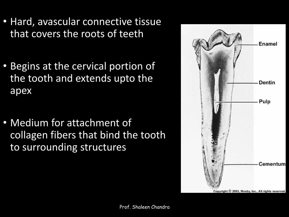
• Hard, avascular connective tissue that covers the roots of teeth
• Begins at the cervical portion of the tooth and extends upto the apex
• Medium for attachment of collagen fibers that bind the tooth to surrounding structures
Prof. Shaleen Chandra

Physical characteristics
Prof. Shaleen Chandra
• Hardness less than dentin
• Colour light yellow• Lacks the translucency of enamel
• Slightly lighter in colour than dentin
• Has been experimentally shown to be permeable to a variety of materials
Prof. Shaleen Chandra

Chemical properties
Prof. Shaleen Chandra
• 45% inorganic material
• 50-55% organic material and water
• Inorganic portion • Mainly calcium and phosphate in form of hydroxyapatite
• Highest fluoride content of all mineralized tissues
• Organic portion • Type I collagen
• Polysaccharides (proteoglycans)
Prof. Shaleen Chandra
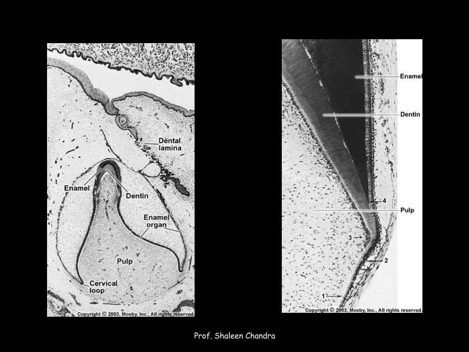
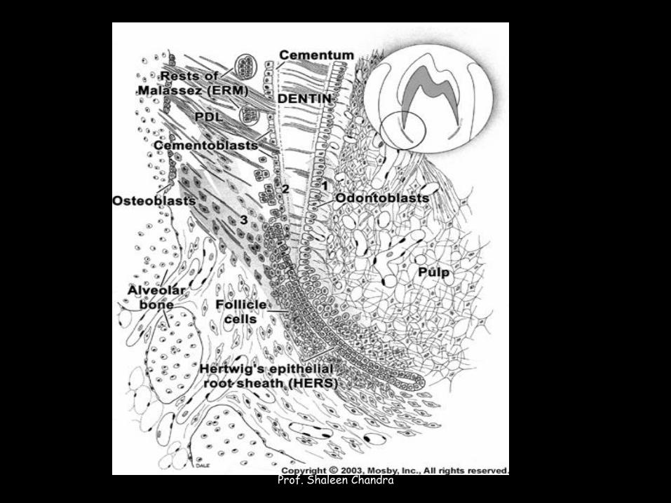
Cementogenesis
Prof. Shaleen Chandra
Prof. Shaleen Chandra
Prof. Shaleen Chandra
• Preceded by deposition of dentin on the inner aspect of HERS
• Breaks occur in the epithelial root sheath allowing the newly formed dentin to come in direct contact with connective tissue of the dental follicle
Prof. Shaleen Chandra
• Loss of continuity of the basal lamina is soon followed by the appearance of collagen fibrils and Cementoblasts between epithelial cells of the root sheath
• Sheath cells migrate towards the dental sac form epithelial Rests of Malassez
Prof. Shaleen Chandra
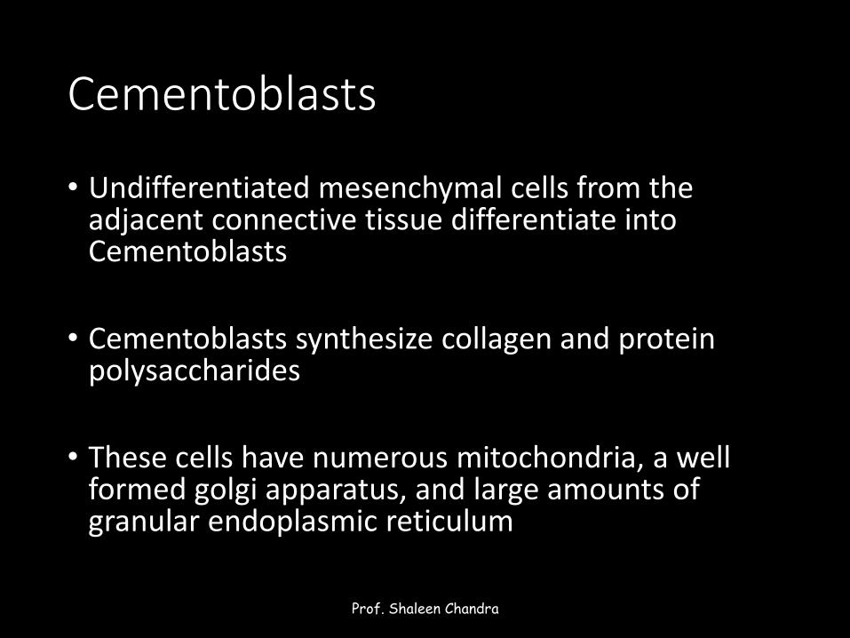
Cementoblasts
• Undifferentiated mesenchymal cells from the adjacent connective tissue differentiate into Cementoblasts
• Cementoblasts synthesize collagen and protein polysaccharides
• These cells have numerous mitochondria, a well formed golgi apparatus, and large amounts of granular endoplasmic reticulum
Prof. Shaleen Chandra
Classification
Prof. Shaleen Chandra
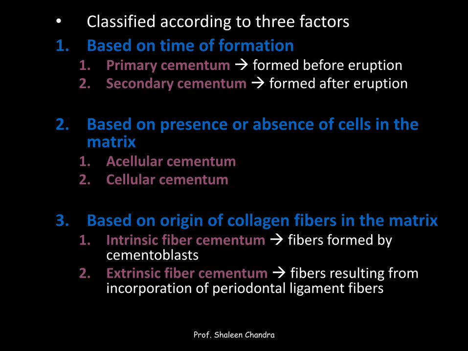
• Classified according to three factors
1. Based on time of formation1. Primary cementum formed before eruption 2. Secondary cementum formed after eruption
2. Based on presence or absence of cells in the matrix
1. Acellular cementum2. Cellular cementum
3. Based on origin of collagen fibers in the matrix1. Intrinsic fiber cementum fibers formed by
cementoblasts2. Extrinsic fiber cementum fibers resulting from
incorporation of periodontal ligament fibers
Prof. Shaleen Chandra
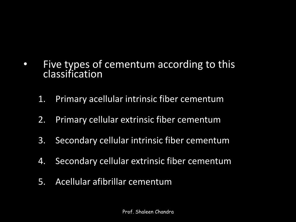
• Five types of cementum according to this classification
1. Primary acellular intrinsic fiber cementum
2. Primary cellular extrinsic fiber cementum
3. Secondary cellular intrinsic fiber cementum
4. Secondary cellular extrinsic fiber cementum
5. Acellular afibrillar cementum
Prof. Shaleen Chandra

Primary acellular intrinsic fiber cementum
• First formed cementum just next to the cementodentinal junction
• 15-20µm in thickness
• All the collagen component is of cementoblastic origin
Prof. Shaleen Chandra
Primary cellular extrinsic fiber cementum
• Fibers of periodontal ligament are inserted into cementum
• Forms the principal tissue of attachment
• Covers at least two thirds of the root
• At cervical margin 50µm thick
• Apically 200µm
Prof. Shaleen Chandra
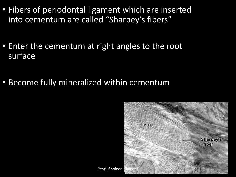
• Fibers of periodontal ligament which are inserted into cementum are called “Sharpey’s fibers”
• Enter the cementum at right angles to the root surface
• Become fully mineralized within cementum
Prof. Shaleen Chandra
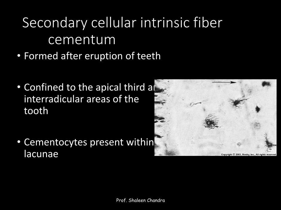
Secondary cellular intrinsic fiber cementum
• Formed after eruption of teeth
• Confined to the apical third and interradicular areas of the tooth
• Cementocytes present within lacunae
Prof. Shaleen Chandra
Secondary cellular extrinsic fiber cementum• Constitutes the bulk of secondary cementum
• Cementocytes within lacunae with processes (canaliculi) directed towards the periodontal ligament
• Has a laminated structure with cementoid on the surface
Prof. Shaleen Chandra
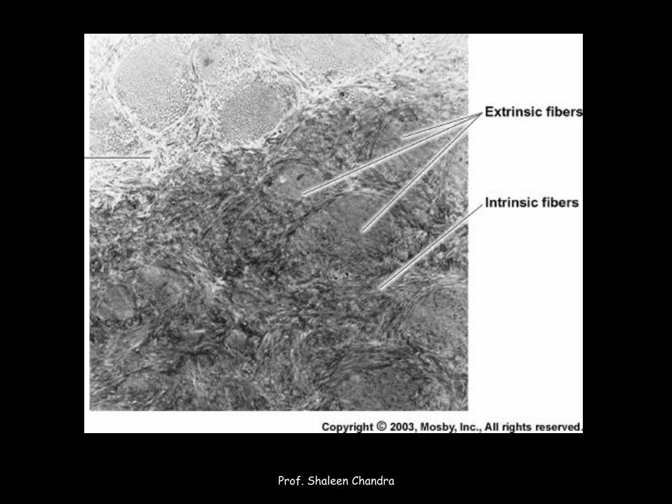
• Contains both intrinsic and extrinsic fibers
• Intrinsic fibers are thinner and run parallel to the root surface
• Extrinsic fibers are larger in diameter and rum perpendicular to the root surface
Prof. Shaleen Chandra
Prof. Shaleen Chandra

Acellular afibrillar cementum
• Found in isolated patches on the enamel surface or close to the cementoenamel junction
• Plays no role in tooth attachment
Prof. Shaleen Chandra
Cementodentinal Junction
Prof. Shaleen Chandra
• Permanent teeth- smooth
• Deciduous teeth – sometimes scalloped
• Provides a firm attachment to the dentin
• Sometimes a layer of intermediate cementum is present between the dentin and cementum• Does not exhibit characteristics of either cementum or
dentin
Prof. Shaleen Chandra
Cementoenamel junction
Prof. Shaleen Chandra
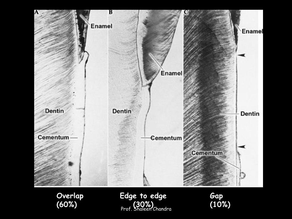
Overlap(60%)
Edge to edge(30%)
Gap(10%)
Prof. Shaleen Chandra
• Gap Junction: enamel epithelium in the cervical portion of the root is delayed in its separation from dentin
• Overlap Junction: when connective tissue cells, probably Cementoblasts, come in contact with enamel they produce a laminated, electron-dense, reticular material termed afibrillar cementum
Prof. Shaleen Chandra
Copyright © 2022 FDOKUMEN