Ontogenetic effects of EEDQ on amphetamine-induced behaviors of rats: role of presynaptic processes
Upload
independentCategory
view
3download
0
Author's personal copy
Acute dextro-amphetamine administration does not alterbrain myo-inositol levels in humans and animals:
MRS investigations at 3 and 18.8 T
Brent M. McGrath a,*, Ryan McKay b, Sanjay Dave a, Peter Seres c, Aalim M. Weljie d,Carolyn M. Slupsky d, Chris C. Hanstock c, Andrew J. Greenshaw a, Peter H. Silverstone a
aDepartment of Psychiatry, University of Alberta, Edmonton, Alberta, Canada T6G 2B7bNational High Field Magnetic Resonance Centre, University of Alberta, Edmonton, Alberta, Canada T6G 2E1
c In Vivo NMR Research Centre, Department of Biomedical Engineering, University of Alberta, Edmonton, Alberta, Canada T6G 2V2dMagnetic Diagnostic Research Centre, University of Alberta, Edmonton, Alberta, Canada T6G 2E1
Received 21 August 2007; accepted 9 April 2008
Available online 18 April 2008
Abstract
The pathophysiological underpinnings of bipolar disorder are not fully understood. However, they may be due in part to changes in thephosphatidylinositol second messenger system (PI-cycle) generally, or changes in myo-inositol concentrations more specifically. Dextro-amphetamine has been used as a model for mania in several human studies as it causes similar subjective and physiological symptoms. Wewanted to determine if dextro-amphetamine altered myo-inositol concentrations in vivo as it would clearly define a mechanism linking putativechanges in the PI-cycle to the subjective psychological changes seen with dextro-amphetamine administration. Fifteen healthy human volunteersreceived a baseline scan, followed by second scan 75 min after receiving a 25 mg oral dose of dextro-amphetamine. Stimulated echo protonmagnetic resonance spectroscopy (MRS) scans were preformed at 3.0 Tesla (T) in the dorsal medial prefrontal cortex (DMPFC). Metabolite datawere adjusted for tissue composition and analyzed using LCModel. Twelve adult male rats were treated acutely with a 5-mg/kg intraperitoneal doseof dextro-amphetamine. After 1 h rats were decapitated and the brains were rapidly removed and frozen until dissection. Rat brains were dissectedinto frontal, temporal, and occipital cortical areas, as well as hippocampus. Tissue was analyzed using a Varian 18.8 T spectrometer. Metaboliteswere identified and quantified using Chenomx Profiler software. The main finding in the present study was that myo-inositol concentrations in theDMPFC of human volunteers and in the four rat brain regions were not altered by acute dextro-amphetamine. While it remains possible that the PI-cycle may be involved in the pathophysiology of bipolar disorder, it is not likely that the subjective and physiological of dextro-amphetamine aremediated, directly or indirectly, via alternations in myo-inositol concentrations.# 2008 Elsevier Ireland Ltd and the Japan Neuroscience Society. All rights reserved.
Keywords: Bipolar disorder; myo-Inositol; Inositol-depletion hypothesis; Mania model; Dextro-amphetamine; Magnetic resonance spectroscopy
1. Introduction
Bipolar disorder (BPD) affects at least 1% of the population,and it is characterized by manic and depressive episodesaccompanied by severe disturbances in cognition, perceptionand behaviour (Goodwin and Jamison 1990; McElroy et al.,1996). While lithium has remained the mainstay for BPD
treatment, other structurally dissimilar medications have beenfound to be effective in acute and prophylactic treatment,particularly valproate (Geddes et al., 2004; Bowden, 2003;Weisler et al., 2004; Herman, 2004; Tohen et al., 2003).Nonetheless, the pathophysiological underpinnings that under-lie these symptoms are not fully understood, although one of themost widely studied hypotheses regarding possible pathophy-siological changes are based on the findings that lithiuminhibits turnover of myo-inositol, an integral component of thephosphatidylinositol second messenger system (PI-cycle), in anuncompetitive manner (Allison and Stewart, 1971; Berridgeand Irvine, 1989; Berridge et al., 1982).
www.elsevier.com/locate/neures
Available online at www.sciencedirect.com
Neuroscience Research 61 (2008) 351–359
* Corresponding author at: Faculty of Medicine, Dalhousie University, Sir
Charles Tupper Medical Building, 5849 University Avenue, PO Box 265,Halifax, NS, Canada B3H 4H7. Tel.: +1 902 401 9409.
E-mail address: [email protected] (B.M. McGrath).
0168-0102/$ – see front matter # 2008 Elsevier Ireland Ltd and the Japan Neuroscience Society. All rights reserved.doi:10.1016/j.neures.2008.04.001
Author's personal copy
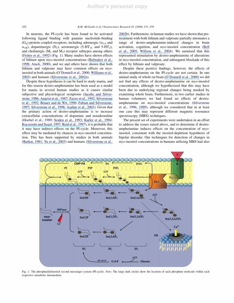
In neurons, the PI-cycle has been found to be activatedfollowing ligand binding with guanine nucleotide-binding(Gq)-protein coupled receptors, including adrenergic (a1A anda1B), dopaminergic (D1), serotonergic (5-HT1C and 5-HT2),and cholinergic (M1 and M3) receptor subtypes among others(Fisher et al., 1992) (Fig. 1). Many studies have shown effectsof lithium upon myo-inositol concentrations (Belmaker et al.,1998; Atack, 2000), and we and others have shown that bothlithium and valproate may have common effects on myo-inositol in both animals (O’Donnell et al., 2000; Williams et al.,2002) and humans (Silverstone et al., 2002a).
Despite these hypotheses it can be hard to study mania, andfor this reason dextro-amphetamine has been used as a modelfor mania in several human studies as it causes similarsubjective and physiological symptoms (Jacobs and Silver-stone, 1986; Angrist et al., 1987; Zacny et al., 1992; Silverstoneet al., 1992; Brauer and de Wit, 1996; Fabian and Silverstone,1997; Silverstone et al., 1998; Asghar et al., 2003). Given thatthe primary action of dextro-amphetamine is to increaseextracellular concentrations of dopamine and noradrenaline(Hoebel et al., 1989; Seiden et al., 1993; Karler et al., 1994;Kuczenski and Segal, 1997; Reid et al., 1997), it is probable thatit may have indirect effects on the PI-cycle. Moreover, thiseffect may be mediated by chances in myo-inositol concentra-tion. This has been supported by studies in both animals(Barkai, 1981; Yu et al., 2003) and humans (Silverstone et al.,
2002b). Furthermore, in human studies we have shown that pre-treatment with both lithium and valproate partially attenuates arange of dextro-amphetamine-induced changes in brainactivation, cognition, and myo-inositol concentration (Bellet al., 2005; Willson et al., 2004). We surmised that thisrepresented stimulation by dextro-amphetamine of alterationsin myo-inositol concentration, and subsequent blockade of thiseffect by lithium and valproate.
Despite these positive findings, however, the effects ofdextro-amphetamine on the PI-cycle are not certain. In oneanimal study of whole rat brain (O’Donnell et al., 2000) we didnot find any effects of dextro-amphetamine on myo-inositolconcentration, although we hypothesized that this may havebeen due to underlying regional changes being masked byexamining whole brain. Furthermore, in two earlier studies inhuman volunteers we had found no effects of dextro-amphetamine on myo-inositol concentrations (Silverstoneet al., 1996, 1999), although we considered that in at leastone case this may represent different magnetic resonancespectroscopy (MRS) techniques.
The present set of experiments were undertaken in an effortto address the issues raised above, and to determine if dextro-amphetamine induces effects on the concentration of myo-inositol, consistent with the inositol-depletion hypothesis ofbipolar disorder. Our techniques for detection of changes inmyo-inositol concentrations in humans utilizing MRS had also
Fig. 1. The phosphatidylinositol second messenger system (PI-cycle). Note: The large dark circles show the location of each phosphate molecule within each
respective metabolic intermediate.
B.M. McGrath et al. / Neuroscience Research 61 (2008) 351–359352

Author's personal copy
improved, allowing us to examine these conflicting results ingreater detail. Our hypotheses for the present study weretherefore that dextro-amphetamine would cause regional brainmyo-inositol changes in rats, opposite to the whole brain effectsreported for lithium and valproate. Secondly, we hypothesizedthat dextro-amphetamine administration would also lead todetectable myo-inositol changes in human volunteers, asmeasured by a novel MRS technique specifically designed tomeasure myo-inositol concentrations in human brain (Kimet al., 2005). If supported, these hypotheses would better definea mechanism linking putative changes in myo-inositolconcentration to the subjective psychological changes seenwith dextro-amphetamine administration, potentially furthersupporting the importance of myo-inositol and the PI-cycle inmood disorders.
2. Materials and methods
2.1. Pre-clinical experiment
Twelve adult male Sprague–Dawley rats (Biosciences Animal Service,
University of Alberta), weighing 350–450 g, were housed in pairs in standardPlexiglas laboratory cages. The rats were provided with food (LabDiet 5001
Rodent Diet, PMI Nutrition International Inc., Brentwood, MO, USA) and
water ad libitum, and were maintained at 20 8C, under a 12-h light/dark cycle
(lights on 07:00–19:00 h), in a humidity-controlled environment. Treatmentwas started 1 week after the rats arrived, giving them an opportunity to
acclimatize to their new environment. This component of the study was
reviewed and approved by the local Animal Policy and Welfare Committee
and carried out in accordance with the guidelines of the Canadian Council onAnimal Care.
2.1.1. Treatment
Each rat received a single, acute intraperitoneal (IP) injection of either5 mg/kg (based on free base weight) dextro-amphetamine (n = 6) (Health and
Welfare Canada, Ottawa, ON, Canada) or saline (n = 6). This dose was
chosen based on other similar studies (Del Arco et al., 1998; Anderzhanova
et al., 2001; Mcgeehan et al., 2004; Choe et al., 2002; Andiarzhanova et al.,2002; Anderzhanova et al., 2002). All injections were administered in 2 ml/
kg volumes of saline. From previous work (Anderzhanova et al., 2002; Mora
and Porras, 1993), at 60 min post injection rats were decapitated. The brains
were rapidly removed and immediately immersed in ice-cold 2-methylbutane(Fisher Scientific, Fairlawn, NJ, USA). The brains were maintained at
!80 8C until brain dissection, tissue extraction and preparation for NMR
analysis.
2.1.2. Brain dissection and preparationWhole brains were dissected into frontal (fcx), temporal (tcx) and occipital
(ocx) cortex, as well as hippocampus (hipp) according to stereotaxic demarca-
tion (Pellegrino et al., 1979). Samples were prepared using a modified version(O’Donnell et al., 2000) of the extraction method described by Bligh and Dyer
(1959). More specifically, brain regions were homogenized in four volumes of
methanol/chloroform (2:1, v/v; Fisher Scientific, Fairlawn, NJ, USA). This was
followed by the subsequent additions of one part of chloroform with homo-genization, and one part of water with homogenization. A standardized (within
brain regions) amount of homogenate (0.7 ml for fcx, 0.4 ml for tcx, 0.45 ml for
ocx, 1 ml for hipp) was transferred to an Eppendorf tube and centrifuged at
1000 rpm for 15 min in a bench top centrifuge (ThermoIEC, Needham Heights,MA, USA). Following centrifugation, a standardized amount of the water/
methanol layer (0.5 ml for fcx, 0.2 ml for tcx, 0.3 ml for ocx, 0.8 ml for hipp)
was transferred to a 12 mm " 75 mm Simport culture tube and maintained at!20 8C overnight. The next day, samples were dried using vacuum centrifuga-
tion (Thermo Electron Corporation, Milford, MA, USA) and then returned to
!20 8C until NMR analysis.
At the time of NMR analysis, dried samples were reconstituted in 0.6 ml ofdH2O and 0.06 ml of deuterated water (D2O) containing 5 mM 2,2-dimethyl-2-
silapentane-5-sulfonate (DSS) as an internal reference standard (Markley et al.,
1998), 100 mM imidazole and 0.2% NaN3 (Chenomx Inc., Edmonton, AB,
Canada), at a pH of approximately 7.
2.1.3. High-field 1H NMR spectroscopy
NMR Spectroscopy was conducted at 37 8C and 18.8 Tesla (T) on a Varian
Inova-800 spectrometer equipped with a 5-mm triple axis gradient HCN probe.One-dimensional single 908 pulse 1H spectra were collected with a water pre-
saturation period of 2 s (gB1 of #150 Hz), sweep widths of 12,000 Hz, and
acquisition times of 2 s. All directly and indirectly detected data sets were zero
filled to twice the number of acquired points. The 1D–1H spectra were apodizedusing a 0.5 Hz line broadening or a cosine weighting function, respectively.
2.1.4. Identification and quantification of brain extract metabolites
Identification and quantification of metabolites from brain extracts weredone using Chenomx Profiler software (Chenomx Inc., www.chenomx.com,
Edmonton, AB, Canada) on 1H NMR spectra of brain extracts. Briefly, Profiler
is linked to a database of metabolite molecules whose unique NMR spectral
signatures are encoded at various spectrometer frequencies including 18.8 T (or800 MHz). Comparison of the rat brain extract NMR spectra to the Chenomx
spectral signature database within Profiler results in a list of compounds
together with their respective concentrations. Metabolites were quantified by
the addition of a known amount of the internal standard DSS (see above) to thebrain extract samples, which also serves as a chemical shift reference (set to
0 ppm). All compounds in the database, including those discussed in this work,
have been verified against known concentrations of reference NMR spectra of
the pure compounds, enabling accurate metabolite identification and concen-tration quantification. Normalized metabolite concentrations were compared
using the Independent samples t-test, with significance evaluated at the a = 0.05
level, using SPSS# (version 11 for OS X).
2.2. Clinical experiment
2.2.1. Subjects
This component of the study was approved by the local ethics board of theUniversity of Alberta Hospital, and all participants gave full informed consent.
Fifteen healthy volunteers (6 male and 9 female) were recruited through a
newsletter advertisement from the university community. Volunteers underwent
a brief medical history, and a detailed semi-structured clinical interview(structured clinical interview for DSM-IV Axis I disorders—SCID) to rule
out past or present psychiatric illness. Volunteers were also assessed on the
Hamilton depression rating scale (HAM-D), the Montgomery-Asberg depres-
sion rating scale (MADRS) and the Young mania rating scale (YMRS). Past andpresent drug and alcohol use was assessed and volunteers were excluded based
on abuse of these substances. In addition, volunteers who had used recreational
drugs in the past 6 months, or amphetamine in the past year were excluded fromparticipating. A magnetic resonance (MR) safety screen was also performed to
ensure safety in undergoing the MR procedure. At 12:00 a.m. on the day of the
scan, volunteers were required to fast until both scans were complete.
2.2.2. Study designAn open-label study design was used. All volunteers underwent a baseline
MRS scan. Upon completion of the first scan, volunteers were administered a
single oral 25 mg dose of dextro-amphetamine (GlaxoSmithKline,Montreal, PQ,CA). The second scan began 75 min after administration of dextro-amphetamine,
which coincides with the peak subjective response to dextro-amphetamine, which
occurs 60–120 min after administration (Asghar et al., 2003).
2.2.3. Physiologic and subjective response measuresResting heart rate and both systolic and diastolic blood pressure were
measured at baseline and at 75 and 150 min after dextro-amphetamine admin-
istration. Subjective measurements of mood were also assessed at baseline andat 75 and 150 min after dextro-amphetamine using a 100 mm visual analogue
scale (VAS) (Folstein and Luria, 1973). The percentage change in VAS scores
from baseline are reported.
B.M. McGrath et al. / Neuroscience Research 61 (2008) 351–359 353

Author's personal copy
2.2.4. Clinical 1H MRS
Data were acquired with a 3.0 T scanner (Magnex Scientific, Abingdon,United Kingdom/SurreyMedical Imaging Systems, Guilford, United Kingdom)
with a 28-cm quadrature birdcage resonator for transmission and reception.
Multislice gradient echo images (repetition time [TR] = 500 ms, echo time[TE] = 22 ms, slice thickness = 5 mm, 11 slices, resolution = 256 " 256) were
used to coregister a 2.5 cm " 2.5 cm " 2.5 cm point-resolved spectroscopy
(PRESS) voxel that encompassed the dorsal medial prefrontal cortex (DMPFC).
The lower border was orientated to a line connecting the anterior and posteriorcommissures. The voxel was centered on the midline, touching the tip of the
genu of the corpus callosum (Fig. 2). The PRESS-selected volume was used for
both tissue segmentation and for acquiringwater-suppressedmetabolite spectra.
Shimming was carried out first with FASTMAP (Gruetter, 1993) to optimize thelinear and non-linear shims over a 5-cm-diameter spherical volume, then with
an automatic optimization of the linear x, y, z shims on the PRESS-selected
voxel at TE = 160 ms and TR = 3000 ms. Typical shimmed water line-widths
were better than 6 Hz (0.05 ppm).Tissue segmentation into grey matter (GM), white matter (WM), and
cerebrospinal fluid (CSF) was performed with a double inversion recovery
sequence to acquire one-dimensional (1D) projections of each T1 compartmentin the PRESS-selected volume (Hanstock and Allen, 2000). Two hyperbolic
secant inversion pulses (110-ms length, bandwidth = 150 Hz) were added to the
PRESS pulse sequence, in which the pulses were 908 sinc-Gauss and 1808optimized-sinc shapes. Before the 908 pulse, a 15-ms spoiler gradient wasapplied to dephase any transverse magnetization resulting from the inversion
pulses. The PRESS parameters were TR = 9 s, TE = 120 ms, two averages with
5-kHz sample frequency, digitized over 128 data points. Shimming over the
PRESS volume was performed by turning the frequency encode gradient off,increasing the digitization to 2048 data points, and decreasing TR = 3 s. This
resulted in typical shimmed line-widths of <0.05 ppm. Prior data were used to
estimate the T1 values for the three brain compartments (GM: 1070 $ 60 ms;WM: 720 $ 30 ms; CSF: 4440 $ 50 ms). With the expression derived by
Redpath and Smith (1994), two pairs of Tinv1 and Tinv2 timings were computed
that gave simultaneous nulls of the CSF compartment with either that from GM
or WM. Twenty-one 1D projections were acquired for each set of inversiontimings. An additional 10 1D projections were acquired with no inversion pulses
and with a TE of 500 ms, which minimized the signal contamination from GM
and WM (<0.2% residual signal after accounting for T2 losses), while main-
taining significant signal from CSF (approximately 50% residual signal).After phase correction of each projection, three-dimensional surface and
contour maps were generated to confirm that the selected Tinv1 and Tinv2 timings
were resulting in simultaneous nulls of CSF with either GM or WM. Subse-
quently, GM and WM projections were selected from the double inversionrecovery 1D projection series. The Tinv1 and Tinv2 timings that were used to
acquire these two series of projections for either GM or WM were then used to
estimate a normalization factor that reflected attenuation due to T1 and T2 lossesfor each projection, thereby fully accounting for all the acquisition timings.
Similarly, a normalization factor was estimated for the CSF projection. Seg-
mentation resulted from first normalizing and then summing the signal across
each of the three projections, such that the relative proportions of GM,WM, and
CSF could be estimated. These proportions were then used to calculate a grey
matter fraction [GM/(GM + WM)] for the voxel. All computations wereperformed within the MATLAB program environment (MathWorks, Natick,
Massachusetts).
The PRESS acquisitions (TE1 = 36 ms, TE2 = 160 ms, TR = 3000 ms)were performed as the sum of 16 subspectra, each of 32 averages, allowing
re-registration to the same frequency reference (the acetyl peak of NAA at
2.023 ppm) to eliminate the effects of frequency drift during the course of the
acquisition.
2.2.5. Identification and quantification of brain metabolites
Metabolite spectra were Fourier transformed, and quantified as ratios tocreatine-phosphocreating and N-acetylaspartate, with the Linear Combination
of Metabolite Basis Spectra (LCModel) program (Provencher, 1993). Ratios
were used because the lack of accurate estimates of T2 relaxation times formyo-
inositol and other strongly coupled spins is an obstacle to absolute quantifica-tion. The basis spectra for LCModel analysis were simulated within the spectral
bandwidth of 1.00–3.90 ppm with numeric methods (Hanstock et al., 2002).
The metabolites included in the basis spectra were N-acetylaspartate, N-
acetylaspartyl-glutamate, creatine, choline, glutamate, glutamine, gamma-ami-nobutyric acid, myo-inositol, glycine, taurine, guanidoacetate, and aspartate.
The in vivo metabolite data were accepted if the signal-to-noise ratio was 11 or
more, and the standard deviation of the fit for the metabolite was %20%. In thepresent study, no subjects were excluded based on the aforementioned criteria.
Metabolite ratios were compared using the paired samples t-test, with sig-
nificance evaluated at the a = 0.05 level, using SPSS# (version 11 for OS X).
3. Results
3.1. Pre-clinical 18.8 T 1H NMR spectroscopy experiment
Fig. 3 depicts a typical 1H spectrum of rat brain fcx extractacquired at 18.8 T, with the DSS peak referenced to 0 ppm. At18.8 T, myo-inositol gives multiplet signals at 3.27, 3.52, 3.62,and 4.06 ppm. The spectral region from 3.50 to 3.65 ppm isexpanded to depict contributions from myo-inositol.
3.1.1. High-field NMR spectroscopy dataMetabolite concentrations are reported in millimolars (mM),
normalized to the DSS standard concentration (0.5 mM)(Table 1). No significant differences between myo-inositolconcentrations in frontal, temporal and occipital cortex andhippocampus were observed between placebo- and dextro-amphetamine-treated rats (independent samples t-test,p > 0.05) (Table 1).
Fig. 2. Localization of a single voxel in the DMPFC.
B.M. McGrath et al. / Neuroscience Research 61 (2008) 351–359354

Author's personal copy
3.2. Clinical 3 T 1H MRS experiment
3.2.1. Subject characteristicsA total of 15 healthy volunteers (6 males, 9 females) who
met the eligibility criteria participated in the present study. Themean age of the group was 28.27 years ($1.22), and their meanweight was 68.69 kg ($4.38). The mean dose of dextro-amphetamine administered was 0.36 mg/kg.
3.2.2. Physiologically and subjective responseAs anticipated from previous studies (Asghar et al., 2003;
Willson et al., 2004), there was a significant dextro-amphetamine-induced increase in heart rate, systolic bloodpressure and diastolic blood pressure (Table 2). The percentagechange in VAS scores from baseline, for each item, are reportedin Table 3. There was a main time effect on the change frombaseline in the subjective measures of anxiety, happiness,alertness, energy, irritability, speed of thought, light-head-edness and restlessness following dextro-amphetamine admin-istration in healthy volunteers. Post hoc analysis, in the form of
paired sample t-tests, showed a significant increase frombaseline to 75 min for all eight items above, and from baselineto 150 min for six items above (Table 3). Physical restlessnessincreased significantly across all three time-points. Theseresults are consistent with dextro-amphetamine having beenadministered at a dose that causes its well-recognizedpsychological effects.
3.2.3. Clinical MRS dataA representative 1H MRS spectra from the DMPFC at 3 T is
shown in Fig. 4, with LCModel fit. No differences in thedistribution of grey matter, white matter and cerebral spinalfluid were found between pre and post dextro-amphetaminescans.
The use of an optimized sequence for myo-inositol acquiredat 3 T provided a well-resolved myo-inositol doublet peak at3.65 ppm. No significant differences between the myo-inositol(m-Ino), myo-inositol/N-acetylaspartate (m-Ino/NAA) or myo-inositol/Creatine-Phosphocreatine (m-Ino/Cr-PCr) peak ratioswere observed when comparing pre- with post-dextro-amphetamine values (paired samples t-test, p > 0.05) (Table 4).
4. Discussion
The present set of experiments were designed to determine ifacute administration of dextro-amphetamine would alter brainmyo-inositol concentrations in either rats or humans. Such afinding would be of significant clinical and experimentalsignificance since it would link the similarity in subjective andphysiological experiences of dextro-amphetamine users tobipolar mania with the purported involvement of myo-inositol,and perhaps the PI-cycle, in bipolar symptomatology. Thiswould provide an important neurochemical model for the study
Fig. 3. Typical 1H magnetic resonance spectroscopy spectra of the rat brain at
18.8 T, with contributions from m-Ino expanded. (a) Region from 4.02 to
4.08 ppm; (b) region from 3.50 to 3.64 ppm; (c) region from 2.24 to 3.30 ppm.m-Ino, myo-inositol.
Table 1
Regional brain 1H MRS metabolite concentration differences between placebo
and dextro-amphetamine treated ratsa
Region Treatment group p-Value
Placebo Dextro-amphetamine
CortexFrontalb 2.203 $ 0.592 2.090 $ 0.445 0.882
Temporal 3.297 $ 0.396 5.172 $ 0.972 0.119
Occipital 3.257 $ 0.657 3.764 $ 0.272 0.500
Sub-cortical
Hippocampus 2.538 $ 0.104 3.279 $ 0.333 0.090
Values represent mean $ S.E.M. Significance was assessed at p < 0.05, using
independent samples t-test.a Concentrations are reported in mM and reflect those of the NMR sample, not
the absolute concentration within the respective brain regions.b Five rats/group.
Table 2Effects of acute dextro-amphetamine on heart rate and blood pressure
Physiological measure Mean (S.E.M.)
Heart rate (beats per min), mean $ S.E.M.a
Baseline 64.3 $ 2.0Pre-MRS (75 min) 73.9 $ 2.5
Post-MRS (150 min) 81.6 $ 2.4
Blood pressure (mm Hg), mean $ S.E.M.b
Systolic
Baseline 117.0 $ 2.2
Pre-MRS (75 min) 131.0 $ 2.0
Post-MRS (150 min) 133.0 $ 2.1
Diastolic
Baseline 68.3 $ 2.5
Pre-MRS (75 min) 76.3 $ 2.0Post-MRS (150 min) 75.7 $ 2.0
Measurements were taken at baseline, 75 min after dextro-amphetamine admin-
istration (Pre-MRS) and again at 150 min after dextro-amphetamine (Post-
MRS).a Heart rate significantly increased from baseline to post-MRS and pre-MRS
to post-MRS, post hoc paired samples t-test p < 0.01.b Blood pressure, both systolic and diastolic, significantly increased from
baseline to pre-MRS and baseline to post-MRS, post hoc paired samples t-testp < 0.05.
B.M. McGrath et al. / Neuroscience Research 61 (2008) 351–359 355

Author's personal copy
of bipolar pathophysiology. That said, the main finding in thepresent set of studies was thatmyo-inositol concentrations werenot altered in vivo by acute dextro-amphetamine administrationin the DMPFC of human volunteers nor in the frontal, temporaland occipital cortex, or in the hippocampus of rats.
These findings are in line with some of our earlier research,which showed no changes in whole rat brain (O’Donnell et al.,2000) or in human volunteers (Silverstone et al., 1996, 1999).However, in another study our findings suggested that dextro-amphetamine increased myo-inositol concentration in volun-teers (Silverstone et al., 2002b). The present findings are morecompelling for several reasons. Firstly, to overcome possible
limitations in our previous animal studies, in the present studyfour brain regions were examined. Three of these regions(frontal and temporal cortex, and hippocampus) have beenshown, clinically, to have alteredmyo-inositol concentrations inbipolar disorder, while the forth region (occipital cortex) hasnot shown such alterations (reviewed in Silverstone et al.,2005). Furthermore, investigation of specific brain regionswould reduce the likelihood of an averaging out effect withrespect to any underlying neurochemical changes, an affect thatmay occur with the use of whole brain. Although, this does notcontrol for effects or changes in intra- versus extra-cellularpools of myo-inositol that may exist within the brain, a level ofcontrol that is not possible when utilizing magnetic resonancespectroscopy. Secondly, the magnet used in the present study isamong the most powerful commercially available for ex vivouse, providing highly resolved spectra. The human volunteerstudy also differed from previous work via a fundamentalimprovement: namely, this study utilized a newly developedsequence specifically optimized for myo-inositol (Kim et al.,2005). This optimized sequence increased the stability andaccuracy of the spectroscopy results, providing more precisemeasurement of brain myo-inositol concentration. Thissequence addresses a major limitation present in the majorityof past clinical MRS studies (Silverstone et al., 2005), whichdid not use an optimized sequence.
Table 3Effects of acute dextro-amphetamine on VAS subjective measures of mood
‘‘0’’ Anchor Mean $ S.E.M. ‘‘100’’ Anchor
Baseline 75 min 150 min
I do not feel anxious at all 22.53 $ 5.43a 41.73 $ 6.81 37.27 $ 6.64 I feel very anxious
I feel very miserable or sad 51.40 $ 8.51a,b 65.73 $ 7.19 72.20 $ 6.91 I feel very happy
I feel mentally slowed 62.07 $ 6.50a,b 80.87 $ 4.83 81.67 $ 4.40 I feel mentally alertI feel physically unwell 82.53 $ 6.77 80.93 $ 5.16 84.20 $ 4.00 Physically I feel fine
I do not feel hungry at all 50.67 $ 6.27 43.53 $ 8.62 43.93 $ 7.40 I feel very hungry
I feel tired and lethargic 61.73 $ 6.00a,b 75.73 $ 5.70 80.53 $ 4.85 I feel very energeticI can concentrate well 24.80 $ 6.66 42.13 $ 7.46 30.80 $ 8.63 I cannot concentrate at all
I feel placid and calm 19.73 $ 4.35a,b 43.20 $ 4.74 36.93 $ 5.40 I feel very irritable
My thoughts are slow 50.07 $ 4.36a,b 71.33 $ 4.66 79.80 $ 4.15 My thoughts are speeded up
I do not feel light headed 24.87 $ 7.83a 48.27 $ 8.15 49.13 $ 8.76 I feel very light-headedI feel physically inactive 43.07 $ 4.35a,b,c 58.33 $ 6.18 71.87 $ 4.40 I feel physically restless
Measurements were taken at baseline, 75 min after dextro-amphetamine administration and again at 150 min after dextro-amphetamine. Scores could range from 0 to
100 (on a 100-mm scale) and are reported as Mean $ S.E.M.a Significant increase from baseline at 75 min, post hoc paired samples t-test p < 0.05.b Significant increase from baseline at 150 min, post hoc paired samples t-test p < 0.05.c Significant increase from 75 min at 150 min, post hoc paired samples t-test p < 0.05.
Fig. 4. A representative 1H MRS spectrum (black line) acquired at 3 T of theDMPFC, with LCModel fit (red line). The baseline is shown in blue. Abbrevia-
tions: Cho – choline; Cr – creatine; Cr-PCr – creatine-phosphocreatine; Glx –
glutamine and glutamate; m-Ino – myo-inositol; NAA – N-acetylaspartate.
Table 4
3 T 1H MRS metabolite peak area ratios for baseline and post dextro-amphe-
tamine scans
Baseline Post D-amphetamine p-Value
Metabolitesa
m-Ino 131.4695 $ 18.5683 123.4837 $ 8.1762 0.667
m-Ino/NAA 1.1569 $ 0.0738 1.1861 $ 0.0606 0.569
m-Ino/Cr-PCr 1.3496 $ 0.0431 1.3145 $ 0.0391 0.401
Values represent mean $ S.E.M., adjusted for grey matter fraction. Significance
from baseline was assessed at p < 0.05 using paired samples t-test.a In machine units.
B.M. McGrath et al. / Neuroscience Research 61 (2008) 351–359356
Author's personal copy
Nonetheless, even with these methodological improvementsand in contrast to both our initial hypotheses, the present studiesdid not lead to detectable changes inmyo-inositol concentrationbetween experimental and control conditions. Thus, from theseresults we conclude that while dextro-amphetamine doesproduce subjective and physiological effects similar to thoseobserved in bipolar mania, it does not appear to induce changesin myo-inositol concentrations. Thus it appears that thesubjective and physiological effects of dextro-amphetamineare not mediated by changes in globally measured myo-inositolconcentrations per se. Moreover, in rats, dextro-amphetaminedoes not induce changes inmyo-inositol concentrations in brainregions purported to be involved in mood regulation. Given thatthis study reports similar findings in both humans and rats,utilizing a similar methodology, it is our contention that thefinding of no change in myo-inositol concentration followingacute dextro-amphetamine administration is a valid one.
Thus, it appears unlikely that alterations in myo-inositolconcentration, in particular, play an important physiologicalrole in the action of dextro-amphetamine. That said, morerecent evidence still suggests that the PI-cycle may be involved,albeit via a more elaborate mechanism. In particular, severalnew targets for investigation have been receiving increasedattention. Among these myo-inositol-1-phosphate synthase(MIP-synthase), sodium-dependent myo-inositol transporters 1and 2 (SMIT1 and SMIT2) and the H+/myo-inositol transporter(HMIT) are all promising avenues of investigation.
MIP-synthase regulates the synthesis of myo-inositol-1-phosphate from glucose-6-phosphate (Fig. 1) and has beenshown to be confined to the vasculature (Wong et al., 1987; DiDaniel et al., 2006). Lithium has been shown to inhibit therecycling of myo-inositol via inhibition of myo-inositolmonophosphatase (IMPase) (Fig. 1) (Allison et al., 1976).Valproate was found to have a similar effect on myo-inositolconcentrations, albeit via the inhibition of MIP-synthase(Vaden et al., 2001). This action has been extended too lessteratogenic derivates of valproate as well (Galit et al., 2007).Interestingly, this group also showed that one of these valproatederivates, valrocemide, attenuated amphetamine-induced activ-ity (rearing) in rats at a degree similar to that seen with lithium(Galit et al., 2007). Recent work has failed to demonstrate anexaggeration of lithium-pilocarpine induced seizures by theaddition of a MIP-synthase inhibitor (Einat et al., 2008). Nordid the MIP-synthase inhibitor result in pilocarpine-inducedseizures when given on it’s own.
SMIT-1 and SMIT-2 are purported to be the primary myo-inositol transporters, exchanging sodium formyo-inositol acrossthe plasmamembrane (VanCalker andBelmaker, 2000). SMIT-1is localized primarily in the choroid plexus (Guo et al., 1997),while SMIT-2 is more widely distributed in the brain (Coadyet al., 2002). In a series of studies using SMIT-1 knockoutmice, ithas been shown that even with a reduction in myo-inositolconcentration well in excess of that generated by lithium, nodepletion in membrane phosphatidylinositiol was observed(Berry et al., 2003, 2004).Moreover, in a subsequent study, itwasfound that SMIT-1 knockout mice did not exhibit the samebehavioural changes as those observed following lithium
treatment inwild-typemice (Shaldubina et al., 2006), suggestingthat changes in myo-inositol concentration is not responsible forthe behavioural effects of lithium. The same research group alsoobserved little difference between SMIT-1 knockout mice andwild-type mice on several measures, including lithium-pilocar-pine seizures, Porsolt forced-swim test and amphetamine-induced hyperactivity (Shaldubina et al., 2007).A recent study inbipolar patients, Willmroth et al. (2007) reported that lithiumtreatment reduced the expression of SMIT mRNA in theneutrophils of bipolar I patients, but not bipolar II patients.Moreover, they found that untreated bipolar I, but not bipolar II,patients had greater SMIT mRNA expression relative to healthycontrols (Willmroth et al., 2007). The significance of this findingis difficult to determine, and may well relate to an, as yet,unknown function of SMIT and myo-inositol.
HMIT is a hydrogen/myo-inositol exchanger that has beenshown to be located in several regions, including the frontalcortex (Uldry et al., 2001). Work by Uldry et al. (2004) haveshown that the localization of HMIT is regulated by severalcellular processes, including depolarization, protein kinase Cactivation and increases in intracellular calcium. This is uniqueto HMIT, as SMITexpression appears to be constitutive and notregulated in this manner. More research is required, beforedefinitive statements regarding the functional importance ofHMIT can be made.
Up until relatively recently, it had been effectivelyimpossible to study directly the changes that happen in thebrains of psychiatric patients, thus requiring the reliance onproxy measures such as plasma or cerebral spinal fluid.However, recent advances in clinical neuroimaging, such asMRS and functional MRI have allowed more direct measures ofbrain activity. That said, there are still numerous technicallimitations with these methodologies, and it is becoming clearerthat we are unlikely to be able to characterize the complicateddisease processes that define these disorders by the measure-ment of a single (or even several) brain metabolites.
Acknowledgements
Regarding technical support and assistance, the authorswould also like to thank Mrs. Gail Rauw of the NeurochemicalResearch Unit, Department of Psychiatry, and Mr. DeryckWebb of the National High Field NMR Centre (NANUC), bothfrom the University of Alberta.
Sponsorship: This work was supported in part by peer-reviewed grants from the Canadian Institutes of HealthResearch (to PHS and BMM) and the Alberta HeritageFoundation for Medical Research (to PHS and BMM).
Conflict of interest
None.
References
Allison, J.H., Stewart, M.A., 1971. Reduced brain inositol in lithium-treated
rats. Nat. New Biol. 233 (43), 267–268.
B.M. McGrath et al. / Neuroscience Research 61 (2008) 351–359 357

Author's personal copy
Allison, J.H., Blisner, M.E., Holland, W.H., Hipps, P.P., Sherman, W.R., 1976.Increased brain myo-inositol 1-phosphate in lithium-treated rats. Biochem.
Biophys. Res. Commun. 71 (2), 664–670.
Anderzhanova, E., Rayevsky, K.S., Saransaari, P., Riitamaa, E., Oja, S.S., 2001.
Effects of sydnocarb and d-amphetamine on extracellular levels of aminoacids in the rat caudate-putamen. Eur. J. Pharmacol. 428 (1), 87–95.
Anderzhanova, E., Rayevsky, K.S., Saransaari, P., Riitamaa, E., Oja, S.S., 2002.
Effects of acute toxic doses of psychostimulants on extracellular levels of
excitatory amino acids and taurine in rats. Comparison of d-amphetamineand sydnocarb. Ann. N.Y. Acad. Sci. 965, 193–203.
Andiarzhanova, E.A., Saransaari, R., Riitamaa, E., Oiia, S.S., Raevskii, K.S.,
2002. Extracellular level of neuroactive amino acids in the rat neostriatumafter treatment with psychostimulants (microdialysis study). Eksp. Klin.
Farmakol. 65 (4), 7–13.
Angrist, B., Corwin, J., Bartlik, B., Cooper, T., 1987. Early pharmacokinetics
and clinical effects of oral dextroamphetamine in normal subjects. Biol.Psychiatry 22, 1357–1368.
Asghar, S.J., Tanay, V.A., Baker, G.B., Greenshaw, A., Silverstone, P.H., 2003.
Relationship of plasma amphetamine levels to physiological, subjective,
cognitive and biochemical measures in healthy volunteers. Hum. Psycho-pharmacol. 18 (4), 291–299.
Atack, J.R., 2000. Lithium, phosphatidylinositol signaling, and bipolar dis-
order: the role of inositol monophosphatase. In: Manji, H.K., Bowden, C.L.,
Belmaker, R.H. (Eds.), Bipolar Medications—Mechanisms of Action.American Psychiatric Press, Inc., Washington, pp. 1–30.
Barkai, A.I., 1981. Myo-inositol turnover in the intact rat brain: Increased
production after d-amphetamine. J. Neurochem. 36 (4), 1485–1491.Bell, E.C., Willson, M.C., Wilman, A.H., Dave, S., Asghar, S.J., Silverstone,
P.H., 2005. Lithium and valproate attenuate dextroamphetamine-induced
changes in brain activation. Human Psychopharmacol. 20 (2), 87–96.
Belmaker, R.H., Agam, G., van Calker, D., Richards, M.H., Kofman, O., 1998.Behavioral reversal of lithium effects by four inositol isomers correlates
perfectly with biochemical effects on PI cycle: depletion by chronic lithium
of brain inositol is specific to hypothalamus, and inositol levels may be
abnormal in postmortem brain from bipolar patients. Neuropsychopharma-cology 19 (3), 220–232.
Berridge, M.J., Irvine, R.F., 1989. Inositol phosphates and cell signaling. Nature
341 (6239), 197–205.Berridge, M.J., Downes, C.P., Hanley, M.R., 1982. Lithium amplifies agonist-
dependent phosphatidylinositol responses in brain and salivary glands.
Biochem. J. 206 (3), 587–595.
Berry, G.T., Wu, S., Buccafusca, R., Ren, J., Gonzales, L.W., Ballard, P.L.,Golden, J.A., Stevens, M.J., Greer, J.J., 2003. Loss of murine Na+/myo-
inositol cotransporter leads to brain myo-inositol depletion and central
apnea. J. Biol. Chem. 278, 18297–18302.
Berry, G.T., Buccafusca, R., Greer, J.J., Eccleston, E., 2004. Phosphoinositidedeficiency due to inositol depletion is not a mechanism of lithium action in
brain. Mol. Genet. Metab. 82, 87–92.
Bligh, E.G., Dyer, W.J., 1959. A rapid method of total lipid extraction and
purification. Can. J. Biochem. Physiol. 37, 911–917.Bowden, C.L., 2003. Valproate. Bipolar Disord. 5, 189–202.
Brauer, L.H., de Wit, H., 1996. Subjective responses to dextroamphetamine
alone and after pimozide pretreatment in normal, healthy volunteers. Biol.Psychiatry 39, 26–32.
Choe, E.S., Chung, K.T., Mao, L., Wang, J.Q., 2002. Amphetamine increases
phosphorylation of extracellular signal-regulated kinase and transcription
factors in the rat striatum via group I metabotropic glutamate receptors.Neuropsychopharmacology 27, 565–575.
Coady, M.J., Wallendorff, B., Gagnon, D.G., Lapointe, J.Y., 2002. Identification
of a novel Na+/myo-inositol cotransporter. J. Biol. Chem. 277, 35219–
35224.Del Arco, A., Martinez, R., Mora, F., 1998. Amphetamine increases extra-
cellular concentrations of glutamate in the prefrontal cortex of the awake
rat: a microdialysis study. Neurochem. Res. 23 (9), 1153–1158.Di Daniel, E., Cheng, L., Maycox, P.R., Mudge, A.W., 2006. The common
inositol-reversible effect of mood stabilizers on neurons does not involve
GSK3 inhibition, myo-inositol-1-phosphate synthase or the sodium-depen-
dent myo-inositol transporters. Mol. Cell Neurosci. 32, 27–36.
Einat, H., Tian, F., Belmaker, R.H., Frost, J.W., 2008.Myo-inositol-1-phosphate(MIP) synthase inhibition: In-vivo study in rats. J. Neural. Transm. 115 (1),
55–58.
Fabian, J.E., Silverstone, P.H., 1997. Diltiazem, a calcium antagonist, partly
attenuates the effects of dextroamphetamine in healthy volunteers. Int. Clin.Psychopharmacol. 12 (2), 113–120.
Fisher, S.K., Heacock, A.M., Agranoff, B.W., 1992. Inositol lipids and signal
transduction in the nervous system: an update. J. Neurochem. 58 (1), 18–38.
Folstein, M.F., Luria, R., 1973. Reliability, validity, and clinical application ofthe Visual Analogue Mood Scale. Psychol. Med. 3, 479–486.
Galit, S., Shirley, M., Ora, K., Belmaker, R.H., Galila, A., 2007. Effects of
valproate derivatives on human brain myo-inositol-1-phosphate (MIP)synthase activity and amphetamine-induced rearing. Pharmacol. Reports
59, 402–407.
Geddes, J.R., Burgess, S., Hawton, K., Jamison, K., Goodwin, G.M., 2004. Long-
term lithium therapy for bipolar disorder: systematic review and meta-analysis of randomized controlled trials. Am. J. Psychiatry 161, 217–222.
Gruetter, R., 1993. Automatic, localized in vivo adjustment of all first- and
second-order shim coils. Magn. Reson. Med. 29, 804–811.
Guo, W., Shimada, S., Tajiri, H., Yamauchi, A., Yamashita, T., Okada, S.,Tohyama, M., 1997. Developmental regulation of Na+/myo-inositol cotran-
sporter gene expression. Brain Res. Mol. Brain Res. 51, 91–96.
Hanstock, C.C., Allen, P.S., 2000. Segmentation of brain from a PRESS
localised single volume using double inversion recovery for simultaneousT1 nulling. In: Proceedings of the 8th Annual Meeting of the International
Society of Magnetic Resonance in Medicine, Denver, USA.
Hanstock, C.C., Coupland, N.J., Allen, P.S., 2002. GABA X2 multiplet mea-sured pre- and post-administration of vigabatrin in human brain. Magn.
Reson. Med. 48, 617–623.
Herman, E., 2004. Lamotrigine: a depression mood stabilizer. Eur. Neuropsy-
chopharmacol. 14, S89–S93.Hoebel, B.G., Hernandez, L., Schwartz, D.H., Mark, G.P., Hunter, G.A., 1989.
Microdialysis studies of brain norepinephrine, serotonin, and dopamine
release during ingestive behavior. Theoretical and clinical implications.
Ann. N.Y. Acad. Sci. 575, 171–191.Jacobs, D., Silverstone, T., 1986. Dextroamphetamine induced arousal in
human subjects as a model for mania. Psychol. Med. 16, 323–329.
Karler, R., Calder, L.D., Thai, L.H., Bedingfield, J.B., 1994. A dopaminergic-glutamatergic basis for the action of amphetamine and cocaine. Brain Res.
658 (1–2), 8–14.
Kim, H., Thompson, R.B., Hanstock, C.C., Allen, P.S., 2005. Variability of
metabolite yield using STEAM or PRESS sequences in vivo at 3.0 T,illustrated with myo-inositol. Magn. Reson. Med. 53 (4), 760–769.
Kuczenski, R., Segal, D.S., 1997. An escalating dose/multiple high-dose binge
pattern of amphetamine administration results in differential changes in the
extracellular dopamine response profiles in caudate-putamen and nucleusaccumbens. J. Neurosci. 17 (11), 4441–4447.
Markley, J.L., Bax, A., Arata, Y., Hilbers, C.W., Kaptein, R., Sykes, B.D.,
Wright, P.E., Wuthrich, K., 1998. Recommendations for the presentation of
NMR structures of proteins and nucleic acids. IUPAC–IUBMB–IUPABInter-Union Task Group on the Standardization of Data Bases of Protein and
Nucleic Acid Structures Determined by NMR Spectroscopy. J. Biomol.
NMR 12 (1), 1–23.McElroy, S.L., Keck Jr., P.E., Strakowski, S.M., 1996. Mania, psychosis and
antipsychotics. J. Clin. Psychiatry 57, 14–26.
Mcgeehan, A.J., Janak, P.H., Olive, M.F., 2004. Effect of the mGluR5 antago-
nist 6-methyl-2-(phenylethynyl)pyridine (MPEP) on the acute locomotorstimulant properties of cocaine, D-amphetamine, and the dopamine reuptake
inhibitor GBR12909 in mice. Psychopharmacology 174, 266–273.
Mora, F., Porras, A., 1993. Effects of amphetamine on the release of excitatory
amino acid neurotransmitters in the basal ganglia of the conscious rat. Can.J. Physiol. Pharmacol. 71, 348–351.
O’Donnell, T., Rotzinger, S., Nakashima, T.T., Hanstock, C.C., Ulrich, M.,
Silverstone, P.H., 2000. Chronic lithium and sodium Valproate bothdecrease the concentration of myo-inositol and increase the concentration
of inositol monophosphates in rat brain. Brain Res. 880, 84–91.
Pellegrino, L.J., Pellegrino, A.S., Cushman, A.J., 1979. A Stereotaxic Atlas of
the Rat Brain. Plenum Press, NewYork.
B.M. McGrath et al. / Neuroscience Research 61 (2008) 351–359358

Author's personal copy
Provencher, S.W., 1993. Estimation of metabolite concentrations from localizedin vivo proton NMR spectra. Magn. Reson. Med. 30, 672–679.
Redpath, T.W., Smith, F.W., 1994. Technical note use of a double inversion
recovery pulse sequence to image selectively grey or white brain matter. Br.
J. Radiol. 67, 1258–1263.Reid, M.S., Hsu Jr., K., Berger, S.P., 1997. Cocaine and amphetamine pre-
ferentially stimulate glutamate release in the limbic system: Studies on the
involvement of dopamine. Synapse 27 (2), 95–105.
Seiden, L.S., Sabol, K.E., Ricaurte, G.A., 1993. Amphetamine: effects oncatecholamine systems and behavior. Ann. Rev. Pharmacol. Toxicol. 33,
639–677.
Shaldubina, A., Johanson, R.A., O’Brien, W.T., Buccafusca, R., Agam, G.,Belmaker, R.H., Klein, P.S., Bersudsky, Y., Berry, G.T., 2006. SMIT1
haploinsufficiency causes brain inositol deficiency without affecting
lithium-sensitive behaviour. Mol. Genet. Metab. 88, 384–388.
Shaldubina, A., Buccafusca, R., Johanson, R.A., Agam, G., Belmaker, R.H.,Berry, G.T., Bersudsky, Y., 2007. Behavioural phenotyping of sodium-myo-
inositol cotransporter heterozygous knockout mice with reduced brain
inositol. Genes Brain Behav. 6, 253–259.
Silverstone, P.H., Pukhovsky, A., Rotzinger, S., 1998. Lithium does notattenuate the effects of dextroamphetamine in healthy volunteers. Psychia-
try Res. 79, 219–226.
Silverstone, P.H., Hanstock, C.C., Fabian, J., Staab, R., Allen, P.S., 1996.
Chronic lithium does not alter human myo-inositol or phosphomonoesterconcentrations as measured by 1H and 31P MRS. Biol. Psychiatry 40 (4),
235–246.
Silverstone, P.H., Johnson, B., Cowen, P.J., 1992. Does ondansetron attenuateamphetamine-induced behaviour in human volunteers? Psychopharmacol..
107 (1) 140–141.
Silverstone, P.H., Rotzinger, S., Pukhovsky, A., Hanstock, C.C., 1999. Effects of
lithium and amphetamine on inositol metabolism in human brain asmeasured by 1H and 31P MRS. Biol. Psychiatry 46, 1634–1641.
Silverstone, P.H., Wu, R.H., O’Donnell, T., Ulrich, M., Asghar, S.J., Hanstock,
C.C., 2002a. Chronic treatment with both lithium and sodium valproate may
normalize phosphoinositol cycle activity in bipolar patients. Human Psy-chopharmacol. Clin. Exp. 17, 321–327.
Silverstone, P.H., O’Donnell, T., Ulrich, M., Asghar, S., Hanstock, C.C., 2002b.
Dextroamphetamine increases phosphoinositol cycle activity in volunteers:an MRS study. Human Psychopharmacol. Clin. Exp. 17, 425–429.
Silverstone, P.H., McGrath, B.M., Wessels, P.H., Bell, E.C., Ulrich, M., 2005.
Current pathophysiological findings in bipolar disorder and in its subtypes.
Curr. Psychiatry Rev. 1, 75–101.
Tohen, M., Ketter, T.A., Zarate, C.A., Suppes, T., Frye, M., Altshuler, L.,Zajecka, J., Schuh, L.M., Risser, R.C., Brown, E., Baker, R.W., 2003.
Olanzapine versus Divalproex sodium for the treatment of acute mania and
maintenance of remission: a 47-week study. Am. J. Psychiatry 160, 1263–
1271.Uldry, M., Ibberson, M., Horisberger, J.D., Chatton, J.Y., Riederer, B.M.,
Thorens, B., 2001. Identification of a mammalian H(+)-myo-inositol sym-
porter expressed predominantly in the brain. EMBO J. 20, 4467–4477.
Uldry, M., Steiner, P., Zurich, M.G., Beguin, P., Hirling, H., Dolci, W., Thorens,B., 2004. Regulated exocytosis of an H(+)/myo-inositol symporter at
synapses and growth cones. EMBO J. 23, 531–540.
Vaden, D.L., Ding, D., Peterson, B., Greenberg, M.L., 2001. Lithium andvalproate decrease inositol mass and increase expression of the yeast INO1
and INO2 genes for inositol biosynthesis. J. Biol. Chem. 276, 15466–15471.
Van Calker, D., Belmaker, R.H., 2000. The high affinity inositol transport
system—implications for the pathophysiology and treatment of bipolardisorder. Bipolar Disord. 2, 102–107.
Weisler, R.H., Kalali, A.H., Ketter, T.A., 2004. A multicenter, randomized,
double-blind, placebo-controlled trial of extended-release carbamazepine
capsules as monotherapy for bipolar disorder patients with manic or mixedepisodes. J. Clin. Psychiatry 65 (4), 478–484.
Williams, R.S., Cheng, L., Mudge, A.W., Harwood, A.J., 2002. A common
mechanism of action for three mood-stabilizing drugs. Nature 417 (6886),
292–295.Willmroth, F., Drieling, T., Lamla, U., Marcushen, M., Wark, H.-J., van Calker,
D., 2007. Sodium-myo-inositol co-transporter (SMIT-1) mRNA is increased
in neutrophils of patients with bipolar I disorder and down-regulated undertreatment with mood stabilizers. Int. J. Neuropsychopharmacol. 10, 63–71.
Willson, M., Wilman, A., Bell, E., Asghar, S., Silverstone, P., 2004. Dex-
troamphetamine causes a change in regional brain activity in vivo during
cognitive tasks: a functional magnetic resonance imaging study of bloodoxygen level-dependent response. Biol. Psychiatry 56 (4), 284–291.
Wong, Y.H., Kalmbach, S.J., Hartman, B.K., Sherman, W.R., 1987. Immuno-
histochemical staining and enzyme activity measurements show myo-ino-
sitol-1-phosphate synthase to be localized in the vasculature of brain. J.Neurochem. 48, 1434–1442.
Yu, M.F., Lin, W.W., Li, L.T., Yin, H.S., 2003. Activation of metabotropic
glutamate receptor 5 is associated with effect of amphetamine on brainneurons. Synapse 50 (4), 334–344.
Zacny, J.P., Bodker, B.K., de Wit, H., 1992. Effects of setting on the subjective
and behavioral effects of d-amphetamine in humans. Addict. Behav. 17 (1),
27–33.
B.M. McGrath et al. / Neuroscience Research 61 (2008) 351–359 359
Copyright © 2022 FDOKUMEN