Organizer Induction Determines Left–Right Asymmetry in Xenopus
Upload
univ-lille1Category
view
3download
0
casaMcbpi6aioClurdA
p
plwtttafCOa
dl
Experimental Cell Research 253, 413–421 (1999)Article ID excr.1999.4662, available online at http://www.idealibrary.com on
Activation of Xenopus Eggs by the Kinase Inhibitor 6-DMAP Suggestsa Differential Regulation of Cyclin B and p39mos Proteolysis
Jean-Francois Bodart,* David Bechard,* Marc Bertout,* Julian Gannon,† Arlette Rousseau,*Jean-Pierre Vilain,* and Stephane Flament*,1
*Centre de Biologie Cellulaire, Laboratoire de Biologie du Developpement, UPRES EA 1033, Universite de Lille 1,SN3, F-59655 Villeneuve d’Ascq Cedex, France; and †Imperial Cancer Research Fund
Clare Hall Laboratories, South Mimms, Hertfordshire, United Kingdom
bs
dpvatlpf
acrmompttamcod
omC(cbpvXadfi
In Xenopus eggs, metaphase II arrest is due to theytostatic factor that maintains a high level of MPFctivity. Kinases are important in this phenomenonince p39mos and MAPK play a part in the cytostaticctivity whereas p34cdc2 is the catalytic subunit ofPF. Fertilization induces a rise in intracellular cal-
ium leading to egg activation that can be mimickedy calcium-increasing agents such as calcium iono-hore. We have performed on Xenopus eggs a biochem-
cal comparison of the effects of the kinase inhibitor-DMAP and the calcium ionophore. Both drugs wereble to induce pronucleus formation but the underly-ng molecular events were different. The inactivationf MAPK occurred earlier in eggs exposed to 6-DMAP.yclins B1 and B2 were stable and p39mos was proteo-
ysed in 6-DMAP-treated eggs while the three proteinsnderwent degradation in A23187-treated ones. Theseesults suggest a differential regulation of ubiquitin-ependent proteolysis of cyclin B and p39mos. © 1999
cademic Press
Key Words: Xenopus; metaphase II arrest; 6-DMAP;roteolysis; calcium; cell cycle.
INTRODUCTION
Full-grown stage VI Xenopus oocytes are arrested atrophase of the first division of meiosis. They are re-eased from this block by progesterone stimulationhich induces them to mature. This event is allowed by
he activation of a cytoplasmic factor called matura-ion-promoting factor (MPF) which is a complex be-ween a Ser/Thr kinase (p34cdc2) and cyclin B [19]. Thectivation of MPF is subjected to a complex regulation;or instance, a positive feedback loop exists betweendc2 and its activating phosphatase Cdc25 [17, 38].nce activated, MPF induces morphological changesccompanying maturation such as germinal vesicle
1 To whom correspondence and reprint requests should be ad-ressed. Fax: 03-20-43-40-38. E-mail: Stephane.Flament@univ-
Cille1.fr.
413
reakdown (GVBD), chromosome condensation, andpindle formation [21].However, meiosis is again arrested at metaphase II
ue to the cytostatic factor (CSF). The product of therotooncogene c-mos (p39mos) [35] and mitogenic-acti-ated protein kinase (MAPK) [15] are involved in CSFctivity. It appears that metaphase arrest depends onhe maintenance of MPF activity which could be stabi-ized by CSF. MAPK could mediate the CSF activity of39mos by preventing the cyclin degradation pathwayrom being turned on [1].
The interaction of the sperm with the egg membranet fertilization leads to an increase in the intracellularalcium which mediates egg activation [3]. The calciumise prevents polyspermy and releases the egg frometaphase II arrest. The completion of meiosis is dem-
nstrated by the second polar body extrusion and fe-ale pronucleus formation. Then, male and female
ronuclei move for karyogamy. The female centrosomehat was lost during oogenesis is replaced by those ofhe sperm which is necessary for pronuclei migrationnd the later cleavages of the embryo [40]. The firstitotic cycle is 65–75 min long whereas the next 11
ycles consist of alternating S and M phases and arenly 30 min long. After midblastula transition, stan-ard mitotic cycles reoccur [32].MPF activity rapidly declines following fertilization
r treatment of eggs with Ca21-increasing agents thatimick the fertilization-associated calcium rise [12].alcium/calmodulin-dependent protein kinase II
CaMK II) has been shown to act downstream of intra-ellular calcium elevation [23, 27]. Cyclin degradationy the ubiquitin pathway [13, 16, 29, 30] as well as34cdc2 dephosphorylation on Thr 161 [22] both inacti-ate MPF at the metaphase/anaphase transition. Ifenopus egg extracts are depleted from their mRNAnd then supplemented with mRNA encoding an un-estructible form of cyclin B, metaphasic spindles areormed that do not disappear even if Ca21 is increasedn the extract [29, 30]. Following fertilization, both
SF and MPF activities disappear but MPF activity0014-4827/99 $30.00Copyright © 1999 by Academic Press
All rights of reproduction in any form reserved.

dpu
pibGmaaeE
ptmngpAdilspp
wethCatwDccmp
twwms
haipLEiftr
EPatts58pbEbudTaTamso
aohfi
mtvm[a
bpfisZLt
P
tTs
wigtttmwlr1
414 BODART ET AL.
ecreases before CSF and cyclin B is degraded before39mos [35, 42]. Proteolysis of p39mos also occurs by thebiquitin pathway [33].6-Dimethylaminopurine (6-DMAP) inhibits phos-
horylation without affecting protein synthesis [31]. Itnhibits maturation in all animal species that haveeen studied. When applied to Xenopus eggs just afterVBD when the oocytes are still in the first division ofeiosis, 6-DMAP induced chromosome decondensation
nd formation of nuclear-like structures [18]. It is alsoble to activate metaphase II-arrested Xenopus eggsven if the drug is applied following microinjection ofGTA which prevents calcium changes [44].The release of metaphase II arrest induced in Xeno-
us eggs by 6-DMAP has never been characterized athe molecular level and an understanding of its actionight be helpful to explain the processes involved in
ormal egg activation. This prompted us to reinvesti-ate the effects of 6-DMAP on Xenopus egg. We com-ared its activity with those of the calcium ionophore23187, which mimics fertilization. This allowed us toemonstrate that even if 6-DMAP and A23187 induceddentically the formation of the pronucleus, the under-ying molecular mechanisms were different. Our re-ults show that cyclin B is stabilized whereas p39mos isroteolyzed. This is discussed in terms of regulation ofroteolysis of these two proteins.
MATERIALS AND METHODS
Handling of oocytes and gametes. Adult Xenopus laevis femalesere purchased from CRBM (CNRS, Montpellier France). To obtainggs, females were primed with 500 IU of human chorionic gonado-ropin (hCG). Mature oviposited eggs were dejellied in 2% cysteine inigh salt Barth (HSB) medium (31.2 mM NaCl, 1.8 mM KCl, 1 mMaCl2, 0.1 mM MgCl2, 1.9 mM NaOH, and 2 mM NaHCO3; bufferedt pH 7.4 with 10 mM Hepes) for 15 min. They were washed threeimes in HSB medium before use. A23187 (Boehringer Mannheim)as used at 10 mM from a stock solution (100 mM) prepared inMSO. 6-DMAP was diluted in HSB and used at 12 mM. This
oncentration was chosen since it was reported to be the most effi-ient; such a concentration of 6-DMAP induced egg activation in 30in [44]. All experiments were performed at 20°C and were always
erformed at least in triplicate.Cytological analysis. Eggs were fixed overnight in Smith’s fixa-
ive, dehydrated, and embedded in paraffin. Sections (7 mm thick)ere stained with nuclear red to detect nuclei and chromosomes andith picroindigo carmine which reveals cytoplasmic structures. Thisethod is fine enough to detect spindles and condensed chromo-
omes, even if they are not located near the plasma membrane [10].Electrophoresis and Western blotting. Eggs (20 per batch) were
omogenized in homogenization buffer [2] and centrifuged for 10 mint 10,000g (4°C) to eliminate yolk platelets. Supernatants were thenncubated at 4°C for 30 min, under constant rotation with 10 ml9CKShs1–Sepharose beads. After a brief centrifugation, 1 vol of 23aemmli sample buffer was added to 1 vol of supernatant for p34cdc2,rk2, and p39mos analysis. Beads were washed three times with
ce-cold bead buffer [2] and treated with 23 Laemmli sample bufferor detection of cyclin B. Eggs proteins were denatured by heatinghe mixture (100°C, 5 min) and then separated by 10% SDS–polyac-
ylamide gel electrophoresis (SDS–PAGE). In the particular case of yrk2/MAPK immunodetection, proteins were resolved by 15% SDS–AGE (prepared from a stock solution containing 29.82% acryl-mide/0.18% bisacrylamide). Such gels allowed a good discrimina-ion between active and inactive MAPK [5]. Separated proteins wereransferred onto nitrocellulose sheets (Schleicher & Schuell) using aemidry apparatus. Nitrocellulose sheets were incubated for 1 h in% lowfat milk in TBS (15 mM Tris, 150 mM NaCl, 0.05% Tween, pH). The sheets were then washed with TBS and incubated for 1 h withrimary antibody. p34cdc2 was detected using the monoclonal anti-ody A17 [13] (1/1000 in TBS). MAPK was detected using the anti-rk2 monoclonal antibody D-2 (Santa Cruz Biotechnology, Heidel-erg, Germany) (1/2500 in TBS). Cyclin B1 detection was performedsing a rabbit polyclonal antibody (1/1000 in TBS). Cyclin B2 wasetected using the monoclonal antibody X121 diluted at 1/1000 inBS. The kinase p39mos was detected using an antiserum raisedgainst the carboxy-terminal part of the enzyme, diluted at 1/1000 inBS (C-237 Santa Cruz Biotechnology). After six washes in TBS, wedded for 1 h either horseradish peroxidase-coupled rabbit anti-ouse IgG antiserum (1/3000 in TBS) or goat anti-rabbit IgG anti-
erum (1/12000 in TBS). After six washes in TBS, horseradish per-xidase was detected with ECL (Amersham).Protein kinase assays. The kinase activity of MPF was measured
fter its purification on p9CKShs1–Sepharose beads by incubating 10 mlf packed-p9CKShs1–sepharose beads for 10 min at 30°C with 1 mg/mlistone H1 and 15 mM [g-32P]ATP (4500 mCi/mmol; 10 mCi/ml), in anal volume of 30 ml [10].The kinase activity of MAPK was measured from MPF-free ho-ogenates (supernatant of p9CKShs1–Sepharose beads). Ten microli-
ers of supernatant was added to 15 ml of a mixture containing oneolume of 2.5 mg/ml myelin basic protein (MBP, Sigma), 1 vol of 20M protein kinase A inhibitor (PKI, Sigma), and 1 vol of 200 mM
g-32P]ATP (4500 mCi/mmol; 10 mCi/ml). The reaction was performedt 30°C for 30 min.Assays were terminated by transferring the tubes into ice. After a
rief centrifugation 15 ml of supernatant was spotted on 2.5 3 3-cmieces of Whatman P81 phosphocellulose paper. Filters were washedve times in 1% phosphoric acid, dried, and transferred in plasticcintillation vials with 1 ml scintillation fluid (Aquasafe 300 plus,insser analytic). [32P]Phosphate incorporation was measured in aKB counter. The kinase activities were measured in triplicate andhe results in cpm were expressed as percentages of the control.
RESULTS
ronucleus Formation
In untreated eggs, a metaphasic spindle anchored inhe plasma membrane was present (Figs. 1A and 1E).he first polar body was often located on an adjacentection.After 30 min of ionophore treatment, a pronucleusas seen instead of a spindle (Fig. 1B). We observed
mportant changes in the distribution of the pigmentranules at this stage (Fig. 1B). After 1 h of ionophorereatment, the pronucleus moved from the cortical areaoward the center of the egg as demonstrated by therail of pigments (Fig. 1C). In both cases (30 and 60in), the pronucleus was rather translucent. After 2 h,e did not find the pronucleus but red structures which
ooked like condensed chromosomes; they were sur-ounded by microtubules and pigment granules (Fig.D). In the 6-DMAP-treated eggs, our cytological anal-
sis revealed the formation of a pronucleus at the same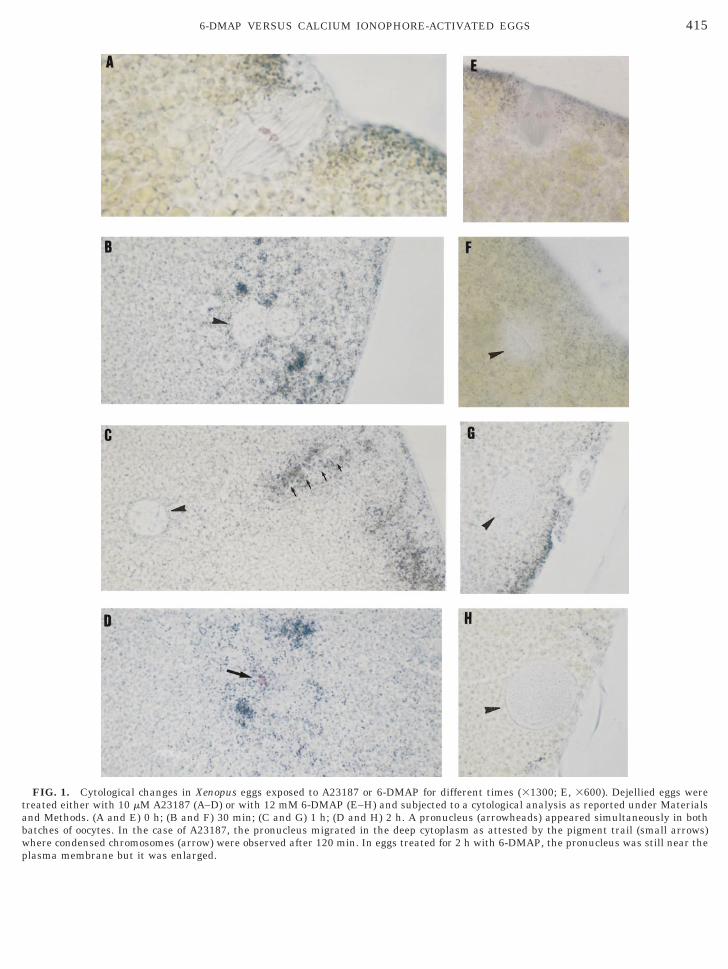
tabwp
4156-DMAP VERSUS CALCIUM IONOPHORE-ACTIVATED EGGS
FIG. 1. Cytological changes in Xenopus eggs exposed to A23187 or 6-DMAP for different times (31300; E, 3600). Dejellied eggs werereated either with 10 mM A23187 (A–D) or with 12 mM 6-DMAP (E–H) and subjected to a cytological analysis as reported under Materialsnd Methods. (A and E) 0 h; (B and F) 30 min; (C and G) 1 h; (D and H) 2 h. A pronucleus (arrowheads) appeared simultaneously in bothatches of oocytes. In the case of A23187, the pronucleus migrated in the deep cytoplasm as attested by the pigment trail (small arrows)here condensed chromosomes (arrow) were observed after 120 min. In eggs treated for 2 h with 6-DMAP, the pronucleus was still near the
lasma membrane but it was enlarged.
tpimcsa
M
aEafwstmt
FiTiftmlita
d(CCSp
m4re
aimaaim
dFci
bbTifo
AAA666
fiwCpdae
416 BODART ET AL.
ime as in the eggs treated with A23187 (Fig. 1F). Thisronucleus stayed in the cortical layer where its sizencreased from 15 mm after 30 min to 50 mm after 120
in (compare Fig. 1F with Fig. 1H). No chromatinondensation was observed in these eggs as demon-trated by the absence of red structures. The resultsre summarized in Table 1.
APK and p34cdc2 Analysis
We analyzed MAPK/Erk2 and p34cdc2 kinases whichre very active in unfertilized eggs. Two forms of therk2 kinase may be detected on Western blots: anctive phosphorylated form and an unphosphorylatedaster migrating inactive form [25]. The other kinasee examined, Cdc2, may also appear as two bands. The
low migrating band is inactive and phosphorylated onhree residues (Thr 14, Tyr 15, and Thr 161). The fastigrating band is either unphosphorylated and inac-
ive or monophosphorylated (Thr 161) and active [35].The results of this Western blot study are shown in
ig. 2. Untreated eggs contained only the slow migrat-ng form of Erk2 and the fast migrating form of Cdc2.he two Erk2 isoforms were present after 25 min of
onophore exposure, but only the dephosphorylatedorm was detected after 40 min. In the case of Cdc2, thewo isoforms were found after 30 min but the fastigrating form alone was present at 120 min. So, fol-
owing calcium ionophore treatment, changes in Erk2mmunodetection profile occurred at the same timehat those of Cdc2 which was the only one to changegain after 120 min.In eggs exposed to 6-DMAP, the changes in Erk2
etection were similar but they appeared more slowlybetween 30 and 50 min). The slower migrating form ofdc2 appeared as early as 20 min but no change in thedc2 immunodetection profile was observed thereafter.o, 6-DMAP induced earlier changes in the electro-horetic mobility of Cdc2.Then, the activities of Cdc2 and Erk2 kinases wereeasured using histone H1 and MBP, respectively, for
0 min following 6-DMAP or A23187 exposure. Theesults are reported in Fig. 3 (mean of three different
TAB
Pronucleus Formation Following
Treatment Total Spindle Cortical pronucleus
23187 10 mM 30 min 16 0 1323187 10 mM 60 min 17 0 623187 10 mM 120 min 21 1 0-DMAP 12 mM 30 min 16 0 14-DMAP 12 mM 60 min 14 0 12-DMAP 12 mM 120 min 15 0 15
xperiments). In eggs treated with A23187, H1 kinase p
ctivity decreased at a stage where changes in themmunodetection profile had not occurred: 50% after 5
in and 4% after 20 min after which the H1 kinasectivity remained low. MBP kinase activity was ingreement with the changes in Erk2 detection on themmunoblots: 82.7% after 20 min and 4.6% after 40
in.In eggs treated with 6-DMAP, H1 kinase activity
ecreased early, as suggested by the Western blot inig. 2: 11.7% after 20 min. MBP kinase activity de-reased more quickly than suggested by the changes ints electrophoretic mobility: 7% after 25 min.
We also measured H1 kinase after 120 min of incu-ation in A23187 since we suspected from the Westernlot data that there had been a reactivation of p34cdc2.he activity that we measured was 47.4% of the activ-
ty found in untreated eggs. In addition, we have per-ormed injections into immature oocytes of cytoplasmbtained from these A23187-treated eggs according to
1
3187 and 6-DMAP Treatments
ytoplasmic pronucleus Condensed chromosomes No structure
0 2 19 1 10 8 120 0 20 0 20 0 0
FIG. 2. Western blot study of p34cdc2 and Erk2 during the 2 h thatollow A23187 or 6-DMAP exposure. In eggs exposed to calciumonophore, a fast migrating form of Erk2 appeared after 25 minhich was the only form to be detected after 40 min. In the case ofdc2, a slow migrating form appeared after 30 min which disap-eared at 120 min. In eggs exposed to 6-DMAP, the changes in Erk2etection were similar but they appeared somehow later (between 30nd 60 min) whereas the slower migrating form of Cdc2 appearedarlier as soon as 20 min. No change in the Cdc2 immunodetection
LE
A2
C
rofile was observed thereafter.
tcofGamat
C
st
bmsisu
ftaglbm
usgap
thttidawts
a6ntda
Xtrbmlp
ta
4176-DMAP VERSUS CALCIUM IONOPHORE-ACTIVATED EGGS
he method previously described [10]. When these re-ipient oocytes were analyzed 4 h later, 29.2% (n 5 24)f them showed GVBD. Control experiments per-ormed with cytoplasm from untreated eggs inducedVBD in 80.3% (n 5 27) of the oocytes. MBP kinasectivity measurement after 120 min of A23187 treat-ent did not reveal the presence of active Erk2 kinase
t this stage (8.7% of maximal activity) as suggested byhe lack of electrophoretic mobility changes.
yclin B and p39mos Analysis
We next analyzed the levels of cyclin B since itsynthesis as well as its degradation can also regulatehe activity of p34cdc2.
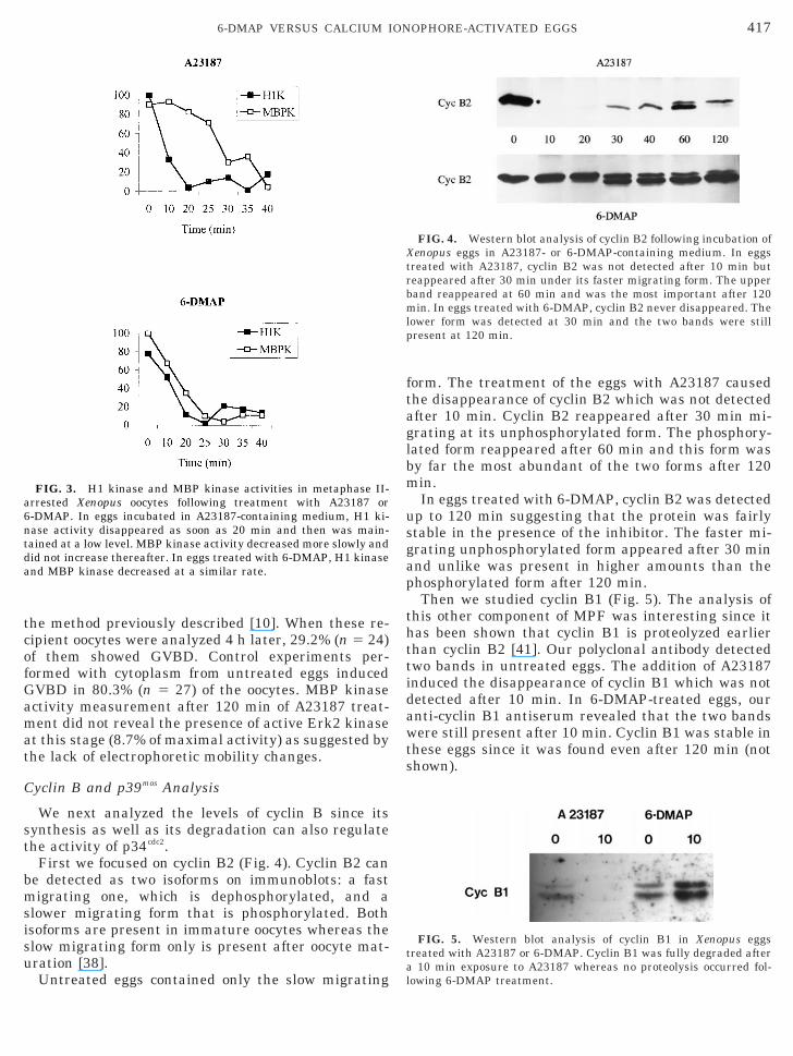
First we focused on cyclin B2 (Fig. 4). Cyclin B2 cane detected as two isoforms on immunoblots: a fastigrating one, which is dephosphorylated, and a
lower migrating form that is phosphorylated. Bothsoforms are present in immature oocytes whereas thelow migrating form only is present after oocyte mat-ration [38].
FIG. 3. H1 kinase and MBP kinase activities in metaphase II-rrested Xenopus oocytes following treatment with A23187 or-DMAP. In eggs incubated in A23187-containing medium, H1 ki-ase activity disappeared as soon as 20 min and then was main-ained at a low level. MBP kinase activity decreased more slowly andid not increase thereafter. In eggs treated with 6-DMAP, H1 kinasend MBP kinase decreased at a similar rate.
Untreated eggs contained only the slow migrating l
orm. The treatment of the eggs with A23187 causedhe disappearance of cyclin B2 which was not detectedfter 10 min. Cyclin B2 reappeared after 30 min mi-rating at its unphosphorylated form. The phosphory-ated form reappeared after 60 min and this form wasy far the most abundant of the two forms after 120in.In eggs treated with 6-DMAP, cyclin B2 was detected
p to 120 min suggesting that the protein was fairlytable in the presence of the inhibitor. The faster mi-rating unphosphorylated form appeared after 30 minnd unlike was present in higher amounts than thehosphorylated form after 120 min.Then we studied cyclin B1 (Fig. 5). The analysis of
his other component of MPF was interesting since itas been shown that cyclin B1 is proteolyzed earlierhan cyclin B2 [41]. Our polyclonal antibody detectedwo bands in untreated eggs. The addition of A23187nduced the disappearance of cyclin B1 which was notetected after 10 min. In 6-DMAP-treated eggs, ournti-cyclin B1 antiserum revealed that the two bandsere still present after 10 min. Cyclin B1 was stable in
hese eggs since it was found even after 120 min (nothown).
FIG. 4. Western blot analysis of cyclin B2 following incubation ofenopus eggs in A23187- or 6-DMAP-containing medium. In eggs
reated with A23187, cyclin B2 was not detected after 10 min buteappeared after 30 min under its faster migrating form. The upperand reappeared at 60 min and was the most important after 120in. In eggs treated with 6-DMAP, cyclin B2 never disappeared. The
ower form was detected at 30 min and the two bands were stillresent at 120 min.
FIG. 5. Western blot analysis of cyclin B1 in Xenopus eggsreated with A23187 or 6-DMAP. Cyclin B1 was fully degraded after
10 min exposure to A23187 whereas no proteolysis occurred fol-
owing 6-DMAP treatment.
il
stinaAt
lpwboiffo
P
sotAbcaAlu
spntmbkoc
psltMpivdedgcat
6tibIms4dsA
M
AffoC(
toadnbm
Xwa
418 BODART ET AL.
Therefore, the formation of the pronucleus that isnduced by 6-DMAP occurs even in the presence of highevels of cyclin B.
Then we studied the levels of p39mos which is synthe-ized during oocyte maturation in response to proges-erone, plays a pivotal role in metaphase II arrest, ands proteolyzed following fertilization or parthenoge-etic activation. We observed that p39mos was degradedfter 30 min of 6-DMAP exposure as in eggs exposed to23187 (Fig. 6). The protein did not reappear thereaf-
er.
DISCUSSION
In this study, we have characterized at the molecularevels the effects of 6-DMAP on Xenopus eggs in com-arison to those of the calcium ionophore A23187hich is well known to mimic fertilization. Such aiochemical analysis had not been performed previ-usly in Xenopus eggs. Our analysis showed that evenf A23187 and 6-DMAP induced identically pronucleusormation, the underlying molecular events were dif-erent. Besides, our results suggest that the proteolysisf cyclin B and p39mos are differentially regulated.
ronucleus Formation
Our cytological study revealed the disappearance ofpindle and condensed chromosomes and the formationf a pronucleus. The time required for this pronucleuso occur (30 min) was similar to that required with23187. Nevertheless we observed a major differenceetween these treatments at later stages: the pronu-leus migrated toward the center of the egg and wasccompanied by a trail of pigments in the case of23187 incubation whereas it stayed in the cortical
ayer when 6-DMAP was used. Such a migration occurs
FIG. 6. Western blot analysis of p39mos following incubation ofenopus eggs in A23187 or 6-DMAP-containing medium. When eggsere treated with A23187 or with 6-DMAP, p39mos was not detectedfter 40 min, and never reappeared, even after a 120 min treatment.
sually following egg fertilization and seems to be as- r
ociated with entry of the sperm. Indeed the maleronucleus is the first to migrate and the female pro-ucleus migrates later and appears to be dependent onhe male aster for its motion [36]. This absence ofigration observed with 6-DMAP might be explained
y the fact that the effects of this drug are mediated byinase inhibition rather than by calcium ions, as dem-nstrated by experiments using EGTA to chelate cal-ium ions [43].Another difference was that the pronucleus was re-
laced by a condensed material stained in red by ourtaining procedure. This red structure which lookedike condensed chromosomes was only detected in eggsreated with A23187. This result was due to a rise inPF activity since we observed an active form of
34cdc2 and a phosphorylated form of cyclin B2 on themmunoblots even if the H1 kinase activity was notery high at the same time. Although chromatin con-ensation occurred after 120 min in A23187-treatedggs, we observed neither cleavage furrows nor spin-les. This is consistent with the fact that in partheno-enetically activated eggs, no cleavage occurs unlessentriole containing fractions or isolated centrosomesre injected at the time of activation [39]. These struc-ures are brought by sperm at fertilization.
The fact that chromatin did not condense in-DMAP-treated eggs after 120 min is correlated withhe absence of p34cdc2 reactivation as shown by thenactive profile of p34cdc2 at this stage on the immuno-lots. This is due to the presence of the inhibitor.ndeed, when eggs are transferred in a 6-DMAP-freeedium after 6-DMAP treatment, chromatin conden-
ation is observed too [our unpublished observations;3]. So, p34cdc2 is reactivated after withdrawal of therug. It is interesting to note that the presence ofpindle is not reported in this case, as observed with23187.
PF and CSF Activities
Our biochemical analysis showed that even if23187 and 6-DMAP induced identically pronucleus
ormation, the underlying molecular events were dif-erent. Indeed, differences were observed in the studyf p34cdc2 and Erk2 kinases. Erk2 is a component ofSF activity which maintains a high level of MPF
p34cdc2–cyclin B) activity in unfertilized eggs [14].In A23187-treated eggs, the modification of the elec-
rophoretic mobility of Erk2 occurred just before thosef p34cdc2. Previous studies with anti-phosphotyrosinentibodies revealed tyrosine phosphorylation of MAPKuring the 25–30 min following fertilization or parthe-ogenetic activation whereas those of p34cdc2 occurredetween 30 and 70 min [8, 27]. Our results based onobility shift changes are in agreement with these
eports. What is the signification of these changes? The

icsHoaicmiymfet
clpdaesMsp
ktepkbwpot6
smisaEttpaltenmbs
aCi
C
Bc6e
towrcittXcmdieo(umBarCn
tttbuap
tpphpaopooa
4196-DMAP VERSUS CALCIUM IONOPHORE-ACTIVATED EGGS
ncrease in electrophoretic mobility of Erk2 was asso-iated to the inactivation of the enzyme as demon-trated by the results of MBP phosphorylation assays.owever, the changes in the immunodetection profile
f p34cdc2 occurred later than the drop in H1 kinasectivity which was observed as soon as 10 min after thencubation in A23187 containing medium. This is be-ause the tyrosine phosphorylation of p34cdc2 reflectsitotic pre-MPF formation rather than the meiotic
nactivation of the kinase. Indeed, tyrosine phosphor-lation of p34cdc2 has been observed during the firstitosis that begins between 60 and 75 min following
ertilization and is completed after 80 min, but thisvent was never observed during the next 11 cell cycleshat are shorter (30 min) [27].
The inactivation of MPF involves the degradation ofyclin B (regulatory subunit) and p34cdc2 dephosphory-ation on its Thr 161 residue [21, 28, 29]. This dephos-horylation of p34cdc2 did not induce mobility shift un-er our SDS–PAGE conditions. However, H1 kinasectivity measurement and cyclin B detection on West-rn blots demonstrate MPF inactivation. So, the re-ults we obtained with calcium ionophore showing thatPF activity decreases before MAPK activity are con-
istent with the fact that the drop in MPF activityrecedes those of CSF activity [41].In 6-DMAP-treated eggs, the decrease in the H1
inase activity was in agreement with the changes inhe electrophoretic mobility of p34cdc2 which occurredarlier than in A23187-treated eggs. Changes in p34cdc2
hosphorylation at Thr 14 and Tyr 15 are due to theinases wee1 and myt1 [6]. There is a feedback loopetween p34cdc2 and its activating phosphatase cdc25hich acts at these two residues [16, 37]. The rephos-horylation of p34cdc2 observed in our study and previ-usly reported in maturing oocytes [17] might be due tohe inhibition of this feedback loop in the presence of-DMAP.Surprisingly, Erk2 was dephosphorylated at the
ame time that in eggs incubated in A23187-containingedium. This dephosphorylation did not reflect the
nactivation of the enzyme since kinase activity mea-urement showed an earlier inhibition probably due to
direct inhibition of the kinase by 6-DMAP. Sincerk2 is dephosphorylated approximately at the same
ime (around 30 min) in both A23187 and 6-DMAP-reated eggs and since we observed no difference in39mos degradation in both treatments (faint detectiont 30 min and then no detection), Erk2 dephosphory-ation might be simply dependent on p39mos degrada-ion. The dephosphorylation of MAPK might also bexplained by the fact that 6-DMAP inhibits other ki-ases upstream in the cascade. Another explanationight be associated to the existence of a feedback loop
etween MAPK and its upregulating enzymes. For in-
tance, MAPK has been shown to phosphorylate p39mos et Ser 3 in vitro and to stimulate p39mos synthesis [24].onsequently, the inhibition of MAPK could modify
ndirectly the activity of its upregulating kinases.
yclin B and p39mos Proteolysis
In contrast to the ready proteolysis of cyclins B1 and2 in eggs treated with A23187, we observed that theseyclins were not degraded in presence of 12 mM-DMAP, even after a 2-h incubation. Why do theseggs not carry out the proteolysis of cyclin B2?One explanation for the absence of cyclin B degrada-
ion might be the lack of calcium release. However, webserved that A23187 addition following 1-h treatmentith 12 mM 6-DMAP did not enhance cyclin B2 deg-
adation (not shown). So, rather than the absence ofalcium release, another effect of 6-DMAP seems tonhibit cyclin B proteolysis. We propose that it is due tohe inhibition of MPF. Indeed, it has been reportedhat when 12 U ml21 of MPF was added to interphaseenopus egg extracts, methionine-labeled sea urchinyclin was rapidly destroyed [7]. In the presence of 1M 6-DMAP in the extracts, sea urchin cyclin degra-
ation was substantially inhibited. 6-DMAP also inhib-ted cyclin degradation in a cell-free system from clammbryos [23]. In addition, biochemical characterizationf the anaphase-promoting complex (APC)/cyclosomea multimeric 20S complex corresponding to an ubiq-itin ligase E3) suggests that the activation of the APCay depend on multiple kinases, including Cdc2/cyclin[19]. The mechanism by which Cdc2/cyclin B triggers
ctivation of the APC remains unclear but seems to beegulated by Xe-p9, a protein belonging to the Suc1/ks family, as demonstrated by experiments using Xe-opus egg extracts [33].Although cyclin B was not proteolysed, we observed
hat p39mos was degraded after 30 min of 6-DMAPreatment that is similar to what occurs in A23187-reated eggs. This result was very surprising becauseoth proteins are thought to be degraded by the ubiq-itin pathway and calcium ions seem to be able toctivate the proteolysis of both proteins [41]. Why39mos is proteolysed in these eggs?Since MPF is inhibited by 6-DMAP treatment and
hat p39mos is proteolyzed in this case, one might pro-ose that active p34cdc2/cyclin B could prevent p39mos
roteolysis. Another explanation might be that the in-ibition by 6-DMAP of a kinase that phosphorylates39mos could allow the dephosphorylation of p39mos byn appropriate phosphatase. Indeed, it has been dem-nstrated that degradation of p39mos required its de-hosphorylation at Ser 3 [32]. However, the proteolysisf p39mos, even in a dephosphorylated state, can occurnly if the ubiquitin degradation machinery has beenctivated. The activation of APC in 6-DMAP-treated
ggs might be obtained by the inhibition of a kinase
taapot
owtNbtfra
6a[gb
atOpra
mCgcgE“F
1
1
1
1
1
1
1
1
1
1
2
2
2
420 BODART ET AL.
hat could allow the dephosphorylation by an appropri-te phosphatase of an inhibitor of APC leading to thectivation of the degradation machinery [40]. This ex-lanation is in agreement with the fact that inhibitorsf tyrosine phosphatases block calcium-induced activa-ion of Xenopus eggs [4].
Differences exist between cyclin B and p39mos prote-lysis. Cyclin B degradation requires a destruction boxhich is necessary for the ubiquitination of cyclin B by
he APC [12, 19]. p39mos proteolysis is mediated by the-terminal Pro2-dependent ubiquitin pathway [32]ut the ubiquitin protein ligase has not been yet iden-ified. The involvement of an ubiquitin ligase distinctrom APC and differently regulated could explain ouresults showing a differential proteolysis of cyclin Bnd p39mos.One might also propose that in Xenopus eggs,
-DMAP induces an increase in intracellular calciums has been shown in oocytes from rhesus monkeys42]. This calcium increase might be sufficient to trig-er p39mos degradation whereas MPF inhibition couldlock cyclin B proteolysis.In conclusion, our study shows that even if A23187
nd 6-DMAP induced identically pronucleus forma-ion, the underlying molecular events were different.ur results suggest that the proteolysis of cyclin B and39mos is differentially regulated. Experiments are cur-ently being performed to elucidate the reason for suchdiscrepancy in the degradation of p39mos and cyclin B.
We thank Dr. Laurent Meijer for helping us in kinase activityeasurement and cyclin B immunodetection as well as Dr. Franckhesnel for his suggestions about ERK 2 detection. We are alsorateful to both of them and to doctor Eve Damiens for their helpfulomments on an early manuscript. These studies were supported byrants from the French “Ministere de l’Education Nationale” (DRED,A No. 1033), the “Association pour la Recherche sur le Cancer,” the
Region Nord–Pas-de-Calais” (Centre de Biologie Cellulaire), and theEDER.
REFERENCES
1. Abrieu, A., Lorca, T., Labbe, J.-C., Morin, N., Keyse, S., andDoree, M. (1996). MAP kinase does not inactivate, but ratherprevents the cyclin degradation pathway from being turned onin Xenopus egg extracts. J. Cell Sci. 109, 239–246.
2. Azzi, L., Meijer, L., Ostvold, A. C., Lew, J., and Wang, J. H.(1994). Purification of a 15 kDa cdk4- and cdk5-binding protein.J. Biol. Chem. 269(6), 13279–13288.
3. Berger, F. (1992). Mechanisms of initiation and propagation ofthe calcium wave during fertilization in deuterostomes. Int. J.Dev. Biol. 36, 245–262.
4. Bodart, J.-F., Bechard, D., Bertout, M., Gannon, J., Rousseau,A., Vilain, J.-P., and Flament, S. (1999). Inhibition of proteintyrosine phosphatases blocks calcium-induced activation ofmetaphase II-arrested oocytes of Xenopus laevis. FEBS Lett.457(2), 175–178.
5. Chesnel, F., Bonnec, G., Tardivel, A., and Boujard, D. (1997).
Comparative effects of insulin on the activation of the Raf/mos-dependent MAP kinase cascade in vitellogenic versus postvitel-logenic Xenopus oocytes. Dev. Biol. 188, 122–133.
6. Coleman, T. R., and Dunphy, W. G. (1994). Cdc2 regulatoryfactors. Curr. Opin. Cell Biol. 6, 877–882.
7. Felix, M.-A., Labbe, J.-C., Doree, M., Hunt, T., and Karsenti, E.(1990). Triggering of cyclin degradation in interphase extractsof amphibian eggs by cdc2 kinase. Nature 346, 379–382.
8. Ferrell, J. E., Wu, M., Gerhart, J. C., and Martin, G. S. (1991).Cell cycle tyrosine phosphorylation of p34cdc2 and a microtu-bule-associated protein kinase homolog in Xenopus oocytes andeggs. Mol. Cell. Biol. 11(4), 1965–1971.
9. Flament, S., Browaeys, E., Rodeau, J. L., Bertout, M., andVilain, J. P. (1996). Xenopus oocyte maturation: Cytoplasmalkalization is involved in germinal vesicle migration. Int. J.Dev. Biol. 40, 471–476.
0. Flament, S., Bodart, J.-F., Browaeys, E., Bertout, M., Rousseau,A., Gannon, J., and Vilain, J.-P. (1997). Procaine-induced mat-uration of Xenopus oocytes is mediated by a transient activationof M-phase promoting factor. Zygote 5, 11–19.
1. Gerhart, J., Wu, M., and Kirschner, M. (1984). Cell cycle dy-namics of an M-phase-specific cytoplasmic factor in Xenopuslaevis oocytes and eggs. J. Cell Biol. 98, 1247–1255.
2. Glotzer, M., Murray, A. W., and Kirschner, M. W. (1991). Cyclinis degraded by the ubiquitin pathway. Nature 349, 132–138.
3. Goodger, N., Gannon, J., Hunt, T., and Morgan, P. R. (1996). Onthe abundance and localization of p34cdc2 in the cells of nor-mal, hyperplasic and malignant human lymphoid and epithe-lial tissues. J. Pathol. 178, 422–428.
4. Haccard, O., Sarcevic, B., Lewellyn, A., Hartley, R., Roy, L.,Izumi, T., Erikson, E., and Maller, J. L. (1993). Induction ofmetaphase arrest in cleaving Xenopus embryos by MAP kinase.Science 262, 1262–1265.
5. Hershko, A. (1991). Methylated ubiquitin inhibits cyclin degra-dation in clam embryo extracts. J. Biol. Chem. 266(25), 16376–16379.
6. Hoffmann, I., Clarke, P. R., Marcote, M. J., Karsenti, E., andDraetta, G. (1993). Phosphorylation and activation of humancdc25-C by cdc2-cyclin B and its involvement in the self-ampli-fication of MPF at mitosis. EMBO J. 12, 53–63.
7. Jessus, C., Rime, H., Haccard, O., Van Lint, J., Goris, J., Mer-levede, W., and Ozon, R. (1991). Tyrosine phosphorylation ofp34cdc2 and p42 during meiotic maturation of Xenopus oocyte:Antagonistic action of okadaic acid and 6-DMAP. Development111, 813–820.
8. King, R. W., Jackson, P. K., and Kirschner, M. W. (1994).Mitosis in transition. Cell 79, 563–571.
9. King, R. W., Peters, J. M., Tugendreich, S., Rolfe, M., Hieter, P.,and Kirschner, M. W. (1995). A 20S complex containing CDC27and CDC16 catalyzes the mitosis-specific conjugaison of ubiq-uitin to cyclin B. Cell 81, 279–288.
0. Kishimoto, T. (1994). Cell reproduction: Induction of M-phaseevents by cyclin-dependent cdc2 kinase. Int. J. Dev. Biol. 38,185–191.
1. Lorca, T., Labbe, J.-C., Devault, A., Fesquet, D., Capony, J.-P.,Cavadore, J.-C., Le Bouffant, F., and Doree, M. (1992). Dephos-phorylation of cdc2 on threonine 161 is required for cdc2 kinaseinactivation and normal anaphase. EMBO J. 11(7), 2381–2390.
2. Lorca, T., Cruzalegui, F. H., Fesquet, D., Cavadore, J.-C., Mery,J., Means, A., and Doree, M. (1993). Calmodulin-dependentkinase II mediates inactivation of MPF and CSF upon fertili-
zation of Xenopus eggs. Nature 366, 270–273.
2
2
2
2
2
2
2
3
3
3
3
3
3
3
3
3
3
4
4
4
4
RR
4216-DMAP VERSUS CALCIUM IONOPHORE-ACTIVATED EGGS
3. Luca, F., and Ruderman, J. (1989). Control of programmedcyclin destruction in a cell-free system. J. Cell Biol. 109, 1895–1909.
4. Matten, W. T., Copeland, T. D., Ahn, N. G., and Vande Woude,G. F. (1996). Positive feedback between MAP kinase and mosduring Xenopus oocyte maturation. Dev. Biol. 179, 485–492.
5. Mordret, G. (1993). MAP kinase kinase: A node connectingmultiple pathways. Biol. Cell 79, 193–207.
6. Morin, N., Abrieu, A., Lorca, T., Martin, F., and Doree, M.(1994). The proteolysis-dependent metaphase to anaphase tran-sition: Calcium/calmodulin-dependent kinase II mediates onsetof anaphase in extracts prepared from unfertilized Xenopuseggs. EMBO J. 13(18), 4343–4352.
7. Murakami, M., and Vande Woude, G. F. (1998). Analysis of theearly embryonic cell cycles of Xenopus: Regulation of cell cyclelength by Xe-wee1 and mos. Development 125, 237–248.
8. Murray, A. W., and Kirschner, M. W. (1989). Cyclin synthesisdrives the early embryonic cell cycle. Nature 339, 275–280.
9. Murray, A. W., Solomon, M. J., and Kirschner, M. W. (1989).The role of cyclin synthesis and degradation in the control ofmaturation promoting factor activity. Nature 339, 280–286.
0. Neant, I., and Guerrier, P. (1988). 6-Dimethylaminopurine-blocks starfish oocyte maturation by inhibiting a relevant pro-tein kinase activity. Exp. Cell Res. 176(1), 68–79.
1. Newport, J., and Kirschner, M. W. (1982). A major developmen-tal transition in early Xenopus embryos. I. characterization andtiming of cellular changes at the midblastula stage. Cell 30(3),675–686.
2. Nishizawa, M., Furuno, N., Okazaki, K., Tanaka, H., Ogawa,Y., and Sagata, N. (1993). Degradation of Mos by the N-termi-nal proline (Pro2)-dependent ubiquitin pathway on fertilizationof Xenopus eggs: Possible significance of natural selection forPro2 in Mos. EMBO J. 12(10), 4021–4027.
3. Patra, D., and Dunphy, W. G. (1998). Xe-p9, a Xenopus Suc1/Cks protein, is essential for the Cdc2-dependent phosphoryla-tion of the anaphase-promoting complex at mitosis. Genes Dev.
12, 2549–2559.4. Sagata, N., Watanabe, N., Vande Woude, G. F., and Ikawa, Y.(1989). The c-mos proto-oncogene product is a cytostatic factorresponsible for meiotic arrest in vertebrates eggs. Nature 342,512–518.
5. Solomon, M. J. (1993). Activation of the various cyclin/cdc2protein kinases. Curr. Opin. Cell Biol. 5, 180–186.
6. Stewart-Savage, J., and Grey, R. D. (1982). The temporal andspatial relationships between cortical contraction, sperm trailformation, and pronuclear migration in fertilized Xenopus eggs.Wilhelm Roux’s Arch. 191, 241–245.
7. Strausfeld, U. P., Fernandez, A., Capony, J.-P., Girard, F.,Lautredou, N., Derancourt, J., Labbe, J.-C., and Lamb, N. J.(1994). Activation of p34cdc2 protein kinase by microinjection ofhuman Cdc25C into mammalian cells: Requirement for priorphosphorylation of Cdc25C by p34cdc2 on sites phosphorylated atmitosis. J. Biol. Chem. 269(8), 5989–6000.
8. Taieb, F., Thibier, C., and Jessus, C. (1997). On cyclins, oocytes,and eggs. Mol. Reprod. Dev. 48, 397–411.
9. Tournier, F., Joly, A., and Bornens, M. (1994). Parthenogenesicproduction of partial blastulas in Xenopus. C. R. Acad. Sci. 317,405–410.
0. Vorlaufer, E., and Peters, J. M. (1998). Regulation of the cyclinB degradation system by an inhibitor of mitotic proteolysis.Mol. Biol. Cell 9, 1817–1831.
1. Watanabe, N., Hunt, T., Ikawa, Y., and Sagata, N. (1991).Independent inactivation of MPF and cytostatic factor (Mos)upon fertilization of Xenopus eggs. Nature 352, 247–248.
2. Wu, G. J., Simerly, C., Zoran, S. S., Funte, L. R., and Schatten,G. (1996). Microtubule and chromatin dynamics during fertili-zation and early development in rhesus monkeys, and regula-tion by intracellular calcium ions. Biol. Reprod. 55, 260–270.
3. Zhang, S. C., and Masui, Y. (1992). Activation of Xenopus laeviseggs in the absence of intracellular Ca activity by the proteinphosphorylation inhibitor, 6-dimethylaminopurine (6-DMAP).
J. Exp. Zool. 263, 317–329.eceived May 25, 1999evised version received August 20, 1999
Copyright © 2022 FDOKUMEN