commercial short holiday breaks in scotland : an analysis of ...
Upload
independentCategory
view
1download
0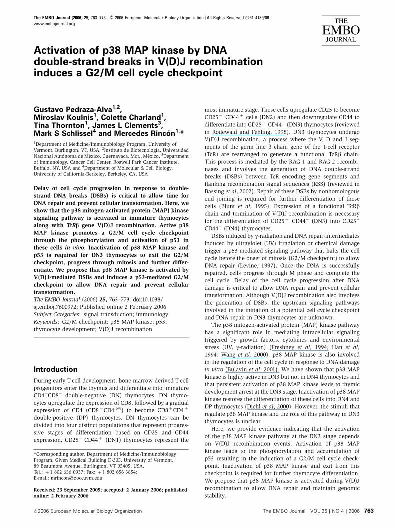
Activation of p38 MAP kinase by DNAdouble-strand breaks in V(D)J recombinationinduces a G2/M cell cycle checkpoint
Gustavo Pedraza-Alva1,2,Miroslav Koulnis1, Colette Charland1,Tina Thornton1, James L Clements3,Mark S Schlissel4 and Mercedes Rincon1,*1Department of Medicine/Immunobiology Program, University ofVermont, Burlington, VT, USA, 2Instituto de Biotecnologıa, UniversidadNacional Autonoma de Mexico. Cuernavaca, Mor., Mexico, 3Departmentof Immunology, Cancer Cell Center, Roswell Park Cancer Institute,Buffalo, NY, USA and 4Department of Molecular & Cell Biology,University of California-Berkeley, Berkeley, CA, USA
Delay of cell cycle progression in response to double-
strand DNA breaks (DSBs) is critical to allow time for
DNA repair and prevent cellular transformation. Here, we
show that the p38 mitogen-activated protein (MAP) kinase
signaling pathway is activated in immature thymocytes
along with TcRb gene V(D)J recombination. Active p38
MAP kinase promotes a G2/M cell cycle checkpoint
through the phosphorylation and activation of p53 in
these cells in vivo. Inactivation of p38 MAP kinase and
p53 is required for DN3 thymocytes to exit the G2/M
checkpoint, progress through mitosis and further differ-
entiate. We propose that p38 MAP kinase is activated by
V(D)J-mediated DSBs and induces a p53-mediated G2/M
checkpoint to allow DNA repair and prevent cellular
transformation.
The EMBO Journal (2006) 25, 763–773. doi:10.1038/
sj.emboj.7600972; Published online 2 February 2006
Subject Categories: signal transduction; immunology
Keywords: G2/M checkpoint; p38 MAP kinase; p53;
thymocyte development; V(D)J recombination
Introduction
During early T-cell development, bone marrow-derived T-cell
progenitors enter the thymus and differentiate into immature
CD4�CD8� double-negative (DN) thymocytes. DN thymo-
cytes upregulate the expression of CD8, followed by a gradual
expression of CD4 (CD8þCD4low) to become CD8þCD4þ
double-positive (DP) thymocytes. DN thymocytes can be
divided into four distinct populations that represent progres-
sive stages of differentiation based on CD25 and CD44
expression. CD25� CD44þ (DN1) thymocytes represent the
most immature stage. These cells upregulate CD25 to become
CD25þ CD44þ cells (DN2) and then downregulate CD44 to
differentiate into CD25þ CD44� (DN3) thymocytes (reviewed
in Rodewald and Fehling, 1998). DN3 thymocytes undergo
V(D)J recombination, a process where the V, D and J seg-
ments of the germ line b chain gene of the T-cell receptor
(TcR) are rearranged to generate a functional TcRb chain.
This process is mediated by the RAG-1 and RAG-2 recombi-
nases and involves the generation of DNA double-strand
breaks (DSBs) between TcR encoding gene segments and
flanking recombination signal sequences (RSS) (reviewed in
Bassing et al, 2002). Repair of these DSBs by nonhomologous
end joining is required for further differentiation of these
cells (Blunt et al, 1995). Expression of a functional TcRbchain and termination of V(D)J recombination is necessary
for the differentiation of CD25þ CD44� (DN3) into CD25�
CD44� (DN4) thymocytes.
DSBs induced by g-radiation and DNA repair-intermediates
induced by ultraviolet (UV) irradiation or chemical damage
trigger a p53-mediated signaling pathway that halts the cell
cycle before the onset of mitosis (G2/M checkpoint) to allow
DNA repair (Levine, 1997). Once the DNA is successfully
repaired, cells progress through M phase and complete the
cell cycle. Delay of the cell cycle progression after DNA
damage is critical to allow DNA repair and prevent cellular
transformation. Although V(D)J recombination also involves
the generation of DSBs, the upstream signaling pathways
involved in the initiation of a potential cell cycle checkpoint
and DNA repair in DN3 thymocytes are unknown.
The p38 mitogen-activated protein (MAP) kinase pathway
has a significant role in mediating intracellular signaling
triggered by growth factors, cytokines and environmental
stress (UV, g-radiation) (Freshney et al, 1994; Han et al,
1994; Wang et al, 2000). p38 MAP kinase is also involved
in the regulation of the cell cycle in response to DNA damage
in vitro (Bulavin et al, 2001). We have shown that p38 MAP
kinase is highly active in DN3 but not in DN4 thymocytes and
that persistent activation of p38 MAP kinase leads to thymic
development arrest at the DN3 stage. Inactivation of p38 MAP
kinase restores the differentiation of these cells into DN4 and
DP thymocytes (Diehl et al, 2000). However, the stimuli that
regulate p38 MAP kinase and the role of this pathway in DN3
thymocytes is unclear.
Here, we provide evidence indicating that the activation
of the p38 MAP kinase pathway at the DN3 stage depends
on V(D)J recombination events. Activation of p38 MAP
kinase leads to the phosphorylation and accumulation of
p53 resulting in the induction of a G2/M cell cycle check-
point. Inactivation of p38 MAP kinase and exit from this
checkpoint is required for further thymocyte differentiation.
We propose that p38 MAP kinase is activated during V(D)J
recombination to allow DNA repair and maintain genomic
stability.Received: 23 September 2005; accepted: 2 January 2006; publishedonline: 2 February 2006
*Corresponding author. Department of Medicine/ImmunobiologyProgram, Given Medical Building D-305, University of Vermont,89 Beaumont Avenue, Burlington, VT 05405, USA.Tel.: þ 1 802 656 0937; Fax: þ 1 802 656 3854;E-mail: [email protected]
The EMBO Journal (2006) 25, 763–773 | & 2006 European Molecular Biology Organization | All Rights Reserved 0261-4189/06
www.embojournal.org
&2006 European Molecular Biology Organization The EMBO Journal VOL 25 | NO 4 | 2006
EMBO
THE
EMBOJOURNAL
THE
EMBOJOURNAL
763

Results
In vivo activation of the p38 MAP kinase induces p53
phosphorylation and accumulation in immature
thymocytes
We have previously shown that persistent activation of p38
MAP kinase in transgenic (Tg) mice expressing a constitutive
active MKK6 mutant (upstream activator of p38 MAP kinase)
blocks thymocyte development at the DN3 stage (Diehl et al,
2000) similar to Rag1 or Rag2 deficiencies (Mombaerts
et al, 1992). Rag1�/� thymocytes are small and arrested in
G0/G1 phase of cell cycle. In contrast, DN3 thymocytes from
MKK6(Glu) Tg mice are large and display a phenotype
characteristic of early promitotic cells (Figure 1A). Despite
their phenotype, MKK6(Glu) thymocytes do not proliferate
in vivo but the presence of a p38 MAP kinase inhibitor
(SB203580) restores proliferation in vitro (Diehl et al,
2000), indicating that inactivation of p38 MAP kinase must
occur for DN3 thymocytes to progress through cell cycle.
It has been shown that activation of p38 MAP kinase
arrests cell cycle at the G2/M checkpoint by phosphorylation
and stabilization of p53 in response to DNA damage in UV-
irradiated fibroblasts (Bulavin et al, 1999; She et al, 2000). To
test whether activation of p38 MAP kinase in thymocytes
leads to the accumulation of p53 in vivo, we examined p53
levels in thymocytes from MKK6(Glu) Tg mice by Western
blot. High levels of p53 were present in MKK6(Glu) thymo-
cytes (Figure 1B). The accumulation of p53 in MKK6(Glu)
thymocytes required p38 MAP kinase activity, as treatment
of these cells in vitro with the selective p38 MAP kinase
inhibitor SB203580 caused a rapid reduction of the p53
protein levels (Figure 1C). In addition, p53 was phosphory-
lated at Ser18 and Ser389 (human Ser15 and Ser392, respec-
tively) in the MKK6(Glu) thymocytes, and the levels of
phosphorylated p53 were diminished in the presence of the
p38 MAP kinase inhibitor (Figure 1D). ATM and Chk1 can
also phosphorylate and activate p53 in response to DNA
damage (reviewed by Caspari, 2000) but no activation of
ATM and Chk1 was detected in MKK6(Glu) thymocytes
(Supplementary Figure S1). In correlation, no significant
phosphorylation of p53 at Ser23 (human Ser20), a Chk1 target
residue (Shieh et al, 1999), was observed in these thymo-
cytes (Supplementary Figure S1). Thus, activation of p38
MAP kinase in vivo induces phosphorylation and accumula-
tion of p53 in DN3 thymocytes.
p38 MAP kinase activation promotes a G2/M cell cycle
checkpoint in DN3 thymocytes
Cell division relies on the expression of cyclins that bind
and activate cyclin-dependent kinases to promote cell cycle
progression towards S phase and later to initiate mitosis.
Association of cyclin B with the cyclin-dependent kinase
Cdc2 induces Cdc2 kinase activity to promote progression
through mitosis (Morgan, 1995). However, during a G2/M
cell cycle checkpoint, cyclin B associates with inactive Cdc2
and these complexes accumulate in the cell, delaying the
progression through mitosis (Poon et al, 1996). To investigate
whether activation of p53 by p38 MAP kinase leads to a G2/M
cell cycle checkpoint in thymocytes from MKK6(Glu) Tg
mice, we examined cyclin B protein levels by Western blot.
Despite the high rate of proliferation of DP thymocytes, very
low levels of cyclin B were present in total wild type (WT)
thymocytes, but higher levels of cyclin B were present in
MKK6(Glu) thymocytes (Figure 2A). In addition, the levels of
cyclin B in MKK6(Glu) thymocytes were higher than those
in WT DN3 thymocytes (Figure 2B). Thus, activation of p38
MAP kinase leads to the accumulation of cyclin B. To
determine whether cyclin B was associated with inactive
Cdc2 in these cells, we examined phosphorylation of Cdc2
at Tyr15 as phosphorylation of this residue is responsible for
inactivation of Cdc2 at the G2/M cell cycle checkpoint
(Booher et al, 1997). Cyclin B was immunoprecipitated
from whole-cell extracts and the presence of phosphorylated
Cdc2 in cyclin B immunoprecipitates was determined by
Western blot. Cyclin B from MKK6(Glu) thymocytes was
associated with Cdc2 kinase phosphorylated at Tyr15
(Figure 2C), indicating that Cdc2 kinase is inactive in these
thymocytes. Moreover, phosphorylation of cyclin B-asso-
ciated Cdc2 kinase was reduced when MKK6(Glu) cells
were treated with SB203580 (Figure 2C). To show that phos-
phorylation of Cdc2 at Tyr15 blocks its kinase activity, cyclin B
was immunoprecipitated from freshly isolated MKK6(Glu)
thymocytes or from MKK6(Glu) thymocytes cultured for the
indicated periods of time in the presence of SB203580 and
Cdc2 kinase activity was determined in vitro using histone H1
as substrate. No Cdc2 kinase activity was present in cyclin B
precipitates from freshly isolated MKK6(Glu) thymocytes
(Figure 2D), but SB203580 restored cyclin B-associated Cdc2
kinase activity (Figure 2D). Analysis of phospho-Cdc2 showed
that the presence of a few dephosphorylated (i.e. activated)
Cdc2 molecules (phospho-Cdc2/total Cdc2 ratio) was suffi-
cient to mediate phosphorylation of a substantial number
of H1 molecules (Figure 2D). These results indicate that p38
MAP kinase promotes the accumulation of inactive cyclin
B–Cdc2 complexes to induce a G2/M cell cycle checkpoint.
The establishment of a G2/M cell cycle checkpoint by
p53 is mediated by inducing transcription of genes encod-
ing proteins such as GADD45, p21 and 14.3.3s that directly
or indirectly inactivate Cdc2 kinase (el-Deiry et al, 1993;
Hermeking et al, 1997; Zhan et al, 1999). To test whether
the induction of the G2/M checkpoint in the MKK6(Glu)
Actin
WT Tg
SB––p53
CB DWT Tg
p53
Actin
WT
SB
Tg
––
p-p53 (Ser389)
p-p53 (Ser18)
Actin
A Rag–/– MKK6(Glu) MKK6 + Rag–/–
Figure 1 p38 MAP kinase activation induces p53 accumulation.(A) Freshly isolated MKK6(Glu), Rag1�/� thymocytes, and a mix ofboth cell types (1:1) were spun, fixed, stained with Giemsa andexamined under light microscopy (� 60). (B) p53 levels weredetermined in whole-cell extracts prepared from freshly isolatedthymocytes from wild type (WT) or MKK6(Glu) transgenic (Tg)mice by Western blot. Thymocytes from WT or MKK6(Glu) (Tg)mice were cultured for 4 h in medium alone (�) or mediumcontaining the p38 MAP kinase inhibitor SB203580 (SB) (5 mM).Whole-cell extracts were prepared and total p53 protein (C) ofphosphorylated p53 (p-p53) on Ser18 or Ser389 and actin (D) levelswere determined by Western blot. Data are representative of threeto four independent experiments.
V(D)J DSBs regulate p38 MAPK and G2/M checkpointG Pedraza-Alva et al
The EMBO Journal VOL 25 | NO 4 | 2006 &2006 European Molecular Biology Organization764
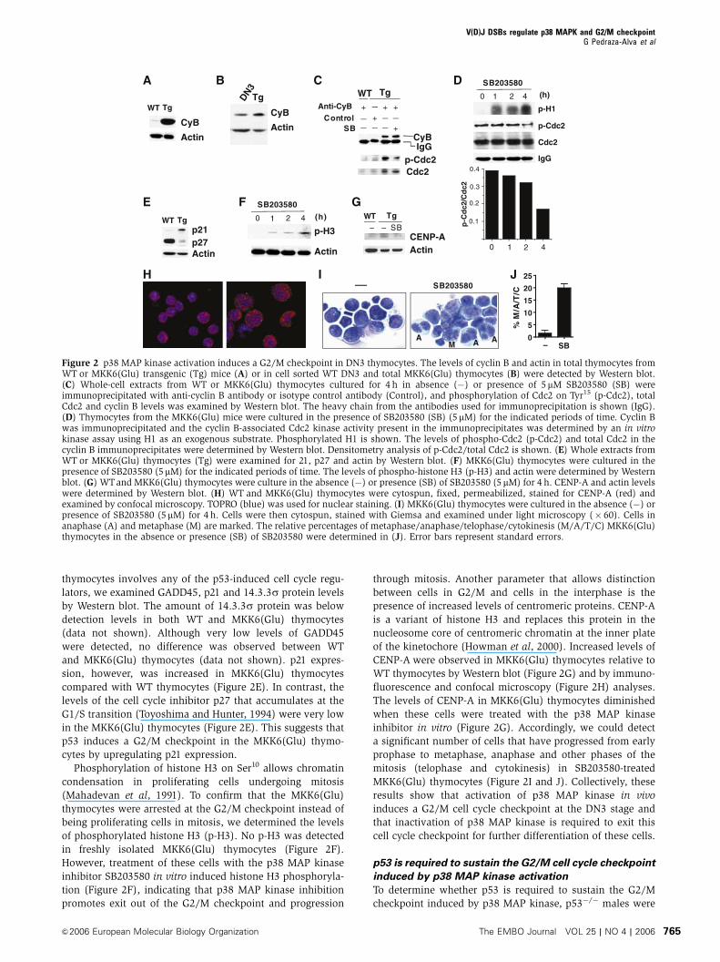
thymocytes involves any of the p53-induced cell cycle regu-
lators, we examined GADD45, p21 and 14.3.3s protein levels
by Western blot. The amount of 14.3.3s protein was below
detection levels in both WT and MKK6(Glu) thymocytes
(data not shown). Although very low levels of GADD45
were detected, no difference was observed between WT
and MKK6(Glu) thymocytes (data not shown). p21 expres-
sion, however, was increased in MKK6(Glu) thymocytes
compared with WT thymocytes (Figure 2E). In contrast, the
levels of the cell cycle inhibitor p27 that accumulates at the
G1/S transition (Toyoshima and Hunter, 1994) were very low
in the MKK6(Glu) thymocytes (Figure 2E). This suggests that
p53 induces a G2/M checkpoint in the MKK6(Glu) thymo-
cytes by upregulating p21 expression.
Phosphorylation of histone H3 on Ser10 allows chromatin
condensation in proliferating cells undergoing mitosis
(Mahadevan et al, 1991). To confirm that the MKK6(Glu)
thymocytes were arrested at the G2/M checkpoint instead of
being proliferating cells in mitosis, we determined the levels
of phosphorylated histone H3 (p-H3). No p-H3 was detected
in freshly isolated MKK6(Glu) thymocytes (Figure 2F).
However, treatment of these cells with the p38 MAP kinase
inhibitor SB203580 in vitro induced histone H3 phosphoryla-
tion (Figure 2F), indicating that p38 MAP kinase inhibition
promotes exit out of the G2/M checkpoint and progression
through mitosis. Another parameter that allows distinction
between cells in G2/M and cells in the interphase is the
presence of increased levels of centromeric proteins. CENP-A
is a variant of histone H3 and replaces this protein in the
nucleosome core of centromeric chromatin at the inner plate
of the kinetochore (Howman et al, 2000). Increased levels of
CENP-A were observed in MKK6(Glu) thymocytes relative to
WT thymocytes by Western blot (Figure 2G) and by immuno-
fluorescence and confocal microscopy (Figure 2H) analyses.
The levels of CENP-A in MKK6(Glu) thymocytes diminished
when these cells were treated with the p38 MAP kinase
inhibitor in vitro (Figure 2G). Accordingly, we could detect
a significant number of cells that have progressed from early
prophase to metaphase, anaphase and other phases of the
mitosis (telophase and cytokinesis) in SB203580-treated
MKK6(Glu) thymocytes (Figure 2I and J). Collectively, these
results show that activation of p38 MAP kinase in vivo
induces a G2/M cell cycle checkpoint at the DN3 stage and
that inactivation of p38 MAP kinase is required to exit this
cell cycle checkpoint for further differentiation of these cells.
p53 is required to sustain the G2/M cell cycle checkpoint
induced by p38 MAP kinase activation
To determine whether p53 is required to sustain the G2/M
checkpoint induced by p38 MAP kinase, p53�/� males were
CyB
Actin
WT Tg
A B
CyB
Actin
DN3
Tg
C
p-Cdc2
CyBIgG
WT Tg
SB
Anti-CyB +Control +
+ +
+
Cdc2
D
p-H1
p-Cdc2
0 1 2 4
SB203580
(h)
IgG
Cdc2
0 1 2 4
0.4
0.3
0.2
0.1p-C
dc2
/Cd
c2
Actin
p21WT Tg
p27
E0 1 2 4
SB203580
(h)
Actin
p-H3
F G
SB203580
AA A
M
WTSB
Tg
––CENP-AActin
H I
0
5
10
15
20
25
– SB
%M
/A/T
/C
J
Figure 2 p38 MAP kinase activation induces a G2/M checkpoint in DN3 thymocytes. The levels of cyclin B and actin in total thymocytes fromWT or MKK6(Glu) transgenic (Tg) mice (A) or in cell sorted WT DN3 and total MKK6(Glu) thymocytes (B) were detected by Western blot.(C) Whole-cell extracts from WT or MKK6(Glu) thymocytes cultured for 4 h in absence (�) or presence of 5mM SB203580 (SB) wereimmunoprecipitated with anti-cyclin B antibody or isotype control antibody (Control), and phosphorylation of Cdc2 on Tyr15 (p-Cdc2), totalCdc2 and cyclin B levels was examined by Western blot. The heavy chain from the antibodies used for immunoprecipitation is shown (IgG).(D) Thymocytes from the MKK6(Glu) mice were cultured in the presence of SB203580 (SB) (5mM) for the indicated periods of time. Cyclin Bwas immunoprecipitated and the cyclin B-associated Cdc2 kinase activity present in the immunoprecipitates was determined by an in vitrokinase assay using H1 as an exogenous substrate. Phosphorylated H1 is shown. The levels of phospho-Cdc2 (p-Cdc2) and total Cdc2 in thecyclin B immunoprecipitates were determined by Western blot. Densitometry analysis of p-Cdc2/total Cdc2 is shown. (E) Whole extracts fromWT or MKK6(Glu) thymocytes (Tg) were examined for 21, p27 and actin by Western blot. (F) MKK6(Glu) thymocytes were cultured in thepresence of SB203580 (5mM) for the indicated periods of time. The levels of phospho-histone H3 (p-H3) and actin were determined by Westernblot. (G) WTand MKK6(Glu) thymocytes were culture in the absence (�) or presence (SB) of SB203580 (5 mM) for 4 h. CENP-A and actin levelswere determined by Western blot. (H) WT and MKK6(Glu) thymocytes were cytospun, fixed, permeabilized, stained for CENP-A (red) andexamined by confocal microscopy. TOPRO (blue) was used for nuclear staining. (I) MKK6(Glu) thymocytes were cultured in the absence (�) orpresence of SB203580 (5 mM) for 4 h. Cells were then cytospun, stained with Giemsa and examined under light microscopy (� 60). Cells inanaphase (A) and metaphase (M) are marked. The relative percentages of metaphase/anaphase/telophase/cytokinesis (M/A/T/C) MKK6(Glu)thymocytes in the absence or presence (SB) of SB203580 were determined in (J). Error bars represent standard errors.
V(D)J DSBs regulate p38 MAPK and G2/M checkpointG Pedraza-Alva et al
&2006 European Molecular Biology Organization The EMBO Journal VOL 25 | NO 4 | 2006 765

crossed with p53þ /�MKK6(Glu) females, but no p53�/�
MKK6(Glu) mice were identified in over more than a hundred
screened littermates, despite the normal litter size. Thus, we
examined the effect of p38 MAP kinase activation in p53
haplo-insufficient mice (p53þ /�). In contrast to p53�/� mice,
p53þ /� mice do not develop tumors, they contain about half
of p53 protein levels and have been extensively used
(Gottlieb et al, 1997; Pani et al, 2002). We therefore examined
whether the reduction of p53 levels (Figure 3A) in thymo-
cytes from p53þ /�MKK6(Glu) mice could restore cell cycle
progression of these cells. Flow cytometry analysis of cell
forward scatter showed that a large proportion of thymocytes
from the p53þ /�MKK6(Glu) mice were smaller than thymo-
cytes from p53þ /þMKK6(Glu) mice (Figure 3B), indicating
that some cells were able to exit the promitotic stage.
Accordingly, decreased levels of p21 were observed in
p53þ /�MKK6(Glu) thymocytes compared to those observed
in p53þ /þMKK6(Glu) thymocytes (Figure 3C).
As we have previously described (Diehl et al, 2000), most
thymocytes from adult p53þ /þ MKK6(Glu) mice display
a CD8þCD4lowCD25þCD44� phenotype characteristic of
fetal immature thymocytes and only a few truly CD4þ
CD8þ DP thymocytes were present in these mice (Figure 3D
and Table I). Similarly, mature single CD4þ and single
CD8þ (CD25-) thymocytes were almost undetectable in
p53þ /þMKK6(Glu) Tg mice (Figure 3D and Table I). In con-
trast, the percentage of a CD4þCD8þ CD25� (truly DP thymo-
cytes) thymocytes in p53þ /�MKK6(Glu) mice was signi-
ficantly higher (Figure 3D and Table I), although it remained
lower than the percentage of DP in WT mice (Figure 3D
and Table I). The fraction of mature single CD4þ and single
CD8þ (CD25-) thymocytes also increased in p53þ /�
MKK6(Glu) mice, indicating that p53 haploinsuficiency
partially restored cell cycle and differentiation of thymocytes
in MKK6(Glu) mice. In addition, the total number of thymo-
cytes in p53þ /�MKK6(Glu) mice was also lower than in
p53þ /þMKK6(Glu) mice (Table I). This was not the result
of increased cell death as the levels of active caspase 3 and
cleaved PARP-1 were similar in p53þ /þMKK6(Glu) and
p53þ /�MKK6(Glu) thymocytes (Supplementary Figure S2).
These results demonstrate that activation of the p38 MAP
kinase induces a G2/M cell cycle checkpoint mediated by
p53 and that inactivation of p53 is required for exiting the
checkpoint and differentiation into DP thymocytes.
DN3 but not DN4 thymocytes undergo
a G2/M checkpoint
If the establishment of a G2/M cell cycle checkpoint is asso-
ciated with V(D)J recombination, large cells with a pheno-
type similar to the MMK6(Glu) thymocyte phenotype should
be present in WT DN3 thymocytes. Flow cytometry analysis
of WT thymocytes showed that some DN3 thymocytes have
an increased cell size compared with DN4 thymocytes that
have already completed recombination (Figure 4A). More-
over, WT DN3 thymocytes contained higher levels of active
p38 MAP kinase than WT DN4 thymocytes (Figure 4B).
Figure 3 Induction of G2/M checkpoint by p38 MAP kinase is mediated by p53. (A) Whole-cell extracts were prepared from freshly isolatedthymocytes from p53þ /þ -WT, p53þ /�-WT, p53þ /þ -MKK6(Glu) transgenic (Tg) or p53þ /�-MKK6(Glu) Tg mice and levels of p53 and actinwere determined by Western blot. (B) Thymocytes were obtained from p53þ /þ -MKK6(Glu) (p53þ /þ Tg) and p53þ /�-MKK6(Glu) (p53þ /�
Tg) mice and cell size was determined by examining forward light scatter by flow cytometry. (C) Whole-cell extracts were prepared from themice described in (A) and p21 and actin levels were determined by Western blot. (D) Thymocytes from the indicated mice were stained forCD4, CD8, CD25 and CD44. Numbers indicate the relative percentages of the specific populations in total thymocytes (central panels) or in thegated populations (right and left panels).
V(D)J DSBs regulate p38 MAPK and G2/M checkpointG Pedraza-Alva et al
The EMBO Journal VOL 25 | NO 4 | 2006 &2006 European Molecular Biology Organization766

In correlation, the levels of p53 were also higher in DN3
thymocytes (Figure 4C). To demonstrate that the presence of
active p38 and p53 in DN3 thymocytes was associated with
the presence of cells at the G2/M cell cycle checkpoint in this
population, we examined the hallmarks of this cell cycle
checkpoint. Although it is well known that DN4 thymocytes
are highly proliferating, cyclin B levels were substantially
higher in DN3 thymocytes than in DN4 thymocytes
(Figure 3D). Furthermore, the levels of phospho-Cdc2 were
also increased in DN3 thymocytes compared with DN4
thymocytes (Figure 4D). Consistent with the presence of
p53 in DN3 thymocytes, the levels of p21 were also elevated
in these cells relative to the levels in DN4 thymocytes
(Figure 4D). In contrast to p21, the levels of the G0/G1 cell
cycle inhibitor p27 were higher in DN4 thymocytes than in
DN3 thymocytes (Figure 4D). To further confirm the presence
p53
Actin
p-p38p38
Iso
dn p38WT
Cel
l nu
mb
er
p53
Cel
l nu
mb
er
p-p38 p53
IsoDN3SDN3L
p-Cdc2
p21
DN4DN3DN4DN3
DN4DN3p27
Actin
CyB
WT dn p380
10
20
30
Lar
ge
DN
3 (%
)
DN3DN4
Cel
l nu
mb
er
Cell size
DN4
DN3
CD4+
DN3
CD4+
DN4
Phospho-H3
Cel
l nu
mb
er
WTdn p38
Cell size
Cel
l nu
mb
er
DN3 DN4
Actin
pH3
28%
17%
A
F
H I J
G
B D E
C
Figure 4 DN3 thymocytes undergo cell cycle arrest at the G2/M checkpoint. (A) WT thymocytes were stained and the cell size (based onforward scatter) of DN3 and DN4 thymocytes was determined by flow cytometry. The levels of phospho-p38 MAP kinase (p-p38) and total p38MAP kinase (p38) (B), p53 and actin (C), CyB, p21, phospho-Cdc2 (p-Cdc2), p27 and actin (D), and phospho-histone H3 (pH3) and actin (E) inWT DN3 and DN4 thymocytes were determined by Western blot. (F) Intracellular staining for phospho-H3 in WT DN3 (filled histogram), DN4(thick line) and single CD4þ (thin line) thymocytes was examined by flow cytometry. (G) Intracellular staining for phospho-p38 MAP kinase(p-p38) or p53 in WT DN3 thymocytes were determined by flow cytometry gating in the CD25þ CD44� CD4– small (DN3S) or large (DN3L)thymocytes. Isotype control antibodies (Iso) on the DN3S population are shown. (H) Thymocytes from dominant-negative p38 MAP kinase Tg(dnp38) or wild-type (WT) mice were stained as above and cell size was determined in the DN3 populations based on forward light scatter byflow cytometry. Numbers indicate the percentage of cells of large size (indicated gate) in the DN3 population from WTand dnp38 Tg mice. Thisis a representative experiment and (I) shows the mean percentage of large-sized cells in the DN3 populations among four independentexperiments (n¼ 4). Error bars represent standard error. (J) Levels of p53 in the DN3 thymocytes from WTand dnp38 Tg mice were determinedby flow cytometry.
Table I p53 haploinsuficiency partially restores MKK6(Glu) thymocyte phenotype
WT Tg
p53+/+ p53+/� p53+/+* p53+/�*
% DPa 83.678.4 78.3715.6 7.272.1 20.777.2% CD4 SPa 8.470.5 7.771.6 1.170.2 2.570.3% CD8 SPa 9.070.2 9.170.3 0.170.1 2.370.9% CD25+ CD44�
b
3.571.4 2.870.37 80.373.4 52.879.2
Total cell numberc 95.573.5 10378.0 20375.7 35.378.5
*The standard error of the mean is shown (n¼ 7). The differences in the mean values for the percentage of DP (P¼ 0.0015), CD4 SP(P¼ 0.0132), CD8 SP (P¼ 0.0168) and CD25+CD44� (P¼ 0.0012) and the total cell number (P¼ 0.0145) between p53+/+-MKK6(Glu) andp53+/�-MKK6(Glu) mice were statistically significant.aThymocytes were obtained from p53+/+-MKK6(Glu) (p53+/+ Tg) and p53+/�-MKK6(Glu) (p53+/� Tg) mice and stained for CD4, CD8, CD25and CD44. The percentage of DP (CD4+CD8+), CD4+ single positive (SP), and CD8+ SP thymocytes was determined in the CD25- population.bCD25 expression was analyzed by flow cytometry in the total thymocyte population.cTotal number of thymocytes (106) in p53+/+-WT, p53+/�-WT, p53+/+-MKK6(Glu) transgenic (Tg) or p53+/�-MKK6(Glu) transgenic mice.
V(D)J DSBs regulate p38 MAPK and G2/M checkpointG Pedraza-Alva et al
&2006 European Molecular Biology Organization The EMBO Journal VOL 25 | NO 4 | 2006 767
of a G2/M cell cycle checkpoint specifically in DN3 cells, we
examined the levels of p-H3. In correlation with the high rate
of proliferation of DN4 thymocytes, p-H3 was present in this
population, but it was practically undetectable in DN3 thymo-
cytes despite the elevated levels of cyclin B (Figure 4E). The
elevated levels of p-H3 in DN4 relative to DN3 thymocytes
was further demonstrated by intracellular staining and
flow cytometry analysis (Figure 4F). The levels of p-H3 in
DN3 thymocytes were comparable with those in mature
single CD4þ thymocytes that do not undergo proliferation
(Figure 4F). Together, these results demonstrate the presence
of cells in G2/M cell cycle checkpoint within the DN3
population that are absent in the DN4 population, although
DN4 cells may have a higher rate of proliferation. To deter-
mine whether the subset of large cells in the DN3 population
(Figure 4A) represents cells undergoing the G2/M cell cycle
checkpoint, the levels of active p38 MAP kinase and p53 in
the small and large DN3 thymocytes from WT mice were
compared. Large DN3 thymocytes contained higher levels of
p53 and active p38 MAP kinase than small DN3 thymocytes
(Figure 4G). In addition, whereas 99% of small DN3 thymo-
cytes were in G0/G1, almost 60% of the large DN3 thymo-
cytes were in G2/M as determined by propidium iodide (PI)
staining (Supplementary Figure S3), confirming that large
DN3 thymocytes represent cells in G2/M cell cycle check-
point.
To examine whether the induction of the G2/M cell cycle
checkpoint in DN3 thymocytes requires p38 MAP kinase
activation, we compared the cell size of DN3 thymocytes
from WT mice with the size of DN3 thymocytes from Tg mice
expressing a dominant-negative p38 MAP kinase mutant
(dnp38) under the control of the proximal Lck promoter.
(Diehl et al, 2000). This mutant inhibits approximately 50%
of the endogenous p38 MAP kinase activity. Analysis of
forward scatter by flow cytometry in DN3 thymocytes
showed that dnp38 Tg mice contained a significantly reduced
fraction of large DN3 thymocytes compared with WT mice
(Figure 4H and I), indicating that there are fewer DN3
thymocytes undergoing G2/M cell cycle arrest. In correlation,
the p53 protein levels in DN3 thymocytes from the dnp38 Tg
mice were lower than in DN3 thymocytes from WT mice
(Figure 4J). Thus, during normal thymocyte development
the activation of p38 MAP kinase at the DN3 stage induces
a p53-mediated G2/M cell cycle checkpoint.
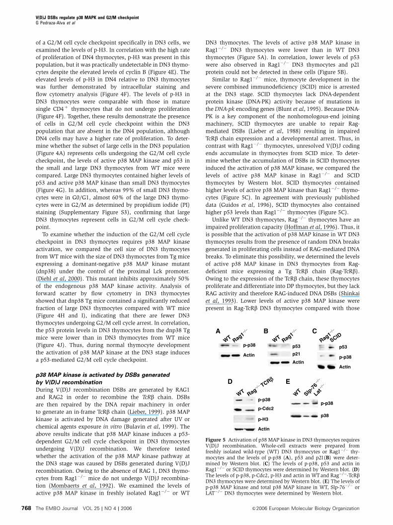
p38 MAP kinase is activated by DSBs generated
by V(D)J recombination
During V(D)J recombination DSBs are generated by RAG1
and RAG2 in order to recombine the TcRb chain. DSBs
are then repaired by the DNA repair machinery in order
to generate an in-frame TcRb chain (Lieber, 1999). p38 MAP
kinase is activated by DNA damage generated after UV or
chemical agents exposure in vitro (Bulavin et al, 1999). The
above results indicate that p38 MAP kinase induces a p53-
dependent G2/M cell cycle checkpoint in DN3 thymocytes
undergoing V(D)J recombination. We therefore tested
whether the activation of the p38 MAP kinase pathway at
the DN3 stage was caused by DSBs generated during V(D)J
recombination. Owing to the absence of RAG 1, DN3 thymo-
cytes from Rag1�/� mice do not undergo V(D)J recombina-
tion (Mombaerts et al, 1992). We examined the levels of
active p38 MAP kinase in freshly isolated Rag1�/� or WT
DN3 thymocytes. The levels of active p38 MAP kinase in
Rag1�/� DN3 thymocytes were lower than in WT DN3
thymocytes (Figure 5A). In correlation, lower levels of p53
were also observed in Rag1�/� DN3 thymocytes and p21
protein could not be detected in these cells (Figure 5B).
Similar to Rag1�/� mice, thymocyte development in the
severe combined immunodeficiency (SCID) mice is arrested
at the DN3 stage. SCID thymocytes lack DNA-dependent
protein kinase (DNA-PK) activity because of mutations in
the DNA-pk encoding genes (Blunt et al, 1995). Because DNA-
PK is a key component of the nonhomologous-end joining
machinery, SCID thymocytes are unable to repair Rag-
mediated DSBs (Lieber et al, 1988) resulting in impaired
TcRb chain expression and a developmental arrest. Thus, in
contrast with Rag1�/� thymocytes, unresolved V(D)J coding
ends accumulate in thymocytes from SCID mice. To deter-
mine whether the accumulation of DSBs in SCID thymocytes
induced the activation of p38 MAP kinase, we compared the
levels of active p38 MAP kinase in Rag1�/� and SCID
thymocytes by Western blot. SCID thymocytes contained
higher levels of active p38 MAP kinase than Rag1�/� thymo-
cytes (Figure 5C). In agreement with previously published
data (Guidos et al, 1996), SCID thymocytes also contained
higher p53 levels than Rag1�/� thymocytes (Figure 5C).
Unlike WT DN3 thymocytes, Rag�/� thymocytes have an
impaired proliferation capacity (Hoffman et al, 1996). Thus, it
is possible that the activation of p38 MAP kinase in WT DN3
thymocytes results from the presence of random DNA breaks
generated in proliferating cells instead of RAG-mediated DNA
breaks. To eliminate this possibility, we determined the levels
of active p38 MAP kinase in DN3 thymocytes from Rag-
deficient mice expressing a Tg TcRb chain (Rag-TcRb).
Owing to the expression of the TcRb chain, these thymocytes
proliferate and differentiate into DP thymocytes, but they lack
RAG activity and therefore RAG-induced DNA DSBs (Shinkai
et al, 1993). Lower levels of active p38 MAP kinase were
present in Rag-TcRb DN3 thymocytes compared with those
CA
p-p38
Actin
WT Rag1−/−
WT Rag1−/−
Rag1−/−B
p-p38
Actin
p53SCID
D
p53
p21
Actin
WT
Slp-7
6−/−
Lat−/−
p-p38
p38
E
WT Rag−/− TCRββ
Actin
p-H3
p-p38
p-Cdc2
Figure 5 Activation of p38 MAP kinase in DN3 thymocytes requiresV(D)J recombination. Whole-cell extracts were prepared fromfreshly isolated wild-type (WT) DN3 thymocytes or Rag1�/� thy-mocytes and the levels of p-p38 (A), p53 and p21(B) were deter-mined by Western blot. (C) The levels of p-p38, p53 and actin inRag1�/� or SCID thymocytes were determined by Western blot. (D)The levels of p-p38, p-Cdc2, p-H3 and actin in WTand Rag�/�-TcRbDN3 thymocytes were determined by Western blot. (E) The levels ofp-p38 MAP kinase and total p38 MAP kinase in WT, Slp-76�/� orLAT�/� DN3 thymocytes were determined by Western blot.
V(D)J DSBs regulate p38 MAPK and G2/M checkpointG Pedraza-Alva et al
The EMBO Journal VOL 25 | NO 4 | 2006 &2006 European Molecular Biology Organization768

found in WT DN3 thymocytes (Figure 5D). Furthermore, the
levels of Cdc2 phosphorylated on Tyr15 (inactive) were also
lower in Rag-TcRb DN3 thymocytes compared with those
observed in WT DN3 thymocytes. In contrast, we found
higher levels of p-H3 in Rag-TcRb DN3 thymocytes than in
WT DN3 thymocytes (Figure 5D), indicating that Rag-TcRbDN3 thymocytes undergo more rapid proliferation probably
due to the absence of cell cycle checkpoint. Thus, random
DNA breaks generated during cell proliferation are not re-
sponsible for the activation of the p38 MAP kinase in WT
DN3 thymocytes. In addition, as pre-TcR is functional in Rag-
TcRb DN3 thymocytes, these results indicate that pre-TcR
signals in the absence of V(D)J recombination are not suffi-
cient to activate p38 MAP kinase in DN3 thymocytes. To
further demonstrate that activation of p38 MAP kinase is
independent of pre-TcR in DN3 thymocytes, we examined the
levels of active p38 MAP kinase in DN3 thymocytes from the
Slp-76 or LAT-deficient mice. Although thymocytes from
these mice recombine TcR-b chain and contain pre-TcR,
they are arrested at the DN3 stage because of the lack of
pre-TcR signaling (Clements et al, 1998; Zhang et al, 1999).
Despite the absence of pre-TcR signaling, DN3 thymocytes
from the Slp-76�/� or the LAT�/� mice contained similar
levels of active p38 MAP kinase to those found in WT DN3
thymocytes (Figure 5E). Together, these results demonstrate
that activation in p38 MAP kinase in DN3 thymocytes
requires V(D)J-mediated DSBs, but does not require pre-TcR
signaling.
V(D)J-generated DSBs are present in post-G1 DN3
thymocytes
Together, these results demonstrate that DSBs generated by
V(D)J recombination are required for the activation of p38
MAP kinase and the induction of a G2/M cell cycle check-
point at the DN3 stage. It has been shown that most of the
DSBs generated during V(D)J recombination occur in G1 and
are repaired at the G1/S checkpoint (Schlissel et al, 1993;
Li et al, 1996). To determine whether the unresolved Rag-
induced DSBs in WT DN3 thymocytes might induce the G2/M
checkpoint, we assayed V(D)J DSBs at the TcRb locus by
ligation-mediated PCR (LM-PCR) (Schlissel et al, 1993). We
isolated DN3 thymocytes from WT mice by cell sorting and
stained them with PI (Supplementary Figure S4). We then
sorted and collected DN3 thymocytes with G0/G1 and S/G2/
M DNA content. To confirm the purity of the sorted popula-
tions, a portion of cells was restained with PI and reanalyzed
by fluorescence-activated cell sorting (FACS) (Supplementary
Figure S4). Sorted G1/G0 and S/G2/M DN3 thymocytes were
immediately lysed to extract genomic DNA and performed
LM-PCR as previously described (Schlissel et al, 1993). As a
control, we used genomic DNA from WT total thymocytes
(positive control) and liver (negative control), as well as
genomic DNA from MKK6(Glu), SCID and RAG1�/� thymo-
cytes. Equal amounts of linker-ligated DNA were assayed for
DSBs at RSS 50 of the Db1 and Db2 gene segments (indicative
of V-to-DJb rearrangement) (Figure 6A). As shown in pre-
vious studies, specific PCR products for 50 Db1 and Db2 RSS
were present in total WT thymocytes and SCID thymocytes,
but were absent in liver cells and Rag-deficient thymocytes
(Figure 6B). PCR products corresponding to the 50 of Db1 and
Db2 broken ends were also detected in both WT G1 and
S/G2/M DN3 thymocytes (Figure 6B). In addition, we also
detected Db1 and Db2 DSBs in MKK6(Glu) thymocytes
(Figure 6B).
To confirm that the LM-PCR products corresponded to
dsDNA breaks at the 50 of Db1 and Db2 RSS, we subjected
the various PCR products to cloning and DNA sequence
analysis. Three clones for each band and each sample were
sequenced. The results (Supplementary Figure S5) confirmed
that the fragment corresponding in size to Db1 was indeed
the 50 region of the Db1 segment and contained the specific
50 RSS. The faint band of higher molecular weight detected
in G1/G0 DN3 thymocytes was nonspecific. The two well-
defined bands detected in the Db2 assay also contained the
region 50 of the Db2 segment (Supplementary Figure S5). The
lower band of the expected size (E) present in all the samples
corresponded to the DSB at the 50 RSS of Db2 (Supplementary
Figure S5). The upper band corresponded to a cryptic RSS
located at 30 of the Db2 gene segment (Figure 6A and
Supplementary Figure S5). Interestingly, this band was pre-
sent in total thymocytes and G1/G0 DN3 thymocytes but it
B
T MKK6
SCID
WT DN3
L G1 G2/M Rag1
−/−
p53
CT M
KK6SCID
WT DN3
L G1 G2/M Rag1
−/−
Dβ1 Jβ1
Dβ2 Jβ2
Dβ2 Jβ2
E
C
A
5′ of Dβ-2
5′ of Dβ-2
C
E
Figure 6 DN3 thymocytes in G2/M contain RAG-mediated DSBs.(A) The schematic diagram represents the TcRb genomic locus (notdrawn to scale). D and J gene segments (boxes) are flanked byconsensus RSS containing 12-bp (open triangles) or 23-bp (blacktriangles) spacers. A cryptic RSS containing a 23-bp was alsoidentified at the 30 of the Db2 gene segment (gray triangle). Thickbars represent the LM-PCR products using primers specific for the 50
of Db1 or Db2 and for the linkers bound to the broken ends. Thinbars represent the probes used for Southern blot analysis of the PCRproducts. (B) LM-PCR was performed using DNA from total WTthymocytes (T), WT liver cells (L), WT G0/G1 DN3 thymocytes(G1), WT S/G2/M DN3 thymocytes (G2/M), MKK6(Glu) DN3thymocytes, SCID DN3 thymocytes and Rag1�/� DN3 thymocytes.PCR products were resolved by Southern blot analysis using 32p-labeled locus-specific primers (Db-1, Db-2). (C) The p53 gene wasamplified from the same amount of DNA used in the LM-PCRreactions (B), and PCR products were separated on agarose gelsand visualized by ethidium bromide staining.
V(D)J DSBs regulate p38 MAPK and G2/M checkpointG Pedraza-Alva et al
&2006 European Molecular Biology Organization The EMBO Journal VOL 25 | NO 4 | 2006 769

was undetectable in S/G2/M DN3 thymocytes probably
because it had already been repaired. To verify that similar
amounts of DNA were used in each LM-PCR, we performed
PCR for p53 genomic DNA as an internal control. Similar
levels of p53 genomic amplification were obtained in all
samples including liver and RAG-deficient thymocytes
where no LM-PCR products were detected (Figure 6C).
These results demonstrate that although some DSBs are
repaired at the G1/S checkpoint in DN3 thymocytes, S/G2/
M DN3 thymocytes contain RAG-mediated DSBs that could
induce a G2/M checkpoint.
If V(D)J recombination is upstream of p38 MAP kinase and
the initiation of the cell cycle checkpoint in DN3 thymocytes,
persistent activation of this pathway in Rag1�/� mice should
be able to activate p53 and induce a G2/M cell cycle check-
point in the absence of V(D)J recombination. To activate p38
MAP kinase in Rag1�/� thymocytes, Rag1�/� mice were
crossed with the MKK6(Glu) Tg mice. Flow cytometry ana-
lysis revealed that similar to Rag1�/� and MKK6(Glu) thymo-
cytes, MKK6(Glu)-Rag1�/� thymocytes were arrested at the
DN3 stage (CD25þCD44�) (Figure 7A). However, in contrast
to Rag1�/� thymocytes that were small and arrested at G0/G1,
MKK6(Glu)-Rag1�/� thymocytes were large (Figure 7B) and
promitotic (data not shown). Furthermore, MKK6(Glu)-
Rag1�/� thymocytes contained elevated levels of p53, p21
and cyclin B compared with Rag1�/� thymocytes as deter-
mined by Western blot (Figure 7C). Interestingly, whereas
thymocytes from the Rag1�/� mice are unable to survive
in vivo and thymi of these mice are very small, DN3 thymo-
cytes from MKK6(Glu)-Rag1�/� mice survived and accumu-
lated in vivo (Figure 7D). Thus, DSBs generated during V(D)J
recombination are required for p38 MAP kinase activation,
but once this pathway is activated, it can trigger the G2/M
cell cycle checkpoint without DSBs. Collectively, these results
show that the activation of p38 MAP kinase is downstream of
DNA damage in the signaling cascade leading to p53 activa-
tion and the induction of a G2/M cell cycle checkpoint during
V(D)J recombination.
Discussion
Although V(D)J recombination is advantageous because it
ensures maximal diversity of the TcR repertoire, it is also
a risk for host cells as genomic aberrations resulting from
un- or mis-repaired DNA breaks may lead to malignancy
(Nacht et al, 1996; Difilippantonio et al, 2000). Thus, an
efficient DNA repair mechanism and the presence of a cell
cycle checkpoint to delay cell division while DNA repair
occurs is essential. Although it has been shown that
RAG-induced DNA breaks are repaired at the G1/S check-
point (Schlissel et al, 1993), the establishment of a G2/M
cell cycle checkpoint in DN3 thymocytes in response to
DSBs resulting from V(D)J recombination has not been
formally demonstrated. Here, we show for the first time
that activation of p38 MAP kinase in response to DSBs
induces a p53-dependent G2/M checkpoint in developing
DN3 thymocytes.
In fibroblasts, phosphorylation of p53 by p38 MAP kinase
promotes the stabilization of p53 and cell cycle arrest at the
G2/M checkpoint in response to DNA damage after UV or
gamma irradiation in vitro (Bulavin et al, 1999; Huang et al,
1999). Here, we demonstrate that activation of p38 MAP
kinase in vivo leads to phosphorylation and accumulation
of p53 and induction of a G2/M cell cycle checkpoint. In
addition to the p53-dependent pathway, it has been recently
shown that p38 MAP kinase can also promote cell cycle arrest
Figure 7 Activation of the p38 MAP kinase pathway in Rag-deficient thymocytes promotes a G2/M cell cycle checkpoint. (A) FACS profilesrepresent the distribution and percentage of CD25�CD44þ , CD25þCD44þ , CD25þCD44� and CD25�CD44� populations in total thymocytesisolated from WT-Rag1�/�, MKK6(Glu) transgenic (Tg)-Rag1þ /þ and Tg-Rag1�/� mice or in gated DN thymocytes from WT-Rag1þ /þ mice.CD4 and CD8 profiles are shown in Supplementary Figure S6. (B) Thymocytes were isolated from the mice described in (A) and cell size wasanalyzed by flow cytometry. (C) The levels of p53, p21, cyclin B and actin were determined by Western blot. (D) Total thymocyte numbers inRag1�/� WT or Rag1�/� MKK6(Glu) Tg mice. The average and standard error of the mean are shown (n¼ 6).
V(D)J DSBs regulate p38 MAPK and G2/M checkpointG Pedraza-Alva et al
The EMBO Journal VOL 25 | NO 4 | 2006 &2006 European Molecular Biology Organization770

at the G2/M checkpoint in vitro by inactivating the Cdc25
phosphatase, resulting in accumulation of inactive Cdc2 and
cell cycle arrest (Bulavin et al, 2001). It is therefore possible
that, together with p53, Cdc25 may also participate in the
establishment of the G2/M checkpoint induced by p38 MAP
kinase in DN3 thymocytes.
It has been proposed that pre-TcR signals can also activate
p38 MAP kinase (Mulroy and Sen, 2001). However, our
results indicate that pre-TcR signals are not required as long
as V(D)J recombination takes place (Figure 5). Because DSBs
generated by DNA-damaging agents promote p38 MAP kinase
activation in vitro (Wang et al, 2000; Bulavin et al, 2001), we
propose that the presence of DSBs generated by of V(D)J
recombination in DN3 thymocytes is the main stimulus that
activates the p38 MAP kinase pathway in vivo. This model is
supported by the presence of active p38 MAP kinase in WT
and SCID DN3 thymocytes, but not in WT DN4, Rag1�/� and
RAG-deficient-TcRb chain Tg thymocytes. Although p38 MAP
kinase is clearly activated in response to DNA damage
in vitro, the molecular mechanisms remain largely unknown.
ATM kinase has been suggested to be responsible for the
activation of the p38 MAP kinase pathway after DNA damage
(Wang et al, 2000). Whether ATM plays a role in the activa-
tion of p38 MAP kinase in response to RAG-induced DSBs
during V(D)J recombination remains to be determined.
However, no difference in ATM activation was observed
between DN3 and DN4 thymocytes (data not shown) arguing
that activation of p38 MAP kinase in DN3 thymocytes is likely
independent of ATM.
The fact that RSS-associated dsDNA breaks have been
preferentially observed in cells in G0/G1 (Schlissel et al,
1993; Li et al, 1996) has lead to the conclusion that V(D)J
recombination occurs only during the G0/G1 phase of the
cell cycle and that DNA damage is repaired exclusively
at the G1/S cell cycle checkpoint (Schlissel et al, 1993;
Guidos et al, 1996). Our results show that WT DN3 thymo-
cytes in S/G2/M phases of the cell cycle contain signal
end DSBs at the TcRb locus. These signal end DSBs can
trigger the G2/M checkpoint to allow signal joint formation
and convert them into inert products (Hiom et al, 1998;
Roth and Craig, 1998), as linear double-stranded DNA can
easily integrate randomly and compromise genomic stability.
Indeed, interchromosomal transposition of signal ends in
human T cells has been recently described (Messier et al,
2003). Although there is no direct evidence that signal ends
induce the same cellular response as coding ends, most
of the DNA repair machinery recognizes DSBs regardless
of whether they are in the chromosomes. It is therefore
logical that signal ends can also induce a G2/M cell cycle
checkpoint.
Thus, we propose that V(D)J recombination at the DN3
stage of thymocyte development triggers activation of the p38
MAP kinase pathway. Activated p38 MAP kinase promotes
a G2/M cell cycle checkpoint through the phosphorylation
and accumulation of p53 to allow DNA repair of deleterious
mutations produced during V(D)J recombination (Figure 8).
Although activation of the p38/p53 pathway at the DN3 stage
may be important for genomic stability, subsequent inactiva-
tion of p38 MAP kinase and termination of the G2/M check-
point is also required for DN3 thymocytes to progress through
mitosis, downregulate CD25 and differentiate into DN4
thymocytes.
Materials and methods
MiceThe MKK6(Glu) Tg and dominant-negative p38 MAP kinase Tgmice have been previously described (Diehl et al, 2000). Rag1�/�,p53�/� and SCID mice were purchased from Jackson Laboratory.Rag2�/�/TcRb chain Tg mice were purchased from TaconicLaboratory. SLP-76�/� and LAT�/� mice have been previousdescribed. Procedures that involved mice were approved byinstitutional guidelines for animal care.
Cell surface staining and cell sortingThe distribution of different thymocyte populations was examinedafter cell surface staining with anti-CD4, CD8, CD25 (Pharmingen),and anti-CD44-PE (Caltag) antibodies by flow cytometry (LSR II,BD). The DN3 and DN4 populations were purified by cell sorting(FacsAria, BD) after four-color staining. To isolate G1 and S/G2/MDN3 thymocytes, total thymocytes were stained (CD4, CD8, CD25and CD44), DN3 thymocytes were sorted, collected and stainedwith PI and sorted again based on DNA content. Post sorted G0/G1and S/G2/M DN3 thymocytes were then collected and lysed forisolation of genomic DNA. Detailed gates, steps and purity of thesorting are described in Supplementary Figure S4. To examine cellcycle in small and large DN3 thymocytes, total thymocytes werestained for four colors as described above, large and small DN3were sorted based on the forward scatter, collected and stained withPI to examine the DNA content. p53 intracellular staining wasperformed after cell surface staining using anti-p53-PE antibody(Pharmingen) as recommended. For phosphorylated p38 MAPkinase intracellular staining, thymocytes were stained for cellsurface markers, fixed with paraformaldehyde, permeabilized with0.05% saponin and stained with an anti-phospho-p38 (Thr180/Tyr182) antibody (Cell Signaling) followed by anti-Rabbit-FITCantibody (Santa Cruz Biotechnology). For detection of phosphory-lated histone H3, cells were fixed, permeabilized (0.1% Tween) andstained using the FITC-anti-phospho-H3 (Cell Signaling). Cell sizewas determined by flow cytometry and analysis of forward lightscatter.
Western blotWhole-cell extracts were prepared in Triton lysis buffer aspreviously described (Pedraza-Alva et al, 1996). Cyclin B1 wasimmunoprecipitated from whole-cell extracts with anti-cyclin B1antibody (Santa Cruz Biotechnology). Immunoprecipitated proteinsor total cell extracts were resolved by SDS–PAGE, transferred tomembranes and immunostained with anti-p53 (Novocastra), anti-p21Waf1, anti-p27Kip, anti-cyclin B1, anti-Cdc2, anti-actin (SantaCruz Biotechnology), anti-phosphorylated Cdc2 (Tyr15), anti-phos-phorylated p38 (Thr180/Tyr182), anti-phosphorylated p53 (Ser15 orSer392) anti-p38 (Cell Signaling), anti-phosphorylated histone H3(Ser10) and anti-CENP-A (Upstate Biotech) antibodies.
Fp38 p-p38*
p53 p-p53*
Cell cyclecheckpoint
p38 p-p38*
p53 p-p53*
pre-TcR
RAGV(D)J DNArepair
Cell cycleprogression
pTα TcRβ
CD25+CD44− CD25−CD44−preTcR+
CD25
?
DN3 DN4
Figure 8 Activation of p38 MAP kinase by V(D)J recombinationleads to the establishment of a p53-dependent G2/M cell cyclecheckpoint to allow repair of RAG-mediated DSBs.
V(D)J DSBs regulate p38 MAPK and G2/M checkpointG Pedraza-Alva et al
&2006 European Molecular Biology Organization The EMBO Journal VOL 25 | NO 4 | 2006 771

Cdc2 kinase assayCyclin B1 precipitates were washed twice with lysis buffer, twicewith kinase buffer (20 mM Tris pH 7.5, 7.5 mM MgCl2, 1 mM DTT)and incubated at 301C for 30 min in 25ml of kinase buffer containing10mCi (g 32P)-ATP (3000 Ci/mmol, Amersham), 100 mM ATP and1mg of histone H1 (Worthington) as exogenous substrate. Thereaction was mixed with an equal volume of 2� SDS–PAGE loadingbuffer. Proteins were separated by SDS–PAGE and 32P-labeledhistone H1. Levels of phospho-histone H1 were determined byphosphoimaging on a FX molecular imager (Bio-Rad).
Ligation-mediated-PCRWT G0/G1 and S/G2/M DN3 thymocytes were isolated by cellsorting, DNA was purified from sorted cells and LM-PCR wasperformed as previously described (Schlissel et al, 1993). Thesequence of the TcRb locus-specific oligonucleotide PCR primersand probes used for LM-PCR has been previously published(Hempel et al, 1998). The control p53 amplification was performedusing the following primers: OIMRO336: 50-ATAGGTCGGCGGTTCAT-30 and OIMRO337: 50-CCCGAGTATCTGGAAGACAG-30. The PCR
products were separated on 1.5% agarose gel and detected byethidium bromide staining. The LM-PCR bands were cut from thegel and the DNA was extracted using the Qiaex II gel extraction kit(Qiagen) following the manufacturer’s instructions. The DNA wasthen ligated directly into the TOPO A/T plasmid (Invitrogen) andtransformed into bacteria. DNA was isolated and sent for sequen-cing using the universal M13 primers.
Supplementary dataSupplementary data are available at The EMBO Journal Online.
Acknowledgements
We thank Karen A Fortner and Oliver Dienz for helpful discussionsand critical reading of the manuscript. This work was supportedby Grant R01 AI051454 (M Rincon) from the National Institute ofHealth. G Pedraza-Alva was partially supported by the SistemaNacional de Investigadores/CONACYT, Mexico.
References
Bassing CH, Swat W, Alt FW (2002) The mechanism and regulationof chromosomal V(D)J recombination. Cell 109 (Suppl): S45–S55
Blunt T, Finnie NJ, Taccioli GE, Smith GC, Demengeot J, GottliebTM, Mizuta R, Varghese AJ, Alt FW, Jeggo PA, Jackson SP (1995)Defective DNA-dependent protein kinase activity is linked toV(D)J recombination and DNA repair defects associated withthe murine scid mutation. Cell 80: 813–823
Booher RN, Holman PS, Fattaey A (1997) Human Myt1 is a cellcycle-regulated kinase that inhibits Cdc2 but not Cdk2 activity.J Biol Chem 272: 22300–22306
Bulavin DV, Higashimoto Y, Popoff IJ, Gaarde WA, Basrur V,Potapova O, Appella E, Fornace Jr AJ (2001) Initiation of aG2/M checkpoint after ultraviolet radiation requires p38 kinase.Nature 411: 102–107
Bulavin DV, Saito S, Hollander MC, Sakaguchi K, Anderson CW,Appella E, Fornace Jr AJ (1999) Phosphorylation of human p53by p38 kinase coordinates N-terminal phosphorylation and apop-tosis in response to UV radiation. EMBO J 18: 6845–6854
Caspari T (2000) How to activate p53. Curr Biol 10: R315–R317Clements JL, Yang B, Ross-Barta SE, Eliason SL, Hrstka RF,
Williamson RA, Koretzky GA (1998) Requirement for the leuko-cyte-specific adapter protein SLP-76 for normal T cell develop-ment. Science 281: 416–419
Diehl NL, Enslen H, Fortner KA, Merritt C, Stetson N, Charland C,Flavell RA, Davis RJ, Rincon M (2000) Activation of the p38mitogen-activated protein kinase pathway arrests cell cycle pro-gression and differentiation of immature thymocytes in vivo.J Exp Med 191: 321–334
Difilippantonio MJ, Zhu J, Chen HT, Meffre E, Nussenzweig MC,Max EE, Ried T, Nussenzweig A (2000) DNA repair protein Ku80suppresses chromosomal aberrations and malignant transforma-tion. Nature 404: 510–514
el-Deiry WS, Tokino T, Velculescu VE, Levy DB, Parsons R,Trent JM, Lin D, Mercer WE, Kinzler KW, Vogelstein B (1993)WAF1, a potential mediator of p53 tumor suppression. Cell 75:817–825
Freshney NW, Rawlinson L, Guesdon F, Jones E, Cowley S, Hsuan J,Saklatvala J (1994) Interleukin-1 activates a novel protein kinasecascade that results in the phosphorylation of Hsp27. Cell 78:1039–1049
Gottlieb E, Haffner R, King A, Asher G, Gruss P, Lonai P, Oren M(1997) Transgenic mouse model for studying the transcriptionalactivity of the p53 protein: age- and tissue-dependent changes inradiation-induced activation during embryogenesis. EMBO J 16:1381–1390
Guidos CJ, Williams CJ, Grandal I, Knowles G, Huang MT, DanskaJS (1996) V(D)J recombination activates a p53-dependent DNAdamage checkpoint in scid lymphocyte precursors. Genes Dev 10:2038–2054
Han J, Lee JD, Bibbs L, Ulevitch RJ (1994) A MAP kinase targetedby endotoxin and hyperosmolarity in mammalian cells. Science265: 808–811
Hempel WM, Stanhope-Baker P, Mathieu N, Huang F, Schlissel MS,Ferrier P (1998) Enhancer control of V(D)J recombination at theTCRbeta locus: differential effects on DNA cleavage and joining.Genes Dev 12: 2305–2317
Hermeking H, Lengauer C, Polyak K, He TC, Zhang L, ThiagalingamS, Kinzler KW, Vogelstein B (1997) 14-3-3 sigma is a p53-regu-lated inhibitor of G2/M progression. Mol Cell 1: 3–11
Hiom K, Melek M, Gellert M (1998) DNA transposition by the RAG1and RAG2 proteins: a possible source of oncogenic translocations.Cell 94: 463–470
Hoffman ES, Passoni L, Crompton T, Leu TM, Schatz DG, Koff A,Owen MJ, Hayday AC (1996) Productive T-cell receptor beta-chain gene rearrangement: coincident regulation of cell cycleand clonality during development in vivo. Genes Dev 10:948–962
Howman EV, Fowler KJ, Newson AJ, Redwards S, MacDonald AC,Kalitis P, Choo KH (2000) Early disruption of centromeric chro-matin organization in centromere protein A (Cenpa) null mice.Proc Natl Acad Sci USA 97: 1148–1153
Huang C, Ma WY, Maxiner A, Sun Y, Dong Z (1999) p38 kinasemediates UV-induced phosphorylation of p53 protein at serine389. J Biol Chem 274: 12229–12235
Levine AJ (1997) p53, the cellular gatekeeper for growth anddivision. Cell 88: 323–331
Li Z, Dordai DI, Lee J, Desiderio S (1996) A conserved degradationsignal regulates RAG-2 accumulation during cell division andlinks V(D)J recombination to the cell cycle. Immunity 5: 575–589
Lieber MR (1999) The biochemistry and biological significance ofnonhomologous DNA end joining: an essential repair process inmulticellular eukaryotes. Genes Cells 4: 77–85
Lieber MR, Hesse JE, Lewis S, Bosma GC, Rosenberg N, Mizuuchi K,Bosma MJ, Gellert M (1988) The defect in murine severe com-bined immune deficiency: joining of signal sequences but notcoding segments in V(D)J recombination. Cell 55: 7–16
Mahadevan LC, Willis AC, Barratt MJ (1991) Rapid histone H3phosphorylation in response to growth factors, phorbol esters,okadaic acid, and protein synthesis inhibitors. Cell 65: 775–783
Messier TL, O’Neill JP, Hou SM, Nicklas JA, Finette BA (2003)In vivo transposition mediated by V(D)J recombinase in humanT lymphocytes. EMBO J 22: 1381–1388
Mombaerts P, Iacomini J, Johnson RS, Herrup K, Tonegawa S,Papaioannou VE (1992) RAG-1-deficient mice have no mature Band T lymphocytes. Cell 68: 869–877
Morgan DO (1995) Principles of CDK regulation. Nature 374:131–134
Mulroy T, Sen J (2001) p38 MAP kinase activity modulates alphabeta T cell development. Eur J Immunol 31: 3056–3063
Nacht M, Strasser A, Chan YR, Harris AW, Schlissel M, Bronson RT,Jacks T (1996) Mutations in the p53 and SCID genes cooperate intumorigenesis. Genes Dev 10: 2055–2066
Pani L, Horal M, Loeken MR (2002) Rescue of neural tube defects inPax-3-deficient embryos by p53 loss of function: implications for
V(D)J DSBs regulate p38 MAPK and G2/M checkpointG Pedraza-Alva et al
The EMBO Journal VOL 25 | NO 4 | 2006 &2006 European Molecular Biology Organization772

Pax-3-dependent development and tumorigenesis. Genes Dev 16:676–680
Pedraza-Alva G, Merida LB, Burakoff SJ, Rosenstein Y (1996) CD43-specific activation of T cells induces association of CD43 to Fynkinase. J Biol Chem 271: 27564–27568
Poon RY, Jiang W, Toyoshima H, Hunter T (1996) Cyclin-dependentkinases are inactivated by a combination of p21 and Thr-14/Tyr-15 phosphorylation after UV-induced DNA damage. J BiolChem 271: 13283–13291
Rodewald HR, Fehling HJ (1998) Molecular and cellular events inearly thymocyte development. Adv Immunol 69: 1–112
Roth DB, Craig NL (1998) VDJ recombination: a transposase goes towork. Cell 94: 411–414
Schlissel M, Constantinescu A, Morrow T, Baxter M, Peng A (1993)Double-strand signal sequence breaks in V(D)J recombination areblunt, 5’-phosphorylated, RAG-dependent, and cell cycle regu-lated. Genes Dev 7: 2520–2532
She QB, Chen N, Dong Z (2000) ERKs and p38 kinase phosphorylatep53 protein at serine 15 in response to UV radiation. J Biol Chem275: 20444–20449
Shieh SY, Taya Y, Prives C (1999) DNA damage-inducible phosphory-lation of p53 at N-terminal sites including a novel site, Ser20,requires tetramerization. EMBO J 18: 1815–1823
Shinkai Y, Koyasu S, Nakayama K, Murphy KM, Loh DY, ReinherzEL, Alt FW (1993) Restoration of T cell development in RAG-2-deficient mice by functional TCR transgenes. Science 259: 822–825
Toyoshima H, Hunter T (1994) p27, a novel inhibitor of G1 cyclin-Cdk protein kinase activity, is related to p21. Cell 78: 67–74
Wang X, McGowan CH, Zhao M, He L, Downey JS, Fearns C, WangY, Huang S, Han J (2000) Involvement of the MKK6-p38gammacascade in gamma-radiation-induced cell cycle arrest. Mol CellBiol 20: 4543–4552
Zhan Q, Antinore MJ, Wang XW, Carrier F, Smith ML, Harris CC,Fornace Jr AJ (1999) Association with Cdc2 and inhibition ofCdc2/Cyclin B1 kinase activity by the p53-regulated proteinGadd45. Oncogene 18: 2892–2900
Zhang W, Sommers CL, Burshtyn DN, Stebbins CC, DeJarnette JB,Trible RP, Grinberg A, Tsay HC, Jacobs HM, Kessler CM, Long EO,Love PE, Samelson LE (1999) Essential role of LAT in T celldevelopment. Immunity 10: 323–332
V(D)J DSBs regulate p38 MAPK and G2/M checkpointG Pedraza-Alva et al
&2006 European Molecular Biology Organization The EMBO Journal VOL 25 | NO 4 | 2006 773
Copyright © 2022 FDOKUMEN