ACGS Best Practice Guidelines for Variant Classification in Rare ...
32
Copyright © ACGS 2020 Page 1 ACGS Best Practice Guidelines for Variant Classification in Rare Disease 2020 Sian Ellard 1,2 , Emma L Baple 2,3,4 , Alison Callaway 5 , Ian Berry 6 , Natalie Forrester 7 , Clare Turnbull 4 , Martina Owens 1 , Diana M Eccles 8 , Stephen Abbs 9 , Richard Scott 4,10 , Zandra C Deans 11 , Tracy Lester 12 , Jo Campbell 13 , William G Newman 14,15 , Simon Ramsden 14 and Dominic J McMullan 16 1. Exeter Genomics Laboratory, Royal Devon & Exeter NHS Foundation Trust, Exeter, EX2 5DW, UK. 2. University of Exeter Medical School, Exeter, EX2 5DW, UK. 3. Department of Clinical Genetics, Royal Devon & Exeter NHS Foundation Trust, Exeter, EX2 5DW, UK. 4. Genomics England, William Harvey Research Institute, Queen Mary University of London, Charterhouse Square, London, EC1M 6BQ, UK 5. Wessex Regional Genetics Laboratory, Salisbury NHS Foundation Trust, Salisbury SP2 8BJ. 6. Yorkshire & North East Genomic Laboratory Hub Central Lab, St James’s University Hospital, Leeds LS9 7TF, UK. 7. Bristol Genetics Laboratory, North Bristol NHS Trust, Bristol BS10 5NB, UK. 8. Wessex Clinical Genetics Service, University Hospital Southampton, Southampton SO16 5YA, UK. 9. East Anglian Medical Genetics Service, Addenbrooke’s Hospital, Cambridge CB2 0QQ, UK. 10. Department of Clinical Genetics, Great Ormond Street Hospital for Children NHS Foundation Trust, London, WC1N 3JH, UK. 11. UK NEQAS for Molecular Genetics, Department of Laboratory Medicine, Royal infirmary of Edinburgh, Edinburgh EH16 4SA, UK. 12. Oxford Genetic Laboratories, Oxford University Hospitals NHS Foundation Trust, Oxford OX3 7LE, UK. 13. Viapath Genetics Laboratory, Viapath Analytics LLP, 5 th Floor Tower Wing, Guy’s Hospital, London SE1 9RT, UK. 14. Manchester Centre for Genomic Medicine, Central Manchester University Hospitals NHS Foundation Trust, Manchester M13 9WL, UK. 15. Evolution and Genomic Science, University of Manchester, Manchester M13 9PL 16. West Midlands Regional Genetics Laboratory, Birmingham Women’s NHS Foundation Trust, Birmingham, B15 2TG, UK. Recommendations ratified by ACGS Quality Subcommittee on 4 th February 2020 1. Document Version History Version Date Description 4.01 04/02/2020 Updated guidelines to replace 2019 version (previous versions 2017 and 2018) 2. Table Of Contents 1. Document Version History .............................................................................................................. 1 2. Table Of Contents ........................................................................................................................... 1 3. Introduction .................................................................................................................................... 2 4. Integration of clinical and scientific data in variant classification .................................................. 4 5. Variant classification: Supplementary notes for use of the ACMG evidence criteria..................... 9 6. Reporting the variant classification .............................................................................................. 16 6.1. Variants of uncertain significance ......................................................................................... 25 6.1.1. Situations where considering reporting a VUS might be appropriate .......................... 26 6.1.2. Situations where reporting a VUS would not be considered appropriate.................... 27
-
Upload
khangminh22 -
Category
Documents
-
view
35 -
download
0
Transcript of ACGS Best Practice Guidelines for Variant Classification in Rare ...

Copyright © ACGS 2020 Page 1
ACGS Best Practice Guidelines for Variant Classification in
Rare Disease 2020
Sian Ellard1,2
, Emma L Baple2,3,4
, Alison Callaway5, Ian Berry
6, Natalie Forrester
7, Clare
Turnbull4, Martina Owens
1, Diana M Eccles
8, Stephen Abbs
9, Richard Scott
4,10, Zandra C
Deans11
, Tracy Lester12
, Jo Campbell13
, William G Newman14,15
, Simon Ramsden14
and Dominic
J McMullan16
1. Exeter Genomics Laboratory, Royal Devon & Exeter NHS Foundation Trust, Exeter, EX2 5DW, UK. 2. University of Exeter Medical School, Exeter, EX2 5DW, UK. 3. Department of Clinical Genetics, Royal Devon & Exeter NHS Foundation Trust, Exeter, EX2 5DW, UK. 4. Genomics England, William Harvey Research Institute, Queen Mary University of London, Charterhouse Square, London,
EC1M 6BQ, UK 5. Wessex Regional Genetics Laboratory, Salisbury NHS Foundation Trust, Salisbury SP2 8BJ. 6. Yorkshire & North East Genomic Laboratory Hub Central Lab, St James’s University Hospital, Leeds LS9 7TF, UK. 7. Bristol Genetics Laboratory, North Bristol NHS Trust, Bristol BS10 5NB, UK. 8. Wessex Clinical Genetics Service, University Hospital Southampton, Southampton SO16 5YA, UK. 9. East Anglian Medical Genetics Service, Addenbrooke’s Hospital, Cambridge CB2 0QQ, UK. 10. Department of Clinical Genetics, Great Ormond Street Hospital for Children NHS Foundation Trust, London, WC1N 3JH,
UK. 11. UK NEQAS for Molecular Genetics, Department of Laboratory Medicine, Royal infirmary of Edinburgh, Edinburgh EH16
4SA, UK. 12. Oxford Genetic Laboratories, Oxford University Hospitals NHS Foundation Trust, Oxford OX3 7LE, UK. 13. Viapath Genetics Laboratory, Viapath Analytics LLP, 5
th Floor Tower Wing, Guy’s Hospital, London SE1 9RT, UK.
14. Manchester Centre for Genomic Medicine, Central Manchester University Hospitals NHS Foundation Trust, Manchester M13 9WL, UK.
15. Evolution and Genomic Science, University of Manchester, Manchester M13 9PL 16. West Midlands Regional Genetics Laboratory, Birmingham Women’s NHS Foundation Trust, Birmingham, B15 2TG, UK.
Recommendations ratified by ACGS Quality Subcommittee on 4
th February 2020
1. Document Version History
Version Date Description
4.01 04/02/2020 Updated guidelines to replace 2019 version (previous versions 2017 and 2018)
2. Table Of Contents
1. Document Version History .............................................................................................................. 1
2. Table Of Contents ........................................................................................................................... 1
3. Introduction .................................................................................................................................... 2
4. Integration of clinical and scientific data in variant classification .................................................. 4
5. Variant classification: Supplementary notes for use of the ACMG evidence criteria ..................... 9
6. Reporting the variant classification .............................................................................................. 16
6.1. Variants of uncertain significance ......................................................................................... 25
6.1.1. Situations where considering reporting a VUS might be appropriate .......................... 26
6.1.2. Situations where reporting a VUS would not be considered appropriate.................... 27

Copyright © ACGS 2020 Page 2
6.1.3. Reporting a heterozygous (likely) pathogenic variant for a gene associated with an
autosomal recessive disease ......................................................................................................... 28
6.1.4. Specific VUS reporting considerations related to WGS, WES and large gene panel tests
…………………………………………………………………………………………………………………………………………………28
6.2. Storage of variant data for future re-analysis ....................................................................... 29
7. Reclassification of variants ............................................................................................................ 29
3. Introduction
In the European Union, a rare disease is defined as rare when it affects less than one in
2000 individuals. Approximately seven thousand rare diseases have been described which
in total affect an estimated 1 in 17 of the UK population (approximately 3.5 million
individuals). Nearly 5000 of these rare diseases are monogenic disorders caused by highly
penetrant variants in a single gene. A molecular genetic diagnosis of a rare disease requires
the identification of a single disease-causing variant (or bi-allelic variants in autosomal
recessive conditions). A prompt and accurate molecular diagnosis can be crucial to the
delivery of optimal care for a patient and their family particularly increasingly in targeting
treatment (Saunders et al 2012). However, diagnosis of a rare genetic disease can be a
challenge and is contingent upon a robust understanding of the molecular aetiology of the
disease. A molecular genetic diagnosis underpins robust disease classification, provision of
prognostic information, accurate risk prediction for relatives, and importantly can indicate the
most appropriate treatment(s), inform access to clinical screening, prevention strategies or
clinical trials and facilitate access to support services and patient-led support groups.
Historically, genetic testing focused on the analysis of one or a small number of genes
indicated by the patient’s phenotype, but the advent of next generation sequencing
technology has revolutionised the scale at which genetic testing can be performed enabling
the analysis of many more genes within the same assay. Large gene panel tests (>100
genes) and whole exome sequencing are routinely available in UK clinical diagnostic
laboratories and whole genome sequencing, first available through the 100,000 Genomes
Project in England, will be commissioned for mainstream clinical care within the NHS in
England in the near future. Deciphering which, if any, of the observed variants are disease-
causing is challenging as each human genome has 3-4 million variants (compared to the
reference human genome sequence). Only a minority are causative of monogenic disease;
most are part of normal human variation or may contribute to an increased or decreased risk
of multi-factorial disease. The gnomAD database (http://gnomad.broadinstitute.org/)
currently includes 17.2 million variants identified by exome sequencing of 125, 748
individuals and 261.9 million variants identified through genome sequencing of 15,708
individuals who were part of various disease-specific and population genetic studies,
(Karczewski et al BioRxiv 2019 https://doi.org/10.1101/531210), but we do not yet have a
comprehensive catalogue of global genetic variation. The focus of these guidelines is the
classification of highly penetrant protein-coding variants. Inferring pathogenicity of non-
coding variants is more complex, but will need to be addressed as a standard of practice in
the future.

Copyright © ACGS 2020 Page 3
In 2015 the American College of Medical Genetics and Genomics (ACMG) and the
Association for Molecular Pathology (AMP) published standards and guidelines for the
interpretation of sequence variants (Richards et al 2015). These guidelines describe a
framework for classifying variants as “pathogenic”, “likely pathogenic”, “uncertain
significance”, “likely benign” or “benign” according to a series of criteria with levels of
evidence defined as very strong, strong, moderate or supporting. They recommend that all
assertions should be classified with respect to a disease and inheritance pattern. The
guidelines also state that a variant of uncertain significance should not be used in clinical
decision making. The consequences of a mis-diagnosis can be harmful not just for the
proband but also their relatives whose clinical management is altered as a consequence of
cascade testing.
Further development of the ACMG/AMP guidelines is being undertaken through the US
ClinGen Sequence Variant Interpretation (SVI) Working Group
(https://www.clinicalgenome.org/working-groups/sequence-variant-interpretation/). Their goal
is to support the refinement and evolution of the guidelines. It was recognised by Richards et
al (2015) that more focused guidance regarding the classification of variants in specific
genes is required given that the applicability and weight assigned to certain criteria may vary
by disease and gene. A number of disease-specific variant expert panels have been
established and are generating disease/gene specific guidelines (for example see Kelly et al
2018 for MYH7-specific guidelines). Work is also underway to consider interpretation and
reporting of variants with reduced penetrance.
High quality, accurate variant interpretation requires scientific knowledge of the gene
structure, function, previously identified variants and disease mechanism in addition to
comprehensive clinical knowledge of the patient and their families’ medical history. The UK’s
National Health Service (NHS) provides a unique opportunity to integrate curated genotype
and phenotype information within a nationally developed database. On 4th November 2016 a
group of NHS clinical scientists and clinical geneticists met to discuss the implementation of
the ACMG guidelines within the UK (see workshop report from the PHG Foundation).
A consensus statement was issued on 11th November 2016 by the Association for Clinical
Genomic Science (see ACGS consensus statement). It recommended adoption of the
ACMG guidelines for germline variant classification and interpretation in UK diagnostic
genetic laboratories performing testing for rare disease and familial cancers.
A “Train the Trainers” workshop was held in February 2017 and attended by representatives
from 24 regional genetics centres across England, Scotland, Wales and Ireland. The aim of
the workshop was to plan the implementation of the ACMG guidelines in a co-ordinated way
in order to achieve accurate usage and hence consistent use of the guidelines across and
within laboratories. Monthly WebEx meetings for rare disease and familial cancer
predisposition were established in 2017 to facilitate variant interpretation for SNVs and
indels through multi-disciplinary case-based discussion and provide an opportunity for
reviewing updates to the guidelines.
Please note that these guidelines are intended for general use in classifying highly-
penetrant variants in patients with rare, monogenic diseases. Disease-specific

Copyright © ACGS 2020 Page 4
guidelines are being developed for disorders where different evidence thresholds are
required, for example familial cancer predisposition and inherited cardiac conditions.
4. Integration of clinical and scientific data in variant classification
Interpretation of a variant for use in clinical decision making requires comprehensive
knowledge of the patient’s phenotype, mode of inheritance for the disease gene, mutational
mechanism (e.g. haploinsufficiency, dominant negative), protein structure/function and the
strength of the gene-disease relationship (Strande et al 2017). With the exception of the
patient’s phenotype data, most of this information can be obtained from the published
literature/databases by a clinical scientist who can also collate the required population data
and in silico predictions of variant effect.
The prior probability that a patient has a disease-causing variant (or variant pair) in a specific
gene is important information that is often not available to the laboratory unless the request
is for a single gene test or testing is being performed to confirm a suspected clinical
diagnosis that is associated with a single gene or small number of genes within a biological
pathway. For disorders where there are clinical diagnostic criteria (for example the Ghent
criteria for Marfan syndrome) it is helpful if the referring clinician indicates whether these
have been met. When requesting large panel tests or exome/genome analysis it can be very
useful for the laboratory if the clinical team provides details regarding the likelihood that a
particular clinical presentation is thought to be monogenic, any specific diagnoses that are
being considered and where feasible, a shortlist of genes that are thought to be of relevance
according to the clinical presentation.
The level of detailed phenotype data provided with the laboratory referral depends upon the
testing scenario and is optimally provided as a set of HPO (Human Phenotype Ontology)
terms, however, it is recognised that certain phenotyping disciplines (e.g. neuroradiology,
skeletal dysplasia) may have their own existing terminology or nosologies which may provide
more detailed and appropriate description than HPO. For disorders where biochemical or
other test results are critical for variant interpretation, this information may be provided via
completion of a laboratory request form for the specific disorder. Phenotype specificity is a
key evidence criterion for variant interpretation and when testing is undertaken at an exome
or genome scale for the diagnosis of very rare disorders, a multi-disciplinary approach is
optimal, involving the referring clinician, clinical scientist and other healthcare professionals
as appropriate. The purpose of the genomic multidisciplinary team (MDT) meeting is to
assess the gene variant(s) identified in the context of the patient’s phenotype data and
ascertain their contribution to the clinical presentation. The multidisciplinary team (MDT)
meeting format is flexible and may be a face-to-face group meeting, video or teleconference,
e-mail correspondence or a telephone conversation between a member of the referring
clinical team and a laboratory scientist responsible for the case.
The key question for the referring clinical team in an MDT discussion is “Does this patient’s
phenotype fit this gene-disease association?”. If so, what is the strength of the evidence to
support the variant classification? Tools to evaluate this aspect of the variant classification
process are in development, for example the Summative Assessment tool within DECIPHER
(https://decipher.sanger.ac.uk/). For variants of uncertain significance, the clinical team may
suggest further tests that result in re-classification of the variant as “likely pathogenic” (or

Copyright © ACGS 2020 Page 5
“likely benign”). These might include further genetic or non-genetic tests, clinical
investigations and/or co-segregation testing.
There are two categories of evidence within the ACMG/AMP guidelines that incorporate
information regarding the patient’s phenotype; the de novo variant assessment, PS2/PM6,
and the phenotype specificity, PP4.
The de novo variant evidence assessment is recorded using the PS2 and PM6 criteria. PS2
is used when both parental relationships have been confirmed, either through trio
exome/genome analysis or using a panel of informative genetic markers, and PM6 is used if
testing for one or both parental relationships has not been undertaken. PS2 and PM6 can
only be used if the patient’s phenotype is consistent with the disease gene association. The
level of evidence applied is determined by the phenotypic specificity. The nature of the
testing strategy should also be considered when applying PS2 and PM6 (see Table 1 for
examples of how to apply these evidence criteria with consideration given to the testing
strategy employed). It is also important to consider the possibility that variants in more than
one gene are contributing to the patient’s clinical presentation (Posey et al 2017).
Table 1: Examples of the use of de novo evidence according to the type of test undertaken and the specificity of the phenotype. Note that trio exome or genome sequencing would reveal non-biological parental relationships. This table should be used in conjunction with the points-based system developed by the ClinGen Sequence Interpretation Group which indicates modification of the evidence strength given multiple reports of de novo events (see https://clinicalgenome.org/site/assets/files/3461/svi_proposal_for_de_novo_criteria_v1_0.pdf).
Type of test Parental relationships confirmed by test
Gene Phenotype Evidence criterion
Single gene followed by parental testing of variant
No NIPBL Classical clinical presentation of Cornelia de Lange including: Facial gestalt, severe global developmental delay/intellectual disability, hirsutism, upper-limb reduction defects, growth retardation and microcephaly
PM6
Trio exome or genome with virtual panel analysis (e.g. DDG2P in DDD study or tiered variants in 100,000 Genomes Project)
Yes NIPBL Classical clinical presentation of Cornelia de Lange including: Facial gestalt, severe global developmental delay/intellectual disability, hirsutism, upper-limb reduction defects, growth retardation and microcephaly
PS2
Gene-agnostic trio exome or genome (variants filtered by mode of inheritance)
Yes NIPBL Classical clinical presentation of Cornelia de Lange including: Facial gestalt, severe global developmental delay/intellectual disability, hirsutism, upper-limb reduction defects, growth retardation and microcephaly
PS2
Trio exome or genome with virtual panel analysis (e.g. DDG2P in DDD study or tiered variants in 100,000 Genomes Project)
Yes NIPBL Severe developmental delay; no other features of Cornelia de Lange
NOT USED
Gene-agnostic trio Yes NIPBL Severe developmental delay; no NOT USED

Copyright © ACGS 2020 Page 6
exome or genome (variants filtered by mode of inheritance)
other features of Cornelia de Lange
Gene panel followed by parental testing of variant
No Many examples
Early infantile epileptic encephalopathy
PM6
Trio exome or genome with virtual panel analysis (e.g. DDG2P in DDD study or tiered variants in 100,000 Genomes Project)
Yes Many examples
Early infantile epileptic encephalopathy
PS2_Moderate
Gene-agnostic trio exome or genome (variants filtered by mode of inheritance)
Yes Many examples
Early infantile epileptic encephalopathy
PS2_Moderate
Trio exome or genome with virtual panel analysis (e.g. DDG2P in DDD study or tiered variants in 100,000 Genomes Project)
Yes Many examples
Non-syndromic Intellectual disability PS2_Supporting
Gene-agnostic trio exome or genome (variants filtered by mode of inheritance)
Yes Many examples
Non-syndromic Intellectual disability PS2_Supporting
PP4 can be used as a supporting piece of evidence when the patient’s phenotype in its
entirety is consistent with a specific genetic aetiology. In some situations it is considered
appropriate to use this evidence criterion at a moderate or strong level after MDT discussion
(see Table 2 below for examples). In order to use PP4 it is essential that (a) all the known
genes associated with the disorder have been analysed using a highly sensitive method (or
methods) appropriate for the reported types of likely pathogenic/pathogenic variants and (b)
variants in these known genes explain the majority of cases with that clinical diagnosis.
The specificity of a phenotype may be supported by the presence of a specific constellation
of recognisable clinical features consistent with the genetic finding, for example facial gestalt
and severe global developmental delay/intellectual disability in a patient with a NIPBL
variant. Where additional more specific phenotypic features are present this can be used as
a moderate piece of evidence (e.g. one of the following additional features; upper-limb
reduction defects, growth retardation and microcephaly).
Circumstances where PP4 might be used as a strong piece of evidence include drug
enzyme or muscle biopsy analysis that is pathognomonic of a specific genetic cause of a
disorder and would in the absence of genetic confirmation be considered a diagnostic
finding.
Although the ACMG/AMP guidelines include the inclusion of functional evidence from
enzymatic assays performed on patient tissue within the PS3 criterion, such data provides
support at the gene rather than variant level, and may be considered more appropriate as
evidence supporting the phenotype specificity. For these reasons we recommend that only
functional evidence at the level of the variant is utilised within the PS3 criterion.

Copyright © ACGS 2020 Page 7
Table 2: Examples of using phenotype specificity as evidence for PP4. *Data from GeneReviews (https://ghr.nlm.nih.gov/) accessed 01/04/2019. **Moog et al J Med Genet 2011. ***See CanVIG guidance for use of PP4 for cancer predisposition gene variants.
Evidence Level
Genetic aetiology
Gene(s) Percentage of cases explained by variants in this gene or gene panel*
Phenotype A strong consensus supporting a clinical diagnosis of the syndrome based on the features described.
Functional evidence (e.g. biochemical, MRI, muscle biopsy)
Supporting Sotos syndrome NSD1 ~90% Facial gestalt and developmental delay/ intellectual disability or childhood overgrowth (height and/or head circumference ≥2 SD above the mean)
N/A
Moderate Sotos syndrome NSD1 ~90% Facial gestalt and developmental delay/intellectual disability and childhood overgrowth (height
and/or head circumference ≥2 SD above the mean)
N/A
Supporting Kabuki syndrome
KMT2D and KDM6A
55-80% Facial gestalt and mild-moderate developmental delay/intellectual disability
N/A
Moderate Kabuki syndrome
KMT2D and KDM6A
55-80% Facial gestalt, mild-moderate developmental delay/intellectual disability and one of the following;
characteristic skeletal anomalies, fetal fingertip pads, postnatal growth deficiency, hyperinsulinism
N/A
Supporting Gorlin syndrome PTCH1 and SUFU
70-85% Facial gestalt and one of the following: BCC before age 30 years or multiple BCCs >5 in a lifetime, multiple jaw keratocysts, palmar or plantar pits, non-specific radiological findings
N/A
Moderate Gorlin syndrome PTCH1 and SUFU
70-85% Facial gestalt and/or two of the following:
BCC before age 30 years or multiple BCCs >5 in a lifetime, multiple jaw keratocysts, palmar or plantar pits, non-specific radiological findings
N/A

Copyright © ACGS 2020 Page 8
Supporting Cornelia de Lange syndrome
RAD21, SMC3, HDAC8 and SMC1A gene panel (when no NIPBL variant identified)
70% Facial gestalt and severe intellectual disability/developmental delay
N/A
Moderate Cornelia de Lange syndrome
NIPBL or RAD21, SMC3, HDAC8 and SMC1A gene panel (if no NIPBL variant identified)
70% Facial gestalt and severe global developmental delay/intellectual disability and one of the following: upper-
limb reduction defects, growth retardation and microcephaly
N/A
Strong Hunter syndrome (MPS II)
IDS Clinical and radiological features consistent with MPS II
Deficient iduronate 2-sulfatase (I2S) enzyme activity in white cells, fibroblasts, or plasma in the presence of normal activity of at least one other sulfatase.
Supporting HNF1A/4A MODY
HNF1A/ HNF4A
N/A Diabetes Improved glycaemic response when treated with sulphonylurea tablets
Strong Calpainopathy CAPN3 84% for cases with severe calpain-3 protein deficiency
Clinical findings consistent with calpainopathy limb girdle muscular dystrophy and raised CK
Consistent muscle biopsy findings and immunoblot analysis identifying calpain-3 protein as absent or severely reduced
Moderate CASK – related pontocerebellar hypoplasia (PCH) in an affected female
CASK N/A PCH, moderate-severe intellectual disability, progressive microcephaly
Classical CASK
neuroimaging findings of PCH differentiating this from other cause of PCH**
Moderate ATRX syndrome ATRX N/A Facial gestalt, severe intellectual disability in an affected male, consistent genital anomalies
HbH inclusion bodies
Supporting ATRX syndrome ATRX N/A Severe, intellectual disability in an affected male Family history
HbH inclusion bodies

Copyright © ACGS 2020 Page 9
compatible with X-linked recessive inheritance
Supporting Multiple Endocrine Neoplasia type 1
MEN1 80-90% for familial cases
Two endocrine tumours; parathyroid, pituitary or gasto-entero-pancreatic tract
Moderate Multiple Endocrine Neoplasia type 1
MEN1 80-90% for familial cases
Two endocrine tumours; parathyroid, pituitary or gasto-entero-pancreatic tract
Somatic loss of heterozygosity at the MEN1 locus***
Moderate Multiple Endocrine Neoplasia type 1
MEN1 80-90% for familial cases
Two endocrine tumours; parathyroid, pituitary or gasto-entero-pancreatic tract and first degree relative also affected
Moderate Hereditary neuropathy with liability to pressure palsies
PMP22 100% Recurrent focal compression neuropathies, family history consistent with autosomal dominant inheritance and absence of diabetes
Prolongation of distal nerve conduction latencies in an individual with clinical features consistent with hereditary neuropathy with liability to pressure palsies
The ACMG/AMP variant classification guidelines may also be applied in interpreting
sequence data from patients with common disease phenotypes where the purpose is to
identify high penetrance genetic predisposition. Examples include familial breast or
colorectal cancer, inherited cardiac conditions and monogenic diabetes. Phenotype and/or
family history data are used to estimate the prior probability of a single highly penetrant gene
accounting for the majority of the phenotype. Phenotypic information is often used to select
patients for genetic testing but additional information to underpin a robust interpretation will
often be lacking in the absence of a family history. Caution is needed since (benign) rare
variants and common phenotypes may coincide frequently, phenocopies are common and
other genetic and environmental factors influence penetrance and phenotype in gene
carriers and non-carriers. As noted above, different evidence thresholds may be required in
these disorders and disease-specific guidelines are being developed for familial cancers and
inherited cardiac conditions. We note that where lower penetrance genes or genetic variants
are included in a gene panel test, any lower penetrance pathogenic variant(s) identified are
unlikely to account for the majority of the phenotype/risk and this should be clearly
articulated.
5. Variant classification: Supplementary notes for use of the ACMG evidence criteria
The assessment of a variant should include phenotype data from all patients currently
identified with the variant; the patient referred for testing, previous patients tested in the
laboratory, published literature and information from variant databases (see Figure 1).

Copyright © ACGS 2020 Page 10
The framework developed by the ACMG team utilises a series of evidence criteria in support
of a pathogenic (P) or benign (B) classification. These are described in tables 3 and 4 of the
publication by Richards et al (2015). The different types of evidence (functional, genetic,
population, in silico etc.) are stratified according to the level of evidence (supporting,
moderate, strong, very strong) and a pathogenicity classification (pathogenic, likely
pathogenic, VUS, likely benign or benign) assigned according to a set of “combining criteria”
according to Table 5 in Richards et al (2015).
Figure 1: The evidence for a variant classification is assessed across all patients for which information is available.
The ACMG guidelines have been transformed into a quantitative Bayesian framework by
Tavtigian et al (2018). Testing of this framework against the “combining criteria” identified
two inconsistencies. First, likely pathogenic rule (i) (one very strong plus one moderate
evidence of pathogenicity) gave a posterior probability of 0.994 which is equivalent to
pathogenic rules (iiia, iiib and iiic). Second, pathogenic rule (ii) (at least two strong criteria in
favour of pathogenicity) gave a posterior probability of 0.975 which is weaker than the other
pathogenic rules which yield a posterior probability of > 0.99. The ACGS recommends that
those variants for which there is one very strong plus one moderate criteria in favour of
pathogenicity are classified as pathogenic. Most frequently these are loss of function
variants predicted to result in nonsense mediated decay that have not been reported in the
gnomAD database. Prior to implementation of the ACMG guidelines they would have been
reported as pathogenic. Note the essential requirement that there is robust evidence to
support loss of function as a known mechanism for the disease. Likewise we recommend
that those variants with evidence for only two strong criteria (posterior probability of 0.975)
are classified as likely pathogenic (requiring an additional one moderate or two supporting
criteria to classify as pathogenic with a posterior probability of 0.994). These updated
combining criteria are summarised in Table 3.
Table 3: Updated combining criteria for classifying pathogenic or likely pathogenic variants from Tavtigian et al (2018). For original version from ACMG/AMP guidelines see Table 5 Richards et al (2015).
Classification Combining rules
Pathogenic (a) 1 Very strong AND

Copyright © ACGS 2020 Page 11
≥1 Strong OR ≥1 Moderate OR ≥2 Supporting
Pathogenic (b) ≥3 Strong
Pathogenic (c) 2 Strong AND ≥1 Moderate OR ≥2 Supporting
Pathogenic (d) 1 Strong AND ≥3 Moderate OR ≥2 Moderate AND ≥2 Supporting OR ≥1 Moderate AND ≥4 Supporting
Likely pathogenic (a) ≥2 Strong
Likely pathogenic (b) 1 Strong AND 1-2 Moderate OR ≥2 Supporting
Likely pathogenic (c) ≥3 Moderate OR 2 Moderate AND ≥2 Supporting OR 1 Moderate AND ≥4 Supporting
The ACMG/AMP guidelines (Richards et al 2015) classify any variant for which there is
conflicting evidence, some in support of and some against pathogenicity, as a variant of
uncertain significance. This is reasonable when the evidence for and against pathogenicity is
of equal strength. Tavtigian et al (2018) suggest an approach that combines the Bayesian
probability but emphasise that expert judgement is always required. For example it is not
appropriate to use missense constraint evidence at the gene level (PP2) for a missense
variant and classify a variant as of uncertain significance when all other evidence suggests
that it is benign.
Table 4 (below) describes additional information to assist with the application of the ACMG
guidelines. These notes must be used in conjunction with the detailed guidance published by
Richards et al (2015) and Jarvik & Browning (2016). The principles of Bayes’ theorem apply
to variant classification in that each item of evidence in support of or against pathogenicity
should be used only once.
Table 4: Supplementary information for classifying pathogenic (P) or benign (B) variants
Evidence criteria (level) supplementary notes PVS1 – (Very Strong) null variant (nonsense, frameshift, canonical ±1 or 2 splice sites, initiation codon, single or multi-exon deletion) in a gene where LOF is a known mechanism of disease
The evidence strength level can be modified depending upon the variant type, location within the gene or any additional evidence for the likelihood of a true null effect. A PVS1 decision tree has been developed by the ClinGen Sequence Variant Interpretation group to support the interpretation of loss of function variants (Tayoun et al 2018). PVS1 can also be used for stop loss variants that abolish the canonical termination codon. In the absence of an in-frame termination codon in the 3’ UTR the mRNA transcript is likely to undergo nonstop mediated decay and PVS1_Very strong can be used. If there is an in-frame termination codon within the 3’UTR then the predicted consequence is a protein with additional amino acids and PM4 (protein length change) can be used (see Figure 2). Note that caution is required when interpreting 3’ nonsense or frameshift variants predicted to escape nonsense mediated decay and consensus spice donor/acceptor site variants predicted to lead to in frame deletions or affecting alternative transcripts. For example the BRCA2 nonsense variant, p.(Lys3326Ter) c.9976A>T, results in loss of the last 93 amino acids of the BRCA2 protein but does not confer a high risk of familial breast cancer

Copyright © ACGS 2020 Page 12
(Mazoyer et al 1996). Nor does the BRCA1 c.594-2A>C slice acceptor site variant (de la Hoya et al 2016). We note that use of the PVS1 decision tree (Tayoun et al 2018) “assumes that the gene/disease association is at a Moderate, Strong, or Definitive clinical validity level (Strande et al 2017)” in addition to LOF being a known mechanism of disease. Gene-disease validity curations at these levels are only available for ~500 genes (https://search.clinicalgenome.org/kb/gene-validity). It is not essential to perform a formal curation for every gene not yet on this list, but laboratories are expected to establish that there is sufficient evidence for the gene/disease association in addition to the LOF mechanism before applying the PVS1 criterion.
PS1 – (Strong) Same amino acid change as a previously established pathogenic variant regardless of nucleotide change
This criterion can be used if there is sufficient evidence for pathogenicity for the same missense variant (ie an amino acid change) caused by a different base substitution. For example the previously reported variants is p.Val12Leu (c.34G>C) and your patient’s variant is p.Val12Leu (c.34G>T) as described by Richards et al (2015).
PS1 may also be used in two other scenarios. First, at a moderate level for initiation codon variants where a different nucleotide substitution affecting the initiation codon has been classified as (likely) pathogenic. Second, at a supporting level for splicing variants where a different nucleotide substitution has been classified as (likely) pathogenic and the variant being assessed is predicted by in silico tools to have a similar or greater deleterious impact on the mRNA/protein function.
PS2 – (Strong) De novo (both maternity and paternity confirmed) in a patient with the disease and no family history
This evidence may be provided either from the patient undergoing testing or a previously identified case. Note that the genotype must be consistent with the phenotype. Mosaicism in either a patient or their parent is evidence of a de novo event. If a de novo variant was identified by trio exome or genome sequencing then maternity and paternity will already have been confirmed by using a bioinformatics pipeline that would reveal
inconsistencies with inheritance. In the situation that a de novo variant is identified by trio exome or genome sequencing a cautious approach is recommended (since every exome typically contains between 1-2 de novo non-synonymous variant and the testing strategy that has been employed will identify these). If the patient’s phenotype is non-specific or there is evidence of significant genetic heterogeneity (e.g. intellectual disability), this criterion should only be used at a lower level. Please see Table 1 for examples. A points-based system has been developed by the ClinGen Sequence Variant Interpretation group to enable this criterion to be used at a stronger level for variants that have been shown to have arisen de novo in multiple index cases (see https://www.clinicalgenome.org/site/assets/files/3461/svi_proposal_for_de_novo_criteria_v1_0.pdf). Please note that the same, not a higher, level of phenotypic specificity should be applied when using this points-based system for variants reported in multiple cases.
PS3 – (Strong) Well-established in vitro or in vivo functional studies supportive of a damaging effect on the gene or gene product Functional studies can include in vitro functional assays for specific variants, for example reporter gene assays
for transcription factors or saturation genome editing to assay missense variants at scale, or investigation of putative splicing variants outside the canonical ±1 or 2 splice sites through mRNA analysis from patient material or use of a minigene splicing assay. Note that PVS1 should not be used for non-canonical splice site variants. Evidence for non-canonical splice site variants should be assessed using PP3 (in silico only) or PS3 (if RNA
studies have been carried out). Where functional data, for example from biochemical testing, provides support at the gene rather than variant level this should be incorporated within the phenotypic specificity criterion (PP4). In silico studies, including
protein modelling, are not considered sufficient evidence for this criterion (but may be incorporated in PM1 evidence). Note that evidence from functional studies must be carefully assessed to determine the data quality, reliability and hence degree of confidence in the results. For example a test that is carried out in a certified diagnostic laboratory, has been replicated in a second centre, or a variant that has undergone multiple functional assessments using different methodologies would provide greater confidence that the variant has a damaging effect upon the gene product. In vitro transfection studies which result in over expression of the protein product and cell studies investigating subcellular location and or function where the physiological relevance of the particular finding(s) has not yet been firmly established should be treated with caution. The use of appropriate control material should also be critically assessed, i.e. a functional assay should bear greater weight when results are equivalent or greater than known pathogenic variants tested in parallel, and when the output differs significantly than normal controls/non-pathogenic variants of the same context.

Copyright © ACGS 2020 Page 13
PS4 – (Strong) The prevalence of the variant in affected individuals is significantly increased compared with the prevalence in controls
Where large cohort studies and meta-analyses are available, a useful resource for calculating odds ratios and confidence intervals to support the use of PS4_Strong is located at https://www.medcalc.org/calc/odds_ratio.php. gnomAD population data can be used for the control population, although this may not be appropriate when there are many cases of the disorder included in the data set, for example in cardiovascular diseases. Case control study data is rarely available for rare diseases, but PS4 can be used as a moderate level of evidence if the variant has been previously identified in multiple (two or more) unrelated affected individuals, or as a supporting level of evidence if previously identified in one unrelated affected individual, and has not been reported in gnomAD (see Note 2 in Table 3, Richards et al 2015). In practice this is most applicable to autosomal dominant disorders where absence from the gnomAD database also allows use of PM2 at moderate level, i.e. both PS4 (moderate or supporting) and PM2 can be used.
PM1 – (Moderate) Located in a mutational hot spot and/or critical and well-established functional domain (e.g. active site of an enzyme) without benign variation
Useful plots of functional domains, gnomAD variants and reported disease-causing variants for a region of a gene are available on the DECIPHER website (see Figure 3) or can be generated using this link). In silico protein modelling data can be included as supporting evidence. PM1 may be upgraded to strong for very specific residues that are critical for protein structure or function. Examples include FBN1 - affects invariant cysteine in EGF-like calcium-binding domain, NOTCH3 - Cysteine substitutions that result in an uneven number of cysteine residues within an EGF-like repeat, COL1A1 or other collagen genes - Glycine substitutions are most common cause of collagen triple helix phenotypes as the glycine in the Gly-X-Y repeat is critical for correct structure, and cysteine or histidine substitutions in C2H4 zinc fingers such as GLI3.
PM2 – (Moderate) Absent from controls (or at extremely low frequency if recessive) in Genome Aggregation Database
It is important to check that the variant position is covered to sufficient read depth in gnomAD. The gnomAD coverage data is available from https://console.cloud.google.com/storage/browser/gnomad-public/release/2.1/coverage. Be aware that indels are less readily identified by next generation sequencing and ascertain whether other indels have been detected within the region. PM2 can be used for autosomal or X-linked recessive disorders if there are no homozygotes/hemizygotes in gnomAD and the allele frequency is not greater than would be predicted for a benign variant with the disease prevalence, penetrance, genetic and allelic heterogeneity. Scientific judgement may be applied in the situation that the variant is sufficiently rare within gnomAD (rather than absent) for an autosomal dominant disorder where a very low number of heterozygotes is consistent with the disease prevalence, penetrance, genetic and allelic heterogeneity. A very useful tool is available at http://cardiodb.org/allelefrequencyapp/ to support this process (Whiffin et al 2017). Application of PM2 at supporting level may be appropriate where a variant is extremely rare in gnomAD but the published population genetics of the disorder are not sufficiently robust to perform reliable calculations of allele frequency. Somatic mosaicism of variants in genes such as DNMT3A and ASXL1 during hematopoietic clonal expansion
can occur with aging in healthy individuals. The age distribution and variant allele frequency can be checked in gnomAD to ascertain whether reported variants may be somatic.
PM3 – (Moderate) For recessive disorders, detected in trans with a pathogenic variant
A points-based system has been developed by the ClinGen Sequence Variant Interpretation group https://www.clinicalgenome.org/site/assets/files/3717/svi_proposal_for_pm3_criterion_-_version_1.pdf. PM3 can be used for the case being assessed if the patient is compound heterozygous and the other variant is (likely) pathogenic (without using PM3). Homozygous occurrences can be included but are reduced by one evidence level to take into consideration the greater prior probability of non-independent allelic segregation.
PM4 – (Moderate) Protein length changes as a result of in-frame deletions/insertions in a non-repeat region or stop-loss variants
This criterion is used for in-frame deletions or insertions and would also apply to a deletion of a small in-frame exon. Caution is recommended for single amino acid in-frame deletions or insertions where this criterion may be used at a supporting level unless there is gene-specific evidence to warrant use at a moderate level. PVS1 is used for out of frame exon deletions and larger in-frame exon deletions that remove a significant proportion of a gene. Please note that PM4 should not be applied if PVS1 is used (Tayoun et al 2018). There is
no fixed definition of small/large as the impact of a deletion will depend on the size of a gene and the gene architecture (including the impact of a deletion on functional domains or regulatory elements). Greater care

Copyright © ACGS 2020 Page 14
should be taken with apparent in-frame exonic insertions/duplications since it is harder to predict their impact at the protein level, and their precise location and orientation may not be known unless demonstrated by whole genome sequencing.
PM5 – (Moderate) Novel missense change at amino acid residue where a different missense change determined to be pathogenic has been seen before
Interpret as “missense change at amino acid residue where a different missense change determined to be pathogenic has been seen before” i.e. the variant does not need to be novel. The previously identified missense variant can be classified as pathogenic or likely pathogenic but if the variant is classified as likely pathogenic and there is only one case reported then we recommend use at supporting level.
PM6 - (Moderate) Assumed de novo, but without confirmation of paternity and maternity
When multiple patients have previously been reported, some with confirmation of parental relationships and others without, this evidence is reported as a single, combined criterion (PS2/PM6). See ClinGen SVI group points-based table as referenced in PS2.
PP1 – (Supporting) Co-segregation with disease in multiple affected family members in a gene definitively known to cause the disease
The thresholds suggested by Jarvik and Browning (2016) should be used. It is important to consider the number of meioses, not the number of informative individuals. Incomplete penetrance, age of onset and phenocopy rates
can be incorporated within the calculation. Note that the level of evidence is increased if there are individuals from multiple unrelated families and the number of informative meioses is summed across the families. For example a supporting level of evidence could be provided either from a single family with 3 informative meioses, two families each with one informative meiosis or one family with 2 informative meiosis plus an additional unrelated case. Note that in the latter cases it would not be appropriate to also use PS4_Supporting. Co-segregation data can be used for autosomal dominant, autosomal recessive, X-linked and imprinted disorders.
PP2 – (Supporting) Missense variant in a gene that has a low rate of benign missense variation and in which missense variants are a common mechanism of disease
ExAC constraint scores have previously been used as evidence for a low rate of benign variation (Lek et al 2016) with Z scores ≥3.09 considered significant. The missense constraint score from gnomAD should now be used (Z score ≥3.09). However it is important to consider constraint for the region encompassing the variant, not just across the entire gene. The DECIPHER database shows regional constraint within the protein view missense constraint track (see Figure 3). New models for calculating regional constraint are being developed (Traynelis et al 2017; Havrilla et al 2019; Samocha et al 2019). Note that it is not appropriate to use PP2 and consequently classify a variant as being of uncertain significance in the scenario that the allele frequency data within gnomAD would classify as likely benign or benign.
PP3 – (Supporting) Multiple lines of computational evidence support a deleterious effect on the gene or gene product (conservation, evolutionary, splicing impact, etc.)
In silico splicing prediction tools can be used as evidence to suggest a significant impact on splicing potential for splice site variants outside the canonical splice acceptor (-1 and -2) and donor (+1 and +2) regions. Variants affecting the last base of an exon or +5 have an increased prior probability of aberrant splicing. PP3 may be used at a supporting level for variants where MaxEntScan predicts >15% reduction compared to reference allele AND SpliceSiteFinder-Like predicts >5% reduction. Note that MaxEnt only predicts aberrations in the Cartegni region (ie 3 bases into exon, ~14 bases into intron) and does not predict native GC splice donor sites (use SpliceSiteFinder-Like for these). PP3 may also be applied where splice prediction algorithms indicate the introduction of a cryptic splice site with the potential to cause aberrant splicing, eg. the introduction of a 3’ (acceptor) site in an intron. PS3 should be used if mRNA analysis is undertaken and demonstrates the presence of an abnormal transcript(s) predicted to result in loss of protein expression. In this situation PP3 would not apply as well since the prediction is not independent evidence. For predicting the impact of missense variants it is likely that a meta-predictor tool (e.g. REVEL, Ioannidis et al 2016 or GAVIN, van der Velde et al 2017) will replace the use of multiple prediction tools that each assess overlapping subsets of the evidence. These tools may be used to generate evidence for PP3 or BP4 (or not used if within a “grey area” where neither apply). Threshold scores for use with meta-predictor tools have not yet been defined but for REVEL they are likely to be around ≥0.7 for PP3 and ≤0.4 for BP4. It is important that any in-house validation studies use a suitably powered set of variants not included in the training sets used to develop the tool.

Copyright © ACGS 2020 Page 15
PP4 – (Supporting) Patient’s phenotype or family history is highly specific for a disease with a single genetic aetiology
This evidence criterion incorporates the prior probability that a patient will have a pathogenic variant in a particular gene or genes and therefore does not need to be limited to diseases where there is a single genetic aetiology. This criterion may also be applied in the scenario where a patient has a rare combination of clinical features for which there are a very limited number of known genetic aetiologies and all those genes have been tested. In certain circumstances where the presenting phenotype is highly specific/pathognomonic of a single genetic aetiology, it may considered appropriate to use this evidence criterion at a moderate or strong level after MDT discussion (see Table 2 for examples). The key consideration with this evidence criteria is the specificity of the phenotype and caution should be exercised when considering phenotypic features which are specific to a disorder that is genetically heterogeneous. Non-specific phenotypes such as intellectual disability, seizure disorder without a specific EEG pattern and subtle abnormalities of the corpus callosum should never be used in isolation as evidence for PP4. The testing strategy used to identify the variant is also important. For example, when a single gene test has been undertaken because the patient’s phenotype is a “good fit” for that specific genetic aetiology, there is a high prior probability that a variant identified within that gene will be causative of the patient’s disease and the test specificity is high. In contrast, when a large panel test for a genetically heterogeneous condition is performed, the overall prior probability for finding a causative variant is the sum of the prior probabilities for each individual gene. Using a gene-agnostic whole exome or genome sequencing strategy with variant filtering by mode of inheritance provides significantly increased specificity compared to a gene panel approach and can be cited as additional evidence.
PP5 – (Supporting) Reputable source recently reports variant as pathogenic, but the evidence is not available to the laboratory to perform an independent evaluation
The ClinGen Sequence Variant Interpretation group recommends that this criterion is not used (Biesecker and Harrison, 2018). This also applies to BP6. Exceptional cases: For genes conferring susceptibility to common cancers, sufficient burden of evidence for classification can typically only be derived from analyses involving large series of enriched cases. The vast majority of such datasets currently reside in large commercial testing laboratories and have not yet been made widely available. Therefore, as an interim measure, in anticipation of collaboration of commercial laboratories within expert groups, we would sanction use of PP5 where a recent classification has been made by such a laboratory of a variant in such a cancer susceptibility gene.
BS1 – (Strong) Allele frequency is greater than expected for disorder
A very useful tool is available to determine whether the allele frequency of the variant is greater than expected for the disorder (Whiffin et al 2017). In the absence of precise information about the disease prevalence and penetrance we recommend using conservative settings (by selecting the highest likely prevalence and the lowest likely penetrance) to see if the variant frequency on the gnomAD database exceeds the maximum credible allele frequency. The tool can be accessed at http://cardiodb.org/allelefrequencyapp/. For an autosomal dominant disorder with high penetrance it is acceptable to use BS1_Strong as stand-alone evidence to classify a variant as likely benign.
BP1 – (Supporting) Missense variant in a gene for which primarily truncating variants are known to cause disease
This criterion can also be used for loss of function variants in a gene where the disease is caused by gain of function variants or dominant negative loss of function variants (e.g. those in the last exon of a gene).
BP4 – (Supporting) BP4 Multiple lines of computational evidence suggest no impact on gene or gene product This criterion should not be used when there is evidence that in silico tools do not show satisfactory performance
for prediction of pathogenic variants in that gene.

Copyright © ACGS 2020 Page 16
Figure 2: Use of PVS1 and PM4 for stop loss variants (courtesy of Kevin Colclough, Royal Devon & Exeter NHS Foundation Trust and including part of the PVS1 decision tree re-drawn from https://www.biorxiv.org/content/early/2018/05/09/313718). NMD=nonsense mediated decay; NSD = nonstop mediated decay
Figure 3: Example plot of KMT2D functional domains, ClinVar variants, proxy population/benign variants and missense constraint from DECIPHER (https://decipher.sanger.ac.uk/gene/KMT2D#overview/protein-info).
6. Reporting the variant classification
The aim of genomic testing for a patient with a rare disease of unknown cause is to provide
a genetic diagnosis by identifying (likely) disease-causing variant(s) that explain the clinical
presentation. The genetic analysis may involve the classification of one or multiple variants
but the genomic laboratory report and any appendices to that report should only describe
those that are relevant, or have likely relevance, to the clinical question being addressed by
the test. An example report is shown in Figure 4. Results included within the genomic
laboratory report will form part of the patient’s clinical record and should be unambiguous to

Copyright © ACGS 2020 Page 17
a non-specialist. In the situation that the testing does not identify (likely) disease-causing
variant(s), the report should clearly state that the result does not exclude a genetic
diagnosis.
Variants are classified as “pathogenic”, “likely pathogenic”, “uncertain significance”, “likely
benign” or “benign” with respect to a disease and inheritance pattern. The evidence and
hence variant classification are dependent upon knowledge at the time of the assessment
and it is important that service users understand that new information may change the
classification.
For pathogenic or likely pathogenic variants, the variant classification must be included
within the results section of the genomic laboratory report together with clear information
regarding the gene-disease association and the mode of inheritance (see Figure 4). We
recommend that the evidence supporting the variant classification is included in an appendix
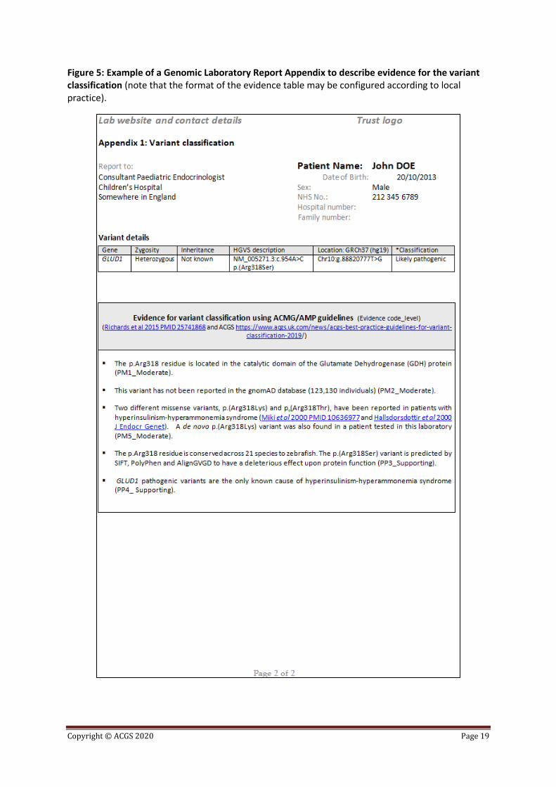
to the report (see example in Figure 5). Variants classified as likely benign or benign should
not be reported.

Copyright © ACGS 2020 Page 18
Figure 4: Example of a Genomic Laboratory Report
Copyright © ACGS 2020 Page 19
Figure 5: Example of a Genomic Laboratory Report Appendix to describe evidence for the variant classification (note that the format of the evidence table may be configured according to local practice).

Copyright © ACGS 2020 Page 20
In Table A we summarise our recommendations for reporting genomic variants. This table
describes which variants to include in the genomic laboratory report, text that should be used
within the result summary box (see Figure 4) and some explanatory notes. Additional
specific notes for recessive disorders are provided in Table B.
Table A: Recommended approach to reporting genomic variants in probands (*see
Figure 6 for sub-classifications of variants of uncertain significance). Note from Richards et
al 2015 that pathogenic is proposed to mean a 99% certainty that the variant is disease-
causing and likely pathogenic equates to 90% certainty.
Classification Variant included in report?
Result summary for genomic laboratory report
Explanatory notes
Pathogenic Yes Genetic diagnosis of disorder X OR Genetic diagnosis of (“Gene name”) - related disorder OR Confirms a genetic diagnosis of disorder X (if there was clinical
suspicion of this specific disorder)
Very high likelihood that the variant is causative of the disorder >99% certainty that the variant is pathogenic
Likely pathogenic
Yes Consistent with a genetic diagnosis of disorder X
High likelihood that the variant is causative of the disorder >90% certainty that the variant is pathogenic
Uncertain significance VUS where further testing or investigations could be considered as the results have the potential to change the classification to likely pathogenic
Yes Inconclusive result – consider further action Clearly describe in the report the action required that could change the classification to likely pathogenic
Further testing or investigations could be undertaken in order to re-classify the variant as likely pathogenic. Examples include:
a) testing parents to determine whether de novo
(PS2/PM6) b) mRNA analysis
for variants predicted to affect normal splicing (PS3)
c) testing affected

Copyright © ACGS 2020 Page 21
relatives to show co-segregation (PP1)
d) biochemical testing (PP4)
e) trial of a treatment that is specific for the genetic aetiology (PP4)
Uncertain significance
*hot/warm/tepid VUS where no further evidence can be obtained 1 strong + 1 supporting
OR 2 moderate + 1
supporting OR 1 moderate + 3
supporting OR 1 strong OR 2 moderate OR 1 moderate + 2
supporting OR 4 supporting OR 1 moderate + 1
supporting OR 3 supporting
Not usually A genetic cause for the patient’s disorder has not been identified
State in the result text that the result does not exclude a genetic diagnosis
These variants should only be reported in exceptional circumstances following MDT discussion (see section 4.1.1).
Uncertain significance
*cool/cold/ice cold VUS 1 moderate OR 2 supporting OR 1 supporting OR No supporting
evidence
No A genetic cause for the patient’s disorder has not been identified
State in the result text that the result does not exclude a genetic diagnosis
These variants are almost invariably unlikely to be disease-causing and are potentially confusing if included in the report. They should only be reported in exceptional circumstances following MDT discussion (see section 4.1.1).
Likely benign
No A genetic cause for the
patient’s disorder has not been identified
State in the result text that the result does not exclude a genetic diagnosis
These variants are not relevant and potentially confusing if included in the report >90% certainty that the variant is benign
Benign No A genetic cause for the patient’s disorder has not been identified
These variants are not relevant and potentially confusing

Copyright © ACGS 2020 Page 22
State in the result text that the result does not exclude a genetic diagnosis
if included in the report >99.9% certainty that the variant is benign
Variant previously reported in the literature as (likely) pathogenic but now classified as (likely) benign
No A genetic cause for the patient’s disorder has not been identified
State in the result text that the result does not exclude a genetic diagnosis
There is no clinical utility in reporting these historical false positive results and potential risk of misinterpretation
A variant type/mechanism that does not fit with the established disease mechanism
No A genetic cause for the patient’s disorder has not been identified
State in the result text that the result does not exclude a genetic diagnosis
For example a loss of function variant in a gene where the disease mechanism is gain of function or is mediated via an effect upon a specific protein structure

Copyright © ACGS 2020 Page 23
Figure 6: Diagram to illustrate the different ways to describe variants of uncertain significance with differing levels of evidence in support of pathogenicity

Copyright © ACGS 2020 Page 24
Table B: Additional recommendations for reporting genomic variants in recessive
disorders
Classification Variant(s) included in report?
Result summary for genomic laboratory report
Explanatory notes
Recessive disorder with two pathogenic variants identified and known to be in trans
Yes Genetic diagnosis of disorder X OR Genetic diagnosis of (“Gene name”)-related disorder OR Confirms a genetic diagnosis of disorder X (if cases where there
was clinical suspicion of this specific disorder)
Very high likelihood that the variants are causative of the disorder >99% certainty that each variant is pathogenic
Recessive disorder with one pathogenic and one likely pathogenic variant identified and known to be in trans
Consistent with a genetic diagnosis of disorder X
High likelihood that the variants are causative of the disorder >90% certainty that each variant is pathogenic
Recessive disorder with two likely pathogenic variants identified and known to be in trans
Consistent with a genetic diagnosis of disorder X
High likelihood that the variants are causative of the disorder >90% certainty that each variant is pathogenic
Recessive disorder with two pathogenic variants identified where not known if variants are in trans
Yes Consistent with a genetic diagnosis of disorder X
High likelihood that the variants are causative of the disorder >99% certainty that each variant is pathogenic
Recessive disorder with one pathogenic and one likely pathogenic variant
Yes Possible genetic diagnosis of disorder X – parental testing required
Likely that the variants are causative of the disorder

Copyright © ACGS 2020 Page 25
identified where not known if variants are in trans
>90% certainty that each variant is pathogenic
Recessive disorder with two likely pathogenic variants identified where not known if variants are in trans
Yes Possible genetic diagnosis of disorder X – parental testing required
Likely that the variants are causative of the disorder >90% certainty that each variant is pathogenic
Recessive disorder with one (likely) pathogenic variant and a hot VUS
Yes Possible genetic diagnosis of disorder X – additional evidence required to confirm or refute this result
When it is not possible to obtain sufficient evidence to classify the VUS as likely pathogenic but all the available clinical, gene-level and variant-level evidence supports the likely diagnosis
Recessive disorder with one (likely) pathogenic variant
Only appropriate in
specific circumstances e.g. patient from a consanguineous
family
See section 6.1.3 Either an incidental carrier finding or a second (likely) pathogenic variant has not been detected
Recessive disorder with one variant of uncertain significance identified
No A genetic cause for the patient’s disorder has not been identified
State in the result text that the result does not exclude a genetic diagnosis
There is no clinical utility in reporting a single heterozygous VUS and potential risk of misinterpretation
6.1. Variants of uncertain significance
The reporting of variants of uncertain significance can be challenging and the consequences
of a misdiagnosis due to misunderstanding the significance of a reported variant of uncertain
significance may have wider implications beyond the proband. It is essential to use clinical
judgement and consider discussion in a multidisciplinary setting (i.e. with the referring
clinician).
With the caveat that current variant classifications are not quantitative, the likelihood of a
variant of uncertain significance being pathogenic is intended to range from 10% to 90%
(Richards et al 2015). Richards et al (2015) also noted that “some laboratories may choose
to sub-classify VUSs, particularly for internal use”. Figure 6 illustrates the different VUS
categories according to the ACMG/AMP guidelines, using posterior probabilities estimated

Copyright © ACGS 2020 Page 26
from a Bayesian approach, on a points scale (derived from the Bayesian approach) and
using a temperature gradient sub-classification which includes the (albeit unofficial) “hot
VUS” description commonly used in the UK. The aim of Figure 6 is to convey the different
levels of uncertainty within the “variant of uncertain significance” category. Whilst
laboratories and clinicians may find it helpful to use VUS sub-classifications, these should
not be included in the genomic laboratory report.
6.1.1. Situations where considering reporting a VUS might be appropriate
Variants of uncertain significance should generally only be considered for reporting
where there is a high level of supporting evidence “hot/warm VUS” and additional
evidence might be obtained to allow re-classification as likely pathogenic.
This might include discussion in a multidisciplinary setting where possible (i.e. with the
referring clinician) to determine whether parental samples might be available to demonstrate
a de novo variant (PS2/PM6) and parental relationships, if there are sufficient affected
relatives (number of informative meiosis) available to show co-segregation (PP1), whether
neuroimaging/muscle biopsy or a biochemical test could provide phenotype specificity
evidence (PP4), whether trial of a treatment that is specific for the genetic aetiology (e.g.
biotin in a patient with biallelic BTH variants) or mRNA analysis in support of aberrant
splicing (PS3). If it is thought that additional evidence could allow re-classification of the
variant as likely pathogenic the initial report should clearly state the further action to be
considered and explain how this might change the variant classification to likely pathogenic.
We recommend that the following text is included in the Result Summary box: “Inconclusive
result – consider further action” and that the further investigations or tests that might be
undertaken are clearly detailed in the “Recommended action” section. This is particularly
important for tests where discussion with the referring clinician is not feasible, for example
high throughput tests that include the analysis of large gene panels in singletons where de
novo variants are a common mechanism of disease. Please note that the emphasis is on
additional testing to obtain evidence in support of pathogenicity. In some cases the new
information will re-classify the variant of uncertain significance as likely benign, but routine
practice should not include additional testing is to prove that a variant with little supporting
evidence in favour of pathogenicity is benign.
Exceptional circumstances
There may be situations in which an MDT discussion concludes that there is clinical utility in
reporting a variant of uncertain significance. This would usually be at the “warm/hot” VUS
level, where it may be impossible to obtain sufficient evidence at this time to reach a variant
classification of likely pathogenic, but where all the available clinical, gene-level and variant-
level evidence supports the likely diagnosis. One scenario might be a rare autosomal
recessive disease with a specific phenotype and one pathogenic or likely pathogenic variant
plus a hot VUS. In this situation we recommend that the following text is included in the
Result Summary box: “Possible genetic diagnosis of disorder X – additional evidence
required to confirm or refute this result”. The results section should include a statement
to the effect that “additional evidence is required to confirm (or refute) this possible
autosomal recessive diagnosis, for example identification of this variant in additional patients
with a similar clinical presentation”.

Copyright © ACGS 2020 Page 27
Where there is moderate evidence for pathogenicity, i.e. a “tepid VUS”, the prior
probability of pathogenicity is particularly important. For example if there are multiple
affected individuals within the family, the specific gene (or biological pathway) was indicated
by the referring clinician or the clinical presentation suggests a very high likelihood of a
monogenic disorder.
Variants of uncertain significance where there is a lower level of supportive evidence
include those for which there is either one moderate, two supporting or one
supporting piece of evidence “cool/cold/ice cold”. In most clinical settings and for most
genes, these “cool/cold/ice cold” variants of uncertain significance are almost invariably
unlikely to be disease-causing and should only be reported in exceptional circumstances.
MDT discussion may be helpful for determining this. Most frequently these are novel
missense variants (PM2 – absent from gnomAD) in genes with missense constraint (PP2) or
for which in silico tools predict a deleterious effect on protein function (PP3). This level of
evidence should be considered circumstantial evidence in the absence of a high level of
phenotypic specificity.
There are some specific exceptional circumstances where a prior decision may be
made by the laboratory and expert clinical team to report certain variants of uncertain
significance in specific genes. Examples include where a well-established specific
pharmacological therapy is recommended for a genetic disorder and a treatment trial may be
considered for a “hot VUS”. For example, low dose sulphonylurea therapy is recommended
for patients with (likely) pathogenic HNF1A or HNF4A variants causing monogenic diabetes
(Pearson et al 2003) and biotin treatment is effective for patients with biallelic (likely)
pathogenic BTH variants.
6.1.2. Situations where reporting a VUS would not be considered appropriate
There are some additional situations where VUSs should not be reported. These
include:
(i) Variants reported in the published literature and mutation databases for which subsequent
scientific evidence has re-classified the variant as likely benign or benign. Examples include
the RET p.(Tyr791Phe) missense variant (Toledo et al 2015) and BRCA1 c.594-2A>C (de la
Hoya et al 2016). There is no clinical utility in reporting these historical false positive results
and past experience has demonstrated the potential for risk of misinterpretation of such
information;
(ii) Variant type/mechanism that does not fit with the established disease mechanism, for
example protein truncating variants predicted to result in nonsense-mediated decay in a
gene where the known disease mechanism is gain of function due to activating missense
variants, or loss of function variants in NOTCH3 in CADASIL, where this variant type is well
established to be benign;
(iii) VUS in a gene only associated with an autosomal recessive disease, where a second
candidate variant has not been detected.

Copyright © ACGS 2020 Page 28
6.1.3. Reporting a heterozygous (likely) pathogenic variant for a gene
associated with an autosomal recessive disease
For single gene testing where the prior probability of a particular autosomal recessive
disorder is high, the finding of a single monoallelic variant in a gene associated with the
autosomal recessive disorder would be reported as “at least a carrier and this result
increases the likelihood of a diagnosis of disorder X”. An example would be a patient with a
positive sweat test undergoing testing for common CFTR variants. This reporting rationale is
based on (a) the prior probability from the phenotype and (b) the incomplete nature of the
test i.e. testing only the most common pathogenic CFTR variants.
When testing large gene panels, if we find a single monoallelic (likely) pathogenic variant in
a gene associated with an autosomal recessive disorder it means that either (a) incidental
carrier status has been revealed or (b) a second variant has been inherited in trans but has
not been detected. If the patient’s phenotype is not compatible with the disorder or biallelic
variants explaining the phenotype have been identified in another gene, then the decision as
to whether to report a finding of incidental carrier status depends upon whether cascade
testing would be offered for relatives and their partners. This is defined by clinical policies
that take into consideration the disease prevalence, whether there are common variants that
could be tested and whether there is known consanguinity. We do not recommend reporting
of carrier status in these conditions as a default approach because such variants are not of
relevance to the clinical presentation. Every individual is likely to be a carrier for multiple rare
diseases and reporting those variants in the context of a test to investigate the cause of the
patient’s rare disorder has greater potential to mislead than to appropriately inform. Work is
underway to develop policy for reporting of incidental findings.
In the situation that we find a single monoallelic (likely) pathogenic variant in a gene
associated with an autosomal recessive disorder and the patient’s phenotype is compatible
with this disorder, then the decision to report will depend upon the phenotypic specificity,
size of the gene panel, known clinical sensitivity of the test and whether a second variant
might be detected by another diagnostic testing method.
6.1.4. Specific VUS reporting considerations related to WGS, WES and large
gene panel tests
Variant classification is dependent upon current knowledge of reported gene-disease
associations. An evidence-based framework for assessing these associations has been
developed by the ClinGen group (Strande et al 2017). Variants identified within a candidate
disease gene, a “gene of uncertain significance” should always be classified as variants of
uncertain significance (Richards et al 2015). Such variants should only be included in the
genomic laboratory report if there is robust evidence for the gene-disease association and
publication is pending. An updated report should be issued after publication to include the
reference.
The advent of exome sequencing has not only identified many new disease genes not
known previously associated with human disease, but also identified novel phenotypes
linked to known disease genes, thereby expanding the phenotypic spectrum. For the
purposes of clinical diagnostic testing, the focus must be on known gene-disease
associations. For example, if a novel missense variant is identified in a gene associated with
a particular set of phenotypic features but the patient’s clinical presentation fits with only a

Copyright © ACGS 2020 Page 29
single/subset of non-specific features of the disorders or is out-with the phenotypic spectrum
associated with the disease gene, we might hypothesise that the restricted clinical
presentation is a consequence of the missense variant being hypomorphic or that the patient
represents an extension of the currently established disease spectrum. In the absence of
other phenotypically-matched patients with hypomorphic variants confirmed by functional
studies, a case series establishing a new phenotype association with the disease gene or
additional robust evidence, such variants should not be reported.
6.2. Storage of variant data for future re-analysis
Next generation sequencing of large gene panels, a whole exome or whole genome will
identify multiple variants of uncertain significance. Bioinformatic analysis pipelines use
different algorithms designed to prioritise rare variants according to criteria such as mode of
inheritance, predicted effect on protein function and presence in disease databases such as
HGMDpro. These pipelines then output lists of annotated genomic variants for manual
review. The number of variants in a prioritised variant list is correlated with the number (and
size) of the genes analysed and the specific filtering parameters that the pipeline uses. The
majority of prioritised variants will be variants of uncertain significance since common
variants classified as likely benign using the BA1 criterion are routinely filtered out. It is also
important to understand that there are many reasons why a disease-causing variant (or
variant pair) may not be included in the prioritised variant list. These include the specified
filtering parameters of the pipeline (genes not in the gene panel used for analysis), variants
within poorly sequenced regions, gene not yet known to be associated with the disease
phenotype, types of variants not detectable by the bioinformatics pipeline, variants within
regulatory elements, deep intronic cryptic splicing variants or mosaic variants. For this
reason it is important to store sequence data in an accessible, secure way in order to permit
future re-analysis within the laboratory. A systematic process for retrieving an individual
patient’s data is essential and is more readily achieved at the laboratory level than by storing
lists of prioritised VUS obtained through a particular pipeline at one defined point in time in
patient notes where there is no robust or reliable mechanism for review of these.
7. Reclassification of variants
Variant data and relevant associated information must be stored within the laboratory in a
way that allows reclassification if required. National guidance is needed to define re-analysis
of stored exome or genome sequence data for patients in whom no analysis is found during
the initial testing process.
Sharing of variant data on a global scale in a manner that conforms to UK information
governance requirements is a goal supported by the ACGS and BSGM (British Society for
Genetic Medicine). The DECIPHER database (https://decipher.sanger.ac.uk/) hosts an NHS
consortium project to allow sharing of variant data in a restricted manner. This allows
member laboratories to identify conflicting classifications for the same variant to enable
submitters to discuss the most appropriate classification based on the available evidence.
Reassessment of a variant that results in reclassification may be prompted by the publication
of new knowledge regarding the variant (or gene-disease association); by a request for a
family member test or as a result of further clinical investigations or evolution of the patient’s
phenotype that questions the original diagnosis.

Copyright © ACGS 2020 Page 30
We propose that reclassification of a variant across categories that fundamentally changes
the clinical relevance – i.e. not from likely benign to benign or likely pathogenic to pathogenic
(or vice versa) – should be shared with other relevant health care professionals. The
laboratory where the new information is generated should liaise with any laboratories (where
this is feasible) that generated the original classification status to ensure consistency across
centres. The new classification data and the basis for this classification should be placed in a
publicly accessible database so that the information is available widely. If the new
classification has potential importance for clinical management e.g. classification of a
BRCA1 or BRCA2 variant that may alter decisions around risk reducing mastectomy, this
decision should be documented (through an MDT) and communicated to the patient’s clinical
team(s) as quickly as possible. For some variants, for example the re-classification of
BRCA1 c.594-2A>C (de la Hoya et al 2016), rapid communication of this information to all
other UK diagnostic genetics laboratories is clearly appropriate (using a designated secure
NHS e-mail address for each laboratory). With the implementation of the NHS England
Genomic Medicine Service a more regulated method for data-sharing will be created in the
near future and could be adopted UK-wide, but ultimately it is the professional responsibility
of the Clinical Genomic community to ensure data is shared responsibly for improved patient
care.
Acknowledgements
We are grateful to Dr Steven Harrison, Professor Heidi Rehm (Harvard, USA) and Dr Helen
Firth (Addenbrooke’s Hospital, Cambridge, UK) for extremely helpful discussions and all
their support. We thank all our colleagues from the Association for Clinical Genomic Science
who have contributed to the revision of these guidelines.
References
Biesecker LG, Harrison SM. The ACMG/AMP reputable source criteria for the interpretation of sequence
variants. Genet Med. 2018 Mar 15. doi: 10.1038/gim.2018.42. [Epub ahead of print] PubMed PMID: 29543229.
de la Hoya M, Soukarieh O, López-Perolio I, Vega A, Walker LC, van Ierland Y, Baralle D, Santamariña M,
Lattimore V, Wijnen J, Whiley P, Blanco A, Raponi M, Hauke J, Wappenschmidt B, Becker A, Hansen TV, Behar
R, Investigators K, Niederacher D, Arnold N, Dworniczak B, Steinemann D, Faust U, Rubinstein W, Hulick PJ,
Houdayer C, Caputo SM, Castera L, Pesaran T, Chao E, Brewer C, Southey MC, van Asperen CJ, Singer CF,
Sullivan J, Poplawski N, Mai P, Peto J, Johnson N, Burwinkel B, Surowy H, Bojesen SE, Flyger H, Lindblom A,
Margolin S, Chang-Claude J, Rudolph A, Radice P, Galastri L, Olson JE, Hallberg E, Giles GG, Milne RL, Andrulis
IL, Glendon G, Hall P, Czene K, Blows F, Shah M, Wang Q, Dennis J, Michailidou K, McGuffog L, Bolla MK,
Antoniou AC, Easton DF, Couch FJ, Tavtigian S, Vreeswijk MP, Parsons M, Meeks HD, Martins A, Goldgar DE,
Spurdle AB. Combined genetic and splicing analysis of BRCA1 c.[594-2A>C; 641A>G] highlights the relevance of
naturally occurring in-frame transcripts for developing disease gene variant classification algorithms. Hum Mol
Genet. 2016 Jun 1;25(11):2256-2268. PubMed PMID: 27008870.
Havrilla JM, Pedersen BS, Layer RM, Quinlan AR. A map of constrained coding regions in the human genome.
Nat Genet. 2019 Jan;51(1):88-95. doi: 10.1038/s41588-018-0294-6. Epub 2018 Dec 10. PubMed PMID:
30531870.
Ioannidis NM, Rothstein JH, Pejaver V, Middha S, McDonnell SK, Baheti S, Musolf A, Li Q, Holzinger E, Karyadi
D, Cannon-Albright LA, Teerlink CC, Stanford JL, Isaacs WB, Xu J, Cooney KA, Lange EM, Schleutker J, Carpten
JD, Powell IJ, Cussenot O, Cancel-Tassin G, Giles GG, MacInnis RJ, Maier C, Hsieh CL, Wiklund F, Catalona WJ,

Copyright © ACGS 2020 Page 31
Foulkes WD, Mandal D, Eeles RA, Kote-Jarai Z, Bustamante CD, Schaid DJ, Hastie T, Ostrander EA, Bailey-Wilson
JE, Radivojac P, Thibodeau SN, Whittemore AS, Sieh W. REVEL: An Ensemble Method for Predicting the
Pathogenicity of Rare Missense Variants. Am J Hum Genet. 2016 Oct 6;99(4):877-885. PubMed PMID.
Jarvik GP, Browning BL. Consideration of Cosegregation in the Pathogenicity Classification of Genomic Variants.
Am J Hum Genet. 2016 Jun 2;98(6):1077-81. doi: 10.1016/j.ajhg.2016.04.003. Epub 2016 May 26. PubMed
PMID: 27236918.
Kelly MA, Caleshu C, Morales A, Buchan J, Wolf Z, Harrison SM, Cook S, Dillon MW, Garcia J, Haverfield E,
Jongbloed JDH, Macaya D, Manrai A, Orland K, Richard G, Spoonamore K, Thomas M, Thomson K, Vincent LM,
Walsh R, Watkins H, Whiffin N, Ingles J, van Tintelen JP, Semsarian C, Ware JS, Hershberger R, Funke
B.cAdaptation and validation of the ACMG/AMP variant classification framework for MYH7-associated
inherited cardiomyopathies: recommendations by ClinGen's Inherited Cardiomyopathy Expert Panel. Genet
Med. 2018 Mar;20(3):351-359. doi: 10.1038/gim.2017.218. Epub 2018 Jan 4. PubMed PMID: 29300372.
Lek M, Karczewski KJ, Minikel EV, Samocha KE, Banks E, Fennell T, O'Donnell-Luria AH, Ware JS, Hill AJ,
Cummings BB, Tukiainen T, Birnbaum DP, Kosmicki JA, Duncan LE, Estrada K, Zhao F, Zou J, Pierce-Hoffman E,
Berghout J, Cooper DN, Deflaux N, DePristo M, Do R, Flannick J, Fromer M, Gauthier L, Goldstein J, Gupta N,
Howrigan D, Kiezun A, Kurki MI, Moonshine AL, Natarajan P, Orozco L, Peloso GM, Poplin R, Rivas MA, Ruano-
Rubio V, Rose SA, Ruderfer DM, Shakir K, Stenson PD, Stevens C, Thomas BP, Tiao G, Tusie-Luna MT, Weisburd
B, Won HH, Yu D, Altshuler DM, Ardissino D, Boehnke M, Danesh J, Donnelly S, Elosua R, Florez JC, Gabriel SB,
Getz G, Glatt SJ, Hultman CM, Kathiresan S, Laakso M, McCarroll S, McCarthy MI, McGovern D, McPherson R,
Neale BM, Palotie A, Purcell SM, Saleheen D, Scharf JM, Sklar P, Sullivan PF, Tuomilehto J, Tsuang MT, Watkins
HC, Wilson JG, Daly MJ, MacArthur DG; Exome Aggregation Consortium.. Analysis of protein-coding genetic
variation in 60,706 humans. Nature. 2016 Aug 18;536(7616):285-91. doi: 10.1038/nature19057. PubMed
PMID: 27535533.
Loeys BL, Dietz HC, Braverman AC, Callewaert BL, De Backer J, Devereux RB, Hilhorst-Hofstee Y, Jondeau G,
Faivre L, Milewicz DM, Pyeritz RE, Sponseller PD, Wordsworth P, De Paepe AM. The revised Ghent nosology
for the Marfan syndrome. J Med Genet. 2010 Jul;47(7):476-85. doi: 10.1136/jmg.2009.072785. PubMed
PMID:20591885.
Mazoyer S, Dunning AM, Serova O, Dearden J, Puget N, Healey CS, Gayther SA, Mangion J, Stratton MR, Lynch
HT, Goldgar DE, Ponder BA, Lenoir GM. A polymorphic stop codon in BRCA2. Nat Genet. 1996 Nov;14(3):253-4.
DOI: 10.1038/ng1196-253 PMID: 8896551
Moog U, Kutsche K, Kortüm F, Chilian B, Bierhals T, Apeshiotis N, Balg S, Chassaing N, Coubes C, Das S, Engels
H, Van Esch H, Grasshoff U, Heise M, Isidor B, Jarvis J, Koehler U, Martin T, Oehl-Jaschkowitz B, Ortibus E, Pilz
DT, Prabhakar P, Rappold G, Rau I, Rettenberger G, Schlüter G, Scott RH, Shoukier M, Wohlleber E, Zirn B,
Dobyns WB, Uyanik G. Phenotypic spectrum associated with CASK loss-of-function mutations. J Med Genet.
2011 Nov;48(11):741-51. doi: 10.1136/jmedgenet-2011-100218. Epub 2011 Sep 27. PubMed PMID: 2195428.
Pearson ER, Starkey BJ, Powell RJ, Gribble FM, Clark PM, Hattersley AT. Genetic cause of hyperglycaemia and
response to treatment in diabetes. Lancet. 2003 Oct 18;362(9392):1275-81. PMID: 14575972
Posey JE, Harel T, Liu P, Rosenfeld JA, James RA, Coban Akdemir ZH, Walkiewicz M, Bi W, Xiao R, Ding Y, Xia F,
Beaudet AL, Muzny DM, Gibbs RA, Boerwinkle E, Eng CM, Sutton VR, Shaw CA, Plon SE, Yang Y, Lupski JR.
Resolution of Disease Phenotypes Resulting from Multilocus Genomic Variation. N Engl J Med. 2017 Jan
5;376(1):21-31. doi: 10.1056/NEJMoa1516767. Epub 2016 Dec 7. PubMed PMID: 27959697.
Richards S, Aziz N, Bale S, Bick D, Das S, Gastier-Foster J, Grody WW, Hegde M, Lyon E, Spector E, Voelkerding
K, Rehm HL; ACMG Laboratory Quality Assurance Committee. Standards and guidelines for the interpretation

Copyright © ACGS 2020 Page 32
of sequence variants: a joint consensus recommendation of the American College of Medical Genetics and
Genomics and the Association for Molecular Pathology. Genet Med. 2015 May;17(5):405-24. doi:
10.1038/gim.2015.30. Epub 2015 Mar 5. PubMed PMID: 25741868.
Samocha KE, Kosmicki JA, Karczewski KJ, O’Donnell-Luria AH, Pierce-Hoffman E, MacArthur DG, Neale BM, Daly
MJ. Regional missense constraint improves variant deleteriousness prediction. bioRxiv
doi: https://doi.org/10.1101/148353.
Saunders CJ, Miller NA, Soden SE, Dinwiddie DL, Noll A, Alnadi NA, Andraws N, Patterson ML, Krivohlavek LA,
Fellis J, Humphray S, Saffrey P, Kingsbury Z, Weir JC, Betley J, Grocock RJ, Margulies EH, Farrow EG, Artman M,
Safina NP, Petrikin JE, Hall KP, Kingsmore SF. Rapid whole-genome sequencing for genetic disease diagnosis in
neonatal intensive care units. Sci Transl Med. 2012 Oct3;4(154):154ra135. doi: 10.1126/scitranslmed.3004041.
PubMed PMID: 23035047.
Strande NT, Rooney Riggs E, Buchanan AH, Ceyhan-Birsoy O, Dwight ST, Goldstein JL, Ghosh R, Seifert BA,
Sneddon TP, Wright MW, Milko LV, Giovanni MA, Murray MF, O'Daniel JM, Ramos EM, Santani AB, Scott AF,
Cherry JM, Plon SE, Rehm HL, Lese Martin C, Berg JS; Evaluating the clinical validity of gene-disease
associations: an evidence-based framework developed by the Clinical Genome Resource. Am J Hum Genet.
2017 Jun 1;100(6):895-906. doi:10.1016/j.ajhg.2017.04.015. Epub 2017 May 25. PubMed PMID: 28552198.
Tavtigian SV, Greenblatt MS, Harrison SM, Nussbaum RL, Prabhu SA, Boucher KM, Biesecker LG; ClinGen
Sequence Variant Interpretation Working Group (ClinGen SVI). Modeling the ACMG/AMP variant classification
guidelines as a Bayesian classification framework. Genet Med. 2018 Sep;20(9):1054-1060.
doi:10.1038/gim.2017.210. Epub 2018 Jan 4. PubMed PMID: 29300386.
Tayoun AA, Pesaran T, DiStefano M, Oza A, Rehm H, Biesecker L, Harrison S, ClinGen Sequence Variant
Interpretation Working Group. Recommendations for Interpreting the Loss of Function PVS1 ACMG/AMP
Variant Criteria. Hum Mutat. 2018 Nov;39(11):1517-1524. doi: 10.1002/humu.23626. Epub 2018 Sep 7. PMID:
30192042.
Toledo RA, Loureço DM Jr, Camacho C, Lindsey S, Cerutti J, Maciel RM, Toledo SP. RET Y791F: alone or
accompanied? Arch Endocrinol Metab. 2015 Oct;59(5):476-7. PMID: 26537412.
Traynelis J, Silk M, Wang Q, Berkovic SF, Liu L, Ascher DB, Balding DJ, Petrovski S. Optimizing genomic medicine
in epilepsy through a gene-customized approach to missense variant interpretation. Genome Res. 2017
Oct;27(10):1715-1729. doi: 10.1101/gr.226589.117. Epub 2017 Sep 1. PubMed PMID: 28864458.
van der Velde KJ, de Boer EN, van Diemen CC, Sikkema-Raddatz B, Abbott KM, Knopperts A, Franke L, Sijmons
RH, de Koning TJ, Wijmenga C, Sinke RJ, Swertz MA. GAVIN: Gene-Aware Variant INterpretation for medical
sequencing. Genome Biol. 2017 Jan 16;18(1):6. doi: 10.1186/s13059-016-1141-7. PubMed PMID: 2809307.
Whiffin N, Minikel E, Walsh R, O'Donnell-Luria A, Karczewski K, Ing AY, Barton PJR, Funke B, Cook SA,
MacArthur DG, Ware JS; Using high-resolution variant frequencies to empower clinical genome interpretation.
Genet Med. 2017 Oct;19(10):1151-1158. doi: 10.1038/gim.2017.26. Epub 2017 May 18. PubMed PMID:
28518168.