Practical Management of Juice Flow and Levels using Model ...

Dias et al. Lipids in Health and Disease 2014, 13:24http://www.lipidworld.com/content/13/1/24
RESEARCH Open Access
Acerola (Malpighia emarginata DC.) juice intakeprotects against alterations to proteins involvedin inflammatory and lipolysis pathways in theadipose tissue of obese mice fed a cafeteria dietFernando Milanez Dias2†, Daniela Dimer Leffa1†, Francine Daumann1, Schérolin de Oliveira Marques2,Thais F Luciano2, Jonathan Correa Possato2, Aline Alves de Santana3, Rodrigo Xavier Neves4, José Cesar Rosa5,Lila Missae Oyama3, Bruno Rodrigues6, Vanessa Moraes de Andrade1, Cláudio Teodoro de Souza2
and Fabio Santos de Lira7*
Abstract
Background: Obesity has been studied as a metabolic and an inflammatory disease and is characterized byincreases in the production of pro-inflammatory adipokines in the adipose tissue.To elucidate the effects of natural dietary components on the inflammatory and metabolic consequences of obesity,we examined the effects of unripe, ripe and industrial acerola juice (Malpighia emarginata DC.) on the relevantinflammatory and lipolysis proteins in the adipose tissue of mice with cafeteria diet-induced obesity.
Materials/methods: Two groups of male Swiss mice were fed on a standard diet (STA) or a cafeteria diet (CAF) for13 weeks. Afterwards, the CAF-fed animals were divided into five subgroups, each of which received a differentsupplement for one further month (water, unripe acerola juice, ripe acerola juice, industrial acerola juice, or vitaminC) by gavage. Enzyme-linked immunosorbent assays, Western blotting, a colorimetric method and histology wereutilized to assess the observed data.
Results: The CAF water (control obese) group showed a significant increase in their adiposity indices andtriacylglycerol levels, in addition to a reduced IL-10/TNF-α ratio in the adipose tissue, compared with the controllean group. In contrast, acerola juice and Vitamin C intake ameliorated the weight gain, reducing the TAG levelsand increasing the IL-10/TNF-α ratio in adipose tissue. In addition, acerola juice intake led to reductions both in thelevel of phosphorylated JNK and to increases in the phosphorylation of IκBα and HSLser660 in adipose tissue.
Conclusions: Taken together, these results suggest that acerola juice reduces low-grade inflammation and amelioratesobesity-associated defects in the lipolytic processes.
Keywords: Cafeteria diet, Malpighia emarginata DC, Adipose tissue, Inflammation, Lipolysis
* Correspondence: [email protected]†Equal contributors7Immunometabolism Research Group, Department of Physical Education,Universidade Estadual Paulista, UNESP, Rua Roberto Simonsen, 305,19060-900 Presidente Prudente, SP, BrazilFull list of author information is available at the end of the article
© 2014 Dias et al.; licensee BioMed Central Ltd. This is an Open Access article distributed under the terms of the CreativeCommons Attribution License (http://creativecommons.org/licenses/by/2.0), which permits unrestricted use, distribution, andreproduction in any medium, provided the original work is properly cited. The Creative Commons Public Domain Dedicationwaiver (http://creativecommons.org/publicdomain/zero/1.0/) applies to the data made available in this article, unless otherwisestated.

Dias et al. Lipids in Health and Disease 2014, 13:24 Page 2 of 9http://www.lipidworld.com/content/13/1/24
IntroductionObesity is characterized by an excessive accumulation ofbody fat due to a chronic state of positive energy balanceresulting from unhealthy dietary habits and sedentaryphysical activity patterns [1]. Adipose tissue plasticity iscoordinated through several steps, and increases in thefatty mass come about through an unbalance betweenlipolytic processes, which involve two principal lipases,hormone sensitive lipase (HSL) and adipose triglyceridelipase (ATGL) [2], and lipogenesis processes, which aremainly controlled by fatty acid synthase (FAS) and acetylCoA carboxylase (ACC) [3].Obesity has been studied as a metabolic and an in-
flammatory disease and is characterized by increases inthe production of pro-inflammatory adipokines in theadipose tissue, which establish systemic low-grade in-flammation [4]. Obesity has also become one of the fore-most problems facing public health; its incidence isassociated with an elevated risk for many types of cancer,cardiovascular diseases, dementia, type 2 diabetes andother co-morbidities.Along with the increasing prevalence of obesity, a
number of drugs have been developed to treat the asso-ciated metabolic disruptions, focusing on achieving in-creased fat mobilization and oxidation and decreased fatabsorption and appetite [5]. However, these drugs havegenerally been unsuccessful due to their low efficacy orlarge side effects [6].In recent years, numerous bioactive compounds in fruits
have been explored for their potential anti-obesity effects;the nutritional components of fruits have the potential toenact a number of positive health effects toward theprevention of lifestyle-related diseases [6,7]. Acerola(Malpighia emaginata DC.) is a fruit found throughoutCentral America and into the northern part of SouthAmerica. This fruit is well known as one of the best nat-ural sources of vitamin C and has become extremelypopular among the health-conscious population [8]. Asidefrom vitamin C, acerola contains other similarly functionalingredients, such as carotenoids, gamma-amino butyricacid (GABA) and polyphenols [9].Dietary bioactive compounds have been demonstrated
to possess anti-inflammatory effects on the adipose tis-sue by several mechanisms [1]. Previous studies [10] and[11] have shown that the nuclear factor kappa B (NFκB)transcription factor is a key mediator of inflammation inadipocyte cells. Currently, studies have shown a close rela-tionship between toll-like receptor 4 (TLR-4) and the acti-vation of the NF-κB pathway, which leads to the elevationof pro-inflammatory adipokine genes and protein expres-sion in adipose tissues [11,12]. In addition, Feingold et al.[13] demonstrated that pro-inflammatory adipokines in-duce lipolysis, principally, IL-6 and TNF-α. However,the opposite is also true; free fatty acid can also lead to
inflammation [14]. In 2005, Suganami et al. [15] showedthat free fatty acids produced by lipolysis in adipocytesactivated monocytes, creating a paracrine loop betweenlipolysis and inflammation. However, at present, weunderstand that adipocytes express several members ofthe family of Toll-Like Receptors (TLRs), which canrecognize free fatty acids and induce the production ofpro-inflammatory adipokines by adipocytes [6].However, relatively little is known about the underlying
mechanisms regulating body weight, lipolytic actions andthe relevant inflammatory status. Thus, the aims of thisstudy were to examine the effects of acerola juice on bodyfat mass and the presence of lipolytic enzymes in adiposetissue in mice fed a cafeteria diet and to observe whetherreducing the fat mass is associated with a diminished levelof low-grade inflammation.
Methods and materialsAnimalsThirty-six male Swiss albino mice, at 25 grams of bodyweight and 5–6 weeks of age, were obtained from the Ani-mal Center from Universidade do Extremo Sul Catarinense(UNESC, Brazil). The experimental protocols for this studywere approved by the Local Ethics Committee for AnimalUse (CEUA) of UNESC, Brazil (register No. 130/2011).The mice were randomized by weight and housed at 6 ani-mals per cage under standard room temperature (22 ± 2°C)under a 12 h light/dark cycle. The animals were acclimatedto their environment for 1 week before the beginning ofthe experiment. Thirty-six animals were divided into 2groups, a control group (6 animals) and cafeteria dietgroup (30 animals). The animals were fed with a standarddiet (STA) or cafeteria diet (CAF) for the 13 weeks. Afterthis period, the animals of the CAF group were subdividedinto five subgroups (6 animals/group): mice treated withvehicle (CAF +water distilled), mice treated with 1 mg/kgof vitamin C (CAF +Vitamin C), and mice treated with0.1 mL/10 g of animal per day with three different acerolajuices: industrial (CAF + Industrial), unripe (CAF +Unripe)and ripe (CAF + Ripe). The industrial acerola juice wasobtained from Da Fruta® (Pernambuco, Brazil). The un-ripe and ripe acerola (Malpighia emerginata DC.) juiceswere purchased from the Nutrilite Farm (Ceará, Brazil).L-ascorbic acid (Chemical Abstract Service RegisterNumber 50-81-7) was purchased from Sigma–Aldrich(Porto Alegre, Brazil), and to obtain the desired finaldose, vitamin C was dissolved in distilled water just be-fore each experiment. All of these diets were adminis-tered to the animals by gavage. The animals wereweighed weekly, and their food intake was recordeddaily (data not shown). After 4 weeks on these differenttreatments, the animals of each group were sacrificed.Then, blood samples were collected to evaluate theTAG levels, and epididymal adipose tissue pads were

Table 1 Macronutrient composition of the diets
Composition Standard diet Cafeteria diet
(2.93 Kcal/g) (4.12 Kcal/g)
g/100 g Kcal/100 g g/100 g Kcal/100 g
Protein 22.00 88.00 7.67 30.68
Carbohydrate 53.00 212.00 41.97 167.88
Saturated fat 4.00 36.00 23.76 213.84
Dias et al. Lipids in Health and Disease 2014, 13:24 Page 3 of 9http://www.lipidworld.com/content/13/1/24
dissected for histology, immunoassay, and western blotanalysis. All adipose tissues (epididymal, mesenteric, andretroperitoneal) were weighed for adiposity index.
Experimental dietsThe STA (Nuvilab CR-1, NUVITAL®, Curitiba, PR, Brazil)provided an energy content of 2.93 kcal/g, and the CAFtotaled 4.12 kcal/g (the constituents of each diet are de-scribed in Table 1). The palatable high-calorie diet (cafe-teria diet) was chosen because mimics the modernpatterns of human food consumption and has been use-fully in other experimental studies to induce obesity inlean animals. This diet was adapted from a diet knownas the cafeteria diet or Western diet, previously de-scribed by Shafat et al. [16]. Both the standard chow andthe experimental diet were replaced daily with freshfood. The animals receiving the cafeteria (hypercaloricdiet) and standard diet had free access to standard chowand water (see weekly menu in Table 2).
Serum triacylglycerol assayFasting animal serum triglyceride levels were assessed bythe colorimetric method with a commercial kit Labtest(Brazil).
TNF-α, and IL-10 protein level determination by ELISAFollowing decapitation, epididymal adipose tissue wasremoved, dissected, homogenized, and centrifuged at12,000 g for 40 min at 4°C. The supernatants were usedfor protein quantification, according to the Bradfordmethod, with bovine serum albumin (BSA) as a refer-ence. The quantitative assessment of TNF-α and IL-10proteins was carried out by ELISA (DuoSet ELISA, R & DSystems, Minneapolis, MN), following the recommendations
Table 2 Weekly menu of animals fed with cafeteria diet
Days of week Food
Monday Mortadella, marshmallow, cheese
Tuesday Chocolate crackers, Doritos®, cho
Wednesday Paçoca peanuts, chow Nuvilab®, m
Thursday Mortadella, chocolate wafer, calf'
Friday, Saturday and Sunday Chocolate crackers, chow Nuvilab
of the manufacturer. All samples were run as duplicates,and the mean value is reported.
Protein analysis by immunoblottingEpididymal adipose tissue was homogenized immediatelyin extraction buffer (1% Triton-X 100, 100 mM Tris,pH 7.4, containing 100 mM sodium pyrophosphate,100 mM sodium fluoride, 10 mM EDTA, 10 mM sodiumvanadate, 2 mM PMSF and 0.1 mg of aprotinin/ml) at 4°C(Polytron MR 2100, Kinematica, Switzerland). The ex-tracts were centrifuged at 9,000 × g and 4°C (5804R,Eppendorf AG, Hamburg, Germany) for 40 min to removeinsoluble materials. The supernatants were used for pro-tein quantification, according to the Bradford method.The proteins were denatured by boiling in Laemmli sam-ple buffer containing 100 mM DTT, run on SDS-PAGE,and transferred to nitrocellulose membranes. The mem-branes were blocked, probed and blotted with primaryantibodies. The antibodies used for immunoblotting werephospho AMPKThr172, phospho HSLserine563, phosphoHSLserine660 (Cell Signaling Biotechnology, Beverly, MA,USA) and perilipina A, CGI-58, ATGL, phospho IκB-αserine32/36, phospho JNK183/Tyr185, NF-κBp50, and TLR4(Santa Cruz Biotechnology, Santa Cruz, CA, USA). Theoriginal membrane was stripped and reblotted with α-tubulin loading protein. Chemiluminescent detection wasperformed with horseradish peroxidase-conjugate second-ary antibodies (Thermo Scientific, Rockford, IL, USA).Autoradiographs of membranes were taken for thevisualization of protein bands. The results of the blotsare presented as direct comparisons of the area of ap-parent bands in autoradiographs and were quantified bydensitometry using the Scion Image software (ScionImage software, ScionCorp, Frederick, MD).
Histological techniqueEpididymal adipose tissue samples were prepared in paraf-fin blocks, which subsequently were subjected to 4 μm-thick histological sectioning for slide preparation, alwaysstained with hematoxylin-eosin. All slides were examinedunder an optical microscope by a pathologist who wasunaware of the origin of the material and of the objectivesof the study. Using a program for image analysis, thecomputer-assisted Image-Pro Plus 6.0 (Media Cybernetics,
chips, chocolate wafer, chow Nuvilab®, water and Guaraná soft drink
w Nuvilab®, sausage hot dog, water and Guaraná soft drink
ortadella, cheese chips, water and Guaraná soft drink
s foot jelly, Doritos®, chow Nuvilab®, water and cola soft drink
®, sausage hot dog, bacon chips, marshmallow, water and cola soft drink

0
5
10
15
20
25
STA CAF+WD CAF+Vit C CAF+IND CAF+UNR CAF+RIP
$
Bod
y w
eigh
t (g)
0
1
2
3
4
5
**
Adi
posi
ty in
dex
(% o
f bo
dy w
eigh
t)
$
STA CAF+WD CAF+Vit C CAF+IND CAF+UNR CAF+RIP
*
0
50
100
150
200
#
$
TAG
(m
g/dL
)
STA CAF+WD CAF+Vit C CAF+IND CAF+UNR CAF+RIP
ba
c
*
$
*# * # *
Figure 1 Effects of cafeteria diet on metabolic parameters. Analysis of body weight (g) (a); adiposity index (% of body weight) (b); andcirculating TAG (mg/dL) (c) of mice fed the standard diet (STA), the cafeteria diet with distilled water (CAF +WD), the cafeteria diet with vitaminC (CAF + Vit C), the cafeteria diet with industrial acerola juice (CAF + IND), the cafeteria diet with the juice of unripe acerolas (CAF + UNR) or thecafeteria diet with the juice of ripe acerolas (CAF + RIP). The results are expressed as means ± SEM (n = 6 per group). $p < 0.05 versus standarddiet; *p < 0.05 versus the cafeteria diet with distilled water; #p < 0.05 versus the cafeteria diet with vitamin C.
0
10
20
30
40
50
60
*
0
25
50
75
100
125
150
175
200
#
0
1
2
3
4
5
* * **
-1g
tp(
FN
Tf o g
/)
ni eor p
-1/
( -
er p
o gn
IL10
)i
t o f
gp
1-F
NT/
1-I
/ g
gf
)i et
p n
oro
p(0
L
STA CAF+WD CAF+Vit C CAF+IND CAF+UNR CAF+RIPSTA CAF+WD CAF+Vit C CAF+IND CAF+UNR CAF+RIP
STA CAF+WD CAF+Vit C CAF+IND CAF+UNR CAF+RIP
$
$
* * **
ab
c
Figure 2 Effects of cafeteria diet on inflammatory parameters in epididymal adipose tissue. TNF-α (pg/μg protein-1) (a); IL-10 (pg/ug protein-1)(b); and IL-10/TNF-α ratio (c) of mice fed the standard diet (STA), the cafeteria diet with distilled water (CAF +WD), the cafeteria diet with vitamin C(CAF + Vit C), the cafeteria diet with industrial acerola juice (CAF + IND), the cafeteria diet with the juice of unripe acerolas (CAF + UNR) or the cafeteriadiet with the juice of ripe acerolas (CAF + RIP). The results are expressed as means ± SEM (n = 6 per group). $p < 0.05 versus standard diet; *p < 0.05versus the cafeteria diet with distilled water; #p < 0.05 versus the cafeteria diet with vitamin C.
Dias et al. Lipids in Health and Disease 2014, 13:24 Page 4 of 9http://www.lipidworld.com/content/13/1/24

Dias et al. Lipids in Health and Disease 2014, 13:24 Page 5 of 9http://www.lipidworld.com/content/13/1/24
Bethesda, MD, USA), we were able to measure the area,mean diameter and perimeter.
Statistical analysisThe data were expressed as means ± SEM. The normal-ity of the variables was evaluated using the Kolmogo-rov–Smirnov test. First, the unpaired t-test was used toevaluate the standard and cafeteria diets. Next, the dif-ferent treatments (water, unripe acerola juice, ripe acer-ola juice, industrial acerola juice, and vitamin C) wereanalyzed using one-way analysis of variance (ANOVA).When ANOVA showed significant differences, post hocanalysis was performed with the Tukey’s test. A probabil-ity of less than 0.05 was considered significant. The soft-ware used to analyze the data was the Statistical Packagefor the Social Sciences (SPSS) version 16.0 for Windows.
ResultsFirst, we evaluated the effects of the cafeteria diet on thebody weight of the mice. The cafeteria diet increased thebody weight in the CAF +WD group when comparedwith standard diet group (p < 0.01). Although the treat-ment not was efficient in reducing the weight of the ex-perimental mice (p > 0.05; Figure 1a), an interestingresult was observed in the adiposity index. The cafeteria
Figure 3 Effects of cafeteria diet on inflammatory molecules in epididNF-κB (c); and TLR4 (d) in the adipose tissue of mice fed the standard dietdiet with vitamin C (CAF + Vit C), the cafeteria diet with industrial acerola ju(CAF + UNR) or the cafeteria diet with the juice of ripe acerola (CAF + RIP).versus standard diet; *p < 0.05 versus the cafeteria diet with distilled water;cafeteria diet with industrial acerola juice.
diet markedly increased the adiposity index in the CAF +WD group when compared with STA group (p < 0.05),while the CAF + Vit C, CAF + IND, CAF +UNR, andCAF + RIP groups exhibited a reduced adiposity indexwhen compared with the CAF +WD group (p < 0.05;Figure 1b). No significant difference was found among thetreated groups (p > 0.05). To determine the adiposityindex, the epididymal, mesenteric and retroperitonealpads were weighed. To observe whether the treatment in-duced increased lipolysis, we assayed the triacylglycerollevels. The TAG levels were increased in the CAF +WDand CAF + Vit C groups when compared with the STAgroup (p < 0.05). However, the TAG levels in the CAF +IND, CAF +UNR, and CAF + RIP groups were reducedwhen compared with the CAF +WD and CAF +Vit Cgroups (p < 0.05; Figure 1c).We observed that the TNF-α levels were increased in
the CAF +WD group when compared with the STAgroup (p < 0.05), although the TNF-α levels were reducedin all of the treated groups when compared with the CAF +WD group (p < 0.05). There was no difference in the TNF-αlevels among the treatment groups (p > 0.05; Figure 2a).When the IL-10 levels (an important anti-inflammatorycytokine) were analyzed, we observed that the IL-10 levelswere unchanged between the CAF +WD and STA groups
ymal adipose tissue. The levels of phosphorylated pIκB (a); pJNK (b);(STA), the cafeteria diet with distilled water (CAF +WD), the cafeteriaice (CAF + IND), the cafeteria diet with the juice of unripe acerolasThe results are expressed as means ± SEM (n = 6 per group). $p < 0.05#p < 0.05 versus the cafeteria diet with vitamin C; @p < 0.05 versus the

Dias et al. Lipids in Health and Disease 2014, 13:24 Page 6 of 9http://www.lipidworld.com/content/13/1/24
(p > 0.05), even though the IL-10 levels were reduced inthe CAF +Vit C group when compared with the CAF +WD group (p < 0.05). In addition, the IL-10 levels werehigher in the CAF + IND group than in the CAF + Vit Cgroup (p < 0.05; Figure 2b). We observed that the IL-10/TNF-α ratio was reduced in the CAF +WD group whencompared with the standard diet group (p < 0.05). How-ever, the IL-10/TNF-α ratio in the CAF + Vit C, CAF +IND, CAF +UNR, and CAF + RIP groups was increasedwhen compared with the CAF +WD group (p < 0.05;Figure 2c).Next, we evaluated the protein levels of the mole-
cules involved in the transduction of pro-inflammatorysignals (pIκ-Bα, pJNK, NF-κB, TLR4), which are
Figure 4 Effects of cafeteria diet on lipolysis molecules in epididymal(b); HSLSer660 (c);and protein levels the Peri A (d); CGI-58 (e); and ATGL (f) idiet with distilled water (CAF +WD), the cafeteria diet with vitamin C (CAFcafeteria diet with the juice of unripe acerolas (CAF + UNR) or the cafeteriaas means ± SEM (n = 6 per group). $p < 0.05 versus standard diet; *p < 0.05cafeteria diet with vitamin C; @p < 0.05 versus the cafeteria diet with indust
presented as representative bands. The pJNK proteinlevels were reduced in the CAF + IND, CAF + UNR,and CAF + RIP groups (p < 0.05; Figure 3b) when com-pared with the CAF +WD group. There was no differ-ence among the groups in the NF-κBp50 protein levels(p > 0.05; Figure 3c). Hoping to evaluate whether lipo-lytic enzymes were altered by the treatment, we evalu-ated the levels of pAMPK, pHSLser563, pHSLser660,PeriA, CGI-58, and ATGL. The results demonstrated ahigher phosphorylation of HSL at the serine 563 and660 sites in the CAF + Vit C and CAF + IND groups(p < 0.05) when compared with the CAF +WD group,suggesting increased lipolytic activity in these groups(Figure 4).
adipose tissue. The levels of phosphorylated AMPKThr172 (a); HSLSer563
n the adipose tissue of mice fed the standard diet (STA), the cafeteria+ Vit C), the cafeteria diet with industrial acerola juice (CAF + IND), thediet with the juice of ripe acerola (CAF + RIP). The results are expressedversus the cafeteria diet with distilled water; #p < 0.05 versus therial acerola juice.
Dias et al. Lipids in Health and Disease 2014, 13:24 Page 7 of 9http://www.lipidworld.com/content/13/1/24
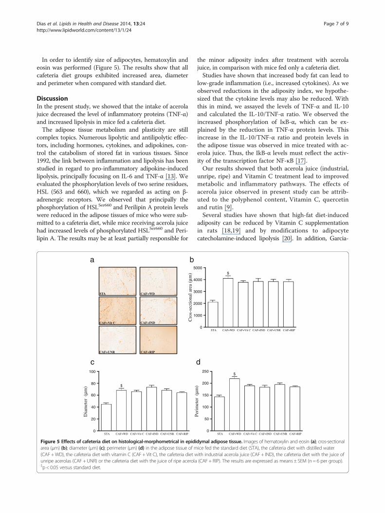
In order to identify size of adipocytes, hematoxylin andeosin was performed (Figure 5). The results show that allcafeteria diet groups exhibited increased area, diameterand perimeter when compared with standard diet.
DiscussionIn the present study, we showed that the intake of acerolajuice decreased the level of inflammatory proteins (TNF-α)and increased lipolysis in mice fed a cafeteria diet.The adipose tissue metabolism and plasticity are still
complex topics. Numerous lipolytic and antilipolytic effec-tors, including hormones, cytokines, and adipokines, con-trol the catabolism of stored fat in various tissues. Since1992, the link between inflammation and lipolysis has beenstudied in regard to pro-inflammatory adipokine-inducedlipolysis, principally focusing on IL-6 and TNF-α [13]. Weevaluated the phosphorylation levels of two serine residues,HSL (563 and 660), which we regarded as acting on β-adrenergic receptors. We observed that principally thephosphorylation of HSLSer660 and Perilipin A protein levelswere reduced in the adipose tissues of mice who were sub-mitted to a cafeteria diet, while mice receiving acerola juicehad increased levels of phosphorylated HSLSer660 and Peri-lipin A. The results may be at least partially responsible for
CAF+WD
CAF+IND
CAF+UNR
STA
CAF+Vit C
CAF+RIP
a
1
2
3
4
5
b
0
20
40
60
80
100
STA CAF+WD CAF+Vit C CAF+IND CAF+UNR CAF+RIP
$
c
Cro
s-se
ctio
nal a
rea
(µm
)
Dia
met
er (
µm)
Figure 5 Effects of cafeteria diet on histological-morphometrical in epidiarea (μm) (b); diameter (μm) (c); perimeter (μm) (d) in the adipose tissue of m(CAF +WD), the cafeteria diet with vitamin C (CAF + Vit C), the cafeteria diet wunripe acerolas (CAF + UNR) or the cafeteria diet with the juice of ripe acerola$p < 0.05 versus standard diet.
the minor adiposity index after treatment with acerolajuice, in comparison with mice fed only a cafeteria diet.Studies have shown that increased body fat can lead to
low-grade inflammation (i.e., increased cytokines). As weobserved reductions in the adiposity index, we hypothe-sized that the cytokine levels may also be reduced. Withthis in mind, we assayed the levels of TNF-α and IL-10and calculated the IL-10/TNF-α ratio. We observed theincreased phosphorylation of IκB-α, which can be ex-plained by the reduction in TNF-α protein levels. Thisincrease in the IL-10/TNF-α ratio and protein levels inthe adipose tissue was observed in mice treated with ac-erola juice. Thus, the IkB-α levels must reflect the activ-ity of the transcription factor NF-κB [17].Our results showed that both acerola juice (industrial,
unripe, ripe) and Vitamin C treatment lead to improvedmetabolic and inflammatory pathways. The effects ofacerola juice observed in present study can be attrib-uted to the polyphenol content, Vitamin C, quercetinand rutin [9].Several studies have shown that high-fat diet-induced
adiposity can be reduced by Vitamin C supplementationin rats [18,19] and by modifications to adipocytecatecholamine-induced lipolysis [20]. In addition, Garcia-
0
000
000
000
000
000
STA CAF+WD CAF+Vit C CAF+IND CAF+UNR CAF+RIP
$
0
50
100
150
200
250
STA CAF+WD CAF+Vit C CAF+IND CAF+UNR CAF+RIP
$
d
Peri
met
er (
µm)
dymal adipose tissue. Images of hematoxylin and eosin (a); cros-sectionalice fed the standard diet (STA), the cafeteria diet with distilled waterith industrial acerola juice (CAF + IND), the cafeteria diet with the juice of(CAF + RIP). The results are expressed as means ± SEM (n = 6 per group).

Dias et al. Lipids in Health and Disease 2014, 13:24 Page 8 of 9http://www.lipidworld.com/content/13/1/24
Diaz et al. [21] demonstrated that Vitamin C supplementationcan modulate an established inflammatory state in theinteraction between adipocytes and macrophages. More-over, resident macrophages produce catecholamines, whichstimulate lipolysis in white adipose tissue [22]. This path-way may be implicated in the central mechanisms of theincreased lipolysis and anti-inflammatory effects of acerolajuice, because IL-4, an anti-inflammatory cytokine, isnecessary for the activation of lipolysis. The associatedincrease in catecholamine production in macrophages[22] may explain the increased level of IκB-α proteinsand reduction in TNF-α protein levels.However, others biocompounds exist in acerola juice
that may be associated with the observed effects on me-tabolism and inflammation. Rivera et al. [23] related thatthe administration of quercetin reduced insulin resistance,dyslipidemia, and hypertension in rats. More recently,Overman et al. [24] reported that quercetin reduces in-flammation in adipose tissue by lowering the infiltrationof macrophages and suppressing NF-κB activation. Weobserved an increased level of phosphorylated Iκ-Bα pro-teins, which may indicate a suppression of NF-κB activation,confirming the observations of Overman and colleagues [24].Hsu et al. [25] showed that the body, liver organ, and
adipose tissue weights of the peritoneal and epididymalfat pads of mice on a high fat diet supplemented withrutin were significantly decreased, compared with thoseon a high fat diet without supplementation. In addition,the authors verified that the serum lipid profiles, insulin,and leptin were significantly decreased after treatmentwith rutin. Recently, Gao et al. [26] showed that rutin isable to block high fat diet-induced obesity, fatty liverand insulin resistance in mice. Further, these beneficialeffects were correlated with a blockade of macrophageinfiltration and chronic inflammation in the adipose tis-sues. In our study, we observed reduced TAG levels inaccordance with the above cited studies.Acerola juice suppresses glucose absorption and blood
glucose elevation after feeding [9]. This decrease in glucosedisposition by the energetic metabolism can be responsible,due to the increased fatty acid utilization, for maintainingthe energetic demand, ameliorating the vicious cycle oflipolysis-inflammation, and mobilizing the free fatty acidsfrom lipolysis to be used as an energy source. However, re-cently, Leffa et al. [27] showed that acerola juice is notable to alter or reverse insulin resistance in mice fed ahigh-fat diet. More studies are needed to achieve a betterunderstanding of the involved mechanisms.Finally, to verify the area of the adipocytes, we evalu-
ated the histology with hematoxylin and eosin (HE). Theresults demonstrated, at least in part, that all cafeteriadiet groups had an increased area of adipocytes whencompared with the STA group, regardless of the effectsof the different treatments.
In conclusion, our results showed that acerola juiceprevents weight gain (measured in terms of body weightand the adiposity index) and dyslipidemia (measuredusing TAG levels) and restores metabolic and inflamma-tory pathways to a normal range. Future studies are neededto better understand the mechanisms involved in the bene-ficial effects associated with acerola juice intake, especiallyin mice fed a cafeteria high-fat diet.
AbbreviationsSTA: Standard diet; CAF: Cafeteria diet; CAF +WD: Cafeteria diet + waterdistilled; CAF + IND: Cafeteria diet + acerola industrial; CAF + UNR: Cafeteriadiet + acerola Unripe; CAF + RIP: Cafeteria diet + acerola Ripe; CAF + VitC: Cafeteria diet + Vitamin C; TAG: Triglyceride; HSL: Hormone sensitive lipase;FAS: Fatty acid synthase; ACC: Acetyl CoA Carboxylase; TLR-4: Toll-likereceptor 4; NFκB: Nuclear factor kappa B; IL-6: Interleukin 6; IL-10: Interleukin10; TNF-α: Tumor necrosis factor alpha; CGI-58: Comparative geneidentification 58; ATGL: Adipose triglyceride lipase; IκB-α: Inhibitor of kappa Balpha; JNK: Jun N-terminal kinases; AMPK: 5′ AMP-activated protein kinase.
Competing interestAll authors declare no conflicts of interest.
Authors’ contributionsFMD, DDL, FD, VMA, contributed to the study design and protocol; SOM,TFL, JCP, AAS and RXN analyzed the samples; and JCR, LMO, CTS, and FSLwrote the manuscript and conducted the statistical analysis, with input andadvice from all authors. All authors read and approved the final manuscript.
AcknowledgmentsThis work was supported by Conselho Nacional de DesenvolvimentoCientífico, Tecnológico (CNPq), and Coordenação de Aperfeiçoamento dePessoal de Nível Superior (Capes).
Author details1Laboratory of Molecular and Cellular Biology, Graduate Programme ofHealth Sciences, Department of Health Sciences, Universidade do ExtremoSul Catarinense, UNESC, 888.06-000 Criciúma, SC, Brazil. 2Laboratory ofExercise Biochemistry and Physiology, Graduate Programme of HealthSciences, Department of Health Sciences, Universidade do Extremo SulCatarinense, UNESC, 888.06-000 Criciúma, SC, Brazil. 3Departamento deFisiologia, Disciplina de Fisiologia da Nutrição, Universidade Federal de SãoPaulo (UNIFESP), 04023-060 São Paulo, SP, Brazil. 4Cancer MetabolismResearch Group, Institute of Biomedical Sciences, University of Sao Paulo(USP), São Paulo, Brazil. 5Immunometabolism Research Group, Institute ofBiomedical Sciences, University of São Paulo (USP), São Paulo, Brazil.6Laboratório do Movimento Humano, Universidade São Judas Tadeu, SãoPaulo, SP, Brazil. 7Immunometabolism Research Group, Department ofPhysical Education, Universidade Estadual Paulista, UNESP, Rua RobertoSimonsen, 305, 19060-900 Presidente Prudente, SP, Brazil.
Received: 29 October 2013 Accepted: 21 January 2014Published: 4 February 2014
References1. Siriwardhana N, Kalupahana NS, Cekanova M, LeMieux M, Greer B,
Moustaid-Moussa N: Modulation of adipose tissue inflammation bybioactive food compounds. J Nutr Biochem 2013, 24(4):613–23.doi:10.1016/j.jnutbio.2012.12.013.
2. Watt MJ, Spriet LL: Triacylglycerol lipases and metabolic control:implications for health and disease. Am J Physiol Endocrinol Metab 2010,299:E162–168.
3. Xiao G, Zhang T, Yu S, Lee S, Calabuig-Navarro V, Yamauchi J, Ringquist S,Dong HH: ATF4 Protein Deficiency Protects against High Fructose-inducedHypertriglyceridemia in Mice. J Biol Chem 2013, 288(35):25350–61.
4. Ouchi N, Parker JL, Lugus JJ, Walsh K: Adipokines in inflammation andmetabolic disease. Nat Rev Immunol 2011, 11(2):85–97.doi: 10.1038/nri2921. Epub 2011 Jan 21. Review.
Dias et al. Lipids in Health and Disease 2014, 13:24 Page 9 of 9http://www.lipidworld.com/content/13/1/24
5. He J, Gao J, Xu M, Ren S, Stefanovic-Racic M, O'Doherty RM, Xie W: PXRablation alleviates diet-induced and genetic obesity and insulinresistance in mice. Diabetes 2013, 62(6):1876–87. doi:10.2337/db12-1039.Epub 2013 Jan 24.
6. Calder PC, Ahluwalia N, Brouns F, Buetler T, Clement K, Cunningham K,Esposito K, Jönsson LS, Kolb H, Lansink M, Marcos A, Margioris A, Matusheski N,Nordmann H, O'Brien J, Pugliese G, Rizkalla S, Schalkwijk C, Tuomilehto J,Wärnberg J, Watzl B, Winklhofer-Roob BM: Dietary factors and low-gradeinflammation in relation to overweight and obesity. Br J Nutr 2011,106(Suppl 3):S5–78. doi:10.1017/S0007114511005460. Review.
7. Cunha CA, Lira FS, Rosa Neto JC, Pimentel GD, Souza GI, da Silva CM,de Souza CT, Ribeiro EB, Sawaya AC, Oller do Nascimento CM, Rodrigues B,de Oliveira Carvalho P, Oyama LM: Green tea extract supplementationinduces the lipolytic pathway, attenuates obesity, and reduceslow-grade inflammation in mice fed a high-fat diet. Mediators Inflamm2013, 2013:635470.
8. Barbalho SM, Damasceno DC, Spada AP, Palhares M, Martuchi KA, Oshiiwa M,Sazaki V, da Silva VS: Evaluation of glycemic and lipid profile of offspring ofdiabetic Wistar rats treated with Malpighia emarginata juice. Exp DiabetesRes 2011, 2011:173647. doi:10.1155/2011/173647. Epub 2011 Jan 23.
9. Hanamura T, Uchida E, Aoki H: Skin-lightening effect of a polyphenolextract from Acerola (Malpighia emarginata DC.) fruit on UV-inducedpigmentation. Biosci Biotechnol Biochem 2008, 72(12):3211–8.Epub 2008 Dec 7.
10. Ajuwon KM, Spurlock ME: Palmitate activates the NF-kappaB transcriptionfactor and induces IL-6 and TNFalpha expression in 3 T3-L1 adipocytes.J Nutr 2005, 135(8):1841–6.
11. Lira FS, Rosa JC, Pimentel GD, Seelaender M, Damaso AR, Oyama LM, doNascimento CO: Both adiponectin and interleukin-10 inhibit LPS-inducedactivation of the NF-κB pathway in 3T3-L1 adipocytes. Cytokine 2012,57(1):98–106. doi: 10.1016/j.cyto.2011.10.001. Epub 2011 Nov 1.
12. Tsukumo DM, Carvalho-Filho MA, Carvalheira JB, Prada PO, Hirabara SM,Schenka AA, Araújo EP, Vassallo J, Curi R, Velloso LA, Saad MJ: Loss-of-functionmutation in Toll-like receptor 4 prevents diet-induced obesity and insulinresistance. Diabetes 2007, 56(8):1986–98. Epub 2007 May 22.
13. Feingold KR, Doerrler W, Dinarello CA, Fiers W, Grunfeld C: Stimulation oflipolysis in cultured fat cells by tumor necrosis factor, interleukin-1, andthe interferons is blocked by inhibition of prostaglandin synthesis.Endocrinology 1992, 130(1):10–6.
14. Estadella D, da Penha Oller do Nascimento CM, Oyama LM, Ribeiro EB,Dâmaso AR, de Piano A: Lipotoxicity: effects of dietary saturated andtransfatty acids. Mediators Inflamm. 2013, 2013:137579.
15. Suganami T, Nishida J, Ogawa Y: A paracrine loop between adipocytesand macrophages aggravates inflammatory changes: role of free fattyacids and tumor necrosis factor alpha. Arterioscler Thromb Vasc Biol 2005,25(10):2062–8. Epub 2005 Aug 25.
16. Shafat A, Murray B, Rumsey D: Energy density in cafeteria dietinduced hyperphagia in the rat. Appetite 2009, 52(1):34–8.doi: 10.1016/j.appet.2008.07.004. Epub 2008 Jul 17.
17. Zamboni M, Di Francesco V, Garbin U, Fratta Pasini A, Mazzali G, Stranieri C,Zoico E, Fantin F, Bosello O, Cominacini L: Adiponectin gene expressionand adipocyte NF-kappaB transcriptional activity in elderly overweightand obese women: inter-relationships with fat distribution, hs-CRP, leptinand insulin resistance. Int J Obes (Lond) 2007, 31(7):1104–9.Epub 2007 Feb 27.
18. Campión J, Milagro FI, Fernández D, Martínez JA: Diferential geneexpression and adiposity reduction induced by ascorbic acidsupplementation in a cafeteria model of obesity. J Physiol Biochem 2006,62(2):71–80.
19. García-Díaz D, Campión J, Milagro FI, Martínez JA: Adiposity dependentapelin gene expression: relationships with oxidative and inflammationmarkers. Mol Cell Biochem 2007, 305(1–2):87–94. Epub 2007 Jun 27.
20. Garcia-Diaz DF, Campion J, Milagro FI, Paternain L, Solomon A, Martinez JA:Ascorbic acid oral treatment modifies lipolytic response and behaviouralactivity but not glucocorticoid metabolism in cafeteria diet-fed rats. ActaPhysiol (Oxf ) 2009, 195(4):449–57. doi: 10.1111/j.1748-1716.2008.01942.x.Epub 2008 Nov 25.
21. Garcia-Diaz DF, Campion J, Quintero P, Milagro FI, Moreno-Aliaga MJ,Martinez JA: Vitamin C modulates the interaction between adipocytesand macrophages. Mol Nutr Food Res 2011, 55(Suppl 2):S257–63.doi: 10.1002/mnfr.201100296. Epub 2011 Jul 28.
22. Nguyen KD, Qiu Y, Cui X, Goh YP, Mwangi J, David T, Mukundan L,Brombacher F, Locksley RM, Chawla A: Alternatively activatedmacrophages produce catecholamines to sustain adaptivethermogenesis. Nature 2011, 480(7375):104–8.
23. Rivera L, Morón R, Sánchez M, Zarzuelo A, Galisteo M: Quercetinameliorates metabolic syndrome and improves the inflammatory statusin obese Zucker rats. Obesity (Silver Spring) 2008, 16(9):2081–7.doi:10.1038/oby.2008.315.
24. Overman A, Chuang CC, McIntosh M: Quercetin attenuates inflammationin human macrophages and adipocytes exposed to macrophage-conditioned media. Int J Obes (Lond). 2011, 35(9):1165–72.doi:10.1038/ijo.2010.272. Epub 2011 Jan 11.
25. Hsu CL, Wu CH, Huang SL, Yen GC: Phenolic compounds rutin ando-coumaric acid ameliorate obesity induced by high-fat diet in rats.J Agric Food Chem 2009, 57(2):425–31. doi:10.1021/jf802715t.
26. Gao M, Ma Y, Liu D: Rutin suppresses palmitic acids-triggered inflammationin macrophages and blocks high fat diet-induced obesity and fatty liver inmice. Pharm Res 2013 [Epub ahead of print].
27. Leffa DD, da Silva J, Daumann F, Dajori AL, Longaretti LM, Damiani AP,de Lira F, Campos F, Ferraz AD, Côrrea DS, de Andrade VM: Corrective effectsof acerola (Malpighia emarginata DC.) juice intake on biochemical andgenotoxical parameters in mice fed on a high-fat diet. Mutat Res 2013:S0027-5107(13)00194-2. doi: 10.1016/j.mrfmmm.2013.11.005.[Epub ahead of print].
doi:10.1186/1476-511X-13-24Cite this article as: Dias et al.: Acerola (Malpighia emarginata DC.) juiceintake protects against alterations to proteins involved in inflammatoryand lipolysis pathways in the adipose tissue of obese mice fed acafeteria diet. Lipids in Health and Disease 2014 13:24.
Submit your next manuscript to BioMed Centraland take full advantage of:
• Convenient online submission
• Thorough peer review
• No space constraints or color figure charges
• Immediate publication on acceptance
• Inclusion in PubMed, CAS, Scopus and Google Scholar
• Research which is freely available for redistribution
Submit your manuscript at www.biomedcentral.com/submit
Copyright © 2022 FDOKUMEN