Longitudinal Consumption of Ergothioneine Reduces ... - MDPI

Accumulation of mtDNA Mutations Disrupts CPC Function
1
Accumulation of Mitochondrial DNA Mutations Disrupts Cardiac Progenitor Cell Function and Reduces Survival
Amabel M. Orogo1, Eileen R. Gonzalez1, Dieter A. Kubli1, Igor L. Baptista1, Sang-Bing Ong1,
Tomas A. Prolla4, Mark A. Sussman3, Anne N. Murphy2, and Åsa B. Gustafsson1,2
From the 1Skaggs School of Pharmacy and Pharmaceutical Sciences, University of California, San Diego,
La Jolla, CA 92093
2Department of Pharmacology, University of California, San Diego, La Jolla, CA 92093
3San Diego Heart Research Institute, San Diego State University, San Diego, CA 92182
4Departments of Genetics and Medical Genetics, University of Wisconsin, Madison, WI 53706
*Running title: Accumulation of mtDNA Mutations Disrupts CPC Function To whom correspondence should be addressed: Dr. Åsa B. Gustafsson, Skaggs School of Pharmacy and Pharmaceutical Sciences, University of California, San Diego, 9500 Gilman Drive MC 0758, La Jolla, CA 92093-0758, USA. Tel : (858) 822-5569 ; Fax : (858) 822-7558; E-mail: [email protected] Keywords: Mitochondria, mitochondrial DNA, cardiac progenitor cells, aging, glycolysis, heart failure, stem cells, differentiation, mitophagy CAPSULE Background: Mitochondrial DNA (mtDNA) mutations accumulate with age but how this impacts cardiac progenitor cell (CPC) function is unknown. Results: mtDNA mutations disrupt mitochondrial function and reduce differentiation potential of CPCs. Conclusion: Preserving mtDNA integrity is critical for CPC homeostasis and regenerative potential. Significance: Increased understanding of pathways regulating progenitor cell function is critical for development of effective cell therapies. ABSTRACT
Transfer of cardiac progenitor cells (CPCs) improves cardiac function in heart failure patients. However, CPC function is reduced with age, limiting their regenerative potential. Aging is associated with numerous changes in cells including accumulation of mitochondrial DNA (mtDNA) mutations, but it is unknown how this impacts CPC function. Here, we demonstrate that acquisition of mtDNA mutations disrupts mitochondrial function, enhances mitophagy, and reduces the
replicative and regenerative capacities of the CPCs. We show that activation of differentiation in CPCs is associated with expansion of the mitochondrial network and increased mitochondrial oxidative phosphorylation. Interestingly, mutant CPCs are deficient in mitochondrial respiration and rely on glycolysis for energy. In response to differentiation, these cells fail to activate mitochondrial respiration. This inability to meet the increased energy demand leads to activation of cell death. These findings demonstrate the consequences of accumulating mtDNA mutations and importance of mtDNA integrity in CPC homeostasis and regenerative potential. INTRODUCTION
The heart contains a pool of c-kit positive cardiac progenitor cells (CPCs) that has the potential to repair the myocardium after injury by secreting paracrine factors, as well as producing new cardiac myocytes and vascular structures (1). However, the functional role of endogenous CPCs in the myocardium and how they contribute to repair are still unclear and controversial. Beltrami et al. initially reported that injection of CPCs into
http://www.jbc.org/cgi/doi/10.1074/jbc.M115.649657The latest version is at JBC Papers in Press. Published on July 16, 2015 as Manuscript M115.649657
Copyright 2015 by The American Society for Biochemistry and Molecular Biology, Inc.
at NA
TIO
NA
L U
NIV
ER
SITY
OF SIN
GA
POR
E on July 21, 2015
http://ww
w.jbc.org/
Dow
nloaded from

Accumulation of mtDNA Mutations Disrupts CPC Function
2
the infarct region leads to generation of new vessels and cardiac myocytes by the CPCs (2). More recently, it was reported that endogenous CPCs are necessary for both cardiac homeostasis and regeneration after injury (3). This study found that cardiac damage induced by isoproterenol treatment leads to CPC activation and commitment to the myocardial cell lineages, including cardiac myocytes. In contrast, a recent study found that differentiation of endogenous CPCs into cardiac myocytes occurs at a very low rate even after injury (4), while another study reported that new cardiac myocytes are generated only from pre-existing myocytes and that endogenous CPCs do not play a critical role in myocardial homeostasis and repair (5). Although the functional role of endogenous CPCs is controversial, it is clear that infusion of CPCs into the injured myocardium leads to repair and improved function. For instance, in animal models of myocardial infarction or doxorubicin-induced cardiomyopathy, injection of CPCs reduces injury and improves left ventricular function (6-8). More importantly, utilization of autologous CPCs in patients with ischemic cardiomyopathy in the SCIPIO clinical trial is showing promising results (9,10). Studies have found that CPC function is reduced with age which reduces their regenerative capacity (11-13). Since stem cell therapies for cardiovascular disease are primarily targeting geriatric patients, the autologous CPCs might have reduced regenerative capacity once they are transplanted back into the patient’s heart. The exact mechanisms underlying the age-related changes in CPC function are still unclear. Therefore, a deeper understanding of the biological processes that regulate CPC function and survival is needed so that more effective therapeutic strategies to repair the heart can be developed.
Mitochondria regulate several key processes including metabolism, heme synthesis, and cell death. Mitochondria are also responsible for producing energy via oxidative phosphorylation (OXPHOS) for cellular development, differentiation, and growth (14-16). Mitochondria contain their own DNA that is replicated independently of the nuclear DNA. The mitochondrial DNA (mtDNA) only encodes 37 genes and 13 of them are subunits of the respiratory complexes or the ATP synthase involved in OXPHOS. Unfortunately, mtDNA is more susceptible to genetic mutations than nuclear DNA
because it is constantly exposed to reactive oxygen species generated by the respiratory chain in the mitochondrial inner membrane. Studies have reported that mtDNA mutations and deletions accumulate with age in various tissues in humans and rodents which can lead to impaired mitochondrial function (17-21). Mutations in mtDNA in the heart have also been demonstrated after treatment with cardiotoxic therapies such as doxorubicin (22) or nucleoside reverse transcriptase inhibitors (23), and after myocardial infarction (24). The contribution of mtDNA mutations to aging has been confirmed by studies in mice expressing a proofreading-deficient mitochondrial DNA polymerase γ (POLG). These mice accumulate mtDNA mutations in cells at a faster rate than wild type mice, which leads to an accelerated aging phenotype and reduced lifespan (25,26). Moreover, accumulation of mtDNA mutations in the heart is associated with increased oxidative damage and apoptosis which results in the development of cardiomyopathy at 13-14 months of age (27). Recent studies have also implicated mitochondria as critical regulators of stem cell function. Embryonic, neuronal and mesenchymal stem cells have been reported to contain few immature mitochondria that are clustered in the perinuclear region, and to rely on glycolysis for energy production (28-30). However, differentiation of stem cells requires metabolic reprogramming to meet the increased energy demand that occurs concomitantly with a shift from cytosolic anaerobic glycolysis to mitochondrial respiration (29,31,32). To date, no attention has been given to the role of mitochondria in CPCs and how accumulation of mtDNA mutations impacts these cells. In this study, we demonstrate that functional mitochondria are critical for CPC function and survival. Accumulation of mtDNA mutations in CPCs leads to disruption of mitochondrial function, reduced proliferation, and increased susceptibility to stress. The mutant CPCs are also unable to transition from glycolysis to oxidative phosphorylation in response to differentiation, which instead leads to activation of cell death. Thus, our studies highlight the critical role of mitochondria for CPC function and the consequences of accumulating mtDNA mutations. EXPERIMENTAL PROCEDURES
at NA
TIO
NA
L U
NIV
ER
SITY
OF SIN
GA
POR
E on July 21, 2015
http://ww
w.jbc.org/
Dow
nloaded from

Accumulation of mtDNA Mutations Disrupts CPC Function
3
CPC Isolation and Culture – All experimental procedures were performed in accordance with institutional guidelines and approved by the Institutional Animal Care and Use Committee of the University of California San Diego. For experiments in Figure 1, C-kit positive CPCs were isolated from 2-month-old male wild-type FVB mice. For all other experiments, CPCs were isolated from male mice homozygous for the PolgD257A mutation on a C57Bl/6 background (25), and corresponding control CPCs were isolated from wild-type, sex-matched litter mates. All c-kit positive adult CPCs were isolated and cultured as previously described (33). To activate differentiation, CPCs were incubated in α-MEM containing 10 nM dexamethasone for up to seven days.
Primary Myoblast Isolation and Culture – Primary myoblasts were isolated from hind limb skeletal muscle of 3-4 month old WT and POLG mice litter mates, using a protocol adapted from rat skeletal muscle (34). Muscles were digested in 0.20% type IA collagenase and 0.25% trypsin in DMEM (GIBCO-Invitrogen), and cells then cultured in growth medium (20% FBS and 1% penicillin-streptomycin in DMEM) on a bed of Matrigel™ (on coverslip, 1 mg/mL, Basement Membrane Matrix; BD Biosciences). Myoblast differentiation was induced 5 days later with 2% horse serum and 1% penicillin-streptomycin in DMEM for 2-4 days.
Echocardiography – Echocardiography was performed as previously described (35) using a Vevo770 In Vivo Micro-Imaging System with an RMV707B 15-45 MHz imaging transducer (VisualSonics Inc.) and parameters quantified using the VisualSonics software.
Quantitative Polymerase Chain Reaction – Genomic DNA was extracted using GenElute Mammalian Genomic DNA Miniprep Kit (Sigma-Aldrich) and PCR amplified with TaqMan Universal Master Mix II. 18s rRNA was used as a control for nuclear DNA and d-loop for mtDNA quantitation, using primer sequences previously described (36). For gene expression assays, RNA was extracted using the RNeasy Mini Kit (Qiagen). cDNA was synthesized using the QuantiTect Reverse Transcription Kit (Qiagen) following manufacturer’s protocols. Taqman primers for PGC-1α, PGC-1β, and GATA-4 were from Integrated DNA Technologies. TaqMan primers for
18s, GATA-6, PECAM1, COX1 (Complex IV Subunit 1), Cox4i1 (Complex IV Subunit 4), and the TaqMan Universal Master Mix II were from Applied Biosystems. qPCR was carried out on a CFX96 Real-Time PCR Detection System (Bio-Rad). Relative amounts of mRNA were normalized to 18s and fold change in gene expression was calculated using the 2(-Delta Delta Ct) method.
Western Blot – Samples were prepared in lysis buffer containing 50 mM Tris-HCl, 150 mM NaCl, 1 mM EGTA, 1 mM EDTA, 1% Triton X-100, and protease inhibitor cocktail (Roche) and run on Invitrogen NuPAGE Bis-Tris gels. The membranes were probed with the following antibodies: MitoProfile Total OXPHOS Rodent WB Antibody cocktail (MitoSciences), PGC-1 (Abcam), COXIV Subunit 4 (Invitrogen), AMPK, p-AMPK, Hexokinase I, Hexokinase II, PKM1/2, GAPDH, LC3A/B (Cell Signaling Technology), DLP1 (BD Biosciences), MFN1, MyoD (Santa Cruz Biotechnology), MFN2, Tubulin (Sigma-Aldrich), and Actin (GeneTex). Membranes were imaged using a ChemiDoc XRS+ System (Bio-Rad).
Mitochondrial Respiration Measurements – Intact mitochondria were isolated from adult mouse hearts and oxygen consumption measurements were taken at 30°C using an Oxygraph apparatus (Hansatech Instruments). Complex I- and Complex II-dependent oxygen consumption were measured as previously described (35). Complex IV-dependent respiration was measured by addition of 1 µM Antimycin A and 0.4 mM TMPD + 1 mM ascorbate. Oligomycin (5 µg/ml) was added to inhibit ATP synthase followed by FCCP (1 µM) to obtain maximal respiration rate. Mitochondrial oxygen consumption of monolayers of CPCs was measured using the Seahorse XF96 analyzer (Seahorse Bioscience), adapted from a previously described protocol (36). CPCs were plated at 2 x 10⁴ or 1 x 10⁴ cells per well 24 hours before measurement of O2 consumption rates. Assays were run in unbuffered, serum-free DMEM containing 10 mM glucose, 3 mM glutamine, and 1 mM pyruvate. CPCs were allowed to equilibrate on the plate before addition of 2 µM oligomycin to measure ATP-linked respiration. Three successive additions of 500 nM FCCP were added to measure maximal respiration. Data was analyzed using Wave for Desktop software (Seahorse Bioscience).
at NA
TIO
NA
L U
NIV
ER
SITY
OF SIN
GA
POR
E on July 21, 2015
http://ww
w.jbc.org/
Dow
nloaded from

Accumulation of mtDNA Mutations Disrupts CPC Function
4
Immunofluorescence Microscopy – Cells were fixed, permeabilized, and blocked as previously described (35). CPCs were stained with antibodies against Tom20 (Santa Cruz Biotechnology), COXIV (Invitrogen), GATA-4 (Santa Cruz Biotechnology), or LAMP2 (Sigma-Aldrich). Myoblasts were stained with antibodies against MyoD and NCAM (Santa Cruz Biotechnology). Cells were incubated in Alexa Fluor 488 or 594 secondary antibodies (Life Technologies), followed by Hoechst 33342 (10 µg/ml, Life Technologies) to stain nuclei. Cells were imaged using a Carl Zeiss Axio Observer Z1, and Z-stacks were acquired using a high-resolution AxioCam MRm digital camera, a 63X oil immersion objective, and Zeiss AxioVision 4.8 software (Carl Zeiss). A minimum of 100 cells per group were counted for each condition.
Brightfield microscopy – Myoblasts were imaged at pre- and post-differentiation using a Carl Zeiss Axio Observer Z1 at 10X magnification. Myotube diameter was quantified using ImageJ software.
Transmission Electron Microscopy – Adult mouse hearts and CPCs were fixed, sectioned, and mounted as previously described (35). Sections were examined on a TECNAI G2 Spirit BioTWIN Transmission Electron Microscope equipped with an Eagle 4k HS digital camera (FEI).
Proliferation Assay – CPCs were seeded in a 12-well tissue culture plate and cell number was determined by counting using a hemocytometer 24 and 72 hrs later. CPCs were also seeded in a 96-well black wall clear bottom plate (Corning) and cell number was measured with the CyQUANT NF Cell Proliferation Assay Kit (Life Technologies). Fluorescence measurements were made using a SpectraMax M3 microplate reader and SoftMax Pro 5.4 software (Molecular Devices).
Cell Death Assay - CPCs were treated with H2O2 (Sigma-Aldrich), Doxorubicin Hydrochloride (Sigma-Aldrich), Sunitinib Malate (BioVision), or 2-deoxyglucose (Sigma-Aldrich). Cells were stained with Hoechst 33342 (10 µg/ml, Life Technologies) and YO-PRO-1 Iodide (100 nM, Life Technologies) for 15 min and immediately imaged using a Carl Zeiss Axio Observer Z1 at 10X magnification, AxioCam MRm digital camera, and Zeiss AxioVision 4.8 software (Carl Zeiss).
Glycolysis Assay – CPCs were seeded in a 12-well tissue culture plate and incubated in growth
medium for 4 days. L-lactate concentration in the growth medium was measured using a Glycolysis Cell-Based Assay Kit (Cayman Chemical) according to manufacturer’s protocol. Colorimetric measurements were made using a SpectraMax M3 microplate reader and SoftMax Pro 5.4 software (Molecular Devices).
ATP Assay – ATP levels were measured using the CellTiter-Glo Luminescent Cell Viability Assay (Promega, Madison, WI) according to the manufacturer’s protocol. CPCs (40,000 cells/100 µl) were added to a 96 well black wall clear bottom plate (Corning). Luminescent measurements were made using a SpectraMax M3 microplate reader and SoftMax Pro 5.4 software (Molecular Devices).
Assessment of mitochondrial membrane potential – CPCs were incubated with tetramethylrhodamine methyl ester (TMRM) (25 nM, Invitrogen) and Hoechst 33342 (10 µg/ml, Life Technologies) for 15 minutes at 37°C. Live cells were examined using a Carl Zeiss Axio Observer Z1 at 40X magnification. Fluorescence intensity was quantified using ImageJ software.
Assessment of autophagy and mitophagy – To assess baseline autophagic flux, CPCs were treated with DMSO or 50 nM Bafilomycin A1 (EMD Millipore) for 2 hours prior to homogenization and western blot analysis. To visualize lysosomes, CPCs were fixed and stained with anti-LAMP2 (Sigma-Aldrich). To assess mitochondrial autophagy, CPCs were infected with an adenovirus encoding GFP-LC3 as previously described (37). Cells were treated with DMSO or 50 nM Bafilomycin A1 for 3 hours, fixed, and stained with anti-Tom20. Cells were imaged at 63X magnification and Z-stacks acquired using a Carl Zeiss Axio Observer Z1. LAMP2 staining was quantified using ImageJ software. Mitophagy was assessed by analyzing colocalization between GFP-LC3 positive autophagosomes and Tom20 labeled mitochondria.
Statistical Analyses - All values are expressed as means + standard error. Student’s t-test was used to compare two sets of data. P values < 0.05 were considered statistically significant. RESULTS
Activation of the differentiation program in CPCs leads to induction of mitochondrial biogenesis – Differentiation is a process that requires energy which is primarily supplied by
at NA
TIO
NA
L U
NIV
ER
SITY
OF SIN
GA
POR
E on July 21, 2015
http://ww
w.jbc.org/
Dow
nloaded from

Accumulation of mtDNA Mutations Disrupts CPC Function
5
mitochondria via oxidative phosphorylation (16). When examining mitochondria in wild-type CPCs from FVBN mice, we found that undifferentiated CPCs contained few mitochondria that were localized primarily in the perinuclear region (Figure 1A). Incubation of CPCs in differentiation medium (DM) for 7 days led to increased cell size, and expansion of the mitochondrial network (Figure 1A). Next, we confirmed that incubation in DM led to upregulation of cardiac lineage markers in CPCs, consistent with previous studies (2,33,38,39). We observed an increase in cells positive for GATA-4, a transcription factor that regulates myocardial differentiation, growth, and survival, after incubation in DM. We found that few cells expressed GATA-4 at Day 0, but there was a significant increase in CPCs stained positively for GATA-4 after 7 days in DM which correlated with an expansion of their mitochondria (Figures 1A and 1B). Real time qPCR confirmed a significant increase in GATA-4 at Day 7 (Figure 1C). There were also significant increases in mRNA encoding the smooth muscle marker GATA-6 and the endothelial marker PECAM1 (Figure 1C). These results indicate that lineage commitment in CPCs is accompanied by expansion of mitochondria.
Next, we assessed whether the increased energy demand associated with activation of differentiation led to an activation of mitochondrial biogenesis. First we confirmed that AMPK, a major regulator of mitochondrial biogenesis in response to energy depletion, was activated during differentiation (Figure 1D). We also found that PGC-1α, a member of the PPARγ co-activator (PGC) family of transcriptional co-activators, was significantly induced in CPCs after incubation in DM (Figure 1E). The activation of AMPK and induction of PGC-1α correlated with an increase in mtDNA content (Figure 1F) and increased levels of mitochondrial OXPHOS proteins (Figure 1G) in the CPCs in DM. These studies suggest that initiation of differentiation in CPCs leads to activation of mitochondrial biogenesis and enhanced mitochondrial oxidative phosphorylation to adapt to increased energy demand.
mtDNA mutations impair CPC proliferation and increase susceptibility to cell death – To further explore the role of mitochondria in CPC function and survival, we isolated CPCs from 2 month-old mice with a proofreading defective mitochondrial DNA polymerase γ
(POLG) and their wild type (WT) litter mates. The POLG mice undergo premature aging and develop cardiomyopathy with a reduced lifespan due to accumulation of mtDNA mutations (25). However, at 2 months of age, we observed no significant differences in body, heart or lung weighs, and cardiac function in POLG mice when compared to age matched WT litter mates (Figures 2A-C). We also found no differences in cardiac mitochondrial content or function at this age between WT and POLG mice (Figures 2D-F). Analysis of mitochondrial ultrastructure by transmission electron microscopy (TEM) confirmed similar mitochondrial content and morphology in WT and POLG hearts at this age (Figure 2G).
We then investigated whether CPCs from these hearts were affected by the mutated POLG. Proliferation is important for self-renewal and maintenance of the CPC population in the heart and we found that the POLG CPCs had significantly reduced proliferation rates compared to WT CPCs (Figure 3A). In addition, the POLG CPCs were more sensitive to cell death after treatment with H2O2 (Figure 3B). In addition, treatment with doxorubicin, an anthracycline topoisomerase inhibitor, or sunitinib, a tyrosine kinase inhibitor, also resulted in increased cell death in POLG CPCs (Figure 3C). Thus, these data suggest that accumulation of mtDNA mutations in the POLG CPCs reduces their proliferative capacity and increases their susceptibility to stress.
mtDNA mutations contribute to abnormal mitochondrial morphology and reduced differentiation potential – Alterations in mitochondrial morphology allow cells to adapt to changes in the cellular environment, including differentiation (40). When examining the effect of accumulating mtDNA mutations on mitochondrial morphology, we discovered that the mutant POLG CPCs contained fragmented mitochondria under baseline conditions (Figures 4A and 4B). Moreover, while WT CPCs increased in cell size and expanded their mitochondrial network after incubation in DM, POLG CPCs maintained their fragmented mitochondria which clustered in the perinuclear region and failed to expand in response to the differentiation stimulus (Figure 4A). The abnormal mitochondrial morphology was also associated with reduced activation of the differentiation program in the POLG CPCs. We observed significantly fewer GATA-4 positive
at NA
TIO
NA
L U
NIV
ER
SITY
OF SIN
GA
POR
E on July 21, 2015
http://ww
w.jbc.org/
Dow
nloaded from

Accumulation of mtDNA Mutations Disrupts CPC Function
6
POLG CPCs compared to WT after incubation in DM (Figure 4C). The POLG CPCs also had significantly reduced levels of GATA-4 transcripts compared to WT CPCs after incubation in DM (Figure 4D). Thus, these data demonstrate that accumulation of mtDNA mutations leads to abnormal mitochondrial morphology and impaired activation of the differentiation program in CPCs.
The fragmented mitochondrial morphology in POLG CPCs prompted us to investigate whether proteins involved in mitochondrial dynamics were altered in the POLG CPCs. Surprisingly, we saw a reduction in the mitochondrial fission protein DRP1 in POLG CPCs after 7 days in DM (Figure 4E). Interestingly, mitochondrial fusion proteins MFN1 and MFN2 were increased in WT CPCs during differentiation, but not in POLG CPCs (Figure 4E). Thus, there is a shift in the balance between fission and fusion proteins towards fusion in WT CPCs, but not in POLG CPCs, upon activation of differentiation. Furthermore, we analyzed mitochondrial ultrastructure in CPCs by TEM and saw drastic differences between WT and POLG CPCs. Mitochondria in undifferentiated WT CPCs had underdeveloped cristae which became more electron-dense upon activation of the differentiation program (Figure 4F). In contrast, mitochondria in POLG CPCs were swollen with abnormal cristae structure under baseline conditions, and failed to increase cristae density after differentiation. Taken together, these data suggest that accumulation of mtDNA mutations leads to impaired mitochondrial dynamics and differentiation program in the CPCs.
Next, we investigated whether other progenitor cells were similarly affected by accumulation of mtDNA mutations. Satellite cells that reside on the basal lamina of the myofiber are important for skeletal muscle maintenance and facilitate repair by giving rise to myoblasts that fuse to form myofibers. Studies have reported that although the number of satellite cells declines with age, these cells still retain their myogenic potential (41,42). We found that myoblasts isolated from WT and POLG litter mates formed myotubes in culture upon differentiation. However, myotube diameter in the POLG mice was significantly smaller than WT (Figure 5A). Both WT and POLG myoblasts expressed the muscle phenotypic marker MyoD pre- and post-differentiation, and the adhesion marker NCAM after differentiation (Figures 5B and
5C). These data suggest that myoblasts from POLG mice are less affected by the presence of a mutant POLG. These myoblasts have sufficient capacity to adapt to metabolic changes during differentiation in order to successfully fuse and form myotubes. However, the smaller myofiber size suggests that defective POLG may affect growth signaling in differentiating myoblasts.
POLG CPCs have impaired mitochondrial biogenesis and energetics – The abnormal mitochondrial morphology and distribution in POLG CPCs are indicators of defective mitochondrial function. Since the primary function of mitochondria is to supply the cell with energy via oxidative phosphorylation, we compared OXPHOS protein levels between WT and POLG CPCs. As previously observed in Figure 1, incubation of WT CPCs in DM resulted in increased expression of OXPHOS proteins. In contrast, POLG CPCs had reduced levels of select OXPHOS proteins, most notably in Complex III and IV subunits (Figure 6A). In fact, we observed no increase in nuclear and mitochondrial encoded subunits of Complex IV in the POLG CPCs after incubation in DM for seven days (Figure 6B). The baseline protein levels were already significantly lower in POLG CPCs compared to WT at Day 0. Interestingly, we found that Complex IV Subunit 1 (Cox1) and Subunit 4 (Cox4i1) transcripts were similar in WT and POLG CPCs at Day 0 (Figure 6C), suggesting that the differences in protein levels between WT and POLG CPCs may be attributed to reduced protein stability in the POLG CPCs. We also found that, in contrast to WT CPCs, the POLG CPCs failed to induce mitochondrial biogenesis factors PGC-1α or PGC-1β to the same extent as WT CPCs during differentiation (Figure 6D). This suggests that accumulation of mtDNA mutations also impairs the mitochondrial biogenesis response.
Our data indicated that POLG CPCs have abnormal mitochondrial structure and contain reduced levels of critical proteins involved in OXPHOS. Therefore, we assessed the level of mitochondrial respiration in WT and POLG CPCs by measuring oxygen consumption using the Seahorse XF Analyzer. In WT CPCs, increased oxygen consumption rate corresponded with increased cell number in response to the mitochondrial uncoupler FCCP. Surprisingly, POLG CPCs had no detectable oxygen consumption regardless of cell number (Figure 7A),
at NA
TIO
NA
L U
NIV
ER
SITY
OF SIN
GA
POR
E on July 21, 2015
http://ww
w.jbc.org/
Dow
nloaded from

Accumulation of mtDNA Mutations Disrupts CPC Function
7
indicating that these cells have shut down mitochondrial respiration. The OXPHOS complexes are also responsible for generating the mitochondrial membrane potential, which is used by the F0F1-ATPase for ATP synthesis (43). We confirmed that the POLG CPCs had significantly reduced mitochondrial membrane potential as assessed by reduced TMRM staining (Figure 7B). TMRM uptake by mitochondria is dependent on the membrane potential. However, cellular ATP levels were similar in WT and POLG CPCs (Figure 7C), suggesting that POLG CPCs are utilizing alternative mechanisms to generate energy. Therefore, we examined whether the POLG CPCs relied on glycolysis to support their energy demands by assessing extracellular lactate, a glycolytic byproduct. We found that the POLG
CPCs had significantly increased extracellular L-lactate compared to WT CPCs (Figure 7D), and addition of 2-deoxyglucose to the growth medium to inhibit glycolysis resulted in rapid death of the POLG CPCs (Figure 7E). This suggests that POLG
CPCs depend solely on glycolysis to meet their energy demands.
Studies have found that differentiation is associated with a metabolic transition from glycolysis to oxidative phosphorylation (29,32). Since our data indicate that the POLG CPCs do not utilize oxidative phosphorylation for ATP production and differentiation is associated with an increase in energy demand, we examined the effect of differentiation on cell survival in these cells. We observed that a significant number of POLG CPCs underwent cell death in DM (Figure 8A). To gain further insight into the metabolic switch that occurs during differentiation, we analyzed levels of glycolytic enzymes in WT and POLG CPCs. We found that mitochondrial associated Hexokinase I increased in WT during differentiation but remained low in POLG CPCs. Hexokinase II, PKM1/2, and GAPDH decreased with time upon incubation in DM in both WT and POLG CPCs (Figures 8B and 8C). Taken together, these results suggest that both WT and POLG CPCs downregulate cytosolic glycolysis during differentiation. However, since POLG CPCs are unable to concurrently activate oxidative phosphorylation, the resulting energy deficiency combined with increased energy demand leads to activation of cell death.
POLG CPCs have increased mitophagy – Loss of mitochondrial membrane potential is a known trigger for mitochondrial autophagy or mitophagy. Mitophagy is a process by which defective mitochondria are sequestered in autophagosomes and subsequently degraded in lysosomes (44). Since mitochondria in the POLG CPCs are impaired, we examined whether these cells had increased levels of mitophagy. First, we assessed whether autophagic flux was altered in POLG CPCs. To assess flux, cells were treated with vehicle or Bafilomycin A1 to inhibit the fusion between autophagosomes and lysosomes. We found that both WT and POLG CPCs accumulated similar levels of the autophagy marker LC3II in the presence of Bafilomycin A1 (Figures 9A and 9B). This suggests that autophagic flux is not different and that a similar number of autophagosomes are formed at steady state in both WT and POLG CPCs. We also assessed whether there were differences in lysosomal activity by LAMP2 staining but we found no significant difference in lysosomal content between WT and POLG CPCs (Figures 9C and 9D). These data suggest that baseline autophagic flux is not altered in POLG CPCs.
Next, we assessed the level of mitophagy in WT and POLG CPCs by monitoring Tom20 labeled mitochondria sequestered in GFP-LC3 positive autophagosomes. Cells were treated with Bafilomycin A1 to prevent degradation of autophagosomes containing mitochondria, as observed by increased formation of GFP-LC3 puncta in both WT and POLG CPCs (Figure 9E). In addition, we found that POLG CPCs exhibited a significantly higher number of GFP-LC3 positive autophagosomes that co-localized with Tom20 labeled mitochondria compared to WT (Figure 9F). This suggests that the rate of mitophagy is higher in POLG CPCs under baseline conditions. Interestingly, we also noted that the GFP-LC3 positive puncta were in general smaller in POLG CPCs than WT CPCs, but why this occurs is currently unknown. DISCUSSION
This study provides important new insights into the critical role of mitochondria in CPC survival and execution of the differentiation program. We demonstrate that acquiring mtDNA mutations causes a disruption in mitochondrial function which leads to a decline in the replicative
at NA
TIO
NA
L U
NIV
ER
SITY
OF SIN
GA
POR
E on July 21, 2015
http://ww
w.jbc.org/
Dow
nloaded from

Accumulation of mtDNA Mutations Disrupts CPC Function
8
and regenerative capacities of the CPCs. Our data show that the mutant POLG CPCs rely solely on glycolysis for energy production and are unable to transition from glycolysis to mitochondrial respiration. Activation of the differentiation program leads to deactivation of glycolysis in both WT and POLG CPCs. However, since the mutant CPCs are unable to meet the increased energy demand, they undergo cell death instead. Thus, our findings demonstrate for the first time the importance of mtDNA integrity in CPC homeostasis and regenerative potential, as well as the consequences of accumulating mtDNA mutations.
Aging is associated with accumulation of mtDNA damage in various tissues including the heart, and studies in mice have confirmed that mtDNA mutations are a contributor rather than a cause of the aging process. Mice with a mutant POLG exhibit multiple premature aging characteristics, including kyphosis, osteoporosis, hair loss and anemia (25,26). These phenotypes arise gradually along with mtDNA mutations, confirming that the accumulation of mtDNA mutations directly contributes to the phenotype. Moreover, the mice develop these phenotypes at an age where heart function is still unaffected (25), suggesting that post-mitotic tissues are less susceptible to mtDNA mutations than tissues with more rapid cellular turnover. Our study suggests that the susceptibility to mtDNA mutations also differs between cells within the same tissue. Adult cardiomyocytes are more protected against accumulation of mtDNA mutations than CPCs, and this protection might be due to their senescent phenotype compared to the proliferating CPCs.
In addition, the finding that cardiac structure and function are not affected by the presence of dysfunctional CPCs in 2-month old POLG mice indicates that a functional pool of CPCs is not required to maintain cardiac homeostasis in young mice. This is consistent with the findings in mice with mutations in the gene encoding c-kit (45). The Kit mutant mice have normal cardiac function at 4 months of age, but at 12 months of age, they display cardiac hypertrophy and significantly impaired LV function compared to WT mice, suggesting that functional CPCs are more important for cardiac homeostasis in the aging myocardium. Interestingly, this study also observed that the Kit mutant mice had reduced myocardial
vascularization. Since some studies suggest that CPCs generate very few myocytes (4,5), it is possible that the c-kit+ CPCs are important for maintaining the vasculature in the aging myocardium rather than forming new myocytes.
Aging is coupled with an increase in senescence and apoptosis of CPCs, which gradually reduces the pool of functional CPCs within the myocardium (46). Decreased CPC number and senescence of the remaining CPCs is associated with the development of cardiac dysfunction and failure with age (47). Thus, our study suggests that accumulation of mtDNA mutations in CPCs with age contributes to their functional decline, and it is possible that the presence of dysfunctional POLG CPCs contributes to the more rapid aging cardiac phenotype in these mice compared to WT mice. After a myocardial infarction, endogenous CPCs migrate to the border zone where they participate in the repair process (33,48). Future studies need to determine whether POLG mice are more susceptible to stress due to the pool of non-functional CPCs.
Stem and progenitor cell populations also exhibit different susceptibility to mtDNA mutations. For instance, hematopoietic stem cells (HSC) and their downstream progeny in POLG mutator mice have similar levels of mtDNA mutations. However, the mtDNA mutations have little effect on the HSCs, whereas the progenitors have impaired differentiation (49). It is speculated that this difference in susceptibility is due to the relative quiescent metabolism and reduced proliferative property of the HSC. In addition, mtDNA mutations lead to reduced self-renewal capacity of embryonic neuronal stem cells. These cells had a slight reduction in respiratory chain complexes I and IV, but no differences in complexes II and III when compared to WT stem cells (50). In contrast, we found a significant reduction in several mitochondrial respiratory complexes in adult POLG CPCs, which may be due to differences between embryonic versus adult cells. Although CPCs exist in niches in the heart where they are relatively quiescent, they still undergo proliferation to maintain the pool. Also, by isolating and expanding the CPCs in culture, we are increasing the frequency of mtDNA mutations and accelerating their aging phenotype.
Our data suggest that mutations in mtDNA-encoded subunits in the POLG CPCs affect
at NA
TIO
NA
L U
NIV
ER
SITY
OF SIN
GA
POR
E on July 21, 2015
http://ww
w.jbc.org/
Dow
nloaded from

Accumulation of mtDNA Mutations Disrupts CPC Function
9
assembly of the respiratory complexes. We noted a significant reduction in both nuclear and mitochondrial encoded Complex IV subunits in the POLG CPCs. It is well established that mutations in one subunit can affect the stability of the entire respiratory complex. Genetic alterations leading to destabilization of one complex can also lead to secondary loss of the other complexes (51-53). Therefore, mutations in only one mtDNA-encoded subunit can have severe consequences for mitochondrial function and cell survival. We also found that mtDNA mutations affect mitochondrial dynamics. It has been reported that mitochondrial fusion is required for differentiation of ESCs into myocytes and disruption of mitochondrial fusion proteins MFN1 and MFN2 in mouse embryonic stem cells impair their differentiation into cardiac myocytes (54). Our data also show that there is an increase in MFN1 and MFN2 proteins while there is a corresponding decrease in DRP1 upon differentiation of CPCs, suggesting that the balance is shifted toward fusion in these cells. In contrast, the balance between fission and fusion is disrupted in POLG CPCs and the mitochondria have adopted a fragmented morphology. Thus, accumulation of mtDNA mutations also impairs mitochondrial dynamics in the CPCs.
Differentiation of cells is associated with a large increase in energy demand (55) and our data demonstrate that there is an increase in proteins involved in OXPHOS and a corresponding decrease in enzymes involved in cytosolic glycolysis in CPCs. The most surprising finding in our study is that the POLG CPCs rely solely on glycolysis for energy production instead of mitochondrial respiration. However, we observed no significant difference in cellular ATP levels between WT and mutant POLG CPCs, suggesting that the undifferentiated POLG CPCs do not rely on mitochondria for ATP production and can compensate for the defect in mitochondrial respiration via glycolysis. Another surprising finding is that the mutant POLG CPCs are unable to switch to mitochondrial respiration in response to differentiation but still turn off the glycolytic pathway. It is likely that the failure to activate mitochondrial respiration leads to activation of the cell death observed upon differentiation of the POLG CPCs. This is in contrast to iPS cells generated from POLG mice which become hyperactive in glycolytic activity during
differentiation to compensate for the defects in OXPHOS (56). Overall, this suggests that acquiring mtDNA mutations have differential effects in the undifferentiated versus the differentiated CPC. CPCs do not require a lot of energy and can rely on glycolysis for self-renewal and proliferation. However, defective mtDNA will lead to a failure in making the metabolic switch to mitochondrial respiration during differentiation, and cell death will be activated instead.
Loss of mitochondrial membrane potential is a known activator of mitophagy (57,58). Our data show that POLG CPCs exhibit increased mitophagy at baseline compared to WT. This is not surprising considering the degree of mitochondrial dysfunction in the mutant CPCs. Although mitophagy appears to be increased in the mutant CPCs, it is not sufficient to replace the dysfunctional mitochondria in these cells. It is known that mitochondrial degradation is coupled to mitochondrial biogenesis (59), but the underlying mechanism of this cross talk is still unclear and under intense investigation. Our data suggest that the mitochondrial biogenesis program is impaired in these cells. Thus, it is possible that the reduced mitochondrial biogenesis also affects the rate of mitochondrial turnover. An imbalance will lead to inefficient turnover of aberrant mitochondria. However, the relationship between mitochondrial degradation and synthesis in the POLG CPCs needs to be further investigated.
The transplantation of autologous CPCs and many other stem cells to treat heart failure are showing promise in clinical trials (9,10,60-62). However, the fitness of the donor cell is a crucial parameter in cell transplantation therapies, regardless of cell type used. Reduced function of the transplanted cells will lead to poor retention and impaired regenerative capacity. Data from our study demonstrate that as mtDNA mutations accumulate with age in CPCs, their therapeutic potential is reduced. Also, as the aging CPCs accumulate mtDNA mutations, they will be less likely to survive transplantation into the hostile environment of a diseased heart. However, the mechanistic basis for the age-related decline in CPC function is very complex and involves multiple changes at the molecular and cellular levels. Although our studies suggest that accumulation of mtDNA damage is an important contributor to the functional decline of CPCs,
at NA
TIO
NA
L U
NIV
ER
SITY
OF SIN
GA
POR
E on July 21, 2015
http://ww
w.jbc.org/
Dow
nloaded from

Accumulation of mtDNA Mutations Disrupts CPC Function
10
additional factors will also contribute to the aging phenotype. For instance, reduced levels of the nucleolar protein nucleostemin contributes to senescence in CPCs (12) and defects in EphA2 signaling in aged CPCs impairs their migration (13). Increased knowledge of the molecular basis for CPC function can be used to select for the most
optimal autologous CPCs to be transplanted back into a patient to ensure maximal myocardial regeneration and repair. Overall, additional identification and characterization of specific pathways that contribute to dysfunction of CPCs, as well as other stem cell populations, will have great impact on the field of regenerative medicine.
ACKNOWLEDGEMENTS Å.B. Gustafsson is supported by an AHA Established Investigator Award, and National Institutes of Health grants R01HL087023, R01HL101217, and P01HL085577. A.M. Orogo is supported in part by the UCSD Graduate Training Program in Cellular and Molecular Pharmacology through an institutional training grant from the National Institute of General Medical Sciences T32GM007752, and National Institutes of Health NRSA Predoctoral Fellowship F31HL123309. M.A. Sussman is supported by NIH grants R01HL067245, R37HL091102, R01HL105759, R01HL113656, R01HL113647, R01HL122525 as well as an award from the Fondation Leducq Transatlantic Network. CONFLICT OF INTEREST The authors declare that they have no conflicts of interest with the contents of this article. AUTHOR CONTRIBUTIONS ÅBG and AMO designed the study, analyzed the experiments, and wrote the paper. AMO, ERG, DAK, ILB, SBO and ANM designed, performed and analyzed the experiments. TAP generated the POLG mice. MAS provided reagents and contributed to experimental design and analysis. All authors reviewed the results and approved the final version of the manuscript. REFERENCES 1. Leri, A., Kajstura, J., and Anversa, P. (2011) Role of cardiac stem cells in cardiac pathophysiology:
a paradigm shift in human myocardial biology Circ Res 109, 941-961 2. Beltrami, A. P., Barlucchi, L., Torella, D., Baker, M., Limana, F., Chimenti, S., Kasahara, H., Rota,
M., Musso, E., Urbanek, K., Leri, A., Kajstura, J., Nadal-Ginard, B., and Anversa, P. (2003) Adult cardiac stem cells are multipotent and support myocardial regeneration Cell 114, 763-776
3. Ellison, G. M., Vicinanza, C., Smith, A. J., Aquila, I., Leone, A., Waring, C. D., Henning, B. J., Stirparo, G. G., Papait, R., Scarfo, M., Agosti, V., Viglietto, G., Condorelli, G., Indolfi, C., Ottolenghi, S., Torella, D., and Nadal-Ginard, B. (2013) Adult c-kit(pos) cardiac stem cells are necessary and sufficient for functional cardiac regeneration and repair Cell 154, 827-842
4. van Berlo, J. H., Kanisicak, O., Maillet, M., Vagnozzi, R. J., Karch, J., Lin, S. C., Middleton, R. C., Marban, E., and Molkentin, J. D. (2014) c-kit+ cells minimally contribute cardiomyocytes to the heart Nature 509, 337-341
5. Senyo, S. E., Steinhauser, M. L., Pizzimenti, C. L., Yang, V. K., Cai, L., Wang, M., Wu, T. D., Guerquin-Kern, J. L., Lechene, C. P., and Lee, R. T. (2013) Mammalian heart renewal by pre-existing cardiomyocytes Nature 493, 433-436
6. Li, Q., Guo, Y., Ou, Q., Chen, N., Wu, W. J., Yuan, F., O'Brien, E., Wang, T., Luo, L., Hunt, G. N., Zhu, X., and Bolli, R. (2011) Intracoronary administration of cardiac stem cells in mice: a new, improved technique for cell therapy in murine models Basic research in cardiology 106, 849-864
7. Fischer, K. M., Cottage, C. T., Wu, W., Din, S., Gude, N. A., Avitabile, D., Quijada, P., Collins, B. L., Fransioli, J., and Sussman, M. A. (2009) Enhancement of myocardial regeneration through genetic engineering of cardiac progenitor cells expressing Pim-1 kinase Circulation 120, 2077-2087
8. De Angelis, A., Piegari, E., Cappetta, D., Marino, L., Filippelli, A., Berrino, L., Ferreira-Martins, J., Zheng, H., Hosoda, T., Rota, M., Urbanek, K., Kajstura, J., Leri, A., Rossi, F., and Anversa, P.
at NA
TIO
NA
L U
NIV
ER
SITY
OF SIN
GA
POR
E on July 21, 2015
http://ww
w.jbc.org/
Dow
nloaded from

Accumulation of mtDNA Mutations Disrupts CPC Function
11
(2010) Anthracycline cardiomyopathy is mediated by depletion of the cardiac stem cell pool and is rescued by restoration of progenitor cell function Circulation 121, 276-292
9. Bolli, R., Chugh, A. R., D'Amario, D., Loughran, J. H., Stoddard, M. F., Ikram, S., Beache, G. M., Wagner, S. G., Leri, A., Hosoda, T., Sanada, F., Elmore, J. B., Goichberg, P., Cappetta, D., Solankhi, N. K., Fahsah, I., Rokosh, D. G., Slaughter, M. S., Kajstura, J., and Anversa, P. (2011) Cardiac stem cells in patients with ischaemic cardiomyopathy (SCIPIO): initial results of a randomised phase 1 trial Lancet 378, 1847-1857
10. Chugh, A. R., Beache, G. M., Loughran, J. H., Mewton, N., Elmore, J. B., Kajstura, J., Pappas, P., Tatooles, A., Stoddard, M. F., Lima, J. A., Slaughter, M. S., Anversa, P., and Bolli, R. (2012) Administration of cardiac stem cells in patients with ischemic cardiomyopathy: the SCIPIO trial: surgical aspects and interim analysis of myocardial function and viability by magnetic resonance Circulation 126, S54-64
11. Mohsin, S., Khan, M., Nguyen, J., Alkatib, M., Siddiqi, S., Hariharan, N., Wallach, K., Monsanto, M., Gude, N., Dembitsky, W., and Sussman, M. A. (2013) Rejuvenation of human cardiac progenitor cells with Pim-1 kinase Circulation research 113, 1169-1179
12. Hariharan, N., Quijada, P., Mohsin, S., Joyo, A., Samse, K., Monsanto, M., De La Torre, A., Avitabile, D., Ormachea, L., McGregor, M. J., Tsai, E. J., and Sussman, M. A. (2015) Nucleostemin rejuvenates cardiac progenitor cells and antagonizes myocardial aging J Am Coll Cardiol 65, 133-147
13. Goichberg, P., Kannappan, R., Cimini, M., Bai, Y., Sanada, F., Sorrentino, A., Signore, S., Kajstura, J., Rota, M., Anversa, P., and Leri, A. (2013) Age-associated defects in EphA2 signaling impair the migration of human cardiac progenitor cells Circulation 128, 2211-2223
14. Nunnari, J., and Suomalainen, A. (2012) Mitochondria: in sickness and in health Cell 148, 1145-1159
15. Atamna, H. (2004) Heme, iron, and the mitochondrial decay of ageing Ageing research reviews 3, 303-318
16. Xu, X., Duan, S., Yi, F., Ocampo, A., Liu, G. H., and Izpisua Belmonte, J. C. (2013) Mitochondrial regulation in pluripotent stem cells Cell metabolism 18, 325-332
17. Dai, D. F., Santana, L. F., Vermulst, M., Tomazela, D. M., Emond, M. J., MacCoss, M. J., Gollahon, K., Martin, G. M., Loeb, L. A., Ladiges, W. C., and Rabinovitch, P. S. (2009) Overexpression of catalase targeted to mitochondria attenuates murine cardiac aging Circulation 119, 2789-2797
18. Corral-Debrinski, M., Horton, T., Lott, M. T., Shoffner, J. M., Beal, M. F., and Wallace, D. C. (1992) Mitochondrial DNA deletions in human brain: regional variability and increase with advanced age Nat Genet 2, 324-329
19. Wanagat, J., Wolff, M. R., and Aiken, J. M. (2002) Age-associated changes in function, structure and mitochondrial genetic and enzymatic abnormalities in the Fischer 344 x Brown Norway F(1) hybrid rat heart J Mol Cell Cardiol 34, 17-28
20. Khaidakov, M., Heflich, R. H., Manjanatha, M. G., Myers, M. B., and Aidoo, A. (2003) Accumulation of point mutations in mitochondrial DNA of aging mice Mutat Res 526, 1-7
21. Bratic, A., and Larsson, N. G. (2013) The role of mitochondria in aging The Journal of clinical investigation 123, 951-957
22. Lebrecht, D., and Walker, U. A. (2007) Role of mtDNA lesions in anthracycline cardiotoxicity Cardiovasc Toxicol 7, 108-113
23. Frerichs, F. C., Dingemans, K. P., and Brinkman, K. (2002) Cardiomyopathy with mitochondrial damage associated with nucleoside reverse-transcriptase inhibitors N Engl J Med 347, 1895-1896
24. Ide, T., Tsutsui, H., Hayashidani, S., Kang, D., Suematsu, N., Nakamura, K., Utsumi, H., Hamasaki, N., and Takeshita, A. (2001) Mitochondrial DNA damage and dysfunction associated with oxidative stress in failing hearts after myocardial infarction Circ Res 88, 529-535
25. Kujoth, G. C., Hiona, A., Pugh, T. D., Someya, S., Panzer, K., Wohlgemuth, S. E., Hofer, T., Seo, A. Y., Sullivan, R., Jobling, W. A., Morrow, J. D., Van Remmen, H., Sedivy, J. M., Yamasoba, T.,
at NA
TIO
NA
L U
NIV
ER
SITY
OF SIN
GA
POR
E on July 21, 2015
http://ww
w.jbc.org/
Dow
nloaded from

Accumulation of mtDNA Mutations Disrupts CPC Function
12
Tanokura, M., Weindruch, R., Leeuwenburgh, C., and Prolla, T. A. (2005) Mitochondrial DNA mutations, oxidative stress, and apoptosis in mammalian aging Science 309, 481-484
26. Trifunovic, A., Wredenberg, A., Falkenberg, M., Spelbrink, J. N., Rovio, A. T., Bruder, C. E., Bohlooly, Y. M., Gidlof, S., Oldfors, A., Wibom, R., Tornell, J., Jacobs, H. T., and Larsson, N. G. (2004) Premature ageing in mice expressing defective mitochondrial DNA polymerase Nature 429, 417-423
27. Dai, D. F., Chen, T., Wanagat, J., Laflamme, M., Marcinek, D. J., Emond, M. J., Ngo, C. P., Prolla, T. A., and Rabinovitch, P. S. (2010) Age-dependent cardiomyopathy in mitochondrial mutator mice is attenuated by overexpression of catalase targeted to mitochondria Aging Cell 9, 536-544
28. Wang, W., Osenbroch, P., Skinnes, R., Esbensen, Y., Bjoras, M., and Eide, L. Mitochondrial DNA integrity is essential for mitochondrial maturation during differentiation of neural stem cells Stem Cells 28, 2195-2204
29. Chung, S., Dzeja, P. P., Faustino, R. S., Perez-Terzic, C., Behfar, A., and Terzic, A. (2007) Mitochondrial oxidative metabolism is required for the cardiac differentiation of stem cells Nat Clin Pract Cardiovasc Med 4 Suppl 1, S60-67
30. Chen, C. T., Shih, Y. R., Kuo, T. K., Lee, O. K., and Wei, Y. H. (2008) Coordinated changes of mitochondrial biogenesis and antioxidant enzymes during osteogenic differentiation of human mesenchymal stem cells Stem Cells 26, 960-968
31. Spitkovsky, D., Sasse, P., Kolossov, E., Bottinger, C., Fleischmann, B. K., Hescheler, J., and Wiesner, R. J. (2004) Activity of complex III of the mitochondrial electron transport chain is essential for early heart muscle cell differentiation Faseb J 18, 1300-1302
32. Chung, S., Arrell, D. K., Faustino, R. S., Terzic, A., and Dzeja, P. P. (2010) Glycolytic network restructuring integral to the energetics of embryonic stem cell cardiac differentiation J Mol Cell Cardiol 48, 725-734
33. Fransioli, J., Bailey, B., Gude, N. A., Cottage, C. T., Muraski, J. A., Emmanuel, G., Wu, W., Alvarez, R., Rubio, M., Ottolenghi, S., Schaefer, E., and Sussman, M. A. (2008) Evolution of the c-kit-positive cell response to pathological challenge in the myocardium Stem Cells 26, 1315-1324
34. Luchessi, A. D., Cambiaghi, T. D., Hirabara, S. M., Lambertucci, R. H., Silveira, L. R., Baptista, I. L., Moriscot, A. S., Costa-Neto, C. M., and Curi, R. (2009) Involvement of eukaryotic translation initiation factor 5A (eIF5A) in skeletal muscle stem cell differentiation Journal of cellular physiology 218, 480-489
35. Kubli, D. A., Zhang, X., Lee, Y., Hanna, R. A., Quinsay, M. N., Nguyen, C. K., Jimenez, R., Petrosyan, S., Murphy, A. N., and Gustafsson, A. B. (2013) Parkin protein deficiency exacerbates cardiac injury and reduces survival following myocardial infarction J Biol Chem 288, 915-926
36. Rikka, S., Quinsay, M. N., Thomas, R. L., Kubli, D. A., Zhang, X., Murphy, A. N., and Gustafsson, A. B. (2011) Bnip3 impairs mitochondrial bioenergetics and stimulates mitochondrial turnover Cell Death Differ 18, 721-731
37. Hanna, R. A., Quinsay, M. N., Orogo, A. M., Giang, K., Rikka, S., and Gustafsson, A. B. (2012) Microtubule-Associated Protein 1 Light Chain 3 (LC3) Interacts with Bnip3 Protein to Selectively Remove Endoplasmic Reticulum and Mitochondria via Autophagy J Biol Chem 287, 19094-19104
38. Linke, A., Muller, P., Nurzynska, D., Casarsa, C., Torella, D., Nascimbene, A., Castaldo, C., Cascapera, S., Bohm, M., Quaini, F., Urbanek, K., Leri, A., Hintze, T. H., Kajstura, J., and Anversa, P. (2005) Stem cells in the dog heart are self-renewing, clonogenic, and multipotent and regenerate infarcted myocardium, improving cardiac function Proceedings of the National Academy of Sciences of the United States of America 102, 8966-8971
39. Bearzi, C., Rota, M., Hosoda, T., Tillmanns, J., Nascimbene, A., De Angelis, A., Yasuzawa-Amano, S., Trofimova, I., Siggins, R. W., Lecapitaine, N., Cascapera, S., Beltrami, A. P., D'Alessandro, D. A., Zias, E., Quaini, F., Urbanek, K., Michler, R. E., Bolli, R., Kajstura, J., Leri, A., and Anversa, P. (2007) Human cardiac stem cells Proceedings of the National Academy of Sciences of the United States of America 104, 14068-14073
at NA
TIO
NA
L U
NIV
ER
SITY
OF SIN
GA
POR
E on July 21, 2015
http://ww
w.jbc.org/
Dow
nloaded from

Accumulation of mtDNA Mutations Disrupts CPC Function
13
40. Kasahara, A., and Scorrano, L. (2014) Mitochondria: from cell death executioners to regulators of cell differentiation Trends in cell biology 24, 761-770
41. Shefer, G., Van de Mark, D. P., Richardson, J. B., and Yablonka-Reuveni, Z. (2006) Satellite-cell pool size does matter: defining the myogenic potency of aging skeletal muscle Developmental biology 294, 50-66
42. Shavlakadze, T., McGeachie, J., and Grounds, M. D. (2010) Delayed but excellent myogenic stem cell response of regenerating geriatric skeletal muscles in mice Biogerontology 11, 363-376
43. Hatefi, Y. (1985) The mitochondrial electron transport and oxidative phosphorylation system Annual review of biochemistry 54, 1015-1069
44. Kubli, D. A., and Gustafsson, A. B. (2012) Mitochondria and mitophagy: the yin and yang of cell death control Circ Res 111, 1208-1221
45. Ye, L., Zhang, E. Y., Xiong, Q., Astle, C. M., Zhang, P., Li, Q., From, A. H., Harrison, D. E., and Zhang, J. J. (2012) Aging Kit mutant mice develop cardiomyopathy PloS one 7, e33407
46. Cesselli, D., Beltrami, A. P., D'Aurizio, F., Marcon, P., Bergamin, N., Toffoletto, B., Pandolfi, M., Puppato, E., Marino, L., Signore, S., Livi, U., Verardo, R., Piazza, S., Marchionni, L., Fiorini, C., Schneider, C., Hosoda, T., Rota, M., Kajstura, J., Anversa, P., Beltrami, C. A., and Leri, A. (2011) Effects of age and heart failure on human cardiac stem cell function Am J Pathol 179, 349-366
47. Torella, D., Rota, M., Nurzynska, D., Musso, E., Monsen, A., Shiraishi, I., Zias, E., Walsh, K., Rosenzweig, A., Sussman, M. A., Urbanek, K., Nadal-Ginard, B., Kajstura, J., Anversa, P., and Leri, A. (2004) Cardiac stem cell and myocyte aging, heart failure, and insulin-like growth factor-1 overexpression Circ Res 94, 514-524
48. Huang, C., Zhang, X., Ramil, J. M., Rikka, S., Kim, L., Lee, Y., Gude, N. A., Thistlethwaite, P. A., Sussman, M. A., Gottlieb, R. A., and Gustafsson, A. B. (2010) Juvenile exposure to anthracyclines impairs cardiac progenitor cell function and vascularization resulting in greater susceptibility to stress-induced myocardial injury in adult mice Circulation 121, 675-683
49. Norddahl, G. L., Pronk, C. J., Wahlestedt, M., Sten, G., Nygren, J. M., Ugale, A., Sigvardsson, M., and Bryder, D. (2011) Accumulating mitochondrial DNA mutations drive premature hematopoietic aging phenotypes distinct from physiological stem cell aging Cell Stem Cell 8, 499-510
50. Ahlqvist, K. J., Hamalainen, R. H., Yatsuga, S., Uutela, M., Terzioglu, M., Gotz, A., Forsstrom, S., Salven, P., Angers-Loustau, A., Kopra, O. H., Tyynismaa, H., Larsson, N. G., Wartiovaara, K., Prolla, T., Trifunovic, A., and Suomalainen, A. (2012) Somatic progenitor cell vulnerability to mitochondrial DNA mutagenesis underlies progeroid phenotypes in Polg mutator mice Cell Metab 15, 100-109
51. Schagger, H., de Coo, R., Bauer, M. F., Hofmann, S., Godinot, C., and Brandt, U. (2004) Significance of respirasomes for the assembly/stability of human respiratory chain complex I J Biol Chem 279, 36349-36353
52. Acin-Perez, R., Bayona-Bafaluy, M. P., Fernandez-Silva, P., Moreno-Loshuertos, R., Perez-Martos, A., Bruno, C., Moraes, C. T., and Enriquez, J. A. (2004) Respiratory complex III is required to maintain complex I in mammalian mitochondria Mol Cell 13, 805-815
53. McKenzie, M., Lazarou, M., Thorburn, D. R., and Ryan, M. T. (2006) Mitochondrial respiratory chain supercomplexes are destabilized in Barth Syndrome patients J Mol Biol 361, 462-469
54. Kasahara, A., Cipolat, S., Chen, Y., Dorn, G. W., 2nd, and Scorrano, L. (2013) Mitochondrial fusion directs cardiomyocyte differentiation via calcineurin and Notch signaling Science 342, 734-737
55. Gaspar, J. A., Doss, M. X., Hengstler, J. G., Cadenas, C., Hescheler, J., and Sachinidis, A. (2014) Unique metabolic features of stem cells, cardiomyocytes, and their progenitors Circ Res 114, 1346-1360
56. Wahlestedt, M., Ameur, A., Moraghebi, R., Norddahl, G. L., Sten, G., Woods, N. B., and Bryder, D. (2014) Somatic cells with a heavy mitochondrial DNA mutational load render induced pluripotent stem cells with distinct differentiation defects Stem Cells 32, 1173-1182
at NA
TIO
NA
L U
NIV
ER
SITY
OF SIN
GA
POR
E on July 21, 2015
http://ww
w.jbc.org/
Dow
nloaded from

Accumulation of mtDNA Mutations Disrupts CPC Function
14
57. Twig, G., Elorza, A., Molina, A. J., Mohamed, H., Wikstrom, J. D., Walzer, G., Stiles, L., Haigh, S. E., Katz, S., Las, G., Alroy, J., Wu, M., Py, B. F., Yuan, J., Deeney, J. T., Corkey, B. E., and Shirihai, O. S. (2008) Fission and selective fusion govern mitochondrial segregation and elimination by autophagy Embo J 27, 433-446
58. Narendra, D., Tanaka, A., Suen, D. F., and Youle, R. J. (2008) Parkin is recruited selectively to impaired mitochondria and promotes their autophagy J Cell Biol 183, 795-803
59. Andres, A. M., Stotland, A., Queliconi, B. B., and Gottlieb, R. A. (2015) A time to reap, a time to sow: mitophagy and biogenesis in cardiac pathophysiology Journal of molecular and cellular cardiology 78, 62-72
60. Hare, J. M., Fishman, J. E., Gerstenblith, G., DiFede Velazquez, D. L., Zambrano, J. P., Suncion, V. Y., Tracy, M., Ghersin, E., Johnston, P. V., Brinker, J. A., Breton, E., Davis-Sproul, J., Schulman, I. H., Byrnes, J., Mendizabal, A. M., Lowery, M. H., Rouy, D., Altman, P., Wong Po Foo, C., Ruiz, P., Amador, A., Da Silva, J., McNiece, I. K., Heldman, A. W., George, R., and Lardo, A. (2012) Comparison of allogeneic vs autologous bone marrow-derived mesenchymal stem cells delivered by transendocardial injection in patients with ischemic cardiomyopathy: the POSEIDON randomized trial Jama 308, 2369-2379
61. Heldman, A. W., DiFede, D. L., Fishman, J. E., Zambrano, J. P., Trachtenberg, B. H., Karantalis, V., Mushtaq, M., Williams, A. R., Suncion, V. Y., McNiece, I. K., Ghersin, E., Soto, V., Lopera, G., Miki, R., Willens, H., Hendel, R., Mitrani, R., Pattany, P., Feigenbaum, G., Oskouei, B., Byrnes, J., Lowery, M. H., Sierra, J., Pujol, M. V., Delgado, C., Gonzalez, P. J., Rodriguez, J. E., Bagno, L. L., Rouy, D., Altman, P., Foo, C. W., da Silva, J., Anderson, E., Schwarz, R., Mendizabal, A., and Hare, J. M. (2014) Transendocardial mesenchymal stem cells and mononuclear bone marrow cells for ischemic cardiomyopathy: the TAC-HFT randomized trial Jama 311, 62-73
62. Karantalis, V., DiFede, D. L., Gerstenblith, G., Pham, S., Symes, J., Zambrano, J. P., Fishman, J., Pattany, P., McNiece, I., Conte, J., Schulman, S., Wu, K., Shah, A., Breton, E., Davis-Sproul, J., Schwarz, R., Feigenbaum, G., Mushtaq, M., Suncion, V. Y., Lardo, A. C., Borrello, I., Mendizabal, A., Karas, T. Z., Byrnes, J., Lowery, M., Heldman, A. W., and Hare, J. M. (2014) Autologous mesenchymal stem cells produce concordant improvements in regional function, tissue perfusion, and fibrotic burden when administered to patients undergoing coronary artery bypass grafting: The Prospective Randomized Study of Mesenchymal Stem Cell Therapy in Patients Undergoing Cardiac Surgery (PROMETHEUS) trial Circulation research 114, 1302-1310
FOOTNOTES The abbreviations used are: CPC, cardiac progenitor cell; mtDNA, mitochondrial DNA; POLG, mitochondrial DNA polymerase gamma; OXPHOS, oxidative phosporylation; DM, differentiation medium; TEM, transmission electron microscopy; H2O2, hydrogen peroxide; Tom20, Translocase of outer membrane; COXIV, mitochondrial Complex IV (cytochrome c oxidase); PGC-1, Peroxisome proliferator-activated receptor gamma coactivator 1; DRP1, dynamin-related protein 1; MFN1/2, mitofusin-1/2; PKM1/2, pyruvate kinase muscle isozymes 1/2; LAMP2, Lysosome-associated membrane protein-2; GFP-LC3, Green Fluorescent Protein-Microtubule-associated Protein 1 Light Chain 3. FIGURE LEGENDS FIGURE 1. Activation of the differentiation program is associated with induction of mitochondrial biogenesis and expansion of the mitochondrial network in CPCs. (A) Mitochondria content increased in CPCs after 7 days of incubation in differentiation medium (DM). The CPCs with increased mitochondrial content were also positive for the cardiac myocyte lineage marker GATA-4. Scale bar = 20 µm. (B) Quantitation of GATA-4 positive CPCs at Day 0 and Day 7 (n=4). (C) Real-time qPCR analysis of GATA-4 (n=6), GATA-6 (n=3) and PECAM1 (n=4) gene expression at Day 0 and Day 7. (D) Phosphorylation of AMPK in CPCs after incubation in DM (n=3). (E) Real-time qPCR analysis of the mitochondrial biogenesis
at NA
TIO
NA
L U
NIV
ER
SITY
OF SIN
GA
POR
E on July 21, 2015
http://ww
w.jbc.org/
Dow
nloaded from

Accumulation of mtDNA Mutations Disrupts CPC Function
15
regulator PGC-1α (n=4) gene expression at Day 0 and Day 7. (F) Real-time qPCR of mtDNA content at Day 0 and Day 7 (n=8). Data is normalized to genomic DNA. (G) Representative western blot of mitochondrial oxidative phosphorylation (OXPHOS) proteins and quantitation of bands (n=6). *p<0.05; **p<0.01; ***p<0.001 vs. Day 0. FIGURE 2. No differences in cardiac and mitochondrial function in WT and POLG mice at 2 months. (A) Average body weight, heart weight/tibia length (HW/TL) and lung weight/tibia length (LW/TL) ratios were measured in age-matched WT and POLG litter mates (n=10). Echocardiography measurements at 2 months of age show no significant differences in (B) % fractional shortening (% FS), % ejection fraction (% EF), (C) left ventricular internal dimension at diastole (LVID; d), and left ventricular internal dimension at systole (LVID; s) (n=7). (D) Representative western blot of mitochondrial biogenesis and OXPHOS proteins in the heart at 2 months. (E) Mitochondria were isolated from the hearts of 2-month old mice. Substrates and inhibitors specific to each respiratory complex were added and oxygen consumption was measured using an oxygen electrode (n=3). (F) Respiratory Control Ratio (RCR) was calculated by dividing State 3/State 4 oxygen consumption. (G) Transmission electron micrographs show normal mitochondrial ultrastructure in the hearts of WT and POLG mice. Scale bar = 1 µm. FIGURE 3. Mutant CPCs exhibit reduced proliferation and increased sensitivity to stress. (A) WT and POLG CPCs were incubated in growth medium for three days and proliferation was determined by assessing changes in cell number (n=4) or by CyQUANT fluorescence (n=9). WT and POLG CPCs were treated with (B) H2O2, (C) Doxorubicin or Sunitinib for 24 hours. Cell death was assessed using YO-PRO-1 staining (n=3). *p<0.05; **p<0.01; ***p<0.001. FIGURE 4. Mutant CPCs have abnormal mitochondrial morphology and reduced activation of the differentiation program. (A) The mitochondrial network in POLG CPCs have a fragmented appearance at both Day 0 and Day 7. Scale bar = 20 µm. Quantitation of (B) fragmented mitochondria (n=3) and (C) GATA-4-positive CPCs (n=3). (D) Real-time qPCR analysis of GATA-4 gene expression at Day 0 and Day 7 in WT and POLG CPCs (n=4). (E) Representative western blots and band quantitation of DRP1 (n=4), MFN1 (n=5), and MFN2 (n=4) in WT and POLG CPCs. (F) Transmission electron micrographs of WT and POLG CPCs under baseline conditions and after incubation in DM. CPCs contain immature mitochondria. Mitochondrial electron density increased in WT CPCs after 7 days in DM. POLG CPCs had abnormal mitochondrial structure and reduced cristae. Scale bar = 500 nm. *p<0.05; **p<0.01; ***p<0.001; n.s.: not significant. FIGURE 5. POLG myoblasts generate smaller skeletal muscle fibers. (A) WT and POLG myoblasts have similar morphology pre-differentiation. After 4 days of differentiation, POLG myotubes had significantly smaller diameter than WT (n=4). Scale bar = 100 µm. (B) Both WT and POLG myoblasts express phenotypic muscle marker MyoD pre- and post-differentiation, and NCAM post-differentiation. Scale bar = 20 µm. (C) Western blot of MyoD protein levels pre- and post-differentiation. **p<0.01 vs. WT. FIGURE 6. Mutant CPCs have reduced levels of both nuclear and mitochondrial encoded OXPHOS proteins and reduced activation of mitochondrial biogenesis. (A) Representative western blots revealed reduced levels of selective OXPHOS subunits in POLG CPCs. (B) Quantitation of mitochondrial-encoded Complex IV Subunit 1 (n=5) and nuclear-encoded Subunit 4 protein expression (n=5). (C) Real-time qPCR analysis of Cox1 (Complex IV Subunit 1) and Cox4i1 (Complex IV Subunit 4) gene expression at baseline (n=3). (D) Real-time qPCR analysis of PGC-1α (n=3) and PGC-1β (n=3) gene expression at Day 0 and Day 7 in WT and POLG CPCs. *p<0.05; **p<0.01; ***p<0.001; n.s.: not significant. FIGURE 7. Mutant CPCs have impaired mitochondrial respiration and reduced membrane potential. (A) Mitochondrial respiration was measured in WT and POLG CPCs using a Seahorse XF
at NA
TIO
NA
L U
NIV
ER
SITY
OF SIN
GA
POR
E on July 21, 2015
http://ww
w.jbc.org/
Dow
nloaded from

Accumulation of mtDNA Mutations Disrupts CPC Function
16
Analyzer. Maximal oxygen consumption rate (OCR) was normalized to cell number (n=4). Cell seeding density: 10K=10,000; 20K=20,000. (B) Representative fluorescent images and quantitation of TMRM fluorescence in WT and POLG CPCs (n=3). Scale bar = 25 µm. A.U.: Arbitrary Units. (C) Analysis of cellular ATP content in WT and POLG CPCs (n=3). (D) L-lactate present in growth medium was measured in WT and POLG CPCs (n=3). (E) 2-deoxyglucose (10 nM) was added to the growth medium to inhibit glycolysis in WT and POLG CPCs. Cell death was assessed using YO-PRO-1 staining (n=3). **p<0.01; ***p<0.001 vs. WT; n.s.: not significant. FIGURE 8. Activation of differentiation leads to downregulation of cytosolic glycolytic enzymes in both WT and POLG CPCs. (A) WT and POLG CPCs were incubated in differentiation medium and cell death was assessed using YO-PRO-1 staining four days later (n=3). (B) Representative western blots of glycolytic enzymes after incubation in DM. (C) Quantitation of Hexokinase I, Hexokinase II, PKM1/2, and GAPDH at different time points after differentiation (n=3). *p<0.05; **p<0.01; ***p<0.001. FIGURE 9. Mutant CPCs have normal autophagic flux but increased mitophagy. (A) Representative western blots show similar levels of LC3II protein in both WT and POLG CPCs after treatment with 50 nM Bafilomycin A1 for 2 hours. (B) Quantitation of LC3II protein levels (n=3). (C) Representative fluorescent images and (D) quantitation of LAMP2 staining in WT and POLG CPCs (n=3). Scale bar = 20 µm. A.U.: Arbitrary Units. (E) Representative fluorescent images and (F) quantitation of GFP-LC3 and Tom20 colocalization per cell in WT and POLG CPCs (n=3). Cells were treated with DMSO or 50 nM Bafilomycin A1 for 3 hours before fixation. Scale bar = 20 µm. *p<0.05; **p<0.01; ***p<0.001; n.s.: not significant.
at NA
TIO
NA
L U
NIV
ER
SITY
OF SIN
GA
POR
E on July 21, 2015
http://ww
w.jbc.org/
Dow
nloaded from

*
0
20
40
60
80
Day 0 Day 7
% G
AT
A-4
+ c
ells
A. B.
C.
D. E.
Day 0
Day 7 DM
COXIV HoechstGATA-4
COXIV GATA-4 Hoechst
F. G.
Total AMPK
Tubulin
p-AMPK
Day: 0 2 4 7
Day: 0 2 4 7
ATP SynthaseCOX III
COX IV
COX II
COX I
Tubulin
Figure 1 - Orogo et al.
**
0
0.5
1
1.5
2
Day 0 Day 7
Fol
d C
han
ge
GA
TA
-4 m
RN
A
*
0
2
4
6
8
10
12
14
Day 0 Day 7
Fol
d C
han
ge
PG
C-1
αm
RN
A
**
0
0.5
1
1.5
2
Day 0 Day 7
Fol
d C
han
ge m
tDN
A
*
0
0.5
1
1.5
2
2.5
3
Day 0 Day 7
Fol
d C
han
ge
GA
TA
-6 m
RN
A *
0
0.5
1
1.5
2
Day 0 Day 7
Fol
d C
han
geP
EC
AM
1 m
RN
A
*
0
0.5
1
1.5
2
Day 0 Day 7
Fol
d C
han
ge
p-A
MP
K/T
otal
AM
PK
****
* ****
00.5
11.5
22.5
33.5
4
Fol
d C
han
ge P
rote
in/
Tu
bu
lin
Day 0 Day 7
Accumulation of mtDNA Mutations Disrupts CPC Function
17
at NA
TIO
NA
L U
NIV
ER
SITY
OF SIN
GA
POR
E on July 21, 2015
http://ww
w.jbc.org/
Dow
nloaded from
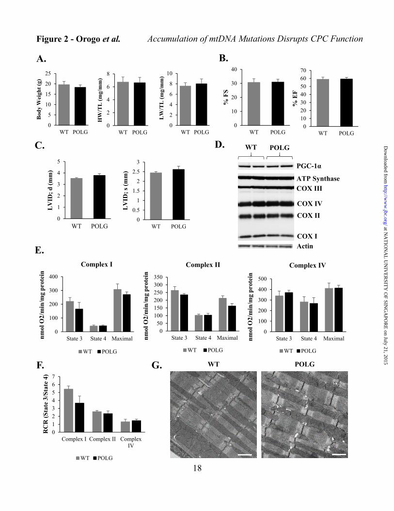
Figure 2 - Orogo et al.
A. B.
C.
0
2
4
6
8
WT POLG
HW
/TL
(m
g/m
m)
0
2
4
6
8
10
WT POLG
LW
/TL
(m
g/m
m)
0
10
20
30
40
WT POLG
% F
S
010203040506070
WT POLG
% E
F
0
1
2
3
4
5
WT POLG
LV
ID;
d (
mm
)
0
0.5
1
1.5
2
2.5
3
WT POLG
LV
ID;
s (m
m)
PGC-1α
ATP Synthase
COX III
COX IV
COX II
COX I
WT POLG
Actin
D.
0
100
200
300
400
State 3 State 4 Maximalnm
ol O
2/m
in/m
g p
rote
in
Complex I
WT POLG
050
100150200250300350
State 3 State 4 Maximalnm
ol O
2/m
in/m
g p
rote
in
Complex II
WT POLG
0
100
200
300
400
500
State 3 State 4 Maximalnm
ol O
2/m
in/m
g p
rote
in
Complex IV
WT POLG
01234567
Complex I Complex II ComplexIV
RC
R (
Sta
te 3
/Sta
te 4
)
WT POLG
E.
G. WT POLG
0
5
10
15
20
25
WT POLG
Bod
y W
eigh
t (g
)
F.
Accumulation of mtDNA Mutations Disrupts CPC Function
18
at NA
TIO
NA
L U
NIV
ER
SITY
OF SIN
GA
POR
E on July 21, 2015
http://ww
w.jbc.org/
Dow
nloaded from

C.
A.
0
0.5
1
1.5
2
2.5
3
3.5
4
4.5
Day 0 Day 3
Fol
d C
han
ge C
yQU
AN
T F
luor
esce
nce
WT POLG
*
0
0.5
1
1.5
2
2.5
3
3.5
4
Day 0 Day 1 Day 3
Fol
d c
han
ge in
Cel
l #
WT POLG
*
*
0
10
20
30
40
50
60
70
0 nM 100 nM 500 nM 1 μM
% C
ell D
eath
Doxorubicin Treatment
WT POLG
**
*
0
10
20
30
40
50
60
70
80
90
0 μM 5 μM 10 μM
% C
ell D
eath
Sunitinib Treatment
WT POLG
*
**
0
10
20
30
40
50
60
0 μM 50 μM 100 μM 200 μM
% C
ell D
eath
H2O2 Treatment
WT POLG
******
**
Figure 3 - Orogo et al. Accumulation of mtDNA Mutations Disrupts CPC Function
19
B.
at NA
TIO
NA
L U
NIV
ER
SITY
OF SIN
GA
POR
E on July 21, 2015
http://ww
w.jbc.org/
Dow
nloaded from

0
10
20
30
40
50
60
70
Day 0 Day 7
% G
AT
A-4
+ C
ells
WT POLG
0
20
40
60
80
100
Day 0 Day 7
% F
ragm
ente
d M
itoc
hon
dri
a
WT POLG
A.
B. C.
Day 0
Day 7 DM
GATA-4
GATA-4
Tom20
Tom20
Merged
Merged
WT CPCs
GATA-4
GATA-4
Tom20
Tom20
Day 0
Day 7 DM
Merged
Merged
POLG CPCs
D.
*** ******
0
0.5
1
1.5
2
2.5
3
3.5
4
4.5
Day 0 Day 7
Fol
d C
han
ge G
AT
A-4
mR
NA
WT POLG
***
Figure 4 - Orogo et al. Accumulation of mtDNA Mutations Disrupts CPC Function
20
at NA
TIO
NA
L U
NIV
ER
SITY
OF SIN
GA
POR
E on July 21, 2015
http://ww
w.jbc.org/
Dow
nloaded from

F.WT POLG
Day 0
Day 7DM
E.
0
0.5
1
1.5
2
2.5
Day 0 Day 7 Day 0 Day 7
WT POLG
Fol
d C
han
ge
MF
N1/
Act
in
*
*
**
0
0.5
1
1.5
2
2.5
Day 0 Day 7 Day 0 Day 7
WT POLG
Fol
d C
han
ge
MF
N2/
Act
in
***
n.s.
0
0.5
1
1.5
2
Day 0 Day 7 Day 0 Day 7
WT POLG
Fol
d C
han
ge
DR
P1/
Act
in *
MFN1
DRP1
Day 0 4 7 0 4 7
WT POLG
MFN2
Actin
Accumulation of mtDNA Mutations Disrupts CPC Function
21
at NA
TIO
NA
L U
NIV
ER
SITY
OF SIN
GA
POR
E on July 21, 2015
http://ww
w.jbc.org/
Dow
nloaded from

Figure 5 - Orogo et al.
**
0
5
10
15
20
WT POLG
Myo
tub
e d
iam
eter
(µ
m)
A.
C.
POLG
WT
Post-diffPre-diff
WT Myoblasts
Post-diff
Pre-diff
MyoD
MyoD
NCAM
NCAM
Merged
Merged
POLG Myoblasts
NCAMMyoD Merged
Pre-diff
Post-diff
MyoD NCAM Merged
MyoD
Actin
Ponceau
POLG Myoblasts
pre post
WT Myoblasts
postpre
MyoD
Actin
Ponceau
B.
Accumulation of mtDNA Mutations Disrupts CPC Function
22
at NA
TIO
NA
L U
NIV
ER
SITY
OF SIN
GA
POR
E on July 21, 2015
http://ww
w.jbc.org/
Dow
nloaded from

0
0.5
1
1.5
2
2.5
3
3.5
4
4.5
Day 0 Day 7 Day 0 Day 7
WT POLG
Fol
d C
han
ge P
GC
-1β
mR
NA
A.
D.
B.
0
0.5
1
1.5
2
2.5
3
Day 0 Day 7 Day 0 Day 7
WT POLGF
old
Ch
ange
C
OX
IVS
ub
1/A
ctin *
n.s.
***
0
0.5
1
1.5
2
2.5
3
3.5
Day 0 Day 7 Day 0 Day 7
WT POLG
Fol
d C
han
ge
CO
XIV
Su
b4/
Act
in ****
n.s.
0
0.2
0.4
0.6
0.8
1
1.2
WT POLG
Rel
ativ
e C
OX
1 m
RN
A
n.s.
0
0.2
0.4
0.6
0.8
1
1.2
WT POLGR
elat
ive
Cox
4i1
mR
NA
n.s.
WT POLG
Day: 0 7 0 7
ATP Synthase
COX III
COX IV Sub 1 (mtDNA encoded)COX II
COX IV Sub 4 (nuclear encoded)Actin
Actin
C.
* *
Figure 6 - Orogo et al.
0
0.5
1
1.5
2
2.5
3
3.5
4
4.5
Day 0 Day 7 Day 0 Day 7
WT POLG
Fol
d C
han
ge P
GC
-1α
mR
NA *
**
*
Accumulation of mtDNA Mutations Disrupts CPC Function
23
at NA
TIO
NA
L U
NIV
ER
SITY
OF SIN
GA
POR
E on July 21, 2015
http://ww
w.jbc.org/
Dow
nloaded from

C. D. E.
A.
***
0
0.5
1
1.5
2
2.5
3
3.5
4
WT POLG
L-L
acta
te (
µM
)/T
otal
P
rote
in (
µg)
0
20
40
60
80
100
0 hr 2 hr 4 hr 6 hr 24 hr
% C
ell D
eath
2-Deoxyglucose Treatment
WT POLG
***0
0.2
0.4
0.6
0.8
1
1.2
WT POLG
Rel
ativ
e M
axim
al
OC
R (
pm
ol/m
in/c
ell)
0500
10001500200025003000350040004500
WT POLG
Rel
ativ
e A
TP
Con
c (n
M) ***
***
**
n.s.
Figure 7 - Orogo et al.
B.
***
0
20
40
60
80
100
120
WT POLG
TM
RM
Flu
ores
cen
ce
(% C
ontr
ol)
WT POLG
Accumulation of mtDNA Mutations Disrupts CPC Function
24
at NA
TIO
NA
L U
NIV
ER
SITY
OF SIN
GA
POR
E on July 21, 2015
http://ww
w.jbc.org/
Dow
nloaded from

Hexokinase I
PKM 1/2
Actin
Hexokinase II
GAPDH
Day 0 4 7 0 4 7
WT POLGA. B.
C.
0
10
20
30
40
50
60
Control DM
% C
ell D
eath
WT POLG
***
0
0.5
1
1.5
2
2.5
3
3.5
4
Day 0 Day 4 Day 7 Day 0 Day 4 Day 7
WT POLG
Fol
d C
han
ge H
exok
inas
e I/
Act
in
***
***
00.20.40.60.8
11.21.41.61.8
2
Day 0 Day 4 Day 7 Day 0 Day 4 Day 7
WT POLG
Fol
d C
han
ge H
exok
inas
e II
/Act
in***
******
**
0
0.2
0.4
0.6
0.8
1
1.2
1.4
1.6
1.8
Day 0 Day 4 Day 7 Day 0 Day 4 Day 7
WT POLG
Fol
d C
han
ge G
AP
DH
/Act
in
**
*** ***
Figure 8 - Orogo et al.
*****
***
0
0.2
0.4
0.6
0.8
1
1.2
Day 0 Day 4 Day 7 Day 0 Day 4 Day 7
WT POLG
Fol
d C
han
ge P
KM
1/2/
Act
in
Accumulation of mtDNA Mutations Disrupts CPC Function
25
at NA
TIO
NA
L U
NIV
ER
SITY
OF SIN
GA
POR
E on July 21, 2015
http://ww
w.jbc.org/
Dow
nloaded from

0
10
20
30
40
50
60
70
80
DMSO Baf A1
# L
C3/
Tom
20 C
oloc
aliz
atio
n
WT POLG
**
***
*
***
0
50
100
150
200
250
300
WT POLG
LA
MP
2 st
ain
ing/
cell
(A
.U.)
n.s.
Figure 9 - Orogo et al.
A.WT POLG
DMSO Baf DMSO Baf
LC3ILC3II
Actin
B.
C. D.
LAMP2
WT POLG
E. F.
LAMP2
00.5
11.5
22.5
33.5
4
DMSO Baf A1 DMSO Baf A1
WT POLG
Fol
d C
han
ge
LC
3II/
Act
in
* *
WT CPCs
GFP-LC3
GFP-LC3
Tom20
Tom20
Merged
Merged
DMSO
Baf A1
POLG CPCsGFP-LC3
GFP-LC3
Tom20
Tom20
Merged
Merged
DMSO
Baf A1
Accumulation of mtDNA Mutations Disrupts CPC Function
26
at NA
TIO
NA
L U
NIV
ER
SITY
OF SIN
GA
POR
E on July 21, 2015
http://ww
w.jbc.org/
Dow
nloaded from

Murphy and Åsa B. GustafssonTomas A. Prolla, Mark A. Sussman, Anne N.A. Kubli, Igor L. Baptista, Sang-Bing Ong, Amabel M. Orogo, Eileen R. Gonzalez, Dieter Cell Function and Reduces SurvivalMutations Disrupts Cardiac Progenitor Accumulation of Mitochondrial DNACell Biology:
published online July 16, 2015J. Biol. Chem.
10.1074/jbc.M115.649657Access the most updated version of this article at doi:
.JBC Affinity SitesFind articles, minireviews, Reflections and Classics on similar topics on the
Alerts:
When a correction for this article is posted•
When this article is cited•
to choose from all of JBC's e-mail alertsClick here
http://www.jbc.org/content/early/2015/07/16/jbc.M115.649657.full.html#ref-list-1
This article cites 0 references, 0 of which can be accessed free at
at NA
TIO
NA
L U
NIV
ER
SITY
OF SIN
GA
POR
E on July 21, 2015
http://ww
w.jbc.org/
Dow
nloaded from
Copyright © 2022 FDOKUMEN