Parasites as a Cause of Keratitis: Need for Increased Awareness
131
Tropical Biomedicine 30(1): 131–140 (2013)
Acanthamoeba genotype T4 detected in naturally-infected
feline corneas found to be in homology with those causing
human keratitis
Ithoi, I.1*, Mahmud, R.1, Abdul Basher, M.H.1, Jali, A.1, Abdulsalam, A.M.1, Ibrahim, J.1 and Mak, J.W.21Department of Parasitology, Faculty of Medicine, University Malaya, Kuala Lumpur, Malaysia,2School of Postgraduate Studies and Research, International Medical University, Kuala Lumpur, Malaysia*Corresponding author email: [email protected] 2nd October 2012; received in revised form 20 January 2013; accepted 24 January 2013
Abstract. A total of 10 out of 65 cornea swab samples from cats with eye symptoms showedAcanthamoeba-like morphology after cultivation. By PCR and DNA sequencing ofAcanthamoeba diagnostic fragment 3 (DF3), all 10 isolates from the positive samples werecategorized into two homologous groups of AfC1 (PM1, PM2, PM3, PF6, KM7, KF8, KMK9)and AfC2 (PM4, PM5, KFK10) due to the presence of bases A354 and G354, respectively.Furthermore, DF3 of AfC1 and AfC2 showed 100% similarity with Genbank reference isolateswith the accession numbers DQ087314, EU146073 and U07401, GU808323, which wereAcanthamoeba castellanii strains genotype T4 originating from human keratitis. This findingsuggests that A. castellani strains have the capability to infect cats and human under favorableconditions.
INTRODUCTION
Acanthamoeba is one of the free-livingbacterivores in nature and it has beenisolated from diverse habitats worldwideincluding soil, sand, water, dust, air, etc.ranging from the tropics to the arctic regions.Acanthamoebiasis in animals was noted inwild squirrels (Lorenzo-Morales et al., 2007),dogs, monkeys, a bull, a kangaroo and anIndian buffalo (Schuster & Visvesvara, 2004).In addition, most of the reported cases indogs were acute to chronic infections of thecentral nervous system (CNS) (Ayers et al.,1972; Pearce et al., 1985; Bauer et al., 1993;Brofman et al., 2003) and multisystemicinfections (Dubey et al., 2005; Kent et al.,2011). Eye infections are rarely reported,although fairly common especially in pets.Eye infections usually occur after the surfaceof the cornea is accidentally scratched orwhen hard particles lodge in the eyes.Common warning signs for eye infection inpets are eye discharge, squinting, redness,
cloudiness, difficulty in keeping the eyelidsopen, and attempts to rub and scratch the eye.In felines, eye infections are frequentlyencountered in veterinary clinics and can becaused by a variety of pathogens includingvirus, bacteria and fungi (Doyle, 2009).However, other pathogens such as free livingamoebae, especially the Acanthamoeba
species, are rarely reported. To the bestof our knowledge, Acanthamoeba has notbeen naturally isolated from animal eyes,including felines (Felis domesticus) excepta patent note (Ledbetter, 2011).
In humans, Acanthamoeba is known asthe causative agent of acanthamoebickeratitis (AK) when it directly infects thecorneas of the eyes following a traumathrough exogenous sources (Jones et al.,1975) and occurs mostly in wearers of alltypes of contact lens (Stehr-Green et al.,1989). Microtrauma to the corneal epitheliumis assumed to be an important factor infacilitating the invasion of Acanthamoeba
(Kinnear, 2004). Others are opportunistic
132
systemic infections, including fatalacanthamoebic granulomatous encephalitis(AGE), nasopharyngeal and cutaneousinfections (Marciano-Cabral & Cabral, 2003).Acanthamoeba infections in humans areincreasing and occurring worldwide. Infectedpets could potentially contribute to theincrease of pathogenic Acanthamoeba bycontaminating the domestic environment andopens an opportunity to infect humans.Therefore, information on the occurrence ofthis amoeba in domestic animals is neededfor the improvement of healthcare structuresin both humans and animals.
MATERIAL AND METHODS
Location of the sampling sites
All selected sampling sites are locatedaround Kuala Lumpur (latitude 3.13900°Nand longitude 101.68686°E), the capital cityof Malaysia. They comprise of four places,namely Society for The Prevention of Crueltyto Animals (SPCA) of Ampang Jaya, NationalZoo of Hulu Kelang, PAWS Animal WelfareSociety of Subang and Kampung Orang Asli(KgOA) of Kuala Pangsun at Hulu Langat,between latitude 3.13044°N–3.21046°Nand longitude 101.55183°E–101.87990°E(Table 1). These sampling sites were selectedbased on the benefits, convenience andpotential implications to public health. Allcats from the National Zoo live individuallyin cages that are cleaned regularly. The SPCAand PAWS shelter the unwanted, abandoned,injured and stray cats around the city of KualaLumpur. Cats at the KgOA of Kuala Pangsunlive freely as pets, in the houses of theMalaysian Aboriginal community.
Ethics statement
The protocol of this study was reviewed andapproved by the Institutional Animal Careand Use Committee of the University ofMalaya (UM IACUC), Kuala Lumpur (EthicsReference number: PAR/29/06/2012/II (R)).Written permission was also obtained fromthe management authorities of the SPCA andNational Zoo. Authorities of the PAWS AnimalWelfare Society and the owners of cats atKgOA agreed verbally without any written
permission. The objectives and protocols ofthe research were thoroughly discussed withthe authorities in charge or the cat owners.
Collection and cultivation of feline
corneal swab samples
A small piece of facial cotton (1.0 cm2) and acotton bud were packed in a 15-mL test tubeand wetted with 1.0 mL of normal saline. Thetube was then autoclaved at 15 lbs pressure,121°C for 15 minutes. The sterile packagingtubes were kept in the refrigerator at 4°C untilused. For sampling, the cold packaging tubeswere placed on ice cubes (or ice pack) in acovered polystyrene box and transported tothe sampling site. Cornea swabs were carriedout among cats (male, female, adult, andkittens) with symptoms of watery eyes withgray discharge and redness. A cold wetcotton-bud was carefully withdrawn byholding the cotton bud handle followed byswabbing of the infected cornea. It was thenplaced back to its original test tube.Precaution was always taken to maintain thepackaging in a cold (10±2°C) condition andto avoid any possible contamination. Theywere then transported to the Laboratory atthe Department of Parasitology, Universityof Malaya. A swabbed cotton-bud and acotton-bed in the packaging tube were placedonto the surface of a non-nutrient agarplate overlaid with a thin layer of liveEscherichia coli, JM109 (Promega), sealedwith a parafilm, placed in a clean containerand incubated at room temperature(26±2°C).
Detection of Acanthamoeba by
morphological and molecular techniques
All culture plates were examined daily forup to 14 days using a light invertedmicroscope (Olympus BX51) before beingdiscarded. The morphology was observedunder a 200 followed by a 400 timesmagnification for specific characteristics ofAcanthamoeba trophozoites (acanthopodiaand pseudopodia) and cysts (wrinkle doublewalled), and were photographed andrecorded. Initially, culture plates withpositive Acanthamoeba-like cells could existas mixed isolates. The positive culture platewas sub-cultured for at least ten times and
133
each sub-culture was carried out by placinga colony of 4-6 cysts onto a new agar platelawned with JM 109. The trophozoites werecultured and harvested for total DNAextraction using the QIAamp DNA mini kit(Qiagen, Hilden, Germany).
Amplification reactions using Acantha-
moeba genus-specific primers, forward JDP1(5’- GGG CCC AGA TCG TTT ACC GTG AA -3’) and reverse JDP2 (5’- TCTC ACA AGCTGC TAG GGG AGT CA -3’) were carried outaccording to the protocol described by(Schroeder et al., 2001). PCR ampliconsknown as ASA.S1 (ranged 423 to 551 bp) werefractioned by 1.5% agarose electrophoresisstained with a TBE (0.5 x Tris-borate EDTA)buffer containing 0.5 µg/ml ethidium bromideand visualized under UV illumination inthe chamber of a BioDoc-It™ ImagingSystem (UVP, Cambridge, United Kingdom).Amplicon sizes were estimated bycomparison with the GeneRuler 100 bpDNA ladder Plus (Fermentas Life Science,Canada).
The ASA.S1 amplicons from 10representative isolates were gel-purifiedusing the QIAquick gel extraction kit (Qiagen,Hilden, Germany), followed by cloning usingthe InsT/AcloneTM PCR product cloning kit(Fermentas). The ASA.S1 or diagnosticfragment 3 (DF3) was inserted into vectors(plasmids, pTZ57R/T) to form a DNArecombinant. The recombinant moleculeswere transformed into Escherichia coli
(strain JM109, Promega) followed byselection of the white colonies carryingrecombinant plasmids with a disruptedβ-galactosidase gene.
The plasmid DNA in selectedrecombinant colonies were confirmed byPCR amplification and were gel-purifiedusing QIAprep® Miniprep kit (Qiagen, Hilden,Germany), followed by sequencing at bothstrands using the amplification primers inan ABI PRISMTM BigdyeTM terminator cyclesequencing ready reaction kit V.3.1 (Korea).The obtained sequences were alignedusing the ClustalW2 software (Labarga et
al., 2007). Each consensus sequence wasblasted against all eukaryotic nucleotidesequences retrieved in the Genbank database(Altschul et al., 1990) to detect the nucleotide
similarities. Phylogenetic analyses wereperformed based on the DF3 sequences ofboth our isolates and references publishedgenotypes using the neighbour-joining ofMEGA version 4 software (Tamura et al.,2007). This was followed by Kimura 2-parameter algorithm and constructed treeby a bootstrap analysis of 1000 replicates.The new data on DNA sequencing of all 10selected isolates of PM1, PM2, PM3, PM4,PM5, PF6, KM7, KF8, KMK9 and KFK10 weredeposited in Genbank with the accessionnumbers JX494391, JX494392, JX494393,JX494394, JX494395, JX494396, JX494397,JX494398, JX494399 and JX494400,respectively.
RESULTS
Microscopic observation of Acantha-
moeba species
The trophozoite and cyst stages wereobserved as early as the second and five daysof cultivation, respectively. Ten (10) isolatesfrom the cornea of 65 cats were successfullygrown in the laboratory. All positive isolateswere from PAWS (6) and KgOA (4), whichwere isolated from 6 adult males, 2 adultfemales, and 1 each of male and female kitten(Table 1). All of these isolates showed goodgrowth at 26±2°C. On the moist agar surface,the trophozoites of all isolates showed thecharacteristics of a spike-like acanthopodiaand pseudopodia that passively protruded atsurface extensions surrounding the cell. Oncontinuous cultivation, the trophozoitesbecame stagnant and slowly transformed toa cyst with wrinkled, thick double walls.
Molecular detection and characterization
of Acanthamoeba species
After the PCR amplification, the Acantha-
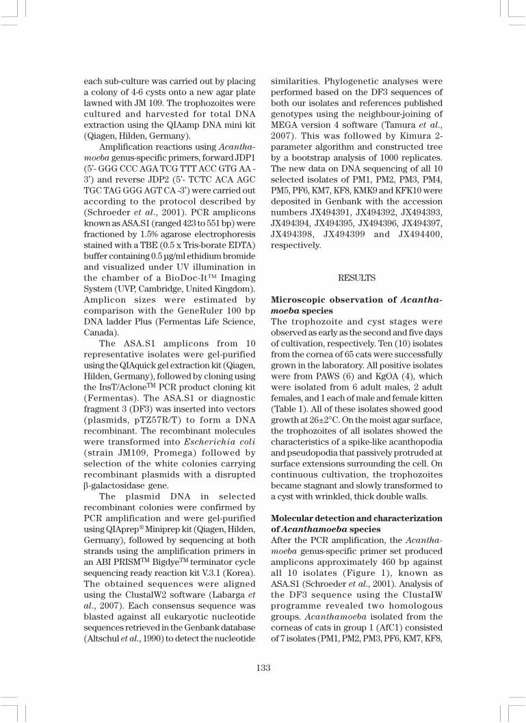
moeba genus-specific primer set producedamplicons approximately 460 bp againstall 10 isolates (Figure 1), known asASA.S1 (Schroeder et al., 2001). Analysis ofthe DF3 sequence using the ClustaIWprogramme revealed two homologousgroups. Acanthamoeba isolated from thecorneas of cats in group 1 (AfC1) consistedof 7 isolates (PM1, PM2, PM3, PF6, KM7, KF8,
134
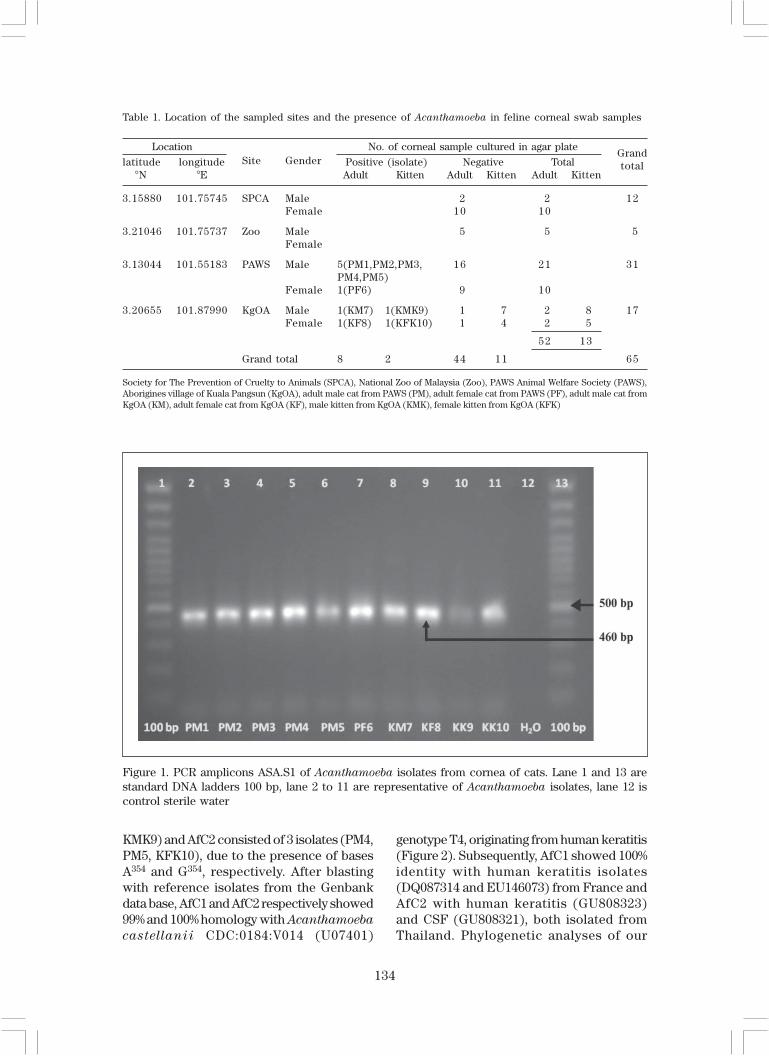
Table 1. Location of the sampled sites and the presence of Acanthamoeba in feline corneal swab samples
LocationSite Gender
No. of corneal sample cultured in agar plateGrand
latitude longitude Positive (isolate) Negative Total total °N °E Adult Kitten Adult Kitten Adult Kitten
3.15880 101.75745 SPCA Male 2 2 12Female 10 10
3.21046 101.75737 Zoo Male 5 5 5Female
3.13044 101.55183 PAWS Male 5(PM1,PM2,PM3, 16 21 31PM4,PM5)
Female 1(PF6) 9 10
3.20655 101.87990 KgOA Male 1(KM7) 1(KMK9) 1 7 2 8 17Female 1(KF8) 1(KFK10) 1 4 2 5
52 13
Grand total 8 2 44 11 65
Society for The Prevention of Cruelty to Animals (SPCA), National Zoo of Malaysia (Zoo), PAWS Animal Welfare Society (PAWS),Aborigines village of Kuala Pangsun (KgOA), adult male cat from PAWS (PM), adult female cat from PAWS (PF), adult male cat fromKgOA (KM), adult female cat from KgOA (KF), male kitten from KgOA (KMK), female kitten from KgOA (KFK)
Figure 1. PCR amplicons ASA.S1 of Acanthamoeba isolates from cornea of cats. Lane 1 and 13 arestandard DNA ladders 100 bp, lane 2 to 11 are representative of Acanthamoeba isolates, lane 12 iscontrol sterile water
KMK9) and AfC2 consisted of 3 isolates (PM4,PM5, KFK10), due to the presence of basesA354 and G354, respectively. After blastingwith reference isolates from the Genbankdata base, AfC1 and AfC2 respectively showed99% and 100% homology with Acanthamoeba
castellanii CDC:0184:V014 (U07401)
genotype T4, originating from human keratitis(Figure 2). Subsequently, AfC1 showed 100%identity with human keratitis isolates(DQ087314 and EU146073) from France andAfC2 with human keratitis (GU808323)and CSF (GU808321), both isolated fromThailand. Phylogenetic analyses of our

135
Fig
ure
2. D
NA
seq
uenc
e of
Acan
tham
oeba
dia
gnos
tic
frag
men
t 3
(DF
3) o
f te
st a
nd G
enba
nk p
ublis
hed
isol
ates

136
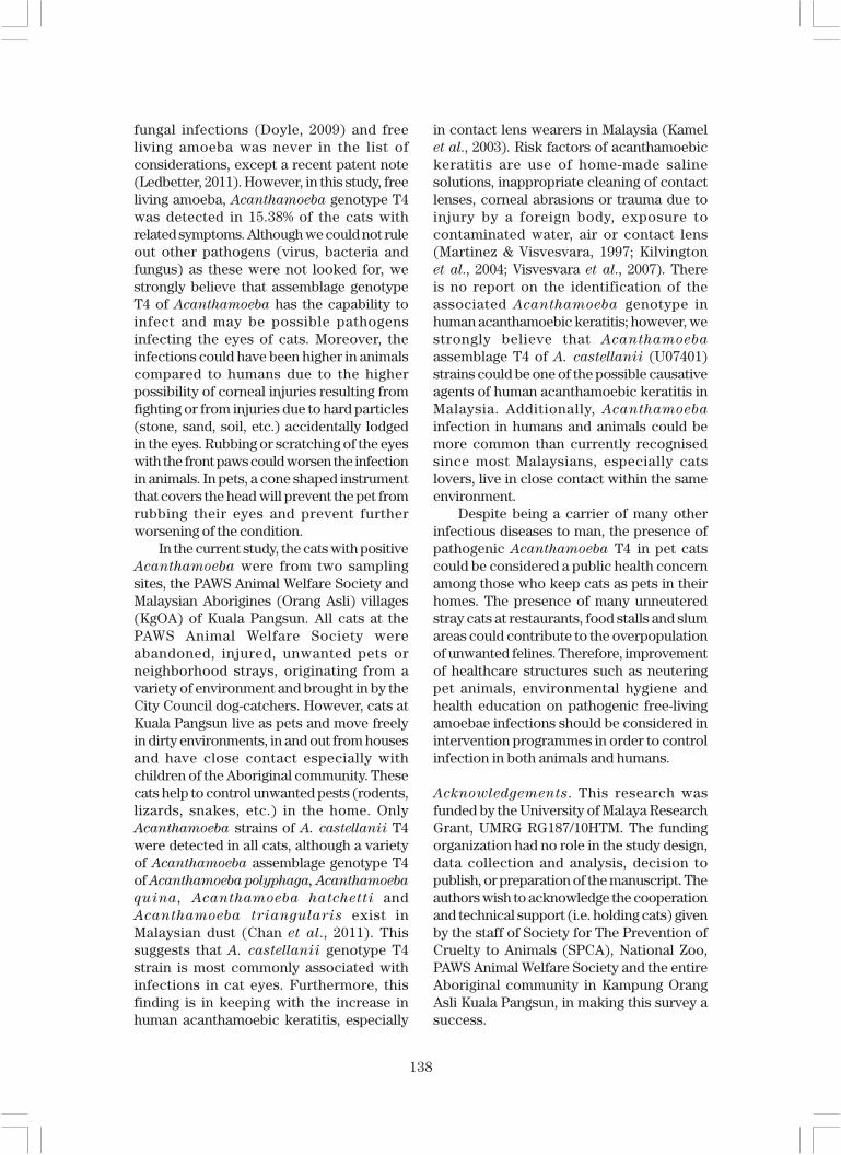
representative and reference isolatesassemblage genotype T1 to T17 showed thatall of the representative isolates wereassemblage Acanthamoeba genotype T4.The relationship between these isolateswas summarized in the phylogenetic treesthat consist of Acanthamoeba assemblagegenotypes T1, T2, T3, T4, T10, T11, T12, T13,T14 and T15 (Figure 3). Genotypes T5, T6,T7, T8, T9, T16 and T17 were excluded asthey are more distant from our representativeisolates.
DISCUSSION
Despite being the causative agent ofacanthameobic keratitis in humansworldwide, Acanthamoeba infection thatcauses feline eye disease is not known. Todetermine whether this amoeba could beconsidered as one of the possible pathogensinfecting the eyes of cats, this study wascarried out to detect Acanthamoeba innaturally-infected feline corneas with
Figure 3. Neighbour-joining tree depicting the relationships between test isolates and reference strainsrepresenting respective genotypes of Acanthamoeba. Numbers at the nodes are percentage-bootstrapping values on 1000 replicates. Genbank accession numbers and genotypes for referencesequences are indicated at the ends of the Acanthamoeba isolates designations
137
symptoms of keratitis. In felines, eyedischarge (often thick and yellow, gray orgreen in colour) is a valuable warning sign ofpathogenic infection which could progressvery quickly and cause permanent eyedamage (Doyle, 2009). Cats with eyedischarge, swelling and redness wereselected and used in our current study. Duringcornea swabbing, the cotton bud used waswet and cold (10±2°C) in order to induce anyAcanthamoeba trophozoites present to roundup without protruding the acanthopodia.Rounded trophozoites may be less adherentto the corneal surface, making them easierto be swabbed out. Using this technicalprecaution, there was an increased chanceof any Acanthamoeba trophozoites and cystsfrom the infected corneas to be cultured. Aftercultivation, 10 out of 65 corneal swab sampleswere identified positive for Acanthamoeba-
like species, based on the morphologicalcharacteristics of the trophozoite and cyststages. Each motile trophozoite showed thecharacteristics of both pseudopodia andacanthopodia, where both structurespassively protruded from surface extensionssurrounding the cell. These structures mayact as fingers that attach to the corneasurface, thus needing a cold object (cottonbud) to release these ‘fingers’ from thecorneal surface. Similarly, when the swabbedcotton bud was placed in the test tube, thecold condition could prevent theAcanthamoeba trophozoites from protrudingacanthopodia and pseudopodia which wouldallow them to adhere to the test tube wall.This would improve the detection rate insamples with lower amoeba cells.
All of these isolates generated ampliconsknown as ASA.S1 by PCR using the genus-specific primer set, JDP1 and JDP2, designedfrom 18S rRNA gene of Acanthamoeba
(Schroeder et al., 2001). DNA sequences ofthese amplicons (or DF3) from eachrepresentative isolate were matched to twohomologous groups of AfC1 (PM1, PM2, PM3,PF6, KM7, KF8, KMK9) and AfC2 (PM4, PM5,KFK10 isolates) due to the presence of basesA354 and G354, respectively. The DF3 regionof 18S rRNA gene was recently used toidentify the sequence variation, in which17 different genotypes (T1-T17) have been
established, T1-T12 (Stothard et al., 1998),T13 (Horn et al., 1999), T14 (Gast, 2001),T15 (Hewett et al., 2003), T16 (Corsaro &Venditti, 2010) and T17 (Nuprasert et al.,2010). Each genotype showed at least6% sequence divergence among differentgenotypes. However, the pathogenicity ofAcanthamoeba could be limited to somegenotypes; genotype T4 occurs mostcommonly with human keratitis (Khan,2006). Similarly, based on the DF3 sequencevariation, the identification of all 10 isolatesfrom the cornea of cats were blasted withAcanthamoeba DNA sequences retrievedfrom the GenBank database, and were allfound to be assemblage genotype T4 withmost matching 99%-100% with 5 isolatesoriginally from human keratitis, ofAcanthamoeba castellanii CDC:0184:V014(U07401) from the USA (Gast et al., 1996),Acanthamoeba spp. (DQ087314, EU146073)from France (NCBI, unpublished), Korea(EF140625) (NCBI, unpublished) andThailand (GU808323) (Nuprasert et al.,2010). Additionally, they matched withAcanthamoeba isolate (GU808321) from theCSF of a patient from Thailand (Nuprasertet al., 2010). Besides that, they have a 99%–100% match with many other referenceisolates from environmental samples isolatedfrom many regions in the world. This suggeststhat all Acanthamoeba isolates in thisstudy (from cornea of cats) are the strains ofAcanthamoeba castellanii genotype T4 thatoccur worldwide and have the capability toinfect humans and animals under favorableconditions.
Most veterinarians believe that felinekeratitis (inflammation of the cornea) resultsfrom foreign objects which lodge in the eye,causing irritation and rubbing. Feline eyedisease associated with vision loss, such askeratitis is a dry eye syndrome leading toreduced production of tears and could beassociated with mucoid discharge. Clinicalsigns include redness, discharge from the eye(discharge is initially watery and as thedisease progresses it turns into thick puss),pain, sensitivity to light (photophobia),swelling, corneal edema, impaired visionor blindness. These symptoms were usuallydiagnosed as due to parasitic, viral or
138
fungal infections (Doyle, 2009) and freeliving amoeba was never in the list ofconsiderations, except a recent patent note(Ledbetter, 2011). However, in this study, freeliving amoeba, Acanthamoeba genotype T4was detected in 15.38% of the cats withrelated symptoms. Although we could not ruleout other pathogens (virus, bacteria andfungus) as these were not looked for, westrongly believe that assemblage genotypeT4 of Acanthamoeba has the capability toinfect and may be possible pathogensinfecting the eyes of cats. Moreover, theinfections could have been higher in animalscompared to humans due to the higherpossibility of corneal injuries resulting fromfighting or from injuries due to hard particles(stone, sand, soil, etc.) accidentally lodgedin the eyes. Rubbing or scratching of the eyeswith the front paws could worsen the infectionin animals. In pets, a cone shaped instrumentthat covers the head will prevent the pet fromrubbing their eyes and prevent furtherworsening of the condition.
In the current study, the cats with positiveAcanthamoeba were from two samplingsites, the PAWS Animal Welfare Society andMalaysian Aborigines (Orang Asli) villages(KgOA) of Kuala Pangsun. All cats at thePAWS Animal Welfare Society wereabandoned, injured, unwanted pets orneighborhood strays, originating from avariety of environment and brought in by theCity Council dog-catchers. However, cats atKuala Pangsun live as pets and move freelyin dirty environments, in and out from housesand have close contact especially withchildren of the Aboriginal community. Thesecats help to control unwanted pests (rodents,lizards, snakes, etc.) in the home. OnlyAcanthamoeba strains of A. castellanii T4were detected in all cats, although a varietyof Acanthamoeba assemblage genotype T4of Acanthamoeba polyphaga, Acanthamoeba
quina, Acanthamoeba hatchetti andAcanthamoeba triangularis exist inMalaysian dust (Chan et al., 2011). Thissuggests that A. castellanii genotype T4strain is most commonly associated withinfections in cat eyes. Furthermore, thisfinding is in keeping with the increase inhuman acanthamoebic keratitis, especially
in contact lens wearers in Malaysia (Kamelet al., 2003). Risk factors of acanthamoebickeratitis are use of home-made salinesolutions, inappropriate cleaning of contactlenses, corneal abrasions or trauma due toinjury by a foreign body, exposure tocontaminated water, air or contact lens(Martinez & Visvesvara, 1997; Kilvingtonet al., 2004; Visvesvara et al., 2007). Thereis no report on the identification of theassociated Acanthamoeba genotype inhuman acanthamoebic keratitis; however, westrongly believe that Acanthamoeba
assemblage T4 of A. castellanii (U07401)strains could be one of the possible causativeagents of human acanthamoebic keratitis inMalaysia. Additionally, Acanthamoeba
infection in humans and animals could bemore common than currently recognisedsince most Malaysians, especially catslovers, live in close contact within the sameenvironment.
Despite being a carrier of many otherinfectious diseases to man, the presence ofpathogenic Acanthamoeba T4 in pet catscould be considered a public health concernamong those who keep cats as pets in theirhomes. The presence of many unneuteredstray cats at restaurants, food stalls and slumareas could contribute to the overpopulationof unwanted felines. Therefore, improvementof healthcare structures such as neuteringpet animals, environmental hygiene andhealth education on pathogenic free-livingamoebae infections should be considered inintervention programmes in order to controlinfection in both animals and humans.
Acknowledgements. This research wasfunded by the University of Malaya ResearchGrant, UMRG RG187/10HTM. The fundingorganization had no role in the study design,data collection and analysis, decision topublish, or preparation of the manuscript. Theauthors wish to acknowledge the cooperationand technical support (i.e. holding cats) givenby the staff of Society for The Prevention ofCruelty to Animals (SPCA), National Zoo,PAWS Animal Welfare Society and the entireAboriginal community in Kampung OrangAsli Kuala Pangsun, in making this survey asuccess.
139
REFERENCES
Altschul, S.F., Gish, W., Miller, W., Myers, E.W.& Lipman, D.J. (1990). Basic localalignment search tool. Journal of
Molecular Biology 215: 403–410.Ayers, K.M., Billups, L. & Garner, F. (1972).
Acanthamoebiasis in a dog. Veterinary
Pathology 9: 221–226.Bauer, R.W., Harrison, L.R. & Watson, C.W.
(1993). Isolation of Acanthamoeba sp.from a greyhoung with pneumonia andgranulomatous amebic encephalitis.Journal of Veterinary Diagnostic
Investigation 5: 386–391.Brofman, P. J., Knostman, K.A.B. & Dibartola,
S.P. (2003). Granulomatous amebicmeningoencephalitis causing thesyndrome of inappropriate secretion ofantidiuretic hormone in a dog. Journal
of Veterinary Internal Medicine 17: 230–234.
Chan, L.L., Mak, J.W., Low, Y.T., Koh, T.T., Ithoi,I. & Mohamed, S.M. (2011). Isolation andcharacterization of Acanthamoeba spp.from air-conditioners in Kuala Lumpur,Malaysia. Acta Tropica 117: 23–30.
Corsaro, D. & Venditti, D. (2010). Phylo-genetic evidence for a new genotype ofAcanthamoeba (Amoebozoa, Acantha-moebida). Parasitology Research 107:233–238.
Doyle, M. (2009). Cat Eye Infections: Causes,
Symptoms, and Treatment of Common
Feline Eye Problems [Online]. internate:suite 101R. Available: http://megandoyle.suite101.com/cat-eye- infections-a114180#ixzz1uTNocwLi.
Dubey, J.P., Benson, J.E., Blakeley, K.T.,Booton, G.C. & Visvesvara, G.S. (2005).Disseminated Acanthamoeba sp. infectionin a dog. Veterinary Parasitology 128:183–187.
Gast, R.J. (2001). Development of anAcanthamoeba-specific reverse dot-blotand the discovery of a new ribotype.Journal of Eukaryotic Microbiology 48:609–615.
Gast, R.J., Ledee, D.R., Fuerst, P.A. & Byers,T.J. (1996). Subgenus systematics ofAcanthamoeba: four nuclear 18S rDNAsequence types. Journal of Eukaryotic
Microbiology 43: 498–504.Hewett, M.K., Robinson, B.S., Monis, P.T.
& Saint, C.P. (2003). Identification of anew Acanthamoeba 18S rRNA genesequence type, corresponding to thespecies Acanthamoeba jacobsi Sawyer,Nerad and Visvesvara, 1992 (Lobosea:Acanthamoebidae). Acta Protozoologica
42: 325–329.Horn, M., Fritsche, T.R., Gautom, R.K.,
Schleifer, K.H. & Wagner, M. (1999). Novelbacterial endosymbionts of Acantha-
moeba spp. related to the Paramecium
caudatum symbiont Caedibacter
caryophilus. Environmental Micro-
biology 1: 357–367.Jones, D.B., Visvesvara, G.S. & Robinson, N.M.
(1975). Acanthamoeba polyphaga keratitisand Acanthamoeba uveitis associatedwith fatal meningoencephalitis. Transac-
tions of the Ophthalmological Societies
of the United Kingdom 95: 221–232.Kamel, A.G.M., Anisah, N., Yusof, S., Faridah,
H., Michael, I., Norhayati, M. & Norazah,A. (2003). Acanthamoeba keratitis is notso rare in Malaysia. Medical Journal of
Malaysia 58: S150Kent, M., Platt, S.R., Rech, R.R., Eagleson, J.S.,
Howerth, E.W., Shoff, M., Fuerst, P.A.,Booton, G., Visvesvara, G.S. & Schatzberg,S.J. (2011). Multisystemic infection withan Acanthamoeba sp in a dog. Journal
of the American Veterinary Medical
Association 238: 1476–1481.Khan, N.A. (2006). Acanthamoeba: Biology
and increasing importance in humanhealth. FEMS Microbiology Reviews 30:564–595.
Kilvington, S., Gray, T., Dart, J., Morlet, N.,Beeching, J.R., Frazer, D.G. & Matheson,M. (2004). Acanthamoeba keratitis: Therole of domestic tap water contaminationin the United Kingdom. Investigative
Ophthalmology and Visual Science 45:165–169.
140
Kinnear, F.B. (2004). Acanthamoeba patho-genicity for corneal cells. Journal of
Infection 49: 310–316.Labarga, A., Valentin, F., Anderson, M. &
Lopez, R. (2007). European bioinformaticsinstitute. Nucleic Acids Research 35: W6–W11.
Ledbetter, E. (2011). Model System of
Acanthamoeba Keratitis Syndrome and
Method for Selecting a Treatment thereof
Cornell University (Suite 310395 Pine
Tree Roa, Ithaca New York, 14850, US).US patent application US2011/024002,Available: http://www.sumobrain.com/patents/WO2011097609.html
Lorenzo-Morales, J., López-Darias, M.,Martínez-Carretero, E. & Valladares, B.(2007). Isolation of potentially patho-genic strains of Acanthamoeba in wildsquirrels from the Canary Islands andMorocco. Experimental Parasitology
117: 74–79.Marciano-Cabral, F. & Cabral, G. (2003).
Acanthamoeba spp. as agents of diseasein humans. Clinical Microbiology Re-
views 16: 273–307.Martinez, J.D. & Visvesvara, G.S. (1997).
Free-living, amphizoic and opportunisticamebas. Brain Pathology 7: 583–598.
Nuprasert, W., Putaporntip, C., Pariyakanok,L. & Jongwutiwes, S. (2010). Identificationof a novel T17 genotype of Acanthamoeba
from environmental isolates and T10genotype causing keratitis in Thailand.Journal of Clinical Microbiology 48:4636–4640.
Pearce, J.R., Powell, H.S., Chandler, F.W.& Visvesvara, G.S. (1985). Amebicmeningoencephalitis caused by Acantha-
moeba castellani in a dog. Journal of the
American Veterinary Medical Associa-
tion 187: 951–952.
Schroeder, J.M., Booton, G.C., Hay, J., Niszl,I.A., Seal, D.V., Markus, M.B., Fuerst, P.A.& Byers, T.J. (2001). Use of subgenic 18Sribosomal DNA PCR and sequencingfor genus and genotype identificationof Acanthamoebae from humans withkeratitis and from sewage sludge.Journal of Clinical Microbiology 39:1903–1911.
Schuster, F.L. & Visvesvara, G.S. (2004). Free-living amoebae as opportunistic andnon-opportunistic pathogens of humansand animals. International Journal for
Parasitology 34: 1001–1027.Stehr-Green, J.K., Bailey, T.M. & Visvesvara,
G. (1989). The epidermiology of Acantha-
moeba keratitis in the United States.American Jounal of Ophthalmology 107:331–336.
Stothard, D.R., Schroeder-Diedrich, J.M.,Awwad, M.H., Gast, R.J., Ledee, D.R.,Rodriguez-Zaragoza, S., Dean, C.L.,Fuerst, P.A. & Byers, T.J. (1998). Theevolutionary history of the genusAcanthamoeba and the identificationof eight new 18S rRNA gene sequencetypes. Journal of Eukaryotic Micro-
biology 45: 45–54.Tamura, K., Dudley, J., Nei, M. & Kumar, S.
(2007). MEGA4: molecular evolutionarygenetics analysis (MEGA) softwareversion 4.0. Molecular Biology and
Evolution 24: 1596–1599.Visvesvara, G.S., Moura, H. & Schuster, F.L.
(2007). Pathogenic and opportunisticfree-living amoebae: Acanthamoeba spp.,Balamuthia mandrillaris, Naegleria
fowleri, and Sappinia diploidea. FEMS
Immunology and Medical Microbiology
50: 1–26.
Copyright © 2022 FDOKUMEN