Current efforts and the potential of nanomedicine in treating fungal keratitis
20
Author Proof 365 Review www.expert-reviews.com ISSN 1746-9899 © 2010 Expert Reviews Ltd 10.1586/EOP.10.19 Taís Gratieri 1 , Guilherme M Gelfuso 1 , Renata FV Lopez 1 and Eliana B Souto †2,3 1 Universidade de São Paulo, Faculdade de Ciências Farmacêuticas de Ribeirão Preto, Av. do Café, s/n. Ribeirão Preto, 14040-903, São Paulo, Brazil 2 Faculty of Health Sciences, Fernando Pessoa University, Rua Carlos da Maia, Nr. 296, Office S.1, P-4200-150 Porto, Portugal 3 Centre of Genetics and Biotechnology, University of Trás-os-Montes and Alto Douro (IBB/CGB-UTAD), PO Box 1013, 5000-801 Vila Real, Portugal † Author for correspondence: Tel.: +351 225 074 630 Fax: +351 225 074 637 [email protected] Fungal infection of the cornea (mycotic or fungal keratitis, keratomycosis) is a serious disease that can lead to loss of vision if not diagnosed and treated promptly and effectively. The pharmacological approach of management of fungal keratitis involves administration of antifungal agents. However, owing to the physiologic constraints of the eye, only a few drugs define sufficient bioavailability. The need for more potent antifungals with increased activity, shorter treatment durations and fewer adverse effects simultaneously stimulates the drive for the development of new antifungal agents with a broader spectrum and improved pharmacokinetic profile, and the development of advanced novel formulations for drug delivery that could increase drug bioavailability while reducing the adverse effects. In this article, the efforts and scientific potential of these two avenues are discussed. First, the classical and novel antifungal drugs are presented. Second, the classical formulations are compared with the advanced novel nanomedicines, and their potential clinical applications are discussed. KEYWORDS: antifungal • fungal keratitis • liposomes • microparticles • nanolipid carriers • nanoparticles • solid lipid nanoparticles Current efforts and the potential of nanomedicine in treating fungal keratitis Expert Rev. Ophthalmol. 5(3), 365–384 (2010) inpatients from January 1999 to December 2004 [12] . Such high incidence is also reported in other places such as India (44%) [13,14] , Brazil [15] , Australia [16] , Thailand (38%) [17] , south Florida (35%) [18] , Nepal (17%) [19] , Saudi Arabia [20] and Ghana (37.6%) [21] . However, in temperate climates, such as Britain and the northern USA, the incidence of fungal keratitis is comparatively low [9–11] . Mycotic keratitis is a serious disease that can lead to loss of vision if not diagnosed and treated promptly and effectively [1] . Regardless of the cause of keratitis, migration of inflammatory cells into the cornea can result in a disruption of the critical condition that maintains transpar- ency, leading to corneal opacification or com- plete blindness [22] . However, the high morbid- ity of this condition is not only related to the migration of inflammatory cells, but also to the physical damage caused by the presence of fun- gal organisms, secondary damage from fungal toxins and enzymes [23] , frequently delayed diag- nosis, and poor response to available therapeutic options [24] . Fungi cannot penetrate the intact healthy cor- neal epithelium and do not enter the cornea from episcleral limbal vessels. Hence, trauma is related Fungal infection of the cornea (mycotic or fun- gal keratitis, keratomycosis) was described for the first time in 1879 in Germany, in a patient who had a corneal ulcer caused by Aspergillus spp. Until 1951, only 63 cases were reported in the literature [1] , but nowadays fungal kera- titis has spread worldwide, with a continuous increase in the number of cases. The distri- bution pattern varies widely with geographic location and season, factors that determine the prevalence of etiological agents. The over- all incidence tends to be higher in tropical and subtropical regions, with Fusarium (20–83.6%), Aspergillus (16.5–75%) and Candida (1–63%) being the most frequent fungi causing keratitis worldwide [2] . While Fusarium and Aspergillus are the most common fungi isolated from patients in the tropics, Candida albicans is the most common pathogen of mycotic keratitis in temperate regions [1,3] . Other pathogens are iso- lated to a minor extent, and include Penicillium (incidence: 0.1–10%), Curvularia (incidence: 2.64–15.7%), Alternaria (incidence: 0.3–5%) and Rhizopus (incidence: 0.06–1%) [4–11] . A study conducted in north China reported that fungal keratitis represented approximately 62% of all cases of severe infective keratitis among the
-
Upload
independent -
Category
Documents
-
view
3 -
download
0
Transcript of Current efforts and the potential of nanomedicine in treating fungal keratitis
Author Pro
of
365
Review
www.expert-reviews.com ISSN 1746-9899© 2010 Expert Reviews Ltd10.1586/EOP.10.19
Taís Gratieri1, Guilherme M Gelfuso1, Renata FV Lopez1 and Eliana B Souto†2,3
1Universidade de São Paulo, Faculdade de Ciências Farmacêuticas de Ribeirão Preto, Av. do Café, s/n. Ribeirão Preto, 14040-903, São Paulo, Brazil2Faculty of Health Sciences, Fernando Pessoa University, Rua Carlos da Maia, Nr. 296, Office S.1, P-4200-150 Porto, Portugal3Centre of Genetics and Biotechnology, University of Trás-os-Montes and Alto Douro (IBB/CGB-UTAD), PO Box 1013, 5000-801 Vila Real, Portugal†Author for correspondence:Tel.: +351 225 074 630 Fax: +351 225 074 637 [email protected]
Fungal infection of the cornea (mycotic or fungal keratitis, keratomycosis) is a serious disease that can lead to loss of vision if not diagnosed and treated promptly and effectively. The pharmacological approach of management of fungal keratitis involves administration of antifungal agents. However, owing to the physiologic constraints of the eye, only a few drugs define sufficient bioavailability. The need for more potent antifungals with increased activity, shorter treatment durations and fewer adverse effects simultaneously stimulates the drive for the development of new antifungal agents with a broader spectrum and improved pharmacokinetic profile, and the development of advanced novel formulations for drug delivery that could increase drug bioavailability while reducing the adverse effects. In this article, the efforts and scientific potential of these two avenues are discussed. First, the classical and novel antifungal drugs are presented. Second, the classical formulations are compared with the advanced novel nanomedicines, and their potential clinical applications are discussed.
Keywords: antifungal • fungal keratitis • liposomes • microparticles • nanolipid carriers • nanoparticles • solid lipid nanoparticles
Current efforts and the potential of nanomedicine in treating fungal keratitisExpert Rev. Ophthalmol. 5(3), 365–384 (2010)
inpatients from January 1999 to December 2004 [12]. Such high incidence is also reported in other places such as India (44%) [13,14], Brazil [15], Australia [16], Thailand (38%) [17], south Florida (35%) [18], Nepal (17%) [19], Saudi Arabia [20] and Ghana (37.6%) [21]. However, in temperate climates, such as Britain and the northern USA, the incidence of fungal keratitis is comparatively low [9–11].
Mycotic keratitis is a serious disease that can lead to loss of vision if not diagnosed and treated promptly and effectively [1]. Regardless of the cause of keratitis, migration of inflammatory cells into the cornea can result in a disruption of the critical condition that maintains transpar-ency, leading to corneal opacification or com-plete blindness [22]. However, the high morbid-ity of this condition is not only related to the migration of inflammatory cells, but also to the physical damage caused by the presence of fun-gal organisms, secondary damage from fungal toxins and enzymes [23], frequently delayed diag-nosis, and poor response to available therapeutic options [24].
Fungi cannot penetrate the intact healthy cor-neal epithelium and do not enter the cornea from episcleral limbal vessels. Hence, trauma is related
Fungal infection of the cornea (mycotic or fun-gal keratitis, keratomycosis) was described for the first time in 1879 in Germany, in a patient who had a corneal ulcer caused by Aspergillus spp. Until 1951, only 63 cases were reported in the literature [1], but nowadays fungal kera-titis has spread worldwide, with a continuous increase in the number of cases. The distri-bution pattern varies widely with geographic location and season, factors that determine the prevalence of etiological agents. The over-all incidence tends to be higher in tropical and subtropical regions, with Fusarium (20–83.6%), Aspergillus (16.5–75%) and Candida (1–63%) being the most frequent fungi causing keratitis worldwide [2]. While Fusarium and Aspergillus are the most common fungi isolated from patients in the tropics, Candida albicans is the most common pathogen of mycotic keratitis in temperate regions [1,3]. Other pathogens are iso-lated to a minor extent, and include Penicillium (incidence: 0.1–10%), Curvularia (incidence: 2.64–15.7%), Alternaria (incidence: 0.3–5%) and Rhizopus (incidence: 0.06–1%) [4–11]. A study conducted in north China reported that fungal keratitis represented approximately 62% of all cases of severe infective keratitis among the

Author Pro
of
Expert Rev. Ophthalmol. 5(3), (2010)366
Review Gratieri, Gelfuso, Lopez & Souto
to most fungal keratitis cases reported in developing countries [5,15,25], especially among agricultural or outdoor workers [26,27] exposed to corneal trauma with plant or soil matter [4,28], which could either introduce the fungus directly into a corneal epi-thelial defect or, alternatively, cause a defect to become infected following trauma. Morbidity in these cases can be aggravated by malnutrition [29] and lack of access to healthcare [30].
Another risk factor for fungal keratitis in industrialized countries is contact lens wear [31]. One hypothesis suggests that microscopic defects are introduced by lens wear that enhance microorganism adherence to the otherwise nonadherent corneal epithelium [32,33]. Candida is the principal cause of keratitis associ-ated with therapeutic contact lenses, although cases by filamen-tous fungi have been reported [34,35]. Recently, there have been epidemic increases in Fusarium keratitis associated with particular contact lens solutions in several parts of the world [36–39].
Less frequently reported risk factors include prolonged use of topical corticosteroids [9,10] and antibacterials, systemic diseases such as diabetes mellitus [40], immunosuppressive diseases [11], prolonged chemo- or immunosuppressive therapy [41], previous eye surgery [42] and chronic eye surface diseases [11].
The diagnosis is difficult since the symptoms are usually non-specific; they include tearing, pain, photophobia, a decrease in vision and redness [43]. Another problem is that features of keratitis caused by yeasts may resemble bacterial keratitis, misleading the diagnosis. In addition, since many of the filamentous fungi grow slowly, the disease often remains unrecognized and untreated for days or weeks until growth is visually detected [32]. In advanced suppurative cases, ulcerative lesions or granular infiltrations in the corneal epithelium may be seen [29]. Pathologic specimens of fila-mentous fungal keratitis demonstrate hyphae following the tissue planes of the cornea, lying parallel to the corneal collagen lamel-lae [32]. Neovascularisation may occur as a result of inflammation, which may lead to severe scarring of the cornea. Associated signs indicating the severity of inflammation include the presence of hypopyon and ciliary injection. It is important to determine the etiologic agent of the corneal ulcer. Diagnosis is usually achieved by scraping material from the base of the ulcer and culturing the material on solid and liquid media [24].
Pharmacological treatmentsThe pharmacological approach to the management of fun-gal keratitis involves the administration of antifungal agents. However, owing to the physiologic constraints of the eye, only a few drugs present adequate bioavailability [44]. Although sur-gical options (e.g., therapeutic keratoplasty) have a high inci-dence of infection recurrence [45], in most cases surgery may be recommended [46]. In extremely severe cases, enucleation or evisceration is needed [47,48].
Until the 1940s, relatively few agents were available for the treatment of systemic fungal infections. Nystatin was the first polyene antifungal to be identified in the late 1940s; however, its use has been discontinued owing to corneal toxicity and poor ocular penetration. By the late 1950s, the broader spectrum, more effective amphotericin B represented a major advance in
the treatment of fungal infections [49,50]. However, its clinical use is associated with numerous adverse effects [51]. The search for newer systemic antifungals led to the discovery of the azoles in the 1960s, with the release of ketoconazole in the early 1980s followed by fluconazole and itraconazole in the early 1990s [52]. These agents were available in oral formulations and demon-strated a relatively improved safety profile compared with that of amphotericin B. Nevertheless, they still present a less than optimal pharmacokinetic profile and, in some cases, a narrow spectrum of activity.
The need for more potent antifungals with increased activity against resistant pathogens, shorter treatment durations and fewer adverse effects stimulates both the drive for the development of new antifungal agents with broader spectrum and better pharmaco-kinetic profile, and the development of advanced novel formula-tions for drug delivery that could increase drug bioavailability while reducing the adverse effects.
In this article, the efforts and potential of these two avenues are discussed. First, the classical and novel antifungal drugs are presented. The classical formulations are then compared with the advanced novel formulations proposed, and the potential of these are discussed.
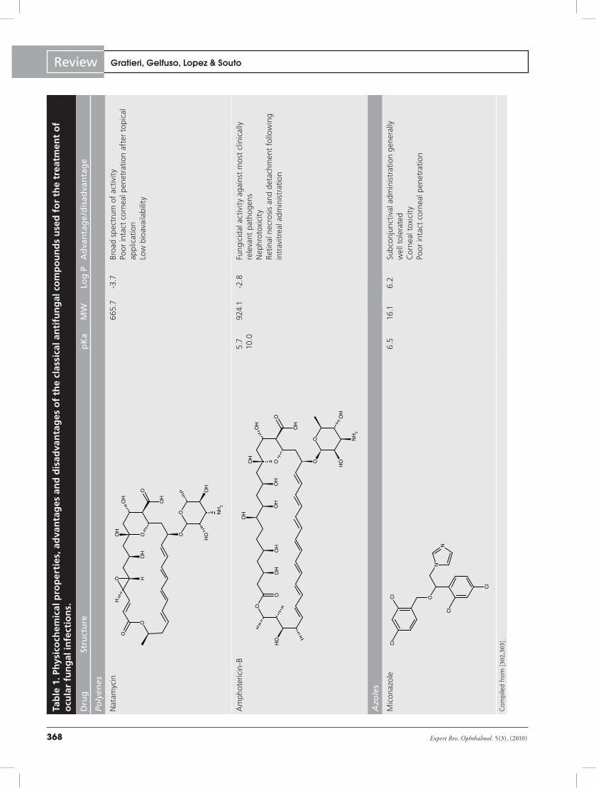
Classical antifungal drugs PolyenesPolyenes exert their antifungal effect by binding to the ergos-terol in the fungal cell membrane, blocking fungal growth or altering membrane permeability [53]. Amphotericin B can also induce oxidative damage, which may contribute to its fungicidal action [54]. The extent of damage to fungal membranes is dose related; however, beyond a certain concentration, human cells may be affected, which accounts for the polyenes’ toxic effects. Natamycin and amphotericin B are the two antifungal agents of this class in current use for the treatment of ophthalmic mycoses.
NatamycinNatamycin, a tetraene polyene, has long been considered the mainstay of treatment for filamentous fungal keratitis [55,56]. As natamycin is poorly soluble in water it is presented as a 5% topi-cal ophthalmic suspension. The initial dosage is normally one drop every hour. Therapy is generally continued for 14–21 days or until there is resolution of active fungal keratitis [301]. It is reported to have a broad spectrum of activity against various fungi, including species of Fusarium, Aspergillus, Candida and Penicillium [2,57,58], although its main limitation is its poor pen-etration after topical application. This has been attributed to the tissue binding, since 97% of the drug that enters the cornea quickly becomes biologically inactivated [59]. Therapeutic concen-trations can still be achieved in the aqueous humor with intense topical administration after removal of the corneal epithelium [59].
Amphotericin B Amphotericin B is a macrolid polyene with two special physico-chemical properties: amphiphilic behavior owing to the apolar and polar sides of the lactone ring and amphoteric behavior owing

Author Pro
of
www.expert-reviews.com 367
ReviewCurrent efforts & the potential of nanomedicine in treating fungal keratitis
to the presence of ionizable carboxyl and amine groups (Table 1). As a consequence of its amphiphilic and zwitterionic nature and the asymmetrical distribution of hydrophobic and hydrophilic groups, amphotericin B is poorly soluble in all aqueous solvents and in many organic solvents [60].
The primary advantages of amphotericin B include its fun-gicidal activity against most clinically relevant pathogens [58] and the low occurrence of resistance [61]. It has been widely administered by intravenous, topical, intracameral and intra-vitreal routes for therapy of ocular infections [56,62–64]. The intravenous administration is the treatment of choice for inva-sive fungal infections, but this route may cause poor corneal bioavailability and severe nephrotoxicity [65]. Similarly to nata-mycin, the corneal penetration of amphotericin B is reduced in the presence of an intact corneal epithelium [59]. The topical regime often includes administration every 30 min for the first 24 h and every hour for the second 24 h, before being slowly tapered according to the clinical response [301]. Subconjunctival injection has been reported to lead to severe toxic effects, and is no longer recommended. Intracameral injections of ampho-tericin B may be an effective adjunctive treatment for fungal keratitis unresponsive to conventional antifungal therapy [66], although cataract may occur [67]. A case reported the use of intrastromal corneal injections combined with intravitreal injection of amphotericin B that led to the eradication of the corneal fungal plaques and the intraocular infection [68]. Intravitreal administration, although commonly used [69], has been reported to cause retinal necrosis and detachment if the injection is not made slowly and exactly in the center of the vitreous, as far as possible from the retina [2].
AzolesThe azoles, discovered in the late 1960s, are totally synthetic. They are inhibitors of a cytochrome P450 fungal enzyme involved in the conversion of lanosterol into ergosterol, an essential sterol in fungal cell membranes. The decrease in ergosterol synthesis leads to increased permeability of the fungal cell membrane, alteration of membrane enzymes, inhibition of growth and death of the fungal cell [70].
The azoles are classified as imidazoles or triazoles, on the basis of whether they have two or three nitrogens in the five-membered azole ring. The imidazoles include clotrimazole, isoconazole, econazole, miconazole and ketoconazole, the last two mostly being used in the treatment of ocular fungal infections. The triazoles include fluconazole and itraconazole.
ImidazolesMiconazole is usually reserved as a second-line drug in the management of fungal keratitis. Very low miconazole levels are obtained in the cornea after intravenous injection, but follow-ing the subconjunctival injection higher levels can be noted in corneas with debridement of corneal epithelium. Similarly, after topical administration the penetration is almost ten times higher in debrided corneas [71]. Topical, subconjunctival and intravenous administrations of miconazole have been reported in the 1980s to
result in successful outcomes [72–75]. Nonetheless, corneal toxicity has been reported, manifesting itself as a row of pinpoint vesicular elevations in the corneal epithelium associated with surround-ing superficial punctate keratitis [74,76]. With the later discovery of other antifungal agents with better pharmacokinetic proper-ties, its use has declined. Notwithstanding, miconazole 1% is one of the most common topical antifungal drugs employed in veterinary cases of fungal keratitis [77–79].
Ketoconazole has pharmacological properties similar to that of miconazole; however, it is absorbable from the gastro intestinal tract and less toxic [80]. It is currently available as oral prepa-ration worldwide. The adult dose of ketoconazole is normally 200–400 mg/day, which can be increased to 800 mg/day. However, gynecomastia, oligospermia and decreased libido have been reported in 5–15% of patients who have been taking 400 mg/day for a long period [301]. Recent in vitro susceptibility studies have shown that the majority of the ocular fungal isolates, including Aspergillus, Candida and some Fusarium species, were sensitive to ketoconazole [81,82]. The oral preparation is often used concomitantly to other topical antifungal agents [83–85], although it can also be administered topically without significant corneal toxicity signs [86]. However, the drawback is its poor water solubility [87]. Case reports can be found in the literature of patients with laboratory-proven fungal corneal infections that were successfully treated with topical ketoconazole. The clinical signs of corneal infection normally disappear after 3–7 weeks of therapy [86].
TriazolesFluconazole is a bistriazole antifungal compound with improved physical and pharmacokinetic properties. Fluconazole is a stable, nontoxic, water-soluble, low-molecular-weight (306.2 Da) com-pound that can be administered by several routes, such as topi-cal [88–92], subconjuntival [93], intravitreal [94] and systemic [95,96]. The subconjunctival regime consists of fluconazole 2% up to 1.0 ml twice daily for at least 5 days [93].
Abbasoglu et al. achieved a fluconazole aqueous humor peak concentration in humans upon single- and multiple-drop appli-cations of a 0.2% solution of 3.35 ± 0.64 and 7.13 ± 0.79 µg/ml, respectively, after 15 min [97]. Antifungal susceptibility tests have reported that among the most common etiological agents in fungal keratitis, Fusarium is the most resistant genera to fluconazole, exhibiting an in vitro MIC of 32–64 µg/ml [98]. Some studies reported lower MIC values for Alternaria alternata (12 µg/ml), Aspergillus (8 µg/ml), Candida (0.2–0.8 µg/ml), Penicillium (4 µg/ml), Curvularia (6–64 µg/ml) and Rhizopus (4–32 µg/ml) [99–102]. Based on this data, topically applied flu-conazole may only be effective for the treatment of less resistant fungi. It is possible that higher aqueous humor concentrations, which would cover the MIC for most pathological agents, could be obtained with multiple-dose administration.
Itraconazole, a dioxolane triazole, is very hydrophobic with a rela-tively higher molecular weight (705.6 Da). It is well absorbed orally, although more than 90% binds to protein in serum [103]. The major drawback of using itraconazole by the oral route for therapy of ocular
Author Pro
of
Expert Rev. Ophthalmol. 5(3), (2010)368
Review Gratieri, Gelfuso, Lopez & SoutoTa
ble
1. P
hys
ico
chem
ical
pro
per
ties
, ad
van
tag
es a
nd
dis
adva
nta
ges
of
the
clas
sica
l an
tifu
ng
al c
om
po
un
ds
use
d f
or
the
trea
tmen
t o
f o
cula
r fu
ng
al in
fect
ion
s.
Dru
gSt
ruct
ure
pK
aM
WLo
g P
Ad
van
tag
e/d
isad
van
tag
e
Poly
enes
Nat
amyc
in6
65.7
-3.7
Bro
ad s
pec
trum
of
acti
vity
Poor
inta
ct c
orn
eal p
enet
rati
on a
fter
to
pica
l ap
plic
atio
nLo
w b
ioav
aila
bilit
y
Am
phot
eric
in-B
5.7
10.0
924
.1-2
.8Fu
ngic
idal
act
ivit
y ag
ains
t m
ost
clin
ical
ly
rele
vant
pat
hog
ens
Nep
hrot
oxic
ity
Retin
al n
ecro
sis
and
det
achm
ent
follo
win
g in
trav
itre
al a
dmin
istr
atio
n
Azo
les
Mic
onaz
ole
6.5
16.1
6.2
Sub
conj
unct
ival
adm
inis
trat
ion
gen
eral
ly
wel
l to
lera
ted
Cor
nea
l tox
icit
yPo
or in
tact
cor
nea
l pen
etra
tion
Co
mp
iled
fro
m [3
02,303].
O
O
O
HO
H
OH
O
OH
O
OH
NH
2
OH
H
O
O
OH
O
OH
O NH
2
OH
OH
OH
OH
O
OO
HO
H
OH
OH
OH
OH
O O
NN
Cl
Cl
O
Cl
Cl

Author Pro
of
www.expert-reviews.com 369
ReviewCurrent efforts & the potential of nanomedicine in treating fungal keratitisTa
ble
1. P
hys
ico
chem
ical
pro
per
ties
, ad
van
tag
es a
nd
dis
adva
nta
ges
of
the
clas
sica
l an
tifu
ng
al c
om
po
un
ds
use
d f
or
the
trea
tmen
t o
f o
cula
r fu
ng
al in
fect
ion
s.
Dru
gSt
ruct
ure
pK
aM
WLo
g P
Ad
van
tag
e/d
isad
van
tag
e
Azo
les
(co
nt.
)
Ket
oco
nazo
le2.
96
.553
1.4
4.3
The
oral
pre
para
tion
is o
ften
use
d co
ncom
itan
tly
to o
ther
to
pica
l ant
ifung
al
agen
ts w
ith
go
od
outc
ome
Poor
wat
er s
olu
bilit
yRe
quire
s ac
id p
H f
or a
bsor
ptio
n
Fluc
onaz
ole
2.0
30
6.2
0.5
Exce
llent
saf
ety
profi
leSt
able
, wat
er-s
olu
ble
Go
od
intr
aocu
lar
pen
etra
tion
Poor
in v
itro
acti
vity
vs
mo
st s
trai
ns o
f A
sper
gillu
s an
d Fu
sariu
m s
pp.
Itra
cona
zole
3.7
705.
65.
6Ex
celle
nt s
afet
y pr
ofile
Hyd
roph
obi
c, h
igh
mo
lecu
lar
wei
ght:
po
or
bio
avai
labi
lyN
arro
w s
pec
trum
cov
erag
e
New
azo
les
Vor
icon
azo
le4
.912
.03
49.3
1.8
2.5
Bro
ader
sp
ectr
um o
f ac
tivi
tyG
reat
er e
ffica
cyRe
vers
ible
dis
turb
ance
of
visi
on a
nd s
kin
rash
es
as c
omm
on s
ide
effe
cts
Rap
id v
itre
al c
lear
ance
Co
mp
iled
fro
m [3
02,303].
O
O
NN
O
O
N
Cl
Cl
H
N
NN N
NN
N
F F
OH
NN
O
O
N
N
N
Cl
Cl
ON
NN
O
N
N
FF F
NO
H
N
N

Author Pro
of
Expert Rev. Ophthalmol. 5(3), (2010)370
Review Gratieri, Gelfuso, Lopez & SoutoTa
ble
1. P
hys
ico
chem
ical
pro
per
ties
, ad
van
tag
es a
nd
dis
adva
nta
ges
of
the
clas
sica
l an
tifu
ng
al c
om
po
un
ds
use
d f
or
the
trea
tmen
t o
f o
cula
r fu
ng
al in
fect
ion
s.
Dru
gSt
ruct
ure
pK
aM
WLo
g P
Ad
van
tag
e/d
isad
van
tag
e
Ech
ino
can
din
s
Cas
pof
ungi
n--
1093
.3-2
.8Fu
ngic
idal
act
ivit
y ag
ains
t flu
cona
zole
-res
ista
nt
fung
i str
ains
Hig
h-m
ole
cula
r-w
eigh
t re
nds
po
or in
tact
co
rnea
l to
pica
l pen
etra
tion
Mic
afun
gin
9.1
1270
.3-0
.4W
ater
so
lubl
eEx
celle
nt in
vitr
o ac
tivi
ty a
gain
st b
oth
Can
dida
an
d A
sper
gillu
sH
igh
-mo
lecu
lar-
wei
ght
rend
s p
oor
inta
ct
corn
eal t
opi
cal p
enet
rati
on
Co
mp
iled
fro
m [3
02,303].
N
OH
N
O
NH
2
OH
O OH
OH
OH
NH
ONH
N H
CH
3
CH
3
OH
O
OH
NHO
H
NH
2
O
O
N H
O
CH
3C
H3
N
N
ON
O
O
NH
OO
H
OO
NH
NH
NH
OH
OOO
H
OH
OH
OS
OH
OH
OO
NH
2
OO
H
O
OH
OH
NH
H

Author Pro
of
www.expert-reviews.com 371
ReviewCurrent efforts & the potential of nanomedicine in treating fungal keratitis
fungal infections is its poor penetration into the cornea, aqueous humor and vitreous compared with fluconazole and ketoconazole. However, oral itraconazole was found to be effective in a case of fungal keratitis of the eye caused by Pichia anomala when used in combination with topical amphotericin B and natamycin [64], and in a case of fungal keratitis caused by Scedosporium apiospermum [104].
Topical itraconazole also proved to be useful for treating infec-tions caused by Aspergillus or Curvularia spp. [105,106]. Topical itraconazole has also been reported as effective in treating ani-mal models of Fusarium keratitis [107], even though its spectrum coverage is narrow against these species [57,82].
AllylaminesAllylamines prevent fungal ergosterol biosynthesis via specific and selective inhibition of fungal squalene epoxidase, thereby inter fering with the integrity of fungal cell membrane [108]. Allylamines are less frequently used in the treatment of ocular fungal infections compared with polyenes and azoles. Antifungal agents belonging to this class include amorolfine, butenafine, naf-tifine and terbinafine, with terbinafine being the most commonly used compound [109].
Novel antifungal drugsNovel antifungal drugs were developed with the aim of solving classical antifungal therapy problems such as severe toxicity (poly-enes), narrow antifungal spectrum (especially against filamen-tous fungi), rapid development of resistance (most azoles), and fungistatic rather than fungicidal effects at the achieved ocular concentrations. Some of these problems are not yet completely solved, but the advances made are presented.
Newer azolesThe newest triazole agents, including ravuconazole, isavucon-azole, posaconazole and voriconazole, are synthetic derivatives of fluconazole but have a significantly broader spectrum of activ-ity [57]. Voriconazole is the better studied compound, and up until now there are no records of the clinical efficacy of the other new azole agents against fungal keratitis, with the exception of a few reports describing the use of posaconazole [110–112].
VoriconazoleVoriconazole is a new antifungal drug derived from fluconazole by the addition of a methyl group to the propyl backbone and by the substitution of a triazole moiety with a fluoropyrimidine group [113]. The molecular alterations conferred to voriconazole a broader spectrum of activity and greater efficacy than its parent compound, fluconazole. However, voriconazole presents more side effects and drug interactions. The most common side effect is a reversible disturbance of vision (photopsia), which may include blurred vision, altered color discrimination and photophobia. These symptoms are related to changes in electroretinogram trac-ings, which revert to normal when treatment with the drug is stopped; no permanent damage to the retina has been noted. Skin rashes are the second most common adverse effect and elevations in hepatic enzyme levels may also occur [114].
The mechanism of action is the same as the other azole agents, but voriconazole also inhibits the 24-methylene dihydrolanasterol demethylation in certain yeast and filamentous fungi [113,114]. The greater efficacy can be confirmed by in vitro susceptibility tests. In general, the MIC of voriconazole for C. albicans is 1–2 log lower than the MIC of fluconazole [115]. It also appears to be very effective in the management of ocular infections caused by many filamentous fungi [40], especially in the management of Aspergillus ocular infections, as compared with other anti fungals [82]. Numerous case reports indicate that voriconazole treatment has been successful where natamycin, amphotericin B or fluconazole have failed, even in cases of drug-resistant fungal keratitis and endophthalmitis [116–122].
Voriconazole is well absorbed following oral administration, with a bioavailability of 90%. A study by Hariprasad et al. demon-strated that orally administered voriconazole achieves therapeutic aqueous and vitreous levels in the noninflamed human eye [123]. After two doses, the mean plasma concentration of voriconazole was 2.13 µg/ml, which resulted in voriconazole concentrations of 0.81 µg/ml in the vitreous and 1.13 µg/ml in the aqueous. The activity spectrum appeared to appropriately encompass the most frequently encountered mycotic species involved in the various causes of fungal endophthalmitis. A similar result was described in a case report of an eye with Scedosporium apiospermum keratitis that went on to corneal transplant. It was found that aqueous voriconazole levels following 12 days of oral treatment in the aqueous humor was 1.8 µg/ml, almost seven times higher than the MIC for that specific strain [124].
Topical therapy may also be used in conjunction with oral ther-apy to increase the amount of drug in the anterior chamber [125]. The topical administration of voriconazole 1% solution every 2 h for 1 day in noninflamed human eyes prior to planned vitrectomy surgery resulted in a mean concentration of 6 µg/ml of the drug in the aqueous and 0.15 µg/ml in the vitreous, demonstrating that the drug penetrates well beyond the cornea when applied topically [126]. These results are in accordance with other studies that applied voriconazole topical solution. Recently, a prospective open-label trial involving ten participants that received topically administered 1% voriconazole solution hourly for four doses or four times a day for 3 days, obtained voriconazole concentrations ranging from 0.1 to 1.1 µg/ml in the vitreous humor [127]. In a similar study, 13 human subjects scheduled for elective anterior segment eye surgery received hourly 2% voriconazole eye drops at 4 h presurgery. Significantly, the voriconazole concentration in the aqueous humor of the eye was similar to that reported for the 1% voriconazole solution, suggestive of concentration-independent absorption through an intact infection-free cornea [128]. This is consistent with observations in a recent animal study, where the voriconazole level in the corneas of horses with fungal keratitis did not change when the administered voriconazole eye drop concentration was changed from 1 to 3% [129]. In addi-tion, in the study conducted by Lau et al. it was also observed that no accumulation of voriconazole in the vitreous humour could be detected with a four-times-a-day dosing regimen, sug-gesting that voriconazole is cleared very rapidly from the posterior

Author Pro
of
Expert Rev. Ophthalmol. 5(3), (2010)372
Review Gratieri, Gelfuso, Lopez & Souto
chamber [127]. This hypothesis is also consistent with results by Shen et al., where the concentration of intravitreal voriconazole at various time points was reported to exhibit exponential decay with a half-life of 2.5 h after single intravitreal injections in a rabbit model [130]. This suggests that in severe cases of fungal keratitis, where pathogens have already spread into the eye or there is a risk of fungal endophthalmitis, considerably higher concentrations of voriconazole or a slow-release formulation would be necessary to sustain therapeutic drug levels in the posterior chamber.
EchinocandinsEchinocandins are lipopeptides that have been synthetically mod-ified from the fermentation broths of various fungi, and have recently emerged as valuable antifungal agents.
They possess a unique mechanism of action, inhibiting b-(1,3)-d-glucan synthase, an enzyme that is necessary for the synthesis of essential components of the cell wall of several fungi. The depletion of these components results in an abnormally weak cell wall unable to withstand osmotic stress [131]. The echinocandins display fungistatic activity against Aspergillus spp. and fungicidal activity against most Candida spp., including strains that are flu-conazole resistant. Overall, resistance to echinocandins is still rare and all agents are well tolerated, with similar adverse effect profiles and few drug–drug interactions [132].
Three echinocandins have been approved by the US FDA, namely caspofungin, micafungin and anidulafungin, but up until now there is no record of anidulafungin applied for the treatment of keratitis.
CaspofunginCaspofungin was the first approved member of the class; it has the most available data and the most indications of the echinocandins [122,133].
Several studies have recently compared the efficacy of topical caspofungin with that of topical amphotericin B. When using an animal model of C. albicans keratitis the authors observed comparable results for 0.5% caspofungin and 0.15% amphoteri-cin B [134]. Similar results were also found for 1% caspofungin and 0.15% amphotericin B topical solutions in an animal model of Fusarium solani keratitis [135].
Since caspofungin has a high molecular weight of 1093.5 Da, the topical administration without corneal epithelium abrasion resulted in no detectable amounts of the drug in the aqueous humor. However, after corneal epithelial abrasion, therapeutic drug levels that cover the MIC of most fungi could be reached [136].
MicafunginMicafungin is a water-soluble echinocandin with excellent in vitro activity against Candida, Aspergillus and some fungi resistant to other antifungal agents [137,138].
Hiraoka et al. evaluated the efficacy of subconjunctival injec-tion of 0.1% micafungin in the treatment of experimental C. albicans keratitis and observed complete healing of the corneal lesions in six out of eight eyes treated [139]. The remaining two eyes where the drug was not effective presented deeper corneal
lesions. Although corneal penetration of micafungin has not been studied yet, the penetration into the deep corneal stroma through an intact epithelial layer seems limited because of its high molecular weight (1292.26 Da). There has been one case report of the clinical application of topical micafungin eyedrops in the treatment of refractory yeast-related corneal ulcers with a satisfac-tory outcome [140]. Moreover, topical instillation of micafungin solution had no apparent toxicity to the cornea [141].
Classical formulationsThe eye is characterized by physiological barriers that limit drug entrance from the blood circulation to its inner structures. These are the blood–aqueous and the blood–retinal barriers [142]. As a consequence, systemic or oral drug therapy requires large drug dosages to reach the site of action in proper amounts, which may cause significant systemic side effects [143]. Intravitreal, periocular and subconjunctival injections could minimize systemic expo-sure of the drug, but the use of these systems is followed by a series of disadvantages. The intravitreally injected drug is rapidly eliminated by the eye’s natural circulatory process and therefore frequent injections may be required. Likewise, large doses are often needed, giving rise to toxicological problems. Besides, there are also relevant side effects, such as pain, discomfort, increased intraocular pressure, intraocular bleeding, increased chances for infection and the risk of retinal detachment. The major complica-tion for intravitreal injection is endophtalmitis, which can result in severe vision loss [144–146]. In addition, ocular injections are not well accepted by patients. The topical administration is the most convenient route for the management of ocular fungal infections, especially for infection affecting the cornea and anterior chamber structures. Therefore, although sometimes not the most efficient, the topical route is the first choice for starting the administration of drugs on the treatment of ocular fungal infections. The classical formulations applied include topical solutions or suspensions in the form of eye drops or ointments in the form of night creams. More recently, lipid complexes of amphotericin B have also been applied.
Topical eye drops In several cases, intensive topical antifungal therapy involves the use of multiple antifungal eye drops in very short administra-tion intervals (e.g., half an hour) [147]. Protection mechanisms of the human eye such as lachrymal secretion and blinking reflex cause rapid drainage of the topically applied eye drops [148]. The short precorneal residence time allied with cornea impermeabil-ity results in low bioavailability, and frequent dosing is usually needed to compensate for the rapid precorneal drug loss.
Water-soluble drugs can be administered in the form of solutions and relatively insoluble drug substances in an aqueous vehicle as a form of suspensions. In this case, the vehicle must contain suitable suspending and dispersing agents to allow good drug redispersibil-ity, maintaining the uniformity of drug dosage. Controlled floccu-lation of suspensions can be accomplished by adding electrolytes, ionic or nonionic surfactants, or even water-soluble polymers [149]. Owing to the particles’ tendency to be retained in the cul-de-sac, the contact time and duration of action of a suspension exceed

Author Pro
of
www.expert-reviews.com 373
ReviewCurrent efforts & the potential of nanomedicine in treating fungal keratitis
those of a solution [150]. The retention may increase with particle size; however, it is recommended that particles should not exceed 10 µm so that they do not cause discomfort.
Several antifungal drugs have been tested in the form of topical eye drops. These drugs may include natamycin [83], amphotericin B [62,107,151], miconazole [74,152], ketoconazole [86], fluconazole [88–92], itraconazole [107], voriconazole [107,125], caspofungin [134,135] and micafungin [140].
The contact time with the target ocular tissue may depend on the physicochemical properties of the drug and the body’s clearance mechanisms, but may also be highly influenced by the vehicle chosen for drug delivery. Even for the newer antifungal compounds, it has been observed that corneal penetration is insuf-ficient. A recent study concluded that to achieve a sustained high level of caspofungin as an effective antifungal therapy for corneal keratitis, the drug should be administered topically every 30 min after removal of the corneal epithelium [136]. However, develop-ing a sustained-release ocular preparation would overcome the requirement for a frequent dosing.
Formulations with enhanced solubility The most important drawback to the formulation of most common antifungal agents is their scarce solubility in water. Such are the cases of amphotericin B (solubility: 0.001 mg/ml; pKa: 5.7) [60], miconazole (solubility: ≤0.00103 mg/ml; pKa: 6.5), ketoconazole (solubility: 0.017 mg/ml; pKa: 6.5) and itracon-azole (solubility: 1.8 mg/ml; pKa: 3.7) [153]. Several attempts have been made to obtain drug formulations suitable for intra-venous and topical ophthalmic administration with adequate drug concentrations.
Cyclodextrins have been used to increase ketoconazole aqueous solubility [154]. When hydroxypropyl b-cyclodextrin was used, it produced more than a twelvefold bioavailability increase after topical instillation in rabbit corneas when compared with the clas-sical ketoconazole suspension [87]. The solubilities of voriconazole, ketoconazole and clotrimazole were also significantly improved with this cyclodextrin in aqueous media [155].
The solubilizer effect of acetate, phosphate and gluconate solu-tions, along with ethanol, glycerol, macrogol 400, propylene gly-col, and surfactants such as polysorbate 20, 60, 80 and sodium taurocholeate, were studied in binary or ternary combinations. Ternary combinations were capable of solubilizing more than 30 mg/ml miconazole and more than 135 mg/ml of ketocon-azole [153]. Nevertheless, for the ocular administration of these solutions further tolerability studies must be performed.
Another example is the colloidal dispersion of amphotericin B with sodium deoxycholate (Fungizone®; Bristol-Myers Squibb Co., NJ, USA), which became available in 1958 for the treat-ment of fungal infections [60]. However, the topical application of such a formulation is known to induce corneal lesions [26,156]. More recent studies have focused on the development of more biocompatible micelles. Micelles composed of a block copolymer poly(2-ethyl-2-oxazoline)-block-poly(aspartic acid) containing amphotericin B (Fungizone) were able to increase drug solubility and efficiency with lower cytotoxicity [157].
Ointments Enhanced ocular retention of oily vehicles has been reported for more than 30 years [158], being attributed to their interaction with the superficial oily layer of the tear film. As a consequence, initial attempts to overcome the poor bioavailability of topically instilled drugs typically involved the use of ointments.
Ointments ensured superior drug bioavailability by increas-ing contact time with the eye, minimizing dilution by tears and resisting nasolachrymal drainage. However, these vehicles have the major drawback of being uncomfortable and causing blurred vision. Consequently, they are mainly used for either adminis-tration overnight or for treatment on the outside and edges of eyelids [159]. A series of antifungal drugs have already been formu-lated in ointments, such as natamycin [152], amphotericin B [160], miconazole [161] and itraconazole [83,84], although in most cases a combined therapy is used.
Lipid complexes To increase the therapeutic index of amphotericin B, lipid complexes were developed. In the commercial drugs Abelcet® (The Liposome Company, NJ, USA) and Amphocil® (Sequus Pharmaceuticals, Inc., CA, USA), amphotericin B has been for-mulated with two phospholipids in a 1:1 drug to lipid molar ratio. Amphotec® (Sequus Pharmaceuticals, Inc., CA, USA) is an amphotericin B formulation with cholesterol sulfate in equimolar concentrations. Amphotec particles resemble discs and have a similar antifungal efficacy to Fungizone but with lower cytotoxic and hemolytic effects. The reduction of renal toxicity has been attributed to the strong affinity of amphotericin B to the choles-terol moieties, which reduces the amount of free amphotericin B in the circulation [60]. A case of Fusarium solani keratitis that progressed to fungal endophthalmitis was successfully treated systemically with the amphotericin B lipid complex Abelcet [162].
Advanced novel formulationsThe clinical efficacy of an antifungal agent in ophthalmic myco-ses depends, to a great extent, on the concentration achieved in the target ocular tissue [163]. Unfortunately, in several cases, topical treatment with classical formulations is not effective enough.
The ability of a drug to penetrate the eye is primarily depen-dent on its physicochemical properties, such as molecular weight, pKa (which determines the nonionized/ionized proportion of the molecule at a certain pH) and log P, which provides information about its lipophilicity.
With respect to drug delivery, the cornea can be divided into three layers, namely the outer epithelium (lipophilic in nature), the stroma (hydrophilic in nature) and the inner endothelium (also lipophilic) [164,165]. In the human eye, the epithelium contains five to seven layers of cells, each connected by tight junctions, which provide a large barrier that is permeable only to small lipophilic molecules. Because the cornea has hydrophilic as well as lipo-philic tissues, it provides an effective bifunctional barrier for the absorption of both lipophilic and hydrophilic compounds. In this way, the overall absorption of moderately lipophilic compounds across the cornea is favored (log P 2–3) [166]. Regardless of the
Author Pro
of
Expert Rev. Ophthalmol. 5(3), (2010)374
Review Gratieri, Gelfuso, Lopez & Souto
administration route, most of the antifungal drugs available do not possess the required physicochemical properties to be absorbed and reach or enter target tissues (Tables 1 & 2).
A promising strategy to overcome these problems involves the development of suitable drug-carrier systems. The in vivo fate of the drug is no longer dependent on the properties of the drug but on the carrier, which should maximize precorneal drug absorption, minimize pre corneal drug loss and allow a controlled and localized release of the active drug, while maintaining the simplicity and convenience of the dosage form.
Since only a limited percentage of the administered drug reaches the target tissue, patient compliance is an important aspect to con-sider when developing an ophthalmic delivery system. As such, attention should be paid to the facility of administration and to the sensorial feeling after the administration, since discomfort (e.g., burning sensation) could induce tear production, followed by drug dilution and drainage through nasolachrymal duct.
Other important aspects to be considered are the retention time, drug-loading capacity and drug protection from metabolic deg-radation. In fact, if the drug-carrier system is able to prolong the retention while loading a sufficient amount of drug in a protected manner, the interval between administrations can be lengthened. For instance, in the case of intravitreal injections, the reduction in the number of injections would also reduce the potential side effects. Apart from these, all the factors that would influence the overall costs should also be considered, such as the possibility of scaling up production, sterilizing, and the physical and chemical storage stability of the product.
Novel colloidal delivery systems such as polymeric nano- and microparticles, liposomes, solid lipid nanoparticles, and nano-structured lipid carriers, are currently being studied in attempt to fulfill all these requirements.
Polymeric micro- & nanoparticlesA controlled-release strategy is to encapsulate the drug in poly-meric microparticles (1–1000 µm) or nanoparticles (1–999 nm). These systems consist of various biocompatible poly-meric matrices in which the drug can be adsorbed, entrapped or covalently attached [167]. Biodegradable and biocompatible syn-thetic polymers such as poly(d,l-lactide-co-glycolide) (PLGA) and polyalkylcya-noacrylates are preferred for nanoparticle production. Nonetheless, use of polysac-charides (e.g., curdlan) and macromol-ecules (e.g., chitosan, albumin and gela-tin) has been very well described in the literature [167–169].
Nanoparticulate technologies in general offer interesting benefits such as solubiliza-tion of hydrophobic drugs, bioavailability improvement, modification of pharmaco-kinetic parameters, and protection of drug molecules from physical, chemical and/or
biological degradation [168]. Increased residence time of drugs and maintenance of their therapeutic concentrations for longer time intervals could reduce the number of subconjunctival and intravit-real injections required in some treatments, while allowing higher doses without toxicity from initial concentration. A drawback is that intravitreal injections of particulate systems may cause vit-real clouding [170]. However, microparticles tend to sink to the lower part of the vitreal cavity, whereas nanoparticles are more likely to cause clouding in the vitreous [145]. It is also suggested that nano particles increase the residence time owing to their bio-adhesive nature, a property that would be especially useful for topi-cal delivery. Different polymers can be used to coat nano particles and improve adhesion. Studies have shown, for example, that the bioavailability of encapsulated indomethacin doubled when poly(e-caprolacton) nanoparticles were coated with chitosan [171]. In addition, microparticles formed of PLGA and poly(ethylene gly-col) (PEG) as a core material and mucoadhesion promoter, respec-tively, showed prolonged residence time in rabbit eyes [172]. In this way, the ideal size and composition of a polymeric colloidal system would depend on the target. For instance, microparticles can be more effective than nanoparticles for intravitreal administration, but if they are larger than 10 µm they could cause an uncomfortable ‘sand-like’ feeling after topical administration [172,173]. In addition, depending on the drug, higher encapsulation efficiency can be obtained in microparticles than nanoparticles.
The encapsulation of antifungal agents in nanoparticulate car-riers has been used with the objective of modifying the pharmaco-kinetics of drugs, resulting in more efficient treatments with fewer side effects. Although there are no records to date of applying these systems for the treatment of ophthalmic fungal infections, they have been studied for the treatment of similar infections in other organs with promising results.
Several recently published works describe the production of nanoparticles containing amphotericin B aiming to control drug delivery and reduce toxicity [174–177]. For example, amphotericin B
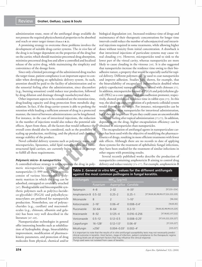
Table 2. General in vitro MIC50 values for the different antifungals against the most common pathogens in fungal keratitis.
Antifungals MIC50 (μg/ml) Ref.
Fusarium Aspergillus Candida
Natamycin 4–8 2–32 4–33† [57,221–224]
Amphotericin B 0.5–32 0.25–2 0.25–1 [57,58,81,82,98,99,137,221,222,225]
Miconazole 8 2 1–10† [58,226]
Ketoconazole 2–16† 0.06–4† 0.008–0.4† [58,81,82]
Fluconazole 32–64 8–64 0.2–51 [58,81,82,98,99,115,225]
Itraconazole 8–32 0.125–4 0.016–0.256 [57,58,82,137,222]
Voriconazole 0.5–12 0.12–0.5 0.008–0.25† [57,115,222,225,227]
Caspofungin 16–128† 0.12–1.0† 0.06–8† [57,225,227]
Micafungin >256† 0.004–0.03† 0.002–4† [225,227]
It is important to note that the results of in vitro antifungal susceptibility tests may not necessarily predict clinical outcome in keratitis. Host factors, stage of infection, patient compliance to the therapeutic regime and drug levels are all important factors that may influence the clinical response. †Fungi used were not isolated from cases of keratitis.

Author Pro
of
www.expert-reviews.com 375
ReviewCurrent efforts & the potential of nanomedicine in treating fungal keratitis
entrapped into PLGA nanoparticles was shown to improve the oral bioavailability and minimize the adverse effects observed in classical systemic amphotericin B therapy [178]. Nonetheless, nanoparticles have also been used for targeting drug delivery. Amphotericin B-loaded PLA-b-PEG nanoparticles coated with polysorbate 80 have been efficiently produced for brain targeting [179]. Since these systems have been shown to efficiently cross the blood–brain barrier, they represent a promising tool for crossing the retinal–blood barrier and increasing intraocular bioavailability after systemic administration. Further studies should be carried out in this area. In addition, intraperitoneal administration of ampho-tericin B nanoparticles based on PLGA and dimercaptosuccinic acid in mice showed antifungal efficacy, fewer undesirable effects and a favorable extended dosing interval [180].
Poly(d,l-lactide-co-glycolide) nanoparticles loaded with vori-conazole were prepared by the emulsion–solvent evaporation technique. The mean particle size was 132.8 nm when using sodium hexametaphosphate to avoid particle agglomeration. Both in vitro and in vivo studies in mice showed greater anti-fungal efficacy of drug-loaded nanoparticles by contrast with the drug alone [181]. Nano- or microparticle production has been described for other antifungal agents, such as fluconazole [182,183] and itraconazole [184].
LiposomesLiposomes are biocompatible and biodegradable phospholipid vesicles formed by one or several lipid bilayers. In each bilayer, the nonpolar fatty acid tails are placed in the interior whereas the polar heads are turned outside, containing an aqueous phase both inside and between the bilayers. Owing to their amphiphilic character, liposomes are able to entrap both hydrophilic and hydrophobic compounds in the aqueous compartments or within the lipid bi layers, respectively [185,186]. Liposomes can provide controlled release of incorporated drugs as the spherical lipid shield formed by bilayer membranes provides a permeability barrier to drug release. In this way, the drug is protected from degradation and clearance, and toxicity resultant from high peak concentration is avoided. This property can be especially useful for posterior segment appli-cations [187]. Similarly to polymeric nano- and microparticles, lipo-somes can minimize some of the adverse side effects encountered by the intraocular administration routes, increasing therapeutic effectiveness [188–190].
Gupta et al., studying the pharmacokinetics of plain and lipo-some-encapsulated fluconazole after intravitreal injection in rab-bit eyes, observed a rapid vitreal clearance and a short half-life (3.08 h) for plain fluconazole, whereas liposome-entrapped flucon-azole showed an extended half-life (23.40 h) [191]. The constant
Table 3. Advantages and disadvantages of advanced novel delivery systems and incorporated antifungal drugs.
Advanced novel formulations
Advantages Disadvantages Incorporated antifungal agents
Ref.
Polymericnanoparticles
May be biocompatible and biodegradableAble to entrap both hydrophilic and hydrophobic drugsControlled releaseProtect drug from metabolic degradationProlonged residence time – bioadhesive properties
Burst effectLimited drug loadingMay cause vitreous cloudingHigh cost
Amphotericin BVoriconazoleFluconazoleItraconazole
[174–180][181][182][184]
Polymeric microparticles
Can be prepared by spray drying – large-scale production May be biocompatible and biodegradableAble to entrap both hydrophilic and hydrophobic drugsControlled releaseProtect drug from metabolic degradationProlonged residence time – bioadhesive properties
Burst effectMay cause uncomfortable sensation if ≥10 μm
Fluconazole [183]
Liposomes Biocompatible and biodegradableAble to entrap both hydrophilic and hydrophobic drugsControlled releaseProtect drug from metabolic degradationProlonged residence time – precorneal and in vitreous
Poor stabilityDifficult to prepare and sterilizeHigh cost
Amphotericin BFluconazole
[194–196][191–193]
SLNs Easy preparation – large-scale production Easy sterilizationImproved ocular bioavailabilityProlonged precorneal residence timeControlled release
Limited drug loading ClotrimazoleKetoconazoleItraconazoleMiconazoleEconazole
[209–211][212][214][215][216]
NLCs Easy preparation – large-scale production Easy sterilizationDrug loading of lipophilic and possibly hydrophilic drugsImproved ocular bioavailabilityProlonged precorneal residence timeControlled release
Hydrophilic drugs can show burst effects
ClotrimazoleKetoconazole
[209,210][212]
NLC: Nanostructured lipid carrier; SLN: Solid lipid nanoparticle.

Author Pro
of
Expert Rev. Ophthalmol. 5(3), (2010)376
Review Gratieri, Gelfuso, Lopez & Souto
terminal elimination of the liposome-loaded drug from the vitreous was seven times less than the plain drug [191]. However, the same authors later discouraged the use of fluconazole as a sole therapy for endophthal-mitis. They reported inferior outcomes for liposome-entrapped fluconazole in a can-didal endophthalmitis rabbit model, prob-ably owing to heterogenous distribuition throughout the vitreous cavity and initial low drug concentration [192].
Liposomal formulations containing flu-conazole for ophthalmic controlled release were also prepared using the reverse-phase evaporation technique [193]. Soya bean phosphatidylcholine and cholesterol in spe-cific weight ratios were used, and selected formulations tested for their in vivo ocular antifungal effect. Conversely, the authors of this work reported that, after in vivo administration in a model of Candida keratitis, fluconazole liposomal formula-tions achieved complete healing in a shorter time than plain fluconazole solution. In addition, the frequency of instillation could be reduced [193].
A reduction in ocular toxicity of subcon-junctival injection of liposomal amphoteri-cin B has also been reported. Comparisons were made with conventional amphoteri-cin B deoxycholate formulation in a rab-bit model. The study reported that subconjunctival injection of amphotericin B deoxycholate formulation or deoxycholate alone induced severe corneal and conjunctival edema with necrosis and infiltration of inflammatory cells, whereas the liposomal formu-lation induced only mild inflammation near the injection site. The authors also observed satisfactory concentrations in corneal stroma after the liposomal formulation injections [194]. In fact, a liposomal formulation named AmBisome® (Vestar, Inc., CA, USA) containing amphotericin B is commercially available. The formulation is supplied lyophilized as a powder and must be recon-stituted in water directly before use, producing liposomes with a mean diameter of 60–70 nm [60]. Because of its hydro phobicity, amphotericin B binds predominantly to the lipid bilayer rather than being placed in the small hydrophilic core of the liposome. The liposomal material consists of hydrogenated soy phospha-tidylcholine and distearoylphosphatidylglycerol. Moreover, the negative charge of the distearoylphosphatidylglycerol can inter-act with the positive amino group of the amphotericin B, form-ing an ionic complex in the bilayers [60]. In addition, a broad antifungal activity spectrum has been defined by the liposomal formulation [195]. In a recent study, the corneal availability fol-lowing systemic administration of parenteral amphotericin B lipid complex or liposomal amphotericin B was compared with that of amphotericin B deoxycholate in a rabbit model [196]. The authors
reported that no drug could be detected in the corneas of the non-inflamed eyes, but in a uveitis-induced model the penetration into the cornea was significantly higher after systemic administra-tion of liposomes, followed by lipid complexes and conventional amphotericin B deoxycholate [196].
It needs to be considered, however, that the type of vesicles formed and the formulations constituents may interfere with the final toxicity and antifungal activity of the drug. It has been observed that small unilamellar vesicles [197–199], multilamellar vesicles [200–202] or large multilamellar vesicles [201] containing amphotericin B perform differently [198]. Similarly, fluconazole showed different MIC values in different vesicle types [198,203]. Inhibition of the antifungal activity of miconazole and ketocon-azole by phospholipids has also been reported. Such an effect seems to be dependent on the phospholipid concentration [198]. Moreover, sterols present in the formulation may interfere with the fungicidal activity of liposomal amphotericin B. It has been observed that ergosterol- and cholesterol-containing liposomes were less effective against C. albicans compared with the sterol-free liposomes [204].
Significant progress has been made in demonstrating the advan-tages of liposome-mediated drug delivery in ophthalmology. In some cases, liposomes have shown to improve efficacy, reduce toxicity, prolong activity and provide site-specific delivery. Despite
Table 4. Aspects to be considered on choosing an ophthalmic delivery system and the performance of advanced novel delivery systems.
Aspects to consider Advanced novel formulations
Nanoparticles Microparticles Liposomes SLNs NLCs
Facility of administration + + + + + + + + + +
Sensorial feeling after administration† (blurred vision, burning sensation, lacrimation)
+ + + + + + + + + + + + + +
Drug loading capacity + + + + + + + - + +
Possibility of drug targeting
+ + + + + + + + + + + + +
Precorneal retention time + + + + + + + + + + + +
In vitreous residence time + + + + + + + + ‡ ‡
Controlled drug release + + + + + + + + + + + +
Avoidance of burst effect - - + + - + +
Avoidance of toxicity + + + + + + + + + + +
Scaling up of production + + + - + + + + + +
Easy to sterilize + + + + + + - + + + + + +
Storage stability + + + - + + + +
It is important to consider the form in which formulations are dispensed. The overall storage stability tends to be significantly higher if the formulations are dispensed in lyophilized form.†The scale indicates the absence of such events, + + + being indicative of the lowest probability of the formulation to cause undesirable sensorial feeling after administration.
‡Not reported. -: Poor; +: Good; + +: Very good; + + +: Excellent; NLC: Nanostructured lipid carrier; SLN: Solid lipid nanoparticle.

Author Pro
of
www.expert-reviews.com 377
ReviewCurrent efforts & the potential of nanomedicine in treating fungal keratitis
these reasons, which make liposomes a potentially useful system for ocular delivery, until now there have been very few attempts to apply them for the treatment of ophthalmic fungal infections. Problems usually encountered were the short shelf life, limited drug-loading capacity, use of aggressive conditions for prepara-tion and sterilization issues [165]. Temperatures required for auto-claving can cause irreversible damage to vesicles while filtration reduces the vesicle to an average of 200 nm, limiting its use to small vesicles.
Solid lipid nanoparticles & nanostructured lipid carriersSolid lipid nanoparticles (SLNs) are the first generation of nanoparticles composed of lipids that are solid at room and body temperatures, stabilized with an emulsifying layer in an aqueous dispersion. They offer the possibility of a controlled drug delivery, since drug mobility in a solid lipid is lower compared with an oily phase. Other advantages of such carriers include the use of physiological compounds in the composition, the fast and effec-tive production process, including the possibility of large-scale production, the avoidance of organic solvents in the production procedures, and the possibility of producing high concentrated lipid suspensions [205]. The main disadvantage, however, is the low drug-loading capacity [206], which is mainly related to the possibility of drug expulsion during storage [207].
Nanostructured lipid carriers (NLCs) are another type of lipid nanoparticle being developed to overcome some limitations of SLNs. NLCs are prepared not only from solid lipids but from a blend of a solid lipid with a certain amount of oil, to maintain a melting point above 40°C. Mixing very different molecules, such as long-chain glycerides of the solid lipid with short-chain glycerides of the liquid lipid, creates crystals with many imperfec-tions [208]. Apart from localizing the drug inbetween fatty acid chains or lipid lamellae, these imperfections provide a location for the additional loading of drug molecules. These drug molecules can then be incorporated in the particle matrix in a molecularly dispersed form, or be arranged in amorphous clusters. There is also more flexibility for modulation of drug release, increasing the drug loading and preventing its leakage.
Lipid nanoparticles (SLNs and NLCs) are interesting systems for the ocular delivery of drugs. Similar to emulsions, they are composed of accepted excipients, and can be produced on a large industrial scale using an established and low-cost homogeniza-tion process. In addition, SLNs and NLCs show the advantages of a solid matrix similar to polymeric nanoparticles, having the ability to protect chemically labile drugs and to modulate release (from very fast to extremely prolonged release). Surface modifications can be used to prolong precorneal residence time. Similarly to liposomes, several SLNs and NLCs have been suc-cessfully prepared for the incorporation of antifungal drugs but aimed for different administration routes, such as transdermal drug delivery.
Clotrimazole-loaded SLNs and NLCs have been prepared by hot high-pressure homogenization with entrapment efficiency higher than 50%. After 3 months of storage at different tempera-tures the mean diameters of SLNs and NLCs remained below
1 µm [209]. The entrapment efficiency and the drug-release pro-file were dependent on the concentration and the lipid mixture employed. NLCs showed higher entrapment efficiency owing to their liquid parts. In agreement with these results, NLCs also depicted a faster release rate in comparison to SLNs with the same lipid concentration. Incorporated clotrimazole in tripalmitine- based SLNs and NLCs stabilized with tyloxapol were also obtained. The particles displayed a spherical shape and a narrow size distribution with a mean diameter smaller than 200 nm [210]. The SLN containing clotrimazole displayed a prolonged release character [211].
Lipid particles containing ketoconazole were also obtained using the hot high-pressure homogenization technique, using Compritol® (Compritol 888 ATO, Gattefossé, Weil am Rhein, Germany) as the solid lipid and the natural antioxidant a-tocoph-erol as the liquid lipid compound for the preparation of NLCs. The authors verified that the SLN matrix was not able to protect the chemically labile ketoconazole against degradation under light exposure. By contrast, the NLCs were able to stabilize the drug, but the aqueous NLC dispersion showed size increase during stor-age. Possible solutions would be light-protected packaging for the SLNs or NLCs physically stabilized in a gel formulation [212]. In accordance, another study revealed that after a shelf life of 2 years, more than 95% of clotrimazole and less than 30% of ketoconazole incorporated in SLNs and NLCs were detected in the developed formulations. Still, these values were shown to be higher than those obtained with reference emulsions of similar composition and droplet sizes [213].
Other antifungal agents that were successfully incorpo-rated in SLNs include itraconazole [214], miconazole [215] and econazole [216].
Therefore, it is expected that in the near future lipid nanopar-ticles will become available for the treatment of ophthalmic fungal infections. Despite the drug-loading difficulties, several compounds commonly used in the treatment of ocular diseases have been incorporated into lipid nanoparticles, such as tobra-mycin [217], gatifloxacin [218], cyclosporine [219] and timolol male-ate [220]. Lipid nanoparticles have shown sustained release and enhancement of drug bioavailability in all such cases [217].
Expert commentaryFor the treatment of ocular fungal infections, one should keep in mind that there are no ideal antifungal agents or administra-tion regimens. As such, the pharmacological treatment should be chosen considering disease-specific conditions, possible side effects, and the drug’s ability to reach the site of infection and achieve therapeutic concentrations.
Few significant advances have been reached in treating oph-thalmic fungal infections. The major problem encountered is the poor water solubility of most of the drugs. Larger mol-ecule sizes (>500 Da) also restrict their intrinsic permeability. Although some formulations with enhanced drug solubility can be easily prepared using cyclodextrins, polymers or suit-able surfactants, these solutions may suffer from the drawback of having low residence time at the ocular surface and being

Author Pro
of
Expert Rev. Ophthalmol. 5(3), (2010)378
Review Gratieri, Gelfuso, Lopez & Souto
rapidly drained. Owing to short residence time and corneal impermeability to most compounds, the topical treatment is often not effective.
Polymeric nano- and microparticles could therefore be a suit-able alternative. Despite not yet being applied for the treatment of fungal keratitis, promising results have been shown for other tar-gets. It is believed that polymeric particles containing antifungal agents could be used to increase drug availability, reduce toxicity and prolong interval of administration. Similarly, liposomes and SLNs offer sustained drug delivery with low toxicity. However, the former represents a challenge when considering large-scale production, whereas the latter has a lower drug-loading capac-ity. NLCs have emerged as a novel delivery system that could incorporate the advantages of those lipid-based delivery systems and overcome their limitations. In the last few years, NLC for-mulations have been successfully prepared for the incorporation of antifungal drugs but have not yet been fully employed in the treatment of ocular diseases. It is expected that in the next few
years more studies will be performed using polymeric particles and lipid-based systems for the ocular route, resulting in more efficient therapeutic options.
Five-year viewIt is expected that in the near future more knowledge will be avail-able on the corneal permeation profile of novel antifungal agents. From that point it is also expected that novel nano medicines would be applied for the ocular delivery of antifungal agents, leading to higher bioavailability and fewer adverse effects.
Financial & competing interests disclosureThe authors would like to thank Fundação de Amparo à Pesquisa do Estado de São Paulo (FAPESP), Brazil, for financial support. The authors have no other relevant affiliations or financial involvement with any organization or entity with a financial interest in or financial conflict with the subject matter or materials discussed in the manuscript apart from those disclosed.
No writing assistance was utilized in the production of this manuscript.
Key issues
• Fungal keratitis occurs throughout the world, but the overall incidence tends to be higher in tropical and subtropical regions. The most frequent fungi causing keratitis worldwide are Fusarium (incidence 20–83.6%), Aspergillus (incidence 16.5–75%) and Candida (incidence 1–63%).
• Fungal keratitis risk factors include trauma, contact lens wear, prolonged use of topical corticosteroids, immunosuppressive diseases, previous eye surgery and chronic eye surface diseases.
• The pharmacological approach of management of fungal keratitis involves the administration of antifungal agents. However, owing to the physiologic constraints of the eye, only a few drugs present adequate bioavailability.
• Classical antifungal drugs act mainly in the fungal cell membrane. The two most commonly used classes are the polyenes and the azoles. The first includes nathamycin and amphotericin B, while the second includes miconazole, ketoconazole, fluconazole and itraconazole.
• Novel drugs have been developed with the aim of solving classical antifungal therapy problems. The novel azole voriconazole is more potent but leads to some adverse effects. The new class echinocandins possesses a broad spectrum but the compounds belonging to this class will probably have low corneal penetration owing to their high molecular weights.
• Nanoparticulated systems containing antifungal drugs could be used to prolong drug delivery and reduce toxicity.
• Liposomes containing antifungal drugs may be useful for intraocular administration. They can minimize some of the adverse side effects encountered by these administration routes and prolong drug residence time, increasing therapeutic effectiveness when no other options are available.
• Antifungal agents have successfully been incorporated into solid lipid nanoparticles and nanostructured lipid carriers, but have not yet been fully employed in the treatment of ocular diseases.
References Papers of special note have been highlighted as:• of interest•• of considerable interest
1 Shukla PK, Kumar M, Keshava GB. Mycotic keratitis: an overview of diagnosis and therapy. Mycoses 51(3), 183–199 (2008).
• Arecentoverviewoffungalkeratitisaspects.Diagnosismethodsandthemostcommontherapiesarepresented.
2 Thomas PA. Current perspectives on ophthalmic mycoses. Clin. Microbiol. Rev. 16(4), 730–797 (2003).
3 Galarreta DJ, Tuft SJ, Ramsay A, Dart JK. Fungal keratitis in London: microbiological and clinical evaluation. Cornea 26(9), 1082–1086 (2007).
4 Bharathi MJ, Ramakrishnan R, Vasu S, Meenakshi R, Palaniappan R. Epidemiological characteristics and laboratory diagnosis of fungal keratitis. A three-year study. Indian J. Ophthalmol. 51(4), 315–321 (2003).
5 Carvalho ACA, Ruthes HI, Maia M et al. Ceratite fúngica no estado do Paraná- Brasil: aspectos epidemiológicos, etiológicos e diagnósticos. Rev. Iberoam. Micol. 18(2), 76–78 (2001).
6 Gopinathan U, Sharma S, Garg P et al. Review of epidemiological features, microbiological diagnosis and treatment outcome of microbial keratitis: experience of over a decade. Indian J. Ophthalmol. 57(4), 273–279 (2009).
7 Panda A, Satpathy G, Nayak N, Kumar S, Kumar A. Demographic pattern, predisposing factors and management of ulcerative keratitis: evaluation of one thousand unilateral cases at a tertiary care centre. Clin. Experiment. Ophthalmol. 35(1), 44–50 (2007).
8 Perez-Balbuena A, Vanzzini-Rosano V, Valadez-Virgen JD, Campos-Moller X. Fusarium keratitis in Mexico. Cornea 28(6), 626–630 (2009).

Author Pro
of
www.expert-reviews.com 379
ReviewCurrent efforts & the potential of nanomedicine in treating fungal keratitis
9 Tanure MA, Cohen EJ, Sudesh S, Rapuano CJ, Laibson PR. Spectrum of fungal keratitis at Wills Eye Hospital, Philadelphia, Pennsylvania. Cornea 19(3), 307–312 (2000).
10 Tuft SJ, Tullo AB. Fungal keratitis in the United Kingdom 2003–2005. Eye 23(6), 1308–1313 (2009).
11 Ritterband DC, Seedor JA, Shah MK, Koplin RS, McCormick SA. Fungal keratitis at the New York Eye and Ear Infirmary. Cornea 25(3), 264–267 (2006).
12 Xie LX, Zhong WX, Shi WY, Sun SY. Spectrum of fungal keratitis in north China. Ophthalmology 113(11), 1943–1948 (2006).
13 Leck AK, Thomas PA, Hagan M et al. Aetiology of suppurative corneal ulcers in Ghana and south India, and epidemiology of fungal keratitis. Br. J. Ophthalmol. 86(11), 1211–1215 (2002).
14 Gopinathan U, Garg P, Fernandes M, Sharma S, Athmanathan S, Rao GN. The epidemiological features and laboratory results of fungal keratitis: a 10-year review at a referral eye care center in south India. Cornea 21(6), 555–559 (2002).
15 Ibrahim MM, Vanini R, Ibrahim FM et al. Epidemiologic aspects and clinical outcome of fungal keratitis in southeastern Brazil. Eur. J. Ophthalmol 19(3), 355–361 (2009).
16 Thew MRJ, Todd B. Fungal keratitis in far north Queensland, Australia. Clin. Experiment. Ophthalmol. 36(8), 721–724 (2008).
17 Sirikul T, Prabriputaloong T, Smathivat A, Chuck RS, Vongthongsri A. Predisposing factors and etiologic diagnosis of ulcerative keratitis. Cornea 27(3), 283–287 (2008).
18 Liesegang TJ, Forster RK. Spectrum of microbial keratitis in South Florida. Am. J. Ophthalmol. 90(1), 38–47 (1980).
19 Upadhyay MP, Karmacharya PC, Koirala S et al. Epidemiologic characteristics, predisposing factors, and etiologic diagnosis of corneal ulceration in Nepal. Am. J. Ophthalmol. 111(1), 92–99 (1991).
20 Khairallah SH, Byrne KA, Tabbara KF. Fungal keratitis in Saudi Arabia. Doc.Ophthalmol. 79(3), 269–276 (1992).
21 Hagan M, Wright E, Newman M, Dolin P, Johnson G. Causes of suppurative keratitis in Ghana. Br J. Ophthalmol. 79(11), 1024–1028 (1995).
22 Hall LR, Lass JH, Diaconu E, Strine ER, Pearlman E. An essential role for antibody in neutrophil and eosinophil recruitment to the cornea: B cell-deficient (microMT)
mice fail to develop Th2-dependent, helminth-mediated keratitis. J. Immunol. 163(9), 4970–4975 (1999).
23 Kaur IP, Rana C, Singh H. Development of effective ocular preparations of antifungal agents. J. Ocul. Pharmacol. Ther. 24(5), 481–493 (2008).
24 FlorCruz NV, Peczon I. Medical interventions for fungal keratitis. Cochrane Database Syst. Rev. 1, CD004241 (2008).
25 Srinivasan M. Fungal keratitis. Curr. Opin. Ophthalmol. 15(4), 321–327 (2004).
26 Thomas PA. Mycotic keratitis – an underestimated mycosis. J. Med. Vet. Mycol. 32(4), 235–256 (1994).
27 Wang LY, Sun ST, Jing Y, Han L, Zhang HM, Yue J. Spectrum of fungal keratitis in central China. Clin. Experiment. Ophthalmol. 37(8), 763–771 (2009).
28 Godoy P, Cano J, Gene J, Guarro J, Hofling-Lima AL, Lopes CA. Genotyping of 44 isolates of Fusarium solani, the main agent of fungal keratitis in Brazil. J. Clin. Microbiol. 42(10), 4494–4497 (2004).
29 Thomas PA. Fungal infections of the cornea. Eye (Lond.) 17(8), 852–862 (2003).
30 Salera CM, Tanure MAG, Lima WTM, Trindade FC, Moreira JA. Perfil das ceratites fúngicas no Hospital São Geraldo Belo Horizonte – MG. Arq. Bras. Oftalmol. 65, 9–13 (2002).
31 Alfonso EC, Miller D, Cantu-Dibildox J, O’Brien TP, Schein OD. Fungal keratitis associated with non-therapeutic soft contact lenses. Am. J. Ophthalmol. 142(1), 154–155 (2006).
32 Klotz SA, Penn CC, Negvesky GJ, Butrus SI. Fungal and parasitic infections of the eye. Clin. Microbiol. Rev. 13(4), 662–685 (2000).
33 Klotz SA, Au YK, Misra RP. A partial-thickness epithelial defect increases the adherence of Pseudomonas aeruginosa to the cornea. Invest. Ophthalmol. Vis. Sci. 30(6), 1069–1074 (1989).
34 Ahearn DG, Zhang S, Stulting RD et al. Fusarium keratitis and contact lens wear: facts and speculations. Med. Mycol. 46(5), 397–410 (2008).
35 Rao SK, Lam PTH, Li EYM, Yuen HKL, Lam DSC. A case series of contact lens-associated Fusarium keratitis in Hong Kong. Cornea 26(10), 1205–1209 (2007).
36 Chang DC, Grant GB, O’Donnell K et al. Multistate outbreak of Fusarium keratitis associated with use of a contact lens solution. JAMA 296(8), 953–963 (2006).
37 Gaujoux T, Chatel MA, Chaumeil C, Laroche L, Borderie VM. Outbreak of contact lens-related Fusarium keratitis in France. Cornea 27(9), 1018–1021 (2008).
38 Khor WB, Aung T, Saw SM et al. An outbreak of Fusarium keratitis associated with contact lens wear in Singapore. JAMA 295(24), 2867–2873 (2006).
39 Gorscak JJ, Ayres BD, Bhagat N et al. An outbreak of Fusarium keratitis associated with contact lens use in the northeastern United States. Cornea 26(10), 1187–1194 (2007).
40 Granados JM, Puerto N, Carrilero MJ. Efficiency of voriconazole in fungal keratitis caused by Candida albicans. Arch. Soc. Esp. Oftalmol. 79(11), 565–568 (2004).
41 Krishnan S, Manavathu EK, Chandrasekar PH. Aspergillus flavus: an emerging non-fumigatus Aspergillus species of significance. Mycoses 52(3), 206–222 (2009).
42 Mendicute J, Orbegozo J, Ruiz M, Saiz A, Eder F, Aramberri J. Keratomycosis after cataract surgery. J. Cataract Refract. Surg. 26(11), 1660–1666 (2000).
43 Nayak N. Fungal infections of the eye: laboratory diagnosis and treatment. Nepal. Med. Coll. J. 10(1), 48–63 (2008).
44 Behrens-Baumann W. Topical antimycotics in ophthalmology. Ophthalmologica 211(Suppl. 1), 33–38 (1997).
45 Ti SE, Scott A, Janardhanan P, Tan DTH. Therapeutic keratoplasty for advanced suppurative keratitis. Am. J. Ophthalmol. 143(5), 755–762 (2007).
46 Yuan XY, Wilhelmus KR, Matoba AY, Alexandrakis G, Miller D, Huang AJW. Pathogenesis and outcome of Paecilomyces keratitis. Am. J. Ophthalmol. 147(4), 691–696 (2009).
47 Banitt M, Berenbom A, Shah M, Buxton D, Milman T. A case of polymicrobial keratitis violating an intact lens capsule. Cornea 27(9), 1057–1061 (2008).
48 Bashir G, Hussain W, Rizvi A. Bipolaris hawaiiensis keratomycosis and endophthalmitis. Mycopathologia 167(1), 51–53 (2009).
49 Gupta AK, Sauder DN, Shear NH. Antifungal agents: an overview. Part I. J. Am. Acad. Dermatol. 30(5 Pt 1), 677–698 (1994).
50 Smith EB. History of antifungals. J. Am. Acad. Dermatol. 23(4 Pt 2), 776–778 (1990).
51 Luke RG, Boyle JA. Renal effects of amphotericin B lipid complex. Am. J. Kidney Dis. 31(5), 780–785 (1998).
Author Pro
of
Expert Rev. Ophthalmol. 5(3), (2010)380
Review Gratieri, Gelfuso, Lopez & Souto
52 Kauffman CA, Carver PL. Antifungal agents in the 1990s. Current status and future developments. Drugs 53(4), 539–549 (1997).
53 te Welscher YM, ten Napel HH, Balagué MM et al. Natamycin blocks fungal growth by binding specifically to ergosterol without permeabilizing the membrane. J. Biol. Chem. 283(10), 6393–6401 (2008).
54 Georgopapadakou NH, Walsh TJ. Human mycoses: drugs and targets for emerging pathogens. Science 264(5157), 371–373 (1994).
55 O’Day DM, Ray WA, Head WS, Robinson RD. Influence of the corneal epithelium on the efficacy of topical antifungal agents. Invest. Ophthalmol. Vis. Sci. 25(7), 855–859 (1984).
56 Loh AR, Hong K, Lee S, Mannis M, Acharya NR. Practice patterns in the management of fungal corneal ulcers. Cornea 28(8), 856–859 (2009).
57 Lalitha P, Shapiro BL, Srinivasan M et al. Antimicrobial susceptibility of Fusarium, Aspergillus, and other filamentous fungi isolated from keratitis. Arch. Ophthalmol. 125(6), 789–793 (2007).
58 Xie LX, Zhai HL, Zhao J, Sun SY, Shi WY, Dong XG. Antifungal susceptibility for common pathogens of fungal keratitis in Shandong Province, China. Am. J. Ophthalmol. 146(2), 260–265 (2008).
59 O’Day DM, Head WS, Robinson RD, Clanton JA. Corneal penetration of topical amphotericin B and natamycin. Curr. Eye Res. 5(11), 877–882 (1986).
60 Torrado JJ, Espada R, Ballesteros MP, Torrado-Santiago S. Amphotericin B formulations and drug targeting. J. Pharm. Sci. 97(7), 2405–2425 (2008).
61 DiDomenico B. Novel antifungal drugs. Curr. Opin. Microbiol. 2(5), 509–515 (1999).
62 Al Assiri A, Al Jastaneiah S, Al Khalaf A, Al Fraikh H, Wagoner MD. Late-onset donor-to-host transmission of Candida glabrata following corneal transplantation. Cornea 25(1), 123–125 (2006).
63 Levy J, Benharroch D, Peled N, Lifshitz T. Blastoschizomyces capitatus keratitis and melting in a corneal graft. Can. J. Ophthalmol. 41(6), 772–774 (2006).
64 Park KA, Ahn K, Chung ES, Chung TY. Pichia anomala fungal keratitis. Cornea 27(5), 619–620 (2008).
65 Khoo SH, Bond J, Denning DW. Administering amphotericin B: a practical approach. J. Antimicrob. Chemother. 33(2), 203–213 (1994).
66 Kaushik S, Ram J, Brar GS, Jain AK, Chakraborti A, Gupta A. Intracameral amphotericin B: initial experience in severe keratomycosis. Cornea 20(7), 715–719 (2001).
67 Yilmaz S, Ture M, Maden A. Efficacy of intracameral amphotericin B injection in the management of refractory keratomycosis and endophthalmitis. Cornea 26(4), 398–402 (2007).
68 Garcia-Valenzuela E, Song CD. Intracorneal injection of amphothericin B for recurrent fungal keratitis and endophthalmitis. Arch. Ophthalmol. 123(12), 1721–1723 (2005).
69 Gupta A, Srinivasan R, Kaliaperumal S, Saha I. Post-traumatic fungal endophthalmitis – a prospective study. Eye 22(1), 13–17 (2008).
70 Koltin Y, Hitchcock CA. The search for new triazole antifungal agents. Curr. Opin. Chem. Biol. 1(2), 176–182 (1997).
71 Foster CS, Stefanyszyn M. Intraocular penetration of miconazole in rabbits. Arch.Ophthalmol. 97(9), 1703–1706 (1979).
72 Anderson RL, Carroll TF, Harvey JT, Myers MG. Petriellidium (Allescheria) boydii orbital and brain abscess treated with intravenous miconazole. Am. J. Ophthalmol. 97(6), 771–775 (1984).
73 Fitzsimons R, Peters AL. Miconazole and ketoconazole as a satisfactory first-line treatment for keratomycosis. Am. J. Ophthalmol. 101(5), 605–608 (1986).
74 Foster CS. Miconazole therapy for keratomycosis. Am. J. Ophthalmol. 91(5), 622–629 (1981).
75 Ishibashi Y, Matsumoto Y, Takei K. The effects of intravenous miconazole on fungal keratitis. Am. J. Ophthalmol. 98(4), 433–437 (1984).
76 Zaidman GW. Miconazole corneal toxicity. Cornea 10(1), 90–91 (1991).
77 Galan A, Martin-Suarez EM, Gallardo JM, Molleda JM. Clinical findings and progression of 10 cases of equine ulcerative keratomycosis (2004–2007). Equine Vet. Educ. 21(5), 236–242 (2009).
78 Myers DA, Lsaza R, Ben Shlomo G, Abbott J, Plummer CE. Fungal keratitis in a gopher tortoise (Gopherus polyphemus). J. Zoo Wildl. Med. 40(3), 579–582 (2009).
79 Andrew SE, Brooks DE, Smith PJ, Gelatt KN, Chmielewski NT, Whittaker CJG. Equine ulcerative keratomycosis: visual outcome and ocular survival in 39 cases (1987–1996). Equine Vet. J. 30(2), 109–116 (1998).
80 Hemady RK, Chu W, Foster CS. Intraocular penetration of ketoconazole in rabbits. Cornea 11(4), 329–333 (1992).
81 Therese KL, Bagyalakshmi R, Madhavan HN, Deepa P. In vitro susceptibility testing by agar dilution method to determine the minimum inhibitory concentrations of amphotericin B, fluconazole and ketoconazole against ocular fungal isolates. Indian J. Med. Microbiol. 24(4), 273–279 (2006).
82 Marangon FB, Miller D, Giaconi JA, Alfonso EC. In vitro investigation of voriconazole susceptibility for keratitis and endophthalmitis fungal pathogens. Am. J. Ophthalmol. 137(5), 820–825 (2004).
83 Manikandan P, Varga J, Kocsube S et al. Mycotic keratitis due to Aspergillus nomius. J. Clin. Microbiol. 47(10), 3382–3385 (2009).
84 Kredics L, Varga J, Kocsube S et al. Infectious keratitis caused by Aspergillus tubingensis. Cornea 28(8), 951–954 (2009).
85 Dursun D, Fernandez V, Miller D, Alfonso EC. Advanced Fusarium keratitis progressing to endophthalmitis. Cornea 22(4), 300–303 (2003).
86 Torres MA, Mohamed J, Cavazos-Adame H, Martinez LA. Topical ketoconazole for fungal keratitis. Am. J. Ophthalmol. 100(2), 293–298 (1985).
87 Zhang JJ, Wang LY, Gao CF, Zhang L, Xia HY. Ocular pharmacokinetics of topically-applied ketoconazole solution containing hydroxypropyl b-cyclodextrin to rabbits. J. Ocul. Pharmacol. Ther. 24(5), 501–506 (2008).
88 Behrens-Baumann W, Klinge B, Ruchel R. Topical fluconazole for experimental Candida keratitis in rabbits. Br. J. Ophthalmol. 74(1), 40–42 (1990).
89 Chung PC, Lin HC, Hwang YS et al. Paecilomyces lilacinus scleritis with secondary keratitis. Cornea 26(2), 232–234 (2007).
90 Sonego-Krone S, Sanchez-Di Martino D, Ayala-Lugo R et al. Clinical results of topical fluconazole for the treatment of filamentous fungal keratitis. Graefes Arch.Clin. Exp. Ophthalmol. 244(7), 782–787 (2006).
91 Holgado S, Luna JD, Juarez CP. Postoperative Candida keratitis treated successfully with fluconazole. Ophthalmic Surg. 24(2), 132 (1993).
92 Sodhi PK, Mehta DK. Fluconazole in managment of ocular infections due to Curvularia. Ann. Ophthalmol. 35(1), 68–72 (2003).
Author Pro
of
www.expert-reviews.com 381
ReviewCurrent efforts & the potential of nanomedicine in treating fungal keratitis
93 Yilmaz S, Maden A. Severe fungal keratitis treated with subconjunctival fluconazole. Am. J. Ophthalmol. 140(3), 454–458 (2005).
94 Su CY, Lin CP, Wang HZ et al. Intraocular use of fluconazole in the management of ocular fungal infection. Kaohsiung J. Med. Sci. 15(4), 218–225 (1999).
95 Avunduk AM, Beuerman RW, Warnel ED, Kaufman HE, Greer D. Comparison of efficacy of topical and oral fluconazole treatment in experimental Aspergillus keratitis. Curr. Eye Res. 26(2), 113–117 (2003).
96 Akler ME, Vellend H, McNeely DM, Walmsley SL, Gold WL. Use of fluconazole in the treatment of candidal endophthalmitis. Clin. Infect. Dis. 20(3), 657–664 (1995).
97 Abbasoglu OE, Hosal BM, Sener B, Erdemoglu N, Gursel E. Penetration of topical fluconazole into human aqueous humor. Exp. Eye Res. 72(2), 147–151 (2001).
98 Li L, Wang Z, Li R, Luo S, Sun X. In vitro evaluation of combination antifungal activity against Fusarium species isolated from ocular tissues of keratomycosis patients. Am. J. Ophthalmol. 146(5), 724–728 (2008).
99 Fungtomc JC, Minassian B, Huczko E, Kolek B, Bonner DP, Kessler RE. In vitro antifungal and fungicidal spectra of a new pradimicin derivative, BMS-181184. Antimicrob. Agents Chemother. 39(2), 295–300 (1995).
100 Gonzalez G. In vitro activities of isavuconazole against opportunistic filamentous and dimorphic fungi. Med. Mycol. 47(1), 71–76 (2009).
101 Li LH, McCarthy P, Hui SW. High-efficiency electrotransfection of human primary hematopoietic stem cells. FASEB J. 15(3), 586–588 (2001).
102 Qiu WY, Yao YF, Zhu YF et al. Fungal spectrum identified by a new slide culture and in vitro drug susceptibility using Etest in fungal keratitis. Curr. Eye Res. 30(12), 1113–1120 (2005).
103 Savani DV, Perfect JR, Cobo LM, Durack DT. Penetration of new azole compounds into the eye and efficacy in experimental Candida endophthalmitis. Antimicrob. Agents Chemother. 31(1), 6–10 (1987).
104 Saracli MA, Erdem U, Gonlum A, Yildiran ST. Scedosporium apiospermum keratitis treated with itraconazole. Med. Mycol. 41(2), 111–114 (2003).
105 Kalavathy CM, Parmar P, Kaliamurthy J et al. Comparison of topical itraconazole 1% with topical natamycin 5% for the treatment of filamentous fungal keratitis. Cornea 24(4), 449–452 (2005).
106 Martinez-Ramos M, Claros B, Vale-Oviedo MA et al. Effect of the vehicle on the topical itraconazole efficacy for treating corneal ulcers caused by Aspergillus fumigatus. Clin. Experiment. Ophthalmol. 36(4), 335–338 (2008).
107 Yavas GF, Oztuerk F, Kusbeci T et al. Antifungal efficacy of voriconazole, itraconazole and amphotericin B in experimental Fusarium solani keratitis. Graefes Arch. Clin. Experiment. Ophthalmol. 246(2), 275–279 (2008).
108 Abdel-Rahman SM, Nahata MC. Oral terbinafine: a new antifungal agent. Ann. Pharmacother. 31(4), 445–456 (1997).
109 Schelenz S, Goldsmith DJA. Aspergillus endophthalmitis: an unusual complication of disseminated infection in renal transplant patients. J. Infect. 47(4), 336–343 (2003).
110 Tu EY, Park AJ. Recalcitrant Beauveria bassiana keratitis: confocal microscopy findings and treatment with posaconazole (noxafil). Cornea 26(8), 1008–1010 (2007).
111 Amiel H, Chohan AAB, Snibson GR, Vajpayee R. Atypical fungal sclerokeratitis. Cornea 27(3), 382–383 (2008).
112 Vyzantiadis TA, Kioumi A, Papadakis E et al. Rhino-cerebral zygomycosis resistant to antimycotic treatment: a case report. Mycoses 52(1), 87–90 (2009).
113 Sabo JA, Abdel-Rahman SM. Voriconazole: a new triazole antifungal. Ann. Pharmacother. 34(9), 1032–1043 (2000).
• Currentlyavailableinformationonthepharmacology,pharmacokinetics,efficacy,adverseeffects,druginteractionsanddosageguidelinesofvoriconazoleispresented.
114 Johnson LB, Kauffman CA. Voriconazole: a new triazole antifungal agent. Clin. Infect. Dis. 36(5), 630–637 (2003).
115 Ruhnke M, Schmidt-Westhausen A, Trautmann M. In vitro activities of voriconazole (UK-109,496) against fluconazole-susceptible and -resistant Candida albicans isolates from oral cavities of patients with human immunodeficiency virus infection. Antimicrob. Agents Chemother. 41(3), 575–577 (1997).
116 Bunya VY, Hammersmith KM, Rapuano CJ, Ayres BD, Cohen EJ. Topical and oral voriconazole in the treatment of fungal keratitis. Am. J. Ophthalmol. 143(1), 151–153 (2007).
117 Creti A, Esposito V, Bocchetti M et al. Voriconazole curative treatment for Acremonium species keratitis developed in a patient with concomitant Staphylococcus aureus corneal infection: a case report. In Vivo 20(1), 169–171 (2006).
118 Durand ML, Kim IK, D’Amico DJ et al. Successful treatment of Fusarium endophthalmitis with voriconazole and Aspergillus endophthalmitis with voriconazole plus caspofungin. Am. J. Ophthalmol. 140(3), 552–554 (2005).
119 Freda R. Use of oral voriconazole as adjunctive treatment of severe cornea fungal infection: case report. Arq. Bras. Oftalmol. 69(3), 431–434 (2006).
120 Kramer M, Kramer MR, Blau H et al. Intravitreal voriconazole for the treatment of endogenous Aspergillus endophthalmitis. Ophthalmology 113(7), 1184–1186 (2006).
121 Al Badriyeh D, Leung L, Davies GE, Stewart K, Kong D. Successful salvage treatment of Scedosporium apiospermum keratitis with topical voriconazole after failure of natamycin. Ann. Pharmacother. 43(6), 1139–1142 (2009).
122 Tu EY. Alternaria keratitis: clinical presentation and resolution with topical fluconazole or intrastromal voriconazole and topical caspofungin. Cornea 28(1), 116–119 (2009).
123 Hariprasad SM, Mieler WF, Holz ER et al. Determination of vitreous, aqueous, and plasma concentration of orally administered voriconazole in humans. Arch. Ophthalmol. 122(1), 42–47 (2004).
124 Nulens E, Eggink C, Rijs AJ, Wesseling P, Verweij PE. Keratitis caused by Scedosporium apiospermum successfully treated with a cornea transplant and voriconazole. J. Clin. Microbiol. 41(5), 2261–2264 (2003).
125 Klont RR, Eggink CA, Rijs AJMM, Wesseling P, Verweij PE. Successful treatment of Fusarium keratitis with cornea transplantation and topical and systemic voriconazole. Clin. Infect. Dis. 40(12), E110–E112 (2005).
126 Vemulakonda GA, Hariprasad SM, Mieler WF, Prince RA, Shah GK, Van Gelder RN. Aqueous and vitreous concentrations following topical administration of 1% voriconazole in humans. Arch. Ophthalmol. 126(1), 18–22 (2008).
127 Lau D, Leung L, Ferdinands M et al. Penetration of 1% voriconazole eye drops into human vitreous humour: a prospective, open-label study. Clin. Experiment. Ophthalmol. 37(2), 197–200 (2009).

Author Pro
of
Expert Rev. Ophthalmol. 5(3), (2010)382
Review Gratieri, Gelfuso, Lopez & Souto
128 Al Badriyeh D, Leung L, Roydhouse T et al. Prospective open-label study of the administration of two-percent voriconazole eye drops. Antimicrob. Agents Chemother. 53(7), 3153–3155 (2009).
129 Clode AB, Davis JL, Salmon J, Michau TM, Gilger BC. Evaluation of concentration of voriconazole in aqueous humor after topical and oral administration in horses. Am. J. Vet. Res. 67(2), 296–301 (2006).
130 Shen YC, Wang MY, Wang CY et al. Clearance of intravitreal voriconazole. Invest. Ophthalmol.Vis. Sci. 48(5), 2238–2241 (2007).
131 Hector RF. Compounds active against cell walls of medically important fungi. Clin. Microbiol. Rev. 6(1), 1–21 (1993).
132 Sucher AJ, Chahine EB, Balcer HE. Echinocandins: the newest class of antifungals. Ann. Pharmacother. 43(10), 1647–1657 (2009).
133 Cleary JD. Echinocandins: pharmacokinetic and therapeutic issues. Curr. Med. Res. Opin. 25(7), 1741–1750 (2009).
134 Goldblum D, Frueh BE, Sarra GM, Katsoulis K, Zimmerli S. Topical caspofungin for treatment of keratitis caused by Candida albicans in a rabbit model. Antimicrob. Agents Chemother. 49(4), 1359–1363 (2005).
135 Ozturk F, Yavas GF, Kusbeci T et al. Efficacy of topical caspofungin in experimental Fusarium keratitis. Cornea 26(6), 726–728 (2007).
136 Vorwerk CK, Tuchen S, Streit F, Binder L, Hofmuller W, Behrens-Baumann W. Aqueous humor concentrations of topically administered caspofungin in rabbits. Ophthalmic Res. 41(2), 102–105 (2009).
137 Tawara S, Ikeda F, Maki K et al. In vitro activities of a new lipopeptide antifungal agent, FK463, against a variety of clinically important fungi. Antimicrob. Agents Chemother. 44(1), 57–62 (2000).
138 Temesgen Z, Barreto J, Vento S. Micafungin: the newest echinocandin. Drugs Today. 45(6), 469–478 (2009).
139 Hiraoka T, Kaji Y, Wakabayashi T, Nanbu PN, Okamoto F, Oshika T. Comparison of micafungin and fluconazole for experimental Candida keratitis in rabbits. Cornea 26(3), 336–342 (2007).
140 Matsumoto Y, Dogru M, Goto E, Fujishima H, Tsubota K. Successful topical application of a new antifungal agent, micafungin, in the treatment of refractory
fungal corneal ulcers: report of three cases and literature review. Cornea 24(6), 748–753 (2005).
141 Hiraoka T, Wakabayashi T, Kaji Y et al. Toxicological evaluation of micafungin ophthalmic solution in rabbit eyes. J. Ocul. Pharmacol. Ther. 21(2), 149–156 (2005).
142 Barar J, Javadzadeh AR, Omidi Y. Ocular novel drug delivery: impacts of membranes and barriers. Expert Opin. Drug Deliv. 5(5), 567–581 (2008).
143 Schalenbourg A, Leys A, de Courten C, Coutteel C, Herbort CP. Corticosteroid-induced central serous chorioretinopathy in patients with ocular inflammatory disorders. Klin. Monbl. Augenheilkd. 219(4), 264–267 (2002).
144 Cunningham MA, Edelman JL, Kaushal S. Intravitreal steroids for macular edema: the past, the present, and the future. Surv. Ophthalmol. 53(2), 139–149 (2008).
145 Del Amo EM, Urtti A. Current and future ophthalmic drug delivery systems. A shift to the posterior segment. Drug Discov. Today 13(3–4), 135–143 (2008).
146 Moshfeghi DM, Kaiser PK, Scott IU et al. Acute endophthalmitis following intravitreal triamcinolone acetonide injection. Am. J. Ophthalmol. 136(5), 791–796 (2003).
147 Kredics L, Varga J, Kocsube S et al. Case of keratitis caused by Aspergillus tamarii. J. Clin. Microbiol. 45(10), 3464–3467 (2007).
148 Wolf EJ, Braunstein A, Shih C, Braunstein RE. Incidence of visually significant pseudophakic macular edema after uneventful phacoemulsification in patients treated with nepafenac. J. Cataract Refract. Surg. 33(9), 1546–1549 (2007).
149 Yasueda S, Inada K, Matsuhisa K, Terayama H, Ohtori A. Evaluation of ophthalmic suspensions using surface tension. Eur. J. Pharm. Biopharm. 57(2), 377–382 (2004).
150 Bourlais CL, Acar L, Zia H, Sado PA, Needham T, Leverge R. Ophthalmic drug delivery systems: recent advances. Prog. Retin. Eye Res. 17(1), 33–58 (1998).
151 Giobbia M, Rossi MC, Conti E et al. Post-traumatic Aspergillus fumigatus keratitis. J. Mycol. Med. 12(1), 32–33 (2002).
152 Suzuki T, Hori N, Miyake T, Hori Y, Mochizuki K. Keratitis caused by a rare fungus, Malassezia restricta. Jpn. J. Ophthalmol. 51(4), 292–294 (2007).
153 Kovacs K, Stampf G, Klebovich I, Antal I, Ludanyi K. Aqueous solvent system for the solubilization azole compounds. Eur. J. Pharm. Sci. 36(2–3), 352–358 (2009).
154 Esclusa-Diaz MT, Guimaraens-Mendez M, Perez-Marcos MB, Vila-Jato JL, Torres-Labandeira JJ. Characterization and in vitro dissolution behaviour of ketoconazole/b- and 2-hydroxypropyl-b-cyclodextrin inclusion compounds. Int. J. Pharm. 143(2), 203–210 (1996).
155 Owens PK, Fell AF, Coleman MW, Berridge JC. Complexation of voriconazole stereoisomers with neutral and anionic derivatised cyclodextrins. J. Incl. Phenom. Macrocycl. Chem. 38(1–4), 133–151 (2000).
156 Cohen T, SauvageonMartre H, Brossard D et al. Amphotericin B eye drops as a lipidic emulsion. Int. J. Pharm. 137(2), 249–254 (1996).
157 Wang CH, Wang WT, Hsiue GH. Development of polyion complex micelles for encapsulating and delivering amphotericin B. Biomaterials 30(19), 3352–3358 (2009).
158 Brauninger GE, Shah DO, Kaufman HE. Direct physical demonstration of oily layer on tear film surface. Am. J. Ophthalmol. 73(1), 132–134 (1972).
159 Nanjawade BK, Manvi FV, Manjappa AS. In situ-forming hydrogels for sustained ophthalmic drug delivery. J. Control. Release 122(2), 119–134 (2007).
160 Hirose H, Terasaki H, Awaya S, Yasuma T. Treatment of fungal corneal ulcers with amphotericin B ointment. Am. J. Ophthalmol. 124(6), 836–838 (1997).
161 Delpalacio A, Perezblazquez E, Cuetara MS et al. Keratomycosis due to Scedosporium-apiospermum. Mycoses 34(11–12), 483–487 (1991).
162 Goldblum D, Frueh BE, Zimmerli S, Bohnke M. Treatment of postkeratitis fusarium endophthalmitis with amphotericin B lipid complex. Cornea 19(6), 853–856 (2000).
163 Manzouri B, Vafidis GC, Wyse RK. Pharmacotherapy of fungal eye infections. Expert Opin. Pharmacother. 2(11), 1849–1857 (2001).
164 Prausnitz MR, Noonan JS. Permeability of cornea, sclera, and conjunctiva: a literature analysis for drug delivery to the eye. J. Pharm. Sci. 87(12), 1479–1488 (1998).
165 Sahoo SK, Dilnawaz F, Krishnakumar S. Nanotechnology in ocular drug delivery. Drug Discov. Today 13(3–4), 144–151 (2008).
166 Wang W, Sasaki H, Chien DS, Lee VHL. Lipophilicity influence on conjunctival drug penetration in the pigmented rabbit: a comparison with corneal penetration. Curr. Eye Res. 10(6), 571–579 (1991).

Author Pro
of
www.expert-reviews.com 383
ReviewCurrent efforts & the potential of nanomedicine in treating fungal keratitis
167 Lockman PR, Mumper RJ, Khan MA, Allen DD. Nanoparticle technology for drug delivery across the blood–brain barrier. Drug Dev. Ind. Pharm. 28(1), 1–13 (2002).
168 Date AA, Joshi MD, Patravale VB. Parasitic diseases: liposomes and polymeric nanoparticles versus lipid nanoparticles. Adv. Drug Deliv. Rev. 59(6), 505–521 (2007).
169 Couvreur P, Vauthier C. Nanotechnology: intelligent design to treat complex disease. Pharm. Res. 23(7), 1417–1450 (2006).
170 Hsu J. Drug delivery methods for posterior segment disease. Curr. Opin. Ophthalmol. 18(3), 235–239 (2007).
171 Calvo P, VilaJato JL, Alonso MJ. Evaluation of cationic polymer-coated nanocapsules as ocular drug carriers. Int. J. Pharm. 153(1), 41–50 (1997).
172 Bin Choy Y, Park JH, Prausnitz MR. Mucoadhesive microparticles engineered for ophthalmic drug delivery. J. Phys. Chem. Solids 69(5–6), 1533–1536 (2008).
173 Chiang CH, Tung SM, Lu DW, Yeh MK. In vitro and in vivo evaluation of an ocular delivery system of 5-fluorouracil microspheres. J. Ocul. Pharmacol. Ther. 17(6), 545–553 (2001).
174 Espuelas MS, Legrand P, Loiseau PM, Bories C, Barratt G, Irache JM. In vitro antileishmanial activity of amphotericin B loaded in poly(e-caprolactone) nanospheres. J. Drug Target. 10(8), 593–599 (2002).
175 Espuelas MS, Legrand P, Irache JM et al. Poly(e-caprolacton) nanospheres as an alternative way to reduce amphotericin B toxicity. Int. J. Pharm. 158(1), 19–27 (1997).
176 Sangeetha S, Venkatesh DN, Adhiyaman R, Santhi K, Suresh B. Formulation of sodium alginate nanospheres containing amphotericin B for the treatment of systemic candidiasis. Trop. J. Pharm. Res. 6(1), 653–659 (2007).
177 Tiyaboonchai W, Limpeanchob N. Formulation and characterization of amphotericin B–chitosan–dextran sulfate nanoparticles. Int. J. Pharm. 329(1–2), 142–149 (2007).
178 Italia JL, Yahya MM, Singh D, Kumar MNVR. Biodegradable nanoparticles improve oral bioavailability of amphotericin B and show reduced nephrotoxicity compared to intravenous Fungizone. Pharm. Res. 26(6), 1324–1331 (2009).
• AmphotericinBentrappedintopoly(lactide-co-glycolide)nanoparticleswasshowntoimprovetheoralbioavailabilityandminimizetheadverseeffectsobservedinclassicalsystemicamphotericinBtherapy.
179 Ren TB, Xu N, Cao CH et al. Preparation and therapeutic efficacy of polysorbate-80-coated amphotericin B/PLA-b-PEG nanoparticles. J. Biomater. Sci. Polym. Ed. 20(10), 1369–1380 (2009).
•• AmphotericinB/poly(lacticacid)-b-poly(ethyleneglycol)nanoparticlescoatedwithpolysorbate80havebeenefficientlyproducedforbraintargeting.
180 Amaral AC, Bocca AL, Ribeiro AM et al. Amphotericin B in poly(lactic-co-glycolic acid) (PLGA) and dimercaptosuccinic acid (DMSA) nanoparticles against paracoccidioidomycosis. J. Antimicrob. Chemother. 63(3), 526–533 (2009).
181 Peng HS, Liu XJ, Lv GX et al. Voriconazole into PLGA nanoparticles: improving agglomeration and antifungal efficacy. Int. J. Pharm. 352(1–2), 29–35 (2008).
182 de Assis DN, Mosqueira VCF, Vilela JMC, Andrade MS, Cardoso VN. Release profiles and morphological characterization by atomic force microscopy and photon correlation spectroscopy of (99m)technetium-fluconazole nanocapsules. Int. J. Pharm. 349(1–2), 152–160 (2008).
183 Rivera PA, Martinez-Oharriz MC, Rubio M, Irache JM, Espuelas S. Fluconazole encapsulation in PLGA microspheres by spray-drying. J. Microencapsul. 21(2), 203–211 (2004).
184 Choi C, Jung H, Nam JP, Park Y, Jang MK, Nah JW. Preparation and characterization of deoxycholic acid-conjugated low molecular weight water-soluble chitosan nanoparticles for hydrophobic antifungal agent carrier. Polymer (Korea) 33(4), 389–395 (2009).
185 Ebrahim S, Peyman GA, Lee PJ. Applications of liposomes in ophthalmology. Surv. Ophthalmol. 50(2), 167–182 (2005).
186 Mehanna MM, Elmaradny HA, Samaha MW. Ciprofloxacin liposomes as vesicular reservoirs for ocular delivery: formulation, optimization, and in vitro characterization. Drug Dev. Ind. Pharm. 35(5), 583–593 (2009).
187 Bejjani RA, Jeanny JC, Bochot A, Behar-Cohen F. The use of liposomes as intravitreal drug delivery system. J. Fr. Ophtalmol. 26(9), 981–985 (2003).
188 Alghadyan AA, Peyman GA, Khoobehi B, Milner S, Liu KR. Liposome-bound cyclosporine: clearance after intravitreal injection. Int. Ophthalmol. 12(2), 109–112 (1988).
189 Barza M, Stuart M, Szoka F Jr. Effect of size and lipid composition on the pharmacokinetics of intravitreal liposomes. Invest. Ophthalmol. Vis. Sci. 28(5), 893–900 (1987).
190 Liu KR, Peyman GA, Khoobehi B, Alkan H, Fiscella R. Intravitreal liposome-encapsulated trifluorothymidine in a rabbit model. Ophthalmology 94(9), 1155–1159 (1987).
191 Gupta SK, Velpandian T, Dhingra N, Jaiswal J. Intravitreal pharmacokinetics of plain and liposome-entrapped fluconazole in rabbit eyes. J. Ocul. Pharmacol. Ther. 16(6), 511–518 (2000).
192 Gupta SK, Dhingra N, Velpandian T, Jaiswal J. Efficacy of fluconazole and liposome entrapped fluconazole for C. albicans induced experimental mycotic endophthalmitis in rabbit eyes. Acta Ophthalmol. Scand. 78(4), 448–450 (2000).
193 Habib FS, Fouad EA, Abdel-Rahman MS, Fathalla D. Liposomes as an ocular delivery system for fluconazole: in vivo study. Bull. Pharmaceut. Sci. 31, 249–263 (2008).
•• FluconazoleliposomalformulationsadministeredinananimalmodelofCandida keratitisdemonstratedmorecompletehealinginashortertimethanplainfluconazolesolution.Thefrequencyofinstillationcouldbereducedfortheliposomalformulation.
194 Kaji Y, Yamamoto E, Hiraoka T, Oshika T. Toxicities and pharmacokinetics of subconjunctival injection of liposomal amphotericin B. Graefes Arch. Clin. Experiment. Ophthalmol. 247(4), 549–553 (2009).
195 Carrillo-Munoz AJ, Quindos G, Tur C et al. Comparative in vitro antifungal activity of amphotericin B lipid complex, amphotericin B and fluconazole. Chemotherapy 46(4), 235–244 (2000).
196 Goldblum D, Rohrer K, Frueh BE, Theurillat R, Thormann W, Zimmerli S. Corneal concentrations following systemic administration of amphotericin B and its lipid preparations in a rabbit model. Ophthalmic Res. 36(3), 172–176 (2004).

Author Pro
of
Expert Rev. Ophthalmol. 5(3), (2010)384
Review Gratieri, Gelfuso, Lopez & Souto
197 Anaissie E, Paetznick V, Proffitt R, Adler-Moore J, Bodey GP. Comparison of the in vitro antifungal activity of free and liposome-encapsulated amphotericin B. Eur. J. Clin. Microbiol. Infect. Dis. 10(8), 665–668 (1991).
198 De Logu A, Fadda AM, Pellerano ML, Diana G, Schivo ML. Prevention by l-a-phosphatidylcholine of antifungal activity in vitro of liposome-encapsulated imidazoles determined by using time–killing curves. Int. J. Antimicrob. Agents 15(1), 43–48 (2000).
199 Lee JW, Amantea MA, Francis PA et al. Pharmacokinetics and safety of a unilamellar liposomal formulation of amphotericin B (AmBisome) in rabbits. Antimicrob. Agents Chemother. 38(4), 713–718 (1994).
200 Szoka FC Jr, Milholland D, Barza M. Effect of lipid composition and liposome size on toxicity and in vitro fungicidal activity of liposome-intercalated amphotericin B. Antimicrob. Agents Chemother. 31(3), 421–429 (1987).
201 Wasan KM, Brazeau GA, Keyhani A, Hayman AC, Lopez-Berestein G. Roles of liposome composition and temperature in distribution of amphotericin B in serum lipoproteins. Antimicrob. Agents Chemother. 37(2), 246–250 (1993).
202 Wasan KM, Rosenblum MG, Cheung L, Lopez-Berestein G. Influence of lipoproteins on renal cytotoxicity and antifungal activity of amphotericin B. Antimicrob. Agents Chemother. 38(2), 223–227 (1994).
203 Singh M, Singh MP, Maiti SN, Gandhi A, Micetich RG, Atwal H. Preparations of liposomal fluconazole and their in vitro antifungal activity. J. Microencapsul. 10(2), 229–236 (1993).
204 Hopfer RL, Mills K, Mehta R et al. In vitro antifungal activities of amphotericin B and liposome-encapsulated amphotericin B. Antimicrob. Agents Chemother. 25(3), 387–389 (1984).
205 Martins S, Sarmento B, Ferreira DC, Souto EB. Lipid-based colloidal carriers for peptide and protein delivery: liposomes versus lipid nanoparticles. Int. J. Nanomedicine 2(4), 595–607 (2007).
206 Wissing SA, Kayser O, Muller RH. Solid lipid nanoparticles for parenteral drug delivery. Adv. Drug Deliv. Rev. 56(9), 1257–1272 (2004).
207 Muller RH, Radtke M, Wissing SA. Solid lipid nanoparticles (SLN) and nanostructured lipid carriers (NLC) in
cosmetic and dermatological preparations. Adv. Drug Deliv. Rev. 54(Suppl. 1), S131–S155 (2002).
208 Muller RH, Radtke M, Wissing SA. Nanostructured lipid matrices for improved microencapsulation of drugs. Int. J. Pharm. 242(1–2), 121–128 (2002).
209 Souto EB, Wissing SA, Barbosa CM, Muller RH. Development of a controlled release formulation based on SLN and NLC for topical clotrimazole delivery. Int. J. Pharm. 278(1), 71–77 (2004).
210 Souto EB, Muller RH. Investigation of the factors influencing the incorporation of clotrimazole in SLN and NLC prepared by hot high-pressure homogenization. J. Microencapsul. 23(4), 377–388 (2006).
211 Souto EB, Muller RH. Rheological and in vitro release behaviour of clotrimazole-containing aqueous SLN dispersions and commercial creams. Pharmazie 62(7), 505–509 (2007).
212 Souto EB, Muller RH. SLN and NLC for topical delivery of ketoconazole. J. Microencapsul. 22(5), 501–510 (2005).
213 Souto EB, Muller RH. The use of SLN (R) and NLC (R) as topical particulate carriers for imidazole antifungal agents. Pharmazie 61(5), 431–437 (2006).
214 Mukherjee S, Ray S, Thakur RS. Design and evaluation of itraconazole loaded solid lipid nanoparticulate system for improving the antifungal therapy. Pak. J. Pharm. Sci. 22(2), 131–138 (2009).
215 Bhalekar MR, Pokharkar V, Madgulkar A, Patil N, Patil N. Preparation and evaluation of miconazole nitrate-loaded solid lipid nanoparticles for topical delivery. AAPS PharmSciTech 10(1), 289–296 (2009).
216 Sanna V, Gavini E, Cossu M, Rassu G, Giunchedi P. Solid lipid nanoparticles (SLN) as carriers for the topical delivery of econazole nitrate: in vitro characterization, ex vivo and in vivo studies. J. Pharm. Pharmacol. 59(8), 1057–1064 (2007).
217 Cavalli R, Gasco MR, Chetoni P, Burgalassi S, Saettone MF. Solid lipid nanoparticles (SLN) as ocular delivery system for tobramycin. Int. J. Pharm. 238(1–2), 241–245 (2002).
218 Kalam MA, Sultana Y, Ali A, Aqil M. Gatifloxacin-loaded solid lipid nanoparticles for topical ocular delivery. J. Pharm. Pharmacol. 61, A75 (2009).
219 Niu M, Shi K, Sun Y, Wang J, Cui F. Preparation of CyA-loaded solid lipid nanoparticles and application
on ocular preparations. J. Drug Deliv. Sci. Technol. 18(4), 293–297 (2008).
220 Attama AA, Reichl S, Muller-Goymann CC. Sustained release and permeation of timolol from surface-modified solid lipid nanoparticles through bioengineered human cornea. Curr. Eye Res. 34(8), 698–705 (2009).
221 Xu Y, Pang GR, Zhao DQ et al. In vitro activity of thimerosal against ocular pathogenic fungi. Antimicrob. Agents Chemother. 54(1), 536–539 (2010).
222 Iqbal NJ, Boey A, Park BJ, Brandt ME. Determination of in vitro susceptibility of ocular Fusarium spp. isolates from keratitis cases and comparison of Clinical and Laboratory Standards Institute M38-A2 and E test methods. Diagn. Microbiol. Infect. Dis. 62(3), 348–350 (2008).
223 Lalitha P, Vijaykumar R, Prajna NV, Fothergill AW. In vitro natamycin susceptibility of ocular isolates of Fusarium and Aspergillus species: comparison of commercially formulated natamycin eye drops to pharmaceutical-grade powder. J. Clin. Microbiol. 46(10), 3477–3478 (2008).
224 Oday DM, Ray WA, Robinson RD, Head WS. Correlation of in vitro and in vivo susceptibility of Candida albicans to amphotericin-B and natamycin. Invest. Ophthalmol. Vis. Sci. 28(3), 596–603 (1987).
225 Ikeda F, Saika T, Sato Y et al. Antifungal activity of micafungin against Candida and Aspergillus spp. isolated from pediatric patients in Japan. Med. Mycol. 47(2), 145–148 (2009).
226 Barasch A, Griffin AV. Miconazole revisited: new evidence of antifungal efficacy from laboratory and clinical trials. Future Microbiol. 3(3), 265–269 (2008).
227 Isham N, Ghannoum MA. Determination of MICs of aminocandin for Candida spp. and filamentous fungi. J. Clin. Microbiol. 44(12), 4342–4344 (2006).
Websites
301 Singh D, Verma A. Keratitis, Fungal: Treatment & Medication http://emedicine.medscape.com/article/1194167-treatment
302 Drug Bank www.drugbank.ca
303 SRC PhysProp Database www.syrres.com