Emotion in twins : an exploratory study - University of Canterbury
Upload
khangminh22Category
view
4download
0
20th International Conference on Dynamical Processes in Excited States of Solids
Christchurch, New Zealand 26-30 August 2019
Abstracts

School of Physical and Chemical Sciences
University of Canterbury
Dodd-Walls Centre for Photonic and Quantum Technologies
Bruker Optics
Elsevier (Poster Prizes)
Thanks to our sponsors

i
20th International Conference on Dynamical Processes in Excited States
of Solids (DPC2019) Christchurch, New Zealand August 26-30 2019
Introduction
Programme
List of Posters
Oral Presentation Abstracts
Monday (1-13)
Tuesday (14-28)
Thursday (29-42)
Friday (43-51)
Poster Presentation Abstracts Monday (52-73)
Tuesday (74-95)
Index
Chateau on the Park – Christchurch A DoubleTree by Hilton 189 Deans Avenue, Riccarton Christchurch 8011, New Zealand

ii

iii
Introduction Welcome to the 20th International Conference on Dynamical Processes in Excited States of Solids. We are pleased to welcome you to Christchurch for exciting presentations and to showcase our city and surroundings. We are ably assisted by our students and postdocs from Canterbury and Otago, who will make you welcome.
Local Organisation
Mike Reid (University of Canterbury) - Chair Jon-Paul Wells (University of Canterbury) - Chair
Programme Committee
Jevon Longdell (University of Otago) - Chair Liu Xiaogang (NUS) Boyang Ding (University of Otago) Michael Fraser (Riken) Cather Simpson (University of Auckland) Neil Manson (ANU) Rose Ahlefeldt (ANU)
International Advisory Committee
Sergey Feofilov, Ioffe Physical-technical Institute, Russia - Chair Luisa Bausá, Universidad Autonoma de Madrid, Spain Dan Boye, Davidson College, USA Xueyuan Chen, Fujian Institute of Research on the Structure of Matter, Fuzhou, China Philippe Goldner, Institute for Chemical Research, Paris, France Jan Hala, Charles University of Praha, Czech Republic Marie-France Joubert, University Claude Bernard, Lyon, France Yoshihiko Kanemitsu, Kyoto University, Japan Takayoshi Kobayashi, University of Tokyo, Japan Neil Manson, Australian National University, Australia Andries Meijerink, University of Utrecht, The Netherlands Alfred Meixner, University of Tubingen, Germany Richard Meltzer, University of Georgia, USA Roger Reeves, University of Canterbury, New Zealand Peter Reineker, University of Ulm, Germany Michael Schreiber, Technische Universitat, Chemnitz, Germany Dezhen Shen, Changchun Institute of Optics, Fine Mechanics and Physics, China
About the DPC Conference DPC is an international conference series held every three years alternately in North America, Europe and Asia with a program focused on theoretical and experimental aspects of excited states dynamics in condensed matter in physics, chemistry and material sciences. The previous four conferences were held in Segovia (Spain), Argonne (USA), Fuzhou (China), and Paris (France) respectively, in 2007, 2010, 2013, and 2016.

iv
Outline and Instructions Sunday, August 25 16:00-19:00: Registration and Reception Monday, Tuesday: Oral Sessions starting at 9:00, Posters early evening. Wednesday: Excursion. Assemble approximately 9:00. Further instructions to follow. Thursday: Oral Sessions starting at 9:00, Banquet from 19:00 (Drinks from 18:30). Friday: Oral Sessions starting at 9:00. Conclude with Lunch.
Oral Presentations: Please have your talk loaded before your session. Allocated times include 5 minutes for questions.
Poster Presentations: You can mount your poster from Monday Morning. Posters will be displayed until the end of Tuesday Evening. You will be assigned to Monday or Tuesday for Presentation. Posters must fit within a 1m width (e.g. A0 in portrait mode). Please use the appropriate attachment mode (pins or velcro depending on the particular board).
Sturge Prize The Sturge Prize Committee, chaired by Philippe Goldner, has awarded the 2019 prize to Dr Jiajia Zhou for her outstanding contribution to the spectroscopy of rare earth based up-conversion nanoparticles. Dr Jiajia Zhou is ARC Discovery Early Career Researcher Award (DECRA) and Chancellor's Postdoctoral Research Fellow at the University of Technology Sydney (UTS).
History of the Sturge Prize
The International Advisory Committee (IAC) of the International Conference on Dynamical Processes in Excited States of Solids (DPC) met in August 2003 in Christchurch, New Zealand on the occasion of the 14th convocation of these meetings and voted unanimously to establish a prize to be awarded in future DPC's in honour of Professor Michael D. Sturge who passed away in 2003.
Professor Sturge made significant contributions to our understanding in many areas of the optical properties of the condensed phases and exercised foresight and leadership in establishing the Journal of Luminescence as the premier depository of s+pectroscopic properties of the solid state. He had a continuing and deep seated concern for the welfare of young students which he demonstrated amply during his tenure at Dartmouth College. For these reasons, the IAC felt it appropriate to establish a prize in his honor which would recognize contributions made by researchers in the field of Condensed Matter Spectroscopy who are in the initial phases of their scientific careers.
Previous Sturge Prize winners: DPC 2005: Irene Georgakoudi DPC 2007: Angel Garcia Adeva DPC 2010: Thierry Chaneliere DPC 2013: Richard Hildner DPC 2016: Haiming Zhu
Poster Prizes Poster prizes will be awarded to graduate students at the Banquet on Thursday.
Poster prizes are sponsored by Elsevier.

DPC19 Programme
Monday Start Length Speaker Title Number9:00 0:30 Welcome Ian Wright Deputy Vice Chancellor, University of Canterbury
Mike Reid
9:30 0:30 Sturge Prize Jiajia Zhou Spectroscopic study of upconversion nanoparticles 110:00 0:30 Morning Tea
10:30 0:30 Invited Renren Deng Energy Management in lanthanide-doped core-shell upconversion nanocrystals 2
11:00 0:20 Jiahua Zhang Suppressing luminescence thermal quenching through energy transfer to thermally stable centers in phosphors
3
11:20 0:20 Maarten Plokker Temperature Dependent Relaxation Dynamics of Luminescent Tm2+-doped Halides for LSC Applications
4
11:40 0:20 Marek Grinberg Non-radiative processes and luminescence quenching in Mn4+ doped phosphors. 5
12:00 1:30 Lunch
13:30 0:30 Invited Yuhei Miyauchi Novel excitonic phenomena in one- and two-dimensional semiconducting nanomaterials and their applications
6
14:00 0:20 Andy Edgar Vibronic Analysis of Photoluminescence from Europium-Doped CsBr X-ray Storage Phosphor
7
14:20 0:20 Masato Sotome Terahertz emission spectroscopy of shift-current in ferroelectric semiconductors 8
14:40 0:20 Eric Chronister Singlet Fission and Triplet Control in Organic Semiconductors 915:00 0:20 Yuting Fu Novel emission of 3F2,3 levels of Tm3+ with ultra-highly thermosensitive
behaviour10
15:20 0:30 Afternoon tea
15:50 0:20 Mariusz Stefanski Laser induced white emission observed from Sr2CeO4/graphene flakes composites
11
16:10 0:20 Dagmara Stefanska Double perovskites prepared using mechanochemical approach – synthesis, structure and optical features
12
16:30 0:20 Shodai Ishii Optical vortex-electron interaction in monolayer transiton metal dichalcogenides 13
16:50 0:10 Break17:00 2:00 Posters19:00
Tuesday9:00 0:40 Plenary Mete Atatüre Solid-state quantum interfaces of spins and photons 149:40 0:30 Invited Thomas Volz Towards quantum polaritonics with fiber cavity polaritons 15
10:10 0:20 Lukasz Marciniak Nanocrystalline luminescent thermometry based on novel excited state absorption principle
16
10:30 0:20 Markus Suta The pathway to an optimum luminescent thermometer – Controlling Boltzmann through excited state dynamics
17
10:50 0:20 Sergey Feofilov Phonon-induced anti-Stokes fluorescence of Cr3+ ions doped crystals excited in one- and multiphonon vibronic sidebands
18
11:10 0:30 Morning Tea
11:40 0:30 Invited Justin Hodgkiss Ultrafast carrier dynamics in metal halide perovskites 1912:10 0:20 Yuao Guo Anomalous intense emission of the 5D0/7F4 transition and local structure of
Eu3+ in β-PbF2 oxyfluoride glass ceramics20
12:30 0:20 Hongbin Liang Luminescence of titanates NaRETiO4 (RE = Y, Gd) and La2MgTiO6 doped with Pr3+
21
12:50 0:20 Tohru Suemoto Observation of intense femtosecond luminescence from bulk gold with microscopic surface roughness
22
13:10 0:20 Tomobumi Mishina Transient Quantum Process in the Interaction of Crystals and Light Pulses 2313:30 1:30 Lunch
15:00 0:30 Invited Feng Wang Combatting concentration quenching in lanthanide-doped upconversion nanoparticles
24
15:30 0:20 Lingdong Sun Tailoring Lanthanide Upconversion Emission via Local Structure Engineering 2515:50 0:20 Hao Dong Tailoring Upconversion Emission in Lanthanide-Doped Core/Shell Nanoparticles 26
16:10 0:30 Afternoon Tea
16:40 0:20 Stefan Lis Selected Lanthanide Doped Nanoluminophores and Multifunctional Nanomaterials Focus on Applications
27
17:00 0:20 Maya Isarov The effect of low temperature coating and annealing synthesis on the optical properties of colloidal CdSe/CdS nano-crystals
28
17:20 0:20 Break17:40 2:00 Posters19:40
Chair: Justin Hodgkiss
Chair: Feng Wang
Chair: Philippe Goldner
Chair: Andries Meijerink
Chair: Jevon Longdell
Day closes
Chair: Sefan Lis
Chair: Andy Edgar
Chair: Marek Grinberg
Day closes

DPC19 Programme
Thursday9:00 0:30 Invited Jeff Thompson Spin dynamics of individually addressed Er3+ ions in a nanophotonic circuit 299:30 0:30 Invited John Bartholomew On-chip quantum technologies using rare-earth ions in crystals 30
10:00 0:20 Guangchong Hu Charge detection mechanism study of single erbium ion in a silicon transistor by pulsed light
31
10:20 0:20 Xiang-Fei Yang Exploring Upconversion of Single Rare Earth Particle in Strong Excitation Field 32
10:40 0:25 Morning Tea
11:05 0:30 Invited Benoit Mahler Colloidal two-dimensional nanocrystals: synthesis and charge carrier dynamics. 33
11:35 0:20 Huanrong Li Luminescent Silver Cluster-Loaded Zeolites 3411:55 0:20 Masanori Koshimizu Initial Relaxation Processes of Excited States in Self-Activated Scintillators Using
Transient Absorption Spectroscopy35
12:15 0:20 Hiroyuki Fukushima Optical and scintillation properties of Ce-doped SrY2O4 single crystals synthesized by the floating zone method
36
12:35 1:30 Lunch
14:05 0:30 Invited Zong-Quan Zhou Coherent Electron and Nuclear Spin Dynamics of Rare Earth Ions at Sub-Kelvin Temperatures
37
14:35 0:20 Manjin Zhong Quantum information processing using frozen core spins in Eu3+:Y2SiO5 3814:55 0:20 Jonathan Everts Microwave to optical photon conversion via fully concentrated rare-earth ion
crystals39
15:15 0:25 Afternoon Tea
15:40 0:30 Invited Rose Ahlefeldt Studying correlated errors in a rare-earth quantum computing system 4016:10 0:20 Gavin King Probing Strong Coupling Between Ions and a Microwave Cavity with Raman
Heterodyne41
16:30 0:20 Philippe Goldner Optical Coherence Time Control by Large Scale Optical Spin Polarization in 171Yb:Y2SiO5
42
16:5019:00
Friday9:00 0:40 Plenary Sejeong Kim Hexagonal Boron Nitride Nanophotonics 439:40 0:20 James Stuart A Quantum Memory at 1550 nm, in Erbium 44
10:00 0:20 Alban Ferrier Ultra-Thin Eu doped Y2O3 Films with optimized Optical Properties for Quantum Technologies
45
10:20 0:20 Alexander Salkeld Optical refrigeration and saturation effects in oxide crystals 4610:40 0:30 Morning Tea
11:10 0:30 Invited Xueyuan Chen The Marriage of Perovskite Quantum Dots with Rare-Earth Emitters 4711:40 0:20 Yongjie Wang 3P0 - 1D2 non-radiative relaxation control via IVCT state in Pr3+-doped
Na2Ln2Ti3O10 (Ln=La, Gd) micro-crystals with triple-layered perovskite structure48
12:00 0:20 Sangeetha Balabhadra The Ytterbium Ion Site Distribution in CaF2:Yb3+ Nanoparticles 4912:20 0:20 Ling Huang Composition-Graded Cesium Lead Halide Perovskite Nanowires with Tunable
Dual-Color Lasing Performance50
12:40 0:20 Andries Meijerink Dark-Bright Exciton Dynamics in Perovskite Nanocrystals 5113:00 0:10 Closing Remarks13:10 1:30 Lunch
Chair: Rose Ahlefeldt
Chair: Xueyuan Chen
Chair: Jon Wells
Chair: Jeff Thompson
Conference Ends
Conference Dinner (Drinks from 18:30)Day closes
Chair: Sergey Feofilov
Chair: Neil Manson

Monday Posters
52 Alizadeh, Yashar Spectroscopy and Crystal field Calculations of Neodymium-doped YttriumOrthosilicate
53 Zhang, Gangyi Synthesis of Yb3+/Er3+ co-doped Ca9Gd2W4O24 crystals for use in opticalthermometry
54 Smith, Kieran Zeeman-Hyperfine Spectra of Ho3+ in C4v Sites in CaF2
55 Kim, Jongsu Metastable-structure silicate shell on silica core by rapid thermal quenching
56 Ma, Zhongying Flame retardancy and afterglow properties of a novel organic-inorganiccomposite
57 Liu, Chao Optical memory based on a laser-written waveguide
58 Xu, Yuejiao Modulation of the Morphology and Luminescence of Lanthanide-dopedNanoparticles
59 Shiratori, Daiki Scintillation properties of Cs2O-BaO-Al2O3-P2O5 glasses
60 Ma, Li Microwave-Cavity and Optical Whispering Gallery Mode Resonator Designfor Rare-Earth Ion Electro-Optic Conversion
61 Martin, Jamin Spectroscopy and Synthesis of CaF2:Eu3+/Eu2+ Nanoparticles
62 Wang, Dan Emission tuning studies in BaMgSiO4: RE (RE = Eu2+, Sr2+) for White LEDs
63 Trejgis, Karolina Singleband ratiometric luminescent thermometer based on ground andexcited states absorption in LaPO4:Nd3+ nanocrystals
64 Kim, Jongsu Electron transport mechanism in electrochemical luminescence of wide bandgap phosphor film electrode
65 Kim, Jongsu Photon-phonon coupling in Y3Al5O12:Ce3+ nanophosphor
66 Solanki, Pratik Spectroscopy of Yb3+/Er3+ co-doped KY3F10 upconverting nanoparticles
67 C K, Jayasankar Down conversion studies in Ce3+/Yb3+-co-doped Ca2SiO4 phosphors fromagricultural waste: Si based solar cell applications
68 Wang, Yuhua Structural design of new Ce3+/Eu2+-doped or co-doped phosphors withexcellent thermal stabilities for WLEDs
69 Ban, Shiliang Optical absorption via exciton interstate transition in asymmetric ZnO/ZnMgOdouble quantum wells with mixed phases
70 Pearce, Matt Quantum processing with rare-earth ensembles in EuCl36D2O
71 Zhong, Jiuping Synthesis and Luminescence Properties of Mono-disperse Sub-20 nmTetragonal Double Tungstates Upconversion Nanocrystals
72 Ishida, Kunio Creation of Phonon Entanglement between Separated Electron-phononsystems
73 Barnett, Peter Simulating Rare-Earth Based Microwave to Optical Upconversion

Tuesday Posters
74 Jobbitt, Nicholas The Intra- and Inter-Site Energy Transfer Dynamics of Sm+3:Y2SiO5
75 Chen, Bing Excitation-Power Sensitivity of Photon Upconversion in NaYbF4:HoNanocrystal
76 Chen, Hang Photoluminescence and cathodoluminescence properties of novel rare-earthfree narrow-band bright green-emitting ZnB2O4:Mn2+ phosphor for LEDs andFEDs
77 Yan, Zuwei Bound Polaron in a Strain GaN/AlxGa1−xN Cylindrical Quantum Dot
78 Bednarkiewicz, Artur Photon avalanche emission in lanthanide doped nanomaterials: features andprospects for applications in luminescent thermometry, super-resolutionimaging and biosensing
79 Liu, Wenjing A double substitution induced Ca(Mg0.8, Al0.2)(Si1.8, Al0.2)O6:Eu2+ phosphorfor w-LEDs
80 Seo, Hyo Jin Luminescence properties of K3Gd5(PO4)6 doped with Bi3+ and Bi3+,Eu3+/Dy3+/Sm3+
81 C K, Jayasankar Efficient NIR quantum cutting and upconversion in Er3+/Yb3+ co-doped zinctellurite glasses for boosting the efficiency of Si-based solar cell
82 Cormack, Madeleine Atomic frequency combs as a filter for scattered light
83 Yanagida, Takayuki VUV spectroscopic and Scintillation Properties of UndopedGd3(AlxGa1−x)5O12 (x = 1, 2, 2.5, 3, 4) Crystals
84 Kim, Jongsu Thermal and concentration dependence in AC-driven powderelectroluminescence
85 Kislov, Alexey Resonant modes associated with Eu impurity in Gd2O3
86 Kimura, Hiromi Optical and thermally stimulated luminescence properties of Cs(Clx, Br1−x)translucent ceramics
87 Akatsuka, Masaki Development of (C6H5C2H4NH3)2Pb1−xMgxBr4 as a two-dimensionalquantum confinement scintillator
88 Lyu, Ze-Yu The Aspect Ratio Dependence of Absorption Anisotropy inLanthanide-Doped Upconversion Emission
89 Ledoux, Gilles Excited state excitation in LiYF4:Yb,Tm nanoparticles
90 Nakauchi, Daisuke Photo- and Radio- luminescence Properties of Undoped and Stabilized HfO2
91 Huang, Lihui Eu3+ doped germanate glass ceramics scintillators containing CaF2
nanocrystals for X-ray detection
92 Seo, Hyo Jin The Effect of Fluxes on Synthesis and Luminescence Properties of AluminatePhosphor
93 Jin, Ming Photon-echo detected nuclear quadrupole resonance spectroscopy
94 Manson, Neil Variation of infrared emission of NV centre in diamond with concentration ofnitrogen
95 Avram, Daniel Time-resolved upconversion emission of single activator and Yb sensitizedbased systems

Abstracts

Spectroscopic study of upconversion nanoparticles
Jiajia Zhou
Institute for Biomedical Materials and Devices (IBMD), Faculty of Science, University of
Technology Sydney, NSW 2007, Australia.
E-mail [email protected]
ABSTRACT
Tremendous progress in nanotechnology has promised advances in the use of luminescent
nanomaterials in imaging, sensing and photonic devices. This translational process relies on the
controllable photophysical properties of the building block – luminescent nanoparticles. Among
various probes, upconversion nanoparticles (UCNPs) are the unique anti-Stokes emission particles,
enabling the conversion of near-infrared light to visible/UV light. In this talk, I will introduce our
recent spectroscopic studies of ensemble and single UCNPs for nanothermometry and optical
multiplexing.
BIOGRAPHY
Jiajia Zhou is an ARC DECRA Fellow and Chancellor’s Postdoctoral Research Fellow at the
University of Technology Sydney (UTS). She received her PhD in Materials Science and Engineering
from Zhejiang University (ZJU). After PhD in 2013, she took a Lectureship at China Jiliang University. In 2016, she moved back to ZJU as a full-time researcher to work with her career mentor
Distinguished Prof. Jianrong Qiu. At the end of 2016 she joined UTS’s research Institute for
Biomedical Materials & Devices (IBMD), under the leadership of Prof. Dayong Jin. Her research
interest focuses on lanthanide spectroscopy and nanophotonics.
Presentation #1Monday 09:30

Energy Management in lanthanide-doped core-shell upconversion nanocrystals
Renren Deng
School of Materials Science and Engineering, Zhejiang University, 38 Zheda Road, Hangzhou 310058, China
Email: [email protected]
The lanthanide-doped materials have been investigated for many years with regard
to their diverse applications ranging from high pulse lasers, solar cells to optical electrodes. The unique optical properties of these materials have rendered their potential usefulness in biological applications such as biolabeling. Driven by this motivation, intense studies have recently been devoted to the development of novel nano-sized (around 1-100 nm in diameter) lanthanide-doped materials which were compliable to biological systems. My research involves the design, fabrication and mechanical studies of lanthanide upconvesion nanocrystals with either unique crystal lattice structures or novel core-shell heterogeneous structures. I am now trying to understand the energy transfer through lanthanide-doped nanoparticles for potential applications such as 3D displays and biological detection. Keywords: Lanthanide-doped nanomaterials, upconversion, core-shell structure, resonance energy transfer References: [1] R. Deng, J. Wang, R. Chen, W. Huang, X. Liu, , J. Am. Chem. Soc. 138 (2016), 15972. [2] R. Deng, F. Qin, R. Chen, W. Huang, M. Hong, X. Liu, Nature Nanotechnol. 10, (2015), 237.
Presentation #2Monday 10:30

Suppressing luminescence thermal quenching through energy transfer to thermally stable centers in phosphors
Jiahua Zhang, Liangliang Zhang, Shuai He, Yu Xiao, Hao Wu, Zhendong Hao
State Key Laboratory of Luminescence and Applications, Changchun Institute of Optics, Fine Mechanics and Physics, Chinese Academy of Sciences, 3888 eastern Nanhu road,
Changchun 130033, China
Co-doping donor ion and acceptor ion in phosphors is an attractive strategy for achieving efficient and color tunable luminescence through energy transfer from donor to acceptor. The co-doped phosphors can be applied in various light sources such as white or near infrared broadband LEDs. It is generally observed that efficient luminescence of acceptor is still realized in case of notable thermal quenching of donor. This means that energy transfer to acceptor can defeat the thermal quenching of donor. To study the competition between the thermal quenching and energy transfer is, therefore, necessary. Here, we report our results in Ba2Lu5B5O17(BLB):Ce3+, Tb3+ green phosphor and Ca2LuZr2Al3O12 (CLZA):Cr3+, Yb3+ near infrared broadband phosphor.
In BLB:Ce3+, Tb3+, the emission color tuning from Ce3+ blue to Tb3+ green was studied as a function of Tb3+ concentration under near UV excitation and well-described based on Ce3+ to Tb3+ energy transfer. The Ce3+ singly doped BLB has a quenching temperature of about 403 K, but the co-doped phosphor shows a quenching temperature for the total luminescence higher than 483K due to efficient energy transfer and thermally stable Tb3+ emission. A relationship between the temperature dependencies of Tb3+ and Ce3+ emissions was obtained in connection with room temperature energy transfer efficiency for the co-doped phosphor. Using the relationship the temperature dependence of Tb3+ emission was well simulated. In CLZA:Cr3+, Yb3+, a broad emission band contributed by Cr3+ and Yb3+ in range of 700 nm -1100 nm was achieved upon Cr3+ excitation by blue light based on Cr3+ to Yb3+ energy transfer. The luminescent properties and energy transfer were studied. The luminescence quenching temperature was promoted from 400 K for Cr3+ singly doped to 600 K for the Cr3+ and Yb3+ co-doped. Similar study as in BLB:Ce3+, Tb3+ was conducted. The temperature dependence of Yb3+ emission was also well simulated for the codoped phosphor. The phosphor converted infrared broadband LED with 60 mW output power under 240 mA forward current was fabricated, exhibiting a potential use as a light source for near infrared applications.
Presentation #3Monday 11:00

Temperature Dependent Relaxation Dynamics of Luminescent Tm
2+-doped Halides for LSC Applications
M.P. Plokker and E. van der Kolk
Luminescence Materials Research Group, Delft University of Technology, Mekelweg 15, 2629JB Delft, The Netherlands
In recent years, Thulium in its 2+ oxidation state has been identified as candidate dopant in halide hosts for Luminescent Solar Concentrators (LSCs) [1]. The materials are able to absorb up to 65% of the solar spectrum. In addition the light emerging from the 4f-4f emission can be used by CIS solar cells for photovoltaic energy conversion. Besides, the large Stokes’ shift between the 4f-4f emission and the 5d-absorption bands of the materials entails virtually no self-absorption losses.
Although the luminescence properties of the Tm2+-doped trihalide perovskites [2,3] and a few Tm2+-doped dihalides [4] have been charted quite intensively, some of the luminescence properties with regard to the LSC application, namely the quantum efficiency (QE), have not been addressed. This has incited us to directly measure the QE values of a series of Tm2+-doped mono- and di-halides and compare these values with a detailed investigation of the relaxation dynamics, which in the end determine the QE-values, as obtained by temperature and time-resolved luminescence intensities of up to five different type of Tm2+ emissions.
For the examined materials, the QE-values are related to the variation in the anion
species from Cl→Br→I that leads to a systematic redshift in the observed emissions
and excitation bands, as caused by nephelauxetic effects. Also the different cation species and coordination geometries are studied that result in a different 5d-crystal field splitting and hence location of the 5d-excitation bands with respect to the emitting 2F5/2 4f-level.
Upon decreasing the temperature to close to 20K, the materials display up to five distinct emission peaks which can be attributed to the 4f12-4f12 and 4f115d1-4f12 transitions of Tm2+. As the temperature is increased, the designated 5d-4f emissions start to undergo quenching. The related processes involve multi-phonon relaxation and inter-configurational crossing with the 2F5/2 top 4f-level. At room temperature most, if not all, of the 5d-4f emissions have quenched completely and only the 4f-4f emission remains.
The findings are ultimately used to constitute a semi-quantitative model that describes the luminescence behaviour of the promising LSC materials.
References: [1] O.M. ten Kate, K.W. Krämer, E. Van der Kolk, Sol. Energy Mater. Sol. Cells 140 (2015) 115-120.
[2] J. Grimm, J.F. Suyver, G. Carver, H.U. Güdel, J. Phys. Chem. B 110 (2006) 2093-2101
[3] J. Grimm, H.U. Güdel, Chem. Phys. Lett. 404 (2005) 40-43
[4] J. Grimm, O.S. Wenger, K.W. Krämer, H.U. Güdel, J. Phys. Chem. B 110 (2006) 101-105
Presentation #4Monday 11:20

Non-radiative processes and luminescence quenching in Mn4+ doped
phosphors.
Marek Grinberg, Tadeusz Lesniewski
Institute of Experimental Physics, Faculty of Mathematics, Physics and Informatics,
University of Gdansk, Wita Stwosza 57, 80–308 Gdansk, Poland
Abstract
We present the temperature dependence of the 2E→4A2 emission of Na2TiF6:Mn4+. Usually
the nonradiative processes diminish the quantum efficiency of the system according to formula
𝑄𝐸(𝑇) =𝑄𝐸(0)
1+𝐴𝑒𝑥𝑝(−𝐸𝑛𝑟
𝑘𝑇)., where A is the ratio of probability of nonradiative to radiative process
and Enr is nonradiative activation energy. Our results are discussed in the framework of the
single configurational coordinate model (SCCD) proposed by Struck and Fonger. We have
critically analyzed the SCCD model and calculated the vibrational overlap integrals directly
from the Manneback recurrence formulas. We have considered the physical origin of shift of
the excited state parabolae in the configurational space as well as possibility different slopes of
the ground and excited electronic manifold. We have compared our results with numerous
results obtained for Mn4+ in other lattices available in the literature. We have found the high
inconsistency between the experimental results and values obtained under the SCCD model.
Moreover, we have found strong correlation between values of A and Enr See Fig. 1
Fig.1 correlation between the experimental
activation energies and the A parameter in ln
scale.
To explain this effect we have considered
interaction with lattice phonons which
density of state is described by power
dependence defined by exponent 𝛼, related to
dimension of phonons space and effective
phonon energy ℏ𝜔𝑒𝑓𝑓 . We found 𝛼 ≈
7.42 and ℏ𝜔𝑒𝑓𝑓=315 cm -1 for Enr < 4000 cm-1 and 𝛼 ≈ 19.73, and ℏ𝜔𝑒𝑓𝑓 =1130 cm-1 for Enr
> 4000 cm-1.
Acknowledgements: Authors would like to thank Professor Ru-Shi Liu’s group (supported by
the Ministry of Science and Technology of Taiwan under contract nos. MOST 107-2113-M-
002-008-MY3 and MOST 107-2923-M-002-004-MY3) for providing Na2TiF6:Mn4+ sample.
This work was also supported by the National Centre for Research and Development Poland
Grant No. PL-TW/V/46/2018
7,0 7,5 8,0 8,5 9,0 9,5 10,00
10
20
30
40
50
Ba2GdSbO6
Sr2MgGe2O7
Mg3Ga2GeO8
Mg6ZnGeGa2O12
ln(A
)
ln(Enr)
Presentation #5Monday 11:40

Novel excitonic phenomena in one- and two-dimensional semiconducting nanomaterials and their applications
Yuhei Miyauchi
Institute of Advanced Energy, Kyoto University, Gokasho, Uji, 611-0011 Japan.
Single-walled carbon nanotubes (SWNTs) and monolayer transition metal dichalcogenides (1L-TMDs) are representative 1D and 2D semiconducting nanomaterials showing various intriguing excitonic phenomena even at high temperature, owing to the large binding energy of low-dimensional excitons on the order of 0.3-0.5 eV. We will discuss novel exciton properties recently found in SWNTs and 1L-TMDs, and their potential applications in bio-imaging, energy conversion, and future optoelectronics. First, we will discuss considerable improvement of exciton photoluminescence (PL)
quantum yield in SWNTs with artificially introduced luminescent defects [1], and that the same materials also show anomalously efficient upconversion PL (UCPL) [2]. The phonon-assisted exciton upconversion process enables SWNTs excited at near-infrared wavelengths longer than ~1050-1200 nm to emit PL shorter than 1000 nm in which common Si-based detectors have finite sensitivity. We demonstrate deep-tissue near-infrared UCPL imaging of living mice with negligible autofluorescence using Si-based detectors. Excitonic thermal radiation property of SWNTs [3] will also be discussed. Because of
their large binding energy, 1D excitons in SWNTs are stable even at temperatures over 1000 K. This unique exciton property, with the very high intrinsic thermal stability of the material itself, enables thermal generation of excitons in semiconducting SWNTs. We observed thermal exciton radiation from individual semiconducting SWNTs with very narrow spectral bandwidth at 1000-2000 K [3], which is promising for their applications in heat-to-light energy conversion devices necessary for efficient thermophotovoltaics. Finally, we will discuss carrier screening effects on Coulomb interactions that cause
valley pseudospin relaxation of 2D excitons in 1L-TMDs [4,5]. We have recently found that the exciton valley relaxation phenomena in 1L-TMDs under various exciton and carrier densities can be comprehensively understood using a unified framework of intervalley exciton scattering via momentum-dependent long range electron–hole exchange interactions screened by 2D carriers [4]. Moreover, we demonstrate that the exciton valley polarization can be tuned by modulating the carrier density [4,5]; these findings may facilitate the development of TMD-based opto-valleytronic devices.
[1] Y. Miyauchi, M. Iwamura, S. Mouri, T. Kawazoe, M. Ohtsu, and K. Matsuda, Nat. Photonics 7, 715 (2013). [2] N. Akizuki, S. Aota, S. Mouri, K. Matsuda, and Y. Miyauchi, Nat. Commun. 6, 8920 (2015). [3] T. Nishihara, A. Takakura, Y. Miyauchi, and K. Itami, Nat. Commun. 9, 3144 (2018). [4] Y. Miyauchi, S. Konabe, F. Wang, W. Zhang, A. Hwang, Y. Hasegawa, L. Zhou, S. Mouri, M. Toh, G. Eda, and K. Matsuda, Nat. Commun. 9, 2598 (2018). [5] K. Shinokita, X. Wang, Y. Miyauchi, K. Watanabe, T. Taniguchi, and K. Matsuda, Adv. Func. Mater. 1900260 (2018).
Presentation #6Monday 13:30

Vibronic Analysis of Photoluminescence from Europium-Doped CsBr X-ray Storage Phosphor
Andy Edgar
School of Chemical and Physical Sciences, Victoria University, Wellington, New Zealand
Europium-doped caesium bromide (CsBr) is a scintillator and a second-generation storage phosphor used for X-ray imaging. The storage phosphor effect relies on the phenomenon of photo-stimulated luminescence (PSL), where, following X-irradiation, electrons trapped at lattice vacancies are stimulated by red light to recombine with trapped holes, resulting in (usually) blue emission. Despite the important practical applications, the spectroscopy of europium in caesium bromide and the mechanism of the storage phosphor effect are poorly understood. There are many unusual observations. No Eu2+ EPR spectrum is observed in CsBr doped with Eu2+. Some authors claim a PSL emission from Eu2+ but no photoluminescence (PL) emission from the same ion (1). Caesium bromide doped with Eu3+ displays PL and PSL characteristic of Eu2+. The active hole trap in PSL has been linked by ENDOR to Eu2+ ions associated with neighbouring H2O molecules, but no associated IR spectrum is observed, just that of surface water (2). In this work, we report on another unusual aspect of this system. The 5d to 4f PL emission of Eu2+ in most compounds comprises a broad band in the blue region of the spectrum, and this is also observed in CsBr:Eu2+ at room temperature. However below ~30K a striking pattern of 30-40 resolved vibronics is observed in those samples which have been prepared to optimise the PSL effect, as shown alongside We present an analysis of this vibronic pattern and an interpretation in terms of europium ions which are coupled to a molecular ion, Br2-. The implications for the mechanism of the PSL effect will be described. References:
1. Schweizer S, Rogulis U, Assmann S, Spaeth JM., Radiat Meas. 2001;33(5):483-6. 2. Loncke F, Vrielinck H, Matthys P, Callens F, Tahon JP, Leblans P.,. Spectroc Acta Pt A-Molec Biomolec Spectr. 2008;69(5):1322-6.
Presentation #7Monday 14:00

Terahertz emission spectroscopy of shift-current in ferroelectric semiconductors
Masato SotomeA, Masao NakamuraA, Makiko OginoB, Yoshio KanekoA, Takahiro MorimotoC, Yang ZhangD,E, Masashi KawasakiA,F, Naoto NagaosaA,F, Yoshinori
TokuraA,F, Naoki OgawaA,G RIKEN CEMSA, Eng., Univ. of Tokyo B, UC BerkeleyC, Max Plank Inst. D, Leibniz InstE, Univ.
of Tokyo, QPECF, JST PRESTOG
Photoexcitation in solids brings about transitions of electrons/holes between different electronic bands. If the solid lacks an inversion symmetry, these electronic transitions support spontaneous photocurrent due to the topological character of the constituting electronic bands; the Berry connection [1]. This photocurrent, termed shift current, is expected to emerge on the time-scale of elementary excitation and proposed to be a major factor for realizing highly-efficient solar cells [2].
To uncover the photocarrier dynamics with and without Berry phase contributions, here we perform terahertz emission spectroscopy [3] by sweeping the excitation photon energy across the band gap (~2.25 eV) of a ferroelectric semiconductor Sn2P2S6 at 294 K [Fig. 1(a)] [4]. The waveforms were explained well by in-gap optical rectification (OR) and shift-current [Fig.1 (b)]. The action spectrum nicely followed that predicted by band calculation [Fig. 1(c)]. The shift current was dominant for above band gap excitation with the averaged charge shift of ~0.3 Å. The photocurrent was found to partly relax backward with the time constant of ~0.4 ps after transferring a portion of charges to the neighbouring sites. Ultrafast spectroscopy of the shift-current dynamics will pave the way to ultrafast photo-detector and solar cell. References: [1] K. T. Butle et. al., Energy Environ. Sci. 8, 838 (2015). [2] T. Morimoto et. al., Sci. Adv. 2, e1501524 (2016). [3] M. Sotome et. al., Proc. Natl. Acad. Sci. USA 116, 1929 (2019). [4] M. Sotome et. al., Appl. Phys. Lett. 116, 1929 (2019).
Fig. 1. Excitation photon energy dependence of THz emission (light polarizations parallel to the ferroelectric polarization P, laser power 0.10 µJ). (a) Experimental waveforms (offset for clarity). (b) Extracted waveforms from two-component dynamic factor analysis. (c) Photoresponse spectra of the two components and the first-principles calculation (shifted by 0.05 eV).
Presentation #8Monday 14:20

DCP 19 abstract submission
Singlet Fission and Triplet Control in Organic Semiconductor Crystals Eric L. Chronister‡a, Chad Cruzb, Christopher Bardeenb
a) University of Nevada Las Vegas; b) University of California, Riverside The control of singlet fission and triplet annihilation is investigated in tetracene crystals. The persistence of quantum beats at high temperature suggests triplets are free to separate while maintaining entanglement. This work explores whether crystalline multilayers can be used to harvest triplets and concentrate excitons produced by singlet fission? Control of triplet diffusion in singlet blocking organic layers have applications to upconversion and OLEDs. This work highlights the dawn of excitonics, i.e. manipulation of triplet excitons analogous to photons. Fission Requirement 1. Energy Conservation The diradical character of the triplet (e.g. large exchange energy) is key to lowering its energy. Many organic molecules fulfill the requirement that 2E(T1)≤ E(S1), e.g. Polyacenes, Rylenes, Polyenes, Isobenzofurans.
Fission Requirement 2. Interacting Molecules There are unanswered questions regarding uphill singlet fission in Tetracene films at low Temp.
Conclusions: • Triplet exciton dynamics controlled using intermediate layers that prevent singlet quenching. • Kinetic charge transfer factors play a role in lack of singlet quenching. e.g. slow hole transfer. • Multiparameter kinetic models can be used to understand how parameters affect performance. • The dawn of excitonics: manipulation of triplet excitons analogous to photons.
Presentation #9Monday 14:40

Novel emission of 3F2,3 levels of Tm3+ with ultra-highly thermosensitive behaviour
Yuting Fu, Lijuan Zhao*, Yuao Guo, Bing Wu, Hua Yu* Key Laboratory of Weak-Light Nonlinear Photonics, Ministry of Education, School of Physics,
Nankai University, Tianjin 300071, China
Up-conversion luminescence (UCL) noncontact optical thermometers based on fluorescence intensity ratio (FIR) technique between two thermally coupled energy levels (TCELs), have attracted significant attention in many recent studies due to its wide range of applications in fast-moving objects, intracellular temperature, and harsh environment [1-3]. The thermal behaviours of emissions originating from 3H4 and 1G4 levels appeal to many researchers before by virtue of more convenient observability. However, there is little research on the thermosensitive behaviour of 3F2,3 levels. Herein, oxyfluoride glass ceramics (GCs) containing β-PbF2 nanocrystals (NCs) doped with Tm3+ and Yb3+ was synthesized by conventional melt-quenching method. For the first time, the Stark sublevels of 3F3 level, 3F3(i)/3F3(j) sublevels, were observed distinctly under 976 nm laser excitation and investigated as thermally coupled energy levels (TCELs). Based on the TCELs of 3F3(i)/3F3(j) sublevels, temperature relative sensitivity Sr can reach 2.05% K-1 at 348 K under phonon-assisted energy transfer (PET) processes from Yb3+ to Tm3+ participation, which is superior to other fluoride FIR thermometers and most oxide FIR thermometers. Furthermore, the strength of PET processes (PETS) bound to Yb3+ varied concentration may play an important role on the value of energy gap (ΔE) determining the value of Sr of the TCELs of 3F3(i)/3F3(j) sublevels in β-PbF2: Tm3+/Yb3+ GCs obtained in experiment. This result opens up new possibilities for the 3F3(i)/3F3(j) sublevels of Tm3+ as TCELs of an optical thermometer with high temperature sensitivity and furnishes a neoteric design for high-sensitive noncontact optical fluoride thermometers by adjusting the ΔE obtained from the temperature-dependent FIR. Key words: Optical thermometers; Stark splitting; Tm3+. Fig.1 (a) Emission spectrum of the β-PbF2: 0.01Tm3+/4.5Yb3+ GCs upon 976 nm laser excitation at room temperature (293 K). (b) Partial emission spectrum of the β-PbF2: 0.01Tm3+/4.5Yb3+ GCs upon 976 nm laser excitation in the temperature range of 358-498 K. Inset is the temperature-dependent FIR with the fitting line (the glaucous symbols and the solid line) and the values of relative sensitivity Sr (the pink symbol). (c) The temperature-dependent FIR based on the TCELs of 3F3(i)/3F3(j) sublevels of β-PbF2: 0.01Tm3+/xYb3+ GCs (x=3, 4.5, 6 mol%) with the fitting line, where the ball symbols are the measure data and the solid line are the fitting curves. References: [1] J. Cai, L. Zhao, F. Hu, X. Wei, Y. Chen, M. Yin, and C. K. Duan, Inorg. Chem. 56, 4039 (2017). [2] S. W. Allison and G. T. Gillies, Rev. Sci. Instrum. 68, 2615 (1997). [3] G. Särner, M. Richter, and M. Aldén, Opt. Lett. 33, 1327 (2008). This work was supported by the National Natural Science Foundation of China (No. 11574164), 111 Project (No. B07013), and National Science Fund for Talent Training in the Basic Science (No. J1103208).
Presentation #10Monday 15:00

Laser induced white emission observed from Sr2CeO4/graphene flakes composites
M. Stefanski, D. Hreniak, W. Strek
Institute of Low Temperature and Structure Research Polish Academy of Sciences Okolna 2 street, 50-422 Wroclaw, Poland
Corresponding author: [email protected]
Over the past decade, graphene has attracted a lot of attention from scientists around the world due to its unique properties. During this time, several methods for obtaining graphene were developed and its production was started on an industrial scale. Moreover, it was found that introduction of graphene into widely used materials significantly improves their properties. Here we present the broadband anti-Stokes laser induced white emission (LIWE) generated from the Sr2CeO4 nanocrystals doped with small amount of the graphene flakes. In order to record LIWE from investigated composites the samples were placed in vacuum atmosphere and illuminated by focused beam of CW laser diode operating upon near infrared range. Characterization of the studied phenomenon assumed measurements of the emission spectra in the function of increasing excitation power density and pressure prevailing in the vacuum chamber (Fig.1a,1d). The results obtained for both measurements showed threshold behaviour typical for the multiphoton absorption (Fig.1b,1e). It was found that both N and p0 parameters strongly depend on the graphene concertation (Fig. 1c,1f). Registered kinetics of LIWE exhibited that the rise times of the white emission decreases with the increase of graphene concentration in the composite. The mechanism responsible for observed effects will be discussed.
0 1 2 100
4
5
6
7
8
400 500 600 700 800 9000
1000
2000
3000
4000
5000
0.1 1102
103
104
105
106
400 500 600 700 800 9000
2000
4000
10-3 10-1 101
6x10⁵
8x10⁵
106
1.2x10⁶
1.4x10⁶
1.6x10⁶
0.0 0.7 1.4 2.10.4
0.6
0.8
1.0
1.2
1.4
1.6
N p
aram
eter
graphene concentration (%)
inte
nsity
(a.u
.)
wavelength (nm)
lexc=975 nmSr2CeO4:0.5%
lexc=975 nmSr2CeO4:0.5%
lexc=975 nm
lexc=975 nmSr2CeO4:0.5%
lexc=975 nm
a b c
d
2.14 W
0.04 W
inte
gral
inte
nsity
(a.u
.)
power (W)
N=6.7
inte
nsity
(a.u
.)
wavelength (nm)
lexc=975 nmSr2CeO4:0.5%
e f
2×10⁻⁴ mbar
1 mbar
inte
gral
inte
nsity
(a.u
.)
pressure (mbar)
p0=1.04
p0 p
aram
eter
graphene concentration (%)
Figure 1. LIWE spectra recorded in function of power density (a) and surroundings pressure (b) and power and pressure
dependencies (b-c, e-f) of investigated composites upon 975 nm excitation line.
Presentation #11Monday 15:50

Double perovskites prepared using mechanochemical approach – synthesis, structure and optical features
D. Stefańska*, T. H.Q. Vu, B. Bondzior, N. Miniajluk, P. J. Dereń
Institute of Low Temperature and Structure Research, Polish Academy of Science, Okólna 2, 50-422 Wroclaw, Poland
*Corresponding author: [email protected]
The family of double perovskites with general formula A2BB’O6 where A2 = Ca, Sr, Ba, Mg, Zn; B= Bi, Gd, La, Y and B’= W, Ti is a large group of materials. Due to their excellent performance such as good chemical stability, high emission intensity and thermal stability they provide growing interest. The A cations are from 8 to 12-fold coordinated while the B and B’ ones are in 6-coordination. These materials crystalize in different crystallographic structure such as cubic, tetragonal, monoclinic and orthorhombic depending on the charge and ionic radii. From this group of compounds we chose Ba2MgWO6 which crystallizes in cubic symmetry with the space group Fm-3m. Appropriate amounts of chemical reagents were milled using planetary ball milling for few hours. The obtained precursor was pre-sintered at 600oC and the final annealing was carried out at 1300oC. The purity of obtained materials was confirmed by XRD powder diffraction. Because of special properties the Eu3+ ions were used as active dopant. Europium ions are commonly known as an optical probe which allow us to check where the dopants are located in the structure. The excitation and emission spectra as well as decay times of the emitting level of dopant were investigated. In particular the influence of Eu3+
concentration on the luminescence properties has been analyzed. Ba2MgWO6 compound is a unique one because it has a high cubic symmetry, and the optical active ions locate at the sites with very high point symmetry in which the electrical dipole transitions are forbidden. The emission spectra recorded at 300 K confirmed this theory. Emission showed intensive and narrow band located at 597 nm which can be assigned to the 5D0 → 7F1 transition. The other transitions from the 5D0 excited level to 7FJ ground state were also visible, however their intensities were significantly lower. Acknowledgements This work was supported by “The National Science Centre” under Grant no. 2017/25/B/ST5/02670, as a part of the research project implementation OPUS13.
Presentation #12Monday 16:10

Optical vortex-electron interaction in monolayer transiton metal dichalcogenides
Shodai Ishii1, Nobuhiko Yokoshi1 and Hajime Ishihara1,2
1 Department of Physics and Electronics, Osaka Prefecture University, Sakai, Osaka, Japan 2 Department of Materials Engineering Science, Osaka University, Toyonaka, Osaka, Japan
Monolayer transition metal dichalcogenides (TMDs) have emerged as materials
with high potential for next electronics. The monolayer TMDs have two valleys at ±K point of the Brillouin zone; the conduction and valence electrons have a valley degree of freedom in addition to charge and spin ones. The valley degree of freedom has been selectively controlled by circularly polarized light because of their valley-contrasting optical selection rule (Fig.1a). Therefore, many researches have used circularly polarized light to open the opto-valleytronics [1].
In controlling the valley degree of freedom, the spin angular momentum of light, i.e., the circular polarization has been focused. On the other hand, light with orbital angular momentum, which is called “optical vortex”, attracts rising attention in the field of optics. This light has the spiral phase structure and the doughnut field intensity [2]. Considering the irradiation of optical vortex to materials, the orbital angular momentum of light transfer to electrons in addition to spin angular momentum of light [3]. Therefore, the optical selection rule and electron motion are different from the case of the circularly polarized light without orbital angular momentum.
In this work, we aim to clarify how the orbital angular momentum of light affect electron motion and to suggest new operations of the valley degree of freedom. To achieve this aim, we first consider the optical transition at ±K point by light with orbital angular momentum (Fig.1b). Compared to the case using plane wave light, the required light polarization for the inter-band transition is reversed. Subsequently, based on this new selection rule, we formulate the interaction between optical vortex and conduction electrons. We believe that using optical vortex will make new operations of the valley degree of freedom possible.
References:
[1] Di Xiao et al., Phys. Rev. Lett. 108, 196802 (2012). [2] L. Allen et al., Phys. Rev. A 45, 8185 (1992). [3] C. T. Schmiegelow et al., Nature Communications 7, 12998 (2016).
Figure 1. Optical inter-band transition by light (a) without orbital angular momentum and (b) with orbital angular momentum. 𝑙 means the orbital angular momentum of light and σ+ (𝜎−) indicates the right-handed ( left-handed ) circular polarization. The
band edges at ±K are mainly dominated by transition metal d-orbitals.
(a) (a) (b)
Presentation #13Monday 16:30

Solid-state quantum interfaces of spins and photons
Mete AtatüreCavendish Laboratory, University of Cambridge, JJ Thomson Ave., Cambridge CB3 0HE UK
Optically active spins in solids offer exciting opportunities as scalable and feasible quantum-opticaldevices. Numerous material platforms, such as diamond, layered materials and semiconductors, areunder investigation, where each platform brings advantages along with challenges. From the photonicsperspective, the brightness and the coherence of light from semiconductor quantum dots remainpractically unchallenged today, while the electronic spin coherence is modest owing to the magneticnoise generated by the nuclear spins of the quantum dot. In this talk, I will present an overview of thecurrent progress to overcome such challenges for solid-state spin-photon interfaces and highlightopportunities to transform their nuclei from nuisance to resource.
Presentation #14Tuesday 09:00

Towards quantum polaritonics with fibre-cavity polaritons
Thomas Volz1,2 1Centre of Excellence on Engineered Quantum Systems (EQUS) 2Department of Physics and Astronomy, Macquarie University,
Sydney, 2109 NSW, Australia
Over the past decade, exciton-polaritons in semiconductor microcavities have attracted a great deal of interest as driven-dissipative quantum fluids [1]. They offer themselves as a versatile platform for performing Hamiltonian simulations with light as well as for experimentally realizing nontrivial out-of-equilibrium phase transitions. The key ingredient at the basis of these phenomena is the fact that polaritons interact with each other. In the regime of large two-body interactions, polaritons can be used to manipulate the quantum properties of a light field. A regime of particular interest that has remained elusive so far is the one for which the interactions are large enough to show up in the system response at the level of few quanta, signified by the presence of quantum correlations between the emitted photons [2].
I will review our work on fibre-cavity polaritons with GaAs quantum wells. In particular, I will discuss our observation of emerging quantum correlations in laser light transmitted through a fiber-cavity polariton system, indicating the onset of the above-mentioned strong interaction regime [3]. In our experiments, we observe a dispersive shape of the photon autocorrelation function including weak antibunching around the polariton resonance which is a characteristic signature of this phenomenon. From the photon autocorrelation data, we are further able to extract a value for the polariton-polariton interaction constant adding to a long standing discussion in the polariton community. Owing to their weak amplitude, the quantum correlations we observe in our system remain far from a fully-developed Fock state of light with low photon number, but they still demonstrate the emergence of time-ordering in the photon stream. Nonetheless, given the underlying physical mechanism, our work acts as a door opener for the emerging field of quantum polaritonics [4]. I will discuss further improvements both on the photonics engineering and the materials engineering side.
In addition to the resonant probing of polaritonic quantum correlations, I will also discuss recent experiments with off-resonant laser excitation: When pumping the exciton reservoir and filtering the emitted light from the lower polariton resonance, we do observe significant quantum correlations. We have developed a model that captures this at first surprising result and that is in fair agreement with observations.
References:
[1] I. Carusotto and C. Ciuti. “Quantum fluids of light”, Rev Mod Phys 85, 299–366 (2013).
[2] A. Verger, C. Ciuti, I. Carusotto. “Polariton quantum blockade in a photonic dot”, Phys Rev B 73, 193306 (2006).
[3] G. Muñoz-Matutano et al, “Emergence of Quantum Correlations from interacting fiber cavity polaritons", Nature Materials 18, 213–218 (2019).
[4] D. Sanvitto and S. Kéna-Cohen, “The road towards polaritonic devices”, Nature Materials 15, 1061 (2016).
Presentation #15Tuesday 09:40

Nanocrystalline luminescent thermometry based on novel excited state absorption principle
K. Trejgis, A. Bednarkiewicz, L. Marciniak
Institute of Low Temperature and Structure Research PAS, Wroclaw, Poland
The capabilities of non-contact temperature readout provided by the luminescent
thermometry technique opens new, unexplored up to date, fields of thermal sensing - such as
non-invasive biomedical thermal imaging from the volume of the tissue with submicrometer
spatial resolution. The most frequently used and the most reliable temperature readouts are
relaying on the emission intensity ratio between two emission bands. Such approach pose some
technical difficulty, because two separate spectral channels (e.g. emission bands quantified by
intensity or images) have to be recorded, which requires spectral separation of the signals used
temperature analysis. This usually happens with either spectrally resolved spectrometers,
mechanically switchable filters, hyperspectral or spectral cameras. These solutions are however
very expensive and / or slow. From measurement technology perspective, it would highly
desirable, to study single emission band under two, different photo-excitations – these light
sources can be switched between with all-electronic way.
Therefore, during this presentation, a new approach of ratiometric temperature sensing will be
presented within which the emission intensity of single emission band upon resonant (with
ground state absorption, GSA) and nonresonant (with the GSA but resonant with excited state
absorption, ESA) will be analyzed for temperature determination. The case study of Eu3+ doped
LiLaP4O12 nanocrystals will be presented as a representative example. Taking advantage from
the temperature dependent increase of 7F2, 7F3 and 7F4 states population, the noncontact
temperature readout may be provided using 620 nm, 650 nm and 690 nm photoexcitation,
respectively. Interestingly, it was found that the relative sensitivity of luminescent thermometer
may be dynamically tuned, by adjusting different ESA mechanisms through increase of
excitation wavelength. In consequence, temperature sensitivity range was extended and ranged
from -150 to 450 oC. The mechanism of temperature dependent ESA was discussed considering
the influence of nanoparticles size and dopant concentration. Moreover the proof-of-concept
experiment of 2D thermal imaging was presented.
Acknowledgements This work was supported by National Science Center Poland (NCN) under project
No. DEC-2017/27/B/ST5/02557.
Presentation #16Tuesday 10:10

The pathway to an optimum luminescent thermometer – Controlling Boltzmann through excited state dynamics
Markus Suta and Andries Meijerink
Condensed Matter and Interfaces, Debye Institute for Nanomaterials Science, Department of Chemistry, Utrecht University, Princetonplein 1, 3584 CC Utrecht, Netherlands
Luminescence (nano)thermometry has evolved to a valuable technique for remote temperature sensing with appreciable spatial resolution below 10 µm [1]. Its applicability has already been successfully proven in, e.g. in situ monitoring of the temperature changes in catalytic reactions [2] or in vivo thermal imaging [3]. Most typically, ratiometric thermometry is employed, which uses an intensity ratio of the emitted light stemming from two thermally coupled excited states for temperature detection if they obey a Boltzmann equilibrium. Trivalent lanthanides with their rich 4fn-based electronic level structure are especially suited for that purpose. In some cases, however, the Boltzmann distribution as a model for the temperature-dependent intensity ratio is doomed to fail for certain temperature ranges where dynamics for relaxation between excited states is too slow compared to radiative decay to reach Boltzmann equilibrium [4]. Moreover, the often-attempted trial-and-error principle for a lanthanide ion and its choice based on a desired spectral window may actually lead to a substantial loss in thermal sensitivity. In this talk, the optimum conditions to gain the maximum thermal sensitivity out of such a thermometer will be demonstrated in line with some real-case examples based on these predictions. Moreover, the physical prerequisites necessary for a ratiometric luminescent thermometer to be in the validity regime of the Boltzmann distribution and potential alternative models will be discussed in a quantitative manner. These considerations aim at a fully predictive model and clear guidelines for the most suitable ratiometric luminescent thermometer based on lanthanide ions dependent on the desirable application range [5]. References: [1] a) D. Jaque, F. Vetrone, Nanoscale 4 (2012), 4301-4326; b) C. D. S. Brites et al., Nanoscale 4 (2012), 4799-4829; c) C. D. S. Brites, A. Millán, L. D. Carlos, Lanthanides in Luminescent Thermometry, in: Handbook on the Physics and Chemistry of Rare Earths (eds.: J.-C. Bünzli, V. K. Pecharsky), Vol. 49, Ch. 281, 2016, pp. 339-427, Elsevier, Amsterdam. [2] a) R. G. Geitenbeek et al., ACS Catal. 8, (2018), 2397-2401; b) R. G. Geitenbeek et al., Chem. Eng. Sci. 198 (2019), 235-240; c) R. G. Geitenbeek, Lab Chip 19 (2019), 1236-1246. [3] a) U. Rocha et al., ACS Nano 7 (2013), 1188-1199; b) A. Benayas et al., Adv. Opt. Mater. 3 (2015), 687-694; c) E. C. Ximendes et al., Nano Lett. 16 (2016), 1695-1703. [4] R. G. Geitenbeek, H. W. de Wijn, A. Meijerink, Phys. Rev. Appl. 10 (2018), 064006. [5] M. Suta, A. Meijerink, to be submitted (2019).
Presentation #17Tuesday 10:30

Phonon-induced anti-Stokes fluorescence of Cr3+ ions doped crystals excited in one- and multiphonon vibronic
sidebands
S.P. Feofilov, A.B. Kulinkin Ioffe Institute, St. Petersburg, 194021, Russia
Finding new doped insulating materials, in which laser cooling employing
phonon-assisted anti-Stokes fluorescence may be obtained, is a challenging problem. Since the first successful laser cooling of Yb3+-doped materials, all experimental observations of laser cooling in insulating solids were performed with 4f-4f transitions of triply-charged rare-earth impurity ions. We discuss the possibility to use the transitions within the 3d3 shell of Cr3+ impurity ions for laser cooling of solids.
The fluorescence of various insulating crystals doped with Cr3+ ions with different energy interval ∆ between the excited 2E and 4T2 states was studied under the excitation in the long-wavelength tail of the absorption spectrum (“laser cooling regime”).
For Cr3+ ions in moderately high octahedral crystal field corresponding to 0<∆< +1000 cm-1 at ambient temperature (300 K) the thermal equilibrium between the metastable 2E and the higher-lying 4T2 states results in dominant contribution of 4T2 - 4A2 transitions to the fluorescence spectrum. The spin-allowed 4T2 -
4A2 fluorescence spectra with a dominant anti-Stokes component were observed under the excitation in the long-wavelength tail of absorption [1].
The spin-forbidden 2E - 4A2 transitions dominate in the fluorescence spectra of Cr3+ ions in very high octahedral crystal field ∆>1000 cm-1 at ambient temperature and in the fluorescence of Cr3+ ions in moderately high octahedral crystal field at lower T. Under the laser excitation within the long-wavelength one-phonon vibronic sidebands of zero-phonon 4A2 - 2E transitions the phonon-assisted anti-Stokes fluorescence spectra were observed at ambient and liquid nitrogen temperature. The spectroscopic results suggest that one-phonon vibronic sidebands of zero-phonon lines in impurity ions fluorescence spectra are of interest from the point of view of achieving optical refrigeration starting at ambient temperature as well as significantly below it.
The temperature changes of optically-excited thermally-isolated samples were monitored with a thermographic camera. Though no optical refrigeration was detected in the presented experiments, the spectroscopic results encourage further studies of electron-phonon bands of Cr3+ ions from the point of view of achieving optical refrigeration.
The authors acknowledge the support from Presidium RAS Program No. 5: Photonic technologies in probing inhomogeneous media and biological objects. References: [1] S.P. Feofilov, A.B. Kulinkin, Optical Materials 75 (2018) 554.
Presentation #18Tuesday 10:50

Ultrafast carrier dynamics in metal halide perovskites
Justin M. Hodgkiss MacDiarmid Institute for Advanced Materials and Nanotechnology, and
Victoria University of Wellington, Wellington, New Zealand
Metal halide perovskites have rapidly emerged as an effective semiconductor for a range of technological applications. Their impressive efficiencies in solar photovoltaic devices have been matched by developments in light emitting devices and lasers. Moreover, these materials are solution processable and their bandgaps are readily tuned by substituting and alloying the constituent ions. This talk will highlight some recent ultrafast spectroscopy studies that illuminate photon-carrier interconversion processes in these materials. First, transient absorption spectroscopy is used to resolve carrier cooling dynamics, a hot phonon bottleneck, and photorefractive effects.1,2 These photorefractive effects influence functionality and obscure access to intrinsic optical spectra, but are difficult to measure and model. Thus, we also introduce white light pulse interferometry – an experimentally straight forward adaptation of transient absorption spectroscopy – to directly probe photoinduced changes to intrinsic optical parameteris on ultrafast timescales.3 Secondly, we applied a novel broadband ultrafast fluorescence spectroscopy to resolve the onset of radiative emission on the sub-picosecond timescale of carrier cooling. We find that, in spite of expected Rashba splitting, carrier recombination is well described by a direct bandgap.4 This experimental observation invalidates widely held views about why perovskites are such effective photovoltaic materials. Finally, we use these tools to examine metal halide perovskite quantum dots, where we find that, aside from the very smallest available quantum dots, most of those studied are in the weakly confined regime, and can be mostly described using the photophysics of the bulk material.5 References: [1] Chen, K., et al. The Journal of Physical Chemistry Letters 2015, 6, 153–158. [2] Price, M. B., et al. Nature communications 2015, 6, 8420. [3] Tamming, R., et al. ACS Photonics 2019, 6, 345–350. [4] Richter, J. M., et al. Advanced Materials 2018, 30. [5] Butkus, J., et al. Chemistry of Materials 2017, 29, 3644–3652.
Presentation #19Tuesday 11:40

Anomalous intense emission of the 5D0/7F4 transition and local structure of Eu3+in β-PbF2 oxyfluoride glass ceramics
Yuao Guo, Lijuan Zhao*, Yuting Fu, Bing Wu, Hua Yu* Key Laboratory of Weak-Light Nonlinear Photonics, Ministry of Education, School of Physics,
Nankai University, Tianjin 300071, China.
Eu3+ doped MeF2 (Me= Ca, Ba, Sr, Pb) as the orange to red emitting nanomaterials for the application of white light-emitting diodes (WLEDs) and luminescent probes in vivo imaging due to their very high quantum efficiency and sufficient absorption strength have attracted much attention [1-2]. However, whether as a orange-reddish emission center or a spectral probe, most researchers focus on 5D0/7F1 (~593 nm) and 5D0/7F2 (~610 nm) transitions of Eu3+ ions. The emission dominated by 5D0/7F4 (~703 nm) transition is rarely mentioned due to its common weak emission. In this work, we observed that in Eu3+ doped β-PbF2 glass ceramics with extremely low phonon energy about 250 cm-1, the anomalous emission intensity of the 5D0/7F4 transitions (Fig. 1(a)), even stronger than those of the 5D0/7F1 and 5D0/7F2 transitions for the first time. Based on the detailed analysis of 5D0/7F4 transition lines, we suggested that Eu3+ ions should occupy the lattice site with D4 symmetry which has never been reported before. Meanwhile, according to the transition lines of 5D0/7FJ (J=0, 1, 2), Eu3+ ions also occupy the lattice site with D4h, C4v symmetry and the number of lattice site with D4h symmetry is dominant. It is precisely because Eu3+ ions occupy multiple sites that determine the its orange-reddish luminescent behaviour in β-PbF2 oxyfluoride glass ceramics. Additionally, by introduction of K+ ions into β-PbF2:Eu3+ system, K+ ions destroyed the lattice symmetry around Eu3+ to some extent resulting in spectral changes of Eu3+, especially 5D0/7F2 transitions (Fig. 1(b)). Eu3+ ions would occupy the lattice site with lower symmetry such as C2v with the addition of K+ ions. But most Eu3+ ions still occupy the lattice site with D4h, D4 symmetry because intensity of 5D0/7FJ (J=1, 2, 4) is comparable which can be attributed to the fact that the just slight distortion coordination polyhedron of EuF8 (Fig. 1(c)). Key words: anomalous 5D0/7F4 transition; local structure; Eu3+
Fig. 1 (a) Emission spectra of Eu-doped GCs with free-K+ doping sample (S0) under the excitation of 393 nm. (b) Emission spectra of Eu-doped GCs with different doping concentration of K+ ions under the excitation of 393 nm. (c) Crystal structure of the β-PbF2 host and the coordination polyhedron of F- anions around Pb2+ cations in β-PbF2. References: [1] G. Zhu, Z. W. Li, C. Wang, X. J. Wang, F. G. Zhou, M. Gao, S. Y. Xin and Y. H. Wang, Dalton Trans., 48 (2019) 16 [2] Y. Y. Li, J. J. Guo, X. H. Liu, T. Aidilibike and W. P. Qin, Phys. Chem. Chem. Phys., 18 (2016) 16094. This work was supported by the National Natural Science Foundation of China (No. 11574164), 111 Project (No. B07013), and National Science Fund for Talent Training in the Basic Sciences (No. J1103208).
Presentation #20Tuesday 12:10

Luminescence of titanates NaRETiO4 (RE = Y, Gd) and La2MgTiO6 doped with Pr3+
Hongbin Liang, Su Zhang, Rui Shi
MOE Key Laboratory of Bioinorganic and Synthetic Chemistry, KLGHI of Environment and Energy Chemistry, School of Chemistry, Sun Yat-sen University, Guangzhou 510275, China
The red-emitting phosphors have attracted a lot of interests for lighting and displays. Commonly, Eu3+ emits red light due to the 5D0 − 7FJ transitions. However, the red colour of Eu3+ ion is intensively affected by the lattice site symmetry. If an Eu3+ ion is located at a centrosymmetric site, the emission of 5D0 − 7F1 transition (near 590 nm) will be stronger and the colour will tend to be on the orange side. Comparatively, a Pr3+ ion can exhibit a prominent red luminescence in some oxide-based lattices upon UV or blue photon excitation, because of the quenching of 3P0 emission [1, 2]. In addition, the optical thermometry has been widely studied and shown actual applications in temperature sensing with noncontact and high-precise characteristics [3-5]. In this report, we present the red emission of Pr3+ doped NaRETiO4 (RE = Y, Gd) and La2MgTiO6 under UV light excitation and/or low-voltage cathode ray excitation. After systematic studies on the concentration/temperature-dependent spectroscopic properties of these series of materials, the potential thermometric applications are demonstrated [6-9]. References: [1] C. De Mello Donegá, A. Meijerink, G. Blasse, J. Phys. Chem. Solids 56 (1995) 673 [2] P. Boutinaud, L. Sarakha, R. Mahiou, P. Dorenbos, Y. Inaguma, J. Lumin. 130 (2010) 1725 [3] W.J. Zhou, F.J. Pan, L. Zhou, D.J. Hou, Y. Huang, Y. Tao, H.B. Liang, Inorg. Chem. 55 (2016) 10415-10424 [4] R. Shi, L.X. Ning, Y. Huang, Y. Tao, L.R. Zheng, Z.B. Li, H.B. Liang, ACS Appl. Mater. Inter. 11 (2019) 9691-9695 [5] R.F. Zhou, C.M. Liu, L.T. Lin, Y. Huang, H.B. Liang, Chem. Eng. J. 369 (2019) 376-385 [6] S. Zhang, H.B. Liang, C.M. Liu, J. Phys. Chem. C 117 (2013) 2216-2221 [7] S. Zhang, H.B. Liang, C.M. Liu, Z.M. Qi, T. Shao, Y.Y. Wang, Opt. Lett. 38 (2013) 612-614 [8] S. Zhang, H.B. Liang, Y.F. Liu, J. Appl. Phys. 115 (2014) 073511 [9] R. Shi, L.T. Lin, P. Dorenbos, H.B. Liang, J. Mater. Chem. C 5 (2017) 10737-10745
Presentation #21Tuesday 12:30

Observation of intense femtosecond luminescence from bulk gold with microscopic surface roughness
Tohru Suemoto1, Ken-ichi Yamanaka2 and Noriaki Sugimoto2
1Toyota Physical and Chemical Research Institute, 2Toyota Central R&D Labs., Inc..
Yokomichi 41-1, Nagakute Aichi, 480-1192, Japan Luminescence is known as a powerful tool for investigating excited state dynamics in semiconductors and insulators. In contrast to these materials, the luminescence in metals has been less studied and the ultrafast response has not been reported, as far as the authors know. Recently, we found subpicosecond luminescence in many elemental metals namely, Au,Ag,Cu,Pt,Pd,Ni,Al,Sn,Zn,Ti and brass [1]. In this report, we show that Au with an appropriate surface roughness show intense luminescence, which carries rich information about the excited state dynamics. The time-resolved luminescence was measured by using upconversion technique under excitation by laser pulses with a time width of 130 fs at 1.19 eV. Figure 1 shows luminescence decay profiles for Au at 0.9 and 0.6 eV, where we can see that the decay is composed of two components and slower at lower energy. The luminescence spectra are very broad spreading from 0.4 to 1.05 eV, reflecting the continuous density of states near the Fermi level contributing to intraband transitions. These behaviours are well understood in terms of a model (see inset of Fig. 1) assuming thermal electrons obeying Fermi-Dirac distribution and a short-lived non-thermal component. In other words, we can deduce important dynamic parameters such as thermalization and cooling time constants. Figure 2 shows the luminescence intensity and surface roughness for Au as functions of absorption at 1.19 eV. The roughness was evaluated by Fourier amplitude at 1/1.4 m-1 of the lateral surface profile. The luminescence is enhanced by roughness for ca.1000 times compared to a flat surface. We can notice a close correlation between these two quantities, indicating that the enhancement is a consequence of the increase of emissivity (absorption) due to roughness. We propose the luminescence approach for excited state dynamics in metals. References: [1] T. Suemoto et al., Excon (June 2018, Nara), Conf. Ultrafast Phenomena (June 2018, Hamburg).
Fig. 2 Luminescence intensity at 0.9 eV and roughness for Au plates with various surfaces.
Lum
ines
cenc
e in
tens
ity
Roughness (F
ourier amplitude)
Rou
ghne
ss (A
rb. u
nits
)
Lum
ines
cenc
e in
tens
ity (C
ount
s/s)
Luminescenceintensity
Roughness
Fig. 1 Luminescence decay profiles for Au measured at 0.9 and 0.6 eV. The calculated results are shown by solid and dashed curves.
Cou
nts/
s
0.6 eV (x10)
0.9 eV
Population
Fermi–Dirac
Nonthermal
Eexc
E
FEne
rgy
Luminescence decay profiles for Au
Presentation #22Tuesday 12:50

Transient Quantum Process in the Interaction of Crystals and Light Pulses
Tomobumi Mishina
Department of Physics, Faculty of Science, Hokkaido University, Sapporo 060-0810, Japan
In the interaction of crystals with light, only the inter-band transitions in the electronic band structures have been considered so far. With respect to coherent phonon phenomena and lattice contraction phenomena observed by ultra-short light pulses, an explanation based on the interaction between the photoexcited carrier and the lattice has been attempted, but a sufficient understanding has not been reached yet. In this work, we show that there exist so-called transient quantum processes in
which the lattice displacement is directly caused by the optical transition if the light pulses are sufficiently shorter than the period of the lattice vibration. The lattice displacement is represented by a coherent state of phonon, which is the
superposition of numerous number states and the short interaction time allows the non-resonant transition between the coherent states as shown in Fig.1. The individual transitions between the number states are small, the whole probability of the superimposed optical transition is large. A schematic view of the lattice displacement due to the light pulse is shown in Fig.2. The momentum of photons constituting the light pulse is extremely small but the momentum contributing to this lattice displacement is the vector potential term appearing in the electromagnetic interaction Hamiltonian. The lattice is displaced from rest position to rest position and the momentum conservation is satisfied. The lattice displacement causes an induced absorption and the absorbed light energy is converted into the elastic energy.
References: [1] Tomobumi Mishina (2008). “Quantum Lattice Contraction Induced by Transient Raman Process”. arXiv:1804.03791.
h
h
Initial
Final
Number States
Fig.1 Optical transition between coherent states.
Fig.2 Lattice displacement caused by light pulse.
Presentation #23Tuesday 13:10

Combatting concentration quenching in lanthanide-doped upconversion nanoparticles
Feng Wang
Department of Materials Science and Engineering, City University of Hong Kong, 83 Tat Chee Avenue, Hong Kong SAR, China and City University of Hong Kong Shenzhen
Research Institute, Shenzhen 518057, China
Lanthanide-doped upconversion nanoparticles that convert low energy excitation into higher-energy emissions are promising for applications in diverse fields ranging from biology and life science to information technology. However, practical use of these nanomaterials is typically constrained by limited emission brightness associated with low concentration of optical centers (or dopants) in most nanoparticles. As dopant concentration increases, interaction of optical centers arises due to a reduced inter-dopant distance, leading to severe concentration quenching. In this talk, I focus on our recent efforts to alleviate concentration quenching in lanthanide-doped nanoparticles through core-shell nanostructural engineering. Examples will be given to demonstrate how to enhance multiphoton upconversion emission for exciting new technological applications. References: [1] Sun, T.; Li, Y.; Ho, W. L.; Zhu, Q; Chen, X.; Jin, L.; Zhu, H.; Huang, B.; Lin, J.;
Little, B. E.; Chu, S. T.;* Wang, F.* Integrating temporal and spatial control of electronic transitions for bright multiphoton upconversion, Nat. Commun. 2019, 10, 1811.
[2] Lin. X; Chen, X; Zhang, W.; Sun, T.; Fang, P.; Liao, Q.; Chen, X.; He, J.; Liu, M.; Wang, F.;* Shi, P.* Core−Shell−Shell Upconversion Nanoparticles with Enhanced Emission for Wireless Optogenetic Inhibition, Nano Lett. 2018, 18, 948.
[3] Chen, X.; Jin, L.; Kong, W.; Sun, T.; Zhang, W.; Liu, X.; Fan, J.; Yu, S. F.;* Wang, F.* Confining energy migration in upconversion nanoparticles towards deep ultraviolet lasing, Nat. Commun. 2016, 7, 10304.
[4] Chen, B.; Liu, Y.; Xiao, Y.; Chen, X.; Li, Y.; Li, M.; Qiao, X.; Fan, X.; Wang, F.* Amplifying Excitation-Power Sensitivity of Photon Upconversion in a NaYbF4:Ho Nanostructure for Direct Visualization of Electromagnetic Hotspots, J. Phys. Chem. Lett. 2016, 7, 4916.
[5] Chen, X.; Peng, D.; Ju, Q.; Wang, F.* Photon upconversion in core-shell nanoparticles, Chem. Soc. Rev. 2015, 44, 1318.
Presentation #24Tuesday 15:00

Tailoring Lanthanide Upconversion Emission via Local Structure Engineering
Ling-Dong Sun, Hao Dong, Chun-Hua Yan
Beijing National Laboratory for Molecular Sciences, State Key Laboratory of Rare Earth Materials Chemistry and Applications, College of Chemistry and Molecular Engineering,
Peking University, Beijing 100871, China
Light emission from lanthanide nanocrystals, ranged from ultraviolet to visible and even the near infrared, are attractive for a broad field of photon conversion applications. Efficient tailoring of the lanthanide emissions, i.e. intensity, selectivity and lifetime, is of great significance for extended applications. We presented facile and effective strategies to tailor the upconversion emission by engineering the local structure, core/shell structure with precisely tuning the composition of the core and shell of the nanocrystals. It was found that the lattice parameter, as well as the coordination number and local symmetry of lanthanides changed with the composition. And the emission selectivity, which is tuned from multiple possibilities to single transitions, is also local structure dependent. Combined with core/shell structure, energy transfer localized in nanodomain benefits to multiphoton process and multiple excitation, and orthogonal excitation and emission integrated in one single particle is achieved. The lanthanide nanocrystals show great promise for displays, multiplex labels and in vivo bioimaging applications. References: [1] H. Dong, L. D. Sun, Y.F. Wang, J. Ke, R. Si, J. W. Xiao, G. M. Lyu, S. Shi, C. H. Yan, J. Am. Chem. Soc. 137 (2015), 6569. [2] H. Dong, L.D. Sun, L. D. Li, R. Si, R. Liu, C. H. Yan, J. Am. Chem. Soc., 139 (2017), 18492. [3] H. Dong, L.D. Sun, W. Feng, Y. Gu, F. Li, C. H. Yan, ACS Nano, 11 (2017), 3289. [4] S. Shi, L.D. Sun, Y. Xue, H. Dong, K. Wu, S. Guo, B. Wu, C.H. Yan, Nano Lett., 18 (2018), 2964.
Presentation #25Tuesday 15:30

Tailoring Upconversion Emission in Lanthanide-Doped Core/Shell Nanoparticles
Hao Dong, Ling-Dong Sun*, Chun-Hua Yan*
Beijing National Laboratory for Molecular Sciences, State Key Laboratory of Rare Earth Materials Chemistry and Applications, College of Chemistry and Molecular Engineering,
Peking University, Beijing 100871, China. E-mail: [email protected], [email protected]
Topological configuration with core/shell geometry is a decent protocol for improving intrinsic properties and integrating unprecedented functionalities.[1] Here, we employ core/shell structure as a versatile platform to enhance upconversion emission efficiency and modulate color output in lanthanide-doped nanoparticles. Enabled by cation exchange between Ca2+ and Na+ ions, uniform monocrystalline cubic CaF2 shell can be grown over hexagonal NaLnF4 nanocrystals (Figure 1a).[2] This heterostructured inert shell promotes the absolute upconversion quantum yield from 0.2% to 3.7%. Meanwhile, it ensures biosafety by suppressing the leakage of lanthanide ions in biological media. With rational integration of luminescent and inert layers in one nanoparticle, controllable interfacial energy transfer can be managed to get excitation orthogonalized upconversion emissions (Figure 1b).[3] Distinct upconversion emission colors and lifetimes are released for multiplexing under respective 980 nm and 808 nm excitations. These findings are enlightening for modulation of diverse properties to meet application requirements.
Figure 1. Tailoring upconversion emission by core/shell structure: (a) emission enhancement by an inert shell, (b) multicolor emission in a multi-layered structure
References:
[1] Carbone, L.; Cozzoli, P. D. Nano Today 5 (2010) 449–493. [2] Dong, H.; Sun, L. D.L; Li, L. D.; Si, R.; Liu, R.; Yan, C. H. J. Am. Chem. Soc. 139 (2017) 18492–18495. [3] Dong, H.; Sun, L. D.; Feng, W.; Gu, Y.; Li, F.; Yan, C. H. ACS Nano 11 (2017) 3289–3297.
Presentation #26Tuesday 15:50

Selected Lanthanide Doped Nanoluminophores and Multifunctional Nanomaterials Focus on Applications
Stefan Lis*, Marcin Runowski, Małgorzata Skwierczyńska, Szymon Goderski,
Natalia Stopikowska, Przemysław Woźny, Teng Zheng Department of Rare Earths, Faculty of Chemistry, Adam Mickiewicz University in Poznań
ul. Uniwersytetu Poznańskiego 8, 61-614 Poznań, Poland
We show here recent results of our investigations concerning nanomaterials based on inorganic matrices, such as fluorides, vanadates, borates and phosphates doped with luminescent lanthanide (Ln3+ or Ln2+) ions. Selected nanoparticles, synthesized under optimal experimental conditions, which exhibit excellent spectroscopic and the required structural properties: pure phase, high crystallinity and homogeneity, with a small and narrow particle size distribution, are effective nanolumonophores and up-converting (UC) luminophores. We demonstrate detailed photophysical characterization and great potential in modern applications of these nanoparticles (NPs) and their surface modified, multifunctional hybrids. Core-shell surface functionalized Ln3+-doped NPs: fluorides or phosphates covered with Au, or Ag as luminescent-plasmonic nanomaterials [1] exhibiting color-tunable emission used for SERS measurements are shown. Novel luminescent–magnetic cellulose microfibers, based on Ln3+-doped fluorides and magnetite nanoparticles (NPs-Fe3O4/SiO2/NH2/PAA/LnF3) [2] and CeF3:Tb3+ NPs and CePO4:Tb3+ nanorods [3] that were used as nanomodifiers of the fibers are presented. Such multifunctional hybrids are excellent materials for textile and documents protection, because it is impossible to counterfeit their authenticity (which can be easily proven using UV light or simple magnetometer) [2]. Cellulose fibers modified with magnetic-upconverting (Fe@Yb,Er) NPs, showing a bright red emission under NIR excitation and magnetic response, were prepared and applied for functionalized textiles [2,3]. Our recent studies showed that luminescence of Ln3+/and/or Ln2+-doped NPs can be successfully used as novel optical contactless sensors of pressure and multifunctional optical sensor (pressure and temperature) for nanomanometry and nanothermometry. The use of NPs as: fluorides (UC NaYF4:Yb3+-Er3+@SiO2 – effective optical, sensor of temp.), phosphates (LaPO4/YPO4:Yb3+-Tm3+), borates (SrB2O4: Sm2+), vanadates, or Ce3+-doped
fluorapatite-Y6Ba4(SiO4)6F2 - sensor of pressure (up to ∼30 GPa) is discussed [4,5]. This work was supported by Polish National Science Centre, grant no. 2016/21/B/ST5/00110. References: [1] S. Goderski, M. Runowski, N. Stopikowska, S. Lis, J. Lumin. 188 (2017) 24. [2] M. Skwierczyńska, M. Runowski, S. Goderski, J. Szczytko, J. Rybusiński, P. Kulpiński, S. Lis, ACS Omega 3 (2018)10383. [3] M. Skwierczyńska, M. Runowski, P. Kulpiński, S. Lis, Carbohydr. Polym. 206 (2019) 742. [4] M. Runowski, P. Wozny, N. Stopikowska, Q. Guo, S. Lis, ACS ACS Appl. Mater. Interfaces 11 (2019) 4131. [5] M. Runowski, N. Stopikowska, D. Szeremeta, S. Goderski, M. Skwierczyńska, S. Lis, ACS Appl. Mater. Interfaces 11 (2019) 13389.
Presentation #27Tuesday 16:40

The effect of low temperature coating and annealing synthesis on the optical properties of colloidal CdSe/CdS nano-crystals
M. Isarov 1,2,3* , A. Sashchiuk 3 and E. Lifshitz 3† 1
The Dodd-Walls Centre for Photonic and Quantum Technologies, New Zealand
2 Department of Physics, University of Otago, Dunedin, New Zealand
3 Nancy and Stephen Grand Technion Energy Program, Schulich Faculty of Chemistry, Russell Berrie
Nanotechnology Institute, Solid State Institute, Technion, Haifa, Israel.
The ability to manipulate the nano-crystal (NC) energy band-gap of metal halide chalcogenides is extensively studied and implemented in various photovoltaic devices [1]. However, the photostability and emission efficiency of these NCs show a strong dependence on their surface quality. Coating a NC with a shell forms a core/shell structure and improves these optical properties. Moreover, the core/shell structure modulates the excited state rates by confining both the electron and the hole inside the NC (known as type-I) or by confining one inside the core and the other inside the shell (type-II). Consequently, the design of the core/shell interface plays a major role on the energy structure band edges. The emission Photo Luminescence (PL) and absorption of CdSe/CdS core/shell NCs were examined in temperature ranges between 4-300K, by using a home-built setup, shown in Fig. 1. The results show that controlled low temperature coating (100°C) and annealing (130°C) of the core/shell interface improve the exciton emission over the surface trap emission by creating an alloyed interface, see Fig. 2, 3. The alloying reduces the crystallographic mismatch in CdSe/CdS NCs and the amount of defect sites. It also induces changes in carrier distribution by forming a graded interface potential, that modifies the CdSe/CdS type I to quasi type II [2]. The improvement of the CdSe/CdS NCs optical properties is crucial their ultimate utilization in photovoltaic devices.
References [1] S. Yakunin et al., “Low-threshold amplified spontaneous emission and lasing from colloidal
nanocrystals of cesium lead halide perovskites”, Nature communications, 6, 8056 (2015). [2] M. Isarov et al., “The effect of low temperature coating and annealing on structural and optical
properties of CdSe/CdS core/shell QDs”, Lithuanian Journal of Physics, 55(4), 297–304 (2016).
* Contact email: [email protected] † Group URL: efratlifshitz.wixsite.com/lifshitz-group
Figure 1: Schematic illustration of the home-built setup for PL and absorption measurements. Figure 2: The effect of the shell coating (BA) and annealing (AA) on the surface trap emission at 100K of CdSe core coated with 3 and 6 CdS monolayers (ML), as given in the legend. The intensity of the PL spectra are normalized to exciton emission. Figure 3: The effect of annealing on the ratio of the PL integrated intensity of surface trap emission to corresponding exciton emission of CdSe core coated (BA) with 3 monolayers (ML) CdS shell and then annealed (AA), as listed in the legend.
Presentation #28Tuesday 17:00

Spin dynamics of individually addressed Er3+ ions in a nanophotonic circuit
Jeff ThompsonPrinceton University
Individually addressed rare earth ions are a promising platform for quantuminformation processing. Erbium is particularly attractive as a single photon sourceand quantum memory for quantum networks, owing to its optical transition at 1.5 µm,in the lowest-loss telecom band. A central challenge to utilizing individual rare earthions is their low photon emission rates, which results from the dipole-forbidden natureof the intra-4f optical transitions. We have demonstrated a solution to this problem bycoupling single Er3+ ions in Y2SiO5 to a silicon nanophotonic circuit, where a photoniccrystal cavity tuned to the ions’ resonance enhances the emission rate by nearlythree orders of magnitude [1]. We have observed single-photon emission fromindividual ions, and more recently, demonstrated efficient readout of a single ion’sspin by using the cavity and an external magnetic field to strongly enhance thecyclicity of the optical transitions [2]. I will discuss these results, as well as ongoingefforts to probe the spin lifetime and coherence of single Er ions in YSO, which showseveral unexpected features. I will also discuss our nascent efforts to develop newhost materials for Erbium, which are free of nuclear spins and allow for theincorporation of Er at low concentrations via ion implantation.
References:
[1] A. M. Dibos, M. Raha, C. M. Phenicie, and J. D. Thompson, Atomic Source of Single Photons in the Telecom Band, Phys. Rev. Lett. 120, 243601 (2018).
[2] M. Raha, C. Phenicie, S. Chen, A. Dibos, and J. D. Thompson, Single-Shot Readout of Erbium Ion Spins Using a Silicon Nanophotonic Device, Manuscript in Preparation (2019).
Presentation #29Thursday 09:00

On-chip quantum technologies using rare-earth ions in crystals
John G. Bartholomew, Jonathan M. Kindem, Jake Rochman, Andrei Ruskuc,
Tian Xie, Ioana Craiciu, Mi Lei, and Andrei Faraon Kavli Nanoscience Institute and Thomas J. Watson, Sr., Laboratory of Applied Physics,
California Institute of Technology, Pasadena, California 91125, USA. &
Institute for Quantum Information and Matter, California Institute of Technology, Pasadena, California 91125, USA.
Quantum technologies offer a fundamentally different method of tackling significant challenges in science and industry. For quantum technologies to provide solutions to these challenges, the quality and scale of entangled qubit networks must continue to grow. A platform that shows considerable promise toward creating quantum networks is solid-state spin systems with optically addressable transitions [1]. Among the many materials under investigation, rare-earth ions (REIs) in crystals are attractive for this purpose because long optical and spin coherence lifetimes have been demonstrated at cryogenic temperatures. In this work, we present our progress in realizing quantum networking technologies using 171Yb3+ ion dopants in yttrium orthovanadate (YVO) coupled to on-chip nanophotonic devices. First, we present our architecture that achieves coherent transduction of photons from microwave frequencies (~3.5 GHz) to the optical domain (~3.04 x 10⁵ GHz = 984.5 nm). The aim of such microwave-to-optical transduction is to provide optical links to establish long distance entanglement between qubits operating in the microwave regime. Our work seeks to build on the use of REI ensembles as magneto-optical transducers in bulk crystals [2] by miniaturizing the device on-chip. We characterize the parameters of this low efficiency device and present experimental efforts toward greater efficiency and scalability. Second, we report on progress on the detection and manipulation of single Yb3+ ions coupled to nanophotonic cavities fabricated in the YVO crystal host. While REIs typically have weak optical transitions that can make direct detection of single ions difficult, the ion emission rate and photon collection efficiency can be significantly increased by coupling to a nanophotonic cavity [3,4]. By achieving a ~70x increase in a Yb3+ ion’s emission rate, we identify single ions through excitation spectroscopy and pulsed second-order autocorrelation measurements. Furthermore, the strong hyperfine coupling between the electron spin and nuclear spin (1/2) in 171Yb3+ present opportunities for direct microwave manipulation of spin transitions, and transitions that are insensitive to magnetic field perturbations at zero applied magnetic field. References: [1] D. D. Awschalom et al., Nat. Photon., 12 (2018) 516-527. [2] X. Fernandez-Gonzalvo et al., Phys. Rev. A, 92 (2015) 62313. [3] A. M. Dibos et al., Phys. Rev. Lett., 120 (2018) 243601. [4] T. Zhong et al., Phys. Rev. Lett., 121 (2018) 183603.
Presentation #30Thursday 09:30

Charge detection mechanism study of single erbium ion in a silicon transistor by pulsed light
Guangchong Hu1, Gabriele G. de Boo1, Chunming Yin1, Matthew J. Sellars2,
Sven Rogge1
1 Center of Excellence for Quantum Computation and Communication Technology, School of Physics, University of New South Wales, Sydney, New South Wales 2052, Australia. 2 Centre of Excellence for Quantum Computation and Communication Technology, RSPE, Australian National University, Canberra, Australian Capital Territory 0200, Australia.
Rare earth ions in solids are broadly utilized in quantum information processing regime by resolving the fluorescence spectrum and absorption spectrum. In our group we have reported the ability to address a single erbium atom in a silicon transistor [1]. Here we would report the charge detection mechanism of a single erbium ion in a silicon transistor by pulsed light. Erbium atoms were implanted in a silicon transistor and then the device was cooled down to 4K. In the continuous wavelength scan, we saw a binary signal when the erbium ion is on resonant, and in the time traces for the on resonant wavelength, we saw a RTS type signal vs time. Based on this feature, instead of using continuous wavelength scan, we utilised Dark-OnRes-Dark-Reset pulse sequences to excite the erbium ion. By the pulsing method, we studied the line width of erbium in silicon transistor, and a 30MHz actual line width was found while the line width is 120MHz or so in continuous wavelength scan. We also could give a upper bound for the optical lifetime of 13/2 excited state around 20us, which is limited by the bandwidth of our setup. References: [1] Yin, C. et al. Optical addressing of an individual erbium ion in silicon. Nature 497, 91–94 (2013).
Presentation #31Thursday 10:00

Exploring Upconversion of Single Rare Earth Particle in Strong Excitation Field
Xiang-Fei Yang, Hao Dong, Ling-Dong Sun*, and Chun-Hua Yan*Beijing National Laboratory of Molecular Science, State Key Laboratory of Rare Earth
Materials Chemistry and Applications, PKU-HKU Joint Laboratory in Rare Earth Materials and Bioinorganic Chemistry, College of Chemistry and Molecular Engineering, Peking
University, Beijing 100781, China. E-mail: [email protected], [email protected]
Thanks to the development of characterization techniques, considerable
research efforts have been devoted to study the upconversion (UC) luminescence of
single lanthanide-doped particle for promising applications such as optical encoding [1]
and sub-diffraction microscopic imaging.[2] Here, we systematically investigate the
upconversion luminescence properties of lanthanide-doped particles under strong
excitation field (~106𝑊/𝑐𝑚2) at single-particle level[3]. Compared to the traditional UC
emissions under low power density, new emission peaks can be observed and multi-
photon processes are found to be stronger. Another interesting finding is the inverted
branch ratio of green emission of NaYF4:Yb/Er, which reveals an important new
transition mechanism of the branch emission at 557 nm. Moreover, the influence of
excitation power density on luminescence lifetimes and related cross relaxation rates
are investigated. These findings provide us a deeper understanding of the UC
mechanism and offer exciting opportunities for lanthanide-doped materials in the field
of multi-color display and anti-counterfeiting.
Figure1.Upconversion emission of single NaYF4:Yb/Er particle in strong excitation field: (a) UC emission spectra, (b) UC decay of green emission.
References:
[1] Y.H. Zhang; X.G. Liu et al. J. Am. Chem. Soc. 136 (2014) 4893[2] Q.Q. Zhan; S. He et al. Nat. Commun. 8 (2017) 1058[3] X.F. Yang; H. Dong; L.D. Sun; C.H. Yan. (to be submitted)
Presentation #32Thursday 10:20

Colloidal two-dimensional nanocrystals: synthesis and charge carrier dynamics.
Benoit Mahler1, Carolina Villamil-Franco2, Elsa Cassette2 1Univ Lyon, Univ Claude Bernard Lyon 1, CNRS, Institut Lumière Matière, F-69622,
VILLEURBANNE, France. 2LIDYL, UMR 9222 CEA-CNRS, Université Paris-Saclay, 91191 Gif-sur-Yvette, France
After years of development, colloidal synthesis is now able to achieve an unprecedented control over the size, shape and crystal structure of nanocrystals produced by this method. In particular, it is now possible to prepare nanosheets or nanoplatelets whose thickness is perfectly controlled at the atomic monolayer scale. In the case of confined semiconductor systems, these colloidal 2D nanocrystals posses a quantum well electronic structure with optoelectronic properties strongly differing from those of colloidal quantum dots.1 Depending on the material, it is now possible to synthesize monolayers (for TMDCs for exemple) or multilayers with a perfect control over their thickness (in the case of CdSe nanoplatelets or hybrid halogenide perovskites). Furthermore, in some cases 2D heterostructures are now achievable using colloidal synthetic schemes, giving an additional way to tune their optoelectronic properties. Using femtosecond transient absorption spectroscopy, we explored the charge carrier dynamics of photogenerated excitons in different colloidal 2D nanocrystals synthesized in the laboratory: CdSe based nanoplatelets and heterostructures2, formamidinium lead-iodide perovskites (FAPI) and WS2 monolayers.3 Depending on the considered 2D nanocrystals, we evidenced a broad range of phenomena, from ultrafast trapping at the defect states to slow carrier cooling that arise from the two dimensional nature of the nanocrystals. We hope to demonstrate that combining the colloidal synthesis toolbox with transient spectroscopy techniques open-up efficient ways to control and characterize finely the photogenerated charge carriers dynamics in colloidal 2D nanostructures. References: (1) Ithurria, S.; Tessier, M. D.; Mahler, B.; Lobo, R. P. S. M.; Dubertret, B.; Efros,
A. L. Nature Materials. 2011, 10, 936–941. (2) Cassette, E.; Pedetti, S.; Mahler, B.; Ithurria, S.; Dubertret, B.; Scholes, G. D. U
Phys. Chem. Chem. Phys. 2017, 19, 8373–8379. (3) Mahler, B.; Hoepfner, V.; Liao, K.; Ozin, G. A. JACS. 2014, 136, 14121–14127.
Presentation #33Thursday 11:05

Luminescent Silver Cluster-Loaded Zeolites
Huanrong Li Hebei Provincial Key Lab of Green Chemical Technology and High Efficient Energy Saving,
School of Chemical Engineering and Technology, Hebei University of Technology, Tianjin, 300130, China
In recent years, silver clusters-loaded zeolites have been one of the hotspots in the fields of luminescent materials. [1-2] Luminescence control and sensing applications are the two major research focuses. Present study has offered the tunable emission from red to blue toward LED phosphor applications and humidity sensing. [3-4] Our group studied the effect of the thermal-treatment on the luminescence of clusters-loaded zeolites and achieved a luminescence material with white emission. [5] The white emitting material was successfully applied on white LEDs. Additionally, we designed another luminescent clusters-loaded zeolites with green emission, which achieved a simple and sensitive formaldehyde sensor based on colorimetric and fluorometric dual mode with naked-eye detection at a low detection line. References: [1] Grandjean, D.; Coutino-Gonzalez, E.; Ngo Tuan, C.; Fron, E.; Baekelant, W.; Aghakhani, S.; Schlexer, P.; D'Acapito, F.; Banerjee, D.; Roeffaers, M. B. J.; Minh Tho, N.; Hofkens, J.; Lievens, P., Science 2018, 361, 686-689. [2] Fenwick, O.; Coutino-Gonzalez, E.; Grandjean, D.; Baekelant, W.; Richard, F.; Bonacchi, S.; De Vos, D.; Lievens, P.; Roeffaers, M.; Hofkens, J.; Samori, P., Nat Mater 2016, 15, 1017-1022. [3] Coutino-Gonzalez, E.; Baekelant, W.; Steele, J. A.; Kim, C. W.; Roeffaers, M. B. J.; Hofkens, J., Accounts Chem Res 2017, 50, 2353-2361. [4] Coutino-Gonzalez, E.; Baekelant, W.; Grandjean, D.; Roeffaers, M. B. J.; Fron, E.; Aghakhani, M. S.; Bovet, N.; Van der Auweraer, M.; Lievens, P.; Vosch, T.; Sels, B.; Hofkens, J., J. Mater. Chem. C. 2015, 3, 11857-11867. [5] Yao, D.C.; Xu, S.; Wang, Y.G.; Li, H.R., Mater Chem Front, DOI:10.1039/C9QM00050J.
Presentation #34Thursday 11:35
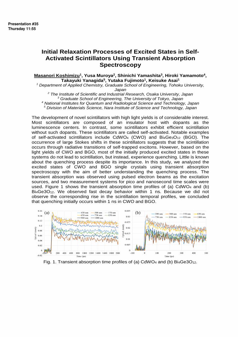
Initial Relaxation Processes of Excited States in Self-Activated Scintillators Using Transient Absorption
Spectroscopy
Masanori Koshimizu1, Yusa Muroya2, Shinichi Yamashita3, Hiroki Yamamoto4, Takayuki Yanagida5, Yutaka Fujimoto1, Keisuke Asai1
1 Department of Applied Chemistry, Graduate School of Engineering, Tohoku University, Japan
2 The Institute of Scientific and Industrial Research, Osaka University, Japan 3 Graduate School of Engineering, The University of Tokyo, Japan
4 National Institutes for Quantum and Radiological Science and Technology, Japan 5 Division of Materials Science, Nara Institute of Science and Technology, Japan
The development of novel scintillators with high light yields is of considerable interest. Most scintillators are composed of an insulator host with dopants as the luminescence centers. In contrast, some scintillators exhibit efficient scintillation without such dopants. These scintillators are called self-activated. Notable examples of self-activated scintillators include CdWO4 (CWO) and Bi4Ge3O12 (BGO). The occurrence of large Stokes shifts in these scintillators suggests that the scintillation occurs through radiative transitions of self-trapped excitons. However, based on the light yields of CWO and BGO, most of the initially produced excited states in these systems do not lead to scintillation, but instead, experience quenching. Little is known about the quenching process despite its importance. In this study, we analyzed the excited states of CWO and BGO single crystals using transient absorption spectroscopy with the aim of better understanding the quenching process. The transient absorption was observed using pulsed electron beams as the excitation sources, and two measurement systems for pico and nanosecond time scales were used. Figure 1 shows the transient absorption time profiles of (a) CdWO4 and (b) Bi4Ge3O12. We observed fast decay behavior within 1 ns. Because we did not observe the corresponding rise in the scintillation temporal profiles, we concluded that quenching initially occurs within 1 ns in CWO and BGO.
Fig. 1. Transient absorption time profiles of (a) CdWO4 and (b) Bi4Ge3O12.
Presentation #35Thursday 11:55

Optical and scintillation properties of Ce-doped SrY2O4 single crystals synthesized by the floating zone method
Hiroyuki Fukushima, Daisuke Nakauchi, Noriaki Kawaguchi, Takayuki Yanagida
Nara Institute of Science and Technology, 8916-5 Ikoma, Nara 630-0192, Japan
Scintillation light is observed by interactions between materials and ionizing radiation such as gamma- or X-rays. The scintillation process is more complex than that of the photoluminescence (PL). After the absorption of ionizing radiations by a material, primary electrons are excited by ionizing radiation, then, numerous secondary electrons are generated by coulomb scattering of primary electrons with host lattice. Finally, secondary electrons are transferred to the emission center, and scintillation light is emitted. In recent, oxide materials have been developed as scintillators owing to their high stability and density [1]. The detection efficiency for high energy photons such as gamma- or X-rays depends on the effective atomic number and density of materials. SrY2O4 has relatively high density (5.28 g/cm3), however, its melting temperature is quite high (2,215 °C) [2]. Thus a report of SrY2O4 single crystal is limited. In this study, the undoped and Ce-doped SrY2O4 single crystals were synthesized using a floating zone furnace and evaluated in PL and scintillation properties. Figure 1 shows the PL contour map of the 1.0 % Ce-doped SrY2O4 single crystal. The broad emission appeared from 430 to 600 nm under the excitation wavelength at around 400 nm. Figure 2 shows the PL decay curve of the 1.0 % Ce-doped SrY2O4 single crystal. The decay curve of the 1.0 % Ce-doped sample was approximated by a sum of two exponential decay components. The primary decay component is an instrumental response, and the value of secondary decay component is about 33 ns which is typical for the 5d-4f transitions of Ce3+. In the conference, further results including scintillation properties will be presented and discussed.
Figure 1. PL excitation (Ex.) / emission (Em.) contour map of 1.0 % Ce-doped SrY2O4 single crystal.
Figure 2. PL decay curve of 1.0 % Ce-doped SrY2O4 single crystal.
References: [1] T. Yanagida, Proc. Japan Acad. Ser. B. 94 (2018) 75–97. [2] J. Philippen et al., J. Cryst. Growth. 459 (2017) 17–22.
Presentation #36Thursday 12:15

Coherent Electron and Nuclear Spin Dynamics of Rare Earth Ions at Sub-KelvinTemperatures
Zong-Quan Zhou
CAS Key Laboratory of Quantum Information, University of Science and Technology of China, CAS, Hefei, 230026, China
Rare earth (RE) ions doped in solids are the state-of-art materials for the realization of optical quantum memories, which are the fundamental building blocks in the large-scale quantum networks. RE ions can be divided into Kramers ions and non-Kramers ions, while the former class has a nonzero electron spin. The Kramers ions based quantum memory has the advantage of supporting wideband quantum storage (100GHz) while the non-Kramers ions can only provide a memory bandwidth of tens MHz. Other advantages of Kramers ions include serving as a microwave memory for a quantum computer and an optical-microwave quantum transducer.
However, compared to the non-Kramers ions, the unquenched electron spins of the Kramers ions experience much larger spin-lattice relaxation (SLR) and spin-spin dipolar interaction. These directly result into low storage efficiency and a short storage time for the Kramers ions based material, thus severely limit its application in quantum information processing. To solve these problems and to fully release the potential of RE ions for quantum information technologies, here we realize the comprehensive enhancement of the population and coherence lifetime for both the electron and nuclear spins of the Kramers ions by constructing a unique instruments which enables the manipulation of solid spins at a temperature of 100 mK.
Presentation #37Thursday 14:05

Quantum information processing using frozen core 𝐄𝐮𝟑$spins in 𝐄𝐮𝟑$𝐘𝟐𝐒𝐢𝐎𝟓
M. Zhong2, RoseL. Ahlefeldt=, and MatthewJ. Sellars=
1. Shenzhen Institute for Quantum Science and Engineering, and Department of Physics, Southern University of Science and Technology, Shenzhen 518055, China.
2. Centre for Quantum Computation and Communication Technology,Research School of Physics and Engineering, The Australian National University, Canberra 0200, Australia
There is growing interest to use nuclear spin states of rare earth ions doped crystals to store and manipulate quantum information due to the potential of optically manipulating these spin states [1][2]. EuE$Y=SiOI is a good candidate for such applications due to its good coherence properties for both hyperfine and optical transitions of the Eu3+ ions [3][4].
In previous work, when we demonstrated a hyperfine coherence time of six hours in this system, we also determined that the interactions between the EuE$ and the nearby host YE$ spins are the remaining dominant decoherence source [4].
Further extension of the coherence time is beneficial for quantum memory applications. Here we present techniques to study the detailed dynamic interactions between the EuE$ ions and YE$ spins in EuE$Y=SiOI with the aim of further extending the hyperfine coherence times of the EuE$ ions through higher precision control over these interactions. Further, we propose a method to initialize the YE$spin states, enabling the YE$spins to be used as a quantum resource for quantum information applications
Figure 1 The measured spin-transition spectrum of Y3+ spins near the Eu3+ ion in a strong magnetic field. The detuning of the Y3+ spin frequencies is produced by the induced magnetic moment on the Eu3+ by the magnetic field. Here p1 - p5 represent different Y3+ sites given by the separation from the Eu3+ ion. References (1) E. Fraval et al., Phys. Rev. Lett. 92 (2005). (2) J. J. Longdell et al., Phys. Rev. B. 95 (2005). (3) F. Konz et al., Phys. Rev. B 68 (2003). (4) M. Zhong et al., Nature 517 (2015).
Y frequency (MHz)
Eu ec
ho am
plitu
de
2.7 2.701 2.702 2.703 2.704 2.705 2.706 2.707 2.708 2.7090
0.1
0.2
0.3
0.4
0.5
0.6
0.7
0.8
0.9
1
p1
p2
p3
p4
p5
Presentation #38Thursday 14:35

Microwave to optical photon conversion via fully concentrated rare-earth ion crystals
Jonathan R. Everts1, Matthew C. Berrington2, Gavin G.G King1, Rose L. Ahlefeldt2, Jevon J. Longdell1
1The Dodd-Walls Centre for Photonic and Quantum Technologies, Department of Physics, University of Otago, New Zealand
2Centre for Quantum Computation and Communication Technology, Laser Physics Centre, The Australian National University, Canberra, Australian Capital Territory 0200, Australia
Most investigations of rare earth ions in solids for quantum information have used rare earth ion doped crystals [1]. Here we analyse the conversion of quantum information from microwave photons to optical frequencies using crystals where the rare earth ions, rather than being dopants, are part of the host crystal. The potential of large ion densities and small linewidths makes such systems very attractive in this application. We show that, as well as high efficiency, large bandwidth conversion is possible. In fact, the collective coupling between the rare earth ions and the optical and microwave cavities is large enough that the limitation on the bandwidth of the devices will instead be the spacing between magnon mode modes in the crystal.
FIG1: The device used to convert microwave photons into optical photons. A rare-earth crystal (doped rare earth crystal for previous device [1], fully concentrated for proposed device) is placed within a microwave resonator and an optical cavity. A static magnetic field is then applied in the �� direction. This controls the frequency of the spin resonance (magnon resonance) Magnon modes are theoretically well studied in materials with isotropic g-tensors, and experimentally well studied in YIG, however little research has been carried out in rare-earth crystals. It is therefore important to obtain a better understanding of the magnon modes within the rare-earth crystals that are suitable for our conversion device. We present preliminary results for investigations carried out in the crystals, Gadolinium Vanadate GdVO4 and Dysprosium Phosphate DyPO4. References: [1] Lewis A. Williamson, Yu-Hui Chen, and Jevon J. Longdell, “Magneto-Optic Modulator with Unit Quantum Efficiency,” Phys. Rev. Lett. 113, 203601 (2014).
Presentation #39Thursday 14:55

Studying correlated errors in a rare-earth quantum computing system
Rose Ahlefeldt
Research School of Physics and Engineering, The Australian National University, Canberra ACT, Australia
The ability to correct errors is a fundamental requirement of any quantum computing system. Typically, error correction protocols work by encoding the information (the logical qubit) across multiple physical qubits, using a set of measurements to identify if any of those qubits have experienced an error, and correcting the error if necessary. Many error correction protocols have been proposed to deal with a range of errors in a range of different qubit systems. It has, however, been difficult to test these protocols on real errors, because systems with enough physical qubits are rare. A particular concern is that most error correction protocols assume uncorrelated errors [1], whereas most physical qubit implementations experience correlated errors [2]. Thus, it is important to test error correction protocols in real systems with real, possibly correlated, errors. I will describe a solid state implementation of quantum computing that could serve as a flexible test-bed for quantum error correction protocols. The physical system is a rare earth crystal stoichiometric in one rare earth, and lightly doped with another. Due to the crystal distortion caused by the dopant, the host ions near the dopant are shifted in optical frequency, creating satellite lines in the spectrum. Each optically resolved satellite line is a potential ensemble qubit. This system is an interesting error correction test-bed because creating small qubit systems, O(10) qubits, will be straightforward. Since the qubits are physically very close, O(10 Å), correlations are expected. I will talk about the sources of error in this system, and discuss how correlations can be characterized. I’ll first discuss static correlations in optical frequency, and then describe measuring correlations in errors on different qubits. References: [1] J. Preskill, Quantum Inf. Comput. 13, (2013) 0181. [2] C. L. Edmunds, C. Hempel, R. Harris, H. Ball, V. Frey, T. M. Stace, and M. J. Biercuk, (2017) arXiv:1712.04954 [quant-ph]
Presentation #40Thursday 15:40

Probing Strong Coupling Between Ions and a MicrowaveCavity with Raman Heterodyne
Gavin King1,Peter Barnett1, John Bartholomew2, Andrei Faraon2, and Jevon Longdell11The Dodd-Walls Centre for Photonic and Quantum Technologies,
Department of Physics, University of Otago, New Zealand.2The California Institute of Technology, Pasadena, California, U.S.A.
Superconducting qubits have transitions in the microwave frequency regime but mi-crowave frequency photons get lost in thermal noise at room temperature. Convertingmicrowave photons into optical photons removes the sensitivity to thermal noise, andallows the transfer of quantum states between distant superconducting qubit basedquantum computers[1, 2, 3]. One method to coherently convert the microwave pho-tons to optical photons is to use a three-level system in a sum-frequency-generationarrangement[4], as schematically shown in the figure.
The rare earth ions are well suited to this method, having narrow optical transi-tions and readily accessible microwave transitions through Zeeman splitting. Erbium inparticular is ideal for this, with optical transitions in the lowest-loss window of silica fi-bre. At 50 ppm in yttrium orthosilicate (YSO) conversion efficiencies of 10−5 have beenseen at 4 K[5], limited by thermal population of the uppe microwave state, parasiticreabsorption from isotopic impurities, and weak coupling between the resonator andions
Recently we measured an isotopically pure 170Er3+ in YSO at around 100 mK. Wesaw strong coupling between the ions and the cavity, conversion using Zeeman split-ting in the excited state, which neatly avoids the problem of thermal population of themicrowave transition, and have measured the spin-lattice relaxation time of the ions.
Figure 1: Using the three-level system in the left panel, we can see upconverted photons usingRaman heterodyne detection (centre) in the excited state of 170Er3+. Inverting the microwaveand optical fields, to look at the ground state, we see strong coupling in the Raman Heterodynesignal (right figure).
References
[1] Siverns JD, Li X, Quraishi Q. Appl Opt. 56(3) (2017) p.B222[2] Rtz H, Luo K-H, Suche H, Silberhorn C. Appl Phys B. 122(1) (2016) p.13.[3] Rtz H, Luo K-H, Suche H, Silberhorn C. Phys Rev Applied. 23(2) (2017) p.024021.[4] Williamson et al., Phys. Rev. Lett. 113, (2014) p.203601.[5] Fernandez-Gonzalvo et al., arXiv:1712.07735v1 (2015)
Presentation #41Thursday 16:10

Optical Coherence Time Control by Large Scale Optical Spin Polarization in 171Yb:Y2SiO5
Sacha Welinski,1 Alexey Tiranov,2 Alban Ferrier,1,3 Mikael Afzelius,2 and
Philippe Goldner1 1Université PSL, Chimie ParisTech, CNRS, Institut de Recherche de Chimie Paris,
75005 Paris, France 2Groupe de Physique Appliquée, Université de Genève, CH-1211 Genève, Switzerland
3Sorbonne Université, 75005 Paris, France
Paramagnetic rare earth ions doped into high quality crystals are promising
systems for broadband optical and microwave quantum memories [1], as well as microwave to optical quantum transducers [2], and could therefore provide new functionalities to e.g. superconducting qubits, one of the leading candidates for quantum computers. Electron spins are however highly sensitive to magnetic noise, which results in short coherence times for optical and spin transitions, unless strong magnetic fields are applied in order to reduce magnetic noise. Alternatively, it is also possible to use ZEFOZ, Zero First Order Zeeman, transitions which have only a quadratic dependence on magnetic field [3]. Surprisingly such transitions occur at zero magnetic field in 171Yb:Y2SiO5 (YSO), which has both ½ electron and nuclear spins [4,5]. As a result, optical and spin T2 up to 180 µs and 1.5 ms have been measured in the absence of magnetic field, properties so far limited to non-Kramers ions like Pr3+. In the latter, spin transitions are in the range of 10s of MHz, whereas splittings of several GHz are found in 171Yb:YSO, allowing e.g. interfacing with microwave photons.
Here, we report on an unusual effect we recently observed: when a narrow linewidth laser (» 1 MHz) is used to burn a spectral hole in 171Yb3+:YSO, the whole inhomogeneously broadened absorption (» 500 MHz) is actually modified on a time scale of a few 10s of seconds. This means that the whole spin population in the spatial volume addressed by the laser can be manipulated, and we were able to obtain strong polarization in one or two spin levels out of four. This is attributed to a combination of strong spin diffusion and slow spin-lattice relaxation, which in turn is favoured at zero magnetic field. Moreover, optical photon echo experiments performed under various spin polarization degrees show optical coherence lifetimes that can be increased from 300 µs (no polarization) up to 800 µs, but also decreased to 120 µs. We expect this ability to dynamically polarize spins on a large scale to enable designing new schemes for obtaining long spin and optical coherence lifetimes in rare earth doped crystals, with applications to a range of optical quantum technologies. References: [1] P. Goldner, A. Ferrier, and O. Guillot-Noël, in Handbook on the Physics and Chemistry of Rare Earths, (Elsevier, 2015), Vol. 46, pp. 1–78. [2] L. A. Williamson et al., Phys Rev. Lett. 113, 203601 (2014). [3] E. Fraval, M. J. Sellars, and J. J. Longdell, Phys. Rev. Lett. 92, 077601 (2004). [4] A. Ortu et al., Nat. Mater. 17, 671–675 (2018). [5] A. Tiranov et al., Phys. Rev. B 98, 195110 (2018).
Presentation #42Thursday 16:30

Hexagonal Boron Nitride Nanophotonics
Sejeong Kim, Johannes E. Fröch, Milos Toth and Igor Aharonovich University of Technology Sydney (UTS) NSW, Australia
Abstract Hexagonal boron nitride (hBN) has emerged as a promising platform, following reports of hyperbolic phonon-polaritons and optically stable, ultra-bright quantum emitters. In this report, we fabricate hBN into high-quality photonic devices such as photonic crystals and microrings to increase light-matter interaction. This is the first direct fabrication of van der Waals crystals into photonic devices with nano-sized features. This can open up promising avenues in many applications in nanophotonics including quantum photonics and optomechanics. Introduction Single photon emitters (SPEs) are key resources for many quantum technologies including quantum computation and quantum communications. To date, the most investigated solid state SPE systems are epitaxial quantum dots that operate primarily at cryogenic temperatures, and colour centres in solids. Despite years of research, the existing systems remain inadequate for practical applications, and the search is on for high-performance quantum emitters. In 2015, the SPE platform expanded to 2D materials. In 2016, hexagonal boron nitride (hBN) emerged as a compelling 2D host of SPEs [1].
SPEs in hBN are promising because they are bright, with more than a million counts per second at room temperature, optically stable at ambient conditions, fully polarized and with a narrow zero photon line (ZPL). Furthermore, hBN is a wide bandgap material, which guarantees optical transparency in the visible and infrared spectral regions. These factors make this material an outstanding candidate for quantum nanophotonics with diverse promising applications.
Here, we demonstrate the first photonic cavities that are entirely consisted of hBN, which is van der Waals crystal with stacked 2D atomic layers [2]. High-Q photonic cavities made of 2D materials enable increased in light-matter interaction which is promising for many nanophotonics applications including quantum photonics, optomechanics [3] and nonlinear optics. References: [1] Igor Aharonovich, Milos Toth, “Quantum emitters in two dimensions”, Science 358, 6360, 2017. [2] Sejeong Kim, Johannes E. Fröch, et. al., “Photonic crystal cavities from hexagonal boron nitride”, Nature communications 9:2623, 2018. [3] Prasoon K Shandilya, Johannes E Fröch, et. al., “Hexagonal boron nitride cavity optomechanics”, arXiv:1809.04023.
Presentation #43Friday 09:00

A Quantum Memory at 1550 nm, in Erbium.
James S. Stuart, Morgan P. Hedges, Matthew J. Sellars Laser Physics Centre,
Research School of Physics and Engineering, ANU.
A quantum memory at the telecommunications wavelength, 1550 nm, could enable global-scale quantum repeater networks. Erbium ions in solid state hosts are a good candidate system for this memory because Er: Y2SiO5 has shown coherence times of over 1 s at the 1538 nm transition [1], allowing for long storage times in a quantum memory. The next step in developing a working quantum repeater is the demonstration of an efficient quantum memory using erbium. I will present an implementation of a quantum memory in Er: Y2SiO5 using the Atomic Frequency Comb (AFC) protocol. The hyperfine transitions of Er: Y2SiO5 can be resolved by applying a large 7 T magnetic field and cooling below 2 K. We use this resolved structure to create the AFC: through optical spin pumping the ensemble is prepared into one hyperfine level, |+7/2>. This greatly increases the optical depth of the crystal (70 dB/cm) and clears out a large spectral area so a spectral comb can be burnt back with little background absorption. The comb is created through hole burning small sub-ensembles down to the |-7/2> hyperfine level, giving very sharp and optically deep spectral features. Initial classical measurements have shown storage times over 1 µs and efficiencies around 10%. Both of these results are an order of magnitude improvement over previous demonstrations in erbium [2, 3]. I will also discuss a recent quantum storage demonstration, the first of its kind in erbium. Finally, I have shown that degradation of spin pumping can occur when optically pumping ensembles with large bandwidths. I will prove that resonant phonons are created, causing spin flips in nearby erbium ions, which greatly increasing hyperfine relaxation. Although this is not a current limitation of the memory, it could affect future demonstrations involving memories with large bandwidths. References: [1] Miloš Rančić, Morgan P. Hedges, Rose L. Ahlefeldt & Matthew J. Sellars, Nature Physics 14 (2018) 50-54. [2] Erhan Saglamyurek, Jeongwan Jin, VarunB.Verma, Matthew D. Shaw, Francesco Marsili, Sae Woo Nam, Daniel Oblak & Wolfgang Tittel Nature Photonics 9 (2015) 83-87. [3] Björn Lauritzen, Jiří Minář, Hugues de Riedmatten, Mikael Afzelius & Nicolas Gisin, Physical Review A 83 (2011) 012318.
Presentation #44Friday 09:40

Ultra-Thin Eu doped Y2O3 Films with optimized Optical Properties for Quantum Technologies
A. Ferrier1,2 A. Tallaire1 M. Scarafagio1,2, N. Harrada1, , P.D. Serrano1,P. Goldner1 1 Institut de Recherche de Chimie Paris (IRCP) PSL Research University, Chimie ParisTech,
CNRS 75005 Paris, France ([email protected]) 2 Sorbonne Universités, UPMC Université Paris 06, 75005 Paris, France
Rare earth (RE) oxides represent a technologically useful class of materials that can address a variety of applications such as photonics, protective coatings, laser media, catalysts, scintillators and phosphors. They have also been highly studied for microelectronics in which high permittivity dielectric films could advantageously replace SiO2 as gate insulators. More recently, RE doped oxide crystals have attracted an increasing attention for quantum technologies [1]. Although promising quantum properties have been demonstrated for Eu doped Y2O3 nanoparticles with spin coherence lifetimes of several ms at 5 K [2], challenges still arise in this form due to the decoherence induced by surface and defects in the material. Alternatively, Y2O3 thin films have emerged as a potential platform for quantum technologies.
In this presentation, we will discuss the optimal conditions to prepare high-crystalline quality Y2O3 thin films by Atomic Layer Deposition (ALD) and Chemical Vapour Deposition (CVD). The choice of growth conditions, the nature of substrate and post treatment will be particularly addressed. The structural and optical properties are thoroughly studied by X-ray diffraction, scanning electronic microscopy, cathodoluminescence, micro-fluorescence and time resolved photoluminescence.
Furthermore, we use high-resolution optical linewidth measurements (i.e Inhomogeneous linewidth) as a powerful tool for material characterization. On the optimized films grown by both ALD and CVD we observe narrow inhomogeneous linewidth of the 7F0 ↔ 5D0 of Eu. These measurements confirm that deposition or post-annealing at high temperature are key parameters in order to obtain high crystalline quality. This study paves the way to the fabrication of ultra-thin RE oxide layers that offer improved optical properties for quantum technologies. References: [1] Goldner, P.; Ferrier A.; Guillot-Noël, O. Handbook on the Physics and Chemistry of Rare Earths 2015, 46, 1-78. [2] Serrano, D.; Karlsson, J.; Fossati, A.; Ferrier, A.; Goldner, P. All-optical control of long-lived nuclear spins in rare-earth doped nanoparticles Nature Communications 2018, 9, 2127. [3] M. Scarafagio, A. Tallaire, K.J. Tielrooij, A. Grishin, M.H. Chavane, F.H. L. Koppens, M. Cassir, P. Goldner and A. Ferrier, Atomic Layer Deposition of nanoscale Eu and Er doped Y2O3 films with optimized optical properties,submitted J. Phys. Chem C 2019.
Presentation #45Friday 10:00

Optical refrigeration and saturation effects in oxide crystals
A.J. Salkeld1*, L. Cheng1, L.B. Andre1, and S.C. Rand1,2 1EECS Department, University of Michigan, Ann Arbor, MI 48109-2099, USA
2Department of Physics, University of Michigan, Ann Arbor, MI 48109, USA * Corresponding author - [email protected]
To date, anti-Stokes fluorescent cooling has been the most successful method in cooling rare-earth-doped crystals, utilizing electric-dipole-forbidden f-f transitions of the dopant. The record minimum temperature of 91 K was achieved in Yb3+:YLF in vacuum [1], and a maximum cooling by 8.8 K was achieved in Yb3+:YAG in air [2]. The chief limitation to ASF cooling is the presence of background impurities which contribute to parasitic absorption and heating, but might be saturable. Here we extend the observation of laser cooling to a new host (Yb:KYW), benchmark its performance against record-breaking results in Yb:YAG, and predict improved cooling performance when saturation takes place.
Our experimental results include open lab environment cooling of Yb:YAG, and Yb:KYW for a variety of dopant concentrations (see Figure 1). Record cooling is observed for these conditions in both materials. We discuss the intensity dependence of impurity absorption and show that saturation of the impurity absorption offers a substantial reduction in the predicted minimum achievable temperature, when impurity absorption is the predominant limiting factor to ASF cooling. We also assess the viability of laser cooling using electric-dipole-allowed transitions of the coolant ion. A candidate material for this study is Ti3+:Al2O3.
(b) Figure 1. Plots of wavelength versus temperature change in K/W and cooling power
for (a) 3% Yb:YAG and (b) 1% Yb:KYW.
References:
[1] S.D. Melgaard, A.R. Albrecht, M.P. Hehlen, and M. Sheik-Bahae. Sci. Rep. 6 (2016), 20380.
[2] E.S. Filho, G. Nemova, S. Loranger, and R. Kashyap. Optics Express 21 (2013), 24711.
Presentation #46Friday 10:20

The Marriage of Perovskite Quantum Dots with Rare-Earth Emitters
Wei Zheng, Zhongliang Gong, Datao Tu, Ping Huang, and Xueyuan Chen*
CAS Key Laboratory of Design and Assembly of Functional Nanostructures, Fujian Institute of Research on the Structure of Matter, Chinese Academy of Sciences, Fuzhou, Fujian 350002, China Perovskite quantum dots (PeQDs) are emerging as a new generation of optoelectronic and photovoltaic materials owing to their outstanding optoelectronic properties such as long carrier diffusion distance, large absorption coefficient, high photoluminescence (PL) quantum yield (QY), narrow emission band, and tunable band gap and PL emission by varying the halide composition.[1,2] The combination of PeQDs with rare-earth (RE) emitters may bring about new optical, electric, thermal, and magnetic properties.[3] In this talk, we shall focus on our recent efforts on the development of PeQDs and RE hybridized materials based on RE-doped PeQDs and PeQDs/RE hetero-structures. Specifically, through sensitization by RE-doped nanoparticles, we have realized full-spectrum photon upconversion tuning in all-inorganic CsPbX3 (X = Cl, Br, and I) PeQDs under low power density excitation, with a color gamut of 140% NTSC, upconversion QYs approaching 0.5%, and significantly lengthened exciton lifetimes from μs to ms scale.[4] We have also developed a novel strategy for fine-tuning the persistent luminescence with a color gamut of 130% NTSC and highly synchronized afterglow decay over 8 h by using CsPbX3 PeQDs as efficient afterglow light conversion materials.[5] These findings provide a general approach for tailoring the upconversion and persistent luminescence by the marriage of PeQDs with RE emitters, thereby opening up new opportunities for PeQDs and RE emitters towards many emerging applications such as solar spectrum conversion, white-emitting persistent light source, complex data storage, and multilevel anti-counterfeiting. Keywords: perovskite quantum dots; rare earths; energy transfer; upconversion luminescence; persistent luminescence References: [1] L. Protesescu, S. Yakunin, M. I. Bodnarchuk, F. Krieg, R. Caputo, C. H. Hendon,
R. X. Yang, A. Walsh, and M.V. Kovalenko*, Nano Lett. 2015, 15, 3692. [2] S. H. Zou, Y. S. Liu*, J. H. Li, C. P. Liu, R. Feng, F. L. Jiang, Y. X. Li, J. Z. Song,
H. B. Zeng*, M. C. Hong*, and X. Y. Chen*, J. Am. Chem. Soc. 2017, 139, 11443. [3] P. Huang, W. Zheng*, Z. L. Gong, W. W. You, J. J. Wei, and X. Y. Chen*, Mater.
Today Nano 2019, 5, 100031. [4] W. Zheng, P. Huang, Z. L. Gong, D. T. Tu, J. Xu, Q. L. Zou, R. F. Li, W. W. You, J.
C. G. Bünzli, and X. Y. Chen*, Nat. Commun. 2018, 9, 3462. [5] Z. L. Gong, W. Zheng*, Y. Gao, P. Huang, D. T. Tu, R. F. Li, J. J. Wei, W. Zhang,
Y. Q. Zhang, and X. Y. Chen*, Angew. Chem. Int. Ed. 2019, DOI: 10.1002/anie.201901045.
Presentation #47Friday 11:10

3P0
1D2 non-radiative relaxation control via IVCT state in
Pr3+
-doped Na2Ln2Ti3O10 (Ln=La, Gd) micro-crystals with triple-layered perovskite structure
Yongjie Wanga, Qi Pengb, Hongbin Liangb, Mikhail G. Brikc,d,e, Andrzej
Suchockia,f
a Institute of Physics, Polish Academy of Sciences, Al. Lotnikow 32/46, 02-668, Warsaw,
Poland bMOE Key Laboratory of Bioinorganic and Synthetic Chemistry, KLGHEI of Environment and
Energy Chemistry, School of Chemistry, Sun Yat-sen University,Guangzhou 510275, China c College of Sciences, Chongqing University of Posts and Telecommunications,
Chongqing 400065, People’s Republic of China d Institute of Physics, University of Tartu, W. Ostwald Str. 1, Tartu 50411, Estonia
e Institute of Physics, Jan Długosz University, Armii Krajowej 13/15, PL-42200
Częstochowa, Poland fInstitute of Physics, Kazimierz Wielki University, Weyssenhoffa 11, 85-072, Bydgoszcz,
Poland
A comparative study on luminescence properties and their nature in relation to
different substitutions at the rare-earth site of Pr3+-doped layered perovskite
Na2Ln2Ti3O10 (Ln=La, Gd) micro-crystals has been reported. At room temperature,
the sample for Ln=La exhibits intense greenish-blue (3P0 3H4) and red (1D2 3H4)
emissions under bandgap excitation, whereas for Ln=Gd the red emission can be
only observed and all 3P0-related emissions were completely quenched. It turns out
that 3P0 1D2 nonradiative relaxation in Na2Ln2Ti3O10 (Ln=La, Gd) critically depends
on the energy location of Pr3+-Ti4+ intervalence charge transfer state (IVCT) and thus
on the location of Pr3+ ground state 3H4 with respect to the top of the valence band.
Temperature-dependent photoluminescence spectra in the 4.5-300 K range reveals a
significant increase of Pr3+ luminescence, which is ascribed to an efficient thermally-
activated energy transfer process from host to Pr3+ ions. Lower energy of self-trapped
exciton state relative to the bottom of conduction band for Ln=Gd is responsible for
thermoluminescence observed at higher temperatures than that for Ln=La sample. In
the case of Ruddlesden-Popper type layered perovskite[1] oxide compounds our
results show a possibility of controlling the 3P0 1D2 nonradiative relaxation of Pr3+
ions and the energy distance of the relatively shallow traps in relation to the bottom of
conduction band, giving a way for specific band-gap engineering in these materials.
References:
[1] Gopalakrishnan, J.; Sivakumar, T.; Ramesha, K.; Thangadurai, V.; Subbanna, G.
N. J. Am. Chem. Soc. 2000, 122, 6237-6241.
Presentation #48Friday 11:40

The Ytterbium Ion Site Distribution in CaF2:Yb3+ Nanoparticles
Sangeetha Balabhadra, Michael F. Reid, Vladimir Golovko, Jon-Paul R. Wells
Dodd-Walls Centre for Photonic and Quantum Technologies, New Zealand.
School of Physical and Chemical Sciences, University of Canterbury, PB 4800, Christchurch 8140, New Zealand
We report on the identification of three distinct single ion sites (having Oh, C4v(F-), C3v(F-) symmetries respectively) as well as preferentially formed clusters centres [1] in CaF2 nanoparticles doped with trivalent ytterbium. The CaF2:Yb3+ nanoparticles were synthesized via a facile hydrothermal method using citrate ions as capping agents. The structural and morphological analyses of as-synthesized nanoparticles were studied using X-ray and microscopy techniques. The effect of Yb3+ molar concentration and nanoparticle sizes on absorption measurements were investigated using a high resolution FTIR spectrometer. Interestingly, increasing the concentration and in particular the size of the particles leads to an increase in the distribution of centres; with smaller particles dominated almost exclusively by cubic centres. Further, we demonstrate selective fluorescence monitoring of preferential clusters to tune high output brightness and output wavelength emission of the nanoparticles, which can be used for applications in optical imaging. [1] V. Petit, P. Camy, J. L. Doualan, X. Portier, R. Moncorgé, Physical Review B - Condensed Matter and Materials Physics 78 (2008) 085131.
Presentation #49Friday 12:00

Composition-Graded Cesium Lead Halide Perovskite Nanowires with Tunable Dual-Color Lasing Performance
Ling Huang, Ling-Dong Sun*, Chun-Hua Yan*
Beijing National Laboratory for Molecular Sciences, State Key Laboratory of Rare Earth Materials Chemistry and Applications, College of Chemistry and Molecular Engineering,
Peking University, Beijing 100871, China, E-mail: [email protected], [email protected]
Cesium lead halide (CsPbX3) perovskite has emerged as a promising low
threshold multicolor laser material; however, realizing wavelength-tunable lasing
output from a single CsPbX3 nanostructure is still constrained by integrating different
composition. For the further development of more than one color lasing from a single
halide perovskite nanostructure, it is significant to construct heterogeneous or
composition-graded perovskite nanostructures. In this work, the direct synthesis of composition-graded CsPbBrxI3−x nanowires
(NWs) is reported through vapor-phase epitaxial growth on mica. The graded
composition along the NW, with an increased Br/I from the center to the ends,
originates from desynchronized deposition of cesium lead halides and controlled
anion-exchange reaction. The graded composition results in varied bandgaps along
the NW, which induce a blue-shifted emission from the center to the ends. As an
efficient gain media, the nanowire exerts position-dependent lasing performance, with
a different color at the ends and center respectively above the threshold. Meanwhile,
dual-color lasing with a wavelength separation of 35 nm is activated simultaneously at
a site with an intermediate composition. This position-dependent dual-color lasing from
a single nanowire makes these metal halide perovskites promising for applications in
nanoscale optical devices.
Figure 1. Confocal laser scanning microscope image of composition-graded CsPbBrxI3−x nanowires (left) and schematic diagram of dual-color lasing (right).
References [1]. L. Huang, Q. Gao, L.-D. Sun, H. Dong, S. Shi, T. Cai, Q. Liao, C.-H. Yan, Adv. Mater. 2018, 1800596.
Presentation #50Friday 12:20

Dark-Bright Exciton Dynamics in Perovskite Nanocrystals
Kunyuan Xu, Jara Vliem and Andries Meijerink CMI, Debye Institute for Nanomaterials Science, Utrecht University, The Netherlands
Lead halide perovskites (LHPs) form an emerging class of materials with promising opto-electronic properties for application in e.g. solar cells, lighting and displays.1 The optical properties for nanocrystalline LHPs are similar to those of chalcogenide semiconductor quantum dots (QDs) and yet markedly different in certain aspects. Last year an unexpected new difference was reported: inversion in the order of the two lowest exciton states by the Rashba effect.2 The lowest excited state in LHP nanocrystals (NCs) was reported not to be the dark exciton state (as in all chalcogenide QDs) but the bright exciton state (see Figure 1).
Here we report temperature dependent exciton lifetimes for CsPbCl3 NCs doped with 0 to 41% Mn2+.3 The exciton emission lifetime increases upon cooling from 300 to 75 K. Upon further cooling a strong and fast sub-ns decay component develops. However, the decay is strongly bi-exponential and a weak slow decay component is observed as well with a ~40-50 ns lifetime below 20 K. These observations are similar to those previously for CsPbX3 NCs in high magnetic fields. The slow component has a much stronger relative intensity in Mn-doped NCs compared to undoped CsPbCl3 NCs. Based on our observations we propose an alternative explanation for the fast sub-ns exciton decay time in CsPbX3 NCs.3 Slow bright–dark state relaxation at cryogenic temperatures gives rise to almost exclusively bright state emission. Incorporation of Mn2+ or high magnetic fields enhances the bright-dark state relaxation and allow for the observation of the long-lived dark state emission at cryogenic temperatures.
Figure 1 – (left) Schematic illustration of inverted bright-dark state splitting in perovskite NCs as proposed in Ref. [2] (figure by dr. F. Rabouw). (right) Alternative model explaining the observation of sub-ns bright state emission by slow bright-dark state relaxation at 4 K. [3] References: [1] Akkerman, Q.A., Rainò, G., Kovalenko, M. and Manna, L., Nat. Mater. 17, 394-405 (2018). [2] Becker, M.A. et al., Nature 189, 189-194 (2018). [3] Xu, K., Vliem, J and Meijerink A., J. Phys. Chem. C 1, 979-984 (2019).
Presentation #51Friday 12:40

SPECTROSCOPY AND CRYSTAL FIELD CALCULATIONS OF NEODYMIUM-DOPED YTTRIUM ORTHOSILICATE
Y. Alizadeh1, J. P. R. Wells1, M. F. Reid1, J. J. Longdell2 and P. P. Goldner3
1The Dodd-Walls Centre for Photonic and Quantum Technologies, Department of Physics and Astronomy, University of Canterbury, PB4800, Christchurch 8140 New Zealand
2The Dodd-Walls Centre for Photonic and Quantum Technologies, Department of Physics, University of Otago, Dunedin, New Zealand
3PSL Research University, Chimie ParisTech-CNRS, Institut de Recherche de Chimie Paris, 75005 Paris, France
Rare-earth-ion doped crystals have been considered as potential candidates for implementing quantum storage in recent years [1], [2], [3], [4].Studies on these materials with the purpose of quantum information processing have been done due to the fact that the hyperfine transitions of these ions show long coherence time or phase memory [2], [5]. In trivalent rare-earth ions the 4f electrons, responsible for optical transitions are shielded by the 5s, 5p electrons which results in a weak ion-lattice coupling and therefore sharp transition lines. The electronic states and nuclear spin couple through hyperfine interaction and can be manipulated using optical and radio frequencies, respectively, while the energy levels are modified by magnetic fields. Coherence time of over six hours for Eu3+ ions in YSO has been achieved through the ZEFOZ (ZEro First-Order Zeeman) method [2]. In these investigations a spin Hamiltonian approach is used to model the hyperfine and magnetic interactions for a particular electronic energy level which is required to locate ZEFOZ points. A process of complex experiments and difficult fittings is needed for each electronic level of every rare-earth ion in the series. With a parametrized crystal field model and adding the magnetic and hyperfine splitting data we can perform such fits. Since the ionic radius and the crystal-field parameters reduce across the rare-earth series [6], [7], [8], [9], the crystal-field parameters for Nd3+ can be scaled up from the Er3+ parameters. Parameters for Site 1 and Site 2 of Er3+:Y2 SiO5 are given by Horvath [1]. The free-ion parameters for Nd3+ were taken from [6]. To perform a complete crystal field calculation the absorption lines and Zeeman g values along all three crystallographic axes (D1,D2,b) of the sample for several multiplets should be obtained. These results have been found for many of the Nd3+ multiplets through Zeeman infrared absorption spectroscopy using a Bruker Vertex 80 FTIR spectrometer in a helium-cooled 4 T superconductor magnet. There are two distinct yttrium sites in YSO structure each with C1 local symmetry. The site with the largest absorbance, labelled Type 1 in [10], has been studied by EPR [11, 12]. In Ref. [12] this site was identified as seven-coordinate by pulsed EPR measurements. The ground state g tensors were taken from [11],[12] and [13] obtained by EPR experiments. The obtained absorption lines were properly assigned to each yttrium site using different methods and analyses. The ground state g values calculated from the Zeeman absorption spectroscopy measurements were compared with the principal g values obtained from the EPR measurements [11,12,13] and some of the absorption lines were assigned to yttrium sites. In a separate infrared absorption measurement the temperature dependence of the Nd3+:Y2 SiO5 spectrum from 10 K to room temperature was studied and with the help of appearing thermal lines of Nd3+ more site assignments were made. To find more reliable site assignments, extensive site-selective emission and excitation measurements were also performed which were consistent with previous assignments. The long-term goal of the work is to obtain parameters for the entire rare-earth series and these parameters will be used to model all possible transitions for all rare-earth ions which have the potential to be used for the storage of quantum coherence.
Presentation #52Monday Poster

20th International Conference on Dynamical Processes in Excited States of Solids
Synthesis of Yb3+/Er3+ co-doped Ca9Gd2W4O24 crystals for use
in optical thermometry
(poster)
Gangyi Zhanga, Qinping Qianga and Yuhua Wang a*
a School of Physical Science and Technology, Lanzhou University, Tianshui South
Road No. 222, Lanzhou, Gansu 730000, China
* E-mail: [email protected]
Ca9Gd2W4O24:Yb3+/Er3+ crystals were synthesized by solid state method. The
structure, morphology, size and upconversion luminescence features had been
characterized. These results indicated that Ca9Gd2W4O24 has a tetragonal cell and has
irregular blocky particle shapes. In upconversion, green (2H11/2, 4S3/2→
4I15/2 and red
(4F9/2→4I15/2) emissions had been observed, both of which occurred via a two-photon
populating process. Additionally, characteristics were studied of the green UC
emission including its temperature ranging from 298 K to 503 K and the sensitivity
was 0.0106 K−1 at 473 K. This result indicated that Ca9Gd2W4O24:Yb3+/Er3+ crystals
may have potential application in high environment as safety sign.
Keywords: Ca9Gd2W4O24, upconversion luminescence, sensitivity
Presentation #53Monday Poster

Zeeman-Hyperfine Spectra of Ho3+ in C4v Sites in CaF2
Kieran M. Smith, Jon-Paul R. Wells, Michael F. Reid*
The Dodd-Walls Centre for Photonic and Quantum Technologies, NZ School of Physical and Chemical Sciences, University of Canterbury,
Christchurch, NZ Email: [email protected]
One of the challenges for practical quantum-information applications, such as encryption and computation, is the preservation of coherence. Coherence may be stored by making use of the nuclear spins of rare-earth (lanthanide) ions, which are coupled to the electronic states via the hyperfine interaction. For example, the storage of quantum coherence for over six hours using magnetic-hyperfine levels of Eu3+ ions in YSO has been achieved by using the ZEFOZ (ZEro First-Order Zeeman) approach, where the direction and magnitude of an applied magnetic field is adjusted to yield a radio-frequency transition that has no first-order dependence on magnetic field variations, and is thus insensitive to magnetic field inhomogeneity [1].
Applications development, such as the location of ZEFOZ points, requires accurate modelling of hyperfine and magnetic interactions, which is usually done using a spin Hamiltonian. Spin-Hamiltonian parameters are not transferrable to other electronic states, or to other ions, so each electronic state requires a different spin Hamiltonian. However, it is possible to model electronic and hyperfine energy using a “crystal field” model, such as our previous work on Ho3+ ions in C4v(F-) sites in CaF2 [2], and recent work on Ho3+ ions in LiYF4 [3]. The parameters from the crystal-field model are transferrable (with small scalings) to other rare-earth ions in the same host crystal. The hyperfine structure of Ho3+ is large enough to be measurable without resorting to complex laser spectroscopy, providing a valuable test case for the investigation of magnetic-hyperfine interactions in rare-earth doped crystals.
In this work we present the Zeeman spectra of the hyperfine levels of Ho3+ ions in C4v(F-) sites in CaF2 for fields of up to 4 Tesla. We obtain excellent agreement between experimental measurements and theoretical predictions of both energies and transition intensities, and demonstrate several cases of hyperfine anti-crossings in the spectra. References: [1] M. Zhong, M. P. Hedges, R. L. Ahlefeldt, J. G. Bartholomew, S. E. Beavan, S. M.Wittig, J. J. Longdell, M. J. Sellars. “Optically addressable nuclear spins in a solid with a six-hour coherence time,” Nature 517 (2015) 177. [2] J. P. R. Wells, G. D. Jones, M. F. Reid, M. N. Popova, and E. P. Chukalina, “Hyperfine patterns of infrared absorption lines of Ho3+ C4v centres in CaF2,” Molecular Physics, 102 (2004) 1367. [3] K. N. Boldyrev, M. N. Popova, B. Z. Malkin, N. M. Abishev, “Direct observation of hyperfine level anticrossings in the optical spectra of a 7LiYF4:Ho3+ single crystal,” Phys. Rev. B 99 (2019) 041105.
Presentation #54Monday Poster

Metastable-structure silicate phosphor shell on silica core by rapid thermal quenching
Hyeonwoo Kang1, Taewook Kang2, Gian Antariksa1, Atar Muhamad1, Sunghoon Lee1, Yongseok Jeong1, Jehong Park1, Youngmoon Yu2,
Jongsu Kim1, 2 1Department of Display Science and Engineering, Pukyong National University, Busan
48513, Republic of Korea 2Interdisciplinary Program of LED and Solid State Light Engineering, Pukyong National
University, Busan 48513, Republic of Korea
Here the stable α-phase and metastable β-phase Zn2SiO4:Mn2+ phosphor shell in
silica core were comparatively investigated by controlling critical synthesis parameters such as highest annealing temperature, cooling rate, and chemical composition ratio. Their metastability was examined by demonstrating the phase transition into the stable structure at the slow thermal annealing and quenching process at a high temperature, and as a consequence the change in emission color. The α-phase and the β-phase Zn2SiO4:Mn2+ phosphors showed the green and yellow emission colors, respectively. Furthermore, optical properties in photoluminescence and electroluminescence were compared in terms of temperature and concentration dependence, and voltage and frequency dependence. References: [1] J. Wan, Z. Wang, X. Chen, L. Mu, W. Yu, Y. Qian, Journal of Luminescence 121
(2006) 32–38. [2] M. Mildea, S. Dembski, A. Osvet, M. Batentschuk, A. Winnacker, G. Sextl,
Materials Chemistry and Physics 148 (2014) 1055-1063. [3] S. Lee, B. Jeon, T. Kang, W. Lee, A. M. Malik, S. Park, J. Lim, B. Park, Y. Jeong,
J. kim, Journal of Luminescence 196 (2018) 290–293.
Acknowledgement: This work was supported by the Development of R&D Professionals on LED
Convergence Lighting for Shipbuilding/Marine Plant and Marine Environments (Project No: N0001363) funded by the Ministry of TRADE, INDUSTRY & ENERGY(MOTIE, Korea)
Presentation #55Monday Poster

20th International Conference on Dynamical Processes in Excited States of Solids
Flame retardancy and afterglow properties of a novel
organic-inorganic composite
(Poster)
Zhongying Ma,Haijie Guo, Yuhua Wang
School of Physical Science and Technology, Lanzhou University, Tianshui South Road No. 222,
Lanzhou, Gansu 730000, China
Key Words: Flame retardancy, long afterglow, yellow emitting
Abstract
A novel multifunctional composite with flame retardancy and yellow-emitting
long-lasting properties was developed. From MCC and TGA, it shows that the
composite incorporated with Ca6BaP4O17: 0.02Eu2+
, 0.015 Ho3+
has not only
outstanding flame retardancy but also excellent thermal stability. Besides,its initial
long-lasting phosphorescence (LLP) intensity can reach about 0.13 cd m2 and its LLP
can last more than 47 h above the recognizable intensity level (0.32 mcd m2)The
results indicate that this composite has the potential to become a novel commercial
LLP phosphor used in the field of emergency lighting and display.
Presentation #56Monday Poster

Optical memory based on a laser-written waveguide
Chao Liu, Zong-Quan Zhou*, Chuan-Feng Li†, Guang-Can Guo
CAS Key Laboratory of Quantum Information, University of Science and Technology of China, CAS, Hefei, 230026, China
We fabricate type-II waveguides in 𝐸𝑢3+151 doped 𝑌2𝑆𝑖𝑂5 crystal using femtosecond-laser micromachining. In waveguide regime, the required laser power for driving the optical transition is decreased by approximately two orders, which indicates the tight confinement of light and the highly increased light-ion interaction. 𝑇2 is measured to be 200 us, which confirms that the material’s coherence property is well preserved. Using atomic frequency combs (AFC) scheme, we demonstrate light storage in excited state with 2 us storage time and 10% storage efficiency. On-demand readout and extended storage time are demonstrated by spin-wave storage. References: [1] Corrielli, G., Seri, A., Mazzera, M., Osellame, R., De Riedmatten, H.(2016) Physical Review Applied, 5 (5)
Presentation #57Monday Poster

Modulation of the Morphology and Luminescence of Lanthanide-doped Nanoparticles
Yuejiao Xu, Ling-Dong Sun*, Chun-Hua Yan*
Beijing National Laboratory for Molecular Sciences, State Key Laboratory of Rare Earth Materials Chemistry and Applications, College of Chemistry and Molecular Engineering,
Peking University, Beijing 100871, China. E-mail: [email protected], [email protected]
The upconversion nanoparticles doped with lanthanide ions show great prospects in bioimaging and theranostic, but it’s efficiency should be further improved.[1] Here, we use injection method to get upconversion nanoparticles with specific crystal face. By changing the composition of precursor injected, we get rhombic dodecahedron nanocrystal exposed with {110} crystal plane and octahedron nanocrystal exposed with {111} crystal plane. The nanoparticles with regular morphology are great candidates for self-assembly supercrystals. Furthermore, we design a Au-UCNP composite structure to modulate the luminescent emission of the lanthanide ions. The gold nanoparticles with different morphology show diverse local surface plasmonic resonance properties and are coated or selectively coated with silica. Then the UCNPs are adhered to the Au nanoparticles through electrostatic attraction with the silica. The upconversion emission is enhanced because of the improved localized density of photonic states near the Au nanoparticle. In addition, the polarization state of the upconversion emission from a single hybrid nanocomposite is analysed. This anisotropic Au-upconversion nanoparticle assemblies are particularly suitable for luminescent orientation sensors.
Figure 1. Modulation of the morphology of lanthanide-doped nanoparticles (a) or tailoring the luminescent emission through compositing with Au nanoparticle
References:
[1] Dong, H.; Sun, L. D.; Yan, C. H. Chem. Rev.19 (2015) 10725–10815.
Presentation #58Monday Poster

Scintillation properties of Cs2O-BaO-Al2O3-P2O5 glasses D. Shiratori, N. Kawaguchi, T, Yanagida
Nara Institute of Science and Technology, Nara, Japan
A scintillator promptly converts the absorbed energy of ionizing radiations into a large number of low energy photons. It is widely applied in many scientific and industrial fields. The material form of a practical scintillator is mainly single crystal, and at present, commercial glass scintillator is only 6Li glass for neutron detection [1]. In general, most commercial glasses for optical applications compose of low atomic number elements. However, in order to detect X-rays, compositions based on heavy elements are required since the detection efficiency of the scintillator for high energy photons depends on the effective atomic number and density. In this study, to increase sensitivity for X-ray, we focused on Cs2O-BaO-Al2O3-P2O5 which contains Cs and Ba as heavy elements and evaluated its radiation response characteristics.
Fig.1 exhibits photoluminescence (PL) excitation and emission map and PL decay curve of the Ce-doped glass sample. We observed a broad emission band from 320 to 450 nm. Based on the PL map, PL decay curve of the Ce-doped sample was observed monitoring at 365 nm under 280 nm excitation. Decay time profile was approximated by a single exponential decay function. The decay time resulted 34 ns, and the origin was ascribed to the 5d-4f transition of Ce3+ [1,2].
Fig. 2 shows scintillation spectra of the non- and Ce-doped samples under X-ray irradiation. The Ce-doped glass sample showed an intense emission from 320 to 450 nm under X-ray irradiation. This emission feature is typical for the 5d-4f transitions of Ce3+. On the other hand, the undoped glass sample also shows the emission from 300 to 450 nm.
Fig. 1 PL/excitation map of Ce-doped glass sample. Inset is PL decay time profile of Ce-doped sample. Excitation and observed wavelength are 280 and 365 nm, respectively.
Fig. 2 Scintillation spectra of the non- and Ce-doped glass samples under X-ray irradiation.
References: [1] J. Czirr, et al., Nucl. Instrum. Meth. A, 424 (1999) 15-19 [2] D. Shiratori et al., J. Mater. Sci. Mater. El. 30 (2019) 2464-2469 [3] Y. Isokawa, et al., Opt. mater. 90 (2019) 187-193
300 400 500 600275
300
325
350
375
400
Ex.[
nm]
Em. [nm]
0 100 200 300 400 500
Ce-doped
Ex : 280 nmEm : 365 nm
IRF
Time [ns]
Coun
ts
200 300 400 500 600Wavelength [nm]
Scin
tilla
tion
inte
nsity
[arb
. uni
t]
Ce-doped undoped
Presentation #59Monday Poster

Microwave-Cavity and Optical Whispering Gallery Mode Resonator Design for Rare-Earth Ion Electro-Optic
Conversion
Li Ma1, Gavin King1, Alfredo Rueda2, Madhuri Kumari1, Jonathon Everts1, Harald G. L. Schwefel1 and Jevon J. Longdell1
1 The Dodd-Walls Centre for Photonic and Quantum Technologies, Department of Physics, University of Otago, New Zealand
2 Institute of Science and Technology Austria, am Campus 1, 3600 Klosterneuburg, Austria
Efficient conversion of signals between the microwave and the optical domain is a key feature required in classical and quantum communication networks. Whispering gallery mode (WGM) resonator is a promising approach to manipulate information at several gigahertz and distribute information at hundreds of terahertz. We aim to embed a high-quality rare-earth ion doped WGM optical resonator into a 3D copper microwave cavity to achieve the efficiency frequency conversion between microwave and optical photons. The schematic draw of the 3D microwave cavity design is shown in Fig. 1, and it was designed with the help of numerical simulations using COMSOL Multiphysics [1]. The microwave resonator is a three-dimensional copper cavity enclosing the optical resonator, designed such that the resonantly enhanced microwave field has maximum overlap with the optical WGM. The cavity contains two protruded copper rings facing each other, which clamp the optical resonator when the cavity is closed. This ensures that the microwave field is focused on the rim of the optical resonator where the optical modes are located, maximizing the overlap between the two vastly different frequencies. Inside the cavity, a prism is placed close to the optical resonator to couple the light evanescently into the WGM resonator disk, and a tuning rod is used to perturb the microwave field for fine adjustment of its resonance frequency. The optical resonator is an Er:YSO WGM resonator with a convex-shaped disk that guides light via total internal reflection along its inner surface, interfering with itself after each roundtrip. We designed the microwave cavity to match the optical resonator’s size about radius of 2.4 mm and thickness of 0.2 mm, resulting in a free spectral range of about 11GHz at the used pump laser 1536 nm.
Figure 1: (a) Typical profile of microwave electric field in z-direction plotted in color. (b) The
top and bottom parts of the microwave cavity with the tuning rod.
References: [1] Rueda, Alfredo, et al. Nature 568.7752 (2019): 378.
Presentation #60Monday Poster

Spectroscopy and Synthesis of CaF2:Eu3+/2+
Nanoparticles
Jamin L.B. Martin1,2, Sangeetha Balabhadra1,2, Jon-Paul R.Wells1,2, Michael F. Reid1,2, Wei Zheng3, and Xueyuan Chen3
1The Dodd-Walls Centre for Photonic and Quantum Technologies, NZ2The School of Physical and Chemical Sciences, University of Canterbury,
Christchurch, NZ3Fujian Institute of Research on the Structure of Matter, Chinese Academy of
Sciences, Fuzhou, Fujian 350002, China
Lanthanide-doped nanoparticles have been shown to be viable candidatesfor use in two-photon microscopy, stable inorganic contrast agents[1], pho-todynamic therapy sensitisers, and thermotherapy for cancer treatment[2].Lanthanide ions doped into bulk CaF2 crystals are known to form a varietyof sites, and to form clusters at concentrations as low as 0.01 mol%, becom-ing the dominant centre by 0.1 mol%[3][4]. This clustering gives enhancedenergy transfer, promising significant improvements in applications requiringup-conversion or down-conversion via energy transfer.
Previous work on lanthanide-doped CaF2 nanoparticles has made use oflow-resolution spectroscopy at high temperatures[5], and was therefore un-able to clearly discriminate between the different sites. In this work wepresent high resolution laser spectroscopy of CaF2:Eu3+ nanoparticles atcryogenic temperatures (10 K), including excitation, emission, and lifetimemeasurements. Samples include core-only and core-shell particles with a va-riety of concentrations and preparation techniques.
All sites previously identified in bulk CaF2:Eu3+ crystals were observed,indicating remarkably bulk-like characteristics. Samples prepared by thermaldecomposition showed the narrowest line-width spectra and were dominatedby high symmetry C4v sites, although cluster sites were found to be present.Both hyrothermal and co-precipitation samples were found to have both highsymmetry C4v and Oh sites, with the hydrothermal samples being predomi-nately dominated by cluster sites.
References
[1] M. Straßer, J. Schrauth, et al. Beilstein journal of nanotechnology, 8:1484–1493, 2017.
[2] X. Wu, G. Chen, J. Shen, Z. Li, Y. Zhang, and G. Han. Bioconjugate chemistry,26(2):166–175, 2014.
[3] K.M. Cirillo-Penn and J.C. Wright. Journal of luminescence, 48:505–508, 1991.
[4] J.P.R. Wells, G.D. Jones, and R.J. Reeves. Journal of luminescence, 72:977–979, 1997.
[5] P. Cortelletti, M. Pedroni, F. Boschi, S. Pin, P. Ghigna, P. Canton, F. Vetrone,and A. Speghini. Crystal Growth & Design, 18(2):686–694, 2018.
1
Presentation #61Monday Poster

20th International Conference on Dynamical Processes in Excited States of Solids
Emission tuning studies in BaMgSiO4: RE (RE = Eu2+
, Sr2+
) for
White LEDs
(Poster)
Dan Wanga,b,*
, Wenjing Liua, Zhiya Zhang
a
a)Department of Materials Science, Lanzhou University, Lanzhou 730000, China
b)State & Local Joint Engineering Laboratory for Light-conversion Materials and Technology,
Lanzhou University, Lanzhou 730000, P. R. China
Key Words: White LEDs, Emission tuning, phosphor
Abstract
White light-emitting diodes (LEDs) is a new generation of illumination owing to
the long lifetime, environmentally friendly characteristics. But the white LEDs have
several drawbacks including the low color-rendering index. Thus the ability to tune
emission color of a luminescent material is great importance for practical applications.
In this work the color tuning in Ba1-xMgSiO4:xEu2+
(0≤x≤0.07) and
Ba0.993-yMgSiO4:0.007Eu2+
, ySr2+
(0≤y≤0.07) are investigated. The emission can be
tuned by increasing Eu2+
concentration and substitution of the host lattice cation Ba2+
by Sr2+
. The emission shows a red shift in Ba1-xMgSiO4:xEu2+
with the increase of
Eu2+
concentration. The red shift results from the preferential occupation of Eu2+
. On
the contrary, the emission of Ba0.993-yMgSiO4:0.007Eu2+
, ySr2+
shows a blue shift
when partial substitution of Ba2+
by Sr2+
. The blue shift is explained by the
preferential occupation of Sr2+
, crystal field splitting of Eu2+
and the electron binding
energy of Eu-O.
Presentation #62Monday Poster

Singleband ratiometric luminescent thermometer based on ground and excited states absorption in LaPO4:Nd3+
nanocrystals
Karolina TREJGIS a), Łukasz MARCINIAK a), Kamila MACIEJEWSKA a), Artur
BEDNARKIEWICZ a) a) Institute of Low Temperature and Structure Research,
Polish Academy of Science, Wrocław, Poland ; [email protected]
Luminescent thermometry (LT) is a novel technique, which enables contactless temperature determination based on a temperature dependent luminescence of
phosphors. In the case of biomedical applications, it is crucial that such a thermometer
reveals biocompatibility; real-time luminescence response; sufficient brightness; high chemical, mechanical and physical stability and high relative sensitivity (SR) to temperature changes. Additionally it should possess absorption and emission bands in the NIR region, what minimizes the absorption and scattering of light on tissues, and thus enhance depth of light penetration. Among different LTs, which have been described in the literature so far, the luminescence intensity ratio based thermometers have shown the highest application potential. Nevertheless, ratiometric method encounters restrictions related to the need for spectral separation of temperature-dependent emission bands to determine actual temperature.
The solution we propose, which addresses these issues, exploits luminescence intensity ratio of a single emission band from LaPO4:Nd3+ nanoparticles upon two distinct photo-excitations – i.e. highly temperature dependent non-resonant excited state absorption (ESA) of 1064 nm is related to resonant absorption with ground state absorption (GSA) 808 nm photoexcitation. According to the Boltzmann rule, the increase of temperature from -150 oC to 300 oC causes the increase of population of 4I11/2 level, what facilitates excited state absorption (ESA) process. Then, upon excitation with 1064 nm line, at temperatures exceeding 50 oC, 100 oC and 150 oC emission bands at 890 nm, 810 nm and 750 nm arise, which are corresponding to the 4F3/2→4I9/2, 4F5/2→4I9/2 and 4F7/2→4I9/2 electronic transition of Nd3+, respectively. The relative sensitivities which were determined from the GSA and ESA excited luminescence, were found to be S1=6.62%/ oC at 50 oC, S2=2.61 %/ oC at 120 oC and S3=3.34 %/ oC at 180 oC for 4F3/2→4I9/2, 4F5/2→4I9/2 and 4F7/2→4I9/2 emission bands, respectively. Acknowledgements: This work was supported by National Science Center Poland (NCN) under project No.
DEC-2017/27/B/ST5/02557.
Presentation #63Monday Poster

Electron transport mechanism in electrochemical luminescence of wide band gap phosphor film electrode
Atar Muhamad1, Taewook Kang2, Gian Antariksa1, Sunghoon Lee1,
Hyeonwoo Kang1, Jehong Park1, Yongseok Jeong1, Heelack Choi2, 3, Jongsu Kim1, 2
1Department of Display Science and Engineering, Pukyong National University, Busan
48513, Republic of Korea
2Interdisciplinary Program of LED and Solid State Lighting Engineering, Pukyong National
University, Busan 48513, Republic of Korea 3Department of Material Science and Engineering, Pukyong National University, Busan
48513, Republic of Korea
The electrochemical luminescence at some silicate phosphor thin films as working
electrode was observed under cathodic voltage for the first time. The phosphor films were deposited on n-type silicon electrode, and rapid thermal annealing was conducted under controlled temperature and time, determining their thickness in the order of several ten nanometer and crystallinity. Here perovskite-structure Pb2+-doped SrSiO3 phosphor thin film on silicon electrode was demonstrated to show deep ultraviolet spectrum (UVB) with the broad peak at 280 nm. It showed the linear voltage and saturating frequency dependences on electrochemical luminescence along generating a lot of bubble on the phosphor-electrolyte interface, the current-luminescence-time profile showed the slow-decayed luminescence only under cathodic polarization and at the voltage parity transition point, but no current flow under anodic polarization. We suggest that the electrochemical luminescence of the wide band gap phosphor film results from the follows; First electron sources are generated by a high field tunnelling of trapped electrons at the silicon-phosphor interface, and secondly they are injected into the phosphor thin film, and thirdly they can move by hopping via intrinsic oxygen vacancy and do impact ionization of Pr3+ activator, and finally they jump up into the electrolyte and form bubbles such as H2 or Cl2. References: [1] T. Ohtake, K. Ohkawa, N. Sonoyama, T. Sakata, Chemical Physics Letters 298
(1998) 395-399. [2] I. Pekgözlü, E. Erdogmus, S. Cubuk, A. S. Basak, Journal of Luminescence 132
(2012) 1394-1399. [3] R.A. Marcus, Marcus, Annual Review of Physical Chemistry 15 (1964) 155–196. Acknowledgement: This research was supported by Basic Science Research Program through the
National Research Foundation of Korea (NRF) funded by the Ministry of Education (NRF-2018R1D1A1A02086123)
Presentation #64Monday Poster

Photon-phonon coupling in Y3Al5O12:Ce3+ nanophosphor
Taewook Kang1, Hyeonwoo Kang2, Gian Antariksa2, Atar Muhamad2, Sunghoon Lee2, Yongseok Jeong2, Youngmoon Yu1, Heelack Choi1, 3,
Jongsu Kim1, 2 1Interdisciplinary Program of LED and Solid State Lighting Engineering, Pukyong National
University, Busan 48513, Republic of Korea 2Department of Display Science and Engineering, Pukyong National University, Busan
48513, Republic of Korea 3Department of Material science and Engineering, Pukyong National University, Busan
48513, Republic of Korea
Yellow Y3Al5O12:Ce3+ nanophosphor with 100 nm in a mean particle size was
synthesized by using a co-precipitation method. The photon-phonon coupling behaviour was investigated by comparing the optical properties of bulk and nano phosphors. The Y3Al5O12:Ce3+ nanophosphor showed the significant narrowing of photoluminescence excitation spectrum. It is the reason why the less photon-phonon coupling with excitation lights is reduced due to less phonon generation at the nanophosphor. Thus, it indicates that the less spectral overlap between excitation and emission spectrum, which can cause the reduction in the reabsorption loss at the shorter-wavelength side at the emission peak. Furthermore, it is notable for the nanophosphor that the lower temperature dependence of emission spectrum was shown compared with the bulk phosphor, indicating that it has less photon-phonon coupling. References: [1] D. Haranath, H. Chander, P. Sharma, S. Singh, Applied Physics Letters 89 (2006)
173118 [2] L. T. Su, A. I. Y. Tok, Y. Zhao, N. Ng, F. Y. C. Boey, J. L. Woodhead, C. J.
Summers, The Journal of Physical Chemistry B 112 (2008) 10830-10832 [3] T. Isobe, ECS Journal of Solid State Science and Technology 2 (2013) R3012-
R3017 Acknowledgement: This work was supported by the Development of R&D Professionals on LED Conver
gence Lighting for Shipbuilding/Marine Plant and Marine Environments (Project No: N0001363) funded by the Ministry of TRADE, INDUSTRY & ENERGY(MOTIE, Korea)
Presentation #65Monday Poster

Spectroscopy of Yb3+/Er3+ co-doped KY3F10 upconverting nanoparticles
Pratik Solanki, Sangeetha Balabhadra, Vladimir Golokov, Mike Reid and
Jon-Paul Wells 1Dodd-Walls Centre for Photonic and Quantum Technologies, New Zealand.
2School of Physical and Chemical Sciences, University of Canterbury, Christchurch, New Zealand.
Ytterbium and erbium co-doped nanoparticles are very well-known for their upconversion phenomenon. They are extensively used in applications such as bio-imaging [1], bio-sensing [2] and drug delivery [3]. However, the fundamental physics which underpins the energy transfer dynamics between the energy absorbing ytterbium and optically reporting erbium ions in these nanomaterials have not been adequately understood.
In this work, we report on preliminary studies of high brightness KY3F10 nanoparticles co-doped with Yb3+ and Er3+. These core only nanoparticles were synthesised using the hydrothermal method. Their crystal structure and morphology were characterized by powder X-ray diffraction (PXRD) and transmission electron microscopy (TEM), from which high crystallinity with a cubic phase and an average particle diameter of 70 nm can be inferred. The nanoparticles are homogeneous, water dispersible and have bright green emission, which can be implemented for optical imaging.
Figure: (a) TEM image and (b) absorption spectrum of KY3F10:Yb3+/Er3+ nanoparticles
High-resolution, temperature dependent absorption measurements were performed using an FTIR, yielding spectra largely consistent with that of the bulk crystal. Upon laser excitation using a 980 nm diode laser, strong visible emission can be observed due to both two and three photon sequential absorption processes yielding intense emission from the 4H11/2, 4S3/2 and 4F9/2 multiplets of trivalent erbium.
References: [1] Chen, G., Qiu, H., Prasad, P. N., Chen, X. Chemical Reviews, 114 (2014): 5161–5214. [2] Sedlmeier, A., Achatz, D. E., Fischer, L. H., Gorris, H. H. and Wolfbeis, O. S. Nanoscale,
4 (2012): 7090–7096. [3] Wang, M., Abbineni, G., Clevenger, A., Mao, C. and Xu, S. Nanomedicine: Nanotechnology,
Biology, and Medicine, 7 (2011): 710–729.
6400 6450 6500 6550 6600 6650 6700 6750 6800
6484 6488 6492 6496 6500 6504 6508
297K
Wavenumber (cm-1)
10K
( 3 cm-1 )
4I15/2→ 4I13/2
Abso
rptio
n in
tens
ity (a
.u.)
Wavenumber (cm-1)
10K100K200K297K
Presentation #66Monday Poster

Down conversion studies in Ce3+/Yb3+-co-doped Ca2SiO4 phosphors fromagricultural waste: Si based solar cell applications
R. Reddappa and C.K. Jayasankar*Department of Physics, Sri Venkateswara University, Tirupati-517 502, India.
Presenting and corresponding author:[email protected]
Despite significant development of the photovoltaic (PV) industry over the pastdecades, the efficient and cost-effective conversion of solar energy into electricity through PVcells remains a daunting task. In this direction, significant research work has been done in rareearth (RE) doped phosphors as down converters based on quantum cutting phenomena. In thisdirection, novel Ca2SiO4 phosphors co-doped with Ce3+/Yb3+ are synthesized from solid-statereaction method with the utilization of agricultural waste materials such as rice husk (SiO2)and egg shells (CaO). An efficient near-infrared quantum cutting phenomenon involving theemission of two NIR photons (981 nm) for each absorption of ultraviolet photon (323nm) have been observed. The above phenomena is due to cooperative energy transfer (CET)from Ce3+ to Yb3+ with efficiency of 25 %. Photoelectric conversion efficiency of siliconsolar cells based on cost-effective RE phosphors will also be reviewed.
Keywords:Ca2SiO4:Ce3+/Yb3+ phosphors; Waste Management; Quantum Cutting; DownConversion; Solar Cell Applications
Presentation #67Monday Poster

20th International Conference on Dynamical Processes in Excited States of Solids
Structural design of new Ce3+
/Eu2+
-doped or co-doped phosphors
with excellent thermal stabilities for WLEDs
(Oral Presentation)
Yuhua Wanga,*, Jianyan Ding
a, Yichao Wang
a
aSchool of Physical Science and Technology, Lanzhou University, Tianshui South Road No. 222,
Lanzhou, Gansu 730000, China
Tel.: +86-931-8912772; Fax: +86-931-8913554 *Email:[email protected]
Key Words: WLED, Phosphor, Thermal stability
Abstract
In widely used phosphor-converted white-light-emitting diodes, thermal stability
is one of the key indexes for phosphors. For realizing excellent thermal stability, the
thermal behaviors of various phosphors have been discussed, which focuses on
single-doped Ce3+
or Eu2+
or co-doped oxides, oxynitrides and nitride-based
phosphors. These results indicate that the luminescence properties would show
abundant changes under varying temperatures, which mainly relies on the crystal
structure of the host. By analyzing the relationship between the thermal behavior and
host lattice of the phosphors, it has been concluded that a rigid crystal structure
combined with a symmetric site is the precondition for phosphors to realize excellent
thermal stability; therefore, we summaries various types of systems with a rigid
framework and several ways to prevent phosphors from undergoing thermal
disturbances. This provides a research direction toward exploring new and stable
phosphors and ways to improve the thermal stability of existing phosphors.
References:
[1] Wang, Y., Ding, J., Wang, Y., Zhou, X., Cao, Y., Ma, B., Li, J.,Wang, Y., Seto, T. & Zhao, Z.
Journal of Materials Chemistry C, 2019, 7(7): 1792-1820.
Presentation #68Monday Poster

Optical absorption via exciton interstate transition in asymmetric ZnO/ZnMgO double quantum wells with mixed
phases
Z. Q. Han, L. Y. Song, Y. H. Zan, S. L. Ban School of Physical Science and Technology, Inner Mongolia University, Hohhot 010021,
China
Mg doped ZnO-based quantum wells (QWs) have attracted attention due to their potential applications in ultraviolet light emitting devices since last decade.[1-2]
The stable structure of ZnO is wurtzite one, within the incorporation of Mg the structure of ternary mixed crystals (TMCs) ZnxMg1-xO will change from a single wurtzite one to a mixed phase with both of wurtzite and zinc-blende. As a result, there are different transitions in wurtzite and zinc-blende structures respectively between the same exciton energy levels and the optical absorption coefficients (OACs) should be studied necessarily in mixed phases.[3]
The optical absorption via exciton interstate transitions in Zn1-xlMgxlO/ZnO/Zn1-
xcMgxcO/ZnO/Zn1-xrMgxrO asymmetric double quantum wells (ADQWs) with mixed phases[4-5] of zinc-blende and wurtzite in Zn1-xMgxO for 0.37<x<0.62 is discussed. The mixed phases are taken into account by our weight model of fitting. The states of excitons are obtained by a finite difference method and a variational procedure in consideration of built-in electric fields (BEFs) and the Hartree potential. The OACs of exciton interstate transitions are obtained by the density matrix method. The results show that Hartree potential bends the conduction and valence bands, whereas a BEF tilts the bands and the combined effect enforces electrons and holes to approach the opposite interfaces to decrease the Coulomb interaction effects between electrons and holes. Furthermore, the OACs indicate a transformation between direct and indirect excitons in zinc-blende ADQWs due to the quantum confinement effects. There are two kinds of peaks corresponding to wurtzite and zinc-blende structures respectively, and the OACs merge together under some special conditions. The computed result of exciton interband emission energies agrees well with a previous experiment. Our conclusions are helpful for further relative theoretical studies, experiments and design of devices consisting of these quantum well structures. This work was supported by the National Natural Science Foundation of China (No. 61764012). References: [1] K. Nakahara, S. Akasaka, H. Yuji, K. Tamura, T. Fujii, Y. Nishimoto, D. Takamizu,
A. Sasaki, T. Tanabe, H. Takasu, H. Amaike, T. Onuma, S. F. Chichibu, A. Tsukazaki, A. Ohtomo, M. Kawasaki, Appl. Phys. Lett. 97 (2010) 013501
[2] J. L. Yang, K. W. Liu, D. Z. Shen, Chin. Phys. B 26 (2017) 047308 [3] A. Djelal, K. Chaibi, N. Tari, K. Zitouni, A. Kadri, Superlatt. Microstruct. 109 (2017)
81 [4] Z. G. Ju, C. X. Shan, C. L. Yang, J. Y. Zhang, B. Yao, D. X. Zhao, D. Z. Shen, X.
W. Fan, Appl. Phys. Lett. 94 (2009) 101902 [5] M. Wei, R. C. Boutwell, J. W. Mares, A. Scheurer, W. V. Schoenfeld, Appl. Phys. Lett. 98 (2011) 261913
Presentation #69Monday Poster

Quantum processing with rare-earth ensembles in EuCl36D2OMatthew J. Pearce, Rose L. Ahlefeldt, Matthew J. SellarsLaser Physics Centre, Research School of Physics and Engineering, ANU, Canberra
We propose using a new spin-cluster system for generating small quantum-processors: the rare earth ions surrounding a dopant in a stochiometric rare earth crystal. Doping a rare earth crystal distorts the local crystalline environment, creating an ensemble of identical clusters of surrounding ions, whose optical and hyperfine frequencies are uniquely determined by their spatial position in the cluster[1], as shown below in Figure 1.
Ensembles of ions in different cluster positions can be used as qubits, with strong local interactions between ions in different qubits. We will describe initial measurements towardsdemonstrating quantum computing in this system, including initialisation of multiple qubits and characterisation of single-qubit gates. Two qubit gates use an optical-frequency shift interaction to enact the gate (as illustrated in Figure 2); we present a characterisation of the interaction for different potential qubits in the spin cluster.
Through application of a magnetic field, it is possible to manipulate oscillator strengths andhyperfine splittings to minimise gate error[2,3], we detail theoretical work into optimising the achievable gate error.
References:[1] RL Ahlefeldt, A. Smith and MJ Sellars, Physical Review B. 80, 205106 (2009)[2] P. Goldner and O. Guillot-Noël, Molecular physics. 102, 1185 (2004).[3] P. Goldner and O. Guillot-Noël, Optical Materials. 28, 21 (2006).
Figure 1: Satellite sites around a dopant ion. Satellite lines can be addressed using their unique optical and hyperfine frequencies.
Qubit 1 Qubit 2
Figure 2: Illustration of the optical shift on a qubit from exciting a neighbouring qubit.
Presentation #70Monday Poster

Synthesis and Luminescence Properties of Mono-disperse Sub-20 nm Tetragonal Double Tungstates Upconversion Nanocrystals
Lu Zheng a, Liujing Xie a, Mei Lin a , Zijun Wang a, Mingmei Wu a, Jiuping Zhong a,*, Guojun Gao b
a. School of Materials, School of Chemistry, Sun Yat-sen University, Guangzhou 510275, China b. Institute of Microstructure Technology, Karlsruhe Institute of Technology, 76344
Eggenstein-Leopoldshafen, Germany
Small and homogeneous nanocrystals (NCs) doped with rare-earth (RE) ions with
high luminescence efficiency, characteristic sharp line emission spectra, low toxicity,
high physical and chemical stabilities, have attracted much attention for applications
in the fields of bio-detection, bio-imaging, drug delivery, tumor diagnosis,
therapeutics and optical thermometery. Yb3+/Er3+ and Yb3+/Ho3+ co-activated
nanocrystal materials including fluorides, oxides, oxyfluorides and oxysulfides have
been frequently reported as excellent upconversion nanocrystals candidates. The
series of tetragonal double tungstates with the formula MRE(WO4)2 (M = alkali
metals) are excellent hosts for the optical materials because they possess compact
packing of crystal lattice, outstanding chemical and physical stabilities. In this work,
The monodisperse and diamond-shaped sub-20 nm NaRE(WO4)2 NCs were
successfully synthesized by a one-step thermolysis method with a special precursor
of hexacarbonyl tungsten. The oriented growth mechanism and luminescence
properties of obtained small and homogeneous diamond-shaped nanocrystals were
investigated. It was found that the small (12 × 29 nm) diamond shaped and ultrathin
(~ 2 nm) NCs are highly monodisperse and can be uniformly dispersed in apolar
solvents, and the NCs show oriented attachment along [001] direction via adhesion
in (001) facets of neighboring NCs. Under excitation of 980 nm continuous laser,
sub-20 nm NaY(WO4)2:Er3+,Yb3+ nanocrystals present a bright green color to naked
eyes and show high temperature resolution of 0.4 K at 293 K and excellent
repeatability of fluorescence intensity ration, which suggested that the
NaY(WO4)2:Er3+,Yb3+ nanocrystals have potential application in nanothermometry.
The effects of SiO2 coating on the upconversion luminescence processes of these
NaRE(WO4)2 NCs codoped rare-earth ions also will be discussed in this work.
*E-mail: [email protected]
Presentation #71Monday Poster

Creation of Phonon Entanglement between Separated Electron-phonon systems
Kunio Ishida
Department of Electric and Electronic Engineering, School of Engineering, Utsunomiya University, Utsunomiya, Japan
Dynamical control of entanglement by external light field is an intriguing problem
which is of importance in the study of light-matter interaction as well as quantum information processing[1]. In particular, recent experiments on diamond crystals have shown that the Raman scattering process contributes to creating phonon entanglement between spatially separated samples[2]. Though the entanglement is evaluated after a certain measurement process in previous studies, it is expected that entangled states are built up during the irradiation of control light. Hence, if we have an information of transient entanglement before measurement, we will obtain a clue to find out appropriate quantities or methods to design the way to control entanglement properties.
In this paper, we numerically study the dynamical behavior of phonon entanglement between non-interacting materials by employing a simple model of electron-phonon systems coupled with quantized light. The Hamiltonian is described by
ℋ = ∑ 𝛺𝑖𝑐𝑖†𝑐𝑖
𝑁𝑖=1 + ∑ [𝜔𝑎𝑗
†𝑎𝑗 + {𝜇(𝑎𝑗† + 𝑎𝑗) + 𝜀}
𝜎𝑧𝑗
+1
2+ {∑ 𝜈𝑖(𝑐𝑖
† + 𝑐𝑖)𝑁𝑖=1 + 𝜆}𝜎𝑥
𝑗]2𝑗=1 ,
where 𝑎𝑗 and 𝑐𝑖 are the annihilation operators for phonons in the j-th electron-phonon
system and photons of the i-th mode, respectively. 𝜎𝑝𝑗 (𝑝 = 𝑥, 𝑦, 𝑧) denotes the Pauli
matrices acting on the two-level electronic states in each system. Since the
nonadiabaticity of the electron-phonon system is taken into account through 𝜆, we calculate the interplay of the nonadiabatic transition and the Raman scattering[3] in the creation dynamics of phonon entanglement. As for the photon mode, we chose three
modes of which the angular frequency 𝛺𝑖 are taken to resonant to the Franck-Condon transition, the Stokes transition, and the anti-Stokes transition. The initial states for photons are chosen to be the coherent states in which the average number of photons are 50-150.
We found out that the quantum mutual information 𝐼𝑚(𝑡) is appropriate to trace the dynamical behavior of entanglement between subsystems of the whole electron-phonon-photon system. The calculated results show that the phonon entanglement
grows with a delay of ~1/ω , though the electron entanglement starts to grow immediately after the irradiation of light. The Huang-Rhys factor 𝜇 is relevant to the delay, since the phonon creation follows the electronic transition by incident light. The
strength of the dipole interaction 𝜈𝑖 is also reflected on the dynamical behavior of 𝐼𝑚(𝑡)
rather than 𝜆 or the Rabi frequency. We also study the rise time of 𝐼𝑚(𝑡) which is also dependent on the value of the parameters, and discuss effective methods of entanglement creation in electron-phonon systems. References [1] K. A. G. Fisher, et al., Phys. Rev. A96 (2017) 012324. [2] K. C. Lee, et al., Science 334 (2011) 1253. [3] K. Ishida, Eur. Phys. J. D (2019) doi: 10.1140/epjd/e2019-90485-5.
Presentation #72Monday Poster

Simulating Rare-Earth Based Microwave to OpticalUpconversion
Peter Barnett, Gavin King, Jevon LongdellUniversity of Otago
Quantum computing and information is a quickly developing field, offering the promiseof revolutions in encryption and algorithms faster than is possible with classicalcomputers. One of the main candidates for quantum information processing aresuperconducting qubits, which operate at microwave frequencies. However, it wouldbe useful to couple qubit devices and transfer quantum information using optical fibers,which would requite optical frequencies. Therefore it would be helpful to be ableto convert between microwave and optical photons, while conserving the quantuminformation.
Conversion devices based on rare-earth ion doped crystals are a promising system tocoherently convert from microwave photons to optical photons.[1] With each rare-earthion acting as a three-level system and one of the optical transitions being coherentlypumped, a microwave input photon will be combined with a pump photon and outputan optical photon of the desired frequency. Erbium ions have optical frequenciesaround 1550 nm, which is the ideal wavelength for sending light via silicon optical fibre.
This project focuses on a theoretical model of such a device, in various configurationsof energy levels, using a master equation approach to simulate the ensemble of three-level atoms. As well as looking at effects which reduce conversion efficiency such asinhomogeneous broadening of the energy levels, temperature and incoherent loss.
4960 4980 5000 5020 5040 5060
(MHz)
205
206
207
208
209
210
Mag
neticField(m
T)
Microwave Input
OpticalPump Optical
Output
Figure 1: Energy level diagram of one of the three-level processes, showing the combiningof the input microwave and pump photons, and output of an optical photon; and the simulatedoptical Raman heterodyne signal due to upconverted photons.
References[1] Williamson et al., Phys. Rev. Lett. 113, (2014) p.203601.
1
Presentation #73Monday Poster

The Intra- and Inter-Site Energy Transfer Dynamics of Sm3+:Y2SiO5
Nicholas L. Jobbitt1,2*, Mike F. Reid1,2, Jon-Paul R. Wells1,2
and Jevon J. Longdell1,3 1The Dodd-Walls Centre for Photonic and Quantum Technologies, NZ
2The School of Physical and Chemical Sciences, University of Canterbury, Christchurch, NZ 3The Department of Physics, University of Otago, Dunedin, NZ
*Contact email: [email protected]
Our study focusses on the low symmetry system of Sm3+:Y2SiO5. Each Y2SiO5 molecule contains two substitutional sites, with each site being occupied by a yttrium ion. These two sites both have a triclinic point-group symmetry of C1 and are distinguished by their coordinate numbers of six and seven respectively, which indicates the number of oxygen ions each site is bonded to [1]. Thus far we have performed a comprehensive crystal-field analysis on Sm3+:Y2SiO5 as such knowledge is required for applications in quantum information storage technologies [2]. The sample used in this study has a relatively high concentration (5,000 ppm), which has led to a small spatial separation of the Sm3+ ions within the Y2SiO5 lattice. We report on the energy transfer dynamics that has arisen due to this small spatial separation through the use of high resolution laser spectroscopy. The intra-site and inter-site nearest neighbour distances with the Y2SiO5 lattice are comparable, with both being around 3.5 Å. This has led to energy transfer both within and between the two sites. Intra-site energy transfer was investigated by exciting and monitoring the luminescence decay of the 4G5/2A1 state as a function of temperature. The luminescence decay curves of both sites were found to be strongly non-exponential, which is highly indicative of intra-site energy transfer as reported in previous works on Sm3+ doped systems [3, 4]. When performing site-selective excitation of isolated spectral features associated with site 1, emission from site 2 was also noted. This is indicative of inter-site energy transfer. Therefore, further studies were performed in order to understand this phenomenon. The luminescence decay of the 4G5/2A1 state was monitored as a function of temperature and excitation wavelength to quantify this inter-site energy transfer. References: [1] B. Maksimov, V. Ilyukhin, Y. A. Kharitonov, and N. Belov, Soviet Physics Crystallography 15, (1971), 806 [2] J. Wesenberg and K. Mølmer, Phys. Rev. A 68, (2003), 012320 [3] M. Yamaga, H. Uno, S-I. Tsuda, J-P. R. Wells, T. P.J. Han, Journal of Luminescence 132, (2012), 1608 [4] T. Luxbacher, H. P. Fritzer, C. D. Flint, Phys. Condens. Matter 7, (1995), 9683
Presentation #74Tuesday Poster

Excitation-Power Sensitivity of Photon Upconversion in NaYbF4:Ho Nanocrystal
Bing Chen
Department of Materials Science and Engineering, City University of Hong Kong, 83 Tat Chee Avenue, Hong Kong SAR, China
Email: [email protected]
Controlling excitation power is the most convenient approach to dynamically tuning upconversion for a variety of studies. Here we present a study of amplifying excitation power-sensitivity of up-conversion in Ho3+ ions in NaYbF4 hosts. Mechanistic investigation reveals that the sensitive response of Ho3+ upconversion to excitation power stems from maximal use of the incident energy enabled by concentrated Yb3+ sensitizers. This allows us to sensitively tune the red-to-green emission intensity ratio from 0.37 to 5.19 by increasing the excitation power from 1.25 to 46.25 Wcm-2. We highlight that the excitation-power sensitive upconversion emission can be exploited to experimentally visualize electromagnetic hotspots. Keywords: Upconversion; holmium; color change; hotspots
Figure 1. Structural illustration, luminescence photos of the NaYF4@NaYbF4:1%Ho@NaYF4 core-shell-shell nanoparticles, and usage for visualized magnetic hotspots. References: [1] Chen, B., Liu, Y., Xiao, Y., Chen, X., Li, Y., Li, M., Qiao, X., Fan, X. and Wang, F., Amplifying excitation-power sensitivity of photon upconversion in a NaYbF4: Ho nanostructure for direct visualization of electromagnetic hotspots. The Journal of Physical Chemistry Letters, 2016, 7(23), pp.4916-4921.
Presentation #75Tuesday Poster

20th International Conference on Dynamical Processes in Excited States of Solids
Photoluminescence and cathodoluminescence properties of novel
rare-earth free narrow-band bright green-emitting ZnB2O4:Mn2+
phosphor for LEDsand FEDs
(Poster)
Hang Chena, Yuhua Wanga,*
aSchool of Physical Science and Technology, Lanzhou University, Tianshui South Road No. 222,
Lanzhou, Gansu 730000, China
Tel.: +86-931-8912772; Fax: +86-931-8913554 *Email:[email protected]
Key Words: Photoluminescence, Cathodoluminscence, Green phosphor, Rare-earth
free, Narrow-band
Abstract
A novel rare-earth free narrow-band green-emitting Mn2+ doped ZnB2O4
phosphor has been synthesized successfully, and the photoluminescence and
cathodoluminescence properties of samples were investigated in detail. The phosphor
can emit bright green light peaking at 541 nm with FWHM about 41 nm under both
UV and blue light excitation. The broad excitation ranges from 235 nm to 535 nm,
especially the strongest excitation locates in blue region. The temperature-dependent
PL properties show that the phosphor has good thermal stability and outstanding color
stability. From the CL spectra, it is obvious that the phosphor also exhibits intense
green emission with high color purity and good color stability under low-voltage
excitation, and the results indicate that the phosphor owns high saturation voltage and
saturation current. In view of the outstanding performance in the PL and CL, the
ZnB2O4:Mn2+ can be considered to apply in both LED and FED devices.
References:
[1] Chen, H., Wang, Y. Chemical Engineering Journal, 2019, 361: 314–321.
Presentation #76Tuesday Poster
Bound Polaron in a Strain GaN/AlxGa1-xN Cylindrical Quantum Dot
Zuwei Yan* and Lei Shi
College of sciences, Inner Mongolia Agricultural University, Hohhot, 010018, P R China
Ⅲ–Ⅴ nitrides and their alloys, with hexagonal wurtzite (WZ) crystal structures and
complicated valence band structures, are characterized by direct and large band gaps. The wide band-gap semiconductors GaN and AlN have attracted much attention for optoelectronic applications in the blue to ultraviolet spectral range in recent years [1,2].
The group-Ⅲ nitrides are commonly produced in the WZ crystal structure with a strong
spontaneous macroscopic polarization [3]. Moreover, strains of the WZ GaN/AlGaN heterostructures, due to large lattice mismatch between GaN and AlGaN, can induce a remarkable piezoelectric polarization. This leads to a strong built-in electric field in order of MV/cm in the heterostructures[4]. Such a strong field will bring about a remarkable reduction of the effective band gaps of quantum wells or quantum dots.
Taking the uniaxial anisotropy and the built-in electric field into account, we calculated the electron-phonon interaction effect on an on-center hydrogenic impurity state in the strain GaN/AlxGa1-xN cylindrical quantum dot, including the interaction between the impurity and phonon modes (LO-like phonon modes and TO-like phonon modes [5,6]). Considering an infinite-confinement potential, the binding energy and polaronic shift were performed as functions of the quantum dot sizes, the anisotropy angle and the Al content with a variational technique. Numerical results show that the binding energy is generally reduced by the e-p and i-p interactions, and the contribution of LO-like phonon to the binding energy is dominant. The binding energy of a bound polaron decreases with the increase of the radius of quantum dot. At the same time, the binding energy slightly decreases with the Al content increasing, and the change of the binding energy is inconspicuous with the variety of anisotropy angle.
Acknowledgements
This work was supported by the National Natural Science Foundation of PR China (Project No. 11364028), the Major Projects of Natural Science Foundation of Inner Mongolia (Project 2013ZD02), the Project of “Prairie excellence” engineering in Inner Mongolia. * corresponding author e-mail: [email protected]
References
[1] C. Bungaro, K. Rapcewicz, J. Bernholc, Phys. Rev. B61, (2000)6720-6725. [2] S. Strite and H. Morkoc, J. Vac. Sci. Technol. B10, 1237-1266 (1992). [3] C. X. Xia, S. Y. Wei, Phys. Lett. A 359, (2006)161-165. [4] A. D. Andreev and E. P. O. Reilly, Phys. Rev. B62, (2000)15851-15870. [5] B. C. Lee, K. W. Kim, M. Dutta, and M. A. Stroscio, Phys. Rev. B56, (1997) 997-
1000. [6] Z.W.Yan, S. L. Ban, and X. X. Liang, Phys. Lett. A326, (2004)157-165.
Presentation #77Tuesday Poster

Photon avalanche emission in lanthanide doped nanomaterials: features and prospects for applications in luminescent thermometry, super-resolution imaging and
biosensing
A. Bednarkiewicz, K. Prorok, L. Marciniak Institute of Low Temperature and Structure Research, Polish Academy of Sciences,
ul.Okolna 2, 50-422 Wroclaw, Poland
Photon avalanche (PA) emission in lanthanide doped dielectric materials is one type of energy up-conversion, which is characterized by very high up-conversion non-linearity (N>5) and very slow rise times (> 1ms) of emission. These features originate from absent ground state absorption, resonant excited state absorption and concomitant energy looping processes, which occur between discreet and metastable energy levels of lanthanides ions. Although, PA phenomenon has been studied in the 90s of XX century for up-conversion lasers, the phenomenon has been rarely observed and studied in nanomaterials.
Based on theoretical modelling of PA phenomenon in Nd3+ doped nanoparticles, and some presumptions existing in scientific experimental results, we model and in consequence predict the PA emission can be potentially useful for such emerging technologies as sensitive ratiometric nano thermometry [1], super-resolution imaging below the diffraction limit of light [2] and Förster Resonance Energy Transfer based bio-sensing with unprecedented sensitivity. Acknowlegments: This work was supported by National Science Center Poland (NCN) under project No. DEC-2017/27/B/ST5/02557. References: [1] NIR-NIR photon avalanche based luminescent thermometry with Nd3+ doped
nanoparticles, L.Marciniak, A.Bednarkiewicz and K.Elzbieciak, J. Mater. Chem. C, 2018, 6, 7568
[2] Photon avalanche in lanthanide doped nanoparticles for biomedical applications: super-resolution imaging, A.Bednarkiewicz, E.Ming-Yue Chan, A.Maria Kotulska, L.Marciniak and K.Prorok, Nanoscale Horizons, 2019 DOI: 10.1039/c9nh00089e
Presentation #78Tuesday Poster

20th International Conference on Dynamical Processes in Excited States of Solids
A double substitution induced Ca(Mg0.8, Al0.2)(Si1.8, Al0.2)O6:Eu2+
phosphor for w-LEDs
(Poster)
Wenjing Liu, Yang Li, Yuhua Wang
Key Laboratory for Special Function Materials and Structural Design of the Ministry of the
Education. Department of Material Science, School of Physical Science and Technology, Lanzhou
University, Lanzhou, 730000, China.
Key Words: Double substitution, Blue-emitting phosphor, w-LEDs
Abstract
A double substitution induced blue-emitting phosphor Ca(Mg0.8, Al0.2)(Si1.8,
Al0.2)O6: Eu2+
(CMAS: Eu2+
) was successfully synthesized by a solid-state reaction
process, and its structure and luminescence properties were investigated in detail. The
crystal structure and chemical composition of the CMAS matrix were analyzed and
determined based on Rietveld refinements and Energy Dispersive Spectroscopy
(EDS). The composition-optimized CMAS: Eu2+
exhibited a strong blue light,
centered at 446 nm upon excitation at 365 nm with the Commission Internationale de
L’Eclairage (CIE) coordinates of (0.144, 0.113). Under 380 nm excitation, the PL
emission intensity area of the optimized phosphor was found to be 46.95% of that of a
commercial BaMgAl10O17:Eu2+
(BAM: Eu2+
) phosphor and the quantum efficiency of
the phosphor is 41.32%. The temperature-dependent PL studies have been
investigated which show the thermal stability of the CMAS: Eu2+
phosphor compared
with that of the CaMgSi2O6:Eu2+
(CMS: Eu2+
) phosphor.
Presentation #79Tuesday Poster

Luminescence properties of K3Gd5(PO4)6 doped with Bi3+
and Bi
3+, Eu
3+/Dy
3+/Sm
3+
Jing Wang, Cuili Chen, Shala Bi, Zutao Fan, and Hyo Jin Seo
Department of Physics and Interdisciplinary Program of Biomedical, Mechanical and Electrical Engineering, Pukyong National University, Busan 608-737,
Republic of Korea
Potassium gadolinium phosphate [K3Gd5(PO4)6] doped with Bi3+, Eu3+, Dy3+, Sm3+
and co-doped with Bi3+/Eu3+, Bi3+/Dy3+, Bi3+/Sm3+ phosphors were prepared by solid state method. Their structural, luminescence, energy transfer and temperature sensing properties have been systematically investigated. Phase purity and morphology of the samples were checked by X-ray powder diffraction and Scanning Electron Microscope. The UV-Vis diffuse spectra, excitation and emission spectra, as well as decay curves were recorded to study the energy transfer mechanism. In particularly, the energy transfer processes from Gd3+ ions to Eu3+, Dy3+, and Sm3+ ions can be found while no energy transfer between Bi3+ and RE (Eu3+, Dy3+, Sm3+) ions can be observed. A probable energy transfer mechanism is proposed. The results of the energy transfer process and the thermal quenching behavior, the emission intensity ratios of Bi3+ and RE (Eu3+, Dy3+, Sm3+) ions present excellent temperature sensing performance. The maximum relative sensitivities of K3Gd5(PO4)6
doped with Bi3+/Eu3+,Bi3+/Dy3+ and Bi3+/Sm3+ reach 2.21, 2.01 and 3.13% K-1,
respectively. The results indicate that K3Gd5(PO4)6:Bi3+,Eu3+/Dy3+/ Sm3+ is promising material for optical temperature sensing. References: [1] Bevara, Samatha, et al. Physical Chemistry Chemical Physics 19.8 (2017): 6030-6041. [2] Li, Kai, and Rik Van Deun. Journal of Alloys and Compounds 787 (2019): 86-95. [3] Chen, Xueyan, et al. RSC Advances 8.62 (2018): 35422-35428.
Presentation #80Tuesday Poster

Efficient NIR quantum cutting and upconversion in Er3+/Yb3+ co-doped zinc telluriteglasses for boosting the efficiency of Si-based solar cell
K. Suresh and C.K. JayasankarDepartment of Physics, Sri Venkateswara University, Tirupati-517 502, India
TeO2+ZnO+Nb2O5+TiO2:Er/Yb glasses were synthesized by conventional melt
quenching technique. Er3+/Yb3+ is one of the best rare earth combinations to down and up-
convert the solar photons. Under 1550 nm excitation, the effect of excitation pump power and
acceptor (Yb3+) concentration on the various energy transfer (ET) mechanisms have been
characterized. ET upconversion as well as excited state absorption ET mechanisms and its
effect on the upconversion emission (1000 nm) properties were explored in detail. On the
other hand, various downconversion mechanisms, resulting in the emission of 1000 nm from
acceptor (Yb3+) under excitation of donor (Er3+) at different wavelengths of 382, 409, 452,
490 and 523 nm were investigated. The excitation, emission and decay results are found to be
significant and indicating that these glasses can boost the efficiency of Si-based solar cells
when applied as conversion layers.
Keywords: Er3+/Yb3+ co-doped tellurite glasses; Quantum Cutting; Upconversion;Photovoltaic Applications.
Presentation #81Tuesday Poster

Atomic frequency combs as a filter for scattered light
M. L. Cormack and J. J. LongdellThe Dodd-Walls Centre for Photonic and Quantum Technologies, Department of Physics, University of
Otago
In this work we present an optical filter that can detect 1MHz sidebands on 606nmlight that has been reflected off a scattering medium. Our motivation is to create aneffective filter for acousto-optic imaging.
Acousto-optic imaging is a developing technique in biological tissue imaging whichaims to combine the depth and resolution of ultrasound imaging with the diagnosticinformation of the optical properties. It uses focused ultrasound in a sample bathed inlaser light to modulate photons that pass through the ultrasound volume. Thesephotons are said to have been “tagged” and contain the optical information from thatspatial location. Difficulties in this method arise due to the need to detect very smallamounts of light with a very small frequency shift typically on the order of a few MHz.Another difficulty in detection is due to the scattering properties of tissue.
Several methods are being explored as methods of detecting the tagged light. Theseinclude speckle contrast analysis [1], heterodyne holography [2], photorefractivecrystal based detection [3] and spectral holeburning methods [4]. While thesetechniques have proved effective each has limitations on their applicability to be usedin living tissue. Quantum memory techniques in rare-earth ion doped crystals havethe potential to accommodate an appropriate filter that is both narrow, accepts lightfrom a wide angle and the ability to collect data within the decorrelation time of tissue(<1ms).
We use praseodymium-doped yttrium orthosilicate due to its remarkably long spectralhole lifetime. The inhomogeneous absorption line is shaped using spectral hole-burning to create two atomic frequency combs (AFC). The AFCs consist of equallyspaced absorping peaks. These can be used to store light temporarily as excitationsin the solid. When excited with an input pulse they emit a photon echo after a timeequal to the inverse of the comb spacing. In this scattered light arrangement ourcurrent echo efficiency is less than 1%.
References:
[1] Li J. and Wang L.V., Appl. Opt. 41 (2002) 2079-2084 [2] Liu Y., Shen Y., Ma C., Shi J. and Wang L.V., Appl. Phys Lett. 108(23) (2016) 231106[3] Laudereau J.-B., À La Guillaume E. B., Servois V., Mariani P., Grabar A. A., TanterM., Gennisson J.-L. and Ramaz F., Journal of Biophotonics 8(5) (2015) 429–436[4] Zhang H., Sabooni M., Rippe L., Kim C., Kröll S., Wang L. V., and Hemmer P. R., Applied Physics Letters 100(13) (2012) 131102.
Presentation #82Tuesday Poster

VUV spectroscopic and Scintillation Properties of Undoped Gd3(AlxGa1-x)5O12 (x = 1, 2, 2.5, 3, 4) Crystals
T. Yanagida1, M. Koshimizu2, N. Kawaguchi1 1Nara Institute of Science and Technology, Japan
2Tohoku University, Japan
Scintillation materials have a function to convert ionizing radiations to low energy photons from vacuum ultra violet (VUV) to nearinfrared (NIR) wavelengths. When ionizing radiations are absorbed by materials, many excited secondary electrons are generated, and they can recombine at luminescence centers to emit scintillation photons. Recently, Ce-doped Gd-based garnet scintillator, namely GAGG (Gd3Al2Ga3O12), was developed by Furukawa and us [1], and GAGG has become a commercial product of Furukawa. GAGG shows a high scintillation light yield (LY) up to 70000 ph/MeV under -ray excitation, and scintillation properties largely change by the ratio of Al and Ga [2]. To understand the origin of high LY and some other properties, investigations on undoped (host) material are important since energy transfer efficiency from the host to emission centers is a key issue for scintillation. However, no study can be found for undoped GAGG on different Al and Ga ratio.
In this work, we synthesized Gd3(AlxGa1-x)5O12 (x = 1, 2, 2.5, 3, 4) single crystals by the floating zone method, and measured VUV spectroscopic properties of them at Synchrotron facility (UVSOR). In addition, undoped Gd3Al2Ga3O12 crystal grown by the Czochralski (CZ) method was given by Furukawa, and we also evaluated it. Figure 1 shows photoluminescence (PL) emission map excited by VUV photons (50~200 nm). We also measured PL lifetime under VUV excitation, scintillation under X- and -ray irradiations, and themolominescence properties. In the conference, these results will be presented.
Figure 1 PL emission and excitation spectra of Gd3(AlxGa1-x)5O12 crystals.
References: [1] T. Yanagida, K. Kamada, Y. Fujimoto, H. Yagi, T. Yanagitani, Opt. Mater., 35 2480 (2013). [2] K. Kamada, T. Yanagida, J. Pejchal, M. Nikl, T. Endo, K. Tsutsumi, Y. Fujimoto, A. Fukabori, A. Yoshikawa J. Phys. D, 44 505104 (2011).
Presentation #83Tuesday Poster

Thermal and concentration dependence in AC-driven powder electroluminescence
Gian Antariksa1, Taewook Kang2, Atar Muhamad1, Hyeonwoo Kang1,
Sunghoon Lee1, Jehong Park1, Yongseok Jeong1, Jongsu Kim1, 2 1Department of Display and Science Engineering, Pukyong National University, Busan
48513, Republic of Korea 2Interdisciplinary Program of LED and Solid State Lighting Engineering, Pukyong National
University, Busan 48513, Republic of Korea
Alternative Current (AC) driven powder electroluminescent device was composed of transparent electrode, Pr3+ doped Y2SiO5 phosphor layer, BaTiO3 dielectric layer, and rear electrode. Its electroluminescence spectrum consisted of the deep ultraviolet peak from f-d transition of Pr3+ ion (broad peak at 280 nm) and several sharp visible peaks from f-f intratransition of Pr3+ (sharp peaks at 490, 510, 570, 650, 670 nm). Its thermal and concentration dependence was investigated in comparison with the photoluminescence spectrum: less thermal quenching, lower optimal concentration, and more brightening at the shorter-wavelength side. This is the reason why the oscillation of AC electric field causes to fluctuating the energy splitting of atomic transition of Pr3+ ions, and as a consequence retarding the energy transfer among them. Furthermore, the time profiles for each peaks were analysed with temperature and frequency; the variation in their decay times were consistent with those in energy transfer rates. References: [1] X. Ouyang, A. H. Kitai, T. Xiao, Journal of Applied Physics 79 (1996) 3229-3234. [2] C. Hu, C. Sun, J. Li, Z. Li, H. Zhang, Z. Jiang, Chemical Physics 325 (2006) 563-
566. [3] M. G. Harwood, P. Popper, D. F. Rushman, Nature 160 (1947) 58-59. Acknowledgement: This research was supported by Basic Science Research Program through the National Research Foundation of Korea (NRF) funded by the Ministry of Education (NRF-2018R1D1A1A02086123)
Presentation #84Tuesday Poster

Resonant modes associated with Eu impurity in Gd2O3
A.N. Kislov and A.F. Zatsepin Institute of Physics and Technology, Ural Federal University, 19 Mira Street,
Ekaterinburg, 620002, Russia
Gadolinium oxide belongs to rare-earth sesquioxides in the lanthanide series and is effectively employed in various technological applications. Over the last decade, interest in europium-doped Gd2O3 has increased due to its specific photoluminescence properties. It is a well-known that localized vibrational excitations can exist in this system. A description of dynamic processes in Eu-doped Gd2O3 is impossible without information on these defect vibrations and the local atomic structure. We will focus on studying the effect of trivalent Eu impurity located at the C2 site on the atomic structure and vibrational spectrum of Gd2O3. Structural distortions and lattice dynamics were calculated within the framework of a cluster approach. To describe the interatomic interactions we used an ionic model with ionic polarizability treated by the realistic shell model. This model assumes a Coulomb interaction between the cores and an interaction between the shells the potential of which includes two components: the Coulomb part and the short-range part, which is described by the Buckingham potential. The potential parameters proposed by Lewis [1] were used in our calculations. The defect structure of Gd2O3:Eu was optimized by minimizing the lattice energy. Then, we have calculated the local symmetrized densities of states (LSDOS) of phonons projected onto symmetric displacement of ions of GdO6 and EuO6 units. The analysis of phonon LSDOS in perfect Gd2O3 and defective Gd2O3:Eu allowed us to estimate the effect of Eu+3 ions on the vibrational spectrum and to determine the frequencies of resonant vibrations. In Eu-doped Gd2O3 the distances between Eu+3 ion and nearest ions are found to be greater than bond lengths in the perfect crystal. According to our calculations Gd+3 and Eu+3 ion motions dominate up to 7 THz. Oxygen movement plays a large role at the high frequencies above 8 THz. When Eu impurity is created, resonant modes are predicted under the A and B representations. References: [1] G.V. Lewis et. al, J. Phys. C: Solid State Phys. 18 (1985) 1149
Presentation #85Tuesday Poster

Optical and thermally stimulated luminescence properties of Cs(Clx, Br1-x) translucent ceramics
Hiromi Kimura1, Takumi Kato1, Masanori Koshimizu2,
Noriaki Kawaguchi1 and Takayuki Yanagida1 1Division of Materials Science, Nara Institute of Science and Technology (NAIST),
8916-5 Takayama-Cho, Ikoma, Nara 630-0192, Japan 2Department of Applied Chemistry, Graduate School of Engineering, Tohoku University,
6-6-07 Aoba, Aramaki, Aoba-ku, Sendai, Miyagi 980-8579, Japan
Storage phosphors have a function to store absorbed energy of ionizing radiation such as X- and γ-rays in the form of carrier trapping at localized trapping centers. The stored energy can be released by stimulation of heat or light to emit photons. The released emission by the heat or light stimulation are called thermally stimulated luminescence (TSL) and optically stimulated luminescence (OSL), respectively. Such phosphors have been used for individual radiation monitoring devices and imaging plates (IPs). Eu-doped CsBr has been put into practical use as IPs, and many investigations on TSL and OSL properties of Eu-doped CsBr have been reported on single crystals [1] and thin films [2]. In recent years, the complex anion compounds have been researched, and it has been reported that emission wavelength or lifetime can be controlled by changing the anion ratio of Cs(Cl, Br) [3]. In addition, our research group have reported that transparent ceramics show better performance for radiation detection than the single crystal counterpart [4]. However, there are no reports on optical, TSL and OSL properties of Cs(Cl, Br) translucent ceramics. In this study, we have synthesized non-doped Cs(Clx, Br1-x) (x = 0, 25, 50, 75, 100) translucent ceramic samples with different anion ratio by the spark plasma sintering method. Subsequently, we have investigated the optical, scintillation and TSL properties. Fig. 1 shows a photograph of Cs(Cl, Br) translucent ceramic samples. It was confirmed that the mesh patterns on the back of the samples were clearly seen through the samples. Fig. 2 shows TSL spectra of the Cs(Cl, Br) translucent ceramic samples measured at 100°C. The broad emission peaks appeared around 430 and 500 nm, and the emission origins would be attributed to some defects [5]. In the conference, we will also report basic optical, VUV-excited PL and TSL properties in detail.
Fig. 1. Synthesized translucent ceramic samples.
Fig. 2. The TSL spectra of the samples measured at 100°C after the samples were irradiated by X-rays (~10 Gy).
References: [1] Y. V. Zorenko, et al., J. Appl. Spectrosc. 73 (2006) 406. [2] A. Ejiri, et al., Solid State Commun. 110 (1999) 575. [3] S. Selvasekarapandian, et al., Mater. Chem. Phys. 89 (2005) 300. [4] T. Kato, et al., Ceram. Int. 42 (2016) 5617. [5] K. Hiromi, et al., J. Ceram. Soc. Jpn. 126 (2018)184.
Presentation #86Tuesday Poster

Development of (C6H5C2H4NH3)2Pb1-xMgxBr4 as a two-dimensional quantum confinement scintillator
M. Akatsuka1, N. Kawano2, N. Kawaguchi1, T. Yanagida1
1. Graduate School of Materials Science, Nara Institute of Science and Technology (NAIST), 8916-5 Takayama-Cho, Ikoma, Nara 630-0192, Japan
2. Graduate School of Engineering Science, Akita University, 1-1 Tegatagakuen-machi, Akita City 010-8502, Japan
The excitons with quantum confinement effect exhibit a high emission intensity, thus scintillators with such exciton luminescence have attracted much attention for a long time. In the previous study, organic-inorganic layered perovskite-type compounds were reported to form a two-dimensional nanostructure, and they showed a high scintillation light yield with fast decay time under X- and gamma-ray irradiations [1]. In this study, we prepared new single crystalline materials such as (C6H5C2H4NH3)Pb1-xMgxBr4 (x=0.05, 0.10, 0.25) by the poor-solvent diffusion method and then evaluated the scintillation properties as a function of the ratio Mg/Pb.
Figure 1 represents X-ray induced scintillation spectra of (C6H5C2H4NH3)Pb1-xMgxBr4. The emission peak appeared around 440 nm owing to exciton from the inorganic layer of the single crystal. Figure 2 shows X-ray induced scintillation decay time profiles of (C6H5C2H4NH3)Pb1-xMgxBr4. The scintillation decay curves were approximated by a sum of three exponential decay functions. The first component was attributed to the recombination of excitons in the organic layers. Furthermore, the origin of the second and third components was ascribed to exciton recombination at shallow traps. Throughout the present work, we confirm that (C6H5C2H4NH3)Pb1-xMgxBr4 samples work as a scintillator.
Figure 1. X-ray induced scintillation emission spectra of (C6H5C2H4NH3)2Pb1-xMgxBr4 samples.
Figure 2. X-ray induced scintillation decay time profiles of (C6H5C2H4NH3)Pb1-xMgxBr4 samples.
References: [1] N. Kawano, M. Koshimizu, G. Okada, Y. Fujimoto, N. Kawaguchi, T. Yanagida, K. Asai,
Sci. Rep. 7 (2017) 14754.
200 300 400 500 600 700
Sci
nti
llat
ion I
nte
nsi
ty (
a.u.)
x=0.05
Wavelength (nm)
x=0.10
x=0.25
0 0.2 0.4 0.6 0.8
Sci
nti
llat
ion I
nte
nsi
ty (
a.u
.)
Time (s)
x=0.05
x=0.10
x=0.25
I(t)=I1(-t/7.7 ns)+I 2(-t/22.2 ns) +I 3(-t/153.0 ns)
I(t)=I1(-t/7.7 ns)+I 2(-t/22.2 ns) +I 3(-t/147.8 ns)
I(t)=I1(-t/7.4 ns)+I 2(-t/22.3 ns) +I 3(-t/152.4 ns)
Presentation #87Tuesday Poster

The Aspect Ratio Dependence of Absorption Anisotropy in Lanthanide-Doped Upconversion Emission
Ze-Yu Lyu, Hao Dong, Ling-Dong Sun*, Chun-Hua Yan*
Beijing National Laboratory for Molecular Sciences, State Key Laboratory of Rare Earth Materials Chemistry and Applications, College of Chemistry and Molecular Engineering,
Peking University, Beijing 100871, China. E-mail: [email protected], [email protected] Polarized light-matter interaction depends critically on the anisotropy of material, such as aspect ratio.[1] Herein, we investigate the polarization behaviour of β-NaYF4:Er crystals with an aspect ratio between 0.33 and 3.5[2]. As shown in Figure 1a, the intensity of upconversion emission changes along with the polarization angle of incident laser with a period of 180°. Enabled by the fast response of avalanche photodiode (APD), we can record corresponding green (525 ±25 nm) and red (650 ± 25 nm) light intensity at each polarization angle (Figure 1b), and both sets have change trend with sinusoidal-alike function. The degree of polarization (DOP=(Imax-Imin)/ (Imax+Imin)) increases as the aspect ratio deviate from 1, at which point the crystal can be considered to be close to isotropic (Figure 1c). Our study uncovers the polarization behaviour of upconversion luminescent, which is helpful for its potential application in diverse areas[3].
Figure 1. Polarization behaviour of upconversion luminescent under 980 nm excitation: (a) spectrum profile at different polarization angle, (b) APD intensity at each angle, (c) DOP versus aspect ratio, each experimental data point was obtained by measuring at least 40 individual crystals. References: [1] Hu, J. T., Li, L. S., Yang, W. D., Manna, L., Wang, L. W., Alivisatos, A. P. Science 292 (2001) 2060-2063. [2] Lyu, Z. Y., Dong, H., Sun, L. D., Yan, C. H., to be published. [3] Zhou, J. J., Chen, G. X., Wu, E., Bi, G., Wu, B. T., Teng, Y., Zhou, S. F., Qiu, J. R. Nano Lett 13 (2013) 2241-2246
Presentation #88Tuesday Poster

Excited state excitation in LiYF4:Yb,Tm nanoparticles
Bhagyesh PUROHIT,a)b) Benoit MAHLER,a) Shashank MISHRA,b) David AMANS,a) Christophe DUJARDIN,a) Yannick GUYOT,a) Marie-France JOUBERT,a)
Stephane DANIELE,c) Gilles LEDOUX,a)
a) Univ Lyon, ILM UMR5306 CNRS-Univ Lyon 1, 10 rue Ada Byron, 69622, Villeurbanne, France
b) Univ Lyon, IRCELyon UMR5256 CNRS-Univ Lyon 1, 2 Av. Albert Einstein, 69626 Villeurbanne, France
c) Univ Lyon, C2P2 UMR5265 CNRS-CPE Lyon-Univ Lyon 1, 43 Bd du 11 Nov. 1918, 69616 Villeurbanne cedex France
Upconversion nanoparticles have attracted great attention in the last years because of their ability to convert infrared photons into visible or even UV photons with sizeable yields. In most studies only the excitation by IR photons in the range of 980nm is studied but in some practical applications, involving for instance solar irradiation, it is important to understand the effect of all wavelengths on the capacity of nanoparticles to convert light. Here we present spectroscopic studies of the UV emission of Yb3+, Tm3+ co-doped nanoparticles of LiYF4 under the combined excitation of IR and visible light. We show that the different steps of excitation are not as straightforward as initially believed and that some neglected levels of Tm3+ play in fact a fundamental role in the upconversion process.
Presentation #89Tuesday Poster

Photo- and Radio- luminescence Properties of Undoped and Stabilized HfO2
D. Nakauchi1, M. Koshimizu2, N. Kawaguchi1, T. Yanagida1
1Nara Institute of Science and Technology, Japan 2Tohoku University, Japan
Scintillators convert incident ionizing radiation into thousands of ultraviolet-visible photons, and they have been used in different fields of radiation measurement including medical imaging, security and so on. Among scintillators, heavy element-based materials with high effective atomic number are advantageous in high energy radiation detection. Up to now, Bi4Ge3O12 scintillator, which were developed in 1973, has been mainly used. In order to develop a scintillator which is a substitute for Bi4Ge3O12, we focus on hafnium-based oxide as a new heavy element material. Since hafnium-based oxide has a quite high melting point of 2400 °C, it is difficult to grow a single crystal; therefore, a few studies have been reported on hafnium-based oxide only in polycrystalline powder or ceramic forms [1]. In this study, undoped HfO2 and stabilized cubic HfO2 bulk crystals were synthesized by the optical Floating Zone technique, which is suitable for crystal growth showing high melting point materials [2], and the photoluminescence and scintillation characteristics were evaluated.
Fig. 1 shows the X-ray induced radioluminescence (RL) spectra of the synthesized single crystals. All the samples exhibit a broad emission around 400-500 nm, which would be owing to oxygen vacancy since it has similar emission wavelength and shape to those of HfO2 reported in the past study [1]. As shown in Fig. 2, the RL decay profiles were evaluated. The decay curve is approximated by a sum of two exponential functions, and the decay time constants are approximately 1 μs. In this presentation, we also report the UV, VUV, X-ray, and gamma-ray induced luminescence and afterglow characteristics to understand the emission mechanisms.
Fig. 1 X-ray induced RL spectra. Fig. 2 X-ray induced RL decay curves. References: [1] E. Rauwel, A. Galeckas, P. Rauwel, Mater. Res. Express 1 (2014) 015035.
[2] D. Nakauchi, G. Okada, N. Kawaguchi, T. Yanagida, Jpn. J. Appl. Phys. 57 (2018) 100307.
200 300 400 500 600 700Wavelength [nm]
Inte
nsi
ty [
arb
. u
nit
]
15% Y2O3
7% Y2O3
undoped
0 10 20 30 40Time [s]
Inte
nsi
ty [
arb
. u
nit
]
15% Y2O3
7% Y2O3
undoped
Presentation #90Tuesday Poster

Eu3+
doped germanate glass ceramics scintillators containing CaF2 nanocrystals for X-ray detection
Lihui Huang,* Jingtao Zhao
College of Materials Science and Engineering, China Jiliang University, Hangzhou 310018, China
In recent years, scintillating glasses have been attractive materials used in many applications such as thermal neutron detection and radiography due to their many advantages like low production cost, large size feasibility and easy shaping of elements compared with the scintillating crystals [1]. However, the existing defects in the glass due to the lack of long range order result in the low light yield and limit their application [2]. Therefore, many efforts have been made to increase the light yield of the glass scintillator. Among them, using fluoride nanocrystals precipitated from rare earth ion doped glass to increase the luminescence efficiency has been proved an effective method [3-5]. In this paper, Eu3+ doped transparent germanate glass ceramics containing CaF2 nanocrystals have been prepared by conventional melt quenching technique with subsequent heat treatment. XRD and TEM were used and verified the formation of CaF2 nanocrystals. Emission spectra, fluorescence decay and X-ray excited luminescence (XEL) were employed to elucidate the optical properties of Eu3+ doped germanate glasses and glass ceramics. Compared to those of Eu3+ doped germanate glass, the photoluminescence and XEL of Eu3+ doped germanate glass ceramics were enhanced remarkably. The results indicate that Eu3+ doped germanate glass ceramic could be a promising scintillating material used in X-ray detection field.
References: [1] N. Chiodini, M. Fasoli, M. Martini, E. Rosetta, G. Spinolo, A. Vedda, M. Nikl, N.
Solovieva, A. Baraldi, and R. Capelletti, Appl. Phys. Lett. 2002, 81, 4374-4376 [2] M. Weber, J. Lumin. 2002,100, 35-45 [3] L. Huang, S. Jia, Y. Li, S. Zhao, D. Deng, H. Wang, G. Jia, Y. Hua, and S. Xu,
Nucl. Instrum. Methods. Phys. Res. Sect. A 2015, 788, 111-115 [4] C. Bocker, S. Bhattacharyya, T. Höche, and C. Rüssel, Acta Mater. 2009, 57,
5956-5963. [5] Q. Luo, X. Qiao, X. Fan, and X. Zhang, J. Am. Ceram. Soc. 2010, 93, 2684-2688
Presentation #91Tuesday Poster

The Effect of Fluxes on Synthesis and Luminescence Properties of Aluminate Phosphor
Cuili Chen, Jing Wang, Shala Bi, Zutao Fan, Hyo Jin Seo* Department of Physics and Interdisciplinary Program of Biomedical, Mechanical and Electrical Engineering, Pukyong National University, Busan 608-737, Republic of Korea. *Corresponding author: Hyo Jin Seo, [email protected] The effect of fluxes on synthesis and luminescence properties on aluminate phosphors is systematically investigated. Several compounds with low melting points NaCl, Li2CO3, KCl, NH4Cl and AlF3 are studied as fluxes in the synthesis process of aluminate SrLaAlO4:Pr3+, which normally needs synthesis at high temperature. It is focused mainly on the effective of fluxes NaCl and Li2CO3 including optimal additive quantity, synthesis temperature and morphology evolution with increasing flux quantity. The crystal structure and morphology of samples are confirmed by X-ray diffraction (XRD) and field emission scanning electron microscope (FE-SEM). The pure crystal phases are successfully obtained with assistant of fluxes at relatively low synthesis temperature. The luminescence properties of SrLaAlO4:Pr3+ are investigated with different type and different quantity of the fluxes. The materials SrLaAlO4:Pr3+
synthesized with different types of the fluxes exhibit different emission intensities. The strongest emission intensity of SrLaAlO4:Pr3+ is obtained with the assistance of the optimal flux quantity. The intensity ratio of emission bands located at 495 and 510 nm is influenced by different additive quantity of flux Li2CO3. In contrast, it only exhibits increasing emission intensities of both bands as adding different quantities of flux NaCl. Additionally, luminescence performance and decay curves of SrLaAlO4:Pr3+ are also investigated in temperature range 10-500 K. References: [1] H. Kawai, T. Abe, T. Hoshina, Japanese Journal of Applied Physics, 20 (1981) 313. [2] Z. Barandiaran, M. Bettinelli, L. Seijo, The journal of physical chemistry letters, 8 (2017) 3095.
Presentation #92Tuesday Poster

Photon-echo detected nuclear quadrupole resonance spectroscopy
Ming Jin, Zong-Quan Zhou, Chuan-Feng Li and Guang-Can Guo
CAS Key Laboratory of Quantum Information, University of Science and Technology of China, CAS, Hefei, 230026, China
Rare-earth-ion-doped crystals (REICs) have drawn great interest over the last decade, especially in quantum computation and communication field. In order to obtain the information of the hyperfine structures of REICs, spectral hole burning and Raman heterodyne detection of nuclear magnetic resonance (NMR) and nuclear quadrupole resonance (NQR) [1-5] are employed in previous works. In this work, we demonstrate a new method of NQR spectroscopy based on the photon-echo. The hyperfine spectra of ground and excited state (5D0) of 151Eu3+ in Y2SiO5 are obtained. This method can distinguish the hyperfine transitions between optical ground state and excited state and is shown to be robust against noise. References: [1] A.A. Kaplyanskii, R.M. Macfarlane (Eds.), Spectroscopy of Solids Containing Rare Earth Ions, North-Holland, Amsterdam, 1987, Ch. 1, pp. 62–67. [2] N.C. Wong, E.S. Kintzer, J. Mlynek, R.G. DeVoe, R.G. Brewer, Raman heterodyne detection of nuclear magnetic resonance, Phys. Rev. B 28 (1983) 4993–5010.3 [3] J. Mlynek, N.C. Wong, R.G. DeVoe, E.S. Kintzer, R.G. Brewer, Raman heterodyne detection of nuclear magnetic resonance, Phys. Rev. Lett. 50 (1983) 993–996. [4] J.J. Longdell, A.L. Alexander, M.J. Sellars, Characterization of the hyperfine interaction in europium-doped yttrium orthosilicate and europium chloride hexahydrate, Phys. Rev. B 74 (2006) 195101. [5] J.J. Longdell, M.J. Sellars, N.B. Manson, Hyperfine interaction in ground and excited states of praseodymium-doped yttrium orthosilicate, Phys. Rev. B 66 (2002) 035101.
Presentation #93Tuesday Poster

Variation of infrared emission of NV centre in diamond with concentration of nitrogen
Neil B Manson, Michael S J Barson, Marcus W Doherty, Morgan
Hedges, Matthew J Sellars
Laser Physic Centre, RSPE, Australian National University, Canberra, ACT, Australia
Optically induced ground state spin polarization of the nitrogen-vacancy arises from spin selective decay from the excited state. Both ground and excited states are spin triplets. When excited for one spin projection ms = 0 the decay is almost entirely to the ms = 0 ground state. For the other spin projection ms = +1 and ms = -1 almost half of the decay is to intermediate spin levels. The subsequent relaxation can be to the ms = 0 ground state and to give spin polarization. However, there is an infrared transition between two intermediate states and the intensity of infrared emission gives an indication of the population in the alternate spin states and, therefore, varies with spin polarization.
The study here is focused on the strength of the infrared emission as it varies with concentration of single substitutional nitrogen Ns in the host diamond. Ns nitrogen does not play a role in the optical decay of single NV centres and the spin polarization in this case is found to be very large. However, for an ensemble of NV centres in 1b diamond the spin polarization is progressively reduced as the concentration of nitrogen is increased. The infrared emission increases with nitrogen concentration. The interest is in establish the mechanism whereby the Ns centres change the optical cycle.
Presentation #94Tuesday Poster

Time-resolved upconversion emission of single activator and Yb sensitized based systems.
Daniel Avrama, Claudiu Colbeaa, Mihaela Floreab and Carmen Tiseanua
aNational Institute for Laser, Plasma and Radiation Physics, P.O. Box MG-36, RO 76900, Bucharest-Magurele, Romania
bNational Institute of Materials Physics, 405A Atomistilor Street, 077125 Magurele-Ilfov, Romania
Despite the impressive number of publications published to date, the mechanisms of upconversion emission in lanthanide based nanoparticles are not fully elucidated. The first step of upconversion process which consists of the successive absorption of photons, can be well described by the upconversion excitation spectra; yet, these are only rarely reported. In this presentation, we describe an all time-resolved approach that correlates absorption and emission in both spectral and temporal domains. We show how the local symmetry and excitation wavelength impacts on the mechanisms composition (ground state absorption followed by excited state excitation or energy transfer) in single activator based UPC systems. In the case of Yb sensitised UPC systems, the time-resolved approach provides direct experimental evidences for the mechanisms populating the key emitting levels. References: [1] M. Florea, D. Avram, V. A. Maraloiu, B. Cojocaru and C. Tiseanu, Nanoscale, 10(37) (2018) 18043-18054. [2] D. Avram, B. Cojocaru, I. Tiseanu, M. Florea and C. Tiseanu, The Journal of Physical Chemistry C, 121(26) (2017) 14274-14284. [3] D. Avram, I. Tiseanu, B. S. Vasile, M. Florea and C. Tiseanu, Scientific reports,8(1) (2018) 18033.
Presentation #95Tuesday Poster

Afzelius, Mikael: Thu 16:30 #42.Aharonovich, Igor: Fri 09:00 #43.Ahlefeldt, Rose: Thu 14:35 #38,
Mon Poster #70, Thu 14:55 #39,Thu 15:40 #40.
Akatsuka, Masaki: Tue Poster #87.Alizadeh, Yashar: Mon Poster #52.Amans, David: Tue Poster #89.Andre, Laura: Fri 10:20 #46.Antariksa, Gian: Mon Poster #65,
Tue Poster #84, Mon Poster #55,Mon Poster #64.
Asai, Keisuke: Thu 11:55 #35.Atatüre, Mete: Tue 09:00 #14.Avram, Daniel: Tue Poster #95.Balabhadra, Sangeetha: Fri 12:00 #49,
Mon Poster #61, Mon Poster #66.Ban, Shiliang: Mon Poster #69.Bardeen, Chris: Mon 14:40 #9.Barnett, Peter: Thu 16:10 #41,
Mon Poster #73.Barson, Michael: Tue Poster #94.Bartholomew, John: Thu 16:10 #41,
Thu 09:30 #30.Bednarkiewicz, Artur: Mon Poster #63,
Tue 10:10 #16, Tue Poster #78.Berrington, Matthew: Thu 14:55 #39.Bi, Shala: Tue Poster #92,
Tue Poster #80.Bondzior, Bartosz: Mon 16:10 #12.Brik, Mikhail G.: Fri 11:40 #48.C K, Jayasankar: Mon Poster #67,
Tue Poster #81.Cassette, Elsa: Thu 11:05 #33.Chen, Bing: Tue Poster #75.Chen, Cuili: Tue Poster #92,
Tue Poster #80.Chen, Hang: Tue Poster #76.Chen, Xueyuan: Fri 11:10 #47,
Mon Poster #61.Cheng, Long: Fri 10:20 #46.Choi, Heelack: Mon Poster #65,
Mon Poster #64.Chronister, Eric: Mon 14:40 #9.Colbea, Claudiu: Tue Poster #95.Cormack, Madeleine: Tue Poster #82.Craiciu, Ioana: Thu 09:30 #30.Cruz, Chad: Mon 14:40 #9.Daniele, Stephane: Tue Poster #89.De Boo, Gabriele G.: Thu 10:00 #31.Deng, Renren: Mon 10:30 #2.Deren, Przemysław J.: Mon 16:10 #12.Ding, Jianyan: Mon Poster #68.Doherty, Marcua: Tue Poster #94.Dong, Hao: Tue Poster #88,
Thu 10:20 #32, Tue 15:50 #26,Tue 15:30 #25.
Dujardin, Christophe: Tue Poster #89.
Edgar, Andy: Mon 14:00 #7.Everts, Jonathan: Thu 14:55 #39.Everts, Jonathon: Mon Poster #60.Fan, Zutao: Tue Poster #92,
Tue Poster #80.Faraon, Andrei: Thu 16:10 #41,
Thu 09:30 #30.Feofilov, Sergey: Tue 10:50 #18.Ferrier, Alban: Thu 16:30 #42,
Fri 10:00 #45.Florea, Mihaela: Tue Poster #95.Fröch, Johannes: Fri 09:00 #43.Fu, Yuting: Tue 12:10 #20,
Mon 15:00 #10.Fujimoto, Yutaka: Thu 11:55 #35.Fukushima, Hiroyuki: Thu 12:15 #36.Gao, Guojun: Mon Poster #71.Goderski, Szymon: Tue 16:40 #27.Goldner, Philippe: Thu 16:30 #42,
Fri 10:00 #45, Mon Poster #52.Golokov, Vladimir: Mon Poster #66.Golovko, Vladimir: Fri 12:00 #49.Gong, Zhongliang: Fri 11:10 #47.Grinberg, Marek: Mon 11:40 #5.Guo, Haijie: Mon Poster #56.Guo, Yuao: Tue 12:10 #20,
Mon 15:00 #10.Guyot, Yannick: Tue Poster #89.Han, Zhiqian: Mon Poster #69.Hao, Zhendong: Mon 11:00 #3.Harrada, Nao: Fri 10:00 #45.He, Shuai: Mon 11:00 #3.Hedges, Morgan: Fri 09:40 #44,
Tue Poster #94.Hodgkiss, Justin: Tue 11:40 #19.Hreniak, Dariusz: Mon 15:50 #11.Hu, Guangchong: Thu 10:00 #31.Huang, Lihui: Tue Poster #91.Huang, Ling: Fri 12:20 #50.Huang, Ping: Fri 11:10 #47.Isarov, Maya: Tue 17:00 #28.Ishida, Kunio: Mon Poster #72.Ishihara, Hajime: Mon 16:30 #13.Ishii, Shodai: Mon 16:30 #13.Jeong, Yongseok: Mon Poster #65,
Tue Poster #84, Mon Poster #55,Mon Poster #64.
Jin, Ming: Tue Poster #93.Jobbitt, Nicholas: Tue Poster #74.Joubert, Marie-France: Tue Poster #89.K, Suresh: Tue Poster #81.Kaneko, Yoshio: Mon 14:20 #8.Kang, Hyeonwoo: Mon Poster #65,
Tue Poster #84, Mon Poster #55,Mon Poster #64.
Kang, Taewook: Mon Poster #65,Tue Poster #84, Mon Poster #55,Mon Poster #64.
Kato, Takumi: Tue Poster #86.Kawaguchi, Noriaki: Tue Poster #86,
Thu 12:15 #36, Tue Poster #87,Tue Poster #90, Mon Poster #59,Tue Poster #83.
Kawano, Naoki: Tue Poster #87.Kawasaki, Masashi: Mon 14:20 #8.Kim, Jongsu: Mon Poster #65,
Tue Poster #84, Mon Poster #55,Mon Poster #64.
Kim, Sejeong: Fri 09:00 #43.Kimura, Hiromi: Tue Poster #86.Kindem, Jonathan: Thu 09:30 #30.King, Gavin: Thu 16:10 #41,
Thu 14:55 #39, Mon Poster #60,Mon Poster #73.
Kislov, Alexey: Tue Poster #85.Koshimizu, Masanori: Tue Poster #86,
Tue Poster #90, Thu 11:55 #35,Tue Poster #83.
Kulinkin, Alexey: Tue 10:50 #18.Kumari, Madhuri: Mon Poster #60.Ledoux, Gilles: Tue Poster #89.Lee, Sunghoon: Mon Poster #65,
Tue Poster #84, Mon Poster #55,Mon Poster #64.
Lei, Mi: Thu 09:30 #30.Lesniewski, Tadeusz: Mon 11:40 #5.Li, Huanrong: Thu 11:35 #34.Li, Yang: Tue Poster #79.Liang, Hongbin: Tue 12:30 #21,
Fri 11:40 #48.Lifshitz, Efrat: Tue 17:00 #28.Lin, Mei: Mon Poster #71.Lis, Stefan: Tue 16:40 #27.Liu, Chao: Mon Poster #57.Liu, Wenjing: Tue Poster #79,
Mon Poster #62.Longdell, Jevon: Thu 16:10 #41,
Tue Poster #82, Thu 14:55 #39,Mon Poster #60, Mon Poster #73,Mon Poster #52.
Lyu, Ze-Yu: Tue Poster #88.Ma, Li: Mon Poster #60.Ma, Zhongying: Mon Poster #56.Maciejewska, Kamila: Mon Poster #63.Mahler, Benoit: Thu 11:05 #33,
Tue Poster #89.Manson, Neil: Tue Poster #94.Marciniak, Lukasz: Tue 10:10 #16,
Tue Poster #78.Marciniak, Łukasz: Mon Poster #63.Martin, Jamin: Mon Poster #61.Meijerink, Andries: Fri 12:40 #51,
Tue 10:30 #17.Miniajluk, Natalia: Mon 16:10 #12.Mishina, Tomobumi: Tue 13:10 #23.Mishra, Shashank: Tue Poster #89.

Miyauchi, Yuhei: Mon 13:30 #6.Morimoto, Takahiro: Mon 14:20 #8.Muhamad, Atar: Mon Poster #65,
Tue Poster #84, Mon Poster #55,Mon Poster #64.
Muroya, Yusa: Thu 11:55 #35.Nagaosa, Naoto: Mon 14:20 #8.Nakamura, Masato: Mon 14:20 #8.Nakauchi, Daisuke: Thu 12:15 #36,
Tue Poster #90.Ogawa, Naoki: Mon 14:20 #8.Ogino, Makiko: Mon 14:20 #8.Park, Jehong: Tue Poster #84,
Mon Poster #55, Mon Poster #64.Pearce, Matt: Mon Poster #70.Peng, Qi: Fri 11:40 #48.Plokker, Maarten: Mon 11:20 #4.Prorok, Katarzyna: Tue Poster #78.Purohit, Bhagyesh: Tue Poster #89.Qiang, Qinping: Mon Poster #53.R, Reddappa: Mon Poster #67.Rand, Stephen: Fri 10:20 #46.Reid, Michael: Mon Poster #54.Reid, Michael F.: Fri 12:00 #49.Reid, Mike: Tue Poster #74,
Mon Poster #52, Mon Poster #66.Rochman, Jake: Thu 09:30 #30.Rueda, Alfredo: Mon Poster #60.Runowski, Marcin: Tue 16:40 #27.Ruskuc, Andrei: Thu 09:30 #30.Salkeld, Alexander: Fri 10:20 #46.Sashchiuk, Aldona: Tue 17:00 #28.Scarafagio, Marion: Fri 10:00 #45.Schwefel, Harald: Mon Poster #60.Sellars, Matthew: Thu 14:35 #38,
Mon Poster #70, Fri 09:40 #44,Tue Poster #94.
Seo, Hyo Jin: Tue Poster #92,Tue Poster #80.
Serrano, Diana: Fri 10:00 #45.Shi, Lei: Tue Poster #77.Shi, Rui: Tue 12:30 #21.Shiratori, Daiki: Mon Poster #59.Skwierczynska, Małgorzata:
Tue 16:40 #27.Smith, Kieran: Mon Poster #54.Solanki, Pratik: Mon Poster #66.Song, Liying: Mon Poster #69.
Sotome, Masato: Mon 14:20 #8.Stefanska, Dagmara: Mon 16:10 #12.Stefanski, Mariusz: Mon 15:50 #11.Stopikowska, Natalia: Tue 16:40 #27.Strek, Wieslaw: Mon 15:50 #11.Stuart, James: Fri 09:40 #44.Suchocki, Andrzej: Fri 11:40 #48.Suemoto, Tohru: Tue 12:50 #22.Sugimoto, Noriaki: Tue 12:50 #22.Sun, Ling-Dong: Mon Poster #58,
Tue Poster #88, Thu 10:20 #32,Tue 15:50 #26.
Sun, Ling-dong: Fri 12:20 #50.Sun, Lingdong: Tue 15:30 #25.Suta, Markus: Tue 10:30 #17.Tallaire, Alexandre: Fri 10:00 #45.Thompson, Jeff: Thu 09:00 #29.Tiranov, Alexey: Thu 16:30 #42.Tiseanu, Carmen: Tue Poster #95.Tokura, Yoshinori: Mon 14:20 #8.Toth, Milos: Fri 09:00 #43.Trejgis, Karolina: Mon Poster #63,
Tue 10:10 #16.Tu, Datao: Fri 11:10 #47.Van der Kolk, Erik: Mon 11:20 #4.Villamil-Franco, Carolina:
Thu 11:05 #33.Vliem, Jara: Fri 12:40 #51.Volz, Thomas: Tue 09:40 #15.Vu, Quan: Mon 16:10 #12.Wang, Dan: Mon Poster #62.Wang, Feng: Tue 15:00 #24.Wang, Jing: Tue Poster #92,
Tue Poster #80.Wang, Yichao: Mon Poster #68.Wang, Yongjie: Fri 11:40 #48.Wang, Yuhua: Mon Poster #68,
Mon Poster #53, Tue Poster #76,Tue Poster #79, Mon Poster #56.
Wang, Zijun: Mon Poster #71.Welinski, Sacha: Thu 16:30 #42.Wells, Jon-Paul: Tue Poster #74,
Mon Poster #54, Mon Poster #61,Mon Poster #66.
Wells, Jon-Paul R.: Fri 12:00 #49.Wells, Jon-Paul Renee:
Mon Poster #52.Wozny, Przemysław: Tue 16:40 #27.
Wu, Bing: Tue 12:10 #20,Mon 15:00 #10.
Wu, Hao: Mon 11:00 #3.Wu, Mingmei: Mon Poster #71.Xiao, Yu: Mon 11:00 #3.Xie, Liujing: Mon Poster #71.Xie, Tian: Thu 09:30 #30.Xu, Kunyuan: Fri 12:40 #51.Xu, Yuejiao: Mon Poster #58.Yamamoto, Hiroki: Thu 11:55 #35.Yamanaka, Ken-ichi: Tue 12:50 #22.Yamashita, Shinichi: Thu 11:55 #35.Yan, Chun-Hua: Mon Poster #58,
Tue Poster #88, Thu 10:20 #32,Tue 15:50 #26, Tue 15:30 #25.
Yan, Chun-hua: Fri 12:20 #50.Yan, Zuwei: Tue Poster #77.Yanagida, Takayuki: Tue Poster #86,
Thu 12:15 #36, Tue Poster #87,Tue Poster #90, Mon Poster #59,Thu 11:55 #35, Tue Poster #83.
Yang, Xiang-Fei: Thu 10:20 #32.Yin, Chunming: Thu 10:00 #31.Yokoshi, Nobuhiko: Mon 16:30 #13.Yu, Hua: Tue 12:10 #20,
Mon 15:00 #10.Yu, Youngmoon: Mon Poster #65,
Mon Poster #55.Zan, Yuhai: Mon Poster #69.Zatsepin, Anatoly: Tue Poster #85.Zhang, Gangyi: Mon Poster #53.Zhang, Jiahua: Mon 11:00 #3.Zhang, Liangliang: Mon 11:00 #3.Zhang, Su: Tue 12:30 #21.Zhang, Yang: Mon 14:20 #8.Zhang, Zhiya: Mon Poster #62.Zhao, Jingtao: Tue Poster #91.Zhao, Lijuan: Tue 12:10 #20,
Mon 15:00 #10.Zheng, Lu: Mon Poster #71.Zheng, Teng: Tue 16:40 #27.Zheng, Wei: Fri 11:10 #47,
Mon Poster #61.Zhong, Jiuping: Mon Poster #71.Zhong, Manjin: Thu 14:35 #38.Zhou, Jiajia: Mon 09:30 #1.Zhou, Zong-Quan: Thu 14:05 #37,
Mon Poster #57, Tue Poster #93.
Copyright © 2022 FDOKUMEN