Absence of penile erections during paradoxical sleep. Peculiar penile events during wakefulness and...
10
Absence of penile erections during paradoxical sleep. Peculiar penile events during wakefulness and slow wave sleep in the armadillo JORGE M. AFFANNI 1,2 , CLAUDIO O. CERVINO 2 and HERNAN J. ALDANA MARCOS 2 1 Instituto de Neurociencia (CONICET-UBA), Departamento de Ciencias Biolo´gicas, Universidad de Buenos Aires, Buenos Aires, Argentina; 2 Facultad de Medicina, Universidad de Moro´n, Moro´n, Argentina Accepted in revised form 18 May 2001; received 2 March 2000 INTRODUCTION The comparative approach of somatic and vegetative mani- festations of sleep oers the possibility of observing variations leading to new findings. The comparative study of penile behavior during sleep in dierent species might be particularly rewarding for the understanding of the functional organization of the brain in that physiological state. Reports on sleep related penile erections in mammalian species dierent from humans are extremely scanty. The first demonstration of paradoxical sleep (PS) erection with experi- mental methods was made by Schmidt et al. (1994) in the rat. As experimental data are lacking the question remains open whether PS erections are a generalized phenomenon of mammalian physiology. In this paper we use the South American armadillo Chaetophractus villosus as an experimental animal. The principal wakefulness and sleep characteristics of this species were reported by Aanni et al. (1968) and Garcı´a Samartino et al. (1974). A peculiar wakefulness rhythm of the olfactory bulbs and olfactory tubercles was described in this mammal (Aanni and Garcı´a Samartino 1984; Cervino 1997; Garcı´a Samartino et al. 1987). Behavioral research on this species was reported by Papini et al. (1984 and 1985).The brain structure was studied by Benı´tez et al. (1994). A remarkable charac- teristic is the absence of pineal gland (Benı´tez et al. 1994). The rectal temperature varies between 35.1 0.4 °C in the morning and 36.1 0.4 °C in the afternoon (Casanave and Aanni 1994). The aim of the present paper is to report the absence of penile erections during PS and the presence of peculiar penile phenomena during wakefulness (W) and slow wave sleep (SWS) in this armadillo. METHODS Ten adult male armadillos, C. villosus (Xenarthra, Dasypodi- dae) weighing 3–4 kg were used. Data on geographical Correspondence: Jorge M. Aanni, Instituto de Neurociencia (INE- UCI), Facultad de Ciencias Exactas y Naturales, Pab. II. Ciudad Universitaria, Av. Cantilo s./n. (1428), Bueonos Aires, Argentina. Tel./fax: 00541 576 3327; e-mail: jorgeaanni@fibertel.com.ar J. Sleep Res. (2001) 10, 219–228 SUMMARY The electroencephalogram (EEG) together with electromyogram (EMG) of the ischiocavernosus, bulbocavernosus and levator penis muscles were chronically monit- ored across behavioral states of the armadillo Chaetophractus villosus. This animal has a very long penis, which exhibits remarkable phenomena during wakefulness (W), slow wave sleep (SWS) and paradoxical sleep (PS). During W it remains retracted within a skin receptacle. During SWS penile protrusion can be observed together with very complex movements. Protrusion is a non erectile event during which the penis remains out of its receptacle but without rigidity. Penile erections are observed only during SWS. Contrasting with other mammals, no erections occur during PS. During this phase the penile muscles share the atonia of the body musculature characteristic of that phase. Some reflections on mechanisms of those penile events are presented. KEYWORDS armadillo, paradoxical sleep, penile erections, slow wave sleep, voluntary erections, wakefulness Ó 2001 European Sleep Research Society 219
Transcript of Absence of penile erections during paradoxical sleep. Peculiar penile events during wakefulness and...

Absence of penile erections during paradoxical sleep.
Peculiar penile events during wakefulnessand slow wave sleep in the armadillo
JORGE M . AFFANN I 1 , 2 , C LAUD IO O . CERV INO 2
and HERNAN J . ALDANA MARCOS 2
1Instituto de Neurociencia (CONICET-UBA), Departamento de Ciencias Biolo gicas, Universidad de Buenos Aires, Buenos Aires, Argentina;2Facultad de Medicina, Universidad de Moro n, Moro n, Argentina
Accepted in revised form 18 May 2001; received 2 March 2000
INTRODUCTION
The comparative approach of somatic and vegetative mani-
festations of sleep o�ers the possibility of observing variations
leading to new ®ndings. The comparative study of penile
behavior during sleep in di�erent species might be particularly
rewarding for the understanding of the functional organization
of the brain in that physiological state.
Reports on sleep related penile erections in mammalian
species di�erent from humans are extremely scanty. The ®rst
demonstration of paradoxical sleep (PS) erection with experi-
mental methods was made by Schmidt et al. (1994) in the rat.
As experimental data are lacking the question remains open
whether PS erections are a generalized phenomenon of
mammalian physiology.
In this paper we use the South American armadillo
Chaetophractus villosus as an experimental animal. The
principal wakefulness and sleep characteristics of this species
were reported by A�anni et al. (1968) and GarcõÂ a Samartino
et al. (1974). A peculiar wakefulness rhythm of the olfactory
bulbs and olfactory tubercles was described in this mammal
(A�anni and GarcõÂ a Samartino 1984; Cervino 1997; GarcõÂ a
Samartino et al. 1987). Behavioral research on this species was
reported by Papini et al. (1984 and 1985).The brain structure
was studied by BenõÂ tez et al. (1994). A remarkable charac-
teristic is the absence of pineal gland (BenõÂ tez et al. 1994).
The rectal temperature varies between 35.1 � 0.4 °C in the
morning and 36.1 � 0.4 °C in the afternoon (Casanave and
A�anni 1994).
The aim of the present paper is to report the absence of
penile erections during PS and the presence of peculiar penile
phenomena during wakefulness (W) and slow wave sleep
(SWS) in this armadillo.
METHODS
Ten adult male armadillos, C. villosus (Xenarthra, Dasypodi-
dae) weighing 3±4 kg were used. Data on geographical
Correspondence: Jorge M. A�anni, Instituto de Neurociencia (INE-
UCI), Facultad de Ciencias Exactas y Naturales, Pab. II. Ciudad
Universitaria, Av. Cantilo s./n. (1428), Bueonos Aires, Argentina.
Tel./fax: 00541 576 3327; e-mail: jorgea�anni@®bertel.com.ar
J. Sleep Res. (2001) 10, 219±228
SUMMARY The electroencephalogram (EEG) together with electromyogram (EMG) of the
ischiocavernosus, bulbocavernosus and levator penis muscles were chronically monit-
ored across behavioral states of the armadillo Chaetophractus villosus. This animal has
a very long penis, which exhibits remarkable phenomena during wakefulness (W), slow
wave sleep (SWS) and paradoxical sleep (PS). During W it remains retracted within a
skin receptacle. During SWS penile protrusion can be observed together with very
complex movements. Protrusion is a non erectile event during which the penis remains
out of its receptacle but without rigidity. Penile erections are observed only during
SWS. Contrasting with other mammals, no erections occur during PS. During this
phase the penile muscles share the atonia of the body musculature characteristic of that
phase. Some re¯ections on mechanisms of those penile events are presented.
KEYWORDS armadillo, paradoxical sleep, penile erections, slow wave sleep,
voluntary erections, wakefulness
Ó 2001 European Sleep Research Society 219

distribution, morphological features and behavior of this
nocturnal species will be found in Cabrera et al. (1940),
Grasse (1955) and Walker (1968).
An additional group of ®ve animals solely implanted for
electroencephalogram (EEG) recording was submitted to one
24 h continuous study. This was performed to ascertain by
simple visual observation, during the dark period, the
occurrence of the penile phenomena observed in the 8 h
sessions. We also used this group for determining the 24 h
percentage of time spent in W, SWS and SP. Three animals
belonging to this group were submitted to 30 °C of ambient
temperature. The observation of penile movements and
erections during the dark period was made under infra-red
light. The arrangement for the observation was as follows: an
array of 49 infra-red light-emitting diodes (LEDs) fed by a
12-V DC source was placed in the roof of the cage at a
distance of 50 cm from the animals. The DC current went on
exactly when the visible light went o� (at 7 PM). A picture
CD camera, model ICD-31 was focused on the animals and
the images were observed by means of a monitor model PM±
9304 (Band W). The camera and the monitor were obtained
from Ikegami Tsushinki Co. Ltd., Utsunomiya, Japan.
Recording of brain electrical activity
The electrode implantation was made under ketamine
hydrocloride (40 mg kg±1, i.m.) and sodium pentobarbital
(35 mg kg±1, i.p.). The skull bones were exposed by removing
a piece of the carapace with a saw. The bones were then
drilled over the rostral and caudal parts of the dorsal
neocortex. Pairs of small stainless steel screws were inserted
into the drilled holes, their tips lying on the dura. Neocortical
electrical activity was bipolarly recorded by means of those
screws.
Bioelectrical signals were digitized with sampling frequency
at 256 Hz. The EEG signals were ®ltered through bandpass
1.6±55 Hz. The bioelectrical signals were recorded and ana-
lysed using Harmonie and Sensa Software (Stellate Systems-
Quebec, Canada-1997).
Recording of EMG activity of penile muscles
Levator penis (LP), bulbocavernosum (BC), ischiocavernosum
(IC) and neck muscles electromyogram (EMG) activity was
recorded by means of electrodes implanted according to the
method described by Holmes et al. (1991), under ketamine and
pentobarbital anesthesia. The electrodes were placed at the
sites indicated in Fig. 1. The EMG of penile muscles was
®ltered through bandpass 25±110 Hz. Penile erection events
and penile movements were de®ned according to polygraphic
and video recordings.
Surgical section of both LP
Both LP muscles were sectioned at the level indicated in Fig. 1,
in animals anesthetized as described above.
General procedures adopted for this study
Following the implantation, the animals were allowed to
recover for 1 week, duringwhich oxitetracycline (7±10 mg kg±1)
was administered to prevent infections under the carapace.
Then, EEG recordings and the study of penile behavior
together with the EMG of penile muscles were made. For those
recordings, each armadillo was kept in a cage provided with a
glass window. The cage remained inside a sound proof room
built as a Faraday cage. Room temperature was 24 � 1 °Cduring the observation period. The animals were maintained
on a 12:12 light±dark with lights on at 07:00. They were fed
with Purina dog chow and water ad libitum. Food and water
renewal plus cleaning of the cage was made at 6 PM.
In the 10 animals, except the additional group, the obser-
vation and recording of penile events, EEG tracings and EMG
of penile muscles were made between 8:00 AM and 4:00 PM.
Each animal was recorded during one 8 h session. Their strong
tendency to sleep in dorsal decubitum greatly facilitates the
observation and monitoring of the penis. Penile events were
continuously observed and recorded on videotapes. The
animals were observed through a video camera and a television
monitor in order to avoid disturbances elicited by the
presence of the experimenter. The most signi®cant events were
photographed.
RESULTS
Brief anatomical description of the penis
This description is necessary in order to understand the results.
The protruded penis (Fig. 1) is very long and in specimens
measuring 35 cm in length, excluding the tail, is approximately
19 cm long. When the animal is awake the penis remains
completely withdrawn and concealed from view within a skin
receptacle which is big enough to entirely contain the penis
(Fig. 2A). When erected, it projects outside in rostral direc-
tion.
A general view of the penile structures is shown in Fig. 1.
This ®gure shows a schematic view of the striate penile
musculature which is composed of the ischiocavernosus muscle
(IC), the bulbocavernosus muscle (BC) and the levator penis
muscle (LP). The IC and BC converge caudo-medially to join
the radix penis. The IC has its origin on the ramus of the
ischium and is inserted on the crus penis. The BC consists of
two symmetrical parts united in a median tendinous raphe. It
arises from the ventral surface of the median raphe. Both parts
encircle the urethral bulb and appear reinserted on the dorsal
surface of the median raphe. The LP is an extraordinarily long
striated muscle which runs over a long way from the sacrum up
to the vicinity of the glans. It arises from the sacrum, whereas
its ®bers descend through the pelvic cavity and converge at the
level of pubic symphysis, passing forwards to the corpus of the
penis. The ®bers run dorsomedially along the dorsum of each
corpus cavernosum ending by an aponeurosis 1 cm before the
glans. The penile extremity forms an angle with the penile
220 J. M. A�anni et al.
Ó 2001 European Sleep Research Society, J. Sleep Res., 10, 219±228

body, an anatomical detail which is relevant for understanding
some phenomena occurring during PS (Fig. 1).
The corpus penis is composed of three elongated masses:
two dorsally located consisting of right and left corpora
cavernosa and another ventrally located represented by the
corpus spongiosum (Fig. 1). The corpus spongiosum is small
along the penile shaft but enlarges distally forming the bulk of
the glans. A thick penile fascia surrounds the erectile tissue and
the two LP muscles.
Brief description of sleep in the armadillo Chaetophractus
villosus
As little is known of sleep in this species, we give here a
summary of its most salient features.
The dominant sleep posture in the laboratory is in dorsal or
lateral decubitus. This permits an easy observation of the penis.
The most striking feature of the sleep pattern in laboratory
conditions is the high percentage and low variability of total
sleep time per day. The percentages of W, SWS and PS of ®ve
animals submitted to 24 h recordings is shown in Table 1.
Periods of uninterrupted sleep of more than 6 h are frequently
seen.
The EEG tracings of W, SWS and PS were described by
A�anni et al. (1968) and by Cervino (1997). Their general
characteristics during W, SWS and PS are similar to those of
other mammals (A�anni et al. 1972) (see Figs 3, 4, 5 and 7). PS
episodes are easily identi®ed by the EEG tracings and the
classical rapid eye movements, muscle atonia, irregular respir-
ation, muscular twitches and movements of the vibrissae.
Theta rhythm is clearly observed in the hippocampus.
A striking characteristic observed during variable periods of
several minutes after the initiation of SWS is the intense tremor
of the four limbs. After those initial minutes, the tremor
disappears. This contrasts with the tremor of the giant
armadillo Priodontes giganteus in which it persists throughout
the entire SWS episodes (A�anni et al. 1972). In our species,
tremor occurs only when the ambient temperature is below
28 °C. It is not continuous and immediately disappears if
arousing stimuli are presented. It also disappears when the
ambient temperature is above 28 °C.For SWS, it is possible to discriminate three stages namely:
(a) SWS1, showing tremor and small slow EEG waves with
very scarce or complete absence of spindles. The slow EEG
waves persist even if the ambient temperature is above 28 °C;(b) SWS2, showing abundant EEG spindles without tremor;
Figure 1. Upper part of the ®gure. Schematic drawing of penile muscles. Note the levator penis (LP) running over a long way from the ventral
aspect of the sacrum up to the penile tip. The relationship between ischiocavernosus, bulbocavernosus and the radix penis can be appreciated. Black
circles indicate the electrode positions. X shows the level of the surgical sections of both LP muscles. Lower part of the ®gure. Transverse sections of
the penis at the level of A (photograph A) and B (photograph B). Note the two levator penis muscles (lp), corpus cavernosum (cc), corpus
spongiosum (cs), urethra (u) and tegument (tg). Note also that at the level of B the corpus cavernosum is greatly reduced (Masson trichrome and
H-E, respectively, 7X).
Penile behaviour during wakefulness and sleep¼ 221
Ó 2001 European Sleep Research Society, J. Sleep Res., 10, 219±228

(c) SWS3, with large delta waves and without tremor.
When the ambient temperature is low (less than 15 °C) theseanimals have high arousal thresholds. However, at the ambient
temperature adopted in this paper (24 � 1 °C) they are very
easily aroused.
Penile behavior and EMG activity of penile muscles
Table 2 shows the di�erent penile events during SWS and PS
episodes with their quantitative features. Before describing the
peculiarities of penile phenomena, a distinction must be made
between penile protrusions and penile erections. Protrusion is
a non erectile phenomenon during which the penis is out from
the skin receptacle remaining in a ¯accid state without rigidity.
Rigidity is attained only during erection. The latter is always
preceded by protrusion.
Figure 2. (A) Penis within the skin receptacle during wakefulness,
(B±D) di�erent postures of the protruded penis during SWS as a
consequence of penile movements.
Table 1 Time percentages spent in W, SWS and PS in ®ve animals
submitted to one 24 h continuous EEG recordings
ARMADILLO 11 12 13 14 15
W % 19.80 15.10 9.00 14.60 16.20
SWS % 59.0 67.2 73.8 66.7 65.3
PS % 21.2 17.7 17.2 18.7 18.5
Table
2Penileeventsduringslowwavesleep(SWS)andparadoxicalsleep(PS).Each
ofthe10anim
alswassubmittedto
one8hsession.Values
are
expressed
inmean�
SD.Note
thaterections
andpenilemovem
ents
are
only
seen
duringSWS.Note
thetotalabsence
ofpenileerectionsduringPS.TFPT:tw
itching¯exionofpeniletip
ARMADIL
LO
12
34
56
78
910
Slowwave
sleep
No.ofepisodes
28
16
18
12
16
20
16
16
22
14
Avg.episode
10.10�
5.40
20.50�
9.30
14.00�
7.23
20.00�
9.00
17.00�
6.30
12.40�
5.00
20.30�
7.40
15.62�
6.90
12.32�
4.93
18.57�
8.43
Duration(m
in)
SWSepisodes
35.71
20.00
77.77
33.33
100.00
50.00
60.00
62.50
36.36
57.14
witherection(%
)No.oferections
4.13
0.75
6.84
2.00
5.86
5.38
2.75
4.61
3.42
3.57
per
hSWS
)1
Avg.erection
2.61�
0.34
2.73�
0.52
2.00�
0.25
2.51�
0.30
2.72�
0.28
1.98�
0.23
2.53�
0.19
2.61�
0.33
1.88�
0.28
2.80�
0.51
Duration(s)
SWSwith
100
100
100
100
100
100
100
100
100
100
Movem
ents
(%)
No.ofmovem
ents
per
hSWS
)1
10.46
5.23
7.80
14.32
4.61
9.56
7.21
12.33
15.03
6.00
Avg.movem
ent
4.14�
2.00
2.36�
1.51
5.63�
2.11
4.51�
2.16
7.54�
2.51
2.25�
1.61
8.31�
3.00
5.63�
2.61
2.49�
0.75
4.69�
2.11
Duration(s)
Paradoxicalsleep
No.ofepisodes
18
14
16
12
14
18
12
10
18
12
Avg.episodeduration(m
in)
5.60�
2.40
6.78�
2.02
7.13�
2.61
7.41�
2.23
8.20�
3.19
5.16�
1.92
2.75�
2.71
12.13�
3.51
5.94�
1.92
8.33�
3.12
Episodes
witherection(%
)0
00
00
00
00
0
Episodes
withTFPT(%
)88.88
71.42
87.50
83.33
57.14
77.77
66.66
80.00
77.77
66.66
222 J. M. A�anni et al.
Ó 2001 European Sleep Research Society, J. Sleep Res., 10, 219±228
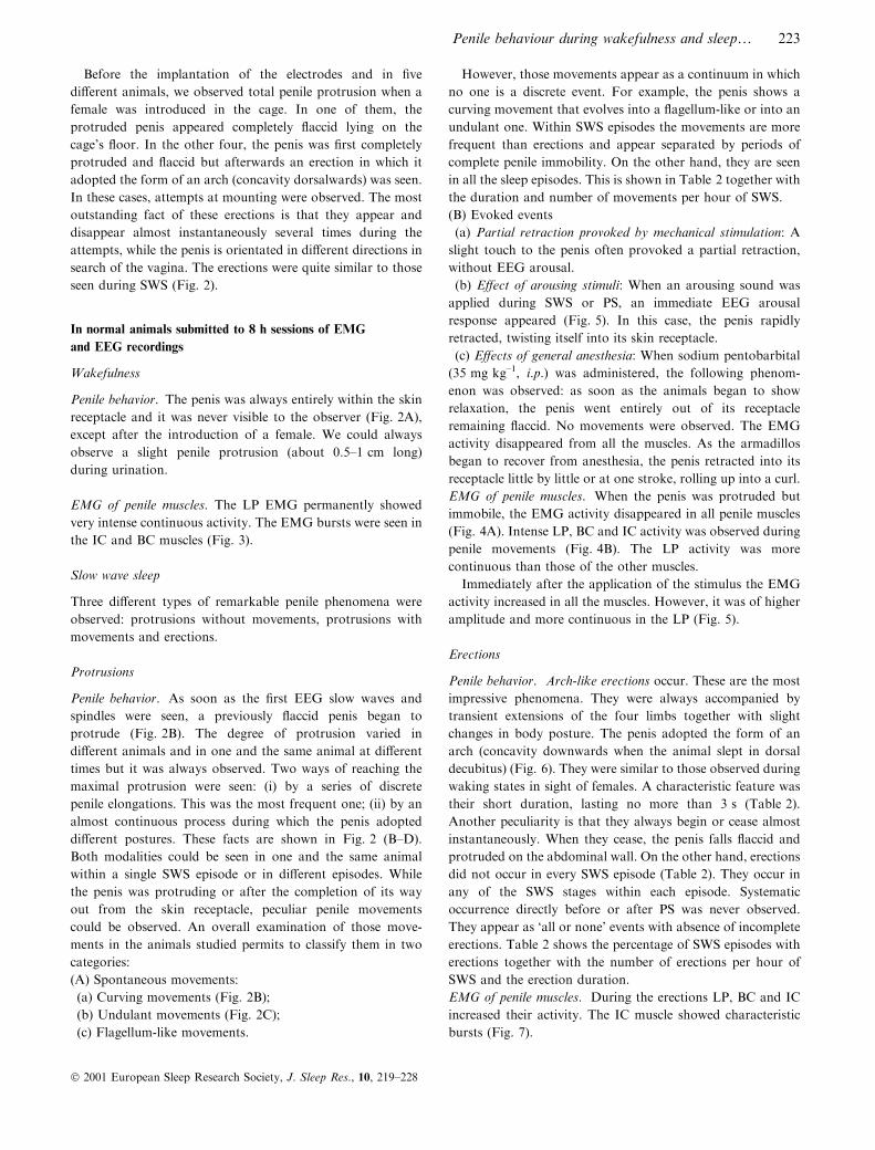
Before the implantation of the electrodes and in ®ve
di�erent animals, we observed total penile protrusion when a
female was introduced in the cage. In one of them, the
protruded penis appeared completely ¯accid lying on the
cage's ¯oor. In the other four, the penis was ®rst completely
protruded and ¯accid but afterwards an erection in which it
adopted the form of an arch (concavity dorsalwards) was seen.
In these cases, attempts at mounting were observed. The most
outstanding fact of these erections is that they appear and
disappear almost instantaneously several times during the
attempts, while the penis is orientated in di�erent directions in
search of the vagina. The erections were quite similar to those
seen during SWS (Fig. 2).
In normal animals submitted to 8 h sessions of EMG
and EEG recordings
Wakefulness
Penile behavior. The penis was always entirely within the skin
receptacle and it was never visible to the observer (Fig. 2A),
except after the introduction of a female. We could always
observe a slight penile protrusion (about 0.5±1 cm long)
during urination.
EMG of penile muscles. The LP EMG permanently showed
very intense continuous activity. The EMG bursts were seen in
the IC and BC muscles (Fig. 3).
Slow wave sleep
Three di�erent types of remarkable penile phenomena were
observed: protrusions without movements, protrusions with
movements and erections.
Protrusions
Penile behavior. As soon as the ®rst EEG slow waves and
spindles were seen, a previously ¯accid penis began to
protrude (Fig. 2B). The degree of protrusion varied in
di�erent animals and in one and the same animal at di�erent
times but it was always observed. Two ways of reaching the
maximal protrusion were seen: (i) by a series of discrete
penile elongations. This was the most frequent one; (ii) by an
almost continuous process during which the penis adopted
di�erent postures. These facts are shown in Fig. 2 (B±D).
Both modalities could be seen in one and the same animal
within a single SWS episode or in di�erent episodes. While
the penis was protruding or after the completion of its way
out from the skin receptacle, peculiar penile movements
could be observed. An overall examination of those move-
ments in the animals studied permits to classify them in two
categories:
(A) Spontaneous movements:
(a) Curving movements (Fig. 2B);
(b) Undulant movements (Fig. 2C);
(c) Flagellum-like movements.
However, those movements appear as a continuum in which
no one is a discrete event. For example, the penis shows a
curving movement that evolves into a ¯agellum-like or into an
undulant one. Within SWS episodes the movements are more
frequent than erections and appear separated by periods of
complete penile immobility. On the other hand, they are seen
in all the sleep episodes. This is shown in Table 2 together with
the duration and number of movements per hour of SWS.
(B) Evoked events
(a) Partial retraction provoked by mechanical stimulation: A
slight touch to the penis often provoked a partial retraction,
without EEG arousal.
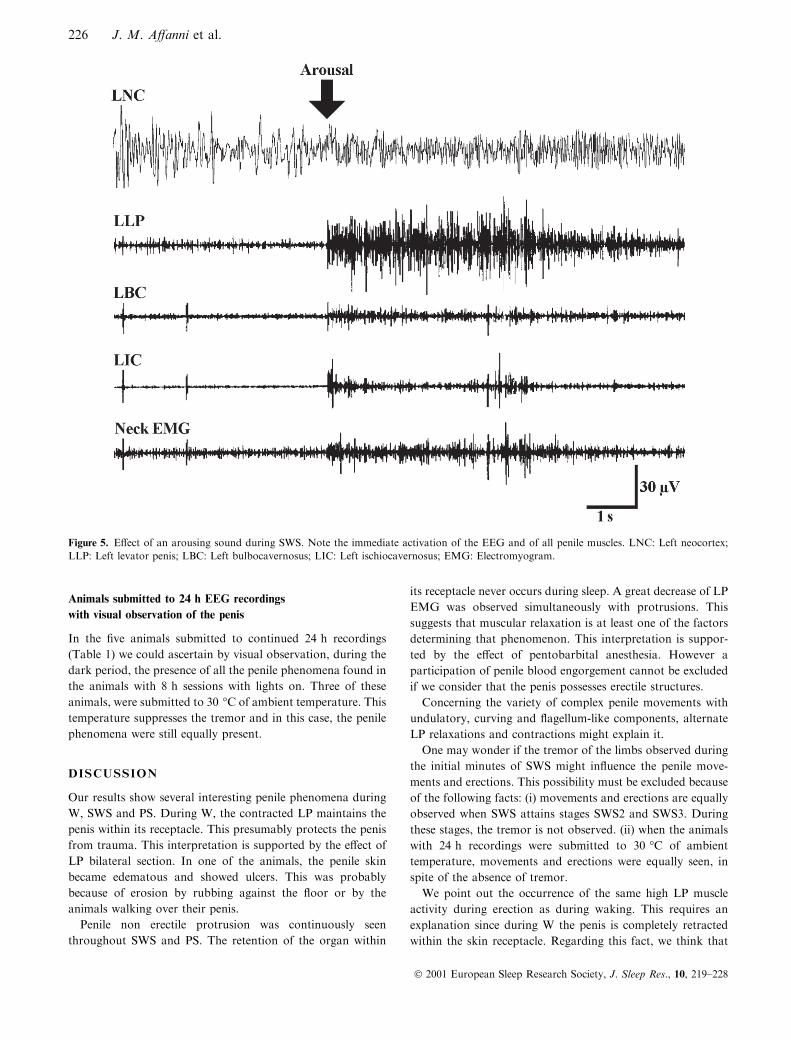
(b) E�ect of arousing stimuli: When an arousing sound was
applied during SWS or PS, an immediate EEG arousal
response appeared (Fig. 5). In this case, the penis rapidly
retracted, twisting itself into its skin receptacle.
(c) E�ects of general anesthesia: When sodium pentobarbital
(35 mg kg±1, i.p.) was administered, the following phenom-
enon was observed: as soon as the animals began to show
relaxation, the penis went entirely out of its receptacle
remaining ¯accid. No movements were observed. The EMG
activity disappeared from all the muscles. As the armadillos
began to recover from anesthesia, the penis retracted into its
receptacle little by little or at one stroke, rolling up into a curl.
EMG of penile muscles. When the penis was protruded but
immobile, the EMG activity disappeared in all penile muscles
(Fig. 4A). Intense LP, BC and IC activity was observed during
penile movements (Fig. 4B). The LP activity was more
continuous than those of the other muscles.
Immediately after the application of the stimulus the EMG
activity increased in all the muscles. However, it was of higher
amplitude and more continuous in the LP (Fig. 5).
Erections
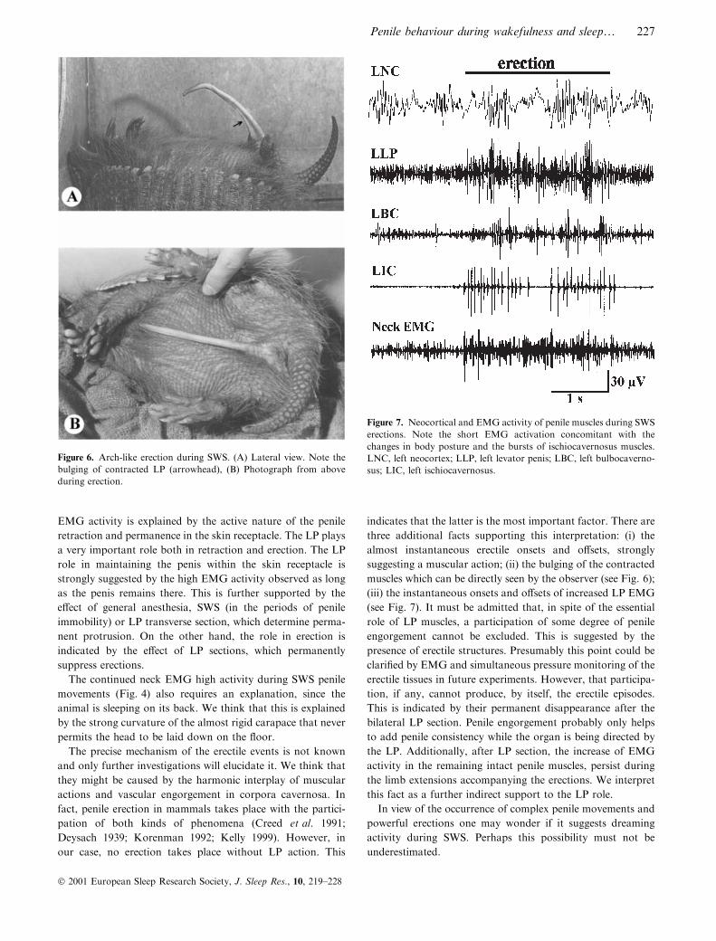
Penile behavior. Arch-like erections occur. These are the most
impressive phenomena. They were always accompanied by
transient extensions of the four limbs together with slight
changes in body posture. The penis adopted the form of an
arch (concavity downwards when the animal slept in dorsal
decubitus) (Fig. 6). They were similar to those observed during
waking states in sight of females. A characteristic feature was
their short duration, lasting no more than 3 s (Table 2).
Another peculiarity is that they always begin or cease almost
instantaneously. When they cease, the penis falls ¯accid and
protruded on the abdominal wall. On the other hand, erections
did not occur in every SWS episode (Table 2). They occur in
any of the SWS stages within each episode. Systematic
occurrence directly before or after PS was never observed.
They appear as `all or none' events with absence of incomplete
erections. Table 2 shows the percentage of SWS episodes with
erections together with the number of erections per hour of
SWS and the erection duration.
EMG of penile muscles. During the erections LP, BC and IC
increased their activity. The IC muscle showed characteristic
bursts (Fig. 7).
Penile behaviour during wakefulness and sleep¼ 223
Ó 2001 European Sleep Research Society, J. Sleep Res., 10, 219±228

Figure 3. (A) Neocortical and EMG activity of penile muscles during W. (B) Neocortical and EMG activity of penile muscles during PS. Note
total absence of EMG activity. The electrocardiogram (ECG) contaminates the tracings. LNC, left neocortex; RNC, right neocortex;
EMG, electromyogram; LLP, left levator penis; RLP, right levator penis; LBC, left bulbocavernosus; RBC, right bulbocavernosus; LIC,
left ischiocavernosus; RIC, right ischiocavernosus.
224 J. M. A�anni et al.
Ó 2001 European Sleep Research Society, J. Sleep Res., 10, 219±228

Paradoxical sleep
Penile behavior. The penis remained protruded and ¯accid
throughout all PS episodes. We never observed erections
during PS episodes (Table 2). Additionally, we never observed
erections in the hundreds of PS episodes recorded from the ®ve
animals visually monitored during the 24 h continuous
recordings. All movements of the corpus penis disappeared
completely. Only one kind of movement was observed from
time to time: a twitching ¯exion of the penile tip (TFPT)
occurring simultaneously with the myoclonic jerks of limbs
and body typically seen during PS episodes (Table 2). Those
¯exions were seen only during PS and their presence consti-
tutes an additional sign of PS episodes.
One outstanding fact is that the penile posture displayed at
the moment of PS initiation persisted unaltered throughout the
PS episode. If, for example, the transition from SWS to PS
occurs when the penis is in a curved posture such as shown in
Fig. 2D it will remain frozen in that posture.
In one exceptional case, a dramatic demonstration of the
absence of penile erection was provided when an observer took
gently the middle part of the penis between his ®ngers. On this
occasion the penis remained hanging in sharp acute angle
between the ®nger tips.
EMG of penile muscles. It was permanently absent from all
penile muscles showing that they share the generalized PS
atonia (Fig. 3).
Animals submitted to LP bilateral section
After the LP section, the penis remained permanently out of
the skin receptacle. It was seen hanging immobile across the
di�erent states wakefulness and sleep. This is represented by:
(i) disappearance of the retraction seen during W or when
sensory stimuli are presented; (ii) neither movements nor SWS
erections appeared whereas the limb extensions accompanying
erections in intact animals continue to occur. However, during
the extensions, the EMG of the remaining intact muscles (IC
and BC) continues to exhibit the increase seen during erections
in normal animals. Those changes became permanent and were
still observed 2 months after the operation. Near the end of
this period, one of the animals showed ulcers and edema in the
penile skin. This animal was immediately sacri®ced to avoid
su�ering.
Figure 4. Neocortical and EMG activity of penile muscles during SWS. (A) Protruded penis without movements, (B) Protruded penis while
performing movements. LNC: Left neocortex; LLP: Left levator penis; LBC: Left bulbocavernosus; LIC: Left ischiocavernosus; EMG:
Electromyogram.
Penile behaviour during wakefulness and sleep¼ 225
Ó 2001 European Sleep Research Society, J. Sleep Res., 10, 219±228
Animals submitted to 24 h EEG recordings
with visual observation of the penis
In the ®ve animals submitted to continued 24 h recordings
(Table 1) we could ascertain by visual observation, during the
dark period, the presence of all the penile phenomena found in
the animals with 8 h sessions with lights on. Three of these
animals, were submitted to 30 °C of ambient temperature. This
temperature suppresses the tremor and in this case, the penile
phenomena were still equally present.
DISCUSSION
Our results show several interesting penile phenomena during
W, SWS and PS. During W, the contracted LP maintains the
penis within its receptacle. This presumably protects the penis
from trauma. This interpretation is supported by the e�ect of
LP bilateral section. In one of the animals, the penile skin
became edematous and showed ulcers. This was probably
because of erosion by rubbing against the ¯oor or by the
animals walking over their penis.
Penile non erectile protrusion was continuously seen
throughout SWS and PS. The retention of the organ within
its receptacle never occurs during sleep. A great decrease of LP
EMG was observed simultaneously with protrusions. This
suggests that muscular relaxation is at least one of the factors
determining that phenomenon. This interpretation is suppor-
ted by the e�ect of pentobarbital anesthesia. However a
participation of penile blood engorgement cannot be excluded
if we consider that the penis possesses erectile structures.
Concerning the variety of complex penile movements with
undulatory, curving and ¯agellum-like components, alternate
LP relaxations and contractions might explain it.
One may wonder if the tremor of the limbs observed during
the initial minutes of SWS might in¯uence the penile move-
ments and erections. This possibility must be excluded because
of the following facts: (i) movements and erections are equally
observed when SWS attains stages SWS2 and SWS3. During
these stages, the tremor is not observed. (ii) when the animals
with 24 h recordings were submitted to 30 °C of ambient
temperature, movements and erections were equally seen, in
spite of the absence of tremor.
We point out the occurrence of the same high LP muscle
activity during erection as during waking. This requires an
explanation since during W the penis is completely retracted
within the skin receptacle. Regarding this fact, we think that
Figure 5. E�ect of an arousing sound during SWS. Note the immediate activation of the EEG and of all penile muscles. LNC: Left neocortex;
LLP: Left levator penis; LBC: Left bulbocavernosus; LIC: Left ischiocavernosus; EMG: Electromyogram.
226 J. M. A�anni et al.
Ó 2001 European Sleep Research Society, J. Sleep Res., 10, 219±228
EMG activity is explained by the active nature of the penile
retraction and permanence in the skin receptacle. The LP plays
a very important role both in retraction and erection. The LP
role in maintaining the penis within the skin receptacle is
strongly suggested by the high EMG activity observed as long
as the penis remains there. This is further supported by the
e�ect of general anesthesia, SWS (in the periods of penile
immobility) or LP transverse section, which determine perma-
nent protrusion. On the other hand, the role in erection is
indicated by the e�ect of LP sections, which permanently
suppress erections.
The continued neck EMG high activity during SWS penile
movements (Fig. 4) also requires an explanation, since the
animal is sleeping on its back. We think that this is explained
by the strong curvature of the almost rigid carapace that never
permits the head to be laid down on the ¯oor.
The precise mechanism of the erectile events is not known
and only further investigations will elucidate it. We think that
they might be caused by the harmonic interplay of muscular
actions and vascular engorgement in corpora cavernosa. In
fact, penile erection in mammals takes place with the partici-
pation of both kinds of phenomena (Creed et al. 1991;
Deysach 1939; Korenman 1992; Kelly 1999). However, in
our case, no erection takes place without LP action. This
indicates that the latter is the most important factor. There are
three additional facts supporting this interpretation: (i) the
almost instantaneous erectile onsets and o�sets, strongly
suggesting a muscular action; (ii) the bulging of the contracted
muscles which can be directly seen by the observer (see Fig. 6);
(iii) the instantaneous onsets and o�sets of increased LP EMG
(see Fig. 7). It must be admitted that, in spite of the essential
role of LP muscles, a participation of some degree of penile
engorgement cannot be excluded. This is suggested by the
presence of erectile structures. Presumably this point could be
clari®ed by EMG and simultaneous pressure monitoring of the
erectile tissues in future experiments. However, that participa-
tion, if any, cannot produce, by itself, the erectile episodes.
This is indicated by their permanent disappearance after the
bilateral LP section. Penile engorgement probably only helps
to add penile consistency while the organ is being directed by
the LP. Additionally, after LP section, the increase of EMG
activity in the remaining intact penile muscles, persist during
the limb extensions accompanying the erections. We interpret
this fact as a further indirect support to the LP role.
In view of the occurrence of complex penile movements and
powerful erections one may wonder if it suggests dreaming
activity during SWS. Perhaps this possibility must not be
underestimated.
Figure 6. Arch-like erection during SWS. (A) Lateral view. Note the
bulging of contracted LP (arrowhead), (B) Photograph from above
during erection.
Figure 7. Neocortical and EMG activity of penile muscles during SWS
erections. Note the short EMG activation concomitant with the
changes in body posture and the bursts of ischiocavernosus muscles.
LNC, left neocortex; LLP, left levator penis; LBC, left bulbocaverno-
sus; LIC, left ischiocavernosus.
Penile behaviour during wakefulness and sleep¼ 227
Ó 2001 European Sleep Research Society, J. Sleep Res., 10, 219±228

The most salient feature of PS is the total absence of penile
erections and movements. The penis remains completely
protruded, ¯accid and immobile throughout that phase. Only
the twitching ¯exion of the penile tip is observable from time
to time, coinciding with the jerks of body muscles character-
istic of PS. We think that this ¯exion is simply an expression of
those jerks in that tip.
The absence of PS erectile events indicates that during the
PS atonia no vascular engorgement is capable of inducing
them. This is indicated by the fact that curved or ¯exed penile
postures displayed at the moment of PS initiation persisted
unaltered throughout the PS episode. This is an important fact
because it demonstrates absence of penile rigidity. In brief, the
absence of PS erections appears because of: (i) atonia of penile
muscles which share the generalized PS atonia; (ii) lack of
vascular phenomena capable of producing penile rigidity by
themselves.
According to our results, it is evident that PS erectile events
are not a universal phenomenon of mammalian physiology.
Without doubt, PS mechanisms are not linked to the neural
system controlling the erections. This fact might lead to
important heuristic consequences.
The repeated instantaneous onsets and o�sets of erections
separated by repeated protrusions in animals submitted to
contact with females strongly suggest that they are voluntary
phenomena. Therefore, they should be under the control of
brain motor areas.
Schmidt et al. (1994) gave the ®rst experimental evidence of
penile erections during PS in the rat. These authors demon-
strated that BC and IC muscles showed EMG characteristic
bursts during PS. This means that those muscles are spared
from the general PS atonia. In sharp contrast with the rat, BC,
IC and LP muscles of this armadillo exhibit complete atonia
during PS. Presumably, this is related to its peculiar erectile
mechanisms depending upon somatic muscles under the
control of brain and spinal motor areas. The absence of penile
erection might be simply because of the fact that penile muscles
share the generalized PS atonia.
Lastly, we wish to point out that the visual observation of
animals with 24 h EEG recordings showed the occurrence of
penile movements and erections during SWS as well as the
complete absence of erections during PS. This indicates that
the penile phenomena reported for the 8 h sessions are also
observed in the 24 h recordings, which include the dark period.
REFERENCES
A�anni, J. and GarcõÂ a Samartino, L. Comparative study of electro-
physiological phenomena in the olfactory bulb of some South
American marsupials and edentates. In: L. Bolis, R. Keynes and
S. Maddrell (Eds) Comparative Physiology of Sensory Systems.
Cambridge University Press, New York, 1984, 315±332.
A�anni, J., GarcõÂ a Samartino, L. and Morita, M. Observaciones sobre
la actividad ele ctrica del neocortex, paleocortex y bulbo olfatorio de
Chaetophractus villosus. Rvta. Soc. Argentina Biol., 1968, 44: 189±
196.
A�anni, J., Lisogorsky, E. and Scaravilli, A. Sleep in the giant South
American armadillo Priodontes giganteus (edentata, mammalia).
Experientia, 1972, 28: 1046±1047.
BenõÂ tez, I., Aldana Marcos, H. and A�anni, J. M. The most salient
features of the encephalon of the Armadillo Chaetophractus villosus.
Com. Biol., 1994, 12: 57±73.
Cabrera, A., and Yepes, J. MamõÂferos Sudamericanos. Comp. Argen-
tina de Editores, Buenos Aires, 1940, 248±250.
Casanave, E. and A�anni, J. M. Body temperature of the armadillo
Chaetophractus villosus (Mammalia, Dasypodidae). Arch. Int.
Physiol. Biochim. Bioph., 1994, 102: 243±246.
Cervino, C. Estudio cuantitativo de dos nuevos ritmos bioeleÂctricos de los
bulbos olfatorios registrados en el armadillo sudamericano Chaetoph-
ractus villosus (Mammalia, Dasypodidae). MS Doctoral Thesis.
Facultad de Ciencias Exactas y Naturales, Universidad de Buenos
Aires, Buenos Aires, 1997, 529, pp.
Creed, K. E., Carati, C. J. and Kreogh, E. J. The physiology of penile
erection. In: S. R. Milligan (Ed.) Oxford Reviews of Reproductive
Biology, Vol. 13. Oxford University Press, New York, 1991.
Deysach, L. J. The comparative morphology of erectile tissue of the
penis with special emphasis on the probable mechanism of erection.
Am. J. Anat., 1939, 64: 111±131.
GarcõÂ a Samartino, L., A�anni, J., Casanave, E., Ferrari, R. and
Iodice, O. On the presence of a peculiar alpha rhythm in the
olfactory tubercle of waking armadillos. Electroenceph. Clin. Neu-
rophysiol., 1987, 66: 185±190.
GarcõÂ a Samartino, L., Scaravilli, A., A�anni, J. and Cinto, R. Estudio
cuantitativo de la vigilia y el suenÄ o en Chaetophractus villosus
(Mammalia, Dasypodidae). Physis, 1974, 33C: 145±150.
Grasse , P. Traite de Zoologie. Masson y Cie, Paris, 1955, TS XVII, F,
1182.
Holmes, G., Chapple, W., Leipheimer, R. and Sachs, B. Electromy-
ographic analysis of male rat perineal muscles during copulation
and re¯exive erections. Physiol. Behav., 1991, 49: 1235±1246.
Kelly, D. A. Expansion of the tunica albuginea during penile in¯ation
in the nine banded armadillo Dasypus novemcimtus. J. Exp. Biol.,
1999, 202: 253±265.
Korenman, S. G. Sexual dysfunction. In: J. D. Wilson and D. W.
Foster (Eds) Williams Textbook of Endocrinology. W.B. Saunders
Co., Phyladelphia, 1992, 1033±1048.
Papini, M., Mustaca, A. and A�anni, J. Discrimination learning in the
Armadillo (Chaetophractus villosus). A study of positional strat-
egies. J. Gen. Psychol., 1985, 112: 119±127.
Papini, M., Mustaca, A. and A�anni, J. Spatial learning in South
American opossums and armadillos. J. Gen. Psychol., 1984, 111:
45±55.
Schmidt, M., Valatx, J., Schmidt, H., Wauquier, A. and Jouvet, M.
Experimental evidence of penile erections during paradoxical sleep
in the rat. Neuroreport, 1994, 5: 561±564.
Walker, E. Mammals of the World, 2nd edn. J. Hopkins Press,
Baltimore, 1968, 482±503.
228 J. M. A�anni et al.
Ó 2001 European Sleep Research Society, J. Sleep Res., 10, 219±228