Bioengineered Skin Intended for Skin Disease Modeling - MDPI
Upload
independentCategory
view
4download
0
ABNORMAL AQUAPORIN-3 PROTEIN EXPRESSION INHYPERPROLIFERATIVE SKIN DISORDERS
Kristen E. Voss2, Roni J. Bollag4, Nicole Fussell2, Charya By2, Daniel J. Sheehan1,4,5, andWendy B. Bollag1,2,3,5,6
1Charlie Norwood VA Medical Center, One Freedom Way, Augusta, GA 309042Institute of Molecular Medicine and Genetics, Georgia Health Sciences University (formerlyMedical College of Georgia), 1120 15th Street, Augusta, GA 309123Department of Physiology, Georgia Health Sciences University (formerly Medical College ofGeorgia), 1120 15th Street, Augusta, GA 309124Department of Pathology, Georgia Health Sciences University (formerly Medical College ofGeorgia), 1120 15th Street, Augusta, GA 309125Department of Medicine (Dermatology), Georgia Health Sciences University (formerly MedicalCollege of Georgia), 1120 15th Street, Augusta, GA 309126Departments of Cell Biology and Anatomy and Orthopaedic Surgery, Georgia Health SciencesUniversity (formerly Medical College of Georgia), 1120 15th Street, Augusta, GA 30912
AbstractNon-melanoma skin cancers (NMSCs) and psoriasis represent common hyperproliferative skindisorders, with approximately one million new NMSC diagnoses each year in the United Statesalone and a psoriasis prevalence of about 2% worldwide. We recently demonstrated that theglycerol channel, aquaporin-3 (AQP3) and the enzyme phospholipase D2 (PLD2) interactfunctionally in epidermal keratinocytes of the skin to inhibit their proliferation. However, othershave suggested that AQP3 is pro-proliferative in keratinocytes and is upregulated in the NMSC,squamous cell carcinoma (SCC). To evaluate the AQP3/PLD2 signaling module in skin diseases,we determined their levels in SCC, basal cell carcinoma (BCC) and psoriasis compared to normalepidermis. Skin biopsies with the appropriate diagnoses (ten normal, five SCC, thirteen BCC andten plaque psoriasis samples) were obtained from the pathology archives and examined byimmunohistochemistry using antibodies recognizing AQP3 and PLD2. In normal epidermisAQP3, an integral membrane protein, was localized mainly to the plasma membrane and PLD2 tothe cell periphery, particularly in suprabasal layers. In BCC, AQP3 and PLD2 levels were reducedcompared to the normal-appearing overlying epidermis. In SCC, AQP3 staining was “patchy,”with areas of reduced AQP3 immunoreactivity exhibiting positivity for Ki67, a marker ofproliferation. PLD2 staining was unchanged in SCC. In psoriasis, AQP3 staining was usuallyobserved in the cytoplasm rather than in the membrane. Also, in the majority of psoriatic samples,PLD2 showed weak immunoreactivity or aberrant localization. These results suggest thatabnormalities in the AQP3/PLD2 signaling module correlate with hyperproliferation in psoriasisand the NMSCs.
To whom correspondence should be addressed: Wendy B. Bollag Georgia Health Sciences University (formerly Medical College ofGeorgia) Department of Physiology 1120 15th Street Augusta, GA 30912 TEL: (706) 721-0698 FAX: (706) [email protected].
CONFLICT OF INTEREST:The authors declare that there are no conflicts of interest.
NIH Public AccessAuthor ManuscriptArch Dermatol Res. Author manuscript; available in PMC 2013 August 26.
Published in final edited form as:Arch Dermatol Res. 2011 October ; 303(8): 591–600. doi:10.1007/s00403-011-1136-x.
NIH
-PA Author Manuscript
NIH
-PA Author Manuscript
NIH
-PA Author Manuscript

Keywords(aquaporin-3); (basal cell carcinoma); epidermis; keratinocytes; (phospholipase D2); skin; (skincancer); (squamous cell carcinoma)
INTRODUCTIONNon-melanoma skin cancer (NMSC) is one of the most common neoplasms in humans, witha million new cases diagnosed each year in the United States alone and an incidence that ison the rise [22]. NMSC can lead to major cosmetic deformities and sporadic mortality. Inthe two most common NMSCs, basal cell carcinoma (BCC) and squamous cell carcinoma(SCC), the major cell type in the epidermis, keratinocytes fail to differentiate and exhibithyperproliferation. Keratinocytes continually regenerate through regulated proliferationfollowed by differentiation into the suprabasal epidermal cell types. In the integumentarydifferentiation program, following cell division, early differentiation gives rise to the stratumspinosum, late differentiation generates the stratum granulosum, and terminal differentiationoccurs in the stratum corneum, the outermost layer of the epidermis (reviewed in [3,1]). Thispattern of proliferation and differentiation is essential for the protective barrier function ofskin. Skin disorders, including NMSCs such as SCC and BCC, can result from dysregulationof the homeostasis of keratinocyte proliferation and differentiation (reviewed in [4,2]).Although environmental insults are known to predispose to skin carcinogenesis, themechanisms underlying the development and progression of BCC and SCC remain unclear.
Similar to the keratinocyte-derived carcinomas BCC and SCC, psoriasis is a commondisabling hyperproliferative skin disorder in which keratinocytes fail to differentiate andproliferate excessively. Although the autoimmune etiology of psoriasis is well-appreciated(reviewed in [9]), with the recent creation of genetically engineered mouse models, it hasbecome clear that changes in the keratinocytes themselves can also trigger a psoriasis-likecondition (reviewed in [12]). For example, mice in which genes for the transcription factorsc-jun and JunB are deleted only in keratinocytes (i.e., an epidermal-targeted conditional c-jun/JunB knockout mouse model) exhibit a hyperproliferative epidermis and othercharacteristics of a psoriasiform phenotype [36]. Thus, current research has suggestedinterplay between keratinocytes and immune cells, with the resulting crosstalk leading tohyperproliferation of keratinocytes in psoriasis [32]. Nevertheless, the mechanismunderlying uncontrolled proliferation and abnormal differentiation of keratinocytes inpsoriasis, as well as the etiology of the disease itself, is still unclear.
Aquaporin-3 (AQP3), an aquaglyceroporin, has recently been ascribed a potential role inskin function (reviewed in [16]). AQP3 is found in the basal cell layer of the epidermis, aswell as suprabasal layers [21], and is an efficient transporter of glycerol and water [34].AQP3 null mice exhibit reduced epidermal glycerol content, impaired skin elasticity,delayed barrier recovery following stratum corneum removal, and delayed wound healing[13]. These phenotypes suggest that AQP3 plays a role in differentiation and proliferation ofkeratinocytes. Interestingly, topical or oral application of glycerol has been shown to correctdefects present in AQP3 null mice including skin hydration, elasticity, and barrier function[14]. Likewise, in the cosmetic industry, glycerol has been used in moisturizers and othertopical skin therapies to promote skin repair ([7] and reviewed in [8]). Our laboratory hasdemonstrated that glycerol, a physiologically relevant primary alcohol, can be utilized byphospholipase D2 (PLD2) to form phosphatidylglycerol both in vitro and in intactkeratinocytes [38]. In addition, PLD2 and AQP3 are colocalized in lipid rafts and co-precipitate from these rafts in a protein-mediated manner [37], and we have proposed thattogether they comprise a signaling module in which AQP3 transports glycerol to PLD2 to
Voss et al. Page 2
Arch Dermatol Res. Author manuscript; available in PMC 2013 August 26.
NIH
-PA Author Manuscript
NIH
-PA Author Manuscript
NIH
-PA Author Manuscript
synthesize phosphatidylglycerol (reviewed in [4]). Phosphatidylglycerol production isincreased by elevated extracellular calcium concentrations that promote keratinocytedifferentiation (and inhibit proliferation), and manipulation of this signaling module, eitherby increasing exogenous glycerol, co-overexpression of AQP3 or direct provision ofphosphatidylglycerol, induces differentiation and inhibits proliferation in rapidly dividingcells [38,5].
On the other hand, in a recent study, Verkman and colleagues [17] demonstrated that AQP3null mice exhibit resistance to skin tumorigenesis, although the described experiments didnot determine whether the effect is cell autonomous (i.e., due to the lack of AQP3 inkeratinocytes) or non-cell autonomous (for instance, related to changes in the inflammatoryresponse in these mice with a global deletion of AQP3). These authors also reported thatRNA interference-mediated knockdown of AQP3 inhibits proliferation in humankeratinocytes [15], suggesting that AQP3 promotes proliferation and is pro-tumorigenic. Insupport of this idea these authors demonstrated that AQP3 protein is strongly expressed inSCC in regions characterized by keratin 14 expression. However, although keratin 14expression is considered a marker of basal, proliferating keratinocytes, Fuchs and colleaguesdemonstrated that in normal epidermis keratin 14 protein is found in both basal andsuprabasal (spinous) layers [30]. Furthermore, keratin 14 staining is observed in SCCs of allstages of differentiation (from poorly differentiated to well differentiated tumors) [30]. Thislatter result is consistent with the findings of Perkins et al. [25], who reported increasedkeratin 14 staining in the more differentiated areas of SCCs, and suggests that keratin 14protein expression cannot be used as a marker of proliferation in SCC.
The potential involvement of AQP3 in other skin diseases is controversial. For example,Olsson et al. [24] and Nakahigashi et al. [23] have reported increased AQP3 expression inatopic eczema (dermatitis) whereas Boury-Jamot et al. [6] demonstrated a down-regulationof AQP3 in eczema (reviewed in [26]). In keratinocytes from depigmented vitiligo lesionsdown-regulation of AQP3 has also been observed, and in conjunction with reductions in thelevels of E-cadherin, β- and γ-catenins and phosphorylated (active) phosphoinositide 3-kinase, this diminished AQP3 protein expression was suggested to decrease keratinocytesurvival. The loss of keratinocytes and keratinocyte-derived growth factors presumablyunderlies the passive cell death of melanocytes resulting in the depigmentation seen invitiligo lesions [20]. On the other hand, to our knowledge there is no information in theliterature concerning the possible role of PLD2 or the AQP3/PLD2 signaling module in skindiseases.
In contrast, our data concerning the differentiation-promoting role of the AQP3/PLD2signaling module [5] in keratinocytes would predict that in hyperproliferative skin diseasesAQP3 levels might be decreased. Alternatively, the ratio of AQP3 to PLD2 and/or thelocalization of these two proteins might be critical for their functional interaction and effectson proliferation versus differentiation. Therefore, the goal of this study was to examine theprotein expression and localization of AQP3 and PLD2 in SCC, BCC and psoriasis relativeto normal epidermis.
MATERIALS AND METHODSTissue Samples
The retrospective characterization of de-identified patient specimens was approved by theMedical College of Georgia institutional review board. Paraffin-embedded tissue sections ofskin with a diagnosis of SCC, BCC, or plaque psoriasis, as determined and verified by adermatopathologist (DJS), and normal epidermis were obtained from the tissue archives ofthe Medical College of Georgia Department of Pathology. The majority of the BCC samples
Voss et al. Page 3
Arch Dermatol Res. Author manuscript; available in PMC 2013 August 26.
NIH
-PA Author Manuscript
NIH
-PA Author Manuscript
NIH
-PA Author Manuscript

were of the nodular type. Normal epidermis was obtained from samples removed duringbreast reduction surgery or from traumatic amputation specimens. Four micron thicksections were cut and mounted on slides by conventional histologic techniques.
AQP3 and PLD2 ImmunohistochemistrySlides for AQP3 staining were deparaffinized, washed twice for 5 minutes in phosphate-buffered saline (PBS), incubated 30 minutes in 3% hydrogen peroxide, washed twice for 5minutes in PBS, incubated 1 hour in 0.3% goat serum, and then incubated overnight withanti-AQP3 (Alomone Labs, Jerusalem, Israel; 1:1000 dilution) in a humidified chamber at4°C. Following primary antibody incubation, an ABC staining kit (Santa CruzBiotechnology, Santa Cruz, CA) was used to visualize immunoreactivity by incubation withan appropriate secondary antibody and development with the chromogen 3,3’-diaminobenzidine (DAB) for 3 minutes. After deparaffinization, slides used for PLD2staining were incubated three times for 10 min in boiling sodium citrate buffer, then stainedwith anti-PLD2 (Abcam, Cambridge, MA; 1:50 dilution) and visualized (DAB for 10minutes) using the same methods as for AQP3. All slides were dehydrated, counterstainedwith hematoxylin, and coverslipped by the Medical College of Georgia Histology CoreFacility.
Ki67 Immunohistochemistry—Ki67 (catalog # M7240, DakoUSA, Carpinteria, CA)immunostaining was performed according to the supplier's protocol. AQP3 and Ki67staining were performed on sequential serial SCC sections.
Statistical Analysis—In the BCC slides, staining intensity in the normal-appearingoverlying epidermis and in the lesion were estimated by three independent observers, with 0representing an absence of staining and 3 representing intense staining. The individualscores were averaged and the difference analyzed for statistical significance with anunpaired Student's t-test using the program InStat (Graphpad, La Jolla, CA).
RESULTSAQP3 Expression
Whereas we have hypothesized that the PLD2/AQP3 signaling module is anti-proliferativeand pro-differentiative [5], Verkman and colleagues have proposed instead that AQP3 ispro-proliferative [15,17]. These authors also report that SCC express high levels of AQP3and suggest that this result also supports the idea that AQP3 promotes proliferation [17]. Inan attempt to resolve this issue, we examined the expression of AQP3 in the non-melanomaskin cancers BCC and SCC by immunohistochemistry. This analysis showed predominantAQP3 expression in the plasma membrane (essentially outlining the cells) of basalkeratinocytes in the normal-appearing overlying epidermis observed in many BCC sections(Figure 1), consistent with the fact that AQP3 is an integral membrane protein. In BCC,AQP3 was down-regulated and in some cases was completely absent despite readilyapparent staining in the normal-appearing overlying epidermis. We observed reduced orabsent AQP3 staining in all 13 of the basal cell carcinomas examined (13/13), with anaverage decrease of approximately 8-fold (Figure 1D) [normal: 2.59 ± 0.14 versus lesion:0.33 ± 0.15 (p<0.001).] T
Next, the staining pattern observed in the normal-appearing overlying epidermis wascompared with that found in normal epidermis obtained from breast reduction surgery ortraumatic amputation. The normal-appearing overlying epidermis and normal epidermis(Figure 2) were shown to have similar AQP3 localization, with well-defined plasma
Voss et al. Page 4
Arch Dermatol Res. Author manuscript; available in PMC 2013 August 26.
NIH
-PA Author Manuscript
NIH
-PA Author Manuscript
NIH
-PA Author Manuscript

membrane staining outlining individual keratinocytes in the majority of the normalepidermal specimens.
In contrast to the clear down-regulation observed in BCC, the results in SCC were lessdefinitive, in that AQP3 immunoreactivity was “patchy,” with some regions of the lesionstaining intensely for AQP3 and others showing little or no staining (Figure 3). Thesefindings were consistent in 5 of 5 lesions examined. AQP3 staining seemed reducedparticularly at the SCC borders, which appeared to be invading the dermis. Interestingly, insequential serial sections the regions showing little AQP3 immunoreactivity stained positivefor the proliferation marker, Ki67 (Figure 4A and B); this suggested that proliferationcorrelates with down-regulation of AQP3 in both BCC and SCC.
We next obtained archival, paraffin-embedded tissue blocks with a diagnosis of plaquepsoriasis and compared AQP3 immunoreactivity in these sections with that in normalepidermis (Figure 2). We found that, in contrast to normal-appearing epidermis overlyingBCC (Figure 1) and the majority of normal epidermis (Figure 2), AQP3 staining in mostpsoriatic lesions was localized intracellularly in most of the lesion rather than to the plasmamembrane, although some membrane localization could be observed in occasionalsuprabasal regions of the hyperproliferative epidermis (Figure 5). We observed intracellularAQP3 localization in 8 out of 10 psoriatic patients. In contrast, AQP3 stained the plasmamembrane of follicular keratinocytes as it does in normal epidermis in many of the psoriasissamples. In the remaining 2 of 10 psoriatic samples, AQP3 staining was localized to the cellmembrane as in normal epidermis with no apparent appreciable expression deficit (data notshown).
PLD2 ExpressionConsistent with the fact that the enzyme is not a transmembrane protein but is insteadmembrane associated, PLD2 expression was predominantly localized to the perinuclearregion and the cell periphery in normal epidermis (Figure 6) and in the normal-appearingepidermis overlying BCC (Figure 7). PLD2 immunoreactivity was distributed throughoutthe basal and suprabasal layers in normal epidermis (Figure 6) as well as in the normal-appearing epidermis overlying BCC (Figure 7) but was reduced in the BCC. This decreasewas again observed in all 13 of the BCCs (13/13), with an approximate 5–fold reduction inBCC relative to the normal-appearing overlying epidermis (Figure 7D) [normal: 2.28 ± 0.18versus BCC: 0.46 ± 0.10 (p<0.001)]. This result suggests that decreased AQP3 and PLD2immunoreactivity is associated with the hyperproliferation observed in BCC. On the otherhand, there was little or no change in PLD2 in SCC as compared to normal epidermis (datanot shown).
In the majority of psoriatic samples (8 of 10), PLD2 staining seemed aberrant, with weakstaining (Figure 8A, C) or largely abnormal localization (Figure 8B, C). In the remaining 2psoriatic samples, PLD2 staining was essentially indistinguishable from that in normalepidermis (Figure 8D). Interestingly, the 2 samples showing normal PLD2 immunoreactivityalso exhibited typical staining for AQP3.
DISCUSSIONOur results suggest that down-regulation of AQP3 in the epidermis may be a marker oftumorigenic potential, since expression of this glycerol channel is decreased in BCC and inthe areas of SCC that are most rapidly proliferating (i.e., Ki67 positive). In the study ofVerkman and colleagues examining AQP3 expression in human SCC, AQP3 was found tobe strongly expressed and co-localized with the basal marker keratin 14; the presence ofAQP3 in SCC was confirmed by immunohistochemistry in our study. However, we found
Voss et al. Page 5
Arch Dermatol Res. Author manuscript; available in PMC 2013 August 26.
NIH
-PA Author Manuscript
NIH
-PA Author Manuscript
NIH
-PA Author Manuscript

that AQP3 protein expression within the epidermis of SCC appeared to be excluded from theatypical keratinocytes corresponded to cells on the inner edge of the epidermis. Moreimportantly, these atypical keratinocytes were proliferating actively, as marked by Ki67positivity. Since keratin 14 protein expression has been observed even in well-differentiatedSCC [30,25], keratin 14 may not be the ideal marker for proliferation, and colocalization ofAQP3 and keratin 14 does not preclude a correspondence with differentiation rather thanproliferation. Thus, our results showing that Ki67 immunoreactivity correlated with reducedAQP3 protein expression suggest that AQP3 may be associated with the differentiationprogram. Indeed, this idea is supported by a recent study [20] demonstrating that RNAinterference-mediated silencing of AQP3 in human keratinocytes results in reducedexpression of keratin 10, a differentiation marker, in response to an elevated extracellularcalcium level, a known inducer of differentiation.
However, the possibility remains that AQP3 (and PLD2) could be pro-proliferative anddown-regulated in hyperproliferative disorders in an attempt to compensate for the excessivegrowth. Indeed, using RNA-interference-mediated knock down of AQP3 levels, Verkmanand colleagues have shown that loss of AQP3 inhibits human keratinocyte proliferation, andadenovirus-mediated AQP3 re-expression returns proliferation values to normal [15]. On theother hand, whether or not AQP3 mediates proliferation or differentiation may depend onwhether or not the protein is associated with PLD2 (and vice versa). Thus, the relativeoverexpression of AQP3 or PLD2 alone may allow proliferation (Qin and Bollag,manuscript in preparation), whereas when regulated in tandem, the interaction of the twoproteins should allow phosphatidylglycerol production and early differentiation.Alternatively, the localization (intracellularly or at the plasma membrane) of this signalingmodule may determine the cellular response, proliferation or differentiation. Indeed, ourprevious immunocytochemistry data suggest a partial colocalization of AQP3 and PLD2,with both plasma membrane and intracellular staining [18]. Similarly, down-regulation ofPLD2 in BCC suggests that this enzyme also may play a role as an anti-proliferative signal,although the same caveat concerning the possibility that the cells down-regulate PLD2 in anattempt to compensate for the hyperproliferation also applies. Thus, the decreased AQP3 inBCC and proliferating regions of SCC provides evidence for the possibility that the AQP3/PLD2 signaling module may act as a tumor suppressor, although obviously additionalstudies to clarify this issue are warranted. Further studies in the epidermis and otherepithelial cancers, such as those of the lung and colon, which also express AQP3 and PLD2[21,27,35,31,28,18,19,33], might elucidate this mechanism and further our understanding ofthe role of AQP3 and PLD2 in epithelial cells.
In the majority of the epidermal samples of plaque psoriasis, another hyperproliferativedisorder of keratinocytes, AQP3 expression was localized diffusely to the cytoplasm asopposed to its plasma membrane localization in normal epidermis. It should be noted that innormal epidermal samples, such intracellular staining was also sometimes observed in basalkeratinocytes, and Sougrat et al. [29] have also reported cytoplasmic staining in basalkeratinocytes of human skin and reconstructed human epidermis. Since AQP3 must be in theplasma membrane in order to transport extracellular glycerol into cells, this result suggeststhat AQP3 transport activity is compromised in keratinocytes in psoriatic lesions and thatphosphatidylglycerol levels should be reduced in this hyperproliferative skin disease.Indeed, it seems possible that localization of AQP3 to an intracellular compartment or theplasma membrane, as well as colocalization with PLD2, could determine whether thisaquaglyceroporin promotes proliferation or differentiation. On the other hand, a minority ofthe psoriatic samples (2 of 10) exhibited apparently normal AQP3 localization. This resultmay not be entirely unexpected, given the complexity and multiple subphenotypes of thisdisease [11], although in this study plaque-type psoriasis was not classified by
Voss et al. Page 6
Arch Dermatol Res. Author manuscript; available in PMC 2013 August 26.
NIH
-PA Author Manuscript
NIH
-PA Author Manuscript
NIH
-PA Author Manuscript
subphenotypes [10]. Additional studies to examine AQP3 and PLD2 immunoreactivity inthese subtypes of plaque psoriasis, as well as other subtypes of psoriasis, seem warranted.
In conclusion, our results are consistent with the idea that the AQP3/PLD2 signaling moduleis abnormal in human skin diseases, with down-regulated levels of both proteins in BCC,decreased AQP3 immunoreactivity in proliferating regions of SCC and anomalousdistribution and/or decreased levels of both proteins in psoriasis compared to normal humanepidermis. Although our results do not prove either a pro-proliferative or a pro-differentiative role for AQP3 and/or PLD2, our data do demonstrate that both AQP3 andPLD2 are abnormally expressed and/or localized in human skin diseases and thus may playa role in or serve as surrogate markers for the pathogenesis of these diseases.
AcknowledgmentsThis project was supported in part by a grant from the National Institutes of Health/ National Institute of Arthritis,Musculoskeletal and Skin Diseases #AR45212. The authors thank Kimberly Smith and Doris Cawley in theGeorgia Research Pathology Services for preparing the sections and performing the Ki67 immunohistochemistryand the Medical College of Georgia Histology Core Facility for assistance with immunohistochemistry.
REFERENCES1. Bikle DD, Pillai S. Vitamin D, calcium and epidermal differentiation. Endocrine Rev. 1993; 14:3–
19. [PubMed: 8491153]
2. Bikle DD. Vitamin D and skin cancer. J Nutr. 2004; 134:3472S–3478S. [PubMed: 15570056]
3. Bollag WB, Dodd ME, Shapiro BA. Protein kinase D and keratinocyte proliferation. Drug NewsPersp. 2004; 17:117–126.
4. Bollag, WB.; Zheng, X. Phospholipase D and keratinocyte biology.. In: Robinson, JW., editor.Trends in Protein Research. Nova Science Publishers, Inc.; New York: 2005. p. 79-118.
5. Bollag WB, Xie D, Zhong X, Zheng X. A potential role for the phospholipase D2-aquaporin-3signaling module in early keratinocyte differentiation: Production of a novel phosphatidylglycerollipid signal. J Invest Dermatol. 2007; 127:2823–2831. [PubMed: 17597824]
6. Boury-Jamot M, Sougrat R, Tailhardat M, Le Varlet B, Bonte F, Dumas M, Verbavatz J-M.Expression and function of aquaporins in human skin: Is aquaporin-3 just a glycerol transporter?Biochim Biophys Acta. 2006; 1758:1034–1042. [PubMed: 16872579]
7. Fluhr JW, Gloor M, Lehmann L, Lazzerinin S, Distante F, Berardesca E. Glycerol acceleratesrecovery of barrier function in vivo. Acta Derm Venereol. 1999; 79:418–421. [PubMed: 10598752]
8. Fluhr JW, Darlenski R, Surber C. Glycerol and the skin: holistic approach to its origin and function.Br J Dermatol. 2008; 159:23–34. [PubMed: 18510666]
9. Ghoreschi K, Weigert C, Röcken M. Immunopathogenesis and role of T cells in psoriasis. ClinDermatol. 2007; 25:574–580. [PubMed: 18021895]
10. Griffiths CEM, Christophers E, Barker JNWN, Chalmers RJG, Chimenti S, Krueger GG, LeonardiC, Menter A, Ortonne J-P, Fry L. A classification of psoriasis vulgaris according to phenotype. BrJ Dermatol. 2007; 156:258–262. [PubMed: 17223864]
11. Gudjonsson JE, Johnston A, Sigmundsdottir H, Valdimarsson H. Immunopathogenic mechanismsin psoriasis. Clin Exp Immunol. 2004; 135:1–8. [PubMed: 14678257]
12. Gudjonsson JE, Johnston A, Dyson M, Valdimarsson H, Elder JT. Mouse models of psoriasis. JInvest Dermatol. 2007; 127:1292–1308. [PubMed: 17429444]
13. Hara M, Ma T, Verkman AS. Selectively reduced glycerol in skin of aquaporin-3 deficient micemay account for impaired skin hydration, elasticity and barrier recovery. J Biol Chem. 2002;277:46616–46621. [PubMed: 12270942]
14. Hara M, Verkman AS. Glycerol replacement corrects defective skin hydration, elasticity, andbarrier function in aquaporin-3-deficient mice. Proc Natl Acad Sci USA. 2003; 100:7360–7365.[PubMed: 12771381]
Voss et al. Page 7
Arch Dermatol Res. Author manuscript; available in PMC 2013 August 26.
NIH
-PA Author Manuscript
NIH
-PA Author Manuscript
NIH
-PA Author Manuscript

15. Hara-Chikuma M, Verkman AS. Aquaporin-3 facilitates epidermal cell migration and proliferationduring wound healing. J Mol Med. 2008; 86:221–231. [PubMed: 17968524]
16. Hara-Chikuma M, Verkman AS. Roles of aquaporin-3 in the epidermis. J Invest Dermatol. 2008;128:2145–2151. [PubMed: 18548108]
17. Hara-Chikuma M, Verkman AS. Prevention of skin tumorigenesis and impairment of epidermalcell proliferation by epidermal cell proliferation by targeted aquaporin-3 gene disruption. Mol CellBiol. 2008; 28:326–332. [PubMed: 17967887]
18. Kang DW, Park MH, Lee YJ, Kim HS, Kwon TK, Park W-S, Min DS. Phorbol ester up-regulatesphospholipase D1 but not phospholipase D2 expression through a PKC/Ras/ERK/NFkB-dependent pathway and enhances matrix metalloproteinase-9 secretion in colon cancer cells. J BiolChem. 2008; 283:4094–4104. [PubMed: 18084005]
19. Kim H, Lee J, Kim S, Shin MK, Mindo S, Shin T. Differential expression of phospholipases D1and D2 in mouse tissues. Cell Biol Int. 2007; 31:148–155. [PubMed: 17085061]
20. Kim N-H, Lee A-Y. Reduced aquaporin3 expression and survival of keratinocytes in thedepigmented epidermis of vitiligo. J Invest Dermatol. 2010; 130:2231–2239. [PubMed: 20428189]
21. Matsuzaki T, Suzuki T, Koyama H, Tanaka S, Takata K. Water channel protein AQP3 is present inepithelia exposed to the environment of possible water loss. J Histochem Cytochem. 1999;47:1275–1286. [PubMed: 10490456]
22. Miller DL, Weinstock MA. Nonmelanoma skin cancer in the United States: incidence. J AmerAcad Dermatol. 1994; 30:774–778. [PubMed: 8176018]
23. Nakahigashi K, Kabashima K, Ikoma A, Verkman AS, Miyachi Y, Hara-Chikuma M.Upregulation of aquaporin-3 is involved in keratinocyte proliferation and epidermal hyperplasia. JInvest Dermatol. Dec 30.2010 e-pub ahead of print.
24. Olsson M, Broberg A, Jernas M, Carlsson L, Rudemo M, Suurküla M, Svensson P-A, Benson M.Increased expression of aquaporin 3 in atopic eczema. Allergy. 2006; 61:1132–1137. [PubMed:16918518]
25. Perkins W, Campbell I, Leigh IM, MaKie RM. Keratin expression in normal skin and epidermalneoplasms demonstrated by a panel of monoclonal antibodies. J Cutan Pathol. 1992; 19:476–482.[PubMed: 1283171]
26. Qin H, Zheng X, Zhong X, Satyaprakash A, Elias PM, Bollag WB. Aquaporin-3 in keratinocytesand skin: Its role and interaction with phospholipase D2. Arch Biochem Biophys. Jan 25.2011 e-pub ahead of print.
27. Sato K, Kobayashi K, Aida S, Tamai S. Bronchiolar expression of aquaporin-3 (AQP3) in rat lungand its dynamics in pulmonary oedema. Pflugers Arch. 2004; 449:106–114. [PubMed: 15248066]
28. Silberstein C, Kierbel A, Amodeo G, Zotta E, Bigi F, Berkowski D, Inbarram C. Functionalcharacterization and localization of AQP3 in the human colon. Braz J Med Biol Res. 1999;32:1303–1313. [PubMed: 10510269]
29. Sougrat R, Morand M, Gondran C, Barre P, Gobin R, Bonte F, Dumas M, Verbavatz JM.Functional expression of AQP3 in human skin epidermis and reconstructed epidermis. J InvestDermatol. 2002; 118:678–685. [PubMed: 11918716]
30. Stoler A, Kopan R, Duvic M, Fuchs E. Use of monospecific antisera and cRNA probes to localizethe major changes in keratin expression during normal and abnormal epidermal differentiation. JCell Biol. 1988; 107:427–446. [PubMed: 2458356]
31. Tanaka M, Inase N, Fushimi K, Ishibashi K, Ichioka M, Sasaki S, Marumo F. Induction ofaquaporin 3 by corticosteroid in a human airway epithelial cell line. Am J Physiol. 1997;273:L1090–L1095. [PubMed: 9374739]
32. Tonel G, Conrad C. Interplay between keratinocytes and immune cells – recent insights intopsoriasis pathogenesis. Int J Biochem Cell Biol. 2008; 41:963–968. [PubMed: 19027868]
33. Wang L, Cummings R, Zhao Y, Kazlauskas A, Sham JK, Morris A, Georas S, Brindley DN,Natarajan V. Involvement of phospholipase D2 in lysophosphatidate-induced transactivation ofplatelet-derived growth factor receptor-beta in human bronchial epithelial cells. J Biol Chem.2003; 278:39931–39940. [PubMed: 12890682]
Voss et al. Page 8
Arch Dermatol Res. Author manuscript; available in PMC 2013 August 26.
NIH
-PA Author Manuscript
NIH
-PA Author Manuscript
NIH
-PA Author Manuscript

34. Yang B, Verkman AS. Water and glycerol permeabilities of aquaporins 1-5 and MIP determinedquantitatively by expression of epitope-tagged constructs in Xenopus oocytes. J Biol Chem. 1997;272:16140–16146. [PubMed: 9195910]
35. Zelenina M, Bondar AA, Zelenin S, Aperia A. Nickel and extracellular acidification inhibit thewater permeability of human aquaporin-3 in lung epithelial cells. J Biol Chem. 2003; 278:30037–30043. [PubMed: 12773542]
36. Zenz R, Efer R, Kenner L, Florin L, Hummerich L, Mehic D, Scheuch H, Angel P, Tschachler E,Wagner EF. Psoriasis-like skin disease and arthritis caused by inducible epidermal deletion of Junproteins. Nature. 2005; 437:369–375. [PubMed: 16163348]
37. Zheng X, Bollag WB. Aquaporin 3 colocates with phospholipase D2 in caveolin-rich membranemicrodomains and is regulated by keratinocyte differentiation. J Invest Dermatol. 2003; 121:1487–1495. [PubMed: 14675200]
38. Zheng X, Ray S, Bollag WB. Modulation of phospholipase D-mediated phosphatidylglycerolformation by differentiating agents in primary mouse epidermal keratinocytes. Biochim BiophysActa. 2003; 1643:25–36. [PubMed: 14654225]
Voss et al. Page 9
Arch Dermatol Res. Author manuscript; available in PMC 2013 August 26.
NIH
-PA Author Manuscript
NIH
-PA Author Manuscript
NIH
-PA Author Manuscript

Figure 1. AQP3 Immunoreactivity was Reduced or Absent in Basal Cell Carcinoma (BCC)Archived paraffin-embedded basal cell carcinoma samples were sectioned (4 μm),deparaffinized, stained with antibodies recognizing AQP3 and visualized with an ABC kitand 3,3’-diaminobenzidine (DAB). Sections were counterstained with hematoxylin. Resultsillustrate three BCCs from different patients and are representative of 13 of 13 BCCs. Theresults were quantified as 3+ (intense staining), 2+ (moderate staining), + (weak staining) or0 (no staining) by three independent observers and the values averaged. Shown in panel D isthe quantitation (means ± SEM) of the 13 BCCs relative to normal-appearing overlyingepidermis with the data analyzed using a student's t-test; *p<0.05. A negative control inwhich the primary antibody was omitted showed no AQP3 staining (data not shown).
Voss et al. Page 10
Arch Dermatol Res. Author manuscript; available in PMC 2013 August 26.
NIH
-PA Author Manuscript
NIH
-PA Author Manuscript
NIH
-PA Author Manuscript

Figure 2. AQP3 Immunoreactivity was Localized Largely to the Plasma Membrane in NormalEpidermis (as in Normal-appearing Epidermis Overlying Basal Cell Carcinoma)Archived paraffin-embedded normal human epidermal samples were sectioned (4 μm) anddeparaffinized. Sections were then stained with an antibody recognizing AQP3 andvisualized with an ABC kit and DAB. Note that AQP3 staining localizes to the plasmamembrane, essentially outlining the cells, as observed also in normal-appearing overlyingepidermis in basal cell carcinoma (Figure 1). AQP3 staining of three patients (representativeof 10) is illustrated in panels A through C and panel D shows a negative control in which theprimary antibody was omitted.
Voss et al. Page 11
Arch Dermatol Res. Author manuscript; available in PMC 2013 August 26.
NIH
-PA Author Manuscript
NIH
-PA Author Manuscript
NIH
-PA Author Manuscript

Figure 3. AQP3 Immunoreactivity was “Patchy” in Squamous Cell Carcinoma (SCC)Archived paraffin-embedded squamous cell carcinoma (SCC) samples were sectioned (4μm) and deparaffinized. Sections were then stained with antibodies recognizing AQP3 andvisualized with an ABC kit and DAB. Sections were counterstained with hematoxylin.Illustrated in panels A through C is AQP3 staining in 3 patients with results representative of5 of 5 SCCs. A negative control in which the primary antibody was omitted showed noAQP3 staining (panel D).
Voss et al. Page 12
Arch Dermatol Res. Author manuscript; available in PMC 2013 August 26.
NIH
-PA Author Manuscript
NIH
-PA Author Manuscript
NIH
-PA Author Manuscript

Voss et al. Page 13
Arch Dermatol Res. Author manuscript; available in PMC 2013 August 26.
NIH
-PA Author Manuscript
NIH
-PA Author Manuscript
NIH
-PA Author Manuscript

Voss et al. Page 14
Arch Dermatol Res. Author manuscript; available in PMC 2013 August 26.
NIH
-PA Author Manuscript
NIH
-PA Author Manuscript
NIH
-PA Author Manuscript
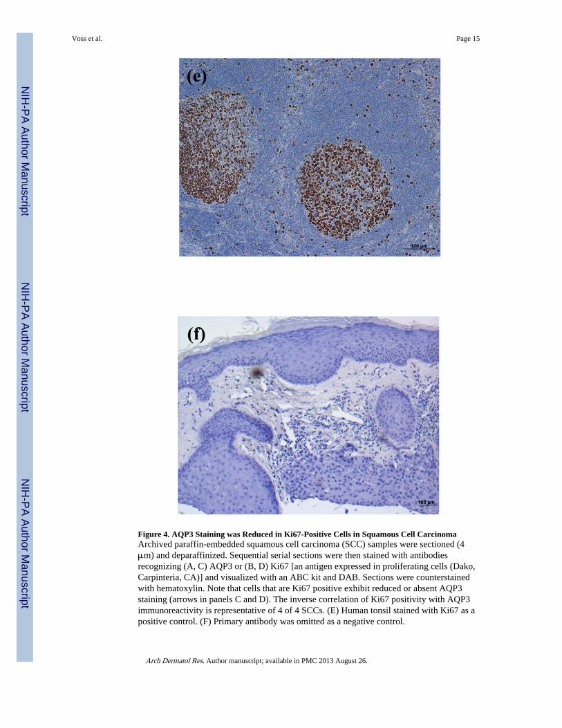
Figure 4. AQP3 Staining was Reduced in Ki67-Positive Cells in Squamous Cell CarcinomaArchived paraffin-embedded squamous cell carcinoma (SCC) samples were sectioned (4μm) and deparaffinized. Sequential serial sections were then stained with antibodiesrecognizing (A, C) AQP3 or (B, D) Ki67 [an antigen expressed in proliferating cells (Dako,Carpinteria, CA)] and visualized with an ABC kit and DAB. Sections were counterstainedwith hematoxylin. Note that cells that are Ki67 positive exhibit reduced or absent AQP3staining (arrows in panels C and D). The inverse correlation of Ki67 positivity with AQP3immunoreactivity is representative of 4 of 4 SCCs. (E) Human tonsil stained with Ki67 as apositive control. (F) Primary antibody was omitted as a negative control.
Voss et al. Page 15
Arch Dermatol Res. Author manuscript; available in PMC 2013 August 26.
NIH
-PA Author Manuscript
NIH
-PA Author Manuscript
NIH
-PA Author Manuscript

Voss et al. Page 16
Arch Dermatol Res. Author manuscript; available in PMC 2013 August 26.
NIH
-PA Author Manuscript
NIH
-PA Author Manuscript
NIH
-PA Author Manuscript

Figure 5. AQP3 Immunoreactivity was Mislocalized in a Majority of Psoriatic Lesions(A through C) Archived paraffin-embedded psoriasis samples were sectioned (4 μm) anddeparaffinized. Sections were then stained with an antibody recognizing AQP3 andvisualized with an ABC kit and DAB. Results from three patients are shown and the stainingpattern is representative of 8 of 10 psoriatic samples examined. (D) A negative control wasperformed by omitting primary antibody. Note that AQP3 is localized to the cytoplasmrather than to the plasma membrane in a majority of the psoriatic epidermis. All sectionswere counterstained with hematoxylin.
Voss et al. Page 17
Arch Dermatol Res. Author manuscript; available in PMC 2013 August 26.
NIH
-PA Author Manuscript
NIH
-PA Author Manuscript
NIH
-PA Author Manuscript

Figure 6. PLD2 Immunoreactivity was Localized Largely to the Cell Periphery in NormalEpidermis (as in Normal-appearing Epidermis Overlying Basal Cell Carcinoma)Archived paraffin-embedded normal human epidermal samples were sectioned (4 μm) anddeparaffinized. Sections were then stained with an antibody recognizing PLD2 andvisualized with an ABC kit and DAB. Note that PLD2 staining localizes to the cellperiphery, as observed also in normal-appearing overlying epidermis in basal cell carcinoma(Figure 8). PLD2 staining of three patients is illustrated in panels A through C, and panel Dshows a negative control in which the primary antibody was omitted.
Voss et al. Page 18
Arch Dermatol Res. Author manuscript; available in PMC 2013 August 26.
NIH
-PA Author Manuscript
NIH
-PA Author Manuscript
NIH
-PA Author Manuscript
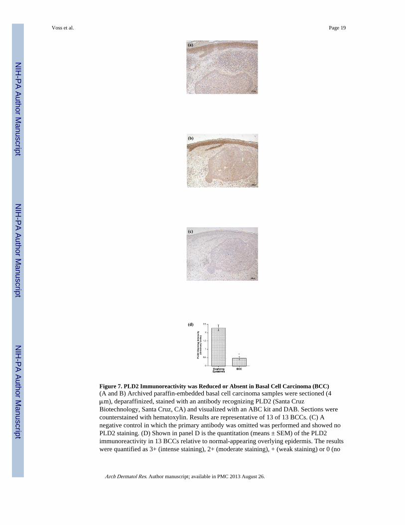
Figure 7. PLD2 Immunoreactivity was Reduced or Absent in Basal Cell Carcinoma (BCC)(A and B) Archived paraffin-embedded basal cell carcinoma samples were sectioned (4μm), deparaffinized, stained with an antibody recognizing PLD2 (Santa CruzBiotechnology, Santa Cruz, CA) and visualized with an ABC kit and DAB. Sections werecounterstained with hematoxylin. Results are representative of 13 of 13 BCCs. (C) Anegative control in which the primary antibody was omitted was performed and showed noPLD2 staining. (D) Shown in panel D is the quantitation (means ± SEM) of the PLD2immunoreactivity in 13 BCCs relative to normal-appearing overlying epidermis. The resultswere quantified as 3+ (intense staining), 2+ (moderate staining), + (weak staining) or 0 (no
Voss et al. Page 19
Arch Dermatol Res. Author manuscript; available in PMC 2013 August 26.
NIH
-PA Author Manuscript
NIH
-PA Author Manuscript
NIH
-PA Author Manuscript

staining) by three independent observers, the values averaged and the data analyzed by anunpaired Student's t-test as described in Methods; *p<0.05.
Voss et al. Page 20
Arch Dermatol Res. Author manuscript; available in PMC 2013 August 26.
NIH
-PA Author Manuscript
NIH
-PA Author Manuscript
NIH
-PA Author Manuscript

Figure 8. PLD2 Immunoreactivity Seemed Aberrant in Psoriatic Lesions(A through C) Archived paraffin-embedded human epidermal samples from psoriaticpatients were sectioned (4 μm) and deparaffinized. Sections were then stained with anantibody recognizing PLD2 and visualized with an ABC kit and DAB. Note that PLD2staining was aberrant (extremely weak) in 8 of 10 psoriasis samples. (D) Immunoreactivityin 1 of the 2 psoriatic samples with essentially normal PLD2 staining is illustrated. Allsections were counterstained with hematoxylin (blue staining).
Voss et al. Page 21
Arch Dermatol Res. Author manuscript; available in PMC 2013 August 26.
NIH
-PA Author Manuscript
NIH
-PA Author Manuscript
NIH
-PA Author Manuscript
Copyright © 2022 FDOKUMEN


![Menschenhaut [Human skin]](https://static.fdokumen.com/doc/165x107/6326d24f24adacd7250b1364/menschenhaut-human-skin.jpg)