AAV Vector-Mediated Microdystrophin Expression in a Relatively Small Percentage of mdx Myofibers...
8
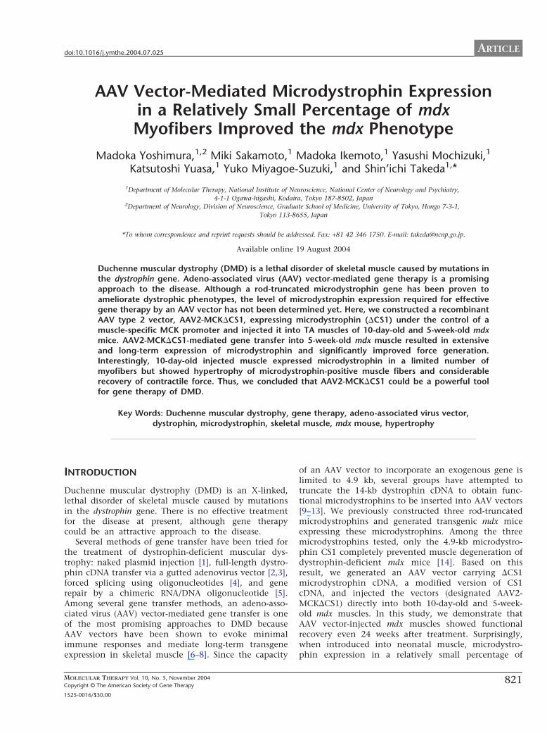
AAV Vector-Mediated Microdystrophin Expression in a Relatively Small Percentage of mdx Myofibers Improved the mdx Phenotype Madoka Yoshimura, 1,2 Miki Sakamoto, 1 Madoka Ikemoto, 1 Yasushi Mochizuki, 1 Katsutoshi Yuasa, 1 Yuko Miyagoe-Suzuki, 1 and Shin’ichi Takeda 1, * 1 Department of Molecular Therapy, National Institute of Neuroscience, National Center of Neurology and Psychiatry, 4-1-1 Ogawa-higashi, Kodaira, Tokyo 187-8502, Japan 2 Department of Neurology, Division of Neuroscience, Graduate School of Medicine, University of Tokyo, Hongo 7-3-1, Tokyo 113-8655, Japan *To whom correspondence and reprint requests should be addressed. Fax: +81 42 346 1750. E-mail: [email protected]. Available online 19 August 2004 Duchenne muscular dystrophy (DMD) is a lethal disorder of skeletal muscle caused by mutations in the dystrophin gene. Adeno-associated virus (AAV) vector-mediated gene therapy is a promising approach to the disease. Although a rod-truncated microdystrophin gene has been proven to ameliorate dystrophic phenotypes, the level of microdystrophin expression required for effective gene therapy by an AAV vector has not been determined yet. Here, we constructed a recombinant AAV type 2 vector, AAV2-MCKDCS1, expressing microdystrophin (DCS1) under the control of a muscle-specific MCK promoter and injected it into TA muscles of 10-day-old and 5-week-old mdx mice. AAV2-MCKDCS1-mediated gene transfer into 5-week-old mdx muscle resulted in extensive and long-term expression of microdystrophin and significantly improved force generation. Interestingly, 10-day-old injected muscle expressed microdystrophin in a limited number of myofibers but showed hypertrophy of microdystrophin-positive muscle fibers and considerable recovery of contractile force. Thus, we concluded that AAV2-MCKDCS1 could be a powerful tool for gene therapy of DMD. Key Words: Duchenne muscular dystrophy, gene therapy, adeno-associated virus vector, dystrophin, microdystrophin, skeletal muscle, mdx mouse, hypertrophy INTRODUCTION Duchenne muscular dystrophy (DMD) is an X-linked, lethal disorder of skeletal muscle caused by mutations in the dystrophin gene. There is no effective treatment for the disease at present, although gene therapy could be an attractive approach to the disease. Several methods of gene transfer have been tried for the treatment of dystrophin-deficient muscular dys- trophy: naked plasmid injection [1], full-length dystro- phin cDNA transfer via a gutted adenovirus vector [2,3], forced splicing using oligonucleotides [4], and gene repair by a chimeric RNA/DNA oligonucleotide [5]. Among several gene transfer methods, an adeno-asso- ciated virus (AAV) vector-mediated gene transfer is one of the most promising approaches to DMD because AAV vectors have been shown to evoke minimal immune responses and mediate long-term transgene expression in skeletal muscle [6–8]. Since the capacity of an AAV vector to incorporate an exogenous gene is limited to 4.9 kb, several groups have attempted to truncate the 14-kb dystrophin cDNA to obtain func- tional microdystrophins to be inserted into AAV vectors [9–13]. We previously constructed three rod-truncated microdystrophins and generated transgenic mdx mice expressing these microdystrophins. Among the three microdystrophins tested, only the 4.9-kb microdystro- phin CS1 completely prevented muscle degeneration of dystrophin-deficient mdx mice [14]. Based on this result, we generated an AAV vector carrying DCS1 microdystrophin cDNA, a modified version of CS1 cDNA, and injected the vectors (designated AAV2- MCKDCS1) directly into both 10-day-old and 5-week- old mdx muscles. In this study, we demonstrate that AAV vector-injected mdx muscles showed functional recovery even 24 weeks after treatment. Surprisingly, when introduced into neonatal muscle, microdystro- phin expression in a relatively small percentage of ARTICLE doi:10.1016/j.ymthe.2004.07.025 MOLECULAR THERAPY Vol. 10, No. 5, November 2004 821 Copyright C The American Society of Gene Therapy 1525-0016/$30.00
-
Upload
independent -
Category
Documents
-
view
3 -
download
0
Transcript of AAV Vector-Mediated Microdystrophin Expression in a Relatively Small Percentage of mdx Myofibers...
ARTICLEdoi:10.1016/j.ymthe.2004.07.025
AAV Vector-Mediated Microdystrophin Expressionin a Relatively Small Percentage of mdxMyofibers Improved the mdx Phenotype
Madoka Yoshimura,1,2 Miki Sakamoto,1 Madoka Ikemoto,1 Yasushi Mochizuki,1
Katsutoshi Yuasa,1 Yuko Miyagoe-Suzuki,1 and Shin’ichi Takeda1,*
1Department of Molecular Therapy, National Institute of Neuroscience, National Center of Neurology and Psychiatry,
4-1-1 Ogawa-higashi, Kodaira, Tokyo 187-8502, Japan2Department of Neurology, Division of Neuroscience, Graduate School of Medicine, University of Tokyo, Hongo 7-3-1,
Tokyo 113-8655, Japan
*To whom correspondence and reprint requests should be addressed. Fax: +81 42 346 1750. E-mail: [email protected].
Available online 19 August 2004
MOLECULA
Copyright C
1525-0016/$
Duchenne muscular dystrophy (DMD) is a lethal disorder of skeletal muscle caused by mutations inthe dystrophin gene. Adeno-associated virus (AAV) vector-mediated gene therapy is a promisingapproach to the disease. Although a rod-truncated microdystrophin gene has been proven toameliorate dystrophic phenotypes, the level of microdystrophin expression required for effectivegene therapy by an AAV vector has not been determined yet. Here, we constructed a recombinantAAV type 2 vector, AAV2-MCKDCS1, expressing microdystrophin (DCS1) under the control of amuscle-specific MCK promoter and injected it into TA muscles of 10-day-old and 5-week-old mdxmice. AAV2-MCKDCS1-mediated gene transfer into 5-week-old mdx muscle resulted in extensiveand long-term expression of microdystrophin and significantly improved force generation.Interestingly, 10-day-old injected muscle expressed microdystrophin in a limited number ofmyofibers but showed hypertrophy of microdystrophin-positive muscle fibers and considerablerecovery of contractile force. Thus, we concluded that AAV2-MCKDCS1 could be a powerful toolfor gene therapy of DMD.
R
Th
30
Key Words: Duchenne muscular dystrophy, gene therapy, adeno-associated virus vector,dystrophin, microdystrophin, skeletal muscle, mdx mouse, hypertrophy
of an AAV vector to incorporate an exogenou
INTRODUCTIONDuchenne muscular dystrophy (DMD) is an X-linked,lethal disorder of skeletal muscle caused by mutationsin the dystrophin gene. There is no effective treatmentfor the disease at present, although gene therapycould be an attractive approach to the disease.
Several methods of gene transfer have been tried forthe treatment of dystrophin-deficient muscular dys-trophy: naked plasmid injection [1], full-length dystro-phin cDNA transfer via a gutted adenovirus vector [2,3],forced splicing using oligonucleotides [4], and generepair by a chimeric RNA/DNA oligonucleotide [5].Among several gene transfer methods, an adeno-asso-ciated virus (AAV) vector-mediated gene transfer is oneof the most promising approaches to DMD becauseAAV vectors have been shown to evoke minimalimmune responses and mediate long-term transgeneexpression in skeletal muscle [6–8]. Since the capacity
THERAPY Vol. 10, No. 5, November 2004
e American Society of Gene Therapy
.00
s gene islimited to 4.9 kb, several groups have attempted totruncate the 14-kb dystrophin cDNA to obtain func-tional microdystrophins to be inserted into AAV vectors[9–13]. We previously constructed three rod-truncatedmicrodystrophins and generated transgenic mdx miceexpressing these microdystrophins. Among the threemicrodystrophins tested, only the 4.9-kb microdystro-phin CS1 completely prevented muscle degeneration ofdystrophin-deficient mdx mice [14]. Based on thisresult, we generated an AAV vector carrying DCS1microdystrophin cDNA, a modified version of CS1cDNA, and injected the vectors (designated AAV2-MCKDCS1) directly into both 10-day-old and 5-week-old mdx muscles. In this study, we demonstrate thatAAV vector-injected mdx muscles showed functionalrecovery even 24 weeks after treatment. Surprisingly,when introduced into neonatal muscle, microdystro-phin expression in a relatively small percentage of
821
ARTICLE doi:10.1016/j.ymthe.2004.07.025
muscle fibers dramatically improved the contractileproperties of dystrophic muscle. This improvement incontractile force was thought to be achieved by hyper-trophied microdystrophin-positive muscle fibers.
RESULTS
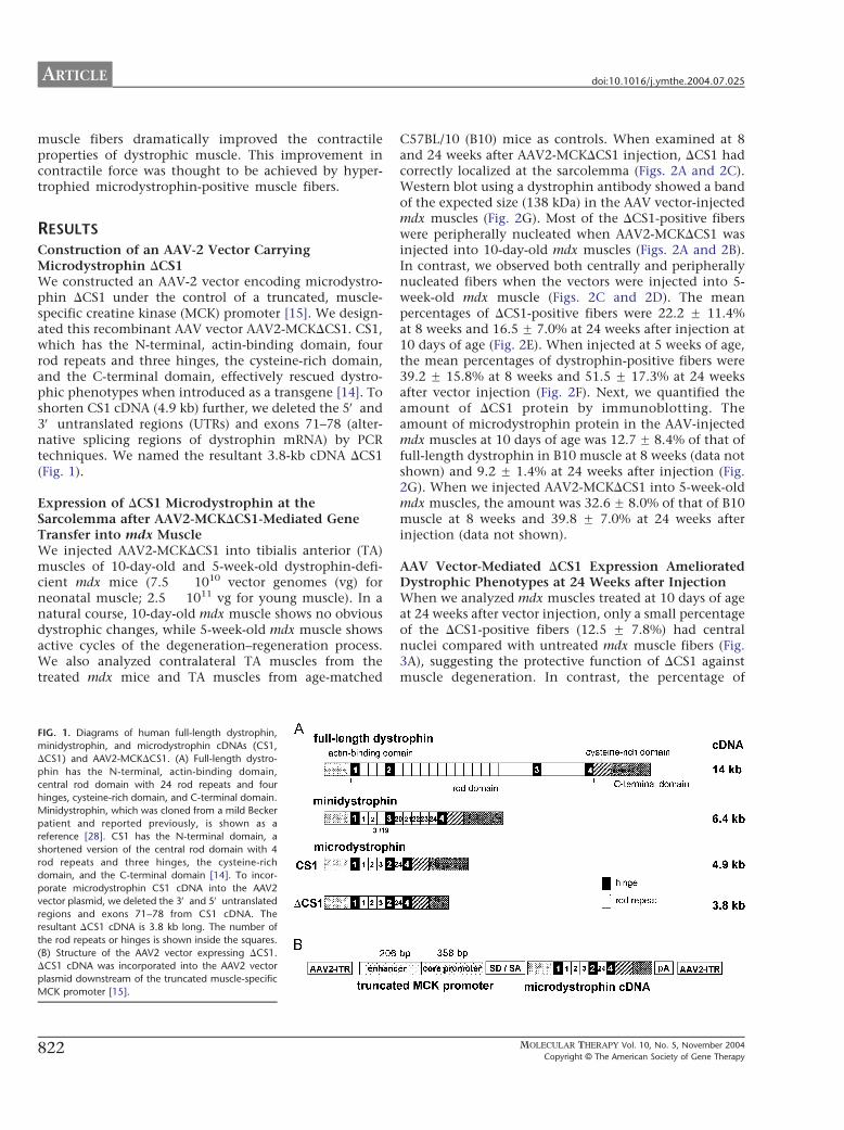
Construction of an AAV-2 Vector CarryingMicrodystrophin DCS1We constructed an AAV-2 vector encoding microdystro-phin DCS1 under the control of a truncated, muscle-specific creatine kinase (MCK) promoter [15]. We design-ated this recombinant AAV vector AAV2-MCKDCS1. CS1,which has the N-terminal, actin-binding domain, fourrod repeats and three hinges, the cysteine-rich domain,and the C-terminal domain, effectively rescued dystro-phic phenotypes when introduced as a transgene [14]. Toshorten CS1 cDNA (4.9 kb) further, we deleted the 5V and3V untranslated regions (UTRs) and exons 71–78 (alter-native splicing regions of dystrophin mRNA) by PCRtechniques. We named the resultant 3.8-kb cDNA DCS1(Fig. 1).
Expression of DCS1 Microdystrophin at theSarcolemma after AAV2-MCKDCS1-Mediated GeneTransfer into mdx MuscleWe injected AAV2-MCKDCS1 into tibialis anterior (TA)muscles of 10-day-old and 5-week-old dystrophin-defi-cient mdx mice (7.5 � 1010 vector genomes (vg) forneonatal muscle; 2.5 � 1011 vg for young muscle). In anatural course, 10-day-old mdx muscle shows no obviousdystrophic changes, while 5-week-old mdx muscle showsactive cycles of the degeneration–regeneration process.We also analyzed contralateral TA muscles from thetreated mdx mice and TA muscles from age-matched
FIG. 1. Diagrams of human full-length dystrophin,
minidystrophin, and microdystrophin cDNAs (CS1,
DCS1) and AAV2-MCKDCS1. (A) Full-length dystro-
phin has the N-terminal, actin-binding domain,
central rod domain with 24 rod repeats and four
hinges, cysteine-rich domain, and C-terminal domain.
Minidystrophin, which was cloned from a mild Becker
patient and reported previously, is shown as a
reference [28]. CS1 has the N-terminal domain, a
shortened version of the central rod domain with 4
rod repeats and three hinges, the cysteine-rich
domain, and the C-terminal domain [14]. To incor-
porate microdystrophin CS1 cDNA into the AAV2
vector plasmid, we deleted the 3V and 5V untranslated
regions and exons 71–78 from CS1 cDNA. The
resultant DCS1 cDNA is 3.8 kb long. The number of
the rod repeats or hinges is shown inside the squares.
(B) Structure of the AAV2 vector expressing DCS1.
DCS1 cDNA was incorporated into the AAV2 vector
plasmid downstream of the truncated muscle-specific
MCK promoter [15].
822
C57BL/10 (B10) mice as controls. When examined at 8and 24 weeks after AAV2-MCKDCS1 injection, DCS1 hadcorrectly localized at the sarcolemma (Figs. 2A and 2C).Western blot using a dystrophin antibody showed a bandof the expected size (138 kDa) in the AAV vector-injectedmdx muscles (Fig. 2G). Most of the DCS1-positive fiberswere peripherally nucleated when AAV2-MCKDCS1 wasinjected into 10-day-old mdx muscles (Figs. 2A and 2B).In contrast, we observed both centrally and peripherallynucleated fibers when the vectors were injected into 5-week-old mdx muscle (Figs. 2C and 2D). The meanpercentages of DCS1-positive fibers were 22.2 F 11.4%at 8 weeks and 16.5 F 7.0% at 24 weeks after injection at10 days of age (Fig. 2E). When injected at 5 weeks of age,the mean percentages of dystrophin-positive fibers were39.2 F 15.8% at 8 weeks and 51.5 F 17.3% at 24 weeksafter vector injection (Fig. 2F). Next, we quantified theamount of DCS1 protein by immunoblotting. Theamount of microdystrophin protein in the AAV-injectedmdx muscles at 10 days of age was 12.7 F 8.4% of that offull-length dystrophin in B10 muscle at 8 weeks (data notshown) and 9.2 F 1.4% at 24 weeks after injection (Fig.2G). When we injected AAV2-MCKDCS1 into 5-week-oldmdx muscles, the amount was 32.6 F 8.0% of that of B10muscle at 8 weeks and 39.8 F 7.0% at 24 weeks afterinjection (data not shown).
AAV Vector-Mediated DCS1 Expression AmelioratedDystrophic Phenotypes at 24 Weeks after InjectionWhen we analyzed mdx muscles treated at 10 days of ageat 24 weeks after vector injection, only a small percentageof the DCS1-positive fibers (12.5 F 7.8%) had centralnuclei compared with untreated mdx muscle fibers (Fig.3A), suggesting the protective function of DCS1 againstmuscle degeneration. In contrast, the percentage of
MOLECULAR THERAPY Vol. 10, No. 5, November 2004
Copyright C The American Society of Gene Therapy
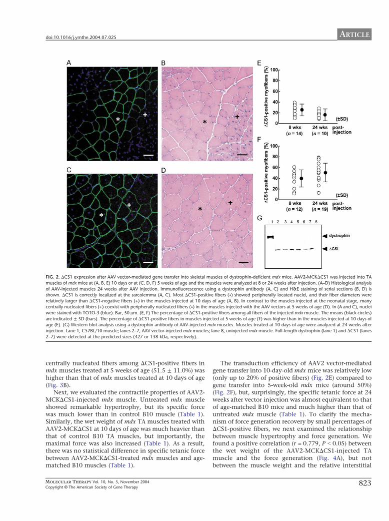
FIG. 2. DCS1 expression after AAV vector-mediated gene transfer into skeletal muscles of dystrophin-deficient mdx mice. AAV2-MCKDCS1 was injected into TA
muscles of mdx mice at (A, B, E) 10 days or at (C, D, F) 5 weeks of age and the muscles were analyzed at 8 or 24 weeks after injection. (A–D) Histological analysis
of AAV-injected muscles 24 weeks after AAV injection. Immunofluorescence using a dystrophin antibody (A, C) and H&E staining of serial sections (B, D) is
shown. DCS1 is correctly localized at the sarcolemma (A, C). Most DCS1-positive fibers (*) showed peripherally located nuclei, and their fiber diameters were
relatively larger than DCS1-negative fibers (+) in the muscles injected at 10 days of age (A, B). In contrast to the muscles injected at the neonatal stage, many
centrally nucleated fibers (+) coexist with peripherally nucleated fibers (*) in the muscles injected with the AAV vectors at 5 weeks of age (D). In (A and C), nuclei
were stained with TOTO-3 (blue). Bar, 50 Am. (E, F) The percentage of DCS1-positive fibers among all fibers of the injected mdx muscle. The means (black circles)
are indicated F SD (bars). The percentage of DCS1-positive fibers in muscles injected at 5 weeks of age (F) was higher than in the muscles injected at 10 days of
age (E). (G) Western blot analysis using a dystrophin antibody of AAV-injected mdx muscles. Muscles treated at 10 days of age were analyzed at 24 weeks after
injection. Lane 1, C57BL/10 muscle; lanes 2–7, AAV vector-injected mdx muscles; lane 8, uninjected mdx muscle. Full-length dystrophin (lane 1) and DCS1 (lanes
2–7) were detected at the predicted sizes (427 or 138 kDa, respectively).
ARTICLEdoi:10.1016/j.ymthe.2004.07.025
centrally nucleated fibers among DCS1-positive fibers inmdx muscles treated at 5 weeks of age (51.5 F 11.0%) washigher than that of mdx muscles treated at 10 days of age(Fig. 3B).
Next, we evaluated the contractile properties of AAV2-MCKDCS1-injected mdx muscle. Untreated mdx muscleshowed remarkable hypertrophy, but its specific forcewas much lower than in control B10 muscle (Table 1).Similarly, the wet weight of mdx TA muscles treated withAAV2-MCKDCS1 at 10 days of age was much heavier thanthat of control B10 TA muscles, but importantly, themaximal force was also increased (Table 1). As a result,there was no statistical difference in specific tetanic forcebetween AAV2-MCKDCS1-treated mdx muscles and age-matched B10 muscles (Table 1).
MOLECULAR THERAPY Vol. 10, No. 5, November 2004
Copyright C The American Society of Gene Therapy
The transduction efficiency of AAV2 vector-mediatedgene transfer into 10-day-old mdx mice was relatively low(only up to 20% of positive fibers) (Fig. 2E) compared togene transfer into 5-week-old mdx mice (around 50%)(Fig. 2F), but, surprisingly, the specific tetanic force at 24weeks after vector injection was almost equivalent to thatof age-matched B10 mice and much higher than that ofuntreated mdx muscle (Table 1). To clarify the mecha-nism of force generation recovery by small percentages ofDCS1-positive fibers, we next examined the relationshipbetween muscle hypertrophy and force generation. Wefound a positive correlation (r = 0.779, P b 0.05) betweenthe wet weight of the AAV2-MCKDCS1-injected TAmuscle and the force generation (Fig. 4A), but notbetween the muscle weight and the relative interstitial
823
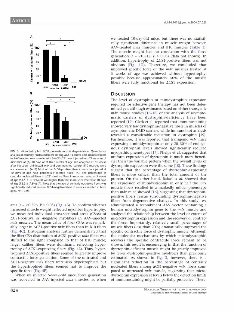
FIG. 3. Microdystrophin DCS1 prevents muscle degeneration. Quantitative
analysis of centrally nucleated fibers among DCS1-positive and -negative fiber
in AAV-injected mdx muscle. AAV2-MCKDCS1 was injected into TA muscles o
mdx mice at (A) 10 days or at (B) 5 weeks of age and analyzed at 24 week
after injection. Uninjected mdx and age-matched control B10 muscles were
also examined. (A, B) Most of the DCS1-positive fibers in muscles injected a
10 days of age have peripherally located nuclei (A). The percentage o
centrally nucleated fibers in DCS1-positive fibers in muscles treated at 5 week
of age (51.5 F 11.0%) (B) was higher than that in muscles treated at 10 day
of age (12.5 F 7.8%) (A). Note that the ratio of centrally nucleated fibers wa
significantly reduced even in DCS1-negative fibers in muscles injected at both
ages. *P b 0.01.
ARTICLE doi:10.1016/j.ymthe.2004.07.025
824
s
f
s
t
f
s
s
s
area (r = �0.596, P N 0.05) (Fig. 4B). To confirm whetherincreased muscle weight reflected myofiber hypertrophy,we measured individual cross-sectional areas (CSAs) ofDCS1-positive or -negative myofibers in AAV-injectedmdx muscles. The mean value of fiber CSAs was remark-ably larger in DCS1-positive mdx fibers than in B10 fibers(Fig. 4C). Histogram analysis further demonstrated thatthe fiber CSA distribution of DCS1-positive mdx fibers wasshifted to the right compared to that of B10 muscle;larger caliber fibers were dominant, reflecting hyper-trophy of DCS1-expressing fibers (Fig. 4E). Thus, hyper-trophied DCS1-positive fibers seemed to greatly improvecontractile force generation. Some of the untreated andDCS1-negative mdx fibers were also hypertrophied, butthe hypertrophied fibers seemed not to improve thespecific force (Fig. 4E).
When we injected 5-week-old mice, force generationwas recovered in AAV-injected mdx muscles, as when
we treated 10-day-old mice, but there was no statisti-cally significant difference in muscle weight betweenAAV-treated mdx muscles and B10 muscles (Table 1).The muscle weight had no correlation with the forcegeneration (r = �0.512, P N 0.05) (data not shown). Inaddition, hypertrophy of DCS1-positive fibers was notobvious (Fig. 4D). Therefore, we concluded thatimproved specific force of the mdx muscles treated at5 weeks of age was achieved without hypertrophy,possibly because approximately 50% of the musclefibers were fully functional for DCS1 expression.
DISCUSSION
The level of dystrophin or minidystrophin expressionrequired for effective gene therapy has not been deter-mined yet, although estimates based on either transgenicmdx mouse studies [16–18] or the analysis of asympto-matic carriers of dystrophin-deficiency have beenreported [19]. Clerk et al. reported that immunostainingshowed very few dystrophin-negative fibers in muscles ofasymptomatic DMD carriers, while immunoblot analysisrevealed a considerable reduction in dystrophin [19].Furthermore, it was reported that transgenic mdx miceexpressing a minidystrophin at only 20–30% of endoge-nous dystrophin levels showed significantly reducedmyopathic phenotypes [17]. Phelps et al. suggested thatuniform expression of dystrophin is much more benefi-cial than the variable pattern when the overall levels ofdystrophin expression were the same [18]. These findingssuggest that the percentage of dystrophin-expressingfibers is more critical than the total amount of theprotein. On the other hand, Rafael et al. showed thatthe expression of minidystrophin in only half the mdxmuscle fibers resulted in a markedly milder phenotypethan mdx mice showed [16], suggesting that dystrophin-positive fibers rescue surrounding dystrophin-negativefibers from degenerative changes. In this study, weadministrated a recombinant AAV vector containing ahuman microdystrophin gene to the mdx muscle andanalyzed the relationship between the level or extent ofmicrodystrophin expression and the recovery of contrac-tile force. Importantly, relatively small percentages ofmuscle fibers (less than 20%) dramatically improved thespecific contractile force of dystrophic muscle. Althoughthe molecular mechanisms by which microdystrophinrecovers the specific contractile force remain to beshown, this result is encouraging in that the function ofdystrophin-deficient muscle might be greatly improvedby fewer dystrophin-positive myofibers than previouslyestimated. As shown in Fig. 3, however, there is asignificant reduction in the percentage of centrallynucleated fibers among DCS1-negative mdx fibers com-pared to untreated mdx muscle, suggesting that micro-dystrophin expression at levels below the detection limitsof immunostaining might be partially protective. There-
MOLECULAR THERAPY Vol. 10, No. 5, November 2004
Copyright C The American Society of Gene Therapy
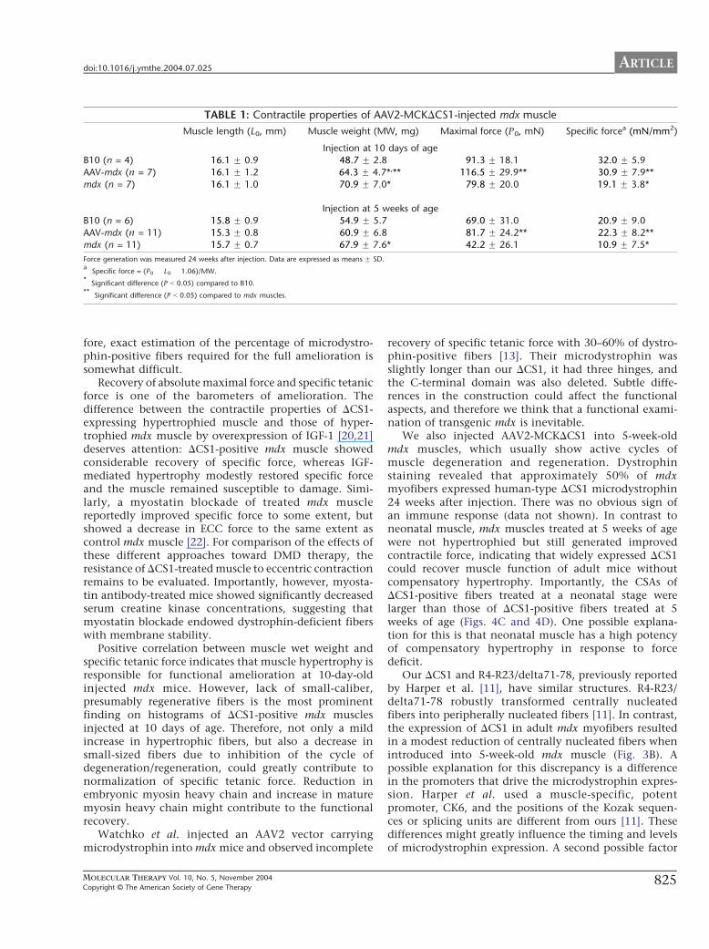
TABLE 1: Contractile properties of AAV2-MCKDCS1-injected mdx muscle
Muscle length (L0, mm) Muscle weight (MW, mg) Maximal force (P0, mN) Specific forcea (mN/mm2)
Injection at 10 days of age
B10 (n = 4) 16.1 F 0.9 48.7 F 2.8 91.3 F 18.1 32.0 F 5.9
AAV-mdx (n = 7) 16.1 F 1.2 64.3 F 4.7*,** 116.5 F 29.9** 30.9 F 7.9**
mdx (n = 7) 16.1 F 1.0 70.9 F 7.0* 79.8 F 20.0 19.1 F 3.8*
Injection at 5 weeks of age
B10 (n = 6) 15.8 F 0.9 54.9 F 5.7 69.0 F 31.0 20.9 F 9.0
AAV-mdx (n = 11) 15.3 F 0.8 60.9 F 6.8 81.7 F 24.2** 22.3 F 8.2**mdx (n = 11) 15.7 F 0.7 67.9 F 7.6* 42.2 F 26.1 10.9 F 7.5*
Force generation was measured 24 weeks after injection. Data are expressed as means F SD.a
Specific force = (P0 � L0 � 1.06)/MW.*
Significant difference (P b 0.05) compared to B10.**
Significant difference (P b 0.05) compared to mdx muscles.
ARTICLEdoi:10.1016/j.ymthe.2004.07.025
fore, exact estimation of the percentage of microdystro-phin-positive fibers required for the full amelioration issomewhat difficult.
Recovery of absolute maximal force and specific tetanicforce is one of the barometers of amelioration. Thedifference between the contractile properties of DCS1-expressing hypertrophied muscle and those of hyper-trophied mdx muscle by overexpression of IGF-1 [20,21]deserves attention: DCS1-positive mdx muscle showedconsiderable recovery of specific force, whereas IGF-mediated hypertrophy modestly restored specific forceand the muscle remained susceptible to damage. Simi-larly, a myostatin blockade of treated mdx musclereportedly improved specific force to some extent, butshowed a decrease in ECC force to the same extent ascontrol mdx muscle [22]. For comparison of the effects ofthese different approaches toward DMD therapy, theresistance of DCS1-treated muscle to eccentric contractionremains to be evaluated. Importantly, however, myosta-tin antibody-treated mice showed significantly decreasedserum creatine kinase concentrations, suggesting thatmyostatin blockade endowed dystrophin-deficient fiberswith membrane stability.
Positive correlation between muscle wet weight andspecific tetanic force indicates that muscle hypertrophy isresponsible for functional amelioration at 10-day-oldinjected mdx mice. However, lack of small-caliber,presumably regenerative fibers is the most prominentfinding on histograms of DCS1-positive mdx musclesinjected at 10 days of age. Therefore, not only a mildincrease in hypertrophic fibers, but also a decrease insmall-sized fibers due to inhibition of the cycle ofdegeneration/regeneration, could greatly contribute tonormalization of specific tetanic force. Reduction inembryonic myosin heavy chain and increase in maturemyosin heavy chain might contribute to the functionalrecovery.
Watchko et al. injected an AAV2 vector carryingmicrodystrophin into mdx mice and observed incomplete
MOLECULAR THERAPY Vol. 10, No. 5, November 2004
Copyright C The American Society of Gene Therapy
recovery of specific tetanic force with 30–60% of dystro-phin-positive fibers [13]. Their microdystrophin wasslightly longer than our DCS1, it had three hinges, andthe C-terminal domain was also deleted. Subtle diffe-rences in the construction could affect the functionalaspects, and therefore we think that a functional exami-nation of transgenic mdx is inevitable.
We also injected AAV2-MCKDCS1 into 5-week-oldmdx muscles, which usually show active cycles ofmuscle degeneration and regeneration. Dystrophinstaining revealed that approximately 50% of mdxmyofibers expressed human-type DCS1 microdystrophin24 weeks after injection. There was no obvious sign ofan immune response (data not shown). In contrast toneonatal muscle, mdx muscles treated at 5 weeks of agewere not hypertrophied but still generated improvedcontractile force, indicating that widely expressed DCS1could recover muscle function of adult mice withoutcompensatory hypertrophy. Importantly, the CSAs ofDCS1-positive fibers treated at a neonatal stage werelarger than those of DCS1-positive fibers treated at 5weeks of age (Figs. 4C and 4D). One possible explana-tion for this is that neonatal muscle has a high potencyof compensatory hypertrophy in response to forcedeficit.
Our DCS1 and R4-R23/delta71-78, previously reportedby Harper et al. [11], have similar structures. R4-R23/delta71-78 robustly transformed centrally nucleatedfibers into peripherally nucleated fibers [11]. In contrast,the expression of DCS1 in adult mdx myofibers resultedin a modest reduction of centrally nucleated fibers whenintroduced into 5-week-old mdx muscle (Fig. 3B). Apossible explanation for this discrepancy is a differencein the promoters that drive the microdystrophin expres-sion. Harper et al. used a muscle-specific, potentpromoter, CK6, and the positions of the Kozak sequen-ces or splicing units are different from ours [11]. Thesedifferences might greatly influence the timing and levelsof microdystrophin expression. A second possible factor
825
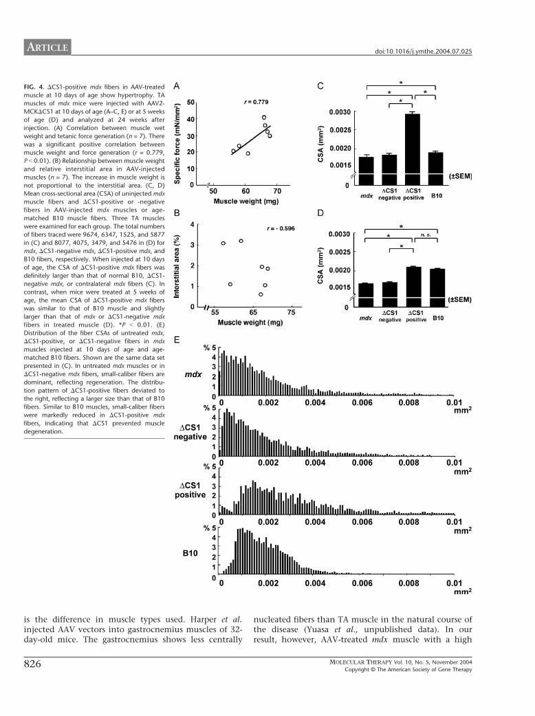
FIG. 4. DCS1-positive mdx fibers in AAV-treated
muscle at 10 days of age show hypertrophy. TA
muscles of mdx mice were injected with AAV2-
MCKDCS1 at 10 days of age (A–C, E) or at 5 weeks
of age (D) and analyzed at 24 weeks after
injection. (A) Correlation between muscle wet
weight and tetanic force generation (n = 7). There
was a significant positive correlation between
muscle weight and force generation (r = 0.779,
P b 0.01). (B) Relationship between muscle weight
and relative interstitial area in AAV-injected
muscles (n = 7). The increase in muscle weight is
not proportional to the interstitial area. (C, D)
Mean cross-sectional area (CSA) of uninjected mdx
muscle fibers and DCS1-positive or -negative
fibers in AAV-injected mdx muscles or age-
matched B10 muscle fibers. Three TA muscles
were examined for each group. The total numbers
of fibers traced were 9674, 6347, 1525, and 5877
in (C) and 8077, 4075, 3479, and 5476 in (D) for
mdx, DCS1-negative mdx, DCS1-positive mdx, and
B10 fibers, respectively. When injected at 10 days
of age, the CSA of DCS1-positive mdx fibers was
definitely larger than that of normal B10, DCS1-
negative mdx, or contralateral mdx fibers (C). In
contrast, when mice were treated at 5 weeks of
age, the mean CSA of DCS1-positive mdx fibers
was similar to that of B10 muscle and slightly
larger than that of mdx or DCS1-negative mdx
fibers in treated muscle (D). *P b 0.01. (E)
Distribution of the fiber CSAs of untreated mdx,
DCS1-positive, or DCS1-negative fibers in mdx
muscles injected at 10 days of age and age-
matched B10 fibers. Shown are the same data set
presented in (C). In untreated mdx muscles or in
DCS1-negative mdx fibers, small-caliber fibers are
dominant, reflecting regeneration. The distribu-
tion pattern of DCS1-positive fibers deviated to
the right, reflecting a larger size than that of B10
fibers. Similar to B10 muscles, small-caliber fibers
were markedly reduced in DCS1-positive mdx
fibers, indicating that DCS1 prevented muscle
degeneration.
ARTICLE doi:10.1016/j.ymthe.2004.07.025
is the difference in muscle types used. Harper et al.injected AAV vectors into gastrocnemius muscles of 32-day-old mice. The gastrocnemius shows less centrally
826
nucleated fibers than TA muscle in the natural course ofthe disease (Yuasa et al., unpublished data). In ourresult, however, AAV-treated mdx muscle with a high
MOLECULAR THERAPY Vol. 10, No. 5, November 2004
Copyright C The American Society of Gene Therapy
ARTICLEdoi:10.1016/j.ymthe.2004.07.025
percentage of centrally nucleated fibers (50%) did notshow dystrophic dysfunction, e.g., decreased force gene-ration. Therefore, centrally nucleated fibers do notdisprove the protective function of DCS1. The percent-age of centrally nucleated fibers would reflect the stateof muscles at the time of injection. Our results areimportant because it is sometimes difficult to start genetherapies before the onset of the clinical course of DMD,and AAV2-MCKDCS1 is expected to show therapeuticeffects even after the onset of disease.
Although we injected high titers of AAV-vectorparticles into the muscle, the transduction efficiencyin 10-day-old mdx muscles was much lower than that in5-week-old mdx muscle. This phenomenon was alsonoticed for the injection of AAV2-CMVLacZ into neo-natal B10 muscle (unpublished results). This differencemight be due to the preferential expression of receptorsor coreceptors for an AAV-2 particle on adult musclefibers. Several molecules, such as heparan sulfate pro-teoglycan [23], aVh5 integrin [24], and dynamin I [25],are proposed to play certain roles in AAV type 2infection, although the expression of these moleculesin muscle fibers during development and aging has notbeen fully determined. Another possibility is dilution ofAAV vectors by rapid growth of neonatal muscle. Whenwe cultured satellite cells prepared from AAV2-CMVLacZ-injected mdx muscle, we observed no bluemyoblasts or myotubes (data not shown). Therefore,proliferation and fusion of nontransduced satellite cells/myoblasts would greatly dilute the microdystrophinprotein.
Our results are promising because AAV vector-medi-ated DCS1 gene transfer had a restorative function fordystrophin-deficient mdx muscle. However, demonstra-tion of the benefits of this gene transfer strategy forhuman DMD patients requires careful testing. Diffe-rences between humans and mice, such as muscle size,life span, or biological properties (especially immuneresponses), should be taken into consideration. A biggeranimal model, e.g., canine X-linked muscular dystrophy[26], will contribute to the preclinical study of genetherapy.
MATERIALS AND METHODS
Constructs of human rod-truncated microdystrophin cDNAs and
generation of AAV vectors expressing microdystrophin. To incorporate
microdystrophin CS1 cDNA (4.9 kb) [14] into an AAV type 2 vector,
we further deleted the 3V and 5V UTRs and exons 71–78 from CS1
cDNA. In brief, DNA fragments of the 5V-terminal and 3V-terminal
regions were independently amplified by PCR to remove exons 71–78
and the 5V and the 3V UTRs and then replace them with corresponding
sequences of CS1 cDNA. The resulting microdystrophin cDNA was 3.8
kb long and designated DCS1. Microdystrophin DCS1 cDNA was then
cloned into an AAV type 2 vector plasmid [15]. The recombinant AAV
vector expressing DCS1 under the control of the truncated muscle-
specific MCK promoter, designated AAV2-MCKDCS1, was then purified
and titrated as previously described [15].
MOLECULAR THERAPY Vol. 10, No. 5, November 2004
Copyright C The American Society of Gene Therapy
Administration of AAV vector to murine skeletal muscle. Fifteen micro-
liters (7.5 � 1010 vg) or 50 Al (2.5 � 1011 vg) of AAV2-MCKDCS1 was
injected directly into the right TA muscles of dystrophin-deficient C57BL/
10 mdx mice at 10 days or 5 weeks of age, respectively. AAV vector-
injected and uninjected mdx muscles and normal muscles of age-matched
C57BL/10 mice were isolated at 8 and 24 weeks after injection.
Contractile properties of AAV2-MCKDCS1-injected TA muscles. Tetanic
force generation was measured and analyzed as described previously with
some modifications [14,27]. The entire TA muscle was removed with its
tibial origin intact, and the distal portion of the TA tendon and its origin
was secured with a 5-O silk suture. The TA was mounted in a vertical tissue
chamber and connected to a force transducer, UL-10GR (Minerva,
Nagano, Japan), and a length servosystem, MM-3 (Narishige, Tokyo,
Japan). Electrical stimulation using a SEN3301 (Nihon Kohden, Tokyo,
Japan) was applied through a pair of platinum wires placed on both sides
of the muscle in physiological soft solution (150 mM NaCl, 4 mM KCl,
2 mM CaCl2, 1 mM MgCl2, 5.6 mM glucose, 5 mM Hepes, pH 7.4, and
0.02 mM d-tubocurarine). Muscle fiber length was adjusted incrementally
by using a micropositioner until peak isometric twitch force responses
were obtained (optimal fiber length (L0)). Maximal tetanic force (P0) was
assessed by stimulation frequencies of 125 pulses/s delivered in 500-ms
duration trains with 2 min intervening between each train. Following two
measurements, the stimulated muscle was weighed after tendon and bone
attachments were removed. All forces were normalized to the physio-
logical cross-sectional area (pCSA), the latter estimated on the basis of the
following formula: muscle wet weight (in mg)/(L0 (in mm) � 1.06 (in mg/
mm3)). The estimated pCSA was used to determine specific tetanic force
(P0/pCSA) of the muscle. After measurement of contractile force, the
muscle was quickly frozen in liquid nitrogen-cooled isopentane for
histopathological analysis.
Histopathological analyses. Histological, immunohistochemical, and
Western blot analyses were performed as described [14]. After blocking
with an M.O.M. kit (Vector Laboratories, Burlingame, CA, USA), dystro-
phin was detected using a monoclonal anti-dystrophin antibody NCL-
DysB (Novocastra, Newcastle, UK; 1:20 dilution) and visualized with
Alexa 488-labeled goat anti-mouse IgG antibody (Molecular Probes,
Eugene, OR, USA) (1:200 dilution). Nuclei were stained with TOTO-3
(Molecular Probes). In some cases, the signal was visualized with
diaminobenzidine and counterstained with hematoxylin. We counted
the number of centrally or peripherally nucleated fibers in dystrophin-
positive or -negative fibers of whole cross sections of TA muscle. In
addition, the CSA of each fiber was measured using an image analysis
system, ImagePro-Plus (Media Cybernetics, Silver Spring, MD, USA). To
evaluate the level of fibrosis, we performed modified Masson trichrome
staining, and the blue-stained area was measured using Image Pro-Plus.
The relative connective tissue area was calculated to the entire muscle
cross-sectional area (%). The signals on immunoblotting were quantitated
using NIH Image.
Statistical analysis. Data were expressed as means F SD or F SEM. If a
significant F ratio was detected by analysis of variance, comparisons
among each group were performed using Fisher’s PLSD. A P value of b0.05
or b0.01 was considered statistically significant. The relation between the
muscle weight and the specific tetanic force was analyzed with Pearson’s
correlation coefficient (P b 0.05).
ACKNOWLEDGMENTS
We are greatly appreciative of Ryoko Nakagawa and Satoru Masuda for their
technical support. We thank Ayako Sakamoto, Kunimasa Arima (Department of
Laboratory Medicine, National Center Hospital for Mental, Nervous and
Muscular Disorders), and Michiko Sagishima (Department of Pathology, School
of Medicine, Teikyo University) for advising on pathological techniques. This
work is supported by Grants-in-Aid from the Center of Excellence, Research on
Nervous and Mental Disorders (10B-1, 13B-1), and Health Sciences Research
Grants for Research on the Human Genome and Gene Therapy (H10-genome-
015, H13-genome-001) from the Ministry of Health, Labor, and Welfare of
827
ARTICLE doi:10.1016/j.ymthe.2004.07.025
Japan, and a Grant-in-Aid for Scientific Research (B) from the Ministry of
Education, Science, Sports, and Culture of Japan.
RECEIVED FOR PUBLICATION FEBRUARY 28, 2004; ACCEPTED JULY 20, 2004.
REFERENCES
1. Acsadi, G., et al. (1991). Human dystrophin expression in mdx mice after intramuscular
injection of DNA constructs. Nature 29: 815 – 818.
2. Gilbert, R., et al. (2001). Dystrophin expression in muscle following gene transfer with
a fully deleted (bguttedQ) adenovirus is markedly improved by trans-acting adenoviral
gene products. Hum. Gene Ther. 20: 1741 – 1755.
3. DelloRusso, C., et al. (2002). Functional correction of adult mdx mouse muscle using
gutted adenoviral vectors expressing full-length dystrophin. Proc. Natl. Acad. Sci. USA
99: 12979 – 12984.
4. Lu, Q. L., et al. (2003). Functional amounts of dystrophin produced by skipping the
mutated exon in the mdx dystrophic mouse. Nat. Med. 9: 1009 – 1014.
5. Bartlett, R. J., et al. (2000). In vivo targeted repair of a point mutation in the
canine dystrophin gene by a chimeric RNA/DNA oligonucleotide. Nat. Biotechnol.
18: 615 – 622.
6. Xiao, X., Li, J., and Samulski, R. J. (1996). Efficient long-term gene transfer into
muscle tissue of immunocompetent mice by adeno-associated virus vector. J. Virol.
70: 8098 – 8108.
7. Kessler, P. D., et al. (1996). Gene delivery to skeletal muscle results in sustained
expression and systemic delivery of a therapeutic protein. Proc. Natl. Acad. Sci. USA 26:
14082 – 14087.
8. Fisher, K. J., et al. (1997). Recombinant adeno-associated virus for muscle directed
gene therapy. Nat. Med. 3: 306 – 312.
9. Yuasa, K., et al. (1998). Effective restoration of dystrophin-associated proteins in
vivo by adenovirus-mediated transfer of truncated dystrophin cDNAs. FEBS Lett.
27: 329 – 336.
10. Wang, B., Li, J., and Xiao, X. (2000). Adeno-associated virus vector carrying human
minidystrophin genes effectively ameliorates muscular dystrophy in mdx mouse model.
Proc. Natl. Acad. Sci. USA 5: 13714 – 13719.
11. Harper, S. Q., et al. (2002). Modular flexibility of dystrophin: implications for gene
therapy of Duchenne muscular dystrophy. Nat. Med. 8: 253 – 261.
12. Fabb, S. A., Wells, D. J., Serpente, P., and Dickson, G. (2002). Adeno-associated virus
vector gene transfer and sarcolemmal expression of a 144 kDa micro-dystrophin
effectively restores the dystrophin-associated protein complex and inhibits myofibre
degeneration in nude/mdx mice. Hum. Mol. Genet. 1: 733 – 741.
828
13. Watchko, J., et al. (2002). Adeno-associated virus vector-mediated minidystrophin
gene therapy improves dystrophic muscle contractile function in mdx mice. Hum. Gene
Ther. 10: 1451 – 1460.
14. Sakamoto, M., et al. (2002). Micro-dystrophin cDNA ameliorates dystrophic pheno-
types when introduced into mdx mice as a transgene. Biochem. Biophys. Res. Commun.
17: 1265 – 1272.
15. Yuasa, K., et al. (2002). Adeno-associated virus vector-mediated gene transfer into
dystrophin-deficient skeletal muscles evokes enhanced immune response against the
transgene product. Gene Ther. 9: 1576 – 1588.
16. Rafael, J., et al. (1994). Prevention of dystrophic pathology in mdx mice by a truncated
dystrophin isoform. Hum. Mol. Genet. 3: 1725 – 1733.
17. Wells, D., et al. (1995). Expression of human full-length and minidystrophin in
transgenic mdx mice: implications for gene therapy of Duchenne muscular dystrophy.
Hum. Mol. Genet. 4: 1245 – 1250.
18. Phelps, S. F., et al. (1995). Expression of full-length and truncated dystrophin mini-
genes in transgenic mdx mice. Hum. Mol. Genet. 4: 1251 – 1258.
19. Clerk, A., et al. (1991). Characterisation of dystrophin in carriers of Duchenne muscular
dystrophy. J. Neurol. Sci. 102: 197 – 205.
20. Gregorevic, P., Plant, D. R., Leeding, K. S., Bach, L. A., and Lynch, G. S.
(2002). Improved contractile function of the mdx dystrophic mouse diaphragm
muscle after insulin-like growth factor-I administration. Am. J. Pathol. 161:
2263 – 2272.
21. Barton, E. R., Morris, L., Musaro, A., Rosenthal, N., and Sweeney, H. L. (2002). Muscle-
specific expression of insulin-like growth factor I counters muscle decline in mdx mice.
J. Cell Biol. 157: 137 – 148.
22. Bogdanovich, S., et al. (2002). Functional improvement of dystrophic muscle by
myostatin blockade. Nature 28: 418 – 421.
23. Summerford, C., and Samulski, R. J. (1998). Membrane-associated heparan sulfate
proteoglycan is a receptor for adeno-associated virus type 2 virions. J. Virol. 72:
1438 – 1445.
24. Summerford, C., Bartlett, J. S., and Samulski, R. J. (1999). AlphaVbeta5 integrin: a co-
receptor for adeno-associated virus type 2 infection. Nat. Med. 5: 78 – 82.
25. Duan, D., et al. (1999). Dynamin is required for recombinant adeno-associated virus
type 2 infection. J. Virol. 73: 10371 – 10376.
26. Shimatsu, Y., et al. (2003). Canine X-linked muscular dystrophy in Japan (CXMDj). Exp.
Anim. 52: 93 – 97.
27. Hosaka, Y., et al. (2002). Alpha1-syntrophin-deficient skeletal muscle exhibits hyper-
trophy and aberrant formation of neuromuscular junctions during regeneration. J. Cell
Biol. 16: 1097 – 1107.
28. England, S. B., et al. (1990). Very mild muscular dystrophy associated with the deletion
of 46% of dystrophin. Nature 343: 180 – 182.
MOLECULAR THERAPY Vol. 10, No. 5, November 2004
Copyright C The American Society of Gene Therapy