Enalapril treatment discloses an early role of angiotensin II in inflammation- and oxidative...
26
Enalapril treatment discloses an early role of angiotensin II in inflammation- and oxidative stress-related muscle damage in dystrophic mdx mice☆ Anna Cozzoli a , Beatrice Nico b , Valeriana Teresa Sblendorio a , Roberta Francesca Capogrosso a , Maria Maddalena Dinardo a , Vito Longo b , Sara Gagliardi c , Monica Montagnani c , and Annamaria De Luca a,⁎ a Unit of Pharmacology, Department of Pharmaco-biology, Faculty of Pharmacy, University of Bari, Italy b Department of Human Anatomy and Histology, Faculty of Medicine, University of Bari, Italy c Department of Biomedical Sciences and Human Oncology, Faculty of Medicine, University of Bari, Italy Abstract Graphical abstract— Highlights—► An early treatment with enalapril was performed in exercised mdx mice. ► In vivo, enalapril increased mouse fore limb strength dose-dependently. ► Ex vivo, enalapril reduced muscular markers of oxidative stress and inflammation. ► Results corroborate an early role of angiotensin II in muscular dystrophy. ► Pre-clinical evidences of therapeutic interest of ACE inhibitors for therapy of DMD. Abstract Inhibitors of angiotensin converting enzymes (ACE) are clinically used to control cardiomyopathy in patients of Duchenne muscular dystrophy. Various evidences suggest potential usefulness of long-term treatment with ACE inhibitors to reduce advanced fibrosis of dystrophic muscle in the ☆ Perspective articles contain the personal views of the authors who, as experts, reflect on the direction of future research in their field. © 2011 Elsevier Ltd. ⁎ Corresponding author at: Sezione di Farmacologia, Dipartimento Farmacobiologico, Università degli Studi di Bari “A. Moro”, Via Orabona 4, Campus, 70125 Bari, Italy. Tel.: +39 080 5442245; fax: +39 080 5442801. [email protected]. This document was posted here by permission of the publisher. At the time of deposit, it included all changes made during peer review, copyediting, and publishing. The U.S. National Library of Medicine is responsible for all links within the document and for incorporating any publisher-supplied amendments or retractions issued subsequently. The published journal article, guaranteed to be such by Elsevier, is available for free, on ScienceDirect. Sponsored document from Pharmacological Research Published as: Pharmacol Res. 2011 November ; 64(5): 482–492. Sponsored Document Sponsored Document Sponsored Document
Transcript of Enalapril treatment discloses an early role of angiotensin II in inflammation- and oxidative...

Enalapril treatment discloses an early role of angiotensin II ininflammation- and oxidative stress-related muscle damage indystrophic mdx mice☆
Anna Cozzolia, Beatrice Nicob, Valeriana Teresa Sblendorioa, Roberta FrancescaCapogrossoa, Maria Maddalena Dinardoa, Vito Longob, Sara Gagliardic, MonicaMontagnanic, and Annamaria De Lucaa,⁎
aUnit of Pharmacology, Department of Pharmaco-biology, Faculty of Pharmacy, University ofBari, ItalybDepartment of Human Anatomy and Histology, Faculty of Medicine, University of Bari, ItalycDepartment of Biomedical Sciences and Human Oncology, Faculty of Medicine, University ofBari, Italy
AbstractGraphical abstract—
Highlights—► An early treatment with enalapril was performed in exercised mdx mice. ► Invivo, enalapril increased mouse fore limb strength dose-dependently. ► Ex vivo, enalaprilreduced muscular markers of oxidative stress and inflammation. ► Results corroborate an earlyrole of angiotensin II in muscular dystrophy. ► Pre-clinical evidences of therapeutic interest ofACE inhibitors for therapy of DMD.
AbstractInhibitors of angiotensin converting enzymes (ACE) are clinically used to control cardiomyopathyin patients of Duchenne muscular dystrophy. Various evidences suggest potential usefulness oflong-term treatment with ACE inhibitors to reduce advanced fibrosis of dystrophic muscle in the
☆Perspective articles contain the personal views of the authors who, as experts, reflect on the direction of future research in their field.© 2011 Elsevier Ltd.⁎Corresponding author at: Sezione di Farmacologia, Dipartimento Farmacobiologico, Università degli Studi di Bari “A. Moro”, ViaOrabona 4, Campus, 70125 Bari, Italy. Tel.: +39 080 5442245; fax: +39 080 5442801. [email protected] document was posted here by permission of the publisher. At the time of deposit, it included all changes made during peerreview, copyediting, and publishing. The U.S. National Library of Medicine is responsible for all links within the document and forincorporating any publisher-supplied amendments or retractions issued subsequently. The published journal article, guaranteed to besuch by Elsevier, is available for free, on ScienceDirect.
Sponsored document fromPharmacological Research
Published as: Pharmacol Res. 2011 November ; 64(5): 482–492.
Sponsored Docum
ent Sponsored D
ocument
Sponsored Docum
ent
mdx mouse model. However, angiotensin II is known to exert pro-inflammatory and pro-oxidativeactions that might contribute to early events of dystrophic muscle degeneration. The present studyhas been aimed at evaluating the effects of an early treatment with enalapril on the pathology signsof exercised mdx mouse model. The effects of 1 and 5 mg/kg enalapril i.p. for 4–8 weeks havebeen compared with those of 1 mg/kg α-methyl-prednisolone (PDN), as positive control. Enalaprilcaused a dose-dependent increase in fore limb strength, the highest dose leading to a recoveryscore similar to that observed with PDN. A dose-dependent reduction of superoxide anionproduction was observed by dihydroethidium staining in tibialis anterior muscle of enalapril-treated mice, approaching the effect observed with PND. In parallel, a significant reduction of theactivated form of the pro-inflammatory Nuclear Factor-kB has been observed in gastrocnemiousmuscle. Histologically, 5 mg/kg enalapril reduced the area of muscle necrosis in bothgastrocnemious muscle and diaphragm, without significant effect on non-muscle area. In parallelno significant changes have been observed in both muscle TGF-β1 and myonuclei positive tophosphorylated Smad2/3. Myofiber functional indices were also monitored by microelectrodesrecordings. A dose-dependent recovery of macroscopic chloride conductance has been observedupon enalapril treatment in EDL muscle, with minor effects being exerted in diaphragm. Howevera modest effect, if any, was found on mechanical threshold, a functional index of calciumhomeostasis. No recovery was observed in creatine kinase and lactate dehydrogenase. Finally theresults suggest the ability of enalapril to blunt angiotensin-II dependent activation of pro-inflammatory and pro-oxidant pathways which may be earlier events with respect to the pro-fibrotic ones, and may in part account for both functional impairment and muscle necrosis. ThePDN-like profile may corroborate the combined use of the two classes of drugs in DMD patientsso to potentiate the beneficial effects at skeletal muscle level, while reducing both spontaneousand PDN-aggravated cardiomyopathy.
AbbreviationsDMD, Duchenne muscular dystrophy; ACE, angiotensin converting enzyme; RAS, renin-angiotensin system; Ang II, angiotensin II; PDN, α-methylprednisolone; NF-kB, nuclear factor-kB; TGF-β1, trasforming growth factor β1; EDL, extensor digitorum longus; CK, creatine kinase;LDH, lactate dehydrogenase; gCl, sarcolemmal chloride conductance; gK, sarcolemmal potassiumconductance; MT, mechanical threshold
KeywordsMuscular dystrophy; Pre-clinical pharmacological tests; Angiotensin-II; Inflammation; Oxidativestress
1 IntroductionDuchenne muscular dystrophy (DMD) is a genetic muscle disorder affecting 1 over 3500male birth and is due to defects of Xp21 gene coding for dystrophin. Animal models doexist, such as the mdx mouse, which are important for pre-clinical studies of pathologymechanisms and therapeutic approaches [1,2]. Dystrophin is a subsarcolemmal proteinproviding a mechanical link between the intracellular cytoskeleton and the extracellularmatrix via the interaction with the dystrophin-associated glycoprotein complex. The absenceof dystrophin leads to a progressive muscle fiber death, failing regeneration and fibrosis,through a complex and not fully understood network of events. An early and chronicinflammatory state plays an important role in degeneration–regeneration events and in thegreater susceptibility to oxidative stress; also the absence of dystrophin leads to adislocalization of nNOS, which contributes to an impaired vasodilatation in contractingmuscles and possibly to the loss of calcium homeostasis [3–5]. All these events influence
Cozzoli et al. Page 2
Published as: Pharmacol Res. 2011 November ; 64(5): 482–492.
Sponsored Docum
ent Sponsored D
ocument
Sponsored Docum
ent
pathology presentation and progression. The absence of dystrophin in cardiac tissue leads tocardiomyocites death and fibrosis with a clinical picture of cardiomyopathy and late heartfailure. Cardiomyopathy is the cause of death in about 10–20% of DMD patients, andrequests specific pharmacological management, among which drugs targeting the renin-angiotensin system (RAS) such as angiotensin (Ang)-converting enzyme (ACE) inhibitors[6,7]. Early treatment of DMD boys with perindopril before the onset of cardiacinvolvement has been found to significantly reduce mortality for both cardiac and noncardiac events, supporting the important therapeutic role of RAS-contrasting drugs [8,9].Accordingly, multicentric clinical trials in DMD patients with Angiotensin (AT)-1 receptorantagonists vs. ACE inhibitors are upcoming. In addition recent pre-clinical experiments onmdx mice underlined the beneficial effect of captopril vs. the deleterious action ofprednisolone on heart function [10]. Inhibition of RAS may also support function andmorphology of dystrophic muscle. Cohn et al. [11], have demonstrated that a chronic 6–9months treatment with losartan, an inhibitor of AT1 receptor of Ang-II, significantlyreduces/prevents fibrosis in skeletal muscle of mdx mice. In parallel, contractile functionand histological profile were ameliorated. In short term, losartan ameliorates muscleregeneration potential of 9-month-old mdx mice after cardiotoxin injury. These effects havebeen directly attributed to the inhibition of TGF-β1, the pro-fibrotic cytokine involved indystrophic muscle fibrosis, due to the documented ability of Ang-II to stimulate TGF-β1production [12,13]. However, Ang-II may also exert TGF-β1 independent effects that cancontribute to fibrosis, to muscle degeneration and to impaired myofiber regeneration [12–14]. In fact Ang-II directly stimulates vasoconstriction and pro-fibrotic Smad signaling andactivates, via protein kinase C/NF-kB, the ubiquitin-proteasome pathway; this latter isknown to play a role in proteolysis occurring in dystrophic muscle [13–16]. In addition,Ang-II, via receptor-mediated pathways, enhances the activity of NADPH-oxidase, leadingto over-production of superoxide anion, the main reactive oxygen species (ROS) producedby the enzyme [17]. The Ang-II induced ROS-dependent signaling may in turn sustaininflammation, reinforcing NF-kB activation and production of pro-inflammatory cytokines[5,17,18]. Interestingly, NADPH-oxidase seems to account for the oxidative stress in heartand muscle of the mdx mouse [19–22], which in turn plays an important role, via NF-kB, indegeneration of dystrophic muscle [4,23,24].
The present research has been focused at evaluating the early role of Ang-II in dystrophicmuscle pathology and, consequently, the usefulness of a timely therapy with RAS-contrasting drugs against muscular, other than cardiac, impairment in DMD patients. To thisaim we performed a 4–8 weeks treatment with the ACE inhibitor enalapril (1–5 mg/kg),during a stage of mdx pathology (from the 5th to the 12th weeks of age) in whichdegeneration–regeneration cycles are prominent phenomena with respect to fibrosis. Thetreatment has been performed in the model of exercised mdx mice, which shows anenhanced mechanical and inflammation-dependent degeneration phase and also in relationto the potential role of RAS in impairing exercise performance [25]. The effects wereevaluated with a multidisciplinary approach, on a large array of in vivo and ex vivo readoutparameters and compared with those produced by α-methyl-prednisolone (PDN), the goldstandard for treatment of DMD boys [7].
2 Materials and methodsAll experiments were conducted in accordance with the Italian Guidelines for the use oflaboratory animals, which conform to the European Community Directive published in 1986(86/609/EEC). Most of the experimental procedures used conform the standard operatingprocedures for pre-clinical tests in mdx mice available onhttp://www.treat-nmd.eu/research/preclinical/SOPs/.
Cozzoli et al. Page 3
Published as: Pharmacol Res. 2011 November ; 64(5): 482–492.
Sponsored Docum
ent Sponsored D
ocument
Sponsored Docum
ent

2.1 In vivo experiments2.1.1 Animal groups, treadmill running and drug treatment—A total of 30 mdxmale mice of 4–5 weeks of age (Charles River Italy-Jackson Laboratories, USA), andhomogeneous for body weight underwent a 30 min running on an horizontal treadmill(Columbus Instruments, USA) at 12 m/min, twice a week (keeping a constant interval of 2–3 days between each trial), for 4–8 weeks, according to standard protocol [19,26]. Thereason for using the chronic treadmill exercise and the relative impact on the murinepathology have been extensively described in previous articles [26–29], then minimizing theneed of an additional control group of untreated non-exercised mdx mice. Thus the groupswere as follows: 8 mdx mice vehicle-treated, 7 mdx mice treated with enalapril at 1 mg/kg,8 mdx mice treated with enalapril at 5 mg/kg and 7 mdx mice treated with prednisolone at1 mg/kg. Age and gender-matching wild type mice (wt, C57/BL10ScSn) were also used forspecific experimental purposes, as indicated in the text. After reviewing the availableinformation, the two doses of enalapril (Sigma–Aldrich-Italy) were chosen in the medium-high therapeutic range and after proper correction for mouse dosing, so to better correlatewith the dose to be used in DMD patients and to avoid false positive/negative [30–32], whilethe dose of PDN has been chosen based on our previous studies [27,28]. The treatmentstarted one day before the beginning of the exercise protocol, and continued until the day ofsacrifice. Each dose of any drug was formulated by proper dilution in sterile water for i.p.injection, so to have the desired drug amount in 0.1 ml/10 g body weight. Drug free-animalswere injected with equal amount of vehicle. Wild-type mice were left free to move in thecage, without additional exercise and monitored at the same time points of mdx animals,according to the experimental need. Every week all mice were monitored for body weightand fore-limb force by means of a grip strength meter (Columbus Instruments, USA); theend of the 4th week was considered for statistical analysis [19,28]. At this time, an exerciseresistance test on treadmill was also performed. All mice were made running on a horizontaltreadmill for 5 min at 5 m/min, then increasing the speed of 1m/min each minute. The totaldistance run by each mouse until exhaustion was measured [19]. At the end of the 4th weekof exercise/treatment the ex vivo experiments were also started. Due to the time-consumingnature of some of the ex vivo experiments, no more than one-two animals could be sacrificedper day. This required to prolong the experimental time window. Thus, the animalscontinued to be exercised/treated until the day of sacrifice but no longer than 8 weeks intotal.
2.2 Ex vivo studies2.2.1 Muscle preparations—Animals of 8–12 weeks belonging to the different groupswere anesthetized with 1.2 g/kg urethane i.p. Extensor digitorum longus (EDL) muscle ofone hind limb and right hemidiaphragm were removed and rapidly placed in the recordingchamber for the electrophysiological recordings. Gastrocnemious (GC) muscles from oneside were removed and processed for histology procedures, while the contralateral ones weresnap frozen in liquid nitrogen and stored at −80 °C until use for biochemical analysis. Thesame procedure was used for the left half-side of diaphragm (DIA), while TA muscles werefrozen in liquid-nitrogen cooled isopentane for immunofluorescence studies.
2.2.2 Electrophysiological recordings by intracellular microelectrodes—EDLmuscles and hemidiaphragm strips were bathed at 30 ± 1 °C in the following normalphysiological solution (in mM): NaCl 148; KCl 4.5; CaCl2 2.0; MgCl2 1.0; NaHCO3 12.0;NaH2PO4 0.44 and glucose 5.55, continuously gassed with 95% O2 and 5% CO2 (pH = 7.2–7.4).
Two intracellular microelectrode current clamp method was used to measure the membraneelectrical properties of muscle fibers, among which membrane resistance (Rm), according to
Cozzoli et al. Page 4
Published as: Pharmacol Res. 2011 November ; 64(5): 482–492.
Sponsored Docum
ent Sponsored D
ocument
Sponsored Docum
ent

the cable equation (fiber input resistance of 140 and 200 Ω cm2, for EDL and DIA,respectively) [26,28]. The total membrane conductance (gm) was calculated as 1/Rm innormal physiological solution, while 1/Rm calculated in a chloride-free solution was thepotassium conductance gK. Chloride conductance (gCl) was calculated as the mean gmminus the mean gK [26,28].
The mechanical threshold (MT) was determined in EDL muscle fibers in the presence oftetrodotoxin (3 μM) using a two microelectrode “point” voltage clamp method [19,28]. Inbrief, the two microelectrodes (spaced about 50 μm) were inserted into the central region ofa superficial fiber, continuously viewed using a stereomicroscope (100× magnification).Depolarizing command pulses of duration ranging from 500 to 5 ms (0.3 Hz) wereprogressively increased in amplitude from the holding potential (H) of −90 mV until visiblecontraction. The threshold membrane potential (V, in mV) was read on a digital sample-and-hold millivoltmeter for each fiber at the various pulse durations t (in ms); mean values ateach t allowed the construction of a “strength-duration” curve. Rheobase voltage (R, in mV)and the rate constant (1/τ, s−1) to reach the rheobase were obtained by a non-linear leastsquare algorithm using the following equation: V = (H − R exp (t/τ))/(1 – exp (t/τ)), where His the holding potential (mV), R, is the rheobase (mV) and τ is the time constant. In thefitting algorithm, each point was weighed by the reciprocal of the variance of that mean Vand the best fit estimates of the parameters R and τ were made [19,28].
2.2.3 Histology and immunohistochemistry—Six-8 μm sections of GC andhemidiaphragm muscles fixed in modified Bouin solution, were stained with toluidin bluewhich allows to distinguish between healthy myofibres (peripherally nucleated fibers),regenerating/regenerated myofibers, showing central nuclei (centrally nucleated fibers),degenerating fibers, associated to the presence of inflammatory infiltrate, as well as the nonmuscle tissue (fibrotic or adipose tissue). Morphometric analysis was performed on tencross-sections from each experimental group by means of at least three animals per group,by using an Image Analysis software (Olympus Italia, Rozzano, Italy) [27,29]. Forimmunohistochemistry, GC muscles were fixed in a 4% paraformaldehyde in phosphate-buffered saline (PBS), then washed in PBS, dehydrated in an ascending ethanol series andembedded in paraffin. Coronal sections of 5 μm were collected on polilysine coated slides,deparaffinized, rehydrated and rinsed for 10 min in tris-buffered saline (TBS) pH 7.2.Endogenous peroxidise was blocked by incubation in 3% H2O2 dissolved in methanol for45 min in the dark. The sections were brought to a boil in 10 mM sodium citrate buffer, pH6, in the dark for 5 min, and after cooling were treated with 5% horse serum in TBS for 1 hand then sequentially incubated with (i) primaries rabbit anti-phospho-Nuclear Factor(NF)-kB p65 subunit antibody (Cell Signaling Technology, Inc., Celbio Italia) and with goatphospho-Smad2/3(Ser 423/425) (Santa Cruz Biotecnology, Inc.) [29] both diluted 1:50 inTBS overnight at 4 °C; (ii) secondary antibody, biotin-labeled swine anti-rabbit IgG andbiotin-labeled swine anti goat (Vector Inc., Burlingame, CA) followed by streptavidin-peroxidase conjugate (Vector Inc.). The developing reaction was performed in 0.05 Macetate buffer pH 5.1, 0.02% 3-amino-9-ethylcarbazole grade II (Sigma Chemical Co.), and0.05% H2O2. Sections were then washed in the same buffer, counterstained with Mayer'shematoxylin for 1 min and mounted in buffered glycerin. Negative controls, obtained bysubstituting normal rabbit serum and normal goat for the primaries antibodies, showed nostaining of the sections.
Dihydroethidium (DHE; Molecular Probes Inc., USA) was used to evaluate superoxide(O2
−) production in muscles in situ [19,22]. DHE freely enters cells where, in the presenceof O2
−, it is oxidized to ethidium bromide that intercalates within nuclear DNA and emitsred fluorescence in proportion to the amount of O2
− present (excitation at 488 nm, emissionat 610 nm). Unfixed, frozen TA muscles were cut into 10-μm-thick cross-sections. Sections
Cozzoli et al. Page 5
Published as: Pharmacol Res. 2011 November ; 64(5): 482–492.
Sponsored Docum
ent Sponsored D
ocument
Sponsored Docum
ent

were pre-hydrated with PBS (10 min) and then incubated with DHE (2 μM, 30 min, 37 °C)in a dark, humidified chamber. Sections were subsequently washed, stained with 4,6-diamidin-2-phenylindol dichlorohydrate (DAPI, Santa Cruz Biotech, Inc., California, USA)to visualize cell nuclei, and mounted on a coverslip. Results were observed with anepifluorescent Zeiss Axiovert TS100 microscope with appropriate filters. Images werecaptured with a CCD camera in conjunction with AxioVision Software (Zeiss) usingidentical imaging settings for each image acquisition. Densitometric analysis for merging ofDHE and DAPI fluorescence was performed using AxioVision Rel 4.6.3 software.Fluorescence was quantified by counting the number of pixels in identical fields for eachgroup and expressed as relative increase with respect to unitary value from wt mice [19].
2.2.4 Determination of transforming growth factor-β1 (TGF-β1)—TGF-β1protein was measured by enzyme-linked immunosorbent assay (ELISA), according to themanufacturer's instructions (R&D System, Minneapolis, MN, USA). Briefly, 10–20 mg oftissue was homogenized in 500 μl of a solution containing 1% Triton X-100, 20 mM Tris pH8.0, 137 mM sodium chloride, 10% glycerol, 5 mM ethylendiaminetetraacetic acid and1 mM phenylmethylsulphonyl fluoride [26]. TGF-β1 levels were expressed as pg of TGF-β1/μg of total protein, measured by classical Bredford analysis.
2.2.5 Creatine kinase and lactate dehydrogenase in plasma and/or blood—Blood was collected by heart puncture soon after animal death in EDTA/heparin rinsedcentrifuged tubes. The blood was centrifuged at 3000 × g for 10 min and plasma wasseparated and immediately used or stored at −20 °C for less than one week. Creatine kinaseand lactate dehydrogenase determinations were performed by standard spectrophotometricanalysis by using diagnostic kits (Sentinel, Farmalab, Italy) [27].
2.3 StatisticsAll data is expressed as mean ± standard error of the mean (S.E.M.) or ±standard deviation(S.D.). The S.E. estimate for the fitted rheobase (R) values (and relative statistical analysis)were obtained as previously described [28]. The size of the S.E. of normalized strengthincrement (ΔNF) has been calculated from the standard error of the mean (S.E.M.) values ofnormalized strength at each time, taking into account the size, as percent of the mean, ofeach S.E.M. and considering the sum of them as indicative of the maximal size of thecalculated S.E. This approach allowed to minimize the unpredictable variability due todifferences in absolute values of the individual differences [33]. Statistical analysis for directcomparison between two means was performed by unpaired Student's t test. Multiplestatistical comparisons between groups, were performed by one-way ANOVA, withBonferroni's t test post hoc correction for allowing a better evaluation of intra- and inter-group variability and avoiding false positive. Pathology-related alterations in mdx groupswere detected as the amount of significant impairment with respect to wt mice. The recoveryscore by the drug treatment, as percentage of change toward the wt value, has beenevaluated according to the Standard Operating Procedures described in the Treat-NMD website (http://www.treat-nmd.eu/research/preclinical/DMD_SOPs).
3 Results and discussion3.1 Effect of enalapril on forelimb strength and resistance to treadmill exercise
The body weight values at the beginning and at the end of the in vivo phase, along with the4-week increments, are shown in Table 1. As can be seen, no significant changes have beenobserved in the increment over time in the various experimental groups, although a slightlylower increment has been observed in enalapril 5 mg/kg and in the PDN-treated groups. Aless increment in body weight with PDN has been also observed in other studies [27]. The
Cozzoli et al. Page 6
Published as: Pharmacol Res. 2011 November ; 64(5): 482–492.
Sponsored Docum
ent Sponsored D
ocument
Sponsored Docum
ent
mdx groups showed slight but not significantly lower values of fore limb strength withrespect to wt animals at the beginning of the experimental protocol. In line with previousobservations, the maximal grip strength was significantly lower after 4 weeks of protocol inthe vehicle-treated mdx animals with respect to wt animals. The treatment with enalaprilwas protective, with a clear dose-dependent effect on both the absolute value and its 4-weekincrement (Table 1). On this latter the recovery score was 42% and 85% with 1 mg/kg and5 mg/kg of enalapril, respectively. A similar protection has been observed with 1 mg/kgPDN (71%). In order to take into account the inter-individual influence of body weight, theforelimb strength values were normalized to body weight both at the beginning (time 0) andafter 4 weeks (time 4) (Table 1). The normalized force increment for each mouse over the 4weeks was then calculated as the difference (normalized force T4) − (normalized force T0)(Fig. 1A). Again, a clear dose-dependent increase in normalized strength increment byenalapril was observed; both enalapril at 5 mg/kg and PDN showed increments innormalized values even higher than that observed in wt mice. To investigate further on thedegree of functional amelioration by the drugs, an acute test of resistance to treadmillexercise was performed at the end of the in vivo phase. As shown in Fig. 1B, the mdx micewere significantly less resistant to exercise with respect to wt. No significant improvementin this parameter has been observed in both enalapril and PDN treated mdx mice, althoughthe enalapril-treated mice run for a greater distance with respect to the other two groups. Invivo fatigability of dystrophic animals is considered due to impaired nitric-oxide dependentvasodilatation [34]; the present results suggest a partial role of Ang II-relatedvasoconstriction in poor exercise-resistance of mdx mice. Also, the results do not support arole of RAS in the poor exercise performance of mdx animals [25].
Apart from a decrease in PDN-treated group, no significant differences were found in theweight of fast-twitch EDL muscle between the experimental groups. Similarly, nosignificant differences were found in the weight of vital organs (liver, kidney, heart andspleen) (Fig. 2), suggesting no gross toxic effects due to the treatments.
3.2 Effect of Enalapril on markers of oxidative stress and inflammationEnalapril acts by contrasting the effects of Ang-II, which is claimed to exert pro-oxidant andpro-inflammatory actions in skeletal muscle. In particular, Ang-II activates NADPH oxidasewith the production of superoxide anion [17]. Importantly, an overexpression of themuscular isoform of the NADPH oxidase, NOX2, and an increased level of superoxideanion do occur in dystrophic muscles [19–21]. In order to evaluate the potential anti-oxidanteffect of enalapril, we tested the ROS-sensitive dye DHE on tibialis anterior (TA) muscle.The specific fluorescence was evaluated by the magenta merging of DAPI and DHE signals,indicating the specific DNA binding of O2
− activated dye. In line with previous data [19], awidespread and intense nuclear staining was observed in mdx muscles (Fig. 3). Reactivitywas observed in both myonuclei and in nuclei of non-muscle cells, likely inflammatoryinfiltrates. A marked reduction of staining has been observed with both enalapril 5 mg/kgand PDN 1 mg/kg. The densitometric analysis clearly evidenced a significant and dose-dependent reduction of reactivity toward the wildtype condition in the enalapril-treatedanimals (Fig. 4). The effect by 5 mg/kg enalapril approached that observed with PDN,suggesting a control of ROS production by enalapril similar to that exerted by a classicalanti-inflammatory approach (Fig. 4). By the way, this is also the first experimental proof thatPDN in fact reduces ROS production in dystrophic muscle.
ROS are main signals for NF-kB activation and this latter is a pro-inflammatorytranscription factor claimed to have a key role in muscular dystrophy [23,24]. Then, weperformed an immunostaining for the phosphorylated NF-kB p65 subunit, the activated formthat undergoes nuclear translocation. In line with previous findings [29], a high percentageof fibers positive for activated NF-kB was found in GC muscle of vehicle-treated mdx mice
Cozzoli et al. Page 7
Published as: Pharmacol Res. 2011 November ; 64(5): 482–492.
Sponsored Docum
ent Sponsored D
ocument
Sponsored Docum
ent

(60 ± 7%; N = 3/9). Interestingly, a marked and significant reduction of positive nuclei wasobserved in enalapril-treated GC mdx muscles. In fact the positivity was observed in only28 ± 13.5% of fibers in the 5 mg/kg treated group (mean ± SD; p < 0.05 by Student's t test)(Fig. 5 upper panel).
3.3 Effect enalapril on histopathology and biochemical markersThe GC and DIA muscles of vehicle-treated mdx mice showed remarkable morphologicalalterations with extensive areas of necrosis, and a wide presence of centronucleated fibers,as a clear index of degeneration–regeneration cycles. The architecture was markedlycompromised due to irregular fiber shape and to the presence of cluster of smallregenerating fibers, of mononuclear infiltrates and of areas of non-muscle tissue (Fig. 5 –lower panel A, B).
The treatment with enalapril led to an amelioration of the histological picture in bothmuscles, in term of a more regular tissue architecture and significant reduction of damagedareas vs. untreated ones (Fig. 5 lower panel C, D). In both muscles, the morphometricanalysis showed a significant decrease of the area of necrosis (Table 2). The decrease innecrosis was similar to the 50–80% decrease observed in both muscles after PDN treatment(data not shown). In GC muscle an increase in the percent of normal peripherally nucleatedfibers vs. centronucleated ones was observed, supporting a decrease in the degeneration–regeneration cycles. This was corroborated by the observation that centronucleated fibers inenalapril-treated GC muscle also showed a trend toward smaller area, while the oppositewas observed in normal fibers (Table 2). Interestingly, in diaphragm, the decrease ofnecrosis was accompanied by an increase in centronucleated fibers; in parallel a significantincrease of the area of both normal and centronucleated fibers was observed (Table 2). Thissuggests a possible role of enalapril in reducing degeneration meanwhile enhancingregeneration efficiency in a muscle-type specific manner. In both muscles, the area coveredby non-muscle tissue, and likely related to fibrosis was only slightly but not significantlyreduced, suggesting minor effects of enalapril on pro-fibrotic pathways. To verify thishypothesis, the total level of TGF-β1 has been measured by ELISA assay. As can be seen inFig. 6 (upper panel), no significant changes were found in the levels of TGF-β1 in GCmuscle of mdx mice treated with enalapril at either dosages. Similarly, in diaphragm theTGF-β1 level were 0.46 ± 0.06 pg/μg (n = 9) and 0.43 ± 0.1 pg/μg (n = 5) in vehicle and5 mg/kg treated enalapril, respectively. In order to better evaluate the effect of enalapril onpro-fibrotic signaling, the percentage of myonuclei positive to activated phosphorylatedSmad2/3 was estimated in GC muscles. While almost no reactivity has been detected in wtGC muscle, vehicle treated mdx myofibers showed an extensive reactivity occurring in bothcentral (by 63%) and peripheral nuclei (by 37%). The reactivity was generally less evidentin GC muscles from enalapril treated mice (5 mg/kg) and in particular in centronucleatedfibers (35% over total positive myofibers); however the quantitative analysis of totalreactivity showed that this trend was not statistical significant, again supporting a minoranti-fibrotic effect of enalapril at this pathology stage (Fig. 6, lower panel).
We next evaluated if the improved in vivo performance, the reduction of muscle necrosisand the reduced markers of oxidative stress were paralleled by an amelioration of thebiochemical markers of pathology. The high plasma level of creatine kinase (CK) is a cleardiagnostic sign of dystrophic muscle damage. None of the treatments exerted significantreduction of plasma CK. Similarly, no significant changes were observed in the plasma levelof lactate dehydrogenase (LDH), taken as a marker of contraction-induced metabolicdistress (Fig. 7). Then, all the values remained significantly higher than those of wt animals(data not shown).
Cozzoli et al. Page 8
Published as: Pharmacol Res. 2011 November ; 64(5): 482–492.
Sponsored Docum
ent Sponsored D
ocument
Sponsored Docum
ent

3.4 Effect of enalapril on functional myofiber parametersFunctional cellular parameters can be taken as biomarkers of muscle state. A specific markerof spontaneous and/or exercise induced myofiber damage is the significant reduction of themacroscopic conductance to chloride ion (gCl). gCl is sensitive to the direct action of pro-inflammatory cytokines; consequently, anti-inflammatory drugs or partial increase indystrophin level exert protective effects on this cellular index [26,28,29,33]. The treatmentwith enalapril lead to a significant and dose-dependent increase in gCl values in EDLmyofibers. The recovery score for this parameter was 69% and 90% in 1 and 5 mg /kgtreated mdx EDL, respectively (Fig. 8A). The effect observed was slightly lower than thatobserved in PDN-treated EDL muscle (recovery score 95%) thus corroborating that it maybe due to the ability of blunting an inflammation-related mechanism. In addition,preliminary results suggest that muscle chloride channels can be a direct target of Ang-IIaction via activation of an AT1 receptor [35]. A clear trend toward a dose-dependentincrement of gCl was also observed in diaphragm as gCl was 1446 ± 86 μS/cm2 (N/n = 5/43); 1524 ± 82 μS/cm2 (N/n = 4/40); 1632 ± 125 μS/cm2 (N/n = 3/19) in vehicle, 1 mg/kg and 5 mg/kg enalapril-treated, respectively. The less efficacy in diaphragm vs. EDLmuscle could be likely related to the possible different mechanism underlying theimpairment of the chloride channel in the two muscle types [26,28]. No significant effect ofenalapril treatment was observed on potassium conductance (gK) in either EDL muscle (Fig.8) or diaphragm (data not shown).
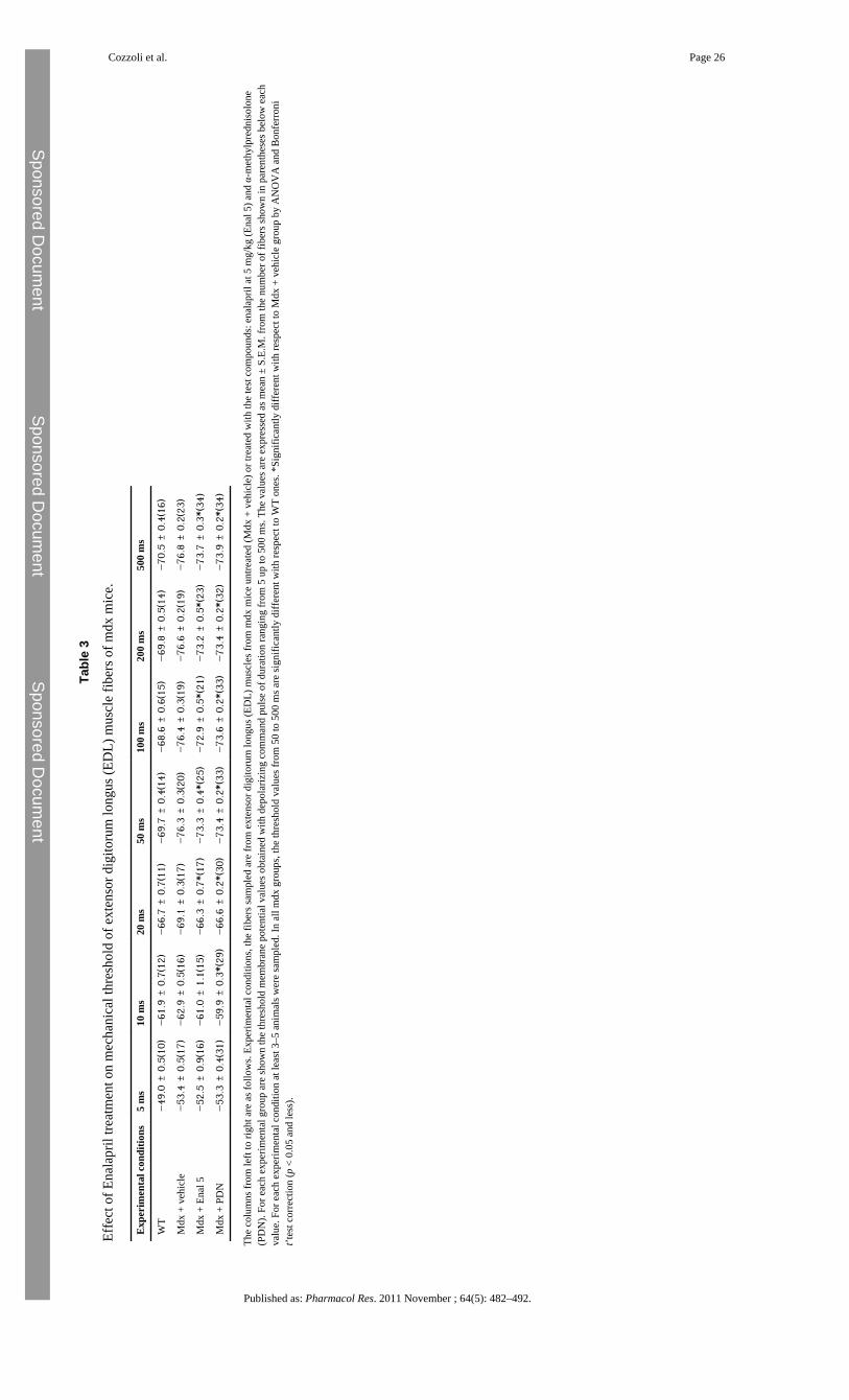
The effects of enalapril were also evaluated on the alteration of the voltage threshold forcontraction (mechanical threshold, MT), a calcium-dependent electrophysiological index ofexcitation–contraction (e–c) coupling mechanism [28]. The alteration of this parameter,along with that of cytosolic calcium level, can be fully contrasted by drugs able to directlyact on the channels that contribute to the enhanced sarcolemmal permeability in mdxmyofibers [19,36]. The threshold potential values of enalapril-treated EDL mdx fibers wereslightly, albeit significantly, shifted toward more positive potentials vs. those of untreatedones, mostly at the long depolarizing steps (from 50 ms ahead) (Table 3). Then, a shift of thestrength-duration curve toward more positive potentials was observed in enalapril-treatedmuscle (Fig. 8B). The rheobase values calculated from the fit were −72.2 ± 1.3 mV and−75.6 ± 1.24 mV for enalapril and vehicle-treated muscles, respectively. The difference wasnot statistically significant (t = 1.8; p > 0.05). Both values were still highly significantlydifferent with respect to that of wild-type myofibers (−66.2 ± 0.47 mV; p < 0.001). Theeffect of 5 mg/kg enalapril almost overlapped that observed with PDN and underlined amodest recovery score (around 35%) on the calcium homeostasis. No effect of either drugwas observed on the time constant to reach the rheobase (data not shown). The result is inline with the observation that other drugs acting as pure anti-inflammatory compounds donot exert any improvement in calcium homeostasis of mdx myofibers, and suggests that themodest benefit of both enalapril and PDN could be related to the anti-oxidant effects, inrelation to the possible cross-talk between ROS and calcium handling mechanisms[20,26,33].
4 ConclusionsACE inhibitors are a widely used class of drugs for cardiovascular disorders due to theirability to reduce both short and long term effects of Ang II on function and re-modelling ofheart and vascular system. Accordingly, ACE inhibitors are already clinically used tocontrast cardiomyopathy occurring in Duchenne patients, and data from clinical trialscorroborate the ability of these drugs in prolonging survival [8,9]. The establishment ofdirect beneficial effect of this class of therapeutics at skeletal muscle level in parallel withthe clarification of their mechanism of action can further reinforce their clinical use inDMD. In fact, data in the literature support a potential involvement of RAS in muscles of
Cozzoli et al. Page 9
Published as: Pharmacol Res. 2011 November ; 64(5): 482–492.
Sponsored Docum
ent Sponsored D
ocument
Sponsored Docum
ent
DMD patients, with over-expression of angiotensin converting enzyme and of AT-1receptor, in parallel with TGF-β1, in regenerating muscle fibers, fibroblasts andinflammatory infiltrates [37]. This is supported by recent evidence of the presence of severalcomponents of RAS in myofibers, corroborating an autocrine production of Ang II inskeletal muscle, potentially triggered by mechanical challenge [38], a critical issue fordystrophic muscle. In addition, long term and indirect activation of systemic RAS, such asthat consequent to congestive heart failure, also plays a role in skeletal muscle atrophy,through direct action of Ang II in regulating the two ubiquitin ligases atrogin-1 and musclering finger-1 (MuRF-1) [16,39]. Although direct evidence about involvement of RAS in themurine pathology is not available, pre-clinical studies in mdx mice claimed a prominent roleof Ang-II-TGFβ-pathway in late occurring fibrosis and failing regeneration [11]. Our resultsfavor a role of Ang-II in early inflammatory phases of muscular dystrophy, supporting awider protective role of ACE inhibitors in dystrophic muscle. Mostly, the results of ourstudy evidenced the ability of enalapril to exert protective effects on mouse strength, whilesignificantly reducing the production of ROS and the activation of NF-kB, pathways thatplay a key role in muscular dystrophy [18,19,23]. Our results are in line with variousevidences showing that Ang-II has pro-inflammatory actions and contributes to insulinresistance in myocytes via ROS-independent and dependent pathways, these latter involvinga receptor and NF-kB-mediated activation of NADPH oxidase [17,40,41]. Interestingly, AngII has been recently claimed to exert a wasting catabolic effect in mouse skeletal muscle viaNADPH oxidase derived ROS [42], while losartan protects against disuse atrophy in rodentsarcopenia via a TGF-β1 independent signaling which rather involves the activation ofinsulin-like growth factor 1 (IGF-1) pathways [43]. Then it is feasible that in dystrophicpathology RAS may contribute to the early inflammatory microenvironment necessary toreinforce the damaging later effects via TGF-β1, meanwhile weakening the IGF-1-dependent regenerative program [12,13,44]. Accordingly, in the present study we found aclear reduction of muscle necrosis by enalapril, with minor, if any, effects on non-muscletissue, TGF-β1 level and p-Smad 2/3 reactivity, supporting earlier actions vs. the inductionof fibrosis. The possibility that enalapril also acts by contrasting functional ischemia cannotbe ruled out, although the modest recovery of resistance to exercise does not support thisview.
However, in contrast with other anti-inflammatory compounds [26,33], enalapril did notreduce CK and LDH. The lack of effect on CK, also routinely observed in our experimentalcondition with PDN [27], has also been observed in long-term losartan treated mdx mice[11]. In a strict sense CK level cannot be taken as a reliable outcome measure fortherapeutics as it might be influenced by the amount of muscle/animal activity, or to otherdrug actions at sarcolemmal level.
Then, it is feasible to conclude that Ang II represents an early upstream signals in triggeringa chronic inflammatory state and dystrophic muscle damage and its early blockade may helpin halting the establishment of auto-reinforcing signals. The results add new evidences aboutthe potential interest of ACE inhibitors for the therapy of DMD patients and support theirearly use. The PDN-similar profile may corroborate the combined use of the two classes ofdrugs in DMD patients so to potentiate the beneficial effects at skeletal muscle level, whilereducing both spontaneous and PDN-aggravated cardiomyopathy [10].
ReferencesGrounds M.D. Radley H.G. Lynch G.S. Nagaraju K. De Luca A. Towards developing standard
operating procedures for pre-clinical testing in the mdx mouse model of Duchenne musculardystrophy. Neurobiol Dis. 2008; 31:1–19. [PubMed: 18499465]
Cozzoli et al. Page 10
Published as: Pharmacol Res. 2011 November ; 64(5): 482–492.
Sponsored Docum
ent Sponsored D
ocument
Sponsored Docum
ent
Willmann R. Possekel S. Dubach-Powell J. Meier T. Ruegg M.A. Mammalian animal models forDuchenne muscular dystrophy. Neuromuscul Disord. 2009; 19:241–249. [PubMed: 19217290]
Hoffman E.P. Dressman D. Molecular pathophysiology and targeted therapeutics for musculardystrophy. Trends Pharmacol Sci. 2001; 22:465–470. [PubMed: 11543874]
Rando T.A. Disatnik M.H. Yu Y. Franco A. Muscle cells from mdx mice have an increasedsusceptibility to oxidative stress. Neuromuscul Disord. 1998; 8:14–21. [PubMed: 9565986]
Whitehead N.P. Yeung E.W. Allen D.G. Muscle damage in mdx (dystrophic) mice: role of calciumand reactive oxygen species. Clin Exp Pharmacol Physiol. 2006; 33:657–662. [PubMed: 16789936]
Bushby K. Straub V. Nonmolecular treatment for muscular dystrophies. Curr Opin Neurol. 2005;18:511–518. [PubMed: 16155433]
Bushby K. Finkel R. Birnkrant D.J. Case L.E. Clemens P.R. Cripe L. Diagnosis and management ofDuchenne muscular dystrophy, part 1: diagnosis, and pharmacological and psychosocialmanagement. Lancet Neurol. 2010; 9:77–93. [PubMed: 19945913]
Duboc D. Meune C. Lerebours G. Devaux J.Y. Vaksmann G. Bécane H.M. Effect of perindopril on theonset and progression of left ventricular dysfunction in Duchenne muscular dystrophy. J Am CollCardiol. 2005; 45:855–857. [PubMed: 15766818]
Duboc D. Meune C. Pierre B. Wahbi K. Eymard B. Toutain A. Perindopril preventive treatment onmortality in Duchenne muscular dystrophy: 10 years’ follow-up. Am Heart J. 2007; 154:596–602.[PubMed: 17719312]
Bauer R. Straub V. Blain A. Bushby K. MacGowan G.A. Contrasting effects of steroids andangiotensin-converting-enzyme inhibitors in a mouse model of dystrophin-deficientcardiomyopathy. Eur J Heart Fail. 2009; 11:463–471. [PubMed: 19233868]
Cohn R.D. van Erp C. Habashi J.P. Soleimani A.A. Klein E.C. Lisi M.T. type 1 receptor blockadeattenuates TGF-beta-induced failure of muscle regeneration in multiple myopathic states. NatMed. 2007; 13:204–210. [PubMed: 17237794]
Berk B.C. Fujiwara K. Lehoux S. ECM remodeling in hypertensive heart disease. J Clin Invest. 2007;117:568–575. [PubMed: 17332884]
Wynn T.A. Cellular and molecular mechanisms of fibrosis. J Pathol. 2008; 214:199–210. [PubMed:18161745]
Martin M.M. Buckenberger J.A. Jiang J. Malana G.E. Knoell D.L. Feldman D.S. TGF-beta1 stimulateshuman AT1 receptor expression in lung fibroblasts by cross talk between the Smad, p38 MAPK,JNK, and PI3K signaling pathways. Am J Physiol Lung Cell Mol Physiol. 2007; 293:L790–L799.[PubMed: 17601799]
Gazzerro E. Assereto S. Bonetto A. Sotgia F. Scarfì S. Pistorio A. Therapeutic potential of proteasomeinhibition in Duchenne and Becker muscular dystrophies. Am J Pathol. 2010; 176:1863–1877.[PubMed: 20304949]
Russell S.T. Wyke S.M. Tisdale M.J. Mechanism of induction of muscle protein degradation byangiotensin II. Cell Signal. 2006; 18:1087–1096. [PubMed: 16257180]
Wei Y. Sowers J.R. Clark S.E. Li W. Ferrario C.M. Stump C.S. Angiotensin II-induced skeletalmuscle insulin resistance mediated by NF-kappaB activation via NADPH oxidase. Am J PhysiolEndocrinol Metab. 2008; 294:E345–351. [PubMed: 18073321]
Arthur P.G. Grounds M.D. Shavlakadze T. Oxidative stress as a therapeutic target during musclewasting: considering the complex interactions. Curr Opin Clin Nutr Metab Care. 2008; 11:408–416. [PubMed: 18542000]
Burdi R. Rolland J.F. Fraysse B. Litvinova K. Cozzoli A. Giannuzzi V. Multiple pathological events inexercised dystrophic mdx mice are targeted by pentoxifylline: outcome of a large array of in vivoand ex vivo tests. J Appl Physiol. 2009; 106:1311–1324. [PubMed: 19131478]
Shkryl V.M. Martins A.S. Ullrich N.D. Nowycky M.C. Niggli E. Shirokova N. Reciprocalamplification of ROS and Ca(2+) signals in stressed mdx dystrophic skeletal muscle fibers.Pflugers Arch. 2009; 458:915–928. [PubMed: 19387681]
Spurney C.F. Knoblach S. Pistilli E.E. Nagaraju K. Martin G.R. Hoffman E.P. Dystrophin-deficientcardiomyopathy in mouse: expression of Nox4 and Lox are associated with fibrosis and alteredfunctional parameters in the heart. Neuromuscul Disord. 2008; 18:371–381. [PubMed: 18440230]
Cozzoli et al. Page 11
Published as: Pharmacol Res. 2011 November ; 64(5): 482–492.
Sponsored Docum
ent Sponsored D
ocument
Sponsored Docum
ent

Williams I.A. Allen D.G. The role of reactive oxygen species in the hearts of dystrophin-deficient mdxmice. Am J Physiol Heart Circ Physiol. 2007; 293:H1969–1977. [PubMed: 17573457]
Acharyya S. Villalta S.A. Bakkar N. Bupha-Intr T. Janssen P.M. Carathers M. Interplay of IKK/NF-kappaB signaling in macrophages and myofibers promotes muscle degeneration in Duchennemuscular dystrophy. J Clin Invest. 2007; 117:889–901. [PubMed: 17380205]
Messina S. Altavilla D. Aguennouz M. Seminara P. Minutoli L. Monici M.C. Lipid peroxidationinhibition blunts nuclear factor-kappaB activation, reduces skeletal muscle degeneration, andenhances muscle function in mdx mice. Am J Pathol. 2006; 168:918–926. [PubMed: 16507907]
Wang P. Fedoruk M.N. Rupert J.L. Keeping pace with ACE: are ACE inhibitors and angiotensin IItype 1 receptor antagonists potential doping agents? Sports Med. 2008; 38:1065–1079. [PubMed:19026021]
De Luca A. Nico B. Liantonio A. Didonna M.P. Fraysse B. Pierno S. A multidisciplinary evaluation ofthe effectiveness of cyclosporine A in dystrophic mdx mice. Am J Pathol. 2005; 166:477–489.[PubMed: 15681831]
Cozzoli A. Rolland J.F. Capogrosso R.F. Sblendorio V.T. Longo V. Simonetti S. Evaluation ofpotential synergistic action of a combined treatment with alpha-methyl-prednisolone and taurineon the mdx mouse model of Duchenne muscular dystrophy. Neuropathol Appl Neurobiol. 2011;37:243–256. [PubMed: 20618838]
De Luca A. Pierno S. Liantonio A. Cetrone M. Camerino C. Fraysse B. Enhanced dystrophicprogression in mdx mice by exercise and beneficial effects of taurine and insulin-like growthfactor-1. J Pharmacol Exp Ther. 2003; 304:453–463. [PubMed: 12490622]
De Luca A. Nico B. Rolland J.F. Cozzoli A. Burdi R. Mangieri D. Gentamicin treatment in exercisedmdx mice: identification of dystrophin-sensitive pathways and evaluation of efficacy in work-loaded dystrophic muscle. Neurobiol Dis. 2008; 32:243–253. [PubMed: 18694830]
Ortiz L.A. Champion H.C. Lasky J.A. Gambelli F. Gozal E. Hoyle G.W. Enalapril protects mice frompulmonary hypertension by inhibiting TNF-mediated activation of NF-kappaB and AP-1. Am JPhysiol Lung Cell Mol Physiol. 2002; 282:L1209–L1221. [PubMed: 12003776]
Reagan-Shaw S. Nihal M. Ahmad N. Dose translation from animal to human studies revisited. FASEBJ. 2008; 22:659–661. [PubMed: 17942826]
Simolin M.A. Pedersen T.X. Bro S. Mäyränpää M.I. Helske S. Nielsen L.B. ACE inhibition attenuatesuremia-induced aortic valve thickening in a novel mouse model. BMC Cardiovasc Disord. 2009;3:9–10.
Pierno S. Nico B. Burdi R. Liantonio A. Didonna M.P. Cippone V. Role of TNF-alpha, but not ofCOX-2 derived eicosanoids, on functional and morphological indices of dystrophic progression inmdx mice: a pharmacological approach. Neuropathol Appl Neurobiol. 2007; 33:344–359.[PubMed: 17493014]
Kobayashi Y.M. Rader E.P. Crawford R.W. Iyengar N.K. Thedens D.R. Faulkner J.A. Sarcolemma-localized nNOS is required to maintain activity after mild exercise. Nature. 2008; 456:511–515.[PubMed: 18953332]
Cozzoli A. Pierno S. Conte Camerino D. De Luca A. Skeletal muscle chloride channel, a biophysicalsensor of dystrophic progression in mdx mouse, is a potential target of pro-inflammatorymediators. Biophys J. 2009; 96:469a–470a.
Rolland J.F. De Luca A. Burdi R. Andreetta F. Confalonieri P. Conte Camerino D. Overactivity ofexercise-sensitive cation channels and their impaired modulation by IGF-1 in mdx native musclefibers: beneficial effect of pentoxifylline. Neurobiol Dis. 2006; 24:466–474. [PubMed: 17010631]
Sun G. Haginoya K. Dai H. Chiba Y. Uematsu M. Hino-Fukuyo N. Intramuscular renin-angiotensinsystem is activated in human muscular dystrophy. J Neurol Sci. 2009; 280:40–48. [PubMed:19232644]
Johnston A.P. Baker J. De Lisio M. Parise G. Skeletal muscle myoblasts possess a stretch-responsivelocal angiotensin signalling system. J Renin Angiotensin Aldosterone Syst. 2010 [Epub ahead ofprint].
Yoshida T. Semprun-Prieto L. Sukhanov S. Delafontaine P. IGF-1 prevents ANG II-induced skeletalmuscle atrophy via Akt- and Foxo-dependent inhibition of the ubiquitin ligase atrogin-1expression. Am J Physiol Heart Circ Physiol. 2010; 298:H1565–1570. [PubMed: 20228261]
Cozzoli et al. Page 12
Published as: Pharmacol Res. 2011 November ; 64(5): 482–492.
Sponsored Docum
ent Sponsored D
ocument
Sponsored Docum
ent

Seshiah P.N. Weber D.S. Rocic P. Valppu L. Taniyama Y. Griendling K.K. Angiotensin II stimulationof NAD(P)H oxidase activity: upstream mediators. Circ Res. 2002; 91:406–413. [PubMed:12215489]
White C.N. Figtree G.A. Liu C.C. Garcia A. Hamilton E.J. Chia K.K. Angiotensin II inhibits the Na+–K+ pump via PKC-dependent activation of NADPH oxidase. Am J Physiol Cell Physiol. 2009;296:C693–700. [PubMed: 19193863]
Semprun-Prieto L.C. Sukhanov S. Yoshida T. Rezk B.M. Gonzalez-Villalobos R.A. Vaughn C.Angiotensin II induced catabolic effect and muscle atrophy are redox dependent. BiochemBiophys Res Commun. 2011 May 6 [Epub ahead of print].
Burks T.N. Andres-Mateos E. Marx R. Mejias R. Van Erp C. Simmers J.L. Losartan restores skeletalmuscle remodeling and protects against disuse atrophy in sarcopenia. Sci Transl Med. 2011;3(82):ra37.
Lanz T.V. Ding Z. Ho P.P. Luo J. Agrawal A.N. Srinagesh H. Angiotensin II sustains braininflammation in mice via TGF-beta. J Clin Invest. 2010; 120:2782–2794. [PubMed: 20628203]
AcknowledgmentsThe financial support of Italian Telethon to the project no. GGP05130 is gratefully acknowledged.
Cozzoli et al. Page 13
Published as: Pharmacol Res. 2011 November ; 64(5): 482–492.
Sponsored Docum
ent Sponsored D
ocument
Sponsored Docum
ent

Fig. 1.Effect of enalapril treatment on fore limb strength and resistance to exercise on mdx mice.(A) The bars show the normalized force increment (Δ normalized force) for wild type (WT)and mdx mice either untreated (vehicle) or treated with enalapril 1 mg/kg (Enal 1), 5 mg/kg(Enal 5) or α-methylprednisolone 1 mg/kg (PDN). Δ normalized force has been calculated asfollows: for each mouse the fore limb strength has been normalized to the respective bodyweight both at the beginning of exercise/treatment protocol (Time 0) and after 4 weeks(Time 4). The difference between the normalized strength values at the two time pointsallowed to calculate for each mouse of each group the increment and to evaluate the effectof either exercise, treatment or both on it. Each bar is the mean ± S.E. (see Section 2) fromthe number of mice in brackets above each bar. Statistical significance between groups wasevaluated by ANOVA test for multiple comparison and Bonferroni t-test post hoc correctionand was as follows: F > 38; p < 0.0001; 10 < F < 40; p < 0.005; significantly different withrespect to *vehicle-treated mdx (p < 0.0001); §enalapril 1 mg/kg treated mice (p < 0.0001).Although not shown in the figure, the values from mdx mouse groups were significantlydifferent with respect to those of WT mice. In (B) is shown the total distance (in m) run bywt and mdx mice treated or not with enalapril 5 mg/kg (Enal 5) or α-methylprednisolone1 mg/kg (PDN) in an exhausting test on treadmill. Each bar is the mean ± S.E.M. from 5 to 6animals. Statistical significance between groups was evaluated by ANOVA test for multiplecomparison (F values) and Bonferroni t-test post hoc correction and was as follows:F = 11.03; p < 0.0005; *significantly different with respect to WT mice (p < 0.005).
Cozzoli et al. Page 14
Published as: Pharmacol Res. 2011 November ; 64(5): 482–492.
Sponsored Docum
ent Sponsored D
ocument
Sponsored Docum
ent
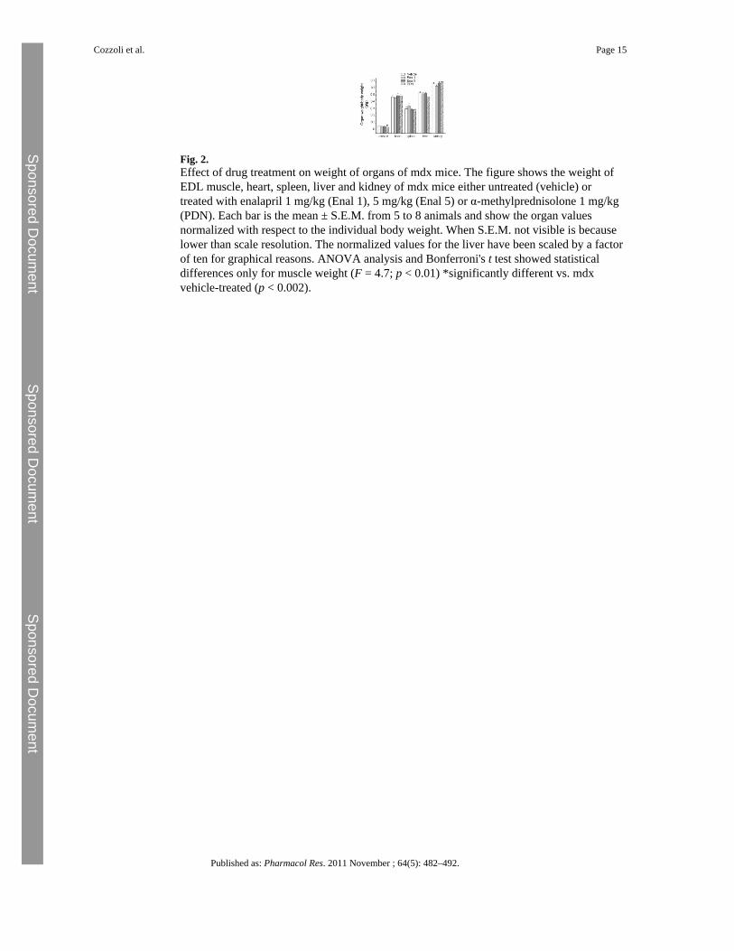
Fig. 2.Effect of drug treatment on weight of organs of mdx mice. The figure shows the weight ofEDL muscle, heart, spleen, liver and kidney of mdx mice either untreated (vehicle) ortreated with enalapril 1 mg/kg (Enal 1), 5 mg/kg (Enal 5) or α-methylprednisolone 1 mg/kg(PDN). Each bar is the mean ± S.E.M. from 5 to 8 animals and show the organ valuesnormalized with respect to the individual body weight. When S.E.M. not visible is becauselower than scale resolution. The normalized values for the liver have been scaled by a factorof ten for graphical reasons. ANOVA analysis and Bonferroni's t test showed statisticaldifferences only for muscle weight (F = 4.7; p < 0.01) *significantly different vs. mdxvehicle-treated (p < 0.002).
Cozzoli et al. Page 15
Published as: Pharmacol Res. 2011 November ; 64(5): 482–492.
Sponsored Docum
ent Sponsored D
ocument
Sponsored Docum
ent

Fig. 3.Effect of drug treatment on superoxide production in TA muscle of mdx mice. In situdetection of O2
− in TA muscles was performed by visualizing red fluorescence in cell nucleiof tissue sections loaded with DHE as described under Section 2. Representative fluorescentphotomicrographs from independent sets of experiments are shown for TA muscle cross-sections at 10× magnification. Merging (magenta fluorescence) of DHE (red fluorescence)and DAPI (blue fluorescence) staining is indicative of O2
− production in cell nuclei frommdx mice either untreated (MDX + vehicle), or treated with α-methylprednisolone 1 mg/kg(MDX + PDN), enalapril 1 mg/kg (MDX + Enal 1) or enalapril 5 mg/kg (MDX + Enal 5).
Cozzoli et al. Page 16
Published as: Pharmacol Res. 2011 November ; 64(5): 482–492.
Sponsored Docum
ent Sponsored D
ocument
Sponsored Docum
ent

Fig. 4.Effect of drug treatment on densitometric determination of superoxide anion production.Densitometric determination of DHE fluorescence in the nuclei of TA muscles from mdxmice treated or not with either enalapril at 1 (Enal 1) and 5 mg/kg (Enal 5) or α-methylprednisolone (PDN) is shown. Each bar is the mean ± S.E.M. of the value of fluorescencerelative to the merging of DHE (red) and DAPI (blue) staining of wt (arbitrarily consideredas unitary) from analysis of 5–16 fields and 3–5 muscle per group (at 40× magnification). Asignificant difference among groups was found by ANOVA analysis (F = 35.57;p < 0.0001), Bonferroni post hoc t-test for individual differences between groups are asfollows: significantly different *vs. vehicle-treated mdx (p < 0.0001) and #vs. enalapil 1 mg/kg treated mdx group (p < 0.05).
Cozzoli et al. Page 17
Published as: Pharmacol Res. 2011 November ; 64(5): 482–492.
Sponsored Docum
ent Sponsored D
ocument
Sponsored Docum
ent
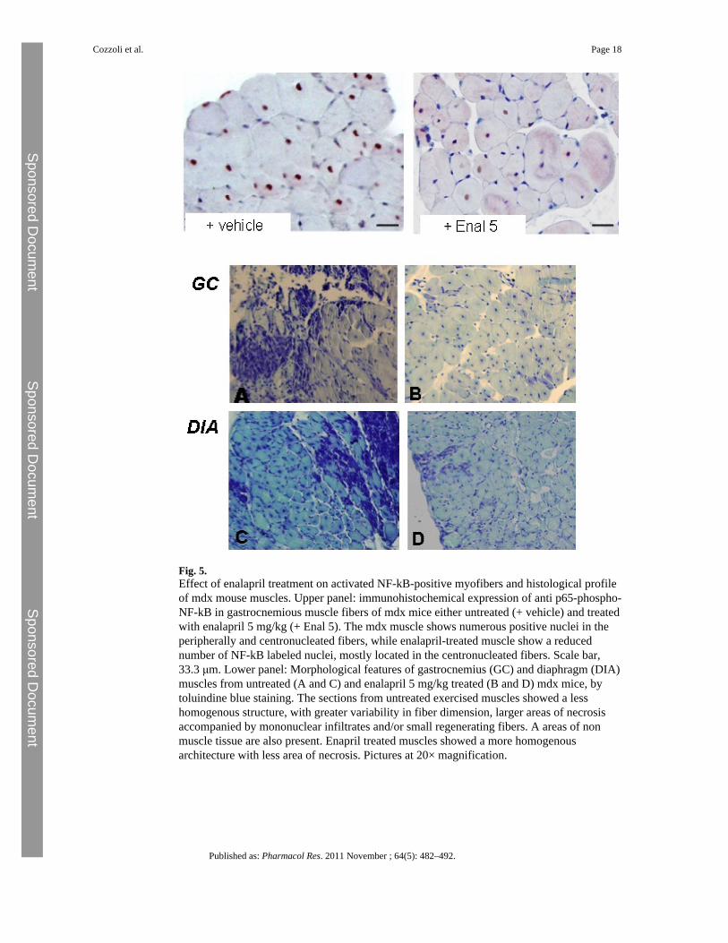
Fig. 5.Effect of enalapril treatment on activated NF-kB-positive myofibers and histological profileof mdx mouse muscles. Upper panel: immunohistochemical expression of anti p65-phospho-NF-kB in gastrocnemious muscle fibers of mdx mice either untreated (+ vehicle) and treatedwith enalapril 5 mg/kg (+ Enal 5). The mdx muscle shows numerous positive nuclei in theperipherally and centronucleated fibers, while enalapril-treated muscle show a reducednumber of NF-kB labeled nuclei, mostly located in the centronucleated fibers. Scale bar,33.3 μm. Lower panel: Morphological features of gastrocnemius (GC) and diaphragm (DIA)muscles from untreated (A and C) and enalapril 5 mg/kg treated (B and D) mdx mice, bytoluindine blue staining. The sections from untreated exercised muscles showed a lesshomogenous structure, with greater variability in fiber dimension, larger areas of necrosisaccompanied by mononuclear infiltrates and/or small regenerating fibers. A areas of nonmuscle tissue are also present. Enapril treated muscles showed a more homogenousarchitecture with less area of necrosis. Pictures at 20× magnification.
Cozzoli et al. Page 18
Published as: Pharmacol Res. 2011 November ; 64(5): 482–492.
Sponsored Docum
ent Sponsored D
ocument
Sponsored Docum
ent

Fig. 6.Effect of enalapil treatment on pro-fibrotic signaling in mdx mouse muscles. The bars in theupper panel show the levels of total TGF-β1 in gastrocnemius muscle, measured by ELISA.Each value is the mean ± S.E.M. from 5 to 9 preparations. Statistical significance has beenevaluated by ANOVA and Bonferroni’t test post hoc correction and was as follows F > 10;p < 0.0001. *Significantly different with respect to WT (p < 0.0001). The middle panelshows representative immunohistochemical expression of p-Smad2/3 in gastrocnemiusmuscles fibers of wt (A), exercised mdx (B) and enalapril (5 mg/kg) treated exercised mdx(C) mice. No labeling of the nuclei and fibers is present in wt muscle (A), while the mdxmuscle shows numerous positive nuclei in the peripherally (B, arrow) and centronucleated
Cozzoli et al. Page 19
Published as: Pharmacol Res. 2011 November ; 64(5): 482–492.
Sponsored Docum
ent Sponsored D
ocument
Sponsored Docum
ent

fibers (B, arrowhead). Enalapril-treated mice (C) show a general trend toward a reducednumber of p-Smad2/3 labeled nuclei (scale bar, 33.3 μm). The quantitative estimation ofpositive myonuclei is represented in the lower panel. The analysis (at least 3 animals/groupand 9 fields/muscle; mean ± S.D.) showed only a slight not significant decrease in pSmad2/3reactivity in enalapril-treated group vs. vehicle treated one. Statistical significance has beenevaluated by ANOVA and Bonferroni's t test post hoc correction and was as follows F > 7;p < 0.03; *significantly different with respect to WT (p < 0.02).
Cozzoli et al. Page 20
Published as: Pharmacol Res. 2011 November ; 64(5): 482–492.
Sponsored Docum
ent Sponsored D
ocument
Sponsored Docum
ent

Fig. 7.Effect of enalapil treatment on plasma level of creatine kinase and lactate dehydrogenase ofmdx mice. Plasma level of creatine kinase (CK) and lactate dehydrogenase (LDH) measuredby standard spectrophotometric analysis. Each columns is the mean ± S.E.M. from 5 to 8animals. No significant statistical differences were observed between values of mdx mice(either treated or not).
Cozzoli et al. Page 21
Published as: Pharmacol Res. 2011 November ; 64(5): 482–492.
Sponsored Docum
ent Sponsored D
ocument
Sponsored Docum
ent
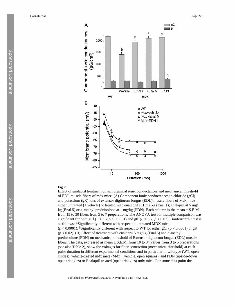
Fig. 8.Effect of enalapril treatment on sarcolemmal ionic conductances and mechanical thresholdof EDL muscle fibers of mdx mice. (A) Component ionic conductances to chloride (gCl)and potassium (gK) ions of extensor digitorum longus (EDL) muscle fibers of Mdx miceeither untreated (+ vehicle) or treated with enalapril at 1 mg/kg (Enal 1); enalapril at 5 mg/kg (Enal 5) or α-methyl prednisolone at 1 mg/kg (PDN). Each column is the mean ± S.E.M.from 15 to 30 fibers from 3 to 7 preparations. The ANOVA test for multiple comparison wassignificant for both gCl (F > 16; p < 0.0001) and gK (F = 3.7; p < 0.02). Bonferroni's t test isas follows: *Significantly different with respect to untreated MDX mice(p < 0.0001); §Significantly different with respect to WT for either gCl (p < 0.0001) or gK(p < 0.02). (B) Effect of treatment with enalapril 5 mg/kg (Enal 5) and α-methylprednisolone (PDN) on mechanical threshold of Extensor digitorum longus (EDL) musclefibers. The data, expressed as mean ± S.E.M. from 10 to 34 values from 3 to 5 preparations(see also Table 2), show the voltages for fiber contraction (mechanical threshold) at eachpulse duration in different experimental conditions and in particular in wildtype (WT, opencircles), vehicle-treated mdx mice (Mdx + vehicle, open squares), and PDN (upside-downopen triangles) or Enalapril treated (open triangles) mdx mice. For some data point the
Cozzoli et al. Page 22
Published as: Pharmacol Res. 2011 November ; 64(5): 482–492.
Sponsored Docum
ent Sponsored D
ocument
Sponsored Docum
ent

standard error bar is not visible being smaller than symbol size. The fit of the experimentaldata lead to the calculation of rheobase voltage values reported in the text.
Cozzoli et al. Page 23
Published as: Pharmacol Res. 2011 November ; 64(5): 482–492.
Sponsored Docum
ent Sponsored D
ocument
Sponsored Docum
ent

Sponsored Docum
ent Sponsored D
ocument
Sponsored Docum
ent
Cozzoli et al. Page 24
Table 1
Effect of enalapril treatment on body weight and fore limb muscle strength of mdx mice.
Groups BW T0 BW T4 ΔBW (T4–T0) Fmax T0 Fmax T4 ΔFmax (T4–T0) NF T0 NF T4
WT (5) 19.7 ± 0.41 25.41 ± 0.8 5.7 ± 0.71 0.122 ± 0.006 0.181 ± 0.01 0.058 ± 0.018 6.25 ± 0.4 7.09 ± 0.33
Mdx + vehicle (8) 17.8 ± 1.24 22.3 ± 1.36* 4.48 ± 0.35 0.113 ± 0.01 0.143 ± 0.01* 0.03 ± 0.009 6.29 ± 0.25 6.33 ± 0.34
Mdx + Enal 1 (7) 16.0 ± 0.71* 20.8 ± 0.57* 4.86 ± 0.54 0.106 ± 0.009 0.148 ± 0.004* 0.042 ± 0.006 6.62 ± 0.49 7.15 ± 0.26
Mdx + Enal 5 (8) 18.8 ± 0.72 22.8 ± 0.59* 4.05 ± 0.77 0.122 ± 0.008 0.176 ± 0.004#§ 0.054 ± 0.007§ 6.49 ± 0.28 7.77 ± 0.22§
Mdx + PDN (7) 19.9 ± 0.99 23.3 ± 0.31 3.44 ± 0.88 0.107 ± 0.01 0.160 ± 0.01 0.050 ± 0.01 5.32 ± 0.38 6.86 ± 0.35§
Columns are as follows: Groups of mice used (in brackets the number of mice per group): wild type C57BL10 (WT); untreated mdx mice(Mdx + vehicle); Mdx mice treated with enalapril 1 mg/kg (Mdx + Enal 1); Mdx mice treated with enalapril 5 mg/kg (Mdx + Enal 5), Mdx micetreated with α-methylprednisolone (Mdx + PDN); body weight, in g at the beginning (T0) and at the end of 4-week-protocol (T4); ΔBW, differencebetween body weights at T4 and T0; Fmax, maximal fore limb strength, in kg at either the beginning (T0) and at the end of 4-week-protocol (T4);ΔFmax: fore limb strength increment between T4 and T0. NF: fore limb strength normalized to body weight at either the beginning (T0) and at theend of 4-week-protocol (T4). Each value is the mean ± S.E.M. For each parameter, the statistical significance between groups was evaluated byANOVA test for multiple comparison (F values) and Bonferroni t-test post hoc correction. Significance at ANOVA test was found for thefollowing parameters BW (F > 3; p < 0.04); Fmax at T4 (F > 3; p < 0.05); NF at T4 (F > 4; p < 0.01). The Bonferroni's test showed statistical
significance as shown in the table with respect to the following groups: *WT (0.002 < p < 0.04); §Mdx + vehicle
(0.002 < p < 0.01), #Mdx + Enalapril 1 mg/kg (p < 0.01).
Published as: Pharmacol Res. 2011 November ; 64(5): 482–492.

Sponsored Docum
ent Sponsored D
ocument
Sponsored Docum
ent
Cozzoli et al. Page 25
Table 2
Effect of Enalapril treatment on histology profile of diaphragm and gastrocnemious muscle of mdx mice.
% NF % CNF Area NF (μ2) Area CNF (μ2) % necrotic area % non-muscle area
GC Mdx vehicle 28 ± 6.6 74 ± 7 832 ± 138 1757 ± 453 5.7 ± 1.9 9.6 ± 3.2
GC Mdx + Enalapril 33 ± 2* 67 ± 6* 1141 ± 543 1496 ± 316 1.2 ± 0.2§ 7.4 ± 4.6
DIA Mdx vehicle 52 ± 6 48 ± 6 699 ± 55 1292 ± 219 8.1 ± 4.7 7.1 ± 3.5
DIA Mdx + Enalapril 46 ± 16 54 ± 4* 830 ± 108* 1598 ± 226* 1.8 ± 0.7** 5.3 ± 2.4
The columns from left to right show the mean values ± S.D. obtained from at least 9 sections from 3 preparations from each experimental group. %NF, is the percent of the normal fibers while % CNF is the percent of centronucleated fibers, i.e. regenerated fiber which replace normalperipherally nucleated fibers and are an index of degeneration–regeneration cycles in the muscle. Area of both normal fibers (NF) andcentronucleated fibers (CNF). The % of necrotic area, is the area composed of necrotic fibers with a large infiltration of inflammatorymononucleated cells, while the % percent of non-muscle area, represents the area of space between fibers or between groups of muscle fibers that isnot stained as muscular tissue and is composed by connective and/or adipose tissue. Significantly different by unpaired Student's t test for
*p < 0.05, **p < 0.001 and §p < 0.0001 vs. muscle-related vehicle-treated mdx group.
Published as: Pharmacol Res. 2011 November ; 64(5): 482–492.
Sponsored Docum
ent Sponsored D
ocument
Sponsored Docum
ent
Cozzoli et al. Page 26
Tabl
e 3
Effe
ct o
f Ena
lapr
il tre
atm
ent o
n m
echa
nica
l thr
esho
ld o
f ext
enso
r dig
itoru
m lo
ngus
(ED
L) m
uscl
e fib
ers o
f mdx
mic
e.
Exp
erim
enta
l con
ditio
ns5
ms
10 m
s20
ms
50 m
s10
0 m
s20
0 m
s50
0 m
s
WT
−49
.0 ±
0.5
(10)
−61
.9 ±
0.7
(12)
−66
.7 ±
0.7
(11)
−69
.7 ±
0.4
(14)
−68
.6 ±
0.6
(15)
−69
.8 ±
0.5
(14)
−70
.5 ±
0.4
(16)
Mdx
+ v
ehic
le−53
.4 ±
0.5
(17)
−62
.9 ±
0.5
(16)
−69
.1 ±
0.3
(17)
−76
.3 ±
0.3
(20)
−76
.4 ±
0.3
(19)
−76
.6 ±
0.2
(19)
−76
.8 ±
0.2
(23)
Mdx
+ E
nal 5
−52
.5 ±
0.9
(16)
−61
.0 ±
1.1
(15)
−66
.3 ±
0.7
*(17
)−73
.3 ±
0.4
*(25
)−72
.9 ±
0.5
*(21
)−73
.2 ±
0.5
*(23
)−73
.7 ±
0.3
*(34
)
Mdx
+ P
DN
−53
.3 ±
0.4
(31)
−59
.9 ±
0.3
*(29
)−66
.6 ±
0.2
*(30
)−73
.4 ±
0.2
*(33
)−73
.6 ±
0.2
*(33
)−73
.4 ±
0.2
*(32
)−73
.9 ±
0.2
*(34
)
The
colu
mns
from
left
to ri
ght a
re a
s fol
low
s. Ex
perim
enta
l con
ditio
ns, t
he fi
bers
sam
pled
are
from
ext
enso
r dig
itoru
m lo
ngus
(ED
L) m
uscl
es fr
om m
dx m
ice
untre
ated
(Mdx
+ v
ehic
le) o
r tre
ated
with
the
test
com
poun
ds: e
nala
pril
at 5
mg/
kg (E
nal 5
) and
α-m
ethy
lpre
dnis
olon
e(P
DN
). Fo
r eac
h ex
perim
enta
l gro
up a
re sh
own
the
thre
shol
d m
embr
ane
pote
ntia
l val
ues o
btai
ned
with
dep
olar
izin
g co
mm
and
puls
e of
dur
atio
n ra
ngin
g fr
om 5
up
to 5
00 m
s. Th
e va
lues
are
exp
ress
ed a
s mea
n ±
S.E.
M. f
rom
the
num
ber o
f fib
ers s
how
n in
par
enth
eses
bel
ow e
ach
valu
e. F
or e
ach
expe
rimen
tal c
ondi
tion
at le
ast 3
–5 a
nim
als w
ere
sam
pled
. In
all m
dx g
roup
s, th
e th
resh
old
valu
es fr
om 5
0 to
500
ms a
re si
gnifi
cant
ly d
iffer
ent w
ith re
spec
t to
WT
ones
. *Si
gnifi
cant
ly d
iffer
ent w
ith re
spec
t to
Mdx
+ v
ehic
le g
roup
by
AN
OV
A a
nd B
onfe
rron
it’t
est c
orre
ctio
n (p
< 0
.05
and
less
).
Published as: Pharmacol Res. 2011 November ; 64(5): 482–492.