A study of serum concentrations and dermal levels of NGF in atopic dermatitis and healthy subjects
16
A study of serum concentrations and dermal levels of NGF in atopic dermatitis and healthy subjects Alexandru D.P. Papoiu a , Hui Wang a , Leigh Nattkemper a,b , Hong Liang Tey a , Yozo Ishiuji a , Yiong-Huak Chan d , Martin Schmelz e , and Gil Yosipovitch a,b,c,* a Department of Dermatology, Wake Forest University School of Medicine, Winston-Salem, NC 27157, USA b Department of Neurobiology and Anatomy, Wake Forest University School of Medicine, Winston- Salem, NC 27157, USA c Department of Regenerative Medicine, Wake Forest University School of Medicine, Winston- Salem, NC 27157, USA d Biostatistics Unit, Yong Loo Lin School of Medicine, National University of Singapore, Clinical Research Centre, 10 Medical Drive, Singapore 117597, Singapore e Department of Anesthesiology and Intensive Care Medicine, Faculty of Clinical Medicine Mannheim, University of Heidelberg, Mannheim, Germany Abstract Nerve growth factor (NGF) was reported to be increased in the serum and skin of atopic dermatitis (AD) patients, to the extent that serum nerve growth factor levels were proposed to serve as a marker of disease severity. We studied NGF levels in the serum and dermis using skin microdialysis and attempted to correlate them with disease severity. We also examined if potential differences between morning and evening levels of NGF can explain the phenomenon of nocturnal itch. In addition, neurogenic inflammation and itch were induced using histamine iontophoresis in lesional and non-lesional skin and the effect of experimental itch on dermal NGF concentration was examined. We found that systemic (serum) and eczematous skin levels of NGF in AD are significantly lower in comparison to healthy controls. Serum NGF decreases from morning to late afternoon in both groups. Interestingly, serum NGF levels were correlated to disease severity in the morning in AD, although the NGF concentration in AD were significantly lower than in the healthy group. The local itch and neurogenic inflammation induction via experimental histamine reduced local NGF levels in the eczema and non-lesional skin in atopics, but not in the healthy controls, where it was slightly increased. The higher the clinical severity of the eczema, a significantly less pronounced effect of neurogenic inflammation on the local levels of NGF was found. The availability of measurable NGF might be reduced by a higher expression of NGF receptors. The fluctuations of NGF levels during the day suggest a complex modulation of this neurotrophin, potentially linked to stress or to an altered neurophysiological mechanism. Keywords Nerve growth factor (NGF) levels; Atopic dermatitis; Itch; Histamine; Daily fluctuation © 2011 Elsevier Ltd. All rights reserved. * Corresponding author at: Department of Dermatology, Wake Forest University School of Medicine, Winston-Salem, NC 27157, USA. Tel.: +1 (336) 716 2901; fax: +1 (336) 716 7732. [email protected] (G. Yosipovitch). Conflict of interest The authors declare none. NIH Public Access Author Manuscript Neuropeptides. Author manuscript; available in PMC 2013 May 21. Published in final edited form as: Neuropeptides. 2011 December ; 45(6): 417–422. doi:10.1016/j.npep.2011.07.008. NIH-PA Author Manuscript NIH-PA Author Manuscript NIH-PA Author Manuscript
Transcript of A study of serum concentrations and dermal levels of NGF in atopic dermatitis and healthy subjects

A study of serum concentrations and dermal levels of NGF inatopic dermatitis and healthy subjects
Alexandru D.P. Papoiua, Hui Wanga, Leigh Nattkempera,b, Hong Liang Teya, Yozo Ishiujia,Yiong-Huak Chand, Martin Schmelze, and Gil Yosipovitcha,b,c,*
aDepartment of Dermatology, Wake Forest University School of Medicine, Winston-Salem, NC27157, USAbDepartment of Neurobiology and Anatomy, Wake Forest University School of Medicine, Winston-Salem, NC 27157, USAcDepartment of Regenerative Medicine, Wake Forest University School of Medicine, Winston-Salem, NC 27157, USAdBiostatistics Unit, Yong Loo Lin School of Medicine, National University of Singapore, ClinicalResearch Centre, 10 Medical Drive, Singapore 117597, SingaporeeDepartment of Anesthesiology and Intensive Care Medicine, Faculty of Clinical MedicineMannheim, University of Heidelberg, Mannheim, Germany
AbstractNerve growth factor (NGF) was reported to be increased in the serum and skin of atopic dermatitis(AD) patients, to the extent that serum nerve growth factor levels were proposed to serve as amarker of disease severity. We studied NGF levels in the serum and dermis using skinmicrodialysis and attempted to correlate them with disease severity. We also examined if potentialdifferences between morning and evening levels of NGF can explain the phenomenon of nocturnalitch. In addition, neurogenic inflammation and itch were induced using histamine iontophoresis inlesional and non-lesional skin and the effect of experimental itch on dermal NGF concentrationwas examined. We found that systemic (serum) and eczematous skin levels of NGF in AD aresignificantly lower in comparison to healthy controls. Serum NGF decreases from morning to lateafternoon in both groups. Interestingly, serum NGF levels were correlated to disease severity inthe morning in AD, although the NGF concentration in AD were significantly lower than in thehealthy group. The local itch and neurogenic inflammation induction via experimental histaminereduced local NGF levels in the eczema and non-lesional skin in atopics, but not in the healthycontrols, where it was slightly increased. The higher the clinical severity of the eczema, asignificantly less pronounced effect of neurogenic inflammation on the local levels of NGF wasfound. The availability of measurable NGF might be reduced by a higher expression of NGFreceptors. The fluctuations of NGF levels during the day suggest a complex modulation of thisneurotrophin, potentially linked to stress or to an altered neurophysiological mechanism.
KeywordsNerve growth factor (NGF) levels; Atopic dermatitis; Itch; Histamine; Daily fluctuation
© 2011 Elsevier Ltd. All rights reserved.*Corresponding author at: Department of Dermatology, Wake Forest University School of Medicine, Winston-Salem, NC 27157,USA. Tel.: +1 (336) 716 2901; fax: +1 (336) 716 7732. [email protected] (G. Yosipovitch).
Conflict of interestThe authors declare none.
NIH Public AccessAuthor ManuscriptNeuropeptides. Author manuscript; available in PMC 2013 May 21.
Published in final edited form as:Neuropeptides. 2011 December ; 45(6): 417–422. doi:10.1016/j.npep.2011.07.008.
NIH
-PA Author Manuscript
NIH
-PA Author Manuscript
NIH
-PA Author Manuscript

1. IntroductionPruritus is known to be exacerbated at night in many systemic and inflammatory skindiseases (Patel et al., 2007). In particular, atopic dermatitis (AD), of which itch is anessential feature of the disease (Stander and Steinhoff, 2002; Koblenzer, 1999), is associatedwith significant nocturnal pruritus (Ebata et al., 1996, 1999). Patients with severe pruritus atnight report a greatly diminished quality of life and impaired sleep (Yosipovitch et al., 2000,2002a, 2002b). The mechanism of night-time itching, however, is not well understood. Inprevious studies, we found that the skin barrier function and skin blood flow (Yosipovitch etal., 1998, 2004) follows a circadian rhythm. Based on these observations, we advanced thehypothesis whether nocturnal pruritus may be related to an increase in the secretion ofneuropeptides or neurotrophins, such as the nerve growth factor (NGF).
It has been reported that levels of NGF are increased in the serum and skin of AD patients(Pincelli et al., 1990; Toyoda et al., 2002) and serum NGF was proposed to be a marker ofdisease severity (Toyoda et al., 2002; Hodeib et al., 2010). However, a recent study did notconcur with these findings (Schulte-Herbrüggen et al., 2007). Serum NGF level was alsoreported to be increased in the evening (at 8 PM) in healthy individuals (Bersani et al.,2004). However, it is known that systemic levels of NGF can be influenced by a variety ofphysiological, pathophysiological, neuroendocrine factors, such as gender, stress, psychiatricconditions/status (Levi-Montalcini et al., 1995; Joachim et al., 2007), and may very well beunder homeostatic and circadian control.
The aims of our study were: (1) to investigate whether serum and dermal NGF levels areincreased in AD and if there is a correlation with disease severity; (2) to investigate whetherserum NGF levels are increased in the evening in AD patients; (3) to determine if histamineinduces an increase in dermal NGF level.
2. Materials and methods2.1. Study population
Adults between18and 55 years of age with moderate to severe AD and age-matched healthyvolunteers were enrolled. The diagnosis of AD was based on the Hanifin and Rajka (1980)criteria and the subjects had to present with eczema involving their forearms. Study subjectswere in good health with no other skin disease. Potential participants were excluded if theyshowed evidence of depression based on the Beck Depression Inventory (BDI > 10), or ifthey worked shifts between8PMand 6AM. The severity of AD was evaluated using theEASI score (Hanifin et al., 2001). The EASI score measures the severity of AD on anumerical scale from 0 (no involvement) to 72 (maximum extension and severity). Subjectswere instructed to discontinue antihistamines, topical and oral corticosteroids, centrallyacting drugs (neuroleptics, antidepressants, gabapentin or mirtazapine etc.) and opioids, atleast 1 week prior to the experimental visit. Thirteen patients with AD (7 males, 6 females;mean age 30.4 ± 9.7, age range 19–55) and 17 healthy volunteers (8 females, 9 males: meanage 30.7 ± 8.9, range 19–52) participated in this study. The study was approved by the localInternal Review Board (IRB) of Wake Forest University Health Sciences and was conductedin accordance with the principles of the Declaration of Helsinki.
2.2. Sample collectionDermal NGF levels were evaluated by skin microdialysis as described previously (Schmelzet al., 1997; Rukwied et al., 2000). Study subjects were accommodated comfortably in aclimate controlled room with a temperature maintained at approximately 23 °C and 43%relative humidity. In AD subjects, a microdialysis probe was inserted into an area of eczema
Papoiu et al. Page 2
Neuropeptides. Author manuscript; available in PMC 2013 May 21.
NIH
-PA Author Manuscript
NIH
-PA Author Manuscript
NIH
-PA Author Manuscript

on the volar forearm and another probe into non-lesional skin, 4 cm away, in a parallelorientation. In healthy subjects, both probes were similarly inserted on the volar aspect ofeither forearm, 4 cm apart in a parallel fashion. In the first hour of skin microdialysis,baseline concentrations of NGF were assessed. In the next hour, histamine iontophoresiswas applied to an area adjacent to the microdialysis catheters to induce itch and to assessdermal NGF levels following itch induction. The microdialysis probes were slowly andgently inserted superficially at the dermal– epidermal junction for a length of 1.5 cm,without the use of local anesthetics. Dual microdialysis was performed using anelectronically-controlled dual pump (World Precision Instruments, Sarasota, Florida)running an isotonic 0.9% saline solution at a flow rate of 4 μl/min. The microdialysiscatheters had a diameter of 0.4 mm and the semi-permeable membranes (Dermal Dialysis,Erlangen, Germany) had a pore size of 3 MDa (permitting free diffusion of macromoleculesup to a molecular weight of 3 MDa). Dialysate samples were immediately frozen at −80 °Cafter collection and were thawed just before ELISA analysis.
Experimental itch was induced by histamine iontophoresis using a portable power DCsource (Perimed, Sweden). The electrodes were applied to skin approximately 1 cm awayfrom the place where the microdialysis catheters were inserted. Histamine 1% in a 2%methylcellulose gel was administered by applying a 200 μA current for 30 s. Blood sampleswere also collected by venipuncture from all subjects at 8.00 AM and 5.00 PM.
2.3. Measurement of free NGF levelsNGF levels in the skin microdialysate and serum samples were measured using the PromegaNGF Emax ELISA kit (Promega, Madison, WI). In accordance with the protocol provided bythe manufacturer, primary polyclonal antibodies were added to Nunc Immuno 96MicroWell™ solid plates (Nalge Nunc International, Rochester, NY) and incubated for 24 hat 4 °C. Sample and block buffer was added to the wells for 1 h and then wells were washed5 times with wash buffer.
2.3.1. Preparation of standard curve—NGF standard was diluted 1:4000 with theNGF block and sample buffer, and a volume of 200 μl was added to first row of columns 11and 12 of a 96-well Nunc plate. Seven 1:2 serial dilutions were performed in these lanes.Samples were added at a 1/10 dilution for serum samples and undiluted for skinmicrodialysate. Each well contained 100 μl and was incubated with shaking for 6 h at roomtemperature. Subsequently, plates were washed 5 times. Secondary monoclonal anti-NGFantibody was added next and the plates were incubated overnight (12 h) at 4 °C. Sampleswere tested in triplicate. A standard curve using stock NGF (provided by the Emax kit) wasgenerated every time ELISA was performed. The standard curve was constructed for everyexperiment and a linear curve fit was obtained. Samples of dialysate that contained NGFdisplayed values within the range of the standard curve.
2.4. Statistical analysisDifferences in NGF concentration between eczema, non-lesional skin in atopics and healthyskin, between morning and evening time points, before and after histamine administrationwere analyzed with a mixed model taking into account subject effects and multiplecomparisons were Bonferroni adjusted using PASW 18.0 software (SPSS, Chicago, IL,USA). Overall daily fluctuations in dermal NGF were also examined as averages collapsedover histamine’s effect. A Spearman correlation analysis of serum and skin dialysate NGFvalues (baseline, post-histamine and % change after histamine) with disease severityexpressed in the EASI scores was performed for both time points.
Papoiu et al. Page 3
Neuropeptides. Author manuscript; available in PMC 2013 May 21.
NIH
-PA Author Manuscript
NIH
-PA Author Manuscript
NIH
-PA Author Manuscript
3. Results3.1. Serum NGF levels
The average serum concentration (±S.D.) of free NGF was 236 ± 38 (at 8 AM), 211 ± 34 pg/ml (at 5 PM) in healthy subjects and 184 ± 28 (8 AM), 161 ± 24 (5 PM) pg/ml in ADsubjects. NGF serum levels were significantly lower (p < 0.001) at both time points in ADsubjects compared to healthy subjects (Table 1). A decrease of NGF serum concentrationsfrom 8 AM to 5 PM was noted in both groups; more specifically, a significant decrease from8 AM to 5 PM was found in the AD group (p = 0.027), but the change was not significant inthe healthy group (Fig. 1).
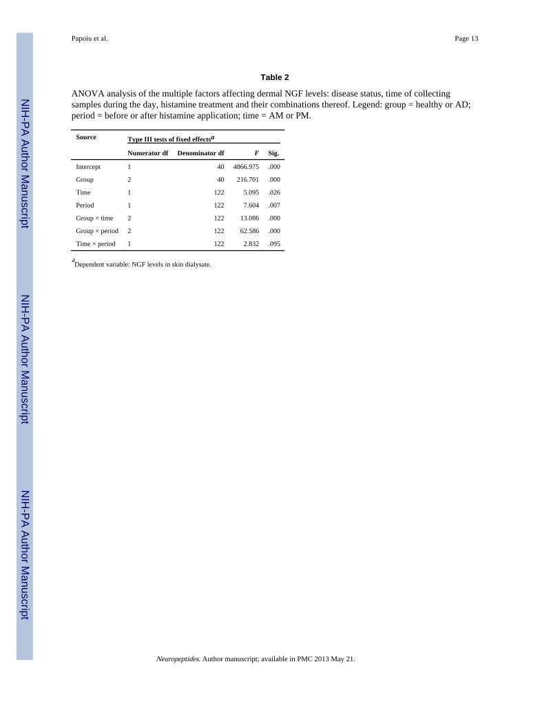
3.2. Dermal NGF levelsWe were interested to answer the following questions, in regards to the skin levels of NGFin the skin: (1) Are these concentrations different in: eczematous areas of atopic skin in ADin comparison with non-lesional areas in AD and healthy skin? (2) Are dermal NGF levelschanging from AM to PM? (3) We also investigated if: the levels of NGF were differentiallyaffected by histamine administration in eczematous, non-lesional skin in AD or healthy skin,and if affirmative, if this response was different at two different time-points during the day(AM or PM). To answer the above questions, a mixed model analysis was performed toassess the effect the interaction terms (time = AM vs. PM, period = pre-histamine vs. post-histamine, group = healthy vs. eczema vs. non-lesional). We found a significant differencein the manner in which NGF levels fluctuated, across all groups overall (p < 0.001), frompre-histamine to post-histamine settings (by period, p = 0.007), by time of day (p = 0.026,AM vs. PM), by group and time (p < 0.001) and by group and period (p < 0.001). (Table 2).
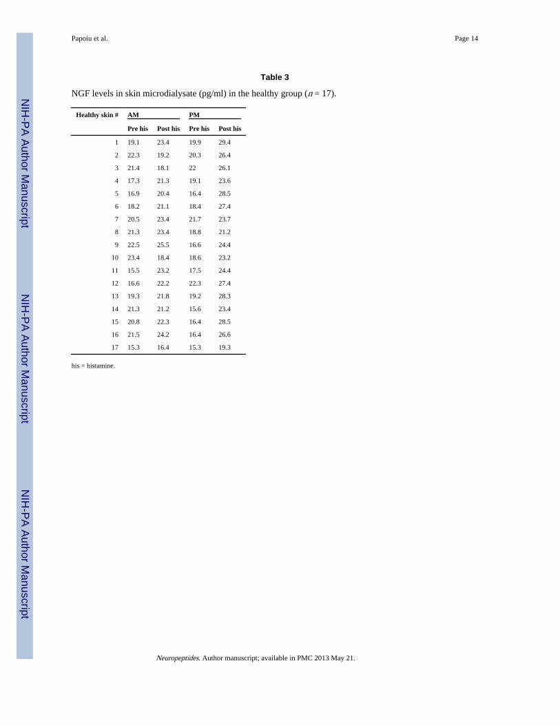
Dermal NGF levels in eczematous skin in AD subjects were consistently found to besignificantly lower compared to non-eczematous skin in AD and the skin of healthy controls(both comparisons, p < 0.001) (Fig. 2, Table 3). There was no significant difference betweenmorning and evening levels in dermal (free) NGF levels, within the same skin area type, inbaseline conditions (Table 4). The NGF concentrations in non-lesional skin in AD subjectsand healthy skin were similar, however there were significant differences revealed by theaction of exogenous histamine. More precisely, administration of histamine increasedmeasurable levels of NGF in healthy skin and decreased NGF levels in non-lesional skin inAD and in the eczematous areas (Fig. 3).
To further dissect the effect of itch and neurogenic inflammation induced experimentallywith histamine on the local dermal NGF concentrations, we performed ANOVA to testwhether % changes from pre-histamine (baseline) to post-histamine values contrastedsignificantly between these 3 groups, and separately, for AM and PM time points.
In the morning (8 AM) healthy skin levels of NGF increased following histamineadministration and the change was significant vs. the change observed in the eczema (p =0.027), but not significantly vs. non-lesional skin in AD. Non-lesional levels of NGFincreased also significantly vs. eczema (p = 0.001). In the evening, the change in levels ofNGF in eczematous skin was significantly different vs. both non-lesional skin in AD andhealthy skin, while the change in AD non-lesional skin was significantly different vs.healthy skin (in other words, all contrasts were significant in a 3-way fashion at 5 PM (p <0.001).
3.3. Correlations between NGF levels and disease severitySerum values of NGF in the morning were significantly correlated with disease severity(Spearman rho coefficient: 0.611*, p = 0.035), while NGF values in the evening (5 PM)
Papoiu et al. Page 4
Neuropeptides. Author manuscript; available in PMC 2013 May 21.
NIH
-PA Author Manuscript
NIH
-PA Author Manuscript
NIH
-PA Author Manuscript

appeared not to be correlated with EASI scores. In other words, the higher the severity, thehigher the serum NGF levels in atopics in the morning. However, AD patients had lowerconcentrations overall than healthy subjects (p = 0.027).
Correlations between dermal NGF levels and disease severity assessed when AM and PMaverage levels (pre-and post-histamine combined) were taken into account, showed that themorning levels of NGF in the non-lesional skin correlated with disease severity (Sperman’srho: 0.631, p = 0.021), while they did not correlate in the eczema or in neither type of area at5 PM. Subsequently, NGF levels were further analyzed for potential correlations withdisease severity, in baseline conditions (pre-histamine) and post-histamine. Thecorresponding effect on NGF concentration following neurogenic inflammation induced byexogenous histamine was also explored in its relationship with the EASI scores Table 5). Inthe evening, in non-lesional skin of AD, baseline NGF levels were significantly correlated (p= 0.03) with disease severity.
4. DiscussionIt was previously reported that increased plasma levels of NGF and substance P in ADpatients correlated with disease severity and plasma NGF was thereby suggested to be amarker of disease severity. In this study, we sought to examine if NGF levels in the serumand dermis correlate with disease severity and nocturnal exacerbation of itch in AD (Patel etal., 2007). We report that the serum concentrations of NGF measured at 8 AM did in factcorrelate with disease severity, although the overall values in the atopic group were lowerthan in the healthy participants. This correlation was not observed when NGF levels weremeasured at 5 PM.
NGF appears to have a clear role in the sprouting of nerve fibers and in the development ofAD lesions. In AD lesions, higher levels of NGF were found in the keratinocytes in theepidermal basal and spinal layers, and an increased expression of NGF receptors (TrkA) wasfound in the epidermis and upper dermis (Dou et al., 2006). The level of NGF in the stratumcorneum was reported to correlate with the severity of itching and eruptions in AD(Yamaguchi et al., 2009). In NC/Nga mice (an atopic mouse model), anti-NGF antibodieswere also shown to significantly inhibit the development of skin lesions, epidermalinnervation, and increase in scratching (Takano et al., 2005, 2007). Our investigations,however, found decreased levels of free NGF levels in the dermis or serum of AD subjectsin comparison with healthy controls. We found that the serum NGF levels in AD subjectswere in effect significantly lower than in controls at both time points (8 AM and 5 PM, p <0.001).
In contrast to several other reports (Toyoda et al., 2002; Hodeib et al., 2010; Yamaguchi etal., 2009; Wang et al., 2008), the results of our study do not support the concept of NGF as areliable marker for the severity of AD, since these correlation patterns seem to vary duringthe day, and overall, NGF (serum) levels in AD are lower in comparison with healthyvolunteers. From this point of view, our results are in concordance with the study of Schulte-Herbrüggen et al. (2007) study, which did not find serum NGF levels to be increased in AD,however, in our experiment the concentrations of NGF at 8 AM appeared positivelycorrelated with the EASI scores.
The drop in NGF levels – as measured by microdialysis – in the axon reflex area afterhistamine application, could be caused by an increase of perfusion and protein extravasationinduced by the release of Substance P(SP) [similarly to the neurogenic protein extravasationseen in the complex regional pain syndrome, CRPS (Kingery, 2010)]. The underlyingmechanism can be related to an amplified NGF-dependent signaling in AD (where lowermeasurable levels could still be masking an increased trkA expression). NGF upregulates SP
Papoiu et al. Page 5
Neuropeptides. Author manuscript; available in PMC 2013 May 21.
NIH
-PA Author Manuscript
NIH
-PA Author Manuscript
NIH
-PA Author Manuscript

expression in C-fibers, and thus, when an axon reflex erythema is induced by histamine, themicrodialysis membranes become engulfed in the area of neurogenic inflammation. Inhealthy volunteers, a (neurogenic) vasodilation can occur with less protein extravasation.Due to a pre-existing upregulation of SP in AD patients, the histamine iontophoresis couldelicit an increased release of SP in the axon reflex area, potentially sufficient to increaseprotein extravasation. This occurrence would dilute local NGF and therefore measurableNGF levels would appear to drop. In the eczema however, an inflammatory proteinextravasation is present, thus, the increased release of SP might not affect the proteinextravasation significantly, as it is already increased.
Histamine was reported to enhance the production of NGF in cultured human keratinocytesin vitro (Kanda and Watanabe, 2003). NGF, on the other hand, may promote the infiltrationof mast cells into inflamed skin (Tam et al., 1997). Mast cells are abundant in the epidermisof AD lesional skin and can be triggered to release histamine and tryptase. This interactionbetween mast cells and NGF may create a vicious cycle, significantly contributing to thedevelopment of itch and skin lesions in AD. In this study, we found that histamine induced aslight elevation of dermal free NGF levels in healthy subjects, and a significant drop(decrease) in NGF levels in atopic subjects, both in non-lesional and eczematous skin. Wecannot, however, exclude the possibility that NGF was released from keratinocytes uponhistamine application, but was bound to its high-affinity receptors (TrkA), which has beenfound to be upregulated in atopic skin. On the other hand, histamine may not play thedominant role in triggering the release of NGF in skin lesions of AD, since it is known thatadministration of antihistamines alone does not result in improvement of AD lesions. Otherstimuli, such as scratching (Yamaoka et al., 2007) and pro-inflammatory mediators, mayhave a more important contribution.
It was previously reported that serum NGF levels vary significantly during the day andevening hours, following a ‘V shaped’ pattern, being increased in the morning (9 AM) andevening (8 PM) and decreased at 1 PM (Bersani et al., 2004). The levels of anotherneurotrophin, the brain derived nerve factor (BDNF), were also found to vary following anultradian rhythm (Raap et al., 2006; Hon et al., 2007). We measured NGF levels in theserum and dermis using 2 time points (8 AM and 5 PM), and we were able to find a decreaseat 5 PM vs. 8 AM, significant in the serum of AD patients but not in healthy subjects. Ittherefore appears unlikely that free NGF could play a direct role in the exacerbation of itchin AD in the evening.
In conclusion, our results suggest that free NGF levels may not serve as a reliable marker fordisease severity in AD and that moreover, free NGF most probably may not cause theexacerbation of pruritus in the evening. We also found that local free NGF in the skin, asmeasured by skin microdialysis, was decreased in atopic patients after the local itchinduction and neurogenic inflammation induced by histamine, while the opposite effectoccurred in the healthy skin.
AcknowledgmentsThis work was supported by an unrestricted grant from Connetics (Stiefel) Corporation to Dr. Gil Yosipovitch, whois also supported by NIAMS Grant 5R01AR055902.
ReferencesBersani G, Iannitelli A, Massoni E, Garavini A, Grilli A, Di Giannantonio M, et al. Ultradian variation
of nerve growth factor plasma levels in healthy and schizophrenic subjects. Int J ImmunopatholPharmacol. 2004; 17 (3):367–372. [PubMed: 15461870]
Papoiu et al. Page 6
Neuropeptides. Author manuscript; available in PMC 2013 May 21.
NIH
-PA Author Manuscript
NIH
-PA Author Manuscript
NIH
-PA Author Manuscript

Dou YC, Hagströmer L, Emtestam L, Johansson O. Increased nerve growth factor and its receptors inatopic dermatitis: an immunohistochemical study. Arch Dermatol Res. 2006; 298 (1):31–37.[PubMed: 16586073]
Ebata T, Aizawa H, Kamide R. An infrared video camera system to observe nocturnal scratching inatopic dermatitis patients. J Dermatol. 1996; 23:153–155. [PubMed: 8935624]
Ebata T, Aizawa H, Kamide R, Niimura M. The characteristics of nocturnal scratching in adults withatopic dermatitis. Br J Dermatol. 1999; 141:82–86. [PubMed: 10417519]
Hanifin JM, Rajka G. Diagnostic features of atopic dermatitis. Acta Derm Venereol (Stockh). 1980;(Suppl 92):44–47.
Hanifin JM, Thurston M, Omoto M, Cherill R, Tofte SJ, Graeber M. The eczema area and severityindex (EASI): assessment of reliability in atopic dermatitis. EASI Evaluator Group. Exp Dermatol.2001; 10 (1):11–18. [PubMed: 11168575]
Hodeib A, El-Samad ZA, Hanafy H, El-Latief AA, El-Bendary A, Abu-Raya A. Nerve growth factor,neuropeptides and cutaneous nerves in atopic dermatitis. Indian J Dermatol. 2010; 55 (2):135–139.[PubMed: 20606880]
Hon KL, Lam MC, Wong KY, Leung TF, Ng PC. Pathophysiology of nocturnal scratching inchildhood atopic dermatitis: the role of brain-derived neurotrophic factor and substance P. Br JDermatol. 2007; 157 (5):922–925. [PubMed: 17725670]
Joachim RA, Kuhlmei A, Dinh QT, Handjiski B, Fischer T, Peters EM, et al. Neuronal plasticity of thebrain–skin connection stress-triggered upregulation of neuropeptides in dorsal root ganglia and skinvia nerve growth factor-dependent pathways. J Mol Med. 2007; 85 (12):1369–1378. [PubMed:17639286]
Kanda N, Watanabe S. Histamine enhances the production of nerve growth factor in humankeratinocytes. J Invest Dermatol. 2003; 121:570–577. [PubMed: 12925217]
Kingery WS. Role of neuropeptide, cytokine, and growth factor signaling in complex regional painsyndrome. Pain Med. 2010; 11 (8):1239–1250. [PubMed: 20704672]
Koblenzer CS. Itching and the atopic skin. J Allergy Clin Immunol. 1999; 104:S109–S113. [PubMed:10482861]
Levi-Montalcini R, Dal Toso R, Della Valle F, Skaper SD, Alberta L. Update of the NGF saga. JNeurol Sci. 1995; 130 (2):119–127. [PubMed: 8586974]
Patel T, Ishiuji Y, Yosipovitch G. Nocturnal itch: why do we itch at night? Acta Derm Venereol. 2007;87 (4):295–298. [PubMed: 17598030]
Pincelli C, Fantini F, Massimi P, Girolomoni G, Seidenari S, Giannetti A. Neuropeptides in skin frompatients with atopic dermatitis: an immunohistochemical study. Br J Dermatol. 1990; 122 (6):745–750. [PubMed: 1695105]
Raap U, Werfel T, Goltz C, Deneka N, Langer K, Bruder M, et al. Circulating levels of brain-derivedneurotrophic factor correlate with disease severity in the intrinsic type of atopic dermatitis.Allergy. 2006; 61 (12):1416–1418. [PubMed: 17073871]
Rukwied R, Lischetzki G, McGlone F, Heyer G, Schmelz M. Mast cell mediators other than histamineinduce pruritus in atopic dermatitis patients: a dermal microdialysis study. Br J Dermatol. 2000;142 (6):1114–1120. [PubMed: 10848733]
Schmelz M, Luz O, Averbeck B, Bickel A. Plasma extravasation and neuropeptide release in humanskin as measured by intradermal microdialysis. Neurosci Lett. 1997; 230:117–120. [PubMed:9259478]
Schulte-Herbrüggen O, Fölster-Holst R, von Elstermann M, Augustin M, Hellweg R. Clinicalrelevance of nerve growth factor serum levels in patients with atopic dermatitis and psoriasis. IntArch Allergy Immunol. 2007; 144 (3):211–216. [PubMed: 17579279]
Stander S, Steinhoff M. Pathophysiology of pruritus in atopic dermatitis: an overview. Exp Dermatol.2002; 11:12–24. [PubMed: 11952824]
Takano N, Sakurai T, Kurachi M. Effects of anti-nerve growth factor antibody on symptoms in theNC/Nga mouse, an atopic dermatitis model. J Pharmacol Sci. 2005; 99(3):277–286. (Epub 2005Nov. 8). [PubMed: 16276037]
Papoiu et al. Page 7
Neuropeptides. Author manuscript; available in PMC 2013 May 21.
NIH
-PA Author Manuscript
NIH
-PA Author Manuscript
NIH
-PA Author Manuscript
Takano N, Sakurai T, Ohashi Y, Kurachi M. Effects of high-affinity nerve growth factor receptorinhibitors on symptoms in the NC/Nga mouse atopic dermatitis model. Br J Dermatol. 2007; 156(2):241–246. [PubMed: 17223862]
Tam SY, Tsai M, Yamaguchi M, Yano K, Butterfield JH, Galli SJ. Expression of functional TrkAreceptor tyrosine kinase in the HMC-1 human mast cell line and in human mast cells. Blood. 1997;90 (5):1807–1820. [PubMed: 9292513]
Toyoda M, Nakamura M, Makino T, Hino T, Kagoura M, Morohashi M. Nerve growth factor andsubstance P are useful plasma markers of disease activity in atopic dermatitis. Br J Dermatol.2002; 147 (1):71–79. [PubMed: 12100187]
Wang IJ, Hsieh WS, Guo YL, Jee SH, Hsieh CJ, Hwang YH, et al. Neuro-mediators as predictors ofpaediatric atopic dermatitis. Clin Exp Allergy. 2008; 38(8):1302–1308. (Epub 2008 May 28).[PubMed: 18510693]
Yamaguchi J, Aihara M, Kobayashi Y, Kambara T, Ikezawa Z. Quantitative analysis of nerve growthfactor (NGF) in the atopic dermatitis and psoriasis horny layer and effect of treatment on NGF inatopic dermatitis. J Dermatol Sci. 2009; 53(1):48–54. (Epub 2008 Oct. 14). [PubMed: 18922683]
Yamaoka J, Di ZH, Sun W, Kawana S. Changes in cutaneous sensory nerve fibers induced by skin-scratching in mice. J Dermatol Sci. 2007; 46(1):41–51. (Epub 2007 Jan 18. Erratum in: J.Dermatol. Sci. 2007). [PubMed: 17239567]
Yosipovitch G, Xiong GL, Haus E, Sackett-Lundeen L, Ashkenazi I, Maibach HI. Time-dependentvariations of the skin barrier function in humans: transepidermal water loss, stratum corneumhydration, skin surface pH, and skin temperature. J Invest Dermatol. 1998; 110:20–23. [PubMed:9424081]
Yosipovitch G, Goon A, Wee J, Chan YH, Goh CL. The prevalence and clinical characteristics ofpruritus among patients with extensive psoriasis. Br J Dermatol. 2000; 143:969–973. [PubMed:11069504]
Yosipovitch G, Ansari N, Goon A, Chan YH, Goh CL. Clinical characteristics of pruritus in chronicidiopathic urticaria. Br J Dermatol. 2002a; 147:32–36. [PubMed: 12100181]
Yosipovitch G, Goon AT, Wee J, Chan YH, Zucker I, Goh CL. Itch characteristics in Chinese patientswith atopic dermatitis using a new questionnaire for the assessment of pruritus. Int J Dermatol.2002b; 41:212–216. [PubMed: 12031029]
Yosipovitch G, Sackett-Lundeen L, Goon A, Yiong HC, Leok GC, Haus E. Circadian and ultradian(12 h) variations of skin blood flow and barrier function in non-irritated and irritated skin-effect oftopical corticosteroids. J Invest Dermatol. 2004; 122:824–829. [PubMed: 15086571]
Papoiu et al. Page 8
Neuropeptides. Author manuscript; available in PMC 2013 May 21.
NIH
-PA Author Manuscript
NIH
-PA Author Manuscript
NIH
-PA Author Manuscript
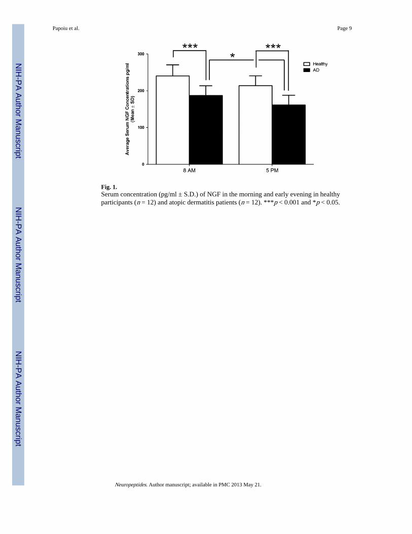
Fig. 1.Serum concentration (pg/ml ± S.D.) of NGF in the morning and early evening in healthyparticipants (n = 12) and atopic dermatitis patients (n = 12). ***p < 0.001 and *p < 0.05.
Papoiu et al. Page 9
Neuropeptides. Author manuscript; available in PMC 2013 May 21.
NIH
-PA Author Manuscript
NIH
-PA Author Manuscript
NIH
-PA Author Manuscript

Fig. 2.Dermal concentrations of NGF (pg/ml ± S.D.) in the morning and early evening in healthyparticipants (n = 17) in comparions with the eczematous areas and non-lesional areas of theskin in atopic dermatitis patients (n = 13). ***p < 0.001 in baseline conditions. Levels of thefree NGF in healthy skin and in non-lesional skin of atopic subjects are both siginificantlyhigher than in the eczematous areas in AD.
Papoiu et al. Page 10
Neuropeptides. Author manuscript; available in PMC 2013 May 21.
NIH
-PA Author Manuscript
NIH
-PA Author Manuscript
NIH
-PA Author Manuscript
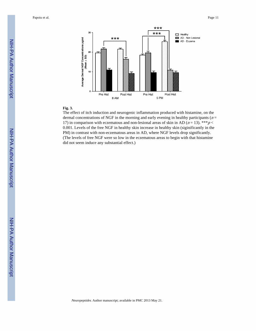
Fig. 3.The effect of itch induction and neurogenic inflammation produced with histamine, on thedermal concentrations of NGF in the morning and early evening in healthy participants (n =17) in comparison with eczematous and non-lesional areas of skin in AD (n = 13). ***p <0.001. Levels of the free NGF in healthy skin increase in healthy skin (siginificantly in thePM) in contrast with non-eczematous areas in AD, where NGF levels drop significantly.(The levels of free NGF were so low in the eczematous areas to begin with that histaminedid not seem induce any substantial effect.)
Papoiu et al. Page 11
Neuropeptides. Author manuscript; available in PMC 2013 May 21.
NIH
-PA Author Manuscript
NIH
-PA Author Manuscript
NIH
-PA Author Manuscript

NIH
-PA Author Manuscript
NIH
-PA Author Manuscript
NIH
-PA Author Manuscript
Papoiu et al. Page 12
Tabl
e 1
NG
F va
lues
mea
sure
d in
ser
um in
hea
lthy
and
AD
sub
ject
s (n
= 1
2), a
nd th
e co
rres
pond
ing
EA
SI s
core
s of
dis
ease
sev
erity
(pg
/ml)
.
AD
#E
ASI
Seru
m v
alue
sH
ealt
hy #
Seru
m v
alue
s
AM
PM
AM
PM
13.
222
019
21
286
244
22.
216
214
22
218
231
313
.422
615
03
288
189
42.
414
413
64
216
218
52.
214
812
85
208
255
614
.622
621
66
268
210
711
.919
015
57
236
234
83.
716
615
68
202
155
99.
618
617
59
287
164
1023
.520
016
710
201
245
1138
.818
415
811
196
180
125.
719
215
912
278
240
Neuropeptides. Author manuscript; available in PMC 2013 May 21.
NIH
-PA Author Manuscript
NIH
-PA Author Manuscript
NIH
-PA Author Manuscript
Papoiu et al. Page 13
Table 2
ANOVA analysis of the multiple factors affecting dermal NGF levels: disease status, time of collectingsamples during the day, histamine treatment and their combinations thereof. Legend: group = healthy or AD;period = before or after histamine application; time = AM or PM.
Source Type III tests of fixed effectsa
Numerator df Denominator df F Sig.
Intercept 1 40 4866.975 .000
Group 2 40 216.701 .000
Time 1 122 5.095 .026
Period 1 122 7.604 .007
Group × time 2 122 13.086 .000
Group × period 2 122 62.586 .000
Time × period 1 122 2.832 .095
aDependent variable: NGF levels in skin dialysate.
Neuropeptides. Author manuscript; available in PMC 2013 May 21.
NIH
-PA Author Manuscript
NIH
-PA Author Manuscript
NIH
-PA Author Manuscript
Papoiu et al. Page 14
Table 3
NGF levels in skin microdialysate (pg/ml) in the healthy group (n = 17).
Healthy skin # AM PM
Pre his Post his Pre his Post his
1 19.1 23.4 19.9 29.4
2 22.3 19.2 20.3 26.4
3 21.4 18.1 22 26.1
4 17.3 21.3 19.1 23.6
5 16.9 20.4 16.4 28.5
6 18.2 21.1 18.4 27.4
7 20.5 23.4 21.7 23.7
8 21.3 23.4 18.8 21.2
9 22.5 25.5 16.6 24.4
10 23.4 18.4 18.6 23.2
11 15.5 23.2 17.5 24.4
12 16.6 22.2 22.3 27.4
13 19.3 21.8 19.2 28.3
14 21.3 21.2 15.6 23.4
15 20.8 22.3 16.4 28.5
16 21.5 24.2 16.4 26.6
17 15.3 16.4 15.3 19.3
his = histamine.
Neuropeptides. Author manuscript; available in PMC 2013 May 21.

NIH
-PA Author Manuscript
NIH
-PA Author Manuscript
NIH
-PA Author Manuscript
Papoiu et al. Page 15
Tabl
e 4
NG
F le
vels
in s
kin
dial
ysat
e (p
g/m
l) in
the
AD
gro
up (
n =
13)
and
cor
resp
ondi
ng E
ASI
sco
res.
EA
SI s
core
AD
non
-les
iona
lA
D e
czem
a
#A
MP
MA
MP
M
Pre
his
Pos
t hi
sP
re h
isP
ost
his
Pre
his
Pos
t hi
sP
re h
isP
ost
his
11.9
121
.216
.318
.713
.411
.57.
5510
.55
11.9
3.2
218
.414
.420
.49.
211
.810
.17.
27.
2
13.4
324
.412
.225
.113
.48.
78.
27.
47.
3
2.2
420
.114
.214
.611
.213
.49.
213
.314
.4
5.7
519
.316
.622
.59.
87.
37.
314
.47.
2
23.5
622
.118
17.7
9.8
11.6
8.06
7.46
8.7
5.6
724
.317
.218
.310
.88.
215
.17.
911
.6
3.7
826
.416
.118
.614
.414
.28.
112
.98.
1
38.8
919
.823
.323
.47.
714
.77.
67.
87.
7
14.6
1024
.621
.519
.27.
97.
97
7.6
7.1
2.2
1118
.815
.517
.69.
415
.815
.27.
210
.8
2.4
1221
.214
.415
.511
.49.
47.
513
.114
.6
9.6
1320
.413
.823
.512
.27.
87.
97.
77.
5
his
= h
ista
min
e.
Neuropeptides. Author manuscript; available in PMC 2013 May 21.

NIH
-PA Author Manuscript
NIH
-PA Author Manuscript
NIH
-PA Author Manuscript
Papoiu et al. Page 16
Table 5
Correlation of various NGF concentrations in systemic circulation or in the skin with disease severity (EASIscores), in the atopic dermatitis group (n = 13).
NGF parameter Correlation type Statistical significance Spearman coeff. p
AM serum NGF level Direct 0.61* 0.037*
PM serum NGF level Direct 0.53 0.08
AM non-lesional skin NGF pre-histamine Direct n.s. n.s.
AM non-lesional skin NGF post-histamine Direct 0.53 0.059
PM non-lesional skin NGF pre-histamine Direct 0.6* 0.03*
PM non-lesional skin NGF post-histamine Inverse n.s. n.s.
AM eczematous skin NGF pre-histamine Inverse n.s. n.s.
AM eczematous skin NGF post-histamine Inverse n.s. n.s.
PM eczematous skin NGF pre-histamine Inverse −0.49 0.088
PM eczematous skin NGF post-histamine Inverse n.s. n.s.
n.s. = non-significant.
*p<0.05.
Neuropeptides. Author manuscript; available in PMC 2013 May 21.