
A Sphingosine1-Phosphate-Activated Calcium Channel Controlling Vascular Smooth Muscle Cell Motility
18
A Sphingosine-1–Phosphate–Activated Calcium Channel Controlling Vascular Smooth Muscle Cell Motility Shang-Zhong Xu,* Katsuhiko Muraki,* Fanning Zeng,* Jing Li,* Piruthivi Sukumar,* Samir Shah, Alexandra M. Dedman, Philippa K. Flemming, Damian McHugh, Jacqueline Naylor, Alex Cheong, Alan N. Bateson, Christopher M. Munsch, Karen E. Porter, David J. Beech Abstract—In a screen of potential lipid regulators of transient receptor potential (TRP) channels, we identified sphingosine-1–phosphate (S1P) as an activator of TRPC5. We explored the relevance to vascular biology because S1P is a key cardiovascular signaling molecule. TRPC5 is expressed in smooth muscle cells of human vein along with TRPC1, which forms a complex with TRPC5. Importantly, S1P also activates the TRPC5–TRPC1 heteromultimeric channel. Because TRPC channels are linked to neuronal growth cone extension, we considered a related concept for smooth muscle. We find S1P stimulates smooth muscle cell motility, and that this is inhibited by E3-targeted anti-TRPC5 antibody. Ion permeation involving TRPC5 is crucial because S1P-evoked motility is also suppressed by the channel blocker 2-aminoethoxydiphenyl borate or a TRPC5 ion-pore mutant. S1P acts on TRPC5 via two mechanisms, one extracellular and one intracellular, consistent with its bipolar signaling functions. The extracellular effect appears to have a primary role in S1P-evoked cell motility. The data suggest S1P sensing by TRPC5 calcium channel is a mechanism contributing to vascular smooth muscle adaptation. (Circ Res. 2006;98:1381-1389.) Key Words: vascular smooth muscle vein sphingosine-1–phosphate transient receptor potential calcium channel S phingosine-1-phosphate (S1P) has emerged as a major endogenous signaling phospholipid with diverse roles in yeast, plants, and mammals. 1 Proposed functions include the regulation of cell proliferation, migration, programmed death, and pathological processes including cancer, asthma, inflam- mation, and trauma. There has been particular interest in the role of S1P in the cardiovascular system, where it accumu- lates in atherosclerotic lesions and plays a role in ischemic preconditioning of the heart. 2–5 S1P is derived from the phosphorylation of sphingosine catalyzed by sphingosine kinase, sphingosine being from ceramide and ceramide from sphingomyelin, a constituent lipid of signaling microdomains of plasma membrane lipid rafts and caveolae. 3 S1P is detected in serum at almost 1 mol/L, although protein binding impacts on the available concentration and local concentra- tions may vary substantially. 6 S1P is quite unusual among signaling molecules in having separate intracellular and extracellular effects. 1,4,7,8 It affects vascular smooth muscle cell migration, 9,10 evokes contraction of rat mesenteric artery, 11 and slows pacemaker activity of the sino-atrial node of the heart. 12 The underlying mechanisms are only partially worked out, but vascular smooth muscle cells respond to S1P with transient followed by sustained elevation of the cytosolic Ca 2 concentration, 10,11,13,14 whereas cardiac myocytes show activation of potassium current and S1P-evoked “Ca 2 deregulation,” depending on extracellular Ca 2 . 12,15 Despite positive effects on Ca 2 sig- naling, the molecular basis of a Ca 2 channel stimulated by S1P is unknown. L-type voltage-gated Ca 2 channels are inhibited by S1P. 12 The Drosophila transient receptor potential (TRP) channel has provided the foundation for discovery of many novel Ca 2 - or Na -permeable plasma membrane channels, 16,17 which are candidates for the less well understood cationic channels of the mammalian cardiovascular system. 18 –23 Searches for activation mechanisms are revealing TRP chan- nels as sensors of temperature, pheromones, osmolarity, and gustatory stimuli. 24,25 However, some TRP channels are expressed outside sensory systems, and activation mecha- nisms are elusive. 17,20 TRPC5 has been associated with the central nervous system and is a regulator of growth cone formation. 26 –28 There is rapid vesicular insertion regulated by growth factors, 29 but this is not the mechanism causing channel opening. TRPC5 may be important outside the Original received July 5, 2005; resubmission received February 13, 2006; revised resubmission received March 23, 2006; accepted April 20, 2006. From the Institute of Membrane and Systems Biology (S.-Z.X., F.Z., J.L., P.S., A.M.D., P.K.F., D.M., J.N., A.C., A.N.B., D.J.B.), University of Leeds, United Kingdom; Cellular Pharmacology (K.M.), School of Pharmacy, Aichi Gakuin University, Nagoya, Japan; School of Medicine (K.E.P.), University of Leeds, United Kingdom; and Yorkshire Heart Centre (S.S., C.M.M.), General Infirmary at Leeds, United Kingdom. *These authors contributed equally to this study. K.M. is a visiting scientist to the University of Leeds. Correspondence to Professor D.J. Beech, Institute of Membrane and Systems Biology, University of Leeds, Leeds, LS2 9JT, UK. E-mail [email protected] © 2006 American Heart Association, Inc. Circulation Research is available at http://circres.ahajournals.org DOI: 10.1161/01.RES.0000225284.36490.a2 1381 Cellular Biology by guest on January 27, 2016 http://circres.ahajournals.org/ Downloaded from by guest on January 27, 2016 http://circres.ahajournals.org/ Downloaded from by guest on January 27, 2016 http://circres.ahajournals.org/ Downloaded from by guest on January 27, 2016 http://circres.ahajournals.org/ Downloaded from by guest on January 27, 2016 http://circres.ahajournals.org/ Downloaded from by guest on January 27, 2016 http://circres.ahajournals.org/ Downloaded from by guest on January 27, 2016 http://circres.ahajournals.org/ Downloaded from by guest on January 27, 2016 http://circres.ahajournals.org/ Downloaded from by guest on January 27, 2016 http://circres.ahajournals.org/ Downloaded from by guest on January 27, 2016 http://circres.ahajournals.org/ Downloaded from
-
Upload
independent -
Category
Documents
-
view
0 -
download
0
Transcript of A Sphingosine1-Phosphate-Activated Calcium Channel Controlling Vascular Smooth Muscle Cell Motility

A Sphingosine-1–Phosphate–Activated Calcium ChannelControlling Vascular Smooth Muscle Cell Motility
Shang-Zhong Xu,* Katsuhiko Muraki,* Fanning Zeng,* Jing Li,* Piruthivi Sukumar,* Samir Shah,Alexandra M. Dedman, Philippa K. Flemming, Damian McHugh, Jacqueline Naylor, Alex Cheong,
Alan N. Bateson, Christopher M. Munsch, Karen E. Porter, David J. Beech
Abstract—In a screen of potential lipid regulators of transient receptor potential (TRP) channels, we identifiedsphingosine-1–phosphate (S1P) as an activator of TRPC5. We explored the relevance to vascular biology because S1Pis a key cardiovascular signaling molecule. TRPC5 is expressed in smooth muscle cells of human vein along withTRPC1, which forms a complex with TRPC5. Importantly, S1P also activates the TRPC5–TRPC1 heteromultimericchannel. Because TRPC channels are linked to neuronal growth cone extension, we considered a related concept forsmooth muscle. We find S1P stimulates smooth muscle cell motility, and that this is inhibited by E3-targetedanti-TRPC5 antibody. Ion permeation involving TRPC5 is crucial because S1P-evoked motility is also suppressed bythe channel blocker 2-aminoethoxydiphenyl borate or a TRPC5 ion-pore mutant. S1P acts on TRPC5 via twomechanisms, one extracellular and one intracellular, consistent with its bipolar signaling functions. The extracellulareffect appears to have a primary role in S1P-evoked cell motility. The data suggest S1P sensing by TRPC5 calciumchannel is a mechanism contributing to vascular smooth muscle adaptation. (Circ Res. 2006;98:1381-1389.)
Key Words: vascular smooth muscle � vein � sphingosine-1–phosphate � transient receptor potential� calcium channel
Sphingosine-1-phosphate (S1P) has emerged as a majorendogenous signaling phospholipid with diverse roles in
yeast, plants, and mammals.1 Proposed functions include theregulation of cell proliferation, migration, programmed death,and pathological processes including cancer, asthma, inflam-mation, and trauma. There has been particular interest in therole of S1P in the cardiovascular system, where it accumu-lates in atherosclerotic lesions and plays a role in ischemicpreconditioning of the heart.2–5 S1P is derived from thephosphorylation of sphingosine catalyzed by sphingosinekinase, sphingosine being from ceramide and ceramide fromsphingomyelin, a constituent lipid of signaling microdomainsof plasma membrane lipid rafts and caveolae.3 S1P is detectedin serum at almost 1 �mol/L, although protein bindingimpacts on the available concentration and local concentra-tions may vary substantially.6
S1P is quite unusual among signaling molecules in havingseparate intracellular and extracellular effects.1,4,7,8 It affectsvascular smooth muscle cell migration,9,10 evokes contractionof rat mesenteric artery,11 and slows pacemaker activity of thesino-atrial node of the heart.12 The underlying mechanismsare only partially worked out, but vascular smooth muscle
cells respond to S1P with transient followed by sustainedelevation of the cytosolic Ca2� concentration,10,11,13,14
whereas cardiac myocytes show activation of potassiumcurrent and S1P-evoked “Ca2� deregulation,” depending onextracellular Ca2�.12,15 Despite positive effects on Ca2� sig-naling, the molecular basis of a Ca2� channel stimulated byS1P is unknown. L-type voltage-gated Ca2� channels areinhibited by S1P.12
The Drosophila transient receptor potential (TRP) channelhas provided the foundation for discovery of many novelCa2�- or Na�-permeable plasma membrane channels,16,17
which are candidates for the less well understood cationicchannels of the mammalian cardiovascular system.18 –23
Searches for activation mechanisms are revealing TRP chan-nels as sensors of temperature, pheromones, osmolarity, andgustatory stimuli.24,25 However, some TRP channels areexpressed outside sensory systems, and activation mecha-nisms are elusive.17,20 TRPC5 has been associated with thecentral nervous system and is a regulator of growth coneformation.26–28 There is rapid vesicular insertion regulated bygrowth factors,29 but this is not the mechanism causingchannel opening. TRPC5 may be important outside the
Original received July 5, 2005; resubmission received February 13, 2006; revised resubmission received March 23, 2006; accepted April 20, 2006.From the Institute of Membrane and Systems Biology (S.-Z.X., F.Z., J.L., P.S., A.M.D., P.K.F., D.M., J.N., A.C., A.N.B., D.J.B.), University of Leeds,
United Kingdom; Cellular Pharmacology (K.M.), School of Pharmacy, Aichi Gakuin University, Nagoya, Japan; School of Medicine (K.E.P.), Universityof Leeds, United Kingdom; and Yorkshire Heart Centre (S.S., C.M.M.), General Infirmary at Leeds, United Kingdom.
*These authors contributed equally to this study.K.M. is a visiting scientist to the University of Leeds.Correspondence to Professor D.J. Beech, Institute of Membrane and Systems Biology, University of Leeds, Leeds, LS2 9JT, UK. E-mail
[email protected]© 2006 American Heart Association, Inc.
Circulation Research is available at http://circres.ahajournals.org DOI: 10.1161/01.RES.0000225284.36490.a2
1381
Cellular Biology
by guest on January 27, 2016http://circres.ahajournals.org/Downloaded from by guest on January 27, 2016http://circres.ahajournals.org/Downloaded from by guest on January 27, 2016http://circres.ahajournals.org/Downloaded from by guest on January 27, 2016http://circres.ahajournals.org/Downloaded from by guest on January 27, 2016http://circres.ahajournals.org/Downloaded from by guest on January 27, 2016http://circres.ahajournals.org/Downloaded from by guest on January 27, 2016http://circres.ahajournals.org/Downloaded from by guest on January 27, 2016http://circres.ahajournals.org/Downloaded from by guest on January 27, 2016http://circres.ahajournals.org/Downloaded from by guest on January 27, 2016http://circres.ahajournals.org/Downloaded from

nervous system because its mRNA species is detected in arange of animal tissues, including human heart and bloodvessels.20,30–32 Furthermore, downregulation of Ca2�-ATPasein cardiac myocytes leads to compensatory upregulation ofTRPC5.33 However, activation signals for TRPC5 remainuncertain. One possibility is that TRPC5 exists to respond topassive depletion of Ca2� stores because human TRPC5activity is enhanced by store depletion,34 and vascular smoothand cardiac muscle cells exhibit store-operated Ca2� en-try.20,35,36 However, in some instances, TRPC5 is unrespon-sive to store depletion,37 and the biological relevance of theoften strong passive store depletion in experimental situationsremains uncertain. On the assumption that key endogenousregulators of TRPC5 were yet to be discovered, we searchedfor novel activators.
Materials and MethodsHuman TissueFreshly discarded human tissue samples were obtained anonymouslyand with informed consent from patients undergoing open heartsurgery in the general infirmary at Leeds. Approval was granted bythe Leeds teaching hospitals local research ethics committee. Saphe-nous vein was transported to the laboratory in Hanks’ solution(in mmol/L): 137 NaCl, 5.4 KCl, 0.01 CaCl2, 0.34 NaH2PO4, 0.44K2HPO4, 8 D-glucose, and 5 HEPES, and processed on the day of theoperation. For RNA isolation and cell culture, medial layer wasextracted by dissection.
cDNA ExpressionFull-length human TRPC5 cDNA (accession number AF054568)was cloned and stably expressed in human embryonic kidney 293cells (HEK 293 cells; T-Rex cells; Invitrogen).34 Cells were grown inDMEM–F12 media (Invitrogen) and supplemented with 10% FBSand penicillin/streptomycin at 37°C in a 5% CO2 incubator. Whereindicated, cells were incubated with tetracycline (Tet�; 1 �g/mL)for 24 to 48 hours to induce the expression of TRPC5. Human
TRPC1 cDNA (accession number X89066; a gift from C. Montell,Johns Hopkins University, Baltimore, Md) was subcloned intoEcoRV site of the bicistronic vector pIRES yellow fluorescentprotein (YFP; BD Clontech), transiently transfected into cells withFuGene 6 (Roche), and subcultured onto coated glass coverslips 24hours later. Cells were then cultured for an additional 24 hours withor without the presence of 1 �g/mL of tetracycline. Dominant-negative (DN) TRPC5 is a triple alanine mutation of the conservedleucine, phenylalanine, tryptophan (LFW) sequence in the ion-pore.40 DN function was confirmed in HEK–TRPC5 cells (supple-mental Figure I, available online at http://circres.ahajournals.org).
Immunofluorescence, Western Blotting,and ImmunoprecipitationCells adhered to polylysine-coated slides were fixed in 2% parafor-maldehyde (30 minutes) and immersed in �20°C methanol (1minute) and 1% BSA with 0.1% Triton X-100 for 1 hour. Incubationin primary antibody was for 2 hours and secondary antibody (goatanti-rabbit IgG-Cy3) for 1 hour. Western blotting protocols weresimilar to those described.18 Small pieces of tissue were placed inPBS containing protease inhibitor cocktail (Sigma) and lysed inLaemmli buffer containing 320 �mol/L dithiothreitol at 80°C to100°C (15 minutes). Proteins were separated on 8% SDS-PAGEgels, transferred to nitrocellulose membrane (Millipore), and probedwith primary antibody and secondary antibody conjugated withhorseradish peroxidase. Membranes were washed with PBS andlabeling detected by ECL plus (Amersham Pharmacia Biotech).Except for T5Chk, anti-TRPC antibodies were custom-made inrabbit (Sigma-Genosys) to unique peptides. T5Chk antibody wasmade in chicken to the C-terminal peptide CVFETWGEACDLLM-HKWGDGQ. T5C3 was made to the C-terminal peptide CK-LLDSSEDVFETWGE and T5E3 to CYETRAIDEPNNCKG. Unlessindicated, antibodies were affinity purified on columns containingimmobilized peptides and dialysed in PBS. T1E3 antibody isdescribed.18 Rabbit anti-protein S100 and the monoclonal antibodyanti-CD31 were from Dako Ltd. For immunoprecipitation (IP) ofsaphenous vein, medial layer was homogenized (Polytron; 2�15 s;setting 6) in 10 volumes of IP buffer containing 20 mmol/LHEPES-NaOH, pH 7.5, 1% Triton X-100, 150 mmol/L NaCl, andprotease inhibitor cocktail. Homogenate was centrifuged at maxi-
Figure 1. Identification of S1P as a novelactivator of human TRPC5. a,Tetracycline-inducible expression ofhuman TRPC5 in HEK 293 cells. T5C3antibody detected (red labeling) TRPC5in induced (Tet�) but not control (Tet�)cells. Cell nuclei were counterstainedwith 4�,6-diamidino-2-phenylindole DAPI(blue). For clarity, the Tet� anti-TRPC5stain is shown without the DAPI overlay.Bar�10 �m. b, Typical single cellresponses to S1P at 1, 3, and 10 �mol/Lin cells expressing TRPC5 (Tet�). Con-trol cell (Tet�) failed to respond to3 �mol/L S1P. c, As in b, but mean dataas the change in Ca2� indicator fluores-cence ratio from the baseline. d, Meandata for responses 5 and 11 minutesafter starting the application of0.1 �mol/L S1P.
1382 Circulation Research June 9, 2006
by guest on January 27, 2016http://circres.ahajournals.org/Downloaded from
mum speed in a microfuge for 20 minutes to remove cell debris. Thesupernatant was washed with Protein A (Sigma) or G (Pierce)sepharose (10 �g) and centrifuged again at maximum speed for 20minutes. The supernatant (200 �g soluble protein) was used for IPwith 2 �g antibody and the mixture rotated overnight at 4°C.
Reverse Transcription–Polymerase Chain ReactionFor RT-PCR, see the online supplement, available athttp://circres.ahajournals.org.
Ca2� ImagingHEK 293 cell recordings were made using fura-PE3 AM and Ca2�
imaging, whereas smooth muscle cell recordings used a 96-wellfluorimetry. Recordings were in standard bath solution containing(in mmol/L): 130 NaCl, 5 KCl, 8 D-glucose, 10 HEPES, 1.2 MgCl2,and 1.5 CaCl2, pH titrated to 7.4 with NaOH. For Ca2�-free solution,CaCl2 was omitted (also see online supplement).
ElectrophysiologyFor whole-cell experiments and outside-out patches, the patchpipette contained (in mmol/L): 135 CsCl, 2 MgCl2, 1 EGTA, 10HEPES, 5 sodium ATP, titrated to pH 7.2 with CsOH (0.1 mmol/Lsodium GTP or 1 mmol/L GDP-�–S was added when specified).Standard bath solution was used. For inside-out patch experiments,the patch pipette solution contained standard bath solution and thebath (superfusion) solution (in mmol/L): 135 CsCl, 2 MgCl2, 1EGTA, 10 HEPES, and 5 sodium ATP, titrated to pH 7.4 with CsOH(also see online supplement).
Cell MotilityHuman saphenous vein smooth muscle cells were prepared byexplant technique and passaged up to 4 times. Cells were grown toconfluence in 24-well plates and DMEM supplemented with 10%FBS and penicillin/streptomycin at 37°C in 5% CO2. Cells werewashed twice with PBS and a linear scrape of �0.3 mm width wasmade through the monolayer with a pipette tip. The cells werecultured with DMEM containing 1% serum and with or without S1P(1 �mol/L), T5E3 antibody (10 �g/mL), 75 �mol/L2-aminoethoxydiphenyl borate (2-APB), or vehicle (methanol orPBS), as specified. After 24 hours, the linear wound was delineatedand the number of cells moving into the wound counted. ForDN-TRPC5 studies, 3 �g of DNA was delivered to cells 48 hoursbefore making the linear wound using the Basic Nucleofector Kit forprimary smooth muscle cells (Amaxa Biosystems). Transfectionefficiency was �70%. For pertussis toxin (1 �g/mL) experiments,preincubation was for 4 hours before application of S1P. As acontrol, pertussis toxin was boiled for 10 minutes before use.
ReagentsUnless indicated, salts and reagents were from Sigma or BDH (BritishDrug House). Sphingosine-1–phosphate (S1P) was purchased fromSigma or Biomol Research Laboratories. The solvent was methanol, andstock concentrations were 10 mmol/L. The final methanol concentrationin the bath (superfusion) solution was �0.1% (v/v), and this was keptconstant throughout (before, during, and after S1P application). Noeffects of methanol were observed. Lithium GDP-�–S, pertussis toxin,and 2-APB were from Sigma. U73122 (1-[6-((17�-3-methoxyestra-1,3,5(10)-trien-17-yl)amino)hexyl]-1H-pyrrole-2,5-dione) and U73343were from Calbiochem.
Data AnalysisHuman tissue/cell experiments were repeated on samples from atleast three patients, yielding similar results. Averaged numerical dataare presented as mean�SEM. Data sets with two groups werecompared by unpaired Student t test and three groups by Tukey–Kramer multiple comparison, with significance indicated by P�0.05(*) or 0.01 (**). For Ca2� imaging, all mean data were based on fourindependent experiments (coverslips) and measurements from 30cells on each coverslip. For patch-clamp experiments, n is the
number of independent experiments on cells or patches. For theinjury assay, n is the number of images used for analysis. Directcomparisons were made on the same batch of cells, with test andcontrol experiments on the same day.
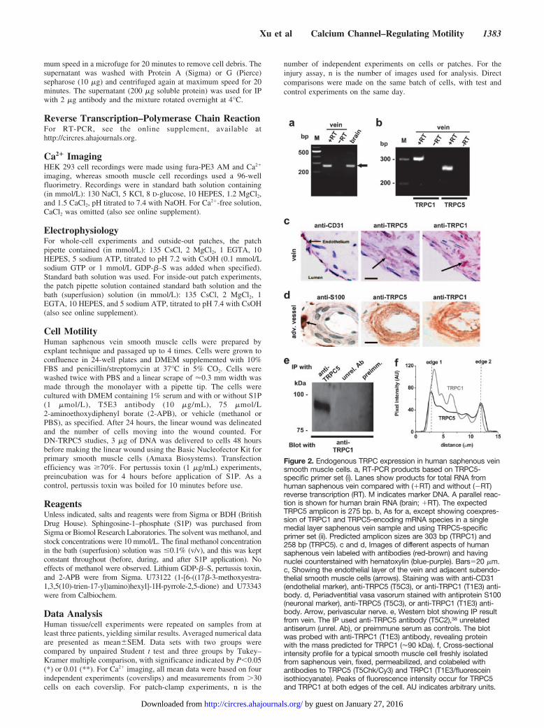
Figure 2. Endogenous TRPC expression in human saphenous veinsmooth muscle cells. a, RT-PCR products based on TRPC5-specific primer set (i). Lanes show products for total RNA fromhuman saphenous vein compared with (�RT) and without (�RT)reverse transcription (RT). M indicates marker DNA. A parallel reac-tion is shown for human brain RNA (brain; �RT). The expectedTRPC5 amplicon is 275 bp. b, As for a, except showing coexpres-sion of TRPC1 and TRPC5-encoding mRNA species in a singlemedial layer saphenous vein sample and using TRPC5-specificprimer set (ii). Predicted amplicon sizes are 303 bp (TRPC1) and258 bp (TRPC5). c and d, Images of different aspects of humansaphenous vein labeled with antibodies (red-brown) and havingnuclei counterstained with hematoxylin (blue-purple). Bars�20 �m.c, Showing the endothelial layer of the vein and adjacent subendo-thelial smooth muscle cells (arrows). Staining was with anti-CD31(endothelial marker), anti-TRPC5 (T5C3), or anti-TRPC1 (T1E3) anti-body. d, Periadventitial vasa vasorum stained with antiprotein S100(neuronal marker), anti-TRPC5 (T5C3), or anti-TRPC1 (T1E3) anti-body. Arrow, perivascular nerve. e, Western blot showing IP resultfrom vein. The IP used anti-TRPC5 antibody (T5C2),38 unrelatedantiserum (unrel. Ab), or preimmune serum as controls. The blotwas probed with anti-TRPC1 (T1E3) antibody, revealing proteinwith the mass predicted for TRPC1 (�90 kDa). f, Cross-sectionalintensity profile for a typical smooth muscle cell freshly isolatedfrom saphenous vein, fixed, permeabilized, and colabeled withantibodies to TRPC5 (T5Chk/Cy3) and TRPC1 (T1E3/fluoresceinisothiocyanate). Peaks of fluorescence intensity occur for TRPC5and TRPC1 at both edges of the cell. AU indicates arbitrary units.
Xu et al Calcium Channel–Regulating Motility 1383
by guest on January 27, 2016http://circres.ahajournals.org/Downloaded from

ResultsS1P Is a Novel Activator of TRPC5To test for novel activators, human TRPC5 was stablyexpressed in HEK 293 cells under a tetracycline-dependentpromoter. The system gave a defined TRPC5 signal in whichTRPC5-expressing cells (tetracycline-induced; Tet�) werecompared directly with control cells from the same batch:cells that do not express TRPC5 (Tet�) as shown byanti-TRPC5 antibody (Figure 1a). We noticed stimulation byS1P in Tet� but not Tet� cells (Figure 1b through 1d).Within a 5-minute application period, S1P was effective at 1to 10 �mol/L. S1P (0.1 �mol/L) was near the threshold foractivation, producing a slow but significant effect (Figure1d).
TRPC5-TRPC1 Heteromultimer in Human Veinand Activation by S1PBecause S1P is a cardiovascular signal, we looked forrelevant expression of TRPC5, focusing on human saphenousvein, which is a coronary artery bypass graft prone to failureattributable to unwanted smooth muscle cell growth. RNAencoding TRPC5 was detected, as in human brain (Figure2a). Because there is strong evidence TRPC1 forms a func-tional heteromultimeric channel with TRPC5,27 we alsolooked for RNA encoding TRPC1 in the same samples; bothRNA species were detected (Figure 2b). Furthermore, TRPC5and TRPC1 proteins were present in subendothelial smoothmuscle cells of the pre-existing venous intima and in smoothmuscle cells of vaso vasorum (Figure 2c and 2d; supplemen-tal Figure II).38 This is important because ion channels formnot only through TRPC5 homomultimerization but alsoTRPC1–TRPC5 heteromultimerization (supplemental FigureIII).27 IP of TRPC5 and TRPC1 in saphenous vein samples(Figure 2e) and colocalization of the proteins at the plasmamembrane (Figure 2f) support the conclusion that TRPC5 and
TRPC1 function together. Therefore, if S1P regulation ofTRPC5 has vascular relevance, it should also activateTRPC1–TRPC5 heteromultimers (Figure 3a). Expression ofhuman TRPC1 was achieved with YFP as a marker oftransfection (Figure 3b). In cells coexpressing TRPC1 andTRPC5, S1P activated a large current (Figure 3c and 3e).Patch-clamp recording was used because the current-voltage(I-V) relationship for the TRPC1-TRPC5 heteromultimerdiffers from that for TRPC5 alone27 (Figure 3d; compare withFigure 5g), enabling us to be confident of studying theheteromultimer. Cells expressing TRPC1 alone were notresponsive to S1P (Figure 3e).
TRPC5 Role in S1P-Evoked MotilityTRPC channels play roles in neuronal growth cone extensionand turning,28 and S1P regulates migration of vascularsmooth muscle cells.9,10 We therefore prepared planar cul-tures of human saphenous vein smooth muscle cells toexplore the potential relevance to cell motility. These cellsexpress TRPC5 and TRPC1 at the surface, as shown bylabeling of cells with antibodies targeted to extracellularepitopes (Figure 4a). Furthermore, S1P evoked Ca2� entryindependently of Ca2� release (Figure 4b). To explore cellmotility, we made a linear deletion in the culture andmeasured the number of cells moving into the vacated spaceover a 24-hour period (Figure 4c). S1P strongly enhanced themovement of cells (Figure 4d).
E3 targeting can be used to design antibodies that blockchannels specifically when added to extracellular solu-tion.42 Such an antibody was described for TRPC5(T5E3).42 T5E3 inhibited the effect of S1P on motility(Figure 4d), showing endogenous TRPC5 is involved.Highly specific chemical blockers of TRPC5 have not beendiscovered, but 2-APB is probably the best option39 andimportantly had an inhibitory effect like T5E3 (Figure 4d).
Figure 3. Activation of the TRPC5–TRPC1 het-eromultimer. a, Schematic illustrating a plan viewof a presumed heterotetrameric arrangement ofTRPC1 (1) and TRPC5 (5) with ion permeationthrough the central pore. b, Example imageshowing transfection of some Tet� HEK 293cells with cDNA encoding TRPC1 and (E)YFP(orange color, white arrow). To reveal all cells,they were loaded with fura-2 indicator dye(green) for illustration purposes. Bar�20 �m. c,Whole-cell patch-clamp recording from a cellcoexpressing TRPC5 and TRPC1, showing acti-vation of current by 2 �mol/L S1P. d, The I-V forthe current induced by S1P in c during a rampchange in voltage from �90 to �100 mV over a1-s period. Note the lack of inflection in the I-Vnear 0 mV, which is a characteristic of TRPC5alone (Figure 5g) but not the TRPC5–TRPC1 het-eromultimer.27,29 e, As for c, but mean�SEMdata (n�3 for each) showing the fold induction ofcurrent at �80 mV in response to methanol vehi-cle (MeOH) or S1P applied to cells expressingTRPC5 and TRPC1 (Tet� with TRPC1/YFP) orTRPC1 alone (Tet� with TRPC1/YFP). Controlwas the pre-MeOH/S1P current.
1384 Circulation Research June 9, 2006
by guest on January 27, 2016http://circres.ahajournals.org/Downloaded from

2-APB blocks ion permeation in TRPC5,39 and thuseffectiveness of this agent suggests S1P-evoked motilitydepends critically on ion flux through the TRPC channelcomplex. As an independent test of this idea, we made anion-pore mutant of TRPC5 that fails to pass current andacts as a DN (supplemental Figure I), presumably becauseit damages ion permeation by entering in the heteromulti-meric complex.40 Transfection of this mutant into vascularsmooth muscle cells inhibited S1P-evoked motility (Figure4e) consistent with the necessity of ion permeation.
Mechanism of Action of Extracellular S1PTo further understand the effect of S1P, we explored itsmechanism of action, initially hypothesizing that it might actrelatively directly, like lysophosphatidylcholine.41 To make adefinitive analysis of TRPC5, we focused on the TRPC5-expressing (Tet�) HEK 293 cells. In the absence of extra-cellular Ca2�, there was little response to S1P, consistent withS1P activating TRPC5-mediated Ca2� influx (Figure 5a and5f). Furthermore, the S1P effect was not prevented bydepletion of calcium stores by thapsigargin (Figure 5b and5f). In control cells in the absence of a lanthanide, we
detected small S1P-evoked Ca2� release signals (data notshown), suggesting that the lanthanide diminished the Ca2�
release event, but also that the cells express G-protein–coupled receptors for S1P.1,4 Often, such receptors couple totheir effectors via pertussis toxin-sensitive Gi/o GTP-bindingproteins.4 Treatment of cells with pertussis toxin inhibited theS1P response (Figure 5c; compare with Figure 5d; Figure 5f).In contrast, TRPC5 activation by carbachol acting at endog-enous muscarinic receptors34 was unaffected. S1P receptorsalso couple to phospholipase C (PLC).4 Consistent with PLCinvolvement, the PLC inhibitor U73122 inhibited the S1Presponse (Figure 5e and 5f), whereas the chemically relatedU73343 (a poor inhibitor of PLC) was ineffective (2 inde-pendent experiments).
S1P also activates TRPC5 in whole-cell voltage-clamprecordings, showing the characteristic double-rectifying I-Vrelationship (Figure 5g34,37). Consistent with G-protein in-volvement, the effect of S1P was prevented when GDP-�–Sreplaced GTP in the patch pipette (Figure 5h). Therefore,extracellular S1P activates TRPC5 via a pertussis toxin-sensitive G-protein pathway, indicating receptor activation37
of TRPC5. The signal after PLC is unknown.
Figure 4. Role in vascular smooth musclecell motility. a, Cultured human saphenousvein smooth muscle cells labeled with anti-TRPC5 (T5E3) or anti-TRPC1 (T1E3) anti-body. The parallel controls were T5E3preadsorbed to its antigenic peptide orT1E3 boiled for 10 minutes before use. Thecells are very flat, so staining is notrestricted to the perimeter of the cells.Bar�20 �m. b, For the same batch of cellsas a, the change in intracellular Ca2� evokedby 1 �mol/L S1P in standard bath solutionwith (Tg Ca) or without (Ca) pretreatmentwith 1 �mol/L thapsigargin (Tg), or in Ca2�-free bath solution without Tg pretreatment (0Ca). Each value is for 16 independent wells.c, Image showing the principle of the cellmotility assay. Cell nuclei are stained withethidium bromide (orange/red). The bracketlabels the area originally stripped of cells bythe pipette tip, and the white dotted lineindicates the boundary. In this example,cells had started to repopulate the space. d,From four independent experiments, themean�SEM number of cells (per 0.3 mm2)repopulating the area 24 hours after chang-ing to 1% serum with methanol vehicle(MeOH; n�36), 1 �mol/L S1P with PBS(n�45), S1P with T5E3 antibody in PBS(n�36), or S1P with 75 �mol/L 2-APB (n�8).e, Cells were transfected with vector lackingor containing DNA encoding DN mutantTRPC5. As in d, cells were counted afterexposure to 1 �mol/L S1P or methanol(n�30 to 42). All comparisons were made inparallel on the same batch of cells.
Xu et al Calcium Channel–Regulating Motility 1385
by guest on January 27, 2016http://circres.ahajournals.org/Downloaded from

Ionotropic Receptor for Intracellular S1PBased on the concept of S1P as both an extracellular andintracellular signaling molecule,7,8 we explored whetherthere is also an intracellular effect on TRPC5. Inside-outmembrane patches were used, enabling exposure of onlythe intracellular face of the channel to the test agent.Unitary events were detected in response to S1P in Tet�but not Tet� cells (Figure 6a). Amplitude histograms showthe unitary current events evoked by S1P were indistin-guishable in size from those evoked by gadolinium (Figure6b), and the I-V relationship, which is linear for the singlechannel current, was the same (Figure 6c). An exampletime series plot for single channel activity shows stimula-tion by S1P (Figure 6d). Some patches contained multiplechannels and thus exhibited macroscopic currents in re-
sponse to S1P with the characteristic double-rectifying I-V(supplemental Figure IV). Mean data show an effect inTet� but not Tet� cells (Figure 6e). In contrast, and alsoin the absence of GTP, S1P had no effect when bath-applied to outside-out patches, whereas lanthanum, whichacts externally,37 activated the channels (Figure 6f).
Effect of Pertussis Toxin on S1P-Evoked MotilityThe above data suggest S1P can activate TRPC5 through twomechanisms. Because activation of S1P receptors can lead toaccumulation of intracellular S1P and S1P may be trans-ported across the membrane,3 the effect of exogenous S1P(Figure 4c through 4e) could reflect extracellular or intracel-lular actions of S1P. However, we found pertussis toxin
Figure 5. Activation via G-protein signaling.All cells were induced to express humanTRPC5 (Tet�), except as indicated in h.S1P and carbachol (CCH) were bath-applied at 3 and 100 �mol/L, respectively.Example traces for single cells are shown.a, Effect of S1P and CCH in the absence ofCa2� added to the bath solution (0 Ca2�). bthrough e, Bath Ca2� (1.5 mmol/L) waspresent throughout. b, S1P response afterpretreatment with 1 �mol/L thapsigargin(Tg) for 0.5 hours, which prevents CCH-evoked Ca2� release.34 c, S1P responseafter preincubation for 4 to 5 hours with 1�g/mL pertussis toxin and 0.5 hours with1 �mol/L thapsigargin. d, As for c, but per-tussis toxin was omitted. e, S1P responseafter pretreatment with 10 �mol/L U73122(and 1 �mol/L thapsigargin) for 0.5 hours. f,Summary data for the experimentsdescribed in a through e. g and h, Whole-cell voltage-clamp recordings from cellsinduced to express TRPC5 (Tet�) or not(Tet�). A 1-s ramp change in voltage from�100 to �100 mV was applied every 10 sfrom a holding potential of �60 mV. g, I-Vdetermined during the voltage-ramp forcurrent induced by bath-applied 10 �mol/LS1P with 0.1 mmol/L GTP in the patchpipette. h, Mean (n�4 to 5) current ampli-tudes at the indicated voltages for whole-cell recordings and bath-applied 10 �mol/LS1P; 0.1 mmol/L GTP or 1 mmol/L GDP-�–S was included in the patch pipette solu-tion, as indicated.
1386 Circulation Research June 9, 2006
by guest on January 27, 2016http://circres.ahajournals.org/Downloaded from

inhibited the effect of S1P on cell motility (Figure 7),suggesting requirement for the extracellular action of S1P.
DiscussionThe study identifies a calcium channel activated by S1P. Thechannel contains TRPC5 protein, which is sufficient for S1Psensitivity and contributes to ion permeation. The finding ledus to discover that S1P evokes vascular smooth muscle cellmotility via a mechanism involving TRPC5. TRPC5 isnatively associated with TRPC1 in these cells, and S1Pactivates channels formed by heteromultimerization ofTRPC5 and TRPC1. This finding does not exclude involve-ment of additional TRP channels linked to TRPC5/TRPC1
and expressed in vascular smooth muscle.20 The relationshipwith cell motility may be a general concept for TRPCchannels because they also regulate neuronal growth coneextension and turning.28 TRPC5 sensitivity to other regula-tors34,37,41 suggests these might in turn act via TRPC5 toregulate cell motility. Intriguingly, and paralleling with thedual signaling function of S1P, TRPC5 is a bipolar target forS1P, showing activation by extracellular and intracellularpathways. The extracellular effect involves G-protein–cou-pled receptor activation, whereas the intracellular effectsurvives in inside-out membrane patches, suggesting TRPC5is an ionotropic receptor for intracellular S1P, and providingone of the few known intracellular targets for S1P. Intrigu-
Figure 6. Ionotropic receptor for intracel-lular S1P. Inside-out (a through e) andoutside-out (f) excised patch recordingsfrom cells expressing TRPC5 (Tet�),except for the control (Tet�) data indi-cated in a, b, and e. a, Original tracesshowing unitary current events inresponse to bath-applied 3 �mol/L S1Pin a patch from a Tet� but not Tet� cell.The calibration bars are 5 pA (vertical)and 25 ms. b, Amplitude histograms. Forthe Tet�/Gd3� data set, the calibrationbars for the example trace are 2 pA (ver-tical) and 5 ms, and Gd3� (10 �mol/L)was in the patch pipette. c, Mean unitarycurrent sizes for S1P- and Gd3�-evokedsingle channel events plotted againstvoltage. Straight lines were fitted, andthe mean unitary conductance was41�1.5 pS (S1P; n�5) and 41.5�1.0 pS(Gd3�; n�6), close to that reported formouse TRPC5.37 d, Time-series plot forsingle channel activity (NPo), showingactivation by bath-applied 3 �mol/L S1P.e, Mean current amplitudes for patchesfrom 9 Tet� and 14 Tet� cells. f, Typicalof 3 experiments, outside-out patch re-cording showing the lack of effect of S1Pbut effect of 50 �mol/L lanthanum (La3�).
Xu et al Calcium Channel–Regulating Motility 1387
by guest on January 27, 2016http://circres.ahajournals.org/Downloaded from

ingly, the extracellular effect of S1P is pertussis toxinsensitive, unlike the carbachol effect. This suggests theagonists act via different signaling cascades, although bothwould seem to require PLC. Another sphingolipid positivelyregulating a calcium channel is extracellular sphingosine (butnot S1P) activating TRPM3.43 This effect is mechanisticallydistinct from that of S1P on TRPC5 because the lipid andchannel are different, the sphingosine effect on TRPM3occurs without involvement of G-proteins, and intracellularsphingosine is ineffective.43
In some studies, TRPC5 shows sensitivity to calcium storedepletion,34 and vascular smooth muscle cells have store-operated calcium entry linked to TRPC channels.18,20 There-fore, activation of TRPC5 by store depletion and intracellularS1P could be linked. Indeed, a previous study suggested S1Pas a signal coupling depleted calcium stores to channels.44
Similarly, S1P could be an intracellular messenger contrib-uting to TRPC5 activation by extracellular S1P or carbacholbecause stimulation of G-protein–coupled receptors elevatesintracellular levels of S1P.45,46
Through screening TRP channels for sensitivity to lipidsignaling molecules, we reveal a previously unappreciatedactivator of TRPC5 channels as well as a mechanism con-tributing to cell motility evoked by the widely studiedsignaling phospholipid S1P. Vascular smooth muscle cellmotility has a central role in the formation and adaptation ofnew arteries and veins, as well as in progression of vasculardiseases including atherosclerosis. Therefore, the data iden-tify a novel and potentially important functional componentand sensing mechanism in vascular biology.
AcknowledgmentsThis work was supported by the Wellcome Trust, Biotechnology andBiological Sciences Research Council, British Heart Foundation,Nuffield Hospital (Leeds), Daiwa Foundation, and Nakatomi HealthFoundation International Exchange Programme.
References1. Spiegel S, Milstien S. Sphingosine-1-phosphate: an enigmatic signaling
lipid. Nat Rev Mol Cell Biol. 2003;4:397–407.2. Levade T, Auge N, Veldman RJ, Cuvillier O, Negre-Salvayre A, Salvayre
R. Sphingolipid mediators in cardiovascular cell biology and pathology.Circ Res. 2001;89:957–968.
3. Saba JD, Hla T. Point-counterpoint of sphingosine 1-phosphate metabo-lism. Circ Res. 2004;94:724–734.
4. Alewijnse AE, Peters SL, Michel MC. Cardiovascular effects ofsphingosine-1-phosphate and other sphingomyelin metabolites. Br JPharmacol. 2004;143:666–684.
5. Jin ZQ, Goetzl EJ, Karliner JS. Sphingosine kinase activation mediatesischemic preconditioning in murine heart. Circulation. 2004;110:1980–1989.
6. Murata N, Sato K, Kon J, Tomura H, Yanagita M, Kuwabara A, Ui M,Okajima F. Interaction of sphingosine 1-phosphate with plasma com-ponents, including lipoproteins, regulates the lipid receptor-mediatedactions. Biochem J. 2000;352 Pt 3:809–815.
7. Payne SG, Milstien S, Spiegel S. Sphingosine-1-phosphate: dual mes-senger functions. FEBS Lett. 2002;531:54–57.
8. Meyer Zu Heringdorf D. Lysophospholipid receptor-dependent and-independent calcium signaling. J Cell Biochem. 2004;92:937–948.
9. Tanski WJ, Nicholl SM, Kim D, Fegley AJ, Roztocil E, Davies MG.Sphingosine-1-phosphate-induced smooth muscle cell migration involvesthe mammalian target of rapamycin. J Vasc Surg. 2005;41:91–98.
10. Bornfeldt KE, Graves LM, Raines EW, Igarashi Y, Wayman G,Yamamura S, Yatomi Y, Sidhu JS, Krebs EG, Hakomori S, et al.Sphingosine-1-phosphate inhibits PDGF-induced chemotaxis of humanarterial smooth muscle cells: spatial and temporal modulation of PDGFchemotactic signal transduction. J Cell Biol. 1995;130:193–206.
11. Bischoff A, Czyborra P, Fetscher C, Meyer Zu Heringdorf D, Jakobs KH,Michel MC. Sphingosine-1-phosphate and sphingosylphosphorylcholineconstrict renal and mesenteric microvessels in vitro. Br J Pharmacol.2000;130:1871–1877.
12. Guo J, MacDonell KL, Giles WR. Effects of sphingosine 1-phosphate onpacemaker activity in rabbit sino-atrial node cells. Pflugers Arch. 1999;438:642–648.
13. Coussin F, Scott RH, Wise A, Nixon GF. Comparison of sphingosine1-phosphate-induced intracellular signaling pathways in vascular smoothmuscles: differential role in vasoconstriction. Circ Res. 2002;91:151–157.
14. Coussin F, Scott RH, Nixon GF. Sphingosine 1-phosphate induces CREBactivation in rat cerebral artery via a protein kinase C-mediated inhibitionof voltage-gated K� channels. Biochem Pharmacol. 2003;66:1861–1870.
15. Nakajima N, Cavalli AL, Biral D, Glembotski CC, McDonough PM, HoPD, Betto R, Sandona D, Palade PT, Dettbarn CA, Klepper RE, SabbadiniRA. Expression and characterization of Edg-1 receptors in rat cardiomyo-cytes: calcium deregulation in response to sphingosine 1-phosphate. EurJ Biochem. 2000;267:5679–5686.
16. Voets T, Talavera K, Owsianik G, Nilius B. Sensing with TRP channels.Nat Chem Biol. 2005;1:85–92.
17. Clapham DE. TRP channels as cellular sensors. Nature. 2003;426:517–524.
18. Xu SZ, Beech DJ. TrpC1 is a membrane-spanning subunit of store-operated Ca2� channels in native vascular smooth muscle cells. Circ Res.2001;88:84–87.
19. Inoue R, Okada T, Onoue H, Hara Y, Shimizu S, Naitoh S, Ito Y, MoriY. The transient receptor potential protein homologue TRP6 is theessential component of vascular alpha(1)-adrenoceptor-activated Ca2�-permeable cation channel. Circ Res. 2001;88:325–332.
20. Beech DJ, Muraki K, Flemming R. Non-selective cationic channels ofsmooth muscle and the mammalian homologues of Drosophila TRP.J Physiol. 2004;559:685–706.
21. Yu Y, Fantozzi I, Remillard CV, Landsberg JW, Kunichika N, PlatoshynO, Tigno DD, Thistlethwaite PA, Rubin LJ, Yuan JX. Enhancedexpression of transient receptor potential channels in idiopathic pulmo-nary arterial hypertension. Proc Natl Acad Sci U S A. 2004;101:13861–13866.
22. Earley S, Waldron BJ, Brayden JE. Critical role for transient receptorpotential channel TRPM4 in myogenic constriction of cerebral arteries.Circ Res. 2004;95:922–929.
23. He Y, Yao G, Savoia C, Touyz RM. Transient receptor potentialmelastatin 7 ion channels regulate magnesium homeostasis in vascularsmooth muscle cells. Role of angiotensin II. Circ Res. 2005;96:207–215.
24. Story GM, Peier AM, Reeve AJ, Eid SR, Mosbacher J, Hricik TR, EarleyTJ, Hergarden AC, Andersson DA, Hwang SW, McIntyre P, Jegla T,Bevan S, Patapoutian A. ANKTM1, a TRP-like channel expressed innociceptive neurons, is activated by cold temperatures. Cell. 2003;112:819–829.
25. Jordt SE, Bautista DM, Chuang HH, McKemy DD, Zygmunt PM,Hogestatt ED, Meng ID, Julius D. Mustard oils and cannabinoids excitesensory nerve fibers through the TRP channel ANKTM1. Nature. 2004;427:260–265.
Figure 7. Effect of pertussis toxin (PTX) on S1P-evoked motility.As for Figure 4d, but showing parallel experiments comparingmethanol vehicle (MeOH; n�4), 1 �mol/L S1P (n�4), and1 �mol/L S1P after pretreatment with 1 �g/mL PTX (n�8) orboiled PTX (n�8).
1388 Circulation Research June 9, 2006
by guest on January 27, 2016http://circres.ahajournals.org/Downloaded from

26. Philipp S, Hambrecht J, Braslavski L, Schroth G, Freichel M, MurakamiM, Cavalie A, Flockerzi V. A novel capacitative calcium entry channelexpressed in excitable cells. EMBO J. 1998;17:4274–4282.
27. Strubing C, Krapivinsky G, Krapivinsky L, Clapham DE. TRPC1 andTRPC5 form a novel cation channel in mammalian brain. Neuron. 2001;29:645–655.
28. Greka A, Navarro B, Oancea E, Duggan A, Clapham DE. TRPC5 is aregulator of hippocampal neurite length and growth cone morphology.Nat Neurosci. 2003;6:837–845.
29. Bezzerides VJ, Ramsey IS, Kotecha S, Greka A, Clapham DE. Rapidvesicular translocation and insertion of TRP channels. Nat Cell Biol.2004;6:709–720.
30. Riccio A, Medhurst AD, Mattei C, Kelsell RE, Calver AR, Randall AD,Benham CD, Pangalos MN. mRNA distribution analysis of human TRPCfamily in CNS and peripheral tissues. Brain Res Mol Brain Res. 2002;109:95–104.
31. Flemming R, Xu SZ, Beech DJ. Pharmacological profile of store-operatedchannels in cerebral arteriolar smooth muscle cells. Br J Pharmacol.2003;139:955–965.
32. Yip H, Chan WY, Leung PC, Kwan HY, Liu C, Huang Y, Michel V, YewDT, Yao X. Expression of TRPC homologs in endothelial cells andsmooth muscle layers of human arteries. Histochem Cell Biol. 2004;122:553–561.
33. Seth M, Sumbilla C, Mullen SP, Lewis D, Klein MG, Hussain A,Soboloff J, Gill DL, Inesi G. Sarco(endo)plasmic reticulum Ca2� ATPase(SERCA) gene silencing and remodeling of the Ca2� signalingmechanism in cardiac myocytes. Proc Natl Acad Sci U S A. 2004;101:16683–16688.
34. Zeng F, Xu SZ, Jackson PK, McHugh D, Kumar B, Fountain SJ, BeechDJ. Human TRPC5 channel activated by a multiplicity of signals in asingle cell. J Physiol. 2004;559:739–750.
35. Hunton DL, Lucchesi PA, Pang Y, Cheng X, Dell’Italia LJ, MarchaseRB. Capacitative calcium entry contributes to nuclear factor of activatedT-cells nuclear translocation and hypertrophy in cardiomyocytes. J BiolChem. 2002;277:14266–14273.
36. Hunton DL, Zou L, Pang Y, Marchase RB. Adult rat cardiomyocytesexhibit capacitative calcium entry. Am J Physiol Heart Circ Physiol.2004;286:H1124–H1132.
37. Plant TD, Schaefer M. TRPC4 and TRPC5: receptor-operated Ca2�-permeable nonselective cation channels. Cell Calcium. 2003;33:441–450.
38. Kumar B, Dreja K, Shah S, Cheong A, Xu SZ, Sukumar P, Naylor J, ForteA, Cipollaro M, McHugh D, Kingston PA, Heagerty AM, Munsch CM,Bergdahl A, Hultgardh-Nilsson A, Gomez MF, Porter KE, Hellstrand P,Beech DJ. Upregulated TRPC1 channel in vascular injury in vivo and itsrole in human neointimal hyperplasia. Circ Res. 2006;98:557–563.
39. Xu SZ, Zeng F, Boulay G, Grimm C, Harteneck C, Beech DJ. Block ofTRPC5 channels by 2-aminoethoxydiphenyl borate: a differential, extra-cellular and voltage-dependent effect. Br J Pharmacol. 2005;145:405–414.
40. Strubing C, Krapivinsky G, Krapivinsky L, Clapham DE. Formation ofnovel TRPC channels by complex subunit interactions in embryonicbrain. J Biol Chem. 2003;278:39014–39019.
41. Flemming PK, Dedman AM, Xu SZ, Li J, Zeng F, Naylor J, Benham CD,Bateson AN, Muraki K, Beech DJ. Sensing of lysophospholipids byTRPC5 calcium channel. J Biol Chem. 2005;281:4977–4982.
42. Xu SZ, Zeng F, Lei M, Li J, Gao B, Xiong C, Sivaprasadarao A, BeechDJ. Generation of functional ion-channel tools by E3 targeting. NatBiotechnol. 2005;23:1289–1293.
43. Grimm C, Kraft R, Schultz G, Harteneck C. Activation of the melastatin-related cation channel TRPM3 by D-erythro-sphingosine. Mol Pharmacol.2005;67:798–805.
44. Itagaki K, Hauser CJ. Sphingosine 1-phosphate, a diffusible calciuminflux factor mediating store-operated calcium entry. J Biol Chem. 2003;278:27540–27547.
45. Meyer zu Heringdorf D, Lass H, Kuchar I, Lipinski M, Alemany R,Rumenapp U, Jakobs KH. Stimulation of intracellular sphingosine-1-phosphate production by G-protein-coupled sphingosine-1-phosphatereceptors. Eur J Pharmacol. 2001;414:145–154.
46. van Koppen CJ, Meyer zu Heringdorf D, Alemany R, Jakobs KH. Sphin-gosine kinase-mediated calcium signaling by muscarinic acetylcholinereceptors. Life Sci. 2001;68:2535–2540.
Xu et al Calcium Channel–Regulating Motility 1389
by guest on January 27, 2016http://circres.ahajournals.org/Downloaded from

4/19/2006 9:08 AM CIRCRESAHA/2006/126730 R2 1
Supplementary Information
A sphingosine-1-phosphate activated calcium channel controlling
vascular smooth muscle cell motility
Shang-Zhong Xu, Katsuhiko Muraki, Fanning Zeng, Jing Li, Piruthivi Sukumar,
Samir Shah, Alexandra M Dedman, Philippa K Flemming, Damian McHugh,
Jacqueline Naylor, Alex Cheong, Alan N Bateson, Christopher M Munsch, Karen E
Porter and David J Beech
Additional Materials and Methods
Immunoprecipitation (IP) using HEK 293 (tsA201) cells. The lysate was prepared as
for saphenous vein except cells were disrupted by sonication in IP buffer with PIC.
Mouse monoclonal anti-FLAG antibody was from Sigma. The tsA-201 cells were co-
transfected with N-terminal FLAG epitope tagged human TRPC1 (fT1) and mouse
TRPC5 (T5, accession number AF029983; a gift from Y Mori) using a calcium
phosphate protocol. Cells were harvested for experiments 36 h after transfection. tsA-
201 cells are a subclone of HEK-293 cells that expresses the simian virus 40 T antigen (a
gift from WA Catterall).
Staining of isolated smooth muscle cells. To isolate single smooth muscle cells from
saphenous vein the medial layer was cut into small pieces (~2×3 mm) and incubated at 37
°C for 1 hr in Hanks’ solution containing 2 mg/ml collagenase (Sigma) and 4 mg/ml

4/19/2006 9:08 AM CIRCRESAHA/2006/126730 R2 2
papain (Sigma). After 3 washes in Hanks’ solution the mixture was mechanically
agitated with a fire-polished glass Pasteur pipette to release cells, which were then fixed
and stained. For immunofluorescence staining, cells adhered to polylysine–coated slides
were fixed in 2% paraformaldehyde (30 min) and immersed in -20°C methanol (1 min)
and 1% BSA with 0.1% Triton X-100 for 1 hour. Incubation in primary antibody was for
2 hr, and secondary antibody (Donkey anti-chicken IgG-CY3, 1:200, Chemicon) for 1 hr.
Fluorescent microscope images were processed with Openlab software (Improvision,
UK).
Block of background signals in HEK 293 cells. Previously we described the use of
extracellular gadolinium (Gd3+) to reduce background signals1. In this study, some
experiments included Gd3+ at 10 µmole/L (whole-cell patch-clamp and 0.1 µmole/L S1P
Ca2+-imaging experiments) or 25 µmole/L (other Ca2+-imaging experiments). The lower
Gd3+ concentration was used in some experiments in an effort to ensure tonic or Gd3+-
facilitated TRPC5 activity did not obscure the S1P response. Gd3+ was not included for
excised patch experiments, unless specified.
RT-PCR. Total RNA was isolated from snap-frozen tissue using a standard TriReagent
protocol and treated with DNAse I (Ambion). One aliquot of 0.5 µg RNA was used for
cDNA synthesis with oligo-dT primed AMV reverse transcriptase (“+RT”) and another
was processed in parallel except for the omission of reverse transcriptase (“-RT”).
Human brain RNA was from Ambion. PCR primer sequences were (forward and
reverse, 5’-3’): TRPC1 (TTAGCGCATGTGGCAA and

4/19/2006 9:08 AM CIRCRESAHA/2006/126730 R2 3
CCACTTACTGAGGCTACTAAT); TRPC5 (i) (GTCATCAAGCAAACGCT and
AGGCTAGAGGGCATTC); TRPC5 (ii) (ACAACCGACTGAAGGG and
CGTAGCACTGAATGGC). Primers were used at 0.5 µmole/L and with 1.5 mmole/L
Mg2+. Thermal cycling was 40 cycles of: 94 °C (30 s); 55 °C (45 s); 72 °C (45 s). PCR
products were electrophoresed on a 4 % agarose gel containing ethidium bromide.
Ca2+ imaging. HEK 293 cells were preincubated with 1 µmole/L fura PE3-AM
(Molecular Probes) at 37 °C for 1 h in standard bath solution, followed by a 30 min wash
period at room temperature. The equipment was previously described2. Images were
sampled every 10 s and analyzed off-line using regions of interest to select individual
cells (Openlab 2 software, Improvision, UK). [Ca2+]i is expressed as the ratio of the
fluorescence emission intensities for 345 and 380 nm excitation (F ratio). Recordings
were made alternately from test and control cells. For saphenous vein smooth muscle
cells, Ca2+ was recorded using a 96-well FlexStation (Molecular Devices). Cells were
loaded with 2 µmole/L fura 2-AM and 0.01% pluronic acid in standard bath solution for
1 h at 37 °C. Fura 2 was excited at 340/380 nm every 4 s. Where indicated, Ca2+ -stores
were depleted by a 0.5-hr pretreatment with 1 µmole/L thapsigargin.
Electrophysiology. Voltage clamp was performed at room temperature with the whole-
cell or excised patch configuration. Signals were amplified with an Axopatch 200A
patch clamp amplifier, controlled with pClamp software 6.0 (Axon Instruments, USA),
and sampled at 3 kHz and filtered at 1 kHz. The recording chamber had a volume of 150
µl and superfusion was continuous at 2-4 ml.min-1.

4/19/2006 9:08 AM CIRCRESAHA/2006/126730 R2 4
0.0
0.5
1.0
receptorstore
DNTRPC5
DNTRPC5
controlcontrol
****
Ampl
itude
(nor
mal
ised
to c
ontr
ol)
Xu et al, Fig S1
Figure S1: Dominant negative (DN) effect of the TRPC5 LFW mutant. HEK-TRPC5
cells were co-transfected with DN-TRPC5 and pDsRed2 (BD Clontech) 48 h prior to
Ca2+-imaging using fura-PE3AM. Expression of wild-type TRPC5 (Tet+) was induced
with tetracycline 24 h prior to imaging. Cells were store-depleted in Ca2+ free solution
containing 1 µM thapsigargin. The store-operated signal (‘store’) was the increase in
intracellular Ca2+ in response to adding 1.5 mM Ca2+ back to the bath solution. The
receptor-operated signal (‘receptor’) was the additional response on subsequent addition
of 0.1 mM carbachol to activate endogenous muscarinic receptors. Cells without red
fluorescence (DsRed2) were assumed to lack DN-TRPC5 and were used as controls.
Data are for 9 independent experiments.

4/19/2006 9:08 AM CIRCRESAHA/2006/126730 R2 5
150 -
100 -
75 -
kDa T5E3
T5-C
hk
control anti-TRPC5
SV c
ell 2
S
V ce
ll 1
a b
Xu et al, Fig S2
Figure S2. Expression of TRPC5 in human saphenous vein smooth muscle cells. (a)
Western blotting on proteins from human saphenous vein, showing labeling of the same
size of protein by two different anti-TRPC5 antibodies: T5-Chk3 or T5E3. The protein is
slightly larger than the predicted mass of TRPC5 (110 kDa), which we suggest results
from endogenous glycosylation of TRPC5. We find brain TRPC5 has a similar higher
mass and deglycosylation reduces the mass to 110 kDa (data not shown). (b) Examples
of immunofluorescence (red) from two freshly isolated human saphenous vein (SV)
smooth muscle cells labeled with (anti-TRPC5) or without (control) T5-Chk antibody.

4/19/2006 9:08 AM CIRCRESAHA/2006/126730 R2 6
IP withan
ti-FLAG
Blot with
unrel. A
b
anti-T
RPC5
250150
100
75
50
kDa
+ - + + T5 & fT1
anti-TRPC5
anti-TRPC1
- T5- fT1
Xu et al, Fig S3
Figure S3. Immunoprecipitation of over-expressed TRPC5 and TRPC1. Western
blot for immunoprecipitation (IP) results from tsA-201 cells mock-transfected (-) or co-
transfected with TRPC5 (T5) and FLAG epitope-tagged TRPC1 (fT1) (+). The IP used
anti-FLAG antibody to bind over-expressed TRPC1, T5C2 antibody to bind TRPC5, or
an unrelated antiserum as a control (unrel. Ab, which was anti-Kir6.2 antiserum
generated in rabbit, as described4). Blots were probed with anti-TRPC5 (T5C2) or anti-
TRPC1 (T1C1; a gift from G Barritt) antibody. Strong bands just under 50 kDa are IgG
from the IP.

4/19/2006 9:08 AM CIRCRESAHA/2006/126730 R2 7
-100 -50 50 100
-100
-50
50
100
150
S1P-
indu
ced
curr
ent (
pA)
mV
Tet+
Xu et al, Fig S4
Figure S4. Current-voltage relationship for TRPC5 activated by intracellular S1P.
Inside-out excised patch recording from a cell expressing TRPC5 (Tet+). S1P was bath-
applied at 10 µmole/L. A 1-s ramp change in voltage from -100 to +100 mV was applied
every 10 s from a holding potential of –60 mV. Current was evoked by S1P and control
(pre-S1P) current subtracted. The I-V is characteristic of TRPC51,5.

4/19/2006 9:08 AM CIRCRESAHA/2006/126730 R2 8
REFERENCES
1. Zeng F, Xu SZ, Jackson PK, McHugh D, Kumar B, Fountain SJ, Beech DJ.
Human TRPC5 channel activated by a multiplicity of signals in a single cell. J Physiol.
2004;559:739-50.
2. Xu SZ, Beech DJ. TrpC1 is a membrane-spanning subunit of store-operated Ca2+
channels in native vascular smooth muscle cells. Circ Res. 2001;88:84-7.
3. Flemming PK, Dedman AM, Xu SZ, Li J, Zeng F, Naylor J, Benham CD,
Bateson AN, Muraki K, Beech DJ. Sensing of lysophospholipids by TRPC5 calcium
channel. J Biol Chem. 2005:281;4977-82.
4. Strubing C, Krapivinsky G, Krapivinsky L, Clapham DE. TRPC1 and TRPC5
form a novel cation channel in mammalian brain. Neuron. 2001;29:645-55.
5. Plant TD, Schaefer M. TRPC4 and TRPC5: receptor-operated Ca2+-permeable
nonselective cation channels. Cell Calcium. 2003;33:441-50.

Cheong, Alan N. Bateson, Christopher M. Munsch, Karen E. Porter and David J. BeechAlexandra M. Dedman, Philippa K. Flemming, Damian McHugh, Jacqueline Naylor, Alex
Shang-Zhong Xu, Katsuhiko Muraki, Fanning Zeng, Jing Li, Piruthivi Sukumar, Samir Shah,Muscle Cell Motility
Phosphate-Activated Calcium Channel Controlling Vascular Smooth−A Sphingosine-1
Print ISSN: 0009-7330. Online ISSN: 1524-4571 Copyright © 2006 American Heart Association, Inc. All rights reserved.is published by the American Heart Association, 7272 Greenville Avenue, Dallas, TX 75231Circulation Research
doi: 10.1161/01.RES.0000225284.36490.a22006;98:1381-1389; originally published online May 4, 2006;Circ Res.
http://circres.ahajournals.org/content/98/11/1381World Wide Web at:
The online version of this article, along with updated information and services, is located on the
http://circres.ahajournals.org/content/suppl/2006/05/05/01.RES.0000225284.36490.a2.DC1.htmlData Supplement (unedited) at:
http://circres.ahajournals.org//subscriptions/
is online at: Circulation Research Information about subscribing to Subscriptions:
http://www.lww.com/reprints Information about reprints can be found online at: Reprints:
document. Permissions and Rights Question and Answer about this process is available in the
located, click Request Permissions in the middle column of the Web page under Services. Further informationEditorial Office. Once the online version of the published article for which permission is being requested is
can be obtained via RightsLink, a service of the Copyright Clearance Center, not theCirculation Researchin Requests for permissions to reproduce figures, tables, or portions of articles originally publishedPermissions:
by guest on January 27, 2016http://circres.ahajournals.org/Downloaded from