A discrete population of IFN λ-expressing BDCA3(hi) dendritic cells is present in human thymus
Upload
independentCategory
view
1download
0
of July 2, 2015.This information is current as
Melanoma Transcription in UvealMHC2TAInducible
γA Role for EZH2 in Silencing of IFN-
Jager and Peter J. Van den ElsenRenske D. M. Steenbergen, Peter J. F. Snijders, Martine J.Wilson, Marja C. J. A. Van Eggermond, Erik Schooten, Tjadine M. Holling, Marloes W. T. Bergevoet, Louis
http://www.jimmunol.org/content/179/8/5317doi: 10.4049/jimmunol.179.8.5317
2007; 179:5317-5325; ;J Immunol
Referenceshttp://www.jimmunol.org/content/179/8/5317.full#ref-list-1
, 19 of which you can access for free at: cites 42 articlesThis article
Subscriptionshttp://jimmunol.org/subscriptions
is online at: The Journal of ImmunologyInformation about subscribing to
Permissionshttp://www.aai.org/ji/copyright.htmlSubmit copyright permission requests at:
Email Alertshttp://jimmunol.org/cgi/alerts/etocReceive free email-alerts when new articles cite this article. Sign up at:
Print ISSN: 0022-1767 Online ISSN: 1550-6606. Immunologists All rights reserved.Copyright © 2007 by The American Association of9650 Rockville Pike, Bethesda, MD 20814-3994.The American Association of Immunologists, Inc.,
is published twice each month byThe Journal of Immunology
by guest on July 2, 2015http://w
ww
.jimm
unol.org/D
ownloaded from
by guest on July 2, 2015
http://ww
w.jim
munol.org/
Dow
nloaded from
A Role for EZH2 in Silencing of IFN-� Inducible MHC2TATranscription in Uveal Melanoma1
Tjadine M. Holling,* Marloes W. T. Bergevoet,* Louis Wilson,* Marja C. J. A. Van Eggermond,*Erik Schooten,* Renske D. M. Steenbergen,‡ Peter J. F. Snijders,‡ Martine J. Jager,†
and Peter J. Van den Elsen2*‡
We investigated the contribution of epigenetic mechanisms in MHC2TA transcriptional silencing in uveal melanoma. Although nocorrelation was observed between impaired CIITA transcript levels after IFN-� induction and DNA methylation of MHC2TApromoter IV (CIITA-PIV), an association was found with high levels of trimethylated histone H3-lysine 27 (3Me-K27-H3) inCIITA-PIV chromatin. The 3Me-K27-H3 modification correlated with a strong reduction in RNA polymerase II-recruitment toCIITA-PIV. Interestingly, we observed that none of these epigenetic modifications affected recruitment of activating transcriptionfactors to this promoter. Subsequently, we demonstrated the presence of the histone methyltransferase EZH2 in CIITA-PIVchromatin, which is known to be a component of the Polycomb repressive complex 2 and able to triple methylate histone H3-lysine27. RNA interference-mediated down-regulation of EZH2 expression resulted in an increase in CIITA transcript levels after IFN-�induction. Our data therefore reveal that EZH2 contributes to silencing of IFN-�-inducible transcription of MHC2TA in uvealmelanoma cells. The Journal of Immunology, 2007, 179: 5317–5325.
D own-regulation of expression of MHC molecules is fre-quently noted on tumor cells. The low or lack of expres-sion of both classes of MHC molecules impairs cellular
immune recognition and frustrates an efficient T cell-mediated tu-mor eradication. The MHC2TA encoded CIITA, is essential for thetranscriptional activation of MHC-II genes, whereas it plays anancillary role in the regulation of MHC-I transcription (1, 2). Pre-vious studies, including ours, have revealed that epigenetic mod-ifications of chromatin play a critical role in the transcriptionalsilencing of MHC2TA and resulting MHC-II genes in cancer. Epi-genetic modifications include methylation of DNA at CpGdinucleotides and posttranslational modifications of histone tailssuch as acetylation and methylation. It has been reported that thelack of IFN-�-induced MHC2TA transcription is associated withCpG dinucleotide methylation of CIITA-PIV and CIITA-PIIIDNA in several cancer cell types (3–11). Besides CpG dinucle-otide methylation, it has been suggested that the lack of IFN-�-induced transcription of MHC2TA in several cancer types is alsoassociated with histone deacetylase activities (12–14).
Histone methylation plays an important role in chromatin dy-namics and gene expression (15). The mechanisms that underliegene repression by histone methylation are known to involvemethylation of histone H3 at lysine 9 (K9-H3) and lysine 27 (K27-
H3), which is catalyzed by the conserved SET domain of varioushistone methyltransferases. The HMTase SUVAR39H1, princi-pally catalyzes trimethylation of K9-H3 (3Me-K9-H3), whereasthe HMTase G9a catalyzes monomethylation and dimethylation ofK9-H3. The HMTase EZH2 catalyzes trimethylation of K27-H3(3Me-K27-H3) (16–19). EZH2 is a component of the Polycombrepressive complex (PRC)23, which is involved in the initiation ofgene silencing (20, 21). All these histone methylation modifi-cations serve as recognition marks for recruitment of additionalepigenetic modification enzymes to chromatin. The 3Me-K9-H3mark, for instance, recruits DNA methyltransferase (Dnmt) 1and 3a through HP1 interactions (22), and also EZH2 has beenfound to interact with Dnmts (23). In this way, the enzymaticmethylation activities that modify histones and DNA are inti-mately linked (22, 23).
Uveal melanoma is the most common malignant tumor of theeye in adults (24). They frequently metastasize leading to a highincidence of tumor-related mortality early in disease (25). It hasbeen documented that MHC-II molecule expression can be ob-served in uveal melanoma lesions, albeit at variable frequenciesand levels of expression (26–29). However, in vitro growth offresh tumor material after enucleation revealed that most of theestablished tumor cell lines did not express MHC-II molecules(28). Furthermore, ocular melanoma cell lines were found gener-ally to lack IFN-�-mediated induction of MHC-II expression (30).This is in line with the notion that primary uveal melanocytes alsolack IFN-�-mediated induction of MHC-II expression (30).
It is well established that transcriptional activation of MHC2TAby IFN-� requires the assembly of IRF-1, Stat-1, and USF-1 totheir respective binding sites in CIITA-PIV (3, 31). On the onehand, the IFN-�-activated factor Stat-1 binds directly with theubiquitously expressed factor USF-1, to the GAS/E box motif in
*Division of Molecular Biology, Department of Immunohematology and BloodTransfusion, Leiden University Medical Center, Leiden, The Netherlands; †Depart-ment of Ophthalmology, Leiden University Medical Center, Leiden, The Netherlands;and ‡Department of Pathology, Vrije Universiteit University Medical Center,Amsterdam, The Netherlands
Received for publication March 9, 2007. Accepted for publication August 10, 2007.
The costs of publication of this article were defrayed in part by the payment of pagecharges. This article must therefore be hereby marked advertisement in accordancewith 18 U.S.C. Section 1734 solely to indicate this fact.1 This research was supported by a grant from the Dutch Cancer Society (RUL 03–2798). R.D.M.S. is a fellow of the Royal Netherlands Academy for Arts and Sciences.2 Address correspondence and reprint requests to Dr. Peter J. van den Elsen, Depart-ment of Immunohematology and Blood Transfusion, Division of Molecular Biology,Building 1, E3-Q, Leiden University Medical Center, Albinusdreef 2, Leiden, TheNetherlands. E-mail address: [email protected]
3 Abbreviations used in this paper: PRC, Polycomb repressive complex; Dnmt,DNA methyltransferase; ChIP, chromatin immunoprecipitation; HDAC, histonedeacetylase.
Copyright © 2007 by The American Association of Immunologists, Inc. 0022-1767/07/$2.00
The Journal of Immunology
www.jimmunol.org
by guest on July 2, 2015http://w
ww
.jimm
unol.org/D
ownloaded from

CIITA-PIV. Indirectly, Stat-1 induces the transcription factorIRF-1, which subsequently participates in the activation of CIITA-PIV through binding to the ISRE in CIITA-PIV (3, 31). Recruit-ment of IRF-1, Stat-1, and USF-1 to CIITA-PIV following IFN-�exposure is severely impaired by CpG dinucleotide methylation asshown in trophoblast cell lines (3). Furthermore, the dynamics ofCIITA-PIV chromatin acetylation modifications after IFN-� treat-ment are severely impaired, which correlates with the CpG dinu-cleotide methylation modification and lack of factor recruitment toCIITA-PIV (3).
In this study, we have evaluated the mechanisms that contributeto silencing of IFN-�-induced expression of MHC-II molecules inuveal melanoma. We show that in OMM1.3 cells the strongly re-duced expression levels of CIITA after IFN-�-induction do notcorrelate with DNA methylation, but with histone H3-lysine 27triple methylation and the presence of EZH2 in CIITA-PIV chro-matin. Consistent with the transcriptionally silent state ofMHC2TA was the lack of RNA polymerase II recruitment intoCIITA-PIV chromatin in this cell line. RNA interference-mediatedsilencing of EZH2 expression resulted in an increment in CIITAexpression levels after IFN-� induction. Together, these observa-tions suggest that EZH2 is involved transcriptional down-regula-tion of IFN-�-induced expression of CIITA in uveal melanoma.
Materials and MethodsCell culture
Mel285 and OMM1.3 cells were obtained from Dr. B. Ksander (SchepensEye Research Institute, Harvard Medical School, Boston, MA), andOCM-1 and OCM-3 were provided by Dr. J. Kan-Mitchell (Wayne StateUniversity, Detroit, MI). All cell lines were derived from individual donorswith different HLA-types. The cell lines Mel 285, OCM-1, and OCM-3were established from the primary tumor, whereas OMM1.3 was estab-lished from a liver metastasis. All uveal melanoma cell lines used in thisstudy expressed MHC-I molecules complexed with �2m at the cell surfaceas detected by W6/32. The HeLa, Tera-2, JEG3, and JAR cell lines werepurchased from the American Type Culture Collection. All cell lines werecultured in IMDM supplemented with 10% heat-inactivated FBS (Greiner),100 U/ml streptomycin, and 100 U/ml penicillin.
Fluorescent-activated cell sorter and Western blot analysis
Cells were harvested, washed with PBS containing 2% FBS, and stainedfor direct immune fluorescence detection of HLA-DR using PE conjugatedmAb (BD Pharmingen). HLA-DQ was detected by indirect fluorescencewith mAb SPVL3 and HLA-DP with the B7.21 mAb (32). The acquisitionwas performed on a FACScan, using the CellQuest program (BD Bio-sciences) for analysis.
For Western blot analysis, total protein samples were prepared by lysing1 � 107 cells/1 ml lysis buffer (250 mM Tris-HCl (pH 7.9), 150 mM NaCl,5 mM EDTA, 0.5% NP40, and 1� protease inhibitor mixture; Sigma-Aldrich) for 30 min on ice. After centrifugation the supernatant was usedto determine protein concentrations using a bicinchoninic acid protein as-say kit (Pierce). Equal amounts were used for SDS-PAGE analysis, and thegels were subsequently blotted and assessed for the presence of HLA-DR�and HLA-DR� protein by Western blot as previously described (33). �-ac-tin Ab (Ab-1; Oncogene Research Products) was used to ensure equalloading.
RNA isolation and RT-PCR analysis
Total RNA was isolated from the melanoma cells using the RNAzolextraction method (Cinna Biotecx Laboratories). RNA samples (2 �g)were transcribed into cDNA using avian myeloblastosis virus reverse tran-scriptase (Promega). Quantification of panCIITA, HLA-DRA, 18S, andGAPDH transcripts was performed on an ICycler IQ system (Bio-RadLaboratories) using the IQ SYBR Green Supermix and the following prim-ers: panCIITA sense, 5�-CCGACACAGACACCATCAAC-3�; panCIITAantisense, 5�-CTTTTCTGCCCAACTTCTGC-3�; HLA-DRA sense, 5�-CAAAGAAGGAGACGGTCTGG-3�; HLA-DRA antisense, 5�-GGCTCTCTCAGTTCCACAGG-3�; 18S sense, 5�-AACGGCTACCACATCCAAGG-3�; 18S antisense, 5�-ACCAGACTTGCCCTCCAATG-3�; GAPDHsense, 5�-GGTCGGAGTCAACGGATTTG-3�; GAPDH antisense, 5�-ATGAGCCCCAGCCTTCTCCAT-3�; EZH2 sense, 5�-GCGCGGGACGAA
GAATAATCAT-3�; EZH2 antisense, 5�-TACACGCTTCCGCCAACAAACT-3�; �2m sense, 5�-TGCTGTCTCCATGTTTGATGTATCT-3�; and�2m antisense, 5�-TCTCTGCTCCCCACCTCTAAGT-3�.
To compare the relative amount of target transcript in different samples,all values were normalized to the appropriately quantified 18S or GAPDHcontrol.
Full-length cDNA synthesized from OMM1.3 CIITA isoform 4 mRNAwas isolated by RT-PCR and cloned into pREP-4 as described previously(34) using the following primer pair: sense, 5�-ATGGCTAGCATGGAGTTGGGGCCCCTAGAAGGTGGCT-3�; antisense, 5�-ATGCTCGAGCAAGGTCCAGCGTGGTTAGTGTCCTCAG-3�.
Promoter constructs and transient transfection
The pGL3-CIITA-PIV reporter constructs was previously described (5).Calcium phosphate transfections were performed with 0.2 � 106 cells and0.5 �g of luciferase pGL3-reporter construct together with 0.1 �g of �-ac-tin-Renilla-reporter construct as an internal control. Twenty-four hours af-ter transfection the cells were either treated with 500 U/ml IFN-� for 48 hor not treated. Functionality of OMM1.3 CIITA was evaluated by activa-tion of the pGL3-DRA promoter-reporter in Tera-2 cells as previouslydescribed (34). Luciferase and Renilla luciferase activity were measuredusing the Dual Luciferase Assay kit according to the manufacturer’s in-structions (Promega) and promoter activity was normalized for transfectionefficiency with the Renilla luciferase activity.
Southern blot analysis
Cells were either untreated or treated with 500 U/ml IFN-� for 48 h beforetotal genomic DNA isolation. DNA isolation, digestion, and Southern blotanalysis were performed as described previously (8). The blots were hy-bridized with a [32P]-labeled CIITA-PIV probe containing nucleotides�355 to � 74 from the transcription start site.
Bisulfite sequencing
Cells were either untreated or treated with 500 U/ml IFN-� for 48 h beforetotal genomic DNA isolation. One microgram of genomic DNA was usedto convert unmethylated CpGs using the EZ DNA Methylation kit fromZymo Research. CIITA-PIV promoter DNA was then amplified usingBSP4.2 (forward primer, 5�-TGGGGATAAGTTTTTTGTAATTTAGGA-3�) and BSP4.3 (reverse primer, 5�-CTACTAATAACCTCTCCCTCCCACCAA-3�) spanning the regulatory region of CIITA-PIV. PCRconditions were: 3 mM MgCl2, Ta of 55°C and 40 cycli. PCR productswere purified using the Qiaquick kit (Qiagen), cloned into pGEMTeasy(Promega), and individual clones were sequenced at the Leiden GenomeTechnology Center.
Chromatin immunoprecipitation (ChIP)
Melanoma cells were either untreated or treated with 500 U/ml IFN-� for48 h before chromatin isolation. Firstly, crosslinking was performed byadding formaldehyde to a final concentration of 1% for 10 min at roomtemperature. The crosslinking was quenched by the addition of 0.125 Mglycine for 5 min. After washing twice with PBS, the cells were harvestedusing 40% Trypsin/EDTA (Greiner) in PBS and scraping. The cells werethen counted, washed once with PBS, and resuspended at a concentrationof 5 � 106 cells/ml in cell lysis buffer (5 mM PIPES (pH 8.0), 0.5%Nonidet P-40, and 85 mM KCl) containing protease inhibitors and kept onice for 10 min. After centrifuging at 3000 rpm for 5 min at 4°C, the cellpellets were resuspended at a concentration of 5 � 106 cells/ml in nuclearlysis buffer (50 mM Tris-HCl (pH 8.1), 10 mM EDTA, and 1% SDS)containing protease inhibitors and kept on ice for 10 min. Chromatin wasthen stored in 1 ml aliquots at �80°C until use.
Chromatin samples were sonicated into fragments with an averagelength of 0.5–3 kb using a Branson 250 sonifier and diluted 10 times usingdilution buffer containing 1% Triton X-100, 2 mM EDTA, 20 mM Tris-HCl (pH 7.9), 50 mM NaCl, and protease inhibitors. Sonicated chromatinwas precleared using preblocked protein A agarose beads (Upstate Bio-technology). Five microgram of Ab (Table I), or no Ab as backgroundcontrol, was added to 2 ml precleared sonicated chromatin (equivalent to2 � 106 cells) and allowed to bind overnight at 4°C. Seventy microliters of50% slurry of preblocked protein A agarose beads (Upstate Biotechnology)were added and allowed to bind for 1–4 h at 4°C. Beads were washed usinglow salt, high salt, LiCl, Tris-EDTA buffers, and chromatin complexeswere released from the beads as described by the manufacturer. Immune-precipitated chromatin complexes were decrosslinked for 4 h at 65°C using0.2M NaCl, and treated with 20 �g/ml RNaseI for 15 min at 37°C. Proteinswere removed by protein K digestion at 45°C for 1 h, followed by phenol/
5318 EZH2 AND SILENCING OF MHC2TA TRANSCRIPTION
by guest on July 2, 2015http://w
ww
.jimm
unol.org/D
ownloaded from

chloroform extraction. Finally, after precipitation chromatin was resus-pended in 30 �l distilled H2O.
The ChIP for EZH2 was performed as described by Baguet et al. (35)with minor modifications. Cells were washed with PBS containing 2% FBSand cross-linked by addition of 1% formaldehyde, 9 mM NaCl, 91 �MEDTA, 45 �M EGTA, and 4.5 mM HEPES. Cross-linking was allowed toproceed at room temperature for 10 min and was terminated with 0.125 Mglycine for 5 min. Cells were rinsed two times with ice-cold PBS contain-ing 2% FBS and resuspended in buffer 1 (1% SDS, 5 mM EDTA, 50 mMTris-HCl (pH 8) protease inhibitors, phosphatase inhibitors, and histonedeacetylase (HDAC) inhibitors) at 10 � 106 cells/ml. Cells were incubatedon ice for 10 min and chromatin was sonicated using a Branson 250 soni-fier. Chromatin was diluted 10-fold with 1% Triton X-100, 2 mM EDTA,150 mM NaCl, 20 mM Tris-HCl (pH 8), protease inhibitors (Sigma-Aldrich), phosphatase inhibitors (Sigma-Aldrich), and HDAC inhibitors(Sigma-Aldrich), precleared with a 50% slurry of preblocked Fast FlowProtein A and G agarose beads (Upstate Biotechnology) for 30 min at 4°C,and rotated overnight at 4°C with 5 �g anti-Ac-H3 or anti-EZH2 (see TableI). Chromatin complexes were extracted by incubation with Fast Flow Pro-tein A and G agarose beads for 1–4 h at 4°C. Further washing and elution
was performed using the same buffers as described above containing pro-tease inhibitors, phosphatase inhibitors, and HDAC inhibitors.
One-tenth of the immune precipitated chromatin was quantified by real-time PCR using an ICycler and SYBR Green Supermix from Bio-Rad, andthe following primers: CIITA-PIV ChIP sense, 5�-TCCTGGCCCGGGGCCTGG-3� (nt �246 till �229 from transcription start site);CIITA-PIV ChIP antisense, 5�-CTGTTCCCCGGGCTCCCGC-3� (nt � 54till � 72 from transcription start site); �2m ChIP sense, 5�-CATGCCTTCTTAAACATCACGAGAC-3� (nt �144 till �120 from transcriptionstart site); �2m ChIP antisense, 5�-CCCCAGCCAATCAGGACAA-3� (nt�10 till �27 from transcription start site); MYT ChIP sense, 5�-GCTGTGGGGAAAGGTAAGTC-3�; MYT ChIP antisense, 5�-ATGTCTCCTCTGTCAGACGC-3� (primer set 9 from Kirmizis et al. (36); GAPDH ChIPsense, 5�-TACTAGCGGTTTTACGGGCG-3� (nt �145 till �126from transcription start site); GAPDH ChIP antisense, 5�-TCGAACAGGAGGAGCAGAGAGCGA-3� (nt � 20 till �3 from transcriptionstart site).
The data presented are derived from two independent ChIP analyseswith real-time PCR performed in duplicate for each ChIP.
FIGURE 1. Protein and RNA expressionof MHC class II and its transactivator inuveal melanoma cell lines. A, Uveal mela-noma cell lines were cultured without IFN-�(�IFN-�) or treated with IFN-� (�IFN-�)for 48 h and cell surface expression of HLA-DR, -DQ, and -DP was determined by FACSanalysis. The filled curves represent the iso-type controls and the open curves representMHC-II staining. B, Western blot analysis ofthe levels of total HLA-DR� and HLA-DR�proteins in uveal melanoma without (�) orwith (�) IFN-� treatment for 48 h. C, Quan-titative PCR analysis of HLA-DRA andCIITA transcript levels in the uveal mela-noma cell lines. RNA was isolated from cellswithout (light bars) or with (dark bars) 48 htreatment with IFN-�. Values were normal-ized to 18S RNA and the amount of HLA-DRA and panCIITA transcripts in HeLawithout IFN-� treatment were set at 1.
Table I. Abs used in ChIP analysis
Name Ab Supplier/Catalog no.
anti-acetyl-histone H3 recognizing acetylatedLys9 and 14
Upstate Biotechnology/06–599
anti-acetyl-histone H4 recognizing acetylatedLys5,8,12,16
Upstate Biotechnology/06–866
anti-trimethyl-histone H3 (Lys27) Upstate Biotechnology/07–449 and T. JenuweinResearch Institute of Molecular Pathology(Vienna, Austria)
anti-trimethyl-histone H3 (Lys9) Upstate Biotechnology/07–442anti-IRF-1 (C-20) Santa Cruz Biotechnology/sc497xanti-STAT1 �p91 (C-24) Santa Cruz Biotechnology/sc345xanti-USF-1 (C-20) Santa Cruz Biotechnology/sc229xanti-Pol II (N-20) Santa Cruz Biotechnology/sc899xanti-EZH2 Upstate Biotechnology/07–689
5319The Journal of Immunology
by guest on July 2, 2015http://w
ww
.jimm
unol.org/D
ownloaded from

RNA interference
Twenty-four hours before transfection cells were plated at 15 � 103 cells/cm2. Dharmacon siRNAs were introduced according to the manufacturerwith a final concentration siRNA of 100 nM in DharmaFECT 3. The trans-fection was repeated after 24 h to enhance EZH2 silencing. Twenty-fourhours after the second transfection cells were either incubated or not with500 U/ml IFN-� and 48 h after the second transfection cells were harvestedfor RNA and protein analysis. Used siRNAs were: EZH2 Duplex (Dhar-macon catalog no. P-002079–01-05) and siGLO Lamin A/C (Dharmaconcatalog no. D-001620–01-05). The level of EZH2 mRNA and proteinknockdown was assessed respectively by real-time PCR analysis and West-ern blot analysis using anti-EZH2 (BD Biosciences catalog no. 612666)and anti-�-actin (Ab-1, Oncogene) as described above. IFN-�-mediatedinduction of CIITA and �2m mRNA following EZH2 knock-down wasquantified by real-time PCR analysis as described above.
ResultsUveal melanoma cell lines display varying MHC-II moleculeexpression characteristics
A panel of established uveal melanoma cell lines was selected onthe basis of their MHC-II expression as determined by FACS anal-ysis. Fig. 1A shows that OMM1.3 and Mel285 cells lack detectablelevels of HLA-DR expression at the cell surface following expo-sure to IFN-�. OCM-3 cells abundantly express HLA-DR in aconstitutive fashion, which contrasts with the weak constitutiveexpression of HLA-DR in OCM-1 cells. Both of these cells man-ifested an increase in IFN-�-induced cell surface expression ofHLA-DR (Fig. 1A). In all cell lines, constitutive and IFN-�-in-duced HLA-DQ and HLA-DP surface expression correlates withHLA-DR surface expression (Fig. 1A).
To rule out the possibility that the lack of HLA-DR surfaceexpression in Mel285 and OMM1.3 cells is due to impairedprotein transport to the cell surface, a Western blot analysis onwhole cell lysates was performed (Fig. 1B). This analysis re-vealed that OMM1.3 and Mel285 cells do not produce theHLA-DR protein in the absence of IFN-�. OMM1.3 cells ex-pressed minute amounts of HLA-DR after IFN-� induction,while this expression was more abundant in Mel285 cells. Fur-thermore, both OCM-1 and OCM-3 cells expressed theHLA-DR protein in the absence of IFN-� induction, albeit tovarying levels, which could be up-regulated by IFN-� congru-ent with the results obtained by FACS analysis.
Subsequently, we investigated the mRNA expression levelsof CIITA and HLA-DRA in these uveal melanoma cell lines byreal-time RT-PCR analysis. In contrast to OCM-3 cells, CIITA
transcripts in the absence of IFN-� were hardly detectable inOMM1.3, Mel285, and OCM-1 cells. However, when comparedwith Mel285, OCM-1, and OCM-3 cells, which showed a clearincrease in IFN-�-induced CIITA expression, CIITA transcriptslevels in OMM1.3 cells remained strongly reduced (Fig. 1C).CIITA transcript levels correlated with the expression of HLA-DRA mRNA, with the exception of Mel285 cells, which hardlydisplayed any induced HLA-DRA transcription (Fig. 1C). Thefinding that Mel285 cells lack expression of HLA-DRA mRNAand protein expression is in line with the observation that theCIITA transcripts expressed in Mel285 cells predominantly en-code for nonfunctional CIITA protein (T. M. Holling and P. J.Van den Elsen, unpublished observations). Moreover, OMM1.3cells were found to express functional CIITA transcripts as de-termined by cloning of full length OMM1.3-CIITA in the eu-karyotic expression vector pREP-4, which was capable to acti-vate a DRA promoter-reporter (data not shown).
Transient promoter-luciferase reporter analyses
To examine whether the severely reduced CIITA transcript levelsin OMM1.3 cells, after IFN-� exposure, are due to the lack oftranscription factors involved in the activation of the principalIFN-�-responsive MHC2TA promoter, CIITA-PIV, we performeda transient transfection assay with a CIITA-PIV promoter-lucif-erase construct. The results of these assays revealed that OMM1.3cells display IFN-�-responsiveness of the CIITA-PIV promoter tolevels similar to those observed in Mel285 and OCM-1 cells (Fig.2). These experiments show that the uveal melanoma cell linestested contain all the necessary transcription factors and signalingpathways to activate CIITA-PIV.
Southern blot and bisulfite sequence analysis
The inability of IFN-� to induce CIITA expression in OMM1.3cells could result from CpG methylation in promoter DNA as hadpreviously been found for other tumor cell types. To examine this,
FIGURE 2. Transcriptional activity of exogenous CIITA-Promoter-IVin uveal melanoma. Uveal melanoma and HeLa cells were transientlytransfected with a CIITA-PIV promoter reporter construct without (lightbars) or with (dark bars) IFN-� treatment for 48 h. Numbers above the darkbars represent the fold induction in CIITA-PIV activity after IFN-� treat-ment. pGL3-basic, which lacks a promoter upstream of the luciferase gene,was used as background control. Promoter activity is indicated as foldinduction relative to the control. Error bars, SD of three independentlyperformed assays.
FIGURE 3. Methylation pattern of the genomic CIITA-PIV region inuveal melanoma. A, Schematic overview of the restriction sites (indicatedas vertical arrows) of the methylation-insensitive MspI (M) enzyme and themethylation sensitive HpaII (H) enzyme at the CIITA-PIV promoter re-gion. Possible generated DNA fragment sizes are indicated as well as thelocation of the probe used. The transcriptional start site is also depicted. B,CIITA-PIV methylation pattern as determined by Southern blot analysis onHindIII/HpaII (H), or HindIII/MspI (M) digested genomic DNA of theuveal melanoma cell lines OMM1.3, Mel285, OCM-1, and OCM-3. C,Mel285 and OCM-1 cells were treated without (�) or with IFN-� (�) for48 h before genomic DNA was isolated. CIITA-PIV methylation patternwas as determined as described under B.
5320 EZH2 AND SILENCING OF MHC2TA TRANSCRIPTION
by guest on July 2, 2015http://w
ww
.jimm
unol.org/D
ownloaded from

we first digested genomic DNA from OMM1.3, Mel285, OCM-1,and OCM-3 uveal melanoma cells with the restriction enzymesHpaII (methylation sensitive) and MspI (methylation insensitive),which cleave CIITA-PIV promoter DNA at the same restrictionsites located outside the transcription factor binding sites for IFN-�-induced CIITA-PIV activation (Figs. 3A and 4A). The Southernblot analysis with the CIITA-PIV probe shows that in OMM1.3cells these sites are unmethylated, which is similar to that inOCM-3 cells (Fig. 3B). Surprisingly, these sites in Mel285 andOCM-1 cells, which do express CIITA mRNA after IFN-� induc-tion, were found to be methylated (Fig. 3B). We then investigatedthe possibility that exposure of Mel285 and OCM-1 cells to IFN-�would influence the methylation status of the promoter IV regionof MHC2TA. However, IFN-� exposure to these uveal melanomacells did not significantly influence the methylation status of thepromoter IV region of MHC2TA (Fig. 3C).
To further evaluate the methylation pattern of CIITA-PIV inmore detail, a bisulfite sequence analysis was performed withgenomic DNA of all cell lines. The CIITA-PIV region containsseveral CpG dinucleotides of which one (CpG no.4) is located inthe E-box, which is a USF-1 binding site. The binding sites forStat-1 (GAS-box) and IRF-1 (ISRE) lack CpG dinucleotides andtheir binding is unlikely to be affected by methylation (Fig. 4A). Inline with the Southern blot analysis, Mel285 and OCM-1 cellsdisplayed an extended CpG dinucleotide methylation pattern,which was not significantly influenced by IFN-� exposure (Fig.4B). Also in line with the Southern blot analysis, the promoter
region in OMM1.3 and OCM-3 cells was found unmethylated andalso not affected by IFN-�. Furthermore, no nucleotide abnormal-ities were observed in the analyzed promoter DNA sequence of allthe cell lines investigated.
Impaired IFN-�-induced MHC2TA transcriptional activation inOMM1.3 cells does not correlate with IFN-�-induced IRF-1 andStat-1 binding, but is associated with strongly reduced RNApolymerase II binding to CIITA-PIV chromatin
To examine whether factors involved in the IFN-�-signaling path-way and mediating CIITA-PIV activation are recruited to CIITA-PIV, we investigated the presence of IRF-1, Stat-1, and USF-1 atCIITA-PIV in the uveal melanoma cell lines OMM1.3, Mel285,OCM-1, and OCM-3 by ChIP assays and real-time PCR analyses.ChIP assays with the promoter of the constitutively expressed andIFN-�-responsive �2m gene and of GAPDH in these cell lineswere used as control (37). Fig. 5 shows that, in IFN-�-uninducedcells, these factors were almost absent in CIITA-PIV chromatin.IFN-� stimulation resulted in recruitment of IRF-1, Stat-1, andUSF-1 to CIITA-PIV chromatin in all uveal melanoma cell linesinvestigated. Because the CpG site in the E-box is mostly unmeth-ylated or partially methylated (as shown for Mel285 and OCM-1cells), it seems that these factors were recruited to CIITA-PIV,independent of the overall methylation status of CIITA-PIV in theuveal melanoma cell lines investigated.
FIGURE 4. CpG dinucleotide methylation pattern in Mel285 and OMM1.3 cells. A, Schematic overview of the CIITA-PIV region. CpG nucleotides areindicated in bold and numbered. Boxes, Known transcription factor binding sites. B, Bisulfite sequencing of the template strand of genomic DNA fromOMM1.3, Mel285, OCM-1, or OMM1.3 cells without IFN-� treatment (upper row) or with 48 h IFN-� treatment (lower row). Numbers refer to the CpGnucleotide numbers in A. At least 10 bisulfite treated CIITA-PIV amplicons were examined in each case. Each sequenced amplicon is represented by ahorizontal row of boxes. Methylated CpGs are indicated by black boxes, unmethylated CpGs are indicated by white boxes.
5321The Journal of Immunology
by guest on July 2, 2015http://w
ww
.jimm
unol.org/D
ownloaded from
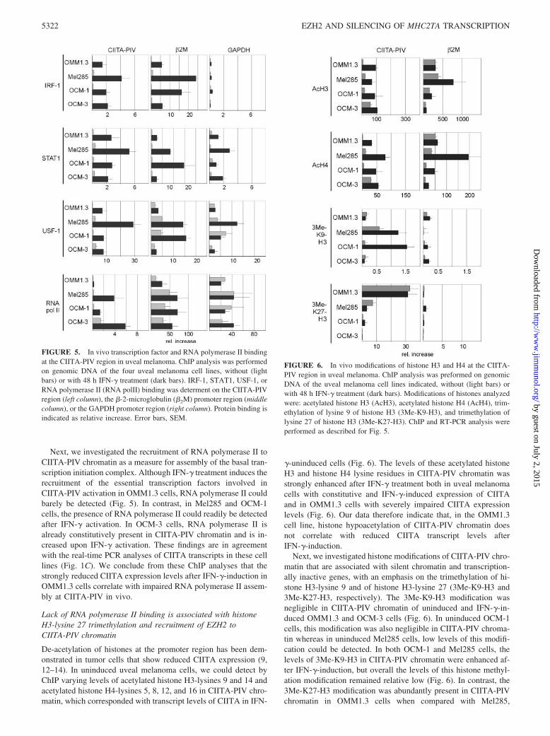
Next, we investigated the recruitment of RNA polymerase II toCIITA-PIV chromatin as a measure for assembly of the basal tran-scription initiation complex. Although IFN-� treatment induces therecruitment of the essential transcription factors involved inCIITA-PIV activation in OMM1.3 cells, RNA polymerase II couldbarely be detected (Fig. 5). In contrast, in Mel285 and OCM-1cells, the presence of RNA polymerase II could readily be detectedafter IFN-� activation. In OCM-3 cells, RNA polymerase II isalready constitutively present in CIITA-PIV chromatin and is in-creased upon IFN-� activation. These findings are in agreementwith the real-time PCR analyses of CIITA transcripts in these celllines (Fig. 1C). We conclude from these ChIP analyses that thestrongly reduced CIITA expression levels after IFN-�-induction inOMM1.3 cells correlate with impaired RNA polymerase II assem-bly at CIITA-PIV in vivo.
Lack of RNA polymerase II binding is associated with histoneH3-lysine 27 trimethylation and recruitment of EZH2 toCIITA-PIV chromatin
De-acetylation of histones at the promoter region has been dem-onstrated in tumor cells that show reduced CIITA expression (9,12–14). In uninduced uveal melanoma cells, we could detect byChIP varying levels of acetylated histone H3-lysines 9 and 14 andacetylated histone H4-lysines 5, 8, 12, and 16 in CIITA-PIV chro-matin, which corresponded with transcript levels of CIITA in IFN-
�-uninduced cells (Fig. 6). The levels of these acetylated histoneH3 and histone H4 lysine residues in CIITA-PIV chromatin wasstrongly enhanced after IFN-� treatment both in uveal melanomacells with constitutive and IFN-�-induced expression of CIITAand in OMM1.3 cells with severely impaired CIITA expressionlevels (Fig. 6). Our data therefore indicate that, in the OMM1.3cell line, histone hypoacetylation of CIITA-PIV chromatin doesnot correlate with reduced CIITA transcript levels afterIFN-�-induction.
Next, we investigated histone modifications of CIITA-PIV chro-matin that are associated with silent chromatin and transcription-ally inactive genes, with an emphasis on the trimethylation of hi-stone H3-lysine 9 and of histone H3-lysine 27 (3Me-K9-H3 and3Me-K27-H3, respectively). The 3Me-K9-H3 modification wasnegligible in CIITA-PIV chromatin of uninduced and IFN-�-in-duced OMM1.3 and OCM-3 cells (Fig. 6). In uninduced OCM-1cells, this modification was also negligible in CIITA-PIV chroma-tin whereas in uninduced Mel285 cells, low levels of this modifi-cation could be detected. In both OCM-1 and Mel285 cells, thelevels of 3Me-K9-H3 in CIITA-PIV chromatin were enhanced af-ter IFN-�-induction, but overall the levels of this histone methyl-ation modification remained relative low (Fig. 6). In contrast, the3Me-K27-H3 modification was abundantly present in CIITA-PIVchromatin in OMM1.3 cells when compared with Mel285,
FIGURE 5. In vivo transcription factor and RNA polymerase II bindingat the CIITA-PIV region in uveal melanoma. ChIP analysis was performedon genomic DNA of the four uveal melanoma cell lines, without (lightbars) or with 48 h IFN-� treatment (dark bars). IRF-1, STAT1, USF-1, orRNA polymerase II (RNA polII) binding was determent on the CIITA-PIVregion (left column), the �-2-microglobulin (�2M) promoter region (middlecolumn), or the GAPDH promoter region (right column). Protein binding isindicated as relative increase. Error bars, SEM.
FIGURE 6. In vivo modifications of histone H3 and H4 at the CIITA-PIV region in uveal melanoma. ChIP analysis was preformed on genomicDNA of the uveal melanoma cell lines indicated, without (light bars) orwith 48 h IFN-� treatment (dark bars). Modifications of histones analyzedwere: acetylated histone H3 (AcH3), acetylated histone H4 (AcH4), trim-ethylation of lysine 9 of histone H3 (3Me-K9-H3), and trimethylation oflysine 27 of histone H3 (3Me-K27-H3). ChIP and RT-PCR analysis wereperformed as described for Fig. 5.
5322 EZH2 AND SILENCING OF MHC2TA TRANSCRIPTION
by guest on July 2, 2015http://w
ww
.jimm
unol.org/D
ownloaded from
OCM-1, and OCM-3 cells (Fig. 6). The 3Me-K9-H3 and 3Me-K27-H3 modifications were hardly detectable in �2m promoterchromatin in any of the uveal melanoma cell lines investigated(Fig. 6).
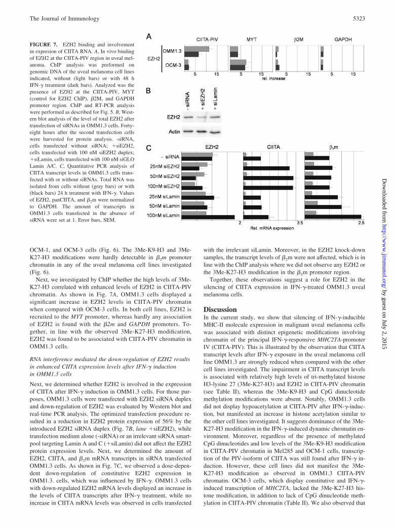
Next, we investigated by ChIP whether the high levels of 3Me-K27-H3 correlated with enhanced levels of EZH2 in CIITA-PIVchromatin. As shown in Fig. 7A, OMM1.3 cells displayed asignificant increase in EZH2 levels in CIITA-PIV chromatinwhen compared with OCM-3 cells. In both cell lines, EZH2 isrecruited to the MYT promoter, whereas hardly any associationof EZH2 is found with the �2m and GAPDH promoters. To-gether, in line with the observed 3Me-K27-H3 modification,EZH2 was found to be associated with CIITA-PIV chromatin inOMM1.3 cells.
RNA interference mediated the down-regulation of EZH2 resultsin enhanced CIITA expression levels after IFN-� inductionin OMM1.3 cells
Next, we determined whether EZH2 is involved in the expressionof CIITA after IFN-� induction in OMM1.3 cells. For those pur-poses, OMM1.3 cells were transfected with EZH2 siRNA duplexand down-regulation of EZH2 was evaluated by Western blot andreal-time PCR analysis. The optimized transfection procedure re-sulted in a reduction in EZH2 protein expression of 56% by theintroduced EZH2 siRNA duplex (Fig. 7B, lane �siEZH2), whiletransfection medium alone (-siRNA) or an irrelevant siRNA smart-pool targeting Lamin A and C (�siLamin) did not affect the EZH2protein expression levels. Next, we determined the amount ofEZH2, CIITA, and �2m mRNA transcripts in siRNA transfectedOMM1.3 cells. As shown in Fig. 7C, we observed a dose-depen-dent down-regulation of constitutive EZH2 expression inOMM1.3. cells, which was influenced by IFN-�. OMM1.3 cellswith down-regulated EZH2 mRNA levels displayed an increase inthe levels of CIITA transcripts after IFN-� treatment, while noincrease in CIITA mRNA levels was observed in cells transfected
with the irrelevant siLamin. Moreover, in the EZH2 knock-downsamples, the transcript levels of �2m were not affected, which is inline with the ChIP analysis where we did not observe any EZH2 orthe 3Me-K27-H3 modification in the �2m promoter region.
Together, these observations suggest a role for EZH2 in thesilencing of CIITA expression in IFN-�-treated OMM1.3 uvealmelanoma cells.
DiscussionIn the current study, we show that silencing of IFN-�-inducibleMHC-II molecule expression in malignant uveal melanoma cellswas associated with distinct epigenetic modifications involvingchromatin of the principal IFN-�-responsive MHC2TA-promoterIV (CIITA-PIV). This is illustrated by the observation that CIITAtranscript levels after IFN-� exposure in the uveal melanoma cellline OMM1.3 are strongly reduced when compared with the othercell lines investigated. The impairment in CIITA transcript levelsis associated with relatively high levels of tri-methylated histoneH3-lysine 27 (3Me-K27-H3) and EZH2 in CIITA-PIV chromatin(see Table II), whereas the 3Me-K9-H3 and CpG dinucleotidemethylation modifications were absent. Notably, OMM1.3 cellsdid not display hypoacetylation at CIITA-PIV after IFN-�-induc-tion, but manifested an increase in histone acetylation similar tothe other cell lines investigated. It suggests dominance of the 3Me-K27-H3 modification in the IFN-�-induced dynamic chromatin en-vironment. Moreover, regardless of the presence of methylatedCpG dinucleotides and low levels of the 3Me-K9-H3 modificationin CIITA-PIV chromatin in Mel285 and OCM-1 cells, transcrip-tion of the PIV-isoform of CIITA was still found after IFN-� in-duction. However, these cell lines did not manifest the 3Me-K27-H3 modification as observed in OMM1.3 CIITA-PIVchromatin. OCM-3 cells, which display constitutive and IFN-�-induced transcription of MHC2TA, lacked the 3Me-K27-H3 his-tone modification, in addition to lack of CpG dinucleotide meth-ylation in CIITA-PIV chromatin (Table II). We also observed that
FIGURE 7. EZH2 binding and involvementin expresion of CIITA RNA. A, In vivo bindingof EZH2 at the CIITA-PIV region in uveal mel-anoma. ChIP analysis was preformed ongenomic DNA of the uveal melanoma cell linesindicated, without (light bars) or with 48 hIFN-� treatment (dark bars). Analyzed was thepresence of EZH2 at the CIITA-PIV, MYT(control for EZH2 ChIP), �2M, and GAPDHpromoter region. ChIP and RT-PCR analysiswere performed as described for Fig. 5. B, West-ern blot analysis of the level of total EZH2 aftertransfection of siRNAs in OMM1.3 cells. Forty-eight hours after the second transfection cellswere harvested for protein analysis. -siRNA,cells transfected without siRNA; �siEZH2,cells transfected with 100 nM siEZH2 duplex;�siLamin, cells transfected with 100 nM siGLOLamin A/C. C, Quantitative PCR analysis ofCIITA transcript levels in OMM1.3 cells trans-fected with or without siRNAs. Total RNA wasisolated from cells without (gray bars) or with(black bars) 24 h treatment with IFN-�. Valuesof EZH2, panCIITA, and �2m were normalizedto GAPDH. The amount of transcripts inOMM1.3 cells transfected in the absence ofsiRNA were set at 1. Error bars, SEM.
5323The Journal of Immunology
by guest on July 2, 2015http://w
ww
.jimm
unol.org/D
ownloaded from

none of the above-mentioned epigenetic modifications interferedwith assembly at CIITA-PIV of transcription factors essential forIFN-�-induced transcription of MHC2TA (Table II). The 3Me-K27-H3 modification correlated with recruitment of EZH2 to CIITA-PIV chromatin in OMM1.3 cells. The observed increase in CIITAtranscript levels after IFN-� treatment of OMM1.3 cells withsiRNA-down-regulated EZH2 expression suggests that this histonemethyl transferase plays a role in MHC2TA transcriptional silenc-ing in OMM1.3 cells.
On the basis of the results obtained with OMM1.3 cells, ourobservations suggest a correlation between the 3Me-K27-H3 mod-ification and impaired RNA polymerase II recruitment to CIITA-PIV. In contrast, the CpG dinucleotide methylation modification inconjunction with low levels of the histone H3-lysine 9 modifica-tion in CIITA-PIV DNA as seen in Mel285 and OCM-1 cells donot greatly interfere with RNA polymerase II recruitment intoCIITA-PIV chromatin. The tri-methylated histone H3-lysine 27modification therefore seems to be the most important epige-netic regulator, which affects CIITA transcription levels afterIFN-�-induction, in uveal melanoma cells.
In all cell lines investigated, recruitment after IFN-� inductionof regulatory factors to CIITA-PIV (i.e., IRF-1, Stat-1, and USF-1)is not impaired by the 3Me-K27-H3 modification or by CpG dinu-cleotide methylation. It should be noted that of the critical regu-latory elements in CIITA-PIV (i.e., ISRE, GAS-box and E-box),only the E-box contains a CpG dinucleotide. In vitro methylationof this E-box CpG affects binding of USF-1 as has been demon-strated in EMSA (3). Therefore, it is of interest to note that of thevarious CpG dinucleotides in CIITA-PIV, the E-box CpG appar-ently is not or partially methylated in vivo (Fig. 4).
Promoter DNA methylation is regarded as a major epigeneticmechanism for silencing gene expression (38). There are two gen-eral mechanisms by which methylation-modified DNA inhibitsgene expression. As mentioned above, methylation-modified CpGdinucleotides in cognate binding sites of specific DNA bindingfactors could interfere in promoter assembly of these factors. Incontrast, the methylation modified CpG dinucleotides in DNAform binding sites of methyl-DNA binding proteins (e.g., MeCP2),which act as transcriptional repressors through their interactionwith histone deacetylases. However, recently it has been docu-mented that gene transcription in some cases occurs despite fullymethylated promoter DNA (39, 40). Furthermore, similar obser-vations have been made with respect to histone deacetylases,which may act for some genes as transcriptional activators ratherthan repressors (41). As mentioned above, we do not observe in-terference in the recruitment of activating transcription factors crit-ical for IFN-�-induced transcriptional activation of CIITA-PIV inuveal melanoma cells in the presence of the observed epigeneticmodifications. This is seemingly in contrast to the observation
made by Morris et al. (3), who showed impaired factor recruitmentto CIITA-PIV after IFN-� exposure in the choriocarcinoma celllines JEG3 and JAR. However, it should be noted that the uvealmelanoma cell panel studied did not contain a cell line displayingboth triple methylated lysine 27 in histone H3 in the presence ofCpG dinucleotide methylation. Therefore, we cannot exclude thepossibility that a combination of the above-mentioned epigeneticmodifications not only affects RNA polymerase II recruitment, butalso recruitment of IFN-�-induced activation factors to CIITA-PIV. This notion is underscored by our recent observation thatbesides dense CpG dinucleotide methylation, CIITA-PIV chroma-tin in JEG3 and JAR cells also contains high levels of the 3Me-K27-H3 modification (T. M. Holling and P. J. Van den Elsen,unpublished observations).
As mentioned before, the enzymatic activities that modify DNAand histones by methylation are intimately linked (22, 23). The3Me-K9-H3 mark recruits Dnmt 1 and 3a through HP1 interac-tions (22), and also EZH2 has been found to interact with Dnmts(23). Therefore, the lack of CpG dinucleotide methylation and ab-sence of the 3Me-K9-H3 mark at CIITA-PIV chromatin inOMM1.3 cells would also infer absence of EZH2-associatedDnmts in OMM1.3 cells. This notion is in line with the recentobservation that the 3Me-K27-H3 modification premarks genes forde novo methylation in cancer (42). It could therefore be arguedthat the epigenetic make-up of the CIITA-PIV region in OMM1.3cells indeed reflects premarking for de novo methylation of DNA,and that this is an intermediate epigenetic state in the completeshut down of Ag presentation functions.
Together, the results from our studies with uveal melanoma celllines suggest that the trimethylation of histone H3-lysine 27 is animportant epigenetic modification, which contributes to stronglyreduced CIITA expression levels after IFN-� induction and whichwould explain the lack of MHC-II molecule expression at the cellsurface.
AcknowledgmentsWe thank Prof. F. Koning and Dr. J. Kooter for critically reading themanuscript; Dr. B. Ksander and Dr. J. Kan-Mitchell for the uveal mela-noma cell lines; and Debbie Kramer for performing the initial bisulfitemodification analysis. Drs. Mark Bix, Yi Zhang, and Thomas Jenuwein foradvice, ChIP protocols and Abs.
DisclosuresThe authors have no financial conflict of interest.
References1. Van den Elsen, P. J., T. M. Holling, H. F. Kuipers, and N. Van der Stoep. 2004.
Transcriptional regulation of antigen presentation. Curr. Opin. Immunol. 16:67–75.
TABLE II. Overview of constitutive and IFN�-induced MHC-II expression and epigenetic modifications atthe CIITA-PIV region in uveal melanoma cell linesa
Cell line OMM1.3 Mel285 OCM-1 OCM-3
IFN� � � � � � � � �HLA-DR protein expression � � � �/� � � � ��DNA methylation � � � � � � � �Transcription factor binding � � � � � � � �RNA polymerase II binding � � � � � � � ��Acetylated histone H3/H4 � �� �/� �� �/� �� � ��3Me-K9-H3 modification � � � �� � �� � �3Me-K27-H3 modification �� �� �/� � � � � �EZH2 binding �� � ND ND ND ND �/� �/�
a �, absent; �, detectable; ��, strongly enhanced; �/�, barely detectable.
5324 EZH2 AND SILENCING OF MHC2TA TRANSCRIPTION
by guest on July 2, 2015http://w
ww
.jimm
unol.org/D
ownloaded from

2. Gobin, S. J., A. Peijnenburg, V. Keijsers, and P. J. Van den Elsen. 1997. Site �is crucial for two routes of IFN �-induced MHC class I transactivation: the ISRE-mediated route and a novel pathway involving CIITA. Immunity 6: 601–611.
3. Morris, A. C., G. W. Beresford, M. R. Mooney, and J. M. Boss. 2002. Kineticsof a � interferon response: expression and assembly of CIITA promoter IV andinhibition by methylation. Mol. Cell. Biol. 22: 4781–4791.
4. Satoh, A., M. Toyota, H. Ikeda, Y. Morimoto, K. Akino, H. Mita, H. Suzuki,Y. Sasaki, T. Kanaseki, Y. Takamura, et al. 2004. Epigenetic inactivation of classII transactivator (CIITA) is associated with the absence of interferon-�-inducedHLA-DR expression in colorectal and gastric cancer cells. Oncogene 23:8876–8886.
5. Van den Elsen, P. J., N. Van der Stoep, H. E. Vietor, L. Wilson, M. van Zutphen,and S. J. Gobin. 2000. Lack of CIITA expression is central to the absence ofantigen presentation functions of trophoblast cells and is caused by methylationof the IFN-� inducible promoter (PIV) of CIITA. Hum. Immunol. 61: 850–862.
6. Van der Stoep, N., P. Biesta, E. Quinten, and P. J. Van den Elsen. 2002. Lack ofIFN-�-mediated induction of the class II transactivator (CIITA) through pro-moter methylation is predominantly found in developmental tumor cell lines. Int.J. Cancer 97: 501–507.
7. Morris, A. C., W. E. Spangler, and J. M. Boss. 2000. Methylation of class IItrans-activator promoter IV: a novel mechanism of MHC class II gene control.J. Immunol. 164: 4143–4149.
8. Holling, T. M., E. Schooten, A. W. Langerak, and P. J. Van den Elsen. 2004.Regulation of MHC class II expression in human T-cell malignancies. Blood 103:1438–1444.
9. Van den Elsen, P. J., T. M. Holling, N. Van der Stoep, and J. M. Boss. 2003.DNA methylation and expression of major histocompatibility complex class I andclass II transactivator genes in human developmental tumor cells and in T cellmalignancies. Clin. Immunol. 109: 46–52.
10. Morimoto, Y., M. Toyota, A. Satoh, M. Murai, H. Mita, H. Suzuki, Y. Takamura,H. Ikeda, T. Ishida, N. Sato, et al. 2004. Inactivation of class II transactivator byDNA methylation and histone deacetylation associated with absence of HLA-DRinduction by interferon-� in haematopoietic tumour cells. Br. J. Cancer 90:844–852.
11. Murphy, S. P., R. Holtz, N. Lewandowski, T. B. Tomasi, and H. Fuji. 2002. DNAalkylating agents alleviate silencing of class II transactivator gene expression inL1210 lymphoma cells. J. Immunol. 169: 3085–3093.
12. Kanaseki, T., H. Ikeda, Y. Takamura, M. Toyota, Y. Hirohashi, T. Tokino,T. Himi, and N. Sato. 2003. Histone deacetylation, but not hypermethylation,modifies class II transactivator and MHC class II gene expression in squamouscell carcinomas. J. Immunol. 170: 4980–4985.
13. Magner, W. J., A. L. Kazim, C. Stewart, M. A. Romano, G. Catalano, C. Grande,N. Keiser, F. Santaniello, and T. B. Tomasi. 2000. Activation of MHC class I, II,and CD40 gene expression by histone deacetylase inhibitors. J. Immunol. 165:7017–7024.
14. Holtz, R., J. C. Choi, M. G. Petroff, J. F. Piskurich, and S. P. Murphy. 2003. ClassII transactivator (CIITA) promoter methylation does not correlate with silencingof CIITA transcription in trophoblasts. Biol. Reprod. 69: 915–924.
15. Santos-Rosa, H., and C. Caldas. 2005. Chromatin modifier enzymes, the histonecode and cancer. Eur. J. Cancer 41: 2381–2402.
16. Stewart, M. D., J. Li, and J. Wong. 2005. Relationship between histone H3 lysine9 methylation, transcription repression, and heterochromatin protein 1 recruit-ment. Mol. Cell. Biol. 25: 2525–2538.
17. Tachibana, M., K. Sugimoto, T. Fukushima, and Y. Shinkai. 2001. Set domain-containing protein, G9a, is a novel lysine-preferring mammalian histone meth-yltransferase with hyperactivity and specific selectivity to lysines 9 and 27 ofhistone H3. J. Biol. Chem. 276: 25309–25317.
18. Cao, R., L. Wang, H. Wang, L. Xia, H. Erdjument-Bromage, P. Tempst,R. S. Jones, and Y. Zhang. 2002. Role of histone H3 lysine 27 methylation inPolycomb-group silencing. Science 298: 1039–1043.
19. Rice, J. C., S. D. Briggs, B. Ueberheide, C. M. Barber, J. Shabanowitz,D. F. Hunt, Y. Shinkai, and C. D. Allis. 2003. Histone methyltransferases directdifferent degrees of methylation to define distinct chromatin domains. Mol. Cell12: 1591–1598.
20. Raaphorst, F. M. 2005. Deregulated expression of Polycomb-group oncogenes inhuman malignant lymphomas and epithelial tumors. Hum. Mol. Genet. 14 SpecNo 1: R93–R100.
21. Cao, R., and Y. Zhang. 2004. The functions of E(Z)/EZH2-mediated methylationof lysine 27 in histone H3. Curr. Opin. Genet. Dev. 14: 155–164.
22. Fuks, F., P. J. Hurd, R. Deplus, and T. Kouzarides. 2003. The DNA methyltrans-ferases associate with HP1 and the SUV39H1 histone methyltransferase. NucleicAcids Res. 31: 2305–2312.
23. Vire, E., C. Brenner, R. Deplus, L. Blanchon, M. Fraga, C. Didelot, L. Morey,A. Van Eynde, D. Bernard, J. M. Vanderwinden, et al. 2006. The Polycombgroup protein EZH2 directly controls DNA methylation. Nature 439: 871–874.
24. Egan, K. M., J. M. Seddon, R. J. Glynn, E. S. Gragoudas, and D. M. Albert. 1988.Epidemiologic aspects of uveal melanoma. Surv. Ophthalmol. 32: 239–251.
25. Diener-West, M., B. S. Hawkins, J. A. Markowitz, and A. P. Schachat. 1992. Areview of mortality from choroidal melanoma, II: a meta-analysis of 5-year mor-tality rates following enucleation, 1966 through 1988. Arch. Ophthalmol. 110:245–250.
26. Lawry, J., Z. Currie, M. O. Smith, and I. G. Rennie. 1999. The correlation be-tween cell surface markers and clinical features in choroidal malignant melano-mas. Eye 13 (Pt 3a): 301–308.
27. Ericsson, C., S. Seregard, A. Bartolazzi, E. Levitskaya, S. Ferrone, R. Kiessling,and O. Larsson. 2001. Association of HLA class I and class II antigen expressionand mortality in uveal melanoma. Invest. Ophthalmol. Visual Sci. 42: 2153–2156.
28. Waard-Siebinga, I., J. Kool, and M. J. Jager. 1995. HLA antigen expression onuveal melanoma cells in vivo and in vitro. Hum. Immunol. 44: 111–117.
29. Jager, M. J., H. M. Hurks, J. Levitskaya, and R. Kiessling. 2002. HLA expressionin uveal melanoma: there is no rule without some exception. Hum. Immunol. 63:444–451.
30. Radosevich, M., Z. Song, J. C. Gorga, B. Ksander, and S. J. Ono. 2004. Epige-netic silencing of the CIITA gene and posttranscriptional regulation of class IIMHC genes in ocular melanoma cells. Invest. Ophthalmol. Visual Sci. 45:3185–3195.
31. Muhlethaler-Mottet, A., W. Di Berardino, L. A. Otten, and B. Mach. 1998. Activa-tion of the MHC class II transactivator CIITA by interferon-� requires cooperativeinteraction between Stat1 and USF-1. Immunity 8: 157–166.
32. Vandekerckhove, B. A., G. Datema, F. Koning, E. Goulmy, G. G. Persijn,J. J. Van Rood, F. H. Claas, and J. E. De Vries. 1990. Analysis of the donor-specific cytotoxic T lymphocyte repertoire in a patient with a long term survivingallograft. Frequency, specificity, and phenotype of donor-reactive T cell receptor(TCR)-��� and TCR-��� clones. J. Immunol. 144: 1288–1294.
33. Kuipers, H. F., P. J. Biesta, T. A. Groothuis, J. J. Neefjes, A. M. Mommaas, andP. J. Van den Elsen. 2005. Statins affect cell-surface expression of major histo-compatibility complex class II molecules by disrupting cholesterol-containingmicrodomains. Hum. Immunol. 66: 653–665.
34. Peijnenburg, A., R. Van den Berg, M. J. Van Eggermond, O. Sanal, J. M. Vossen,A. M. Lennon, C. Alcaide-Loridan, and P. J. Van den Elsen. 2000. DefectiveMHC class II expression in an MHC class II deficiency patient is caused by anovel deletion of a splice donor site in the MHC class II transactivator gene.Immunogenetics 51: 42–49.
35. Baguet, A., X. Sun, T. Arroll, A. Krumm, and M. Bix. 2005. Intergenic tran-scription is not required in Th2 cells to maintain histone acetylation and tran-scriptional permissiveness at the Il4-Il13 locus. J. Immunol. 175: 8146–8153.
36. Kirmizis, A., S. M. Bartley, A. Kuzmichev, R. Margueron, D. Reinberg,R. Green, and P. J. Farnham. 2004. Silencing of human polycomb target genes isassociated with methylation of histone H3 Lys 27. Genes Dev. 18: 1592–1605.
37. Gobin, S. J., P. Biesta, and P. J. Van den Elsen. 2003. Regulation of human �2-microglobulin transactivation in hematopoietic cells. Blood 101: 3058–3064.
38. Klose, R. J., and A. P. Bird. 2006. Genomic DNA methylation: the mark and itsmediators. Trends Biochem. Sci. 31: 89–97.
39. Tong, Y., T. Aune, and M. Boothby. 2005. T-bet antagonizes mSin3a recruitmentand transactivates a fully methylated IFN-� promoter via a conserved T-boxhalf-site. Proc. Natl. Acad. Sci. USA 102: 2034–2039.
40. Kim, S., J. Kang, B. M. Evers, and D. H. Chung. 2004. Interferon-� inducescaspase-8 in neuroblastomas without affecting methylation of caspase-8 pro-moter. J. Pediatr. Surg. 39: 509–515.
41. Nusinzon, I., and C. M. Horvath. 2005. Histone deacetylases as transcriptionalactivators? Role reversal in inducible gene regulation. Sci. STKE 2005:re11.
42. Schlesinger, Y., R. Straussman, I. Keshet, S. Farkash, M. Hecht, J. Zimmerman,E. Eden, Z. Yakhini, E. Ben Shushan, B. E. Reubinoff, et al. 2007. Polycomb-mediated methylation on Lys27 of histone H3 pre-marks genes for de novo meth-ylation in cancer. Nat. Genet. 39: 232–236.
5325The Journal of Immunology
by guest on July 2, 2015http://w
ww
.jimm
unol.org/D
ownloaded from
Copyright © 2022 FDOKUMEN