Role of calcineurin in thrombin-mediated endothelial cell contraction
FEBS 29337 FEBS Letters 579 (2005) 1591–1596
A reassessment of the inhibitory capacity of human FKBP38on calcineurin
Matthias Weiwad1, Frank Edlich1, Frank Erdmann, Franziska Jarczowski, Susann Kilka,Madlen Dorn, Arndt Pechstein, Gunter Fischer*
Max-Planck Research Unit for Enzymology of Protein Folding, Weinbergweg 22, D-06120 Halle/Saale, Germany
Received 7 September 2004; revised 26 November 2004; accepted 23 December 2004
Available online 11 February 2005
Edited by Judit Ovadi
Abstract The microbial peptidomacrolide FK506 affects manyeukaryotic developmental and cell signaling programs via calci-neurin inhibition. Prior formation of a complex betweenFK506 and intracellular FK506-binding proteins (FKBPs) isthe precondition for the interaction with calcineurin. A puzzlingdifference has emerged between the mammalian multidomainprotein hFKBP38 and other FKBPs. It was shown thathFKBP38 not only binds to calcineurin but also inhibits the pro-tein phosphatase activity of calcineurin on its own [Shirane, M.and Nakayama, K.I. (2003) Nature Cell Biol. 5, 28–37]. Inher-ent calcineurin inhibition by hFKBP38 would completely elimi-nate the need for FK506 in controlling many signaltransduction pathways. To address this issue, we have character-ized the functional and physical interactions between calcineurinand hFKBP38. A recombinant hFKBP38 variant and endoge-nous hFKBP38 were tested both in vitro and in vivo. The proteinsneither directly inhibited calcineurin activity nor affected NFATreporter gene activity in SH-SY5Y and Jurkat cells. In addition,a direct physical interaction between calcineurin and hFKBP38was not detected in co-immunoprecipitation experiments. How-ever, hFKBP38 indirectly affected the subcellular distributionof calcineurin by interaction with typical calcineurin ligands, asexemplified by the anti-apoptotic protein Bcl-2. Our data suggestthat hFKBP38 cannot substitute for the FKBP/FK506 complexin signaling pathways controlled by the protein phosphataseactivity of calcineurin.� 2005 Federation of European Biochemical Societies. Publishedby Elsevier B.V. All rights reserved.
Keywords: Serine/threonine protein phosphatase; Calcineurin;Calmodulin; FK506-binding protein
1. Introduction
Calcineurin (Protein phosphatase 2B, CaN) links a revers-
ible intracellular Ca2+/calmodulin signal to the dephosphoryl-
ation reaction of many important proteins such as the
transcription factors NFAT, MEF2, Elk-1, as well as NO syn-
thase and the regulatory RII subunit of cAMP dependent pro-
tein kinase. Consequently, this enzyme couples calcium
signaling to apoptosis and transcriptional control in the
*Corresponding author. Fax: +49 345 55 11972.E-mail address: [email protected] (G. Fischer).
1 Both authors contributed equally to this work.
0014-5793/$30.00 � 2005 Federation of European Biochemical Societies. Pu
doi:10.1016/j.febslet.2004.12.098
immune, nervous and cardiovascular systems [1–3]. When dis-
tributed into the cytoplasm of mammalian cells the microbial
compounds FK506 and cyclosporin A (CsA) potently inhibit
CaN by generating ternary complexes with their cellular tar-
gets FK506-binding proteins (FKBPs) and cyclophilins,
respectively [4,5]. In immunosuppression, the protein compo-
nent of the inhibitory complex of FK506 is thought to be
FKBP12, thus, forming the FK506/FKBP12/CaN complex.
The ubiquitously distributed FKBP12 is the prototypic
FKBP-type peptidyl–prolyl cis/trans isomerase (PPIase) [6].
The FK506/FKBP12 complex, but not the drug alone binds
to and tightly inhibits CaN in that the interface between the
catalytic (CaNA) and the regulatory (CaNB) subunit of CaN
is targeted by a composite drug–protein binding surface. In
the case of FK506/FKBP12 and CsA/cyclophilin18, the con-
tacting atoms do not directly interfere with the active site of
CaNA. Rather, the restricted access of protein substrates to
the active site underlying ternary complex formation consti-
tutes the molecular basis for CaN inhibition [7–9].
Since these microbial drugs are not normal constituents of
mammalian cells they are not available for the mammalian
immunophilins to bind, and to perform a control of CaN
activity from endogenously produced resources. This raises
the question as to whether mammalian cells also contain com-
pounds potentially targeting CaN in a FK506-like manner.
Interestingly, there are derivatives of the microbial drugs that
can inhibit CaN by their own, thus, circumventing the need for
prior binding of the presenter proteins. It is a conformational
change in the drug derivative that gives rise to a complete CaN
interacting surface in the drug alone [10,11]. The conforma-
tional changes must allow replacement of important immuno-
philin–CaN contacting sites in the ternary complex with new
drug-based interaction sites. A similar situation could be ex-
pected for the immunophilin component of the ternary com-
plex. It is interesting to speculate, based on the small
‘‘effector domain’’ of FK506 [7], about the possibility of a pep-
tide segment of a larger congener of FKBP12 capable of mim-
icking the FK506 ‘‘effector domain’’ in its CaN interfering
conformation. Such a FKBP molecule would be expected to
realize an inherent inhibitor of CaN.
Recently, inhibition of the RII phosphopeptide dephospho-
rylating activity of calcineurin was observed for an anti-hem-
agglutinin epitope immunoprecipitate of recombinant
hemagglutinin epitope-tagged hFKBP38 giving rise to the the-
ory that hFKBP38 by its own is able to mimic the composite
surface of the FK506–FKBP12 complex. It was concluded that
hFKBP38 forms the endogenous molecule of human cells that
blished by Elsevier B.V. All rights reserved.

1592 M. Weiwad et al. / FEBS Letters 579 (2005) 1591–1596
substitutes for the microbial ligand as represented by FK506
and CsA in CaN inhibition. However, adequate confirmation
of CaN inhibition using untagged hFKBP38 in isolation is still
lacking [12,13]. hFKBP38, a protein of 355 residues, belongs to
those group of FKBP containing a PPIase domain, tetratrico-
peptide repeats and a putative Ca2+/calmodulin binding site
[6,14]. For a constitutive FKBP-like CaN inhibitor an increase
of the threshold for entry to apoptosis was suggested [15]. It
would be crucial to know whether hFKBP38 controls protein
dephosphorylation by CaN, since the FKBP-like immunophi-
lins were found to be involved in neuroprotection and neuro-
regeneration, processes that are tightly linked to apoptotic
signals [16,17]. In this study, we test the finding that hFKBP38
is a CaN inhibitor by its own in vitro and in vivo. However, the
present study shows that hFKBP38 neither interacts in vitro
nor in vivo with CaN. Therefore hFKBP38 does not act as
inherent inhibitor of CaN.
Furthermore, hFKBP38 inhibits CaN protein phosphatase
activity in the presence of FK506. This observation supports
the notion that hFKBP38 behaves overall in a manner similar
to other immunophilins.
2. Materials and methods
Enzymes used: human FKBP381-336, human calmodulin, humanBcl-2, were recombinantly expressed by using a pET28a-vector inE. coli Rosettae cells. Human calcineurin was recombinantlyexpressed and purified according Mondragon et al. [18]. Maltose-bind-ing protein–Bcl-2 fusion protein was purchased from Sigma (Deis-enhofen, Germany). The hFKBP38 antibody was an affinity purifiedsection 4 polyclonal rabbit antibody against the purified hFKBP38 do-main (amino acids 1–166). Peptide substrates used were obtained fromBachem (Heidelberg, Germany). FK506 was purchased from calbio-chem (La Jolla, CA).
2.1. Calcineurin activity assayThe calcineurin phosphatase assay was performed as described by
Baumgrass et al. [19]. Direct inhibition of 5 nM recombinant humancalcineurin by hFKBP381-336 was measured in the presence of 50 nMand 1 lM CaM at various concentrations of the PPIase (0–20 lM).Inhibition of 5 nM calcineurin by hFKBP381-336 was assayed at con-stant concentrations of 30 lM FK506 and 20 lM CaM in dependenceon the hFKBP381-336 concentration. Due to the tight interaction be-tween hFKBP381-336 and FK506, the concentration of the complex isalmost identical to the hFKBP381-336 concentration used. Calcineurinactivity was expressed relative to a reference without effectors. Errorbars represent S.D. from the mean (n = 3).
Fig. 1. Effect of hFKBP38 on CaN phosphatase activity. (A) CaN(5 nM) activity was measured in the presence of various concentrationsof hFKBP381-336 in buffer containing either 50 nM CaM (black bars)or 1 lM CaM (grey bars) using [33P]-labeled 19-residue phosphopep-tide substrate (RII phosphopeptide). (B) E. coli cell lysate containing
1-336
2.2. Measurement of PPIase activityPPIase activity was determined using protease-coupled assays as de-
scribed elsewhere [20]. Typically, hFKBP381-336 PPIase activity wasmeasured in a reaction mixture containing 1 lM hFKBP381-336,5 lM recombinant human CaM and 5 mM CaCl2. hFKBP381-336
was tested using the oligopeptide succinyl-AFPF-4-nitroanilide assubstrate.
recombinant hFKBP38 was subjected to CaM–Sepharose in thepresence of 2 mM CaCl2. Bound protein was eluted by 2 mM EGTAand subsequently analysed by SDS–PAGE with Coomassie Bluestaining; 1 – crude extract, 2 – washing step, 3 – eluat. (C) Native gelelectrophoresis experiment was performed with samples containing 1 –hFKBP381-336, 2 – hFKBP381-336 preincubated with CaM, and 3 –hFKBP381-336 preincubated with CaM and CaCl2. Samples weresubjected to a 12.5% native gel. Migration of hFKBP381-336 wasanalysed by Western blot using rabbit anti-hFKBP38 antibody. (D)Residual CaN (5 nM) activity was determined in the presence of 30 lMFK506 and 20 lM CaM at varying concentrations of FKBP12 (greybars) and hFKBP38 (black bars).
2.3. Protein–protein interaction in vitroNative gel electrophoresis experiments; Native PAGEwas performed
according to Laemmli�s gel systemwithout SDS and b-mercaptoethanolat a constant temperature of 4 �C. CaM-binding assay; CaM–Sephar-ose (Amersham-Pharmacia Biotech) was preequilibrated in Ca2+-con-taining buffer A (25 mM Tris/HCl, pH 7.5, 200 mM NaCl, 1 mMDTT, 2 mM CaCl2). Subsequently, 1 ml crude E. coli lysate containingoverexpressed hFKBP381-336 was incubated with CaM–Sepharose.After washing, bound proteins were eluted with buffer A containing2 mM EGTA. Samples were subjected to 12.5% SDS–PAGE and
analysed by Coomassie Blue staining. Co-immunoprecipitation; Celllysis and co-immunoprecipitation experiments were performedaccording to manufactures protocols of the immunoprecipitationstarter kit (Amersham Bioscience). Bcl-2 binding assay; 40 ll of 6 lMmaltose-binding protein–Bcl-2 fusion protein (MBP–Bcl-2) was
M. Weiwad et al. / FEBS Letters 579 (2005) 1591–1596 1593
subjected to 40 ll amylose-resin (New England Biolabs, Beverly, MA)and incubated for 30 min. Thereafter, beads were washed twice withbuffer B (25 mM Tris–HCl, pH 7.5, 200 mM NaCl, 1 mM DTT) andincubate with CaN. Subsequently, beads were incubated for 1 h with40 ll reaction mixture containing Ca2+/CaM, hFKBP381-336 andCa2+/CaM, hFKBP381-336, Ca2+/CaM and GPI1046, FKBP381-336 orFKBP12. Bcl-2-boundCaNwas eluted with 300 mMmaltose, subjectedto 12.5% SDS–PAGE and analysed by Western blotting using mouseanti-CaN antibody.
2.4. NFAT reporter gene assayTo test the effect of hFKBP381-336 overexpression on CaN phospha-
tase activity SH-SY5Y and Jurkat cells transfected with NFAT-lucifer-ase reporter plasmid (Stratagene, Netherlands) were co-transfected byelectroporation (Amaxa, Cologne, Germany) either with pcDNA4/HisMax C vector or a construct of this vector containing the sequenceof hFKBP38. SH-SY5Y cells were cultured in DMEM (Biochrom, Ber-lin, Germany) supplemented with 2 mM LL-glutamine and 10% (v/v)heat-inactivated FCS in a humidified incubator at 37 �C in 10% (v/v)CO2. Jurkat cells were cultured in RPMI 1640 with 10% FCS and2 mM LL-glutamine at 37 �C in 5% CO2. Cells were stimulated with2 lM ionomycin for 5 h. After cell lysis the protein content of each ly-sate was determined by the Bradford method. Equivalent amounts ofprotein were applied to determine the level of the extracted luciferasefrom the cells by bioluminescence measurement using the luciferase as-say system (Promega, Mannheim, Germany). In addition, a b-galacto-sidase plasmid was co-transfected as internal standard.
2.5. ImmunohistochemistrySH-SY5Y cells grown on coverslips were fixed in 4% paraformalde-
hyde in phosphate buffered saline (PBS) for 30 min at room tempera-ture. After washing in PBS cells were permeabilized 5 min on ice with0.2% Triton X-100 in PBS containing 10% FCS, blocked with PBScontaining 10% FCS for 1 h, incubated with PBS containing primaryantibody (1:50 dilution for hamster anti-human Bcl-2 Ab (BD Biosci-ences Pharmingen)), 1:50 dilution for mouse anti-human CaN Ab(Sigma) and incubated with PBS containing secondary antibody(1:100 dilution for FITC-conjugated goat anti-mouse antibody IgGand 1:100 dilution for Cy5-conjugated goat anti-hamster antibody(Biomol, Hamburg, Germany)) and 10% FCS for 30 min. Cells wereanalysed by confocal laser scanning microscopy (Carl Zeiss LSM410) at 750-fold magnification.
3. Results and discussion
3.1. Effect of hFKBP38 on CaN phosphatase activity in vitro
Human FKBP38 was previously identified for being an
intrinsic CaN inhibitor by measurement of CaN activity after
applying immunoprecipitated HA-tagged hFKBP38 expressed
in HeLa cells [12]. Unfortunately, this report lacked a determi-
nation of inhibition constants, hFKBP38 concentration ap-
plied in the phosphatase assay or other biochemical data
verifying the described interaction.
Therefore, we tested with purified proteins the inhibition of
CaN activity by hFKBP381-336 in the absence of FK506 in or-
der to determine the inhibition constant of this interaction.
Calcineurin activity was measured using a scintillation proxim-
ity assay according to Baumgrass et al. [19]. A [33P]-labeled
biotinylated 19-residue peptide of a partial sequence of the
RII subunit of the bovine PKA (RII phosphopeptide) was
used as substrate. When performed in the presence of 50 nM
Ca2+/CaM a slight decrease in CaN activity was found at high
concentrations of hFKBP381-336 (Fig. 1A, black bars). This
small effect on CaN activity was abolished at 1 lM CaM
and was found to be strictly dependent on the CaM concentra-
tion (Fig. 1A, grey bars). These findings are in strict contrast to
previously reported data [12].
Recent data from our lab indicate that hFKBP38 interacts
with CaM in a calcium-dependent manner (Fig. 1B and C).
Thus, both Ca2+/CaM-binding proteins, CaN and hFKBP38,
may compete for the same cofactor in the CaN activity assay
resulting in a weak CaN inhibition but only under Ca2+/
CaM limiting conditions. To gain insight in the putative pre-
senter protein function of hFKBP38, CaN activity was investi-
gated in the presence of the FK506 and hFKBP381-336. At
20 lM complex concentration residual CaN activity was about
60% of the control (Fig. 1D). Therefore, the potency of the
gain-of-function, which is reported to be critical for the immu-
nosuppressive activity of FK506, is similar to that observed for
the FK506/FKBP52 complex, and about 200-fold lower than
that for the FK506/FKBP12 complex [14].
In addition, we tested whether CaN influences the PPIase
activity of hFKBP38 in a standard assay [20] using 1 lMhFKBP381-336, 5 lM CaM, 5 mM Ca2+ and CaN concentra-
tions in a range of 200 nM–2 lM. We were not able to
detect any influence of CaN on hFKBP38 activity (data
not shown).
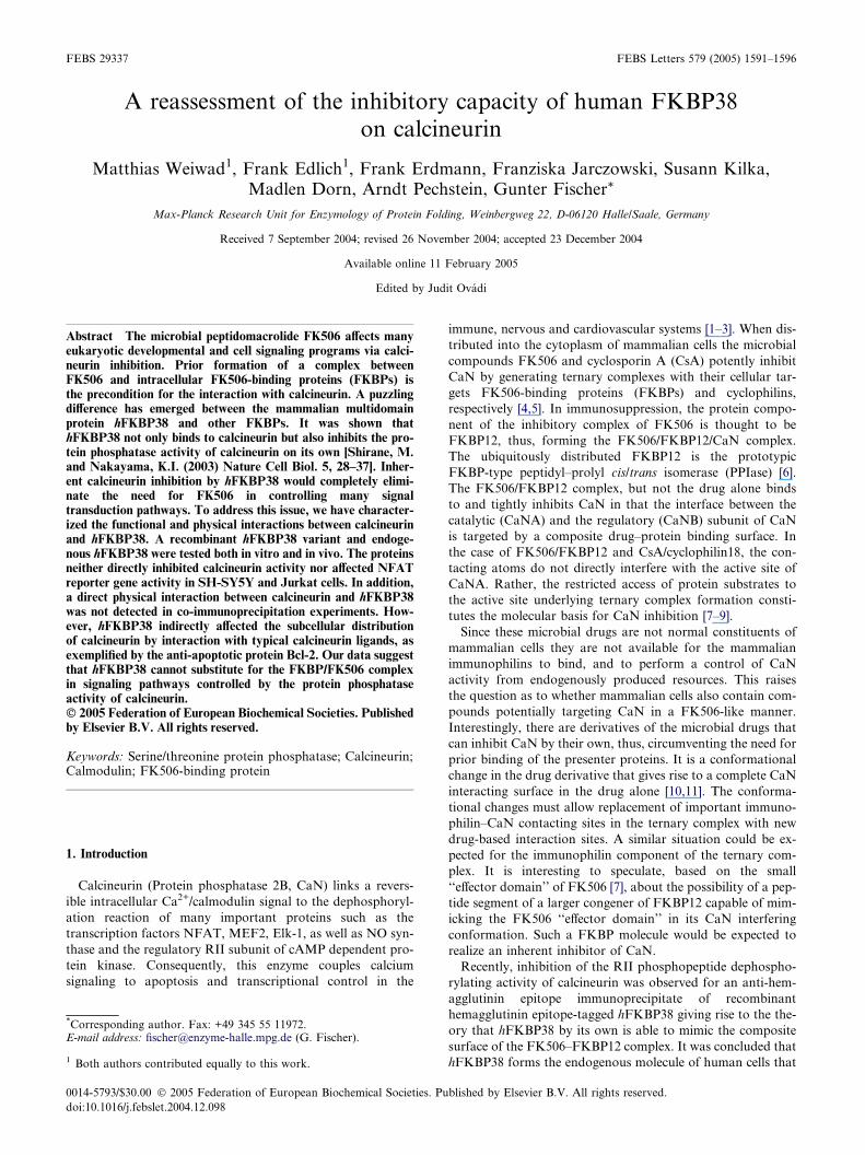
3.2. Effect of hFKBP38 on CaN phosphatase activity in vivo
Based on these findings, we were interested in the effect of
hFKBP38 on CaN activity in vivo. Therefore, we established
a NFAT reporter gene assay in Jurkat and SH-SY5Y cells
examining the CaN activity in living cells. Cells were co-
transfected either with pcDNA4/HisMax C vector or a con-
struct of this vector containing the full length sequence of
hFKBP38 (Fig. 2A). In order to raise intracellular calcium lev-
els and activate CaN activity cells were stimulated with 2 lMionomycin for 5 h. As negative control stimulated cells were
treated with the specific CaN inhibitor CsA. GPI1046 was used
to affect hFKBP38. Due to application of the calcium iono-
phore ionomycin NFAT dephosphorylation increases. In the
presence of CsA dephosphorylation levels were comparable
to those in unstimulated cells. In SH-SY5Y cells neither over-
expression of hFKBP38 nor addition of the immunophilin li-
gand GPI1046 had a significant effect on CaN phosphatase
activity. However, in Jurkat cells CaN activity increases upon
hFKBP38 overexpression, whereas application of GPI1046 re-
sulted in a slight decrease of NFAT dephosphorylation (Fig.
2B and C). The difference between both cell lines seems to be
a result of different levels of endogenous hFKBP38 (Fig.
2A). Assuming particular high concentrations of hFKBP38
in neuronal cells as previously reported [14], hFKBP38 overex-
pression would cause small additional effects. These results im-
ply that hFKBP38 does not inhibit CaN protein phosphatase
activity in vivo.
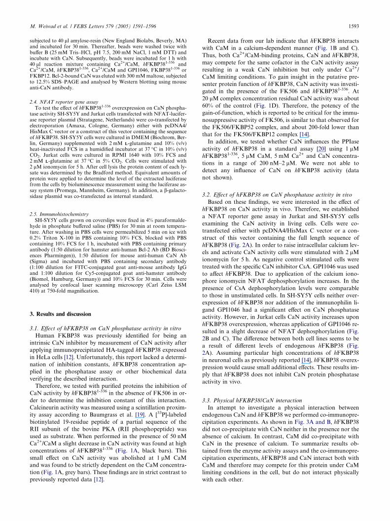
3.3. Physical hFKBP38/CaN interaction
In attempt to investigate a physical interaction between
endogenous CaN and hFKBP38 we performed co-immunopre-
cipitation experiments. As shown in Fig. 3A and B, hFKBP38
did not co-precipitate with CaN neither in the presence nor the
absence of calcium. In contrast, CaM did co-precipitate with
CaN in the presence of calcium. To summarize results ob-
tained from the enzyme activity assays and the co-immunopre-
cipitation experiments, hFKBP38 and CaN interact both with
CaM and therefore may compete for this protein under CaM
limiting conditions in the cell, but do not interact physically
with each other.

1594 M. Weiwad et al. / FEBS Letters 579 (2005) 1591–1596
3.4. Influence of hFKBP38 on CaN–Bcl-2 interactions
To verify results obtained with NFAT reporter gene assay in
Jurkat cells that are not based on direct interaction between
hFKBP38 and CaN, we were interested in determining whether
Bcl-2 might serve as the missing link between hFKBP38 and
the regulation of CaN activity, because reported data [12,21]
describe interactions of CaN and hFKBP38 with Bcl-2.
Fig. 3. CaN does not interact with hFKBP38. Co-immunoprecipita-tion; SH-SY5Y cell lysate was incubated with (A) mouseanti-calcineurin antibody and (B) rabbit anti-hFKBP38 antibody.Antibody/protein complexes were bound to protein G sepharose.Samples were washed. Pellet (P) and Supernatant (S) were subjected toSDS–PAGE and analysed by Western blot using mouse anti-CaMantibody and (A) rabbit anti-hFKBP38 antibody or (B) mouse anti-calcineurin antibody. b-Actin was used as loading control.
Using a binding assay, where a MBP–Bcl-2 fusion protein
was bound to amylose resin, direct binding of CaN to Bcl-2
was observed. hFKBP38 did not affect this interaction. How-
ever, hFKBP381-336 was able to interfere with CaN–Bcl-2
interactions upon addition of Ca2+/CaM. The effect of
hFKBP38 application on CaN–Bcl-2 interaction was abolished
in the presence of the neuroimmunophilin ligand GPI1046.
Neither hFKBP12 nor CaM alone did affect this interaction
(Fig. 4A).
In order to verify whether the observed disruption of the
Bcl-2–CaN interaction by hFKBP38 has an effect on CaN sub-
cellular distribution, we performed an experiment observing
the localization of Bcl-2 and CaN in SH-SY5Y cells (Fig.
4B). In order to increase cellular calcium concentrations cells
were stimulated by ionomycin. The neuroimmunophilin ligand
GPI1046 was applied to stimulated cells to affect hFKBP38
and therefore preventing disruption of Bcl-2/CaN interactions.
In unstimulated cells Bcl-2 and CaN are predominantly co-
localized. Upon calcium rise induced by calcium ionophore
ionomycin CaN mainly redistributed from Bcl-2. Application
of GPI1046 prevents CaN redistribution due to intracellular
Ca2+ rise, indicating that hFKBP38 may cause an increased
proportion of CaN in the cytosol and therefore led to higher
Fig. 2. hFKBP38 does not affect NFAT reporter gene activity.(A) SH-SY5Y and Jurkat cells were transfected with NFAT-luciferasereporter plasmid and co-transfected either with pcDNA4/HisMax Cvector (grey bar) or a construct of this vector containing the sequenceof hFKBP38 (black bar). Expression of hFKBP38 was analysed byWestern blot using mouse anti-HisTag antibody. Endogenous andartificially expressed hFKBP38 was analysed by Western blot usingrabbit anti-hFKBP38 antibody. b-Actin was used as loading control.Jurkat (B) and SH-SY5Y cells (C) were stimulated with 2 lMionomycin for 5 h. In some cases, cells were preincubated with theneuroimmunophilin ligand GPI1046 and the calcineurin inhibitorCsA for 30 min. After cell lysis, the amount of the extracted luciferasefrom the cells was determined by bioluminescence measurement.Activities are expressed as n-fold over basal. The data presented aremeans ± S.D. of three independent experiments.
b

Fig. 4. Ca2+/CaM complexed hFKBP381-336 disrupts Bcl-2–CaN interaction. (A) MBP–Bcl-2 fusion protein was immobilized on amylose beads andincubated with CaN. hFKBP381-336 did not interfere with the detected CaN/Bcl-2 interaction. In the presence of Ca2+/CaM hFKBP381-336 disruptedCaN/Bcl-2 complexes. Upon addition of the neuroimmunophilin ligand GPI1046 this effect was abolished. hFKBP12 did not interfere with the CaN/Bcl-2 interaction. Bcl-2-bound CaN was eluted with 300 mMmaltose, subjected to 12.5% SDS–PAGE and analysed by Western blotting using mouseanti-CaN antibody. (B) Subcellular distribution of CaN and Bcl-2 in SH-SY5Y neuroblastoma cells was analysed by immunostaining with FITC-conjugated goat anti-mouse antibody against mouse anti-CaN antibody and Cy5-conjugated goat anti-hamster antibody against hamster anti-Bcl-2antibody. Nuclei were stained with DAPI. Cells were treated for 5 h with 2 lM ionomycin and 2 lM GPI1046. Bcl-2 and CaN co-localize inunstimulated SH-SY5Y cells. After application of the calcium ionophore ionomycin no co-localization is observed, whereas after addition of 2 lMGPI1046 CaN co-localizes with Bcl-2.
M. Weiwad et al. / FEBS Letters 579 (2005) 1591–1596 1595
CaN activity detected in the presence of high cellular calcium
concentrations.
4. Conclusion
The results obtained with co-immunoprecipitation experi-
ments, CaN activity assay, and the NFAT reporter gene assay
show that hFKBP38 cannot function as a binding partner or
inherent inhibitor of CaN. Upon formation of the immunophi-
lin/immunosuppressant complex CaN protein phosphatase
activity is inhibited. Therefore, hFKBP38 behaves as other
immunophilins by inhibiting CaN in complex with the immu-
nosuppressive drug whereas the protein by its own does not.
References
[1] Feske, S., Okamura, H., Hogan, P.G. and Rao, A. (2003) Ca2+/calcineurin signalling in cells of the immune system. Biochem.Biophys. Res. Commun. 311, 1117–1132.
[2] Groth, R.D., Dunbar, R.L. and Mermelstein, P.G. (2003)Calcineurin regulation of neuronal plasticity. Biochem. Biophys.Res. Commun. 311, 1159–1171.
[3] Vega, R.B., Bassel-Duby, R. and Olson, E.N. (2003) Control ofcardiac growth and function by calcineurin signaling. J. Biol.Chem. 278, 36981–36984.
[4] Gothel, S.F. and Marahiel, M.A. (1999) Peptidyl–prolyl cis–transisomerases, a superfamily of ubiquitous folding catalysts. Cell.Mol. Life Sci. 55, 423–436.
[5] Schreiber, S.L. andCrabtree, G.R. (1992) Themechanism of actionof cyclosporin A and FK506. Immunol. Today 13, 136–142.
[6] Fischer, G. and Aumuller, T. (2003) Regulation of peptide bondcis/trans isomerization by enzyme catalysis and its implication inphysiological processes. Rev. Physiol. Biochem. Pharmacol. 148,105–150.
[7] Griffith, J.P., et al. (1995) X-ray structure of calcineurin inhibitedby the immunophilin-immunosuppressant FKBP12–FK506 com-plex. Cell 82, 507–522.
[8] Huai, Q., Kim, H.Y., Liu, Y., Zhao, Y., Mondragon, A., Liu,J.O. and Ke, H. (2002) Crystal structure of calcineurin–cyclophi-lin–cyclosporin shows common but distinct recognition ofimmunophilin–drug complexes. Proc. Natl. Acad. Sci. USA 99,12037–12042.
[9] Jin, L. and Harrison, S.C. (2002) Crystal structure of humancalcineurin complexed with cyclosporin A and human cyclophilin.Proc. Natl. Acad. Sci. USA 99, 13522–13526.
[10] Zhang, Y.X., Raumgrass, R., Schutkowski, M. and Fischer, G.(2004) Branches on the alpha-C atom of cyclosporin A residue 3result in direct calcineurin inhibition and rapid cyclophilin 18binding. Chembiochem 5, 1006–1009.

1596 M. Weiwad et al. / FEBS Letters 579 (2005) 1591–1596
[11] Baumgrass, R., Zhang, Y., Erdmann, F., Thiel, A., Weiwad, M.,Radbruch, A. and Fischer, G. (2004) Substitution in position 3 ofcyclosporin A abolishes the cyclophilin-mediated gain-of-functionmechanism but not immunosuppression. J. Biol. Chem. 279,2470–2479.
[12] Shirane, M. and Nakayama, K.I. (2003) Inherent calcineurininhibitor FKBP38 targets Bcl-2 to mitochondria and inhibitsapoptosis. Nature Cell Biol. 5, 28–37.
[13] Shirane, M. and Nakayama, K.I. (2004) Immunophilin FKBP38,an inherent inhibitor of calcineurin, targets Bcl-2 to mitochondriaand inhibits apoptosis. Nippon Rinsho – Japanese J. Clin. Med.62, 405–412.
[14] Lam, E., Martin, M. and Wiederrecht, G. (1995) Isolation of acDNA encoding a novel human FK506-binding protein homologcontaining leucine zipper and tetratricopeptide repeat motifs.Gene 160, 297–302.
[15] Adrain, C., Creagh, E.M. and Martin, S.J. (2003) Defying death:showing Bcl-2 the way home. Nature Cell Biol. 5, 9–11.
[16] Guo, X., Dillman 3rd, J.F., Dawson, V.L. and Dawson, T.M.(2001) Neuroimmunophilins: novel neuroprotective and neurore-generative targets. Ann. Neurol. 50, 6–16.
[17] Tanaka, K. and Ogawa, N. (2004) Possibility of non-immunosuppressive immunophilin ligands as potential thera-peutic agents for Parkinson�s disease. Curr. Pharm. Des. 10,669–677.
[18] Mondragon, A., Griffith, E.C., Sun, L., Xiong, F., Armstrong, C.and Liu, J.O. (1997) Overexpression and purification of humancalcineurin alpha from Escherichia coli and assessment of catalyticfunctions of residues surrounding the binuclear metal center.Biochemistry 36, 4934–4942.
[19] Baumgrass, R., Weiwad, M., Erdmann, F., Liu, J.O., Wunderlich,D., Grabley, S. and Fischer, G. (2001) Reversible inhibition ofcalcineurin by the polyphenolic aldehyde gossypol. J. Biol. Chem.276, 47914–47921.
[20] Fischer, G., Wittmann-Liebold, B., Lang, K., Kiefhaber, T.and Schmid, F.X. (1989) Cyclophilin and peptidyl–prolyl cis–trans isomerase are probably identical proteins. Nature 337,476–478.
[21] Shibasaki, F., Kondo, E., Akagi, T. and McKeon, F. (1997)Suppression of signalling through transcription factor NF-ATby interactions between calcineurin and Bcl-2. Nature 386,728–731.
Copyright © 2022 FDOKUMEN