A REAL-TIME PCR-BASED ASSAY FOR DETECTION OF WUCHERERIA BANCROFTI DNA IN BLOOD AND MOSQUITOES
15
A REAL-TIME PCR-BASED ASSAY FOR DETECTION OF WUCHERERIA BANCROFTI DNA IN BLOOD AND MOSQUITOES RAMAKRISHNA U. RAO * , LAURA J. ATKINSON, REDA M. R. RAMZY, HANAN HELMY, HODA A. FARID, MOSES. J. BOCKARIE, MELINDA SUSAPU, SANDRA J. LANEY, STEVEN A. WILLIAMS, and GARY J. WEIL Infectious Diseases Division, Department of Internal Medicine, Washington University School of Medicine, St. Louis, Missouri; Research & Training Center on Vectors of Diseases, Ain Shams University, Cairo, Egypt; Papua New Guinea Institute of Medical Research, Madang, Papua New Guinea; Department of Biological Sciences, Smith College, Northampton, Massachusetts Abstract We developed and evaluated real-time polymerase chain reaction (PCR) assays for detecting Wuchereria bancrofti DNA in human blood and in mosquitoes. An assay based on detection of the W. bancrofti “LDR” repeat DNA sequence was more sensitive than an assay for Wolbachia 16S rDNA. The LDR-based assay was sensitive for detecting microfilarial DNA on dried membrane filters or on filter paper. We also compared real-time PCR with conventional PCR (C-PCR) for detecting W. bancrofti DNA in mosquito samples collected in endemic areas in Egypt and Papua New Guinea. Although the two methods had comparable sensitivity for detecting filarial DNA in reference samples, real-time PCR was more sensitive than C-PCR in practice with field samples. Other advantages of real-time PCR include its high-throughput capacity and decreased risk of cross- contamination between test samples. We believe that real-time PCR has great potential as a tool for monitoring progress in large-scale filariasis elimination programs. INTRODUCTION Bancroftian filariasis (caused by the nematode parasite Wuchereria bancrofti) is a serious tropical disease that can lead to chronic, disabling conditions such as lymphedema, elephantiasis, and genital deformities. Microscopy has been used since the time of Manson in the 19th century to show microfilariae in human blood and to detect filarial larvae in mosquitoes. 1 Recent diagnostic advances in lymphatic filariasis (LF) have included development of sensitive immunoassays for detecting parasite antigens or antibodies in human blood 2,3 and methods for detecting parasite DNA in blood and mosquitoes. 4–15 These advances are timely in view of the diagnostic needs of the Global Program for Elimination of Lymphatic Filariasis (GPELF), which aims to eliminate filariasis as a public health problem in 83 countries by the year 2020. 16–18 GPELF has relied heavily on antigen detection as a method for identifying and mapping areas to be targeted for mass drug administration (MDA). * Address correspondence to R. U. Rao, Infectious Diseases Division, Department of Internal Medicine, Washington University School of Medicine, Box 8051, 660 S Euclid Avenue, St. Louis, MO 63110. E-mail: [email protected]. Authors’ addresses: Ramakrishna U. Rao, Laura J. Atkinson, and Gary J. Weil, Infectious Diseases Division, Department of Internal Medicine, Washington University School of Medicine, St. Louis, MO 63110, E-mails: [email protected], [email protected], and [email protected]. Reda M. R. Ramzy, Hanan Helmy, and Hoda A. Farid, Research & Training Center on Vectors of Diseases, Ain Shams University, Abbassia, Cairo 11566, Egypt, E-mails: [email protected], [email protected], and [email protected]. Moses J. Bockarie, Papua New Guinea Institute of Medical Research, Madang, Papua New Guinea, and the Center for Global Health and Diseases, Case-Western Reserve University, Cleveland, OH 44106, E-mail: [email protected]. Melinda Susapu, Papua New Guinea Institute of Medical Research, Madang, Papua New Guinea, E-mail: [email protected]. Sandra J. Laney and Steven A. Williams, Department of Biological Sciences, Smith College, Northampton, MA 01063. E-mails: [email protected] and [email protected]. NIH Public Access Author Manuscript Am J Trop Med Hyg. Author manuscript; available in PMC 2008 January 14. Published in final edited form as: Am J Trop Med Hyg. 2006 May ; 74(5): 826–832. NIH-PA Author Manuscript NIH-PA Author Manuscript NIH-PA Author Manuscript
-
Upload
independent -
Category
Documents
-
view
0 -
download
0
Transcript of A REAL-TIME PCR-BASED ASSAY FOR DETECTION OF WUCHERERIA BANCROFTI DNA IN BLOOD AND MOSQUITOES
A REAL-TIME PCR-BASED ASSAY FOR DETECTION OFWUCHERERIA BANCROFTI DNA IN BLOOD AND MOSQUITOES
RAMAKRISHNA U. RAO*, LAURA J. ATKINSON, REDA M. R. RAMZY, HANAN HELMY, HODAA. FARID, MOSES. J. BOCKARIE, MELINDA SUSAPU, SANDRA J. LANEY, STEVEN A.WILLIAMS, and GARY J. WEILInfectious Diseases Division, Department of Internal Medicine, Washington University School ofMedicine, St. Louis, Missouri; Research & Training Center on Vectors of Diseases, Ain ShamsUniversity, Cairo, Egypt; Papua New Guinea Institute of Medical Research, Madang, Papua NewGuinea; Department of Biological Sciences, Smith College, Northampton, Massachusetts
AbstractWe developed and evaluated real-time polymerase chain reaction (PCR) assays for detectingWuchereria bancrofti DNA in human blood and in mosquitoes. An assay based on detection of theW. bancrofti “LDR” repeat DNA sequence was more sensitive than an assay for Wolbachia 16SrDNA. The LDR-based assay was sensitive for detecting microfilarial DNA on dried membranefilters or on filter paper. We also compared real-time PCR with conventional PCR (C-PCR) fordetecting W. bancrofti DNA in mosquito samples collected in endemic areas in Egypt and PapuaNew Guinea. Although the two methods had comparable sensitivity for detecting filarial DNA inreference samples, real-time PCR was more sensitive than C-PCR in practice with field samples.Other advantages of real-time PCR include its high-throughput capacity and decreased risk of cross-contamination between test samples. We believe that real-time PCR has great potential as a tool formonitoring progress in large-scale filariasis elimination programs.
INTRODUCTIONBancroftian filariasis (caused by the nematode parasite Wuchereria bancrofti) is a serioustropical disease that can lead to chronic, disabling conditions such as lymphedema,elephantiasis, and genital deformities. Microscopy has been used since the time of Manson inthe 19th century to show microfilariae in human blood and to detect filarial larvae inmosquitoes.1 Recent diagnostic advances in lymphatic filariasis (LF) have includeddevelopment of sensitive immunoassays for detecting parasite antigens or antibodies in humanblood2,3 and methods for detecting parasite DNA in blood and mosquitoes.4–15 Theseadvances are timely in view of the diagnostic needs of the Global Program for Elimination ofLymphatic Filariasis (GPELF), which aims to eliminate filariasis as a public health problemin 83 countries by the year 2020.16–18 GPELF has relied heavily on antigen detection as amethod for identifying and mapping areas to be targeted for mass drug administration (MDA).
* Address correspondence to R. U. Rao, Infectious Diseases Division, Department of Internal Medicine, Washington University Schoolof Medicine, Box 8051, 660 S Euclid Avenue, St. Louis, MO 63110. E-mail: [email protected]’ addresses: Ramakrishna U. Rao, Laura J. Atkinson, and Gary J. Weil, Infectious Diseases Division, Department of InternalMedicine, Washington University School of Medicine, St. Louis, MO 63110, E-mails: [email protected], [email protected], [email protected]. Reda M. R. Ramzy, Hanan Helmy, and Hoda A. Farid, Research & Training Center on Vectors of Diseases, AinShams University, Abbassia, Cairo 11566, Egypt, E-mails: [email protected], [email protected], [email protected]. Moses J. Bockarie, Papua New Guinea Institute of Medical Research, Madang, Papua New Guinea, and theCenter for Global Health and Diseases, Case-Western Reserve University, Cleveland, OH 44106, E-mail: [email protected] Susapu, Papua New Guinea Institute of Medical Research, Madang, Papua New Guinea, E-mail: [email protected]. SandraJ. Laney and Steven A. Williams, Department of Biological Sciences, Smith College, Northampton, MA 01063. E-mails:[email protected] and [email protected].
NIH Public AccessAuthor ManuscriptAm J Trop Med Hyg. Author manuscript; available in PMC 2008 January 14.
Published in final edited form as:Am J Trop Med Hyg. 2006 May ; 74(5): 826–832.
NIH
-PA Author Manuscript
NIH
-PA Author Manuscript
NIH
-PA Author Manuscript
Other tools are needed to effectively monitor the effect of MDA programs and to determinewhether filariasis transmission has been interrupted.
Preliminary studies have shown the potential value of molecular xenodiagnosis (MX, detectionof parasite DNA in mosquitoes by polymerase chain reaction [PCR]) as a tool for assessingchanges in parasite prevalence rates in endemic populations after MDA.13 This methodrequires collection of representative samples of mosquitoes, efficient isolation of total DNAfrom mosquito pools, amplification of parasite DNA sequences, and detection of the amplifiedproduct. A number of groups have reported success using species-specific primers and PCRto amplify a 188-bp non-coding DNA sequence in W. bancrofti (the “SspI” repeat DNAsequence).4–6 The amplified product can be detected by agarose gel electrophoresis, enzyme-linked immunosorbent assay (ELISA), or by DNA test strips.4,13,19 Despite the potentialvalue of this technology, MX has not been a practical choice for use by endemic countries formonitoring filariasis elimination programs; no government’s national filariasis eliminationprogram uses this method for monitoring at this time. The main barrier to widespread adoptionof this technology has been that the laboratory infrastructure required for the test is not widelyavailable in filariasis-endemic countries. Technical barriers also should be mentioned. Currentmethods for MX are inefficient and labor-intensive, and in practice, testing is slow. Therefore,additional work is needed to further simplify MX for filariasis so that it can be a viable, practicaltool for monitoring large filariasis elimination programs.
With these goals in mind, the purpose of this study was to explore the use of real-time PCRfor detecting filarial DNA. We performed preliminary studies with several target sequences tooptimize the real-time PCR assays, and we evaluated the performance of these tests with severaltypes of field samples.
MATERIALS AND METHODSDetection of microfilaremia and filarial antigenemia
Blood samples were collected in the Egyptian villages of Tahoria (TH, in Qalubyiagovernorate) and Kafr El Bahary (KB; Giza governorate). Approximately 10% of thehouseholds were studied each year in each village; different randomly selected householdsamples (~500 people per village) were studied each year. These repeated, cross-sectionalsurveys were performed before the first round of MDA and approximately 7–9 months aftereach round of MDA. Finger prick blood samples were collected for detection of W.bancrofti antigenemia with rapid-format card tests. Subjects with positive filariasis antigentests were tested for microfilaremia by membrane filtration of 1 mL of venous blood collectedbetween 9:00 pm and 1:00 am. The MF prevalence rate was defined as the number of peoplewith microfilaremia divided by the number of people tested for filarial antigenemia.
DNA isolationWuchereria bancrofti DNA was recovered from dried nucleopore membranes (5-μm pore size;Nucleopore, Pleasanton, CA) that had been used to filter venous blood from humans withmicrofilaremia. DNA isolated from these filters is assumed to be largely parasite DNA withsome human DNA from cells trapped on the filters. Genomic DNA (gDNA) was extractedusing Wizard Genomic DNA kits (Promega, Madison, WI) into 200 μL of water as per themanufacturer’s instructions. The quality and quantity of gDNA was assessed byspectrophotometry (GeneQuant; Pharmacia Biotech, Cambridge, UK). The Wizard Kit wasalso used to isolate DNA from Dirofilaria immitis and Brugia malayi adult worms and fromuninfected Aedes aegypti mosquitoes.
RAO et al. Page 2
Am J Trop Med Hyg. Author manuscript; available in PMC 2008 January 14.
NIH
-PA Author Manuscript
NIH
-PA Author Manuscript
NIH
-PA Author Manuscript
DNA was also extracted from dried blood in sample application pads from used filarial antigencard tests (ICT Filariasis; AMRAD ICT, French’s Forest, NSW, Australia; and Filariasis Nowkits; Binax, Portland, ME). These cards were selected from tests performed in Egypt duringthe years 2000–2004. Sample application pads contain cells and microfilariae (when present)from 100-μL blood samples. All blood samples were collected between 9:00 pm and 1:00 am.We also studied sample application pads from cards that had been tested with plasma insteadof whole blood. Individual pads were carefully lifted off of the cards with sterile surgical blades.To avoid contamination, new blades were used for each pad. Total gDNA was extracted fromthese pads using QIAamp DNA kits (Qiagen, Valencia, CA).
Mosquito collection and DNA extractionMethods used for collection of blood-engorged Culex pipiens from randomly selected housesin filariasis-endemic areas in Egypt have been previously described.5,14,19 Cx. pipiensmosquitoes were collected from approximately 100 randomly selected houses per village inKB and TH villages in Egypt in 2000 and 2003. The 2000 collection was performed beforeany MDA for filariasis. The 2003 collection was performed approximately 9 months after thethird annual round of the Egyptian government’s MDA program (single dosediethylcarbamazine and albendazole with coverage of ~85% of the eligible population, whichexcluded children less than 2 years of age and pregnant women). Mosquitoes were tested byhousehold pool with 5 to 25 mosquitoes per pool.
Anopheles punctulatus mosquitoes were collected from villages in a filariasis-endemic area inPapua New Guinea (Usino-Bundi district in Madang province) using CDC light traps withoutCO2 placed inside houses. Mosquitoes were collected from three villages (Buksak, Iguruwe,and Naru). Female mosquitoes were sorted into two separate pools (engorged or gravid versushost-seeking) from each collection site. One hundred sixty-two mosquito pools from PapuaNew Guinea were tested in this study. The mean number of mosquitoes per pool was 7.4(median, 4.5; range, 1–22).
Genomic DNA was isolated from mosquitoes in Egypt and Papua New Guinea as previouslydescribed.11 These samples were tested in the endemic country laboratories for W. bancroftiDNA with conventional PCR, and aliquots of the DNA samples were coded and sent to St.Louis for blinded testing by real-time PCR.
Real-time PCR assays for detection of W. bancrofti and Wolbachia DNAPreliminary studies showed that NV1 and NV2 primers used for amplification of the SspI targetsequence by conventional PCR were not suitable for the real-time PCR assay.6 We proceededto develop real-time PCR assays based on two other target sequences. The first of these, the“long DNA repeat” of W. bancrofti (LDR; GenBank accession no. AY297458) was used as adetection target with blood and mosquito gDNA templates. The second target studied was 16SrDNA (GenBank accession no. AF093510) from Wolbachia endosymbiont bacteria present infilarial worms.20 Conditions were optimized to amplify the LDR and Wolbachia 16S rDNAtargets with primers and probes specific for these sequences. The primers (LDR1, LDR2) andTaqMan probe designed by Primer Express software (Applied Biosystems, Foster City, CA)for the LDR target sequence are shown in Figure 1. The following sequences were used todetect the Wolbachia 16S rDNA target sequence: forward primer, 5′-ccagcagccgcggtaat-3′;reverse primer, 5′-cgccctttacgcccaat-3′; probe, 5′-cggagagggctagcgttattcggaatt-3′. All primersand probes were synthesized commercially by Integrated DNA Technologies (Coralville, IA).The probes were labeled with the reporter dye FAM (6-carboxyfluorescein) at the 5′ end andthe quencher dye TAMRA (6-carboxytetramethylrhodamine) at the 3′ end. Primers wereunlabeled. Real-time PCR reactions were performed with 12.5 μL of TaqMan master mix(Applied Biosystems) along with 450 nmol/L of each primer and 125 nmol/L probe in a final
RAO et al. Page 3
Am J Trop Med Hyg. Author manuscript; available in PMC 2008 January 14.
NIH
-PA Author Manuscript
NIH
-PA Author Manuscript
NIH
-PA Author Manuscript
volume of 25 μL. Two microliters of gDNA isolated from mosquitoes, from used nucleoporemembranes, or from used filariasis card test sample application pads was mixed with PCRmaster mix in 96-well MicroAmp optical plates (Applied Biosystems). Extracted gDNA fromD. immitis worms, B. malayi worms, Ae. aegypti (uninfected, laboratory reared) mosquitoes,Escherichia coli, and human DNA (Sigma Chemical Co., St. Louis, MO) were also tested (10and 1 ng per reaction) to determine the specificity of the real-time PCR assay. Thermal cyclingand data analysis were done with an ABI Prism 7000 instrument using SDS software (AppliedBiosystems). Water was used as a negative control, and DNA from W. bancrofti microfilariae(MF) served as a positive control sample in all real-time PCR runs. All real-time PCR reactionswere carried out in duplicate, and cycle threshold (Ct) values for each sample were determinedaccording to the manufacturer’s instructions. All real-time PCR assays with DNA from driedhuman blood samples or from mosquito pools were performed blindly with coded samples,and results were compared later with results previously obtained by conventional PCR (C-PCR) in Egypt and PNG.
C-PCR assay to detect Wuchereria bancrofti DNA in mosquitoesC-PCR for detection of the SspI repeat DNA (Gen-Bank accession no. L20344) was performedin endemic country laboratories in Egypt and Papua New Guinea essentially as previouslydescribed.6 Briefly, this method uses primers NV1 and NV2 to amplify a 188-bp product ingDNA from W. bancrofti (Figure 1). C-PCR in the endemic country laboratories used a HotStarTaq PCR kit (Qiagen) with NV1 and NV2 primers. PCR thermal cycling conditions for HotStarPCR were 95°C for 15 minutes and 54°C 5 for minutes, followed by 72°C for 30 seconds, 94°C for 20 seconds, and 54°C for 30 seconds for 35 cycles with a 72°C 5-min extension step.Water and “no template” controls were used in all PCR runs. PCR products were detected byagarose gel electrophoresis.
Detection of W. bancrofti microfilariae in night blood samplesBlood was collected by finger prick from consenting volunteers between 9:00 pm and 1:00am. Microfilariae were detected by microscopic examination of Giemsa-stained 50-μL-thicksmears. In some cases, MFs were detected by nucleopore membrane filtration of 1 mL venousblood.5
Ethical clearanceStudies involving human subjects were reviewed and approved by institutional review boardsat Washington University School of Medicine and at Ain Shams University. Informed consentwas obtained from all participants.
Data analysisThe relationship between MF counts by membrane filter and Ct values was assessed by thenon-parametric Spearman rank correlation test.
RESULTSDetection of W. bancrofti and Wolbachia DNA using real-time PCR
Preliminary technical studies were performed with serial dilutions of gDNA obtained fromMF-positive membrane filters. Ct values obtained with the LDR primers and probe wereinversely proportional to the amounts of DNA template tested, and reaction efficiencies wereclose to 100% (slope, −3.56; R2 = 0.98; Figure 2). Serial 10-fold dilutions of gDNA isolatedfrom MF-positive membrane filters were tested by real-time PCR with LDR reagents and byC-PCR with SspI primers to compare the sensitivities of the two methods. The limit of detection(1.4 × 10⁵ dilution factor) was the same for both methods.
RAO et al. Page 4
Am J Trop Med Hyg. Author manuscript; available in PMC 2008 January 14.
NIH
-PA Author Manuscript
NIH
-PA Author Manuscript
NIH
-PA Author Manuscript
Real-time PCR results obtained with primers and probes specific for the LDR andWolbachia 16S rDNA target sequences for template from 19 MF-positive membrane filtersare shown in Figure 3. MF counts for these filters varied from 26 to 1,794 per mL blood (mean,447; median, 241). The LDR assay was significantly more sensitive than the Wolbachia assay,both in terms of Ct values and the number of samples with positive signals. Ct values wereinversely correlated with MF counts, as expected. However, the relationship was onlystatistically significant for the LDR assay.
Specificity of W. bancrofti DNA detection by real-time PCRTo assess the specificity of real-time PCR, 10 and 1 ng of gDNA from human blood, Ae.aegypti mosquitoes, D. immitis adult worms, B. malayi adult worms, and W. bancrofti MF wereused as templates for real-time PCR with the LDR primers and probe. Only the gDNA fromW. bancrofti MF produced positive signals. Thus, this assay is specific for W. bancrofti DNA.Real-time PCR with primers and probe for Wolbachia 16S rDNA produced positive signalswith templates containing W. bancrofti and B. malayi DNA but not E. coli DNA, indicatingthe assay is specific for Wolbachia DNA.
Sensitivity of real-time PCR for detecting W. bancrofti DNA in dried blood from filariasisantigen card tests
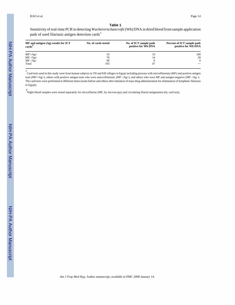
Forty-seven of 193 card test sample application pads with dried blood were positive for W.bancrofti DNA by real-time PCR with LDR reagents (Table 1). Parasite DNA was detected inall 33 samples from subjects with microfilaremia (100%) and in 14 of 70 (20%) sample padsfrom amicrofilaremic subjects with positive filarial antigen tests. Presumably, some of thesesubjects had low-level microfilaremia that was not detected by microscopic examination ofstained thick blood smears. No parasite DNA was detected in 90 sample application pads fromsubjects with negative tests for microfilaremia and filarial antigenemia. In addition, no parasiteDNA was detected in 12 sample application pads from MF-positive subjects whose antigencard tests had been performed with plasma instead of blood. Thus, we found no evidence offree parasite DNA in plasma from MF carriers.
DNA from all of the dried blood samples that had W. bancrofti DNA (detected by real-timePCR with LDR reagents) were retested by real-time PCR with the Wolbachia 16S rDNAprimers and probe. Only 7 of 47 (14.8%) of these samples were positive for Wolbachia DNA.This confirmed the lower sensitivity of the Wolbachia-based assay relative to the LDR assay.
Sensitivity of real-time PCR and C-PCR for detecting W. bancrofti DNA in Cx. pipiens andAn. punctulatus mosquitoes
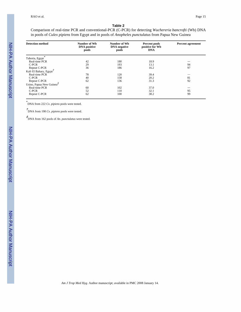
Mosquito results are shown in Table 2. Real-time PCR (with LDR reagents) detected manymore positive pools than C-PCR. All samples with discordant results were retested by bothmethods. All real-time PCR results were confirmed. However, many samples that were initiallyscored as negative by C-PCR and positive by real-time PCR were found to be positive by C-PCR after repeat testing; agreement between the two methods was fairly good when results ofrepeat C-PCR are considered.
Effects of MDA on mosquito infection ratesFigure 4 shows effects of three rounds of MDA on mosquito pool infection rates for twoEgyptian villages. Note that infection parameters were significantly higher in village KB thanin TH before the initiation of MDA. MDA had dramatic effects on mosquito pool infectionrates in both villages. No infected mosquito pools were detected after three annual rounds ofMDA in TH, whereas infections persisted in KB with a 77.5% reduction in the percentage ofpositive mosquito pools relative to the pre-MDA baseline value. Moreover, percent agreements
RAO et al. Page 5
Am J Trop Med Hyg. Author manuscript; available in PMC 2008 January 14.
NIH
-PA Author Manuscript
NIH
-PA Author Manuscript
NIH
-PA Author Manuscript
between LDR assay and C-PCR on mosquito samples after MDA were 100% (TH) and 98%(KB).
DISCUSSIONThe purpose of this study was to explore the value of real-time PCR for detecting filarial DNAin field samples. A recent publication from Thailand showed that filarial DNA could bedetected by real-time PCR with two probes and melting curve analysis.21 However, the authorsdid not report results of specificity testing or results obtained with field samples.
In contrast to results reported by Lulitanond et al.,21 our preliminary studies showed that real-time PCR did not work well with NV1 and NV2 primers and a TaqMan probe directed to theSspI target sequence used for C-PCR. Therefore, we focused on studies to optimize conditionsfor detecting parasite DNA using the LDR and Wolbachia 16S target sequences. Our resultsshowed that the LDR assay was sensitive, specific, and efficient, with a dose–response curvethat was linear over a range of five orders of magnitude. We then evaluated the LDR detectionassay with a panel of DNA samples isolated from membrane filters with known numbers ofW. bancrofti MF visualized by microscopy. It was impressive that all membranes producedpositive DNA signals after storage for periods ranging from 6 months to 4 years. LDR Ct valueswere significantly (inversely) correlated with MF counts, but the correlation was onlymoderately strong. This may reflect partial degradation of parasite DNA on old membranes,variable recovery of parasite DNA from membranes, or the possible presence of PCR inhibitorson membranes.
The LDR real-time PCR assay was also sensitive for detecting parasite DNA in human blooddried on used ICT card sample application pads. Indeed, PCR detection was more sensitive fordetecting infections than microscopy performed with blood from the same subjects, becausesome samples from amicrofilaremic subjects with positive filarial antigen tests had positiveDNA tests. Specificity was excellent; positive tests were not seen with blood samples fromamicrofilaremic subjects with negative antigen tests or with blood samples from MF carriersthat was collected during the day. The latter result suggests that DNA detected by PCR isderived from intact MF and not from “free DNA” in plasma. The card test results suggestmultiple new uses for these cards when they are tested with blood samples collected at timesthat correspond to peak MF levels. First, selected samples can be tested by real-time PCR todetermine whether subjects with positive antigen tests have circulating MF. Second, used cardsfrom different places and times provide a valuable archive of parasite DNA that may be usefulfor DNA-based studies of drug resistance or parasite polymorphism.
The sensitivity of real-time PCR for detection of Wolbachia 16S DNA was much lower thanthe LDR assay for detecting filarial DNA. If the two reactions are equally efficient, thedifference in Ct values (~10 cycles) suggests at least a 1,000-fold difference in copy numberper cell for the two target sequences. Differences in stability of eukaryotic and bacterial DNAsequences on dried filters or differences in efficiency of isolation of DNA template from theparasite and bacteria may have contributed to the difference in sensitivity.22–24 While thenumber of LDR repeats in W. bancrofti gDNA presumably is constant throughout the parasitelife cycle, this may not be the case for the Wolbachia target sequence. Recently, it has beenreported that microfilariae have fewer Wolbachia than other parasite stages.25 This would tendto limit the diagnostic value of Wolbachia DNA for detecting MF in blood samples.
Our project used large panels of mosquito DNA extracts from Egypt and Papua New Guineato compare the sensitivity of real-time PCR (LDR target) with C-DNA (SspI target). The twomethods had the same sensitivity with a standard template of isolated parasite DNA in ourlaboratory in St. Louis. However, the real-time PCR assay was more sensitive for detecting
RAO et al. Page 6
Am J Trop Med Hyg. Author manuscript; available in PMC 2008 January 14.
NIH
-PA Author Manuscript
NIH
-PA Author Manuscript
NIH
-PA Author Manuscript
W. bancrofti DNA in field samples relative to C-PCR performed in endemic countrylaboratories. This evaluation provided a useful, real-world comparison of the two DNAdetection methods. In practice, C-PCR requires subjective scoring of bands in agarose gels,and we found that technicians had been reluctant to score very faint or questionable bands aspositives. Repeat C-PCR resolved discrepancies in most cases. However, there were a fewcases where repeated testing verified discrepancies with some samples only positive by LDRreal-time PCR and (a few) others only positive by C-PCR for the SspI sequence. Additionalstudies are needed to determine whether variability in LDR and SspI repeat sequences accountfor these discrepancies.
Beyond the technical comparison of the two DNA detection methods, the decreases in Egyptianmosquito pool infection rates documented by LDR real-time PCR after MDA were impressiveand consistent with C-PCR results. These results suggest that MDA had a major effect onparasite prevalence rates in the Egyptian villages studied, and they show the potential value ofreal-time PCR for monitoring the impact of MDA in filariasis elimination programs.
Looking ahead to practicalities, we should point out that real-time PCR is comparable in costto C-PCR (~$2 per mosquito pool including DNA isolation, target amplification, and detectionof amplified product). Costs for the instrumentation and reagents for real-time PCR aredecreasing over time; additional research is needed to further optimize methods to reduce costs.However, in terms of the data produced, MX by real-time PCR or C-PCR is far preferable(more sensitive and efficient) to traditional dissection with microscopy for detecting filarialinfections in mosquito populations and as a tool for monitoring late stages of filariasiselimination programs. In comparing real-time PCR with C-PCR for this purpose, we favorreal-time PCR because of its increased sensitivity with field samples, lower labor requirements,reduced potential for contamination in the laboratory (no need to separately analyze PCRproducts in the laboratory), and much higher throughput capability. Coupled with advances inmosquito collection methods and methods for DNA isolation, we believe that MX by real-timePCR (perhaps in regional reference laboratories) will prove to be a practical tool for monitoringfilariasis elimination programs.
Acknowledgements
The authors would like to acknowledge the efforts of field teams and technical staff at Ain Shams University, Cairo,Egypt, and at the Papua New Guinea Institute of Medical Research in Madang. We thank K. Curtis for technical help.
Financial support: This work was supported by National Institutes of Health Grant AI 35855.
References1. Grove DI, Warren KS, Mahmoud AA. Algorithms in the diagnosis and management of exotic diseases.
VI. The filariases. J Infect Dis 1975;132:340–352. [PubMed: 1099150]2. Weil GJ, Lammie PJ, Weiss N. The ICT filariasis test: a rapid-format antigen test for diagnosis of
bancroftian filariasis. Parasitol Today 1997;13:401–404. [PubMed: 15275155]3. Lammie P, Weil G, Rahmah N, Kaliraj P, Steel C, Goodman D, Lakshmikanthan V, Ottesen E.
Recombinant antigen based assays for the diagnosis and surveillance of lymphatic filariasis - amulticenter trial. Filaria J 2004;3:9. [PubMed: 15347425]
4. Williams S, Laney S, Bierwert L, Saunders L, Boakye D, Fischer P, Goodman D, Helmy H, Hoti S,Vasuki V, Lammie P, Plichart C, Ramzy R, Ottesen E. Development and standardization of a rapidPCR-based method for the detection of Wuchereria bancrofti in mosquitoes for xenomonitoring thehuman prevalence of bancroftian filariasis. Ann Trop Med Parasitol 2002;96:S41–S46. [PubMed:12625916]
5. Ramzy R, Farid H, Kamal H, Ibrahim G, Morsy Z, Faris R, Weil G. A polymerase chain reaction-based assay for detection of Wuchereria bancrofti in human blood and Culex pipiens. Trans Roy SocTrop Med Hyg 1997;91:156–160. [PubMed: 9196756]
RAO et al. Page 7
Am J Trop Med Hyg. Author manuscript; available in PMC 2008 January 14.
NIH
-PA Author Manuscript
NIH
-PA Author Manuscript
NIH
-PA Author Manuscript
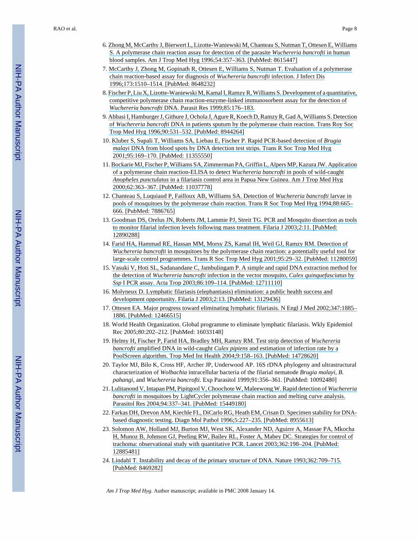
6. Zhong M, McCarthy J, Bierwert L, Lizotte-Waniewski M, Chanteau S, Nutman T, Ottesen E, WilliamsS. A polymerase chain reaction assay for detection of the parasite Wuchereria bancrofti in humanblood samples. Am J Trop Med Hyg 1996;54:357–363. [PubMed: 8615447]
7. McCarthy J, Zhong M, Gopinath R, Ottesen E, Williams S, Nutman T. Evaluation of a polymerasechain reaction-based assay for diagnosis of Wuchereria bancrofti infection. J Infect Dis1996;173:1510–1514. [PubMed: 8648232]
8. Fischer P, Liu X, Lizotte-Waniewski M, Kamal I, Ramzy R, Williams S. Development of a quantitative,competitive polymerase chain reaction-enzyme-linked immunosorbent assay for the detection ofWuchereria bancrofti DNA. Parasit Res 1999;85:176–183.
9. Abbasi I, Hamburger J, Githure J, Ochola J, Agure R, Koech D, Ramzy R, Gad A, Williams S. Detectionof Wuchereria bancrofti DNA in patients sputum by the polymerase chain reaction. Trans Roy SocTrop Med Hyg 1996;90:531–532. [PubMed: 8944264]
10. Kluber S, Supali T, Williams SA, Liebau E, Fischer P. Rapid PCR-based detection of Brugiamalayi DNA from blood spots by DNA detection test strips. Trans R Soc Trop Med Hyg2001;95:169–170. [PubMed: 11355550]
11. Bockarie MJ, Fischer P, Williams SA, Zimmerman PA, Griffin L, Alpers MP, Kazura JW. Applicationof a polymerase chain reaction-ELISA to detect Wuchereria bancrofti in pools of wild-caughtAnopheles punctulatus in a filariasis control area in Papua New Guinea. Am J Trop Med Hyg2000;62:363–367. [PubMed: 11037778]
12. Chanteau S, Luquiaud P, Failloux AB, Williams SA. Detection of Wuchereria bancrofti larvae inpools of mosquitoes by the polymerase chain reaction. Trans R Soc Trop Med Hyg 1994;88:665–666. [PubMed: 7886765]
13. Goodman DS, Orelus JN, Roberts JM, Lammie PJ, Streit TG. PCR and Mosquito dissection as toolsto monitor filarial infection levels following mass treatment. Filaria J 2003;2:11. [PubMed:12890288]
14. Farid HA, Hammad RE, Hassan MM, Morsy ZS, Kamal IH, Weil GJ, Ramzy RM. Detection ofWuchereria bancrofti in mosquitoes by the polymerase chain reaction: a potentially useful tool forlarge-scale control programmes. Trans R Soc Trop Med Hyg 2001;95:29–32. [PubMed: 11280059]
15. Vasuki V, Hoti SL, Sadanandane C, Jambulingam P. A simple and rapid DNA extraction method forthe detection of Wuchereria bancrofti infection in the vector mosquito, Culex quinquefasciatus bySsp I PCR assay. Acta Trop 2003;86:109–114. [PubMed: 12711110]
16. Molyneux D. Lymphatic filariasis (elephantiasis) elimination: a public health success anddevelopment opportunity. Filaria J 2003;2:13. [PubMed: 13129436]
17. Ottesen EA. Major progress toward eliminating lymphatic filariasis. N Engl J Med 2002;347:1885–1886. [PubMed: 12466515]
18. World Health Organization. Global programme to eliminate lymphatic filariasis. Wkly EpidemiolRec 2005;80:202–212. [PubMed: 16033148]
19. Helmy H, Fischer P, Farid HA, Bradley MH, Ramzy RM. Test strip detection of Wuchereriabancrofti amplified DNA in wild-caught Culex pipiens and estimation of infection rate by aPoolScreen algorithm. Trop Med Int Health 2004;9:158–163. [PubMed: 14728620]
20. Taylor MJ, Bilo K, Cross HF, Archer JP, Underwood AP. 16S rDNA phylogeny and ultrastructuralcharacterization of Wolbachia intracellular bacteria of the filarial nematode Brugia malayi, B.pahangi, and Wuchereria bancrofti. Exp Parasitol 1999;91:356–361. [PubMed: 10092480]
21. Lulitanond V, Intapan PM, Pipitgool V, Choochote W, Maleewong W. Rapid detection of Wuchereriabancrofti in mosquitoes by LightCycler polymerase chain reaction and melting curve analysis.Parasitol Res 2004;94:337–341. [PubMed: 15449180]
22. Farkas DH, Drevon AM, Kiechle FL, DiCarlo RG, Heath EM, Crisan D. Specimen stability for DNA-based diagnostic testing. Diagn Mol Pathol 1996;5:227–235. [PubMed: 8955613]
23. Solomon AW, Holland MJ, Burton MJ, West SK, Alexander ND, Aguirre A, Massae PA, MkochaH, Munoz B, Johnson GJ, Peeling RW, Bailey RL, Foster A, Mabey DC. Strategies for control oftrachoma: observational study with quantitative PCR. Lancet 2003;362:198–204. [PubMed:12885481]
24. Lindahl T. Instability and decay of the primary structure of DNA. Nature 1993;362:709–715.[PubMed: 8469282]
RAO et al. Page 8
Am J Trop Med Hyg. Author manuscript; available in PMC 2008 January 14.
NIH
-PA Author Manuscript
NIH
-PA Author Manuscript
NIH
-PA Author Manuscript

25. McGarry HF, Egerton GL, Taylor MJ. Population dynamics of Wolbachia bacterial endosymbiontsin Brugia malayi. Mol Biochem Parasitol 2004;135:57–67. [PubMed: 15287587]
RAO et al. Page 9
Am J Trop Med Hyg. Author manuscript; available in PMC 2008 January 14.
NIH
-PA Author Manuscript
NIH
-PA Author Manuscript
NIH
-PA Author Manuscript
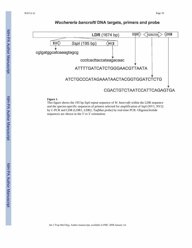
Figure 1.This figure shows the 195 bp SspI repeat sequence of W. bancrofti within the LDR sequenceand the species-specific sequences of primers selected for amplification of SspI (NV1, NV2)by C-PCR and LDR (LDR1, LDR2, TaqMan probe) by real-time PCR. Oligonucleotidesequences are shown in the 5′ to 3′ orientation.
RAO et al. Page 10
Am J Trop Med Hyg. Author manuscript; available in PMC 2008 January 14.
NIH
-PA Author Manuscript
NIH
-PA Author Manuscript
NIH
-PA Author Manuscript
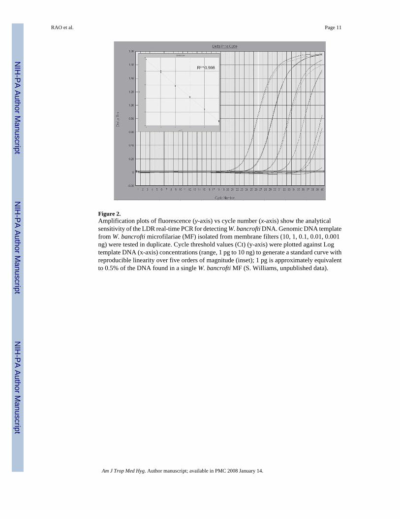
Figure 2.Amplification plots of fluorescence (y-axis) vs cycle number (x-axis) show the analyticalsensitivity of the LDR real-time PCR for detecting W. bancrofti DNA. Genomic DNA templatefrom W. bancrofti microfilariae (MF) isolated from membrane filters (10, 1, 0.1, 0.01, 0.001ng) were tested in duplicate. Cycle threshold values (Ct) (y-axis) were plotted against Logtemplate DNA (x-axis) concentrations (range, 1 pg to 10 ng) to generate a standard curve withreproducible linearity over five orders of magnitude (inset); 1 pg is approximately equivalentto 0.5% of the DNA found in a single W. bancrofti MF (S. Williams, unpublished data).
RAO et al. Page 11
Am J Trop Med Hyg. Author manuscript; available in PMC 2008 January 14.
NIH
-PA Author Manuscript
NIH
-PA Author Manuscript
NIH
-PA Author Manuscript
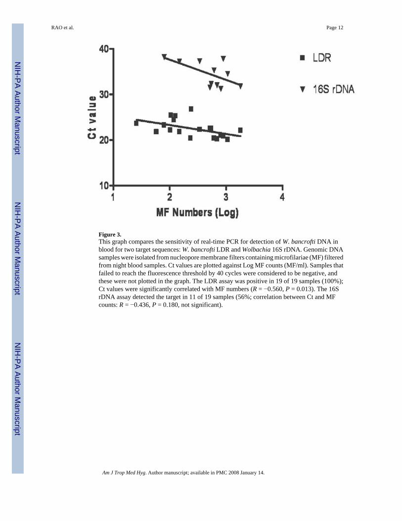
Figure 3.This graph compares the sensitivity of real-time PCR for detection of W. bancrofti DNA inblood for two target sequences: W. bancrofti LDR and Wolbachia 16S rDNA. Genomic DNAsamples were isolated from nucleopore membrane filters containing microfilariae (MF) filteredfrom night blood samples. Ct values are plotted against Log MF counts (MF/ml). Samples thatfailed to reach the fluorescence threshold by 40 cycles were considered to be negative, andthese were not plotted in the graph. The LDR assay was positive in 19 of 19 samples (100%);Ct values were significantly correlated with MF numbers (R = −0.560, P = 0.013). The 16SrDNA assay detected the target in 11 of 19 samples (56%; correlation between Ct and MFcounts: R = −0.436, P = 0.180, not significant).
RAO et al. Page 12
Am J Trop Med Hyg. Author manuscript; available in PMC 2008 January 14.
NIH
-PA Author Manuscript
NIH
-PA Author Manuscript
NIH
-PA Author Manuscript
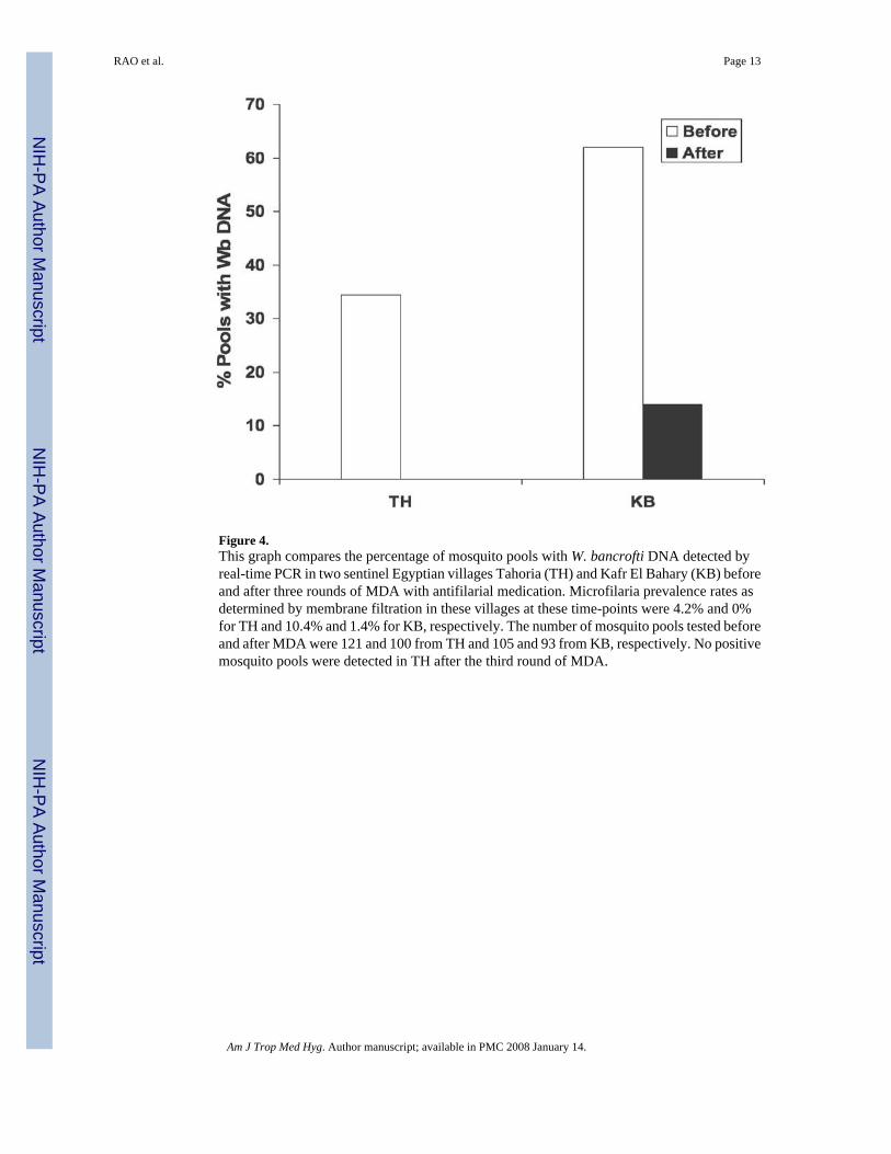
Figure 4.This graph compares the percentage of mosquito pools with W. bancrofti DNA detected byreal-time PCR in two sentinel Egyptian villages Tahoria (TH) and Kafr El Bahary (KB) beforeand after three rounds of MDA with antifilarial medication. Microfilaria prevalence rates asdetermined by membrane filtration in these villages at these time-points were 4.2% and 0%for TH and 10.4% and 1.4% for KB, respectively. The number of mosquito pools tested beforeand after MDA were 121 and 100 from TH and 105 and 93 from KB, respectively. No positivemosquito pools were detected in TH after the third round of MDA.
RAO et al. Page 13
Am J Trop Med Hyg. Author manuscript; available in PMC 2008 January 14.
NIH
-PA Author Manuscript
NIH
-PA Author Manuscript
NIH
-PA Author Manuscript
NIH
-PA Author Manuscript
NIH
-PA Author Manuscript
NIH
-PA Author Manuscript
RAO et al. Page 14
Table 1Sensitivity of real-time PCR in detecting Wuchereria bancrofti (Wb) DNA in dried blood from sample applicationpads of used filariasis antigen detection cards*
MF and antigen (Ag) results for ICTcards†
No. of cards tested No. of ICT sample padspositive for Wb DNA
Percent of ICT sample padspositive for Wb DNA
MF+/Ag+ 33 33 100MF−/Ag+ 70 14 20MF−/Ag− 90 0 0Total 193 47 —
*Card tests used in this study were from human subjects in TH and KB villages in Egypt including persons with microfilaremia (MF) and positive antigen
tests (MF+/Ag+), others with positive antigen tests who were amicrofilaremic (MF−/Ag+), and others who were MF and antigen-negative (MF−/Ag−).The card tests were performed at different times (some before and others after initiation of mass drug administration for elimination of lymphatic filariasisin Egypt).
†Night blood samples were tested separately for microfilariae (MF, by microscopy) and circulating filarial antigenemia (by card test).
Am J Trop Med Hyg. Author manuscript; available in PMC 2008 January 14.
NIH
-PA Author Manuscript
NIH
-PA Author Manuscript
NIH
-PA Author Manuscript
RAO et al. Page 15
Table 2Comparison of real-time PCR and conventional-PCR (C-PCR) for detecting Wuchereria bancrofti (Wb) DNAin pools of Culex pipiens from Egypt and in pools of Anopheles punctulatus from Papua New Guinea
Detection method Number of WbDNA positive
pools
Number of WbDNA negative
pools
Percent poolspositive for Wb
DNA
Percent agreement
Tahoria, Egypt* Real-time PCR 42 180 18.9 — C-PCR 29 193 13.1 94 Repeat C-PCR 36 186 16.2 97Kafr El Bahary, Egypt† Real-time PCR 78 120 39.4 — C-PCR 40 158 20.2 81 Repeat C-PCR 62 136 31.3 92Usino, Papua New Guinea‡ Real-time PCR 60 102 37.0 — C-PCR 52 110 32.1 95 Repeat C-PCR 62 100 38.2 99
*DNA from 222 Cx. pipiens pools were tested.
†DNA from 198 Cx. pipiens pools were tested.
‡DNA from 162 pools of An. punctulatus were tested.
Am J Trop Med Hyg. Author manuscript; available in PMC 2008 January 14.