A Pictorial Review of Atlantoaxial Rotatory Fixation: Key Points for the Radiologist
12
Clinical Radiology (2001) 56: 947–958 doi:10.1053/crad.2001.0679, available online at http://www.idealibrary.com on Pictorial Review A Pictorial Review of Atlanto-axial Rotatory Fixation: Key Points for the Radiologist CLARE J. ROCHE*, MICHAEL O’MALLEY {, JOHN C. DORGAN {, HELEN M. CARTY* *Department of Radiology, Alder Hey Children’s Hospital, Liverpool, U.K. and {Department of Orthopaedics, Alder Hey Children’s Hospital, Liverpool, U.K. Received: 10 July 2000 Revised: 16 October 2000 Accepted: 3 November 2000 Atlanto-axial rotatory fixation (AARF) is a rare condition which occurs more commonly in children than in adults. The terminology can be confusing and the condition is also known as ‘atlanto-axial rotatory subluxation’ and ‘atlanto-axial rotary dislocation’. Rotatory fixation is the preferred term, how- ever, as in most cases the fixation occurs within the normal range of rotation of the joint. By definition, therefore, the joint is neither subluxed nor dislocated. AARF is a cause of acquired torticollis. Diagnosis can be dicult and is often delayed. The radiologist plays a key role in confirming the diagnosis. The classification system proposed by Fielding in 1977 is most frequently used and will be discussed in detail. Given that this classification system was devised in the days before computed tomography (CT), as well as the fact that combined atlanto-axial and atlanto-occipital rotatory subluxation (AORF) is omitted from the classification, we propose a modification to the classification of this rare but significant disorder. The radiological findings in six cases of AARF will be illustrated, including a case with associated atlanto-occipital subluxation. The pertinent literature is reviewed and a more comprehensive classification system proposed. The imaging approach to diagnosis and the orthopaedic approach to management will be discussed. Roche, C. J. et al. (2001). Clinical Radiology 56, 947–958. # 2001 The Royal College of Radiologists Key words: atlas, axis, dislocation, spine, CT. Torticollis or ‘twisted/wry neck’ may be due to a variety of conditions. It is helpful to subdivide the causes of torticollis into two groups: (1) disorders of rotation of the atlanto- axial joint resulting in fixed or limited rotation of the neck; and (2) other disorders causing limited rotation of the neck without primarily involving the atlanto-axial joint. The first group includes rotatory fixation of the atlanto- axial joint which may occur spontaneously or secondary to trauma, congenital anomalies of the upper cervical spine and arthritides [1–5]. This group also includes ‘Grisel’s syndrome’ which is defined as non-traumatic atlanto-axial subluxation secondary to ligamentous laxity and inflam- mation following infection or surgery in the head and neck region [4]. The second group includes conditions where the primary abnormality is in the sternomastoid muscle, such as congenital fibrosis of the sternomastoid and acquired benign paroxysmal torticollis. Contraction of the sternomastoid muscle results in torticollis. Locally painful neck conditions such as lymphadenitis and underlying disorders such as tumours in the cervical spine, posterior fossa and cervical cord may also result in secondary sternomastoid contraction and spasm [1,6]. In this group of diverse conditions contraction of the sternomastoid muscle is the deforming force, resulting in secondary rotation at the atlanto-axial joint and torticollis. Clinical examination in these patients will reveal contraction of the sternomastoid on the side opposite the direction of head rotation, the ‘short’ sternomastoid. Conversely, when the torticollis is due to an underlying disorder of atlanto-axial rotation sternomas- toid spasm is found on the side to which the head is rotated, the ‘long’ sternomastoid. The spasm in this case represents an attempt to correct the deformity [1–3,6,7]. 0009-9260/01/120947+12 $35.00/0 # 2001 The Royal College of Radiologists Author for correspondence and guarantor of study: Helen M. Carty, Radiology Department, Alder Hey Children’s Hospital, Eaton Road, Liverpool L12 2AP, U.K. Fax: 44(0)151 2525 533; E-mail: [email protected]
-
Upload
independent -
Category
Documents
-
view
1 -
download
0
Transcript of A Pictorial Review of Atlantoaxial Rotatory Fixation: Key Points for the Radiologist

Clinical Radiology (2001) 56: 947±958doi:10.1053/crad.2001.0679, available online at http://www.idealibrary.com on
Pictorial Review
A Pictorial Review of Atlanto-axial Rotatory Fixation:Key Points for the Radiologist
CLARE J. ROCHE*, MICHAEL O'MALLEY{, JOHN C. DORGAN{, HELEN M. CARTY*
*Department of Radiology, Alder Hey Children's Hospital, Liverpool, U.K. and {Department of Orthopaedics, Alder HeyChildren's Hospital, Liverpool, U.K.
Received: 10 July 2000 Revised: 16 October 2000 Accepted: 3 November 2000
Atlanto-axial rotatory ®xation (AARF) is a rare condition which occurs more commonly in childrenthan in adults. The terminology can be confusing and the condition is also known as `atlanto-axialrotatory subluxation' and `atlanto-axial rotary dislocation'. Rotatory ®xation is the preferred term, how-ever, as in most cases the ®xation occurs within the normal range of rotation of the joint. By de®nition,therefore, the joint is neither subluxed nor dislocated. AARF is a cause of acquired torticollis. Diagnosiscan be di�cult and is often delayed. The radiologist plays a key role in con®rming the diagnosis.The classi®cation system proposed by Fielding in 1977 is most frequently used and will be discussed indetail. Given that this classi®cation system was devised in the days before computed tomography (CT),as well as the fact that combined atlanto-axial and atlanto-occipital rotatory subluxation (AORF) isomitted from the classi®cation, we propose a modi®cation to the classi®cation of this rare but signi®cantdisorder.The radiological ®ndings in six cases of AARF will be illustrated, including a case with associatedatlanto-occipital subluxation. The pertinent literature is reviewed and a more comprehensiveclassi®cation system proposed. The imaging approach to diagnosis and the orthopaedic approach tomanagement will be discussed. Roche, C. J. et al. (2001). Clinical Radiology 56, 947±958.
# 2001 The Royal College of Radiologists
Key words: atlas, axis, dislocation, spine, CT.
Torticollis or `twisted/wry neck' may be due to a variety ofconditions. It is helpful to subdivide the causes of torticollisinto two groups: (1) disorders of rotation of the atlanto-axial joint resulting in ®xed or limited rotation of the neck;and (2) other disorders causing limited rotation of the neckwithout primarily involving the atlanto-axial joint.
The ®rst group includes rotatory ®xation of the atlanto-axial joint which may occur spontaneously or secondary totrauma, congenital anomalies of the upper cervical spineand arthritides [1±5]. This group also includes `Grisel'ssyndrome' which is de®ned as non-traumatic atlanto-axialsubluxation secondary to ligamentous laxity and in¯am-mation following infection or surgery in the head and neckregion [4].
The second group includes conditions where the primaryabnormality is in the sternomastoid muscle, such ascongenital ®brosis of the sternomastoid and acquired benignparoxysmal torticollis. Contraction of the sternomastoidmuscle results in torticollis. Locally painful neck conditionssuch as lymphadenitis and underlying disorders such astumours in the cervical spine, posterior fossa and cervicalcord may also result in secondary sternomastoid contractionand spasm [1,6]. In this group of diverse conditionscontraction of the sternomastoid muscle is the deformingforce, resulting in secondary rotation at the atlanto-axialjoint and torticollis. Clinical examination in these patientswill reveal contraction of the sternomastoid on the sideopposite the direction of head rotation, the `short'sternomastoid. Conversely, when the torticollis is due toan underlying disorder of atlanto-axial rotation sternomas-toid spasm is found on the side to which the head is rotated,the `long' sternomastoid. The spasm in this case representsan attempt to correct the deformity [1±3,6,7].
0009-9260/01/120947+12 $35.00/0 # 2001 The Royal College of Radiologists
Author for correspondence and guarantor of study: Helen M. Carty,Radiology Department, Alder Hey Children's Hospital, Eaton Road,Liverpool L12 2AP, U.K. Fax: �44(0)151 2525 533; E-mail:[email protected]
948 CLINICAL RADIOLOGY
THE ATLANTO-AXIAL JOINT
The atlanto-axial joint is unique and complex. It is themost active joint in the body, moving approximately 600times per hour [8].The normal range of cervical rotation is approximately
908 to either side and half this rotation occurs at theatlanto-axial joint [5]. The facet joints between the atlas andthe axis are almost horizontal, allowing excellent rotation atthe expense of bony stability. The transverse ligament,running behind the odontoid, prevents excessive anteriormotion of the atlas on the axis. The paired alar ligamentswhich run from the posterolateral tip of the odontoid to theoccipital condyles limit rotation of the atlas on the axis. Theright alar ligament prevents excessive rotation to the leftand vice versa [9,10]. The alar ligaments also act assecondary stabilizers, preventing anterior shift [1,11](Fig. 1). Cadaver studies have shown that when thetransverse ligament is cut there is anterior subluxation of4 mm, with further subluxation prevented by the alarligaments. If the alar ligaments are also cut then muchgreater forward subluxation occurs and there is alsoexcessive rotation of the atlas on the axis [11].The physiological range of rotation of the atlas on the
axis is 25±538 to either side [12±18]. Rotation of 4 568 or aright±left di�erence of4 88 is said to indicate hypermobi-lity. Rotation of 5 288 is reported to indicate hypomobility[16].
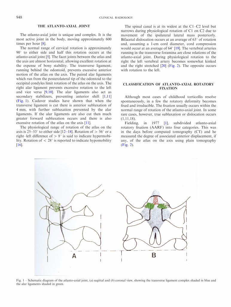
The spinal canal is at its widest at the C1±C2 level butnarrows during physiological rotation of C1 on C2 due tomovement of the ipsilateral lateral mass posteriorly.Bifacetal dislocation occurs at an average of 638 of rotationand, assuming a 1-cm cord diameter, cord compressionwould occur at an average of 648 [19]. The vertebral arteriesrunning in the transverse foramina are close relations of theatlanto-axial joint. During physiological rotation to theright the left vertebral artery becomes somewhat kinkedand the right stretched [20] (Fig. 2). The opposite occurswith rotation to the left.
CLASSIFICATION OF ATLANTO-AXIAL ROTATORYFIXATION
Although most cases of childhood torticollis resolvespontaneously, in a few the rotatory deformity becomes®xed and irreducible. The ®xation usually occurs within thenormal range of rotation of the atlanto-axial joint. In somerare cases, however, true subluxation or dislocation occurs(1,11,18).
Fielding, in 1977 [1], subdivided atlanto-axialrotatory ®xation (AARF) into four categories. This wasin the days before computed tomography (CT) and hemeasured the degree of associated anterior displacement, ifany, of the atlas on the axis using plain tomography(Fig. 2).
Fig. 1 ± Schematic diagram of the atlanto-axial joint, (a) sagittal and (b) coronal view, showing the transverse ligament complex shaded in blue andthe alar ligaments shaded in green.
PICTORIAL REVIEW 949
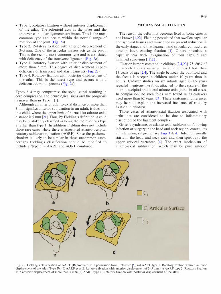
. Type 1. Rotatory ®xation without anterior displacementof the atlas. The odontoid acts as the pivot and thetransverse and alar ligaments are intact. This is the mostcommon type and occurs within the normal range ofrotation of the joint (Fig. 2a).
. Type 2. Rotatory ®xation with anterior displacement of3±5 mm. One of the articular masses acts as the pivot.This is the second most common type and is associatedwith de®ciency of the transverse ligament (Fig. 2b).
. Type 3. Rotatory ®xation with anterior displacement ofmore than 5 mm. This degree of displacement impliesde®ciency of transverse and alar ligaments (Fig. 2c).
. Type 4. Rotatory ®xation with posterior displacement ofthe atlas. This is the rarest type and occurs with ade®cient odontoid process (Fig. 2d).
Types 2±4 may compromise the spinal canal resulting incord compression and neurological signs and the prognosisis graver than in Type 1 [1].Although an anterior atlanto-axial distance of more than
3 mm signi®es anterior subluxation in an adult, it does notin a child, where the upper limit of normal for atlanto-axialdistance is 5 mm [21]. Thus, by Fielding's de®nition, a childmay be mistakenly classi®ed as being the more serious type2 rather than type 1. In addition Fielding does not includethose rare cases where there is associated atlanto-occipitalrotatory subluxation/®xation (AORF). Since the pathome-chanism is likely to be similar in these uncommon cases,perhaps Fielding's classi®cation should be modi®ed toinclude a `type 5' ± AARF and AORF combined.
MECHANISM OF FIXATION
The reason the deformity becomes ®xed in some cases isnot known [1,22]. Fielding postulated that swollen capsularand synovial tissues and muscle spasm prevent reduction inthe early stages and that ligament and capsular contracturesdevelop later, causing ®xation [1]. Others postulate acapsular tear with invagination of torn capsule andin¯amed synovium [18,22].
Fixation is more common in children [2,4,23]; 75±80% ofall reported cases occurred in children aged less than13 years of age [2,4]. The angle between the odontoid andthe facets is steeper in children under 10 years than inadults. Cadaver studies on six infants aged 0±3.5 yearsrevealed meniscus-like folds attached to the capsule of theatlanto-occipital and lateral atlanto-axial joints in all cases.In comparison, no such folds were found in 23 cadaversaged more than 62 years [24]. These anatomical di�erencesmay help to explain the increased incidence of rotatory®xation in children.
Those cases of atlanto-axial ®xation associated witharthritides are considered to be due to in¯ammatorydisruption of the ligament complex.
Grisel's syndrome, or atlanto-axial subluxation followinginfection or surgery in the head and neck region, constitutesan interesting subgroup (see Figs 3 & 4). Infection usuallystarts in the head and neck area and then spreads to theupper cervical vertebrae [4]. The exact mechanism ofatlanto-axial subluxation, which may be pure anterior
Fig. 2 ± Fielding's classi®cation of AARF (Reproduced with permission from Reference [5]) (a) AARF type 1. Rotatory ®xation without anteriordisplacement of the atlas. Type 3b. (b) AARF type 2. Rotatory ®xation with anterior displacement of 3±5 mm. (c) AARF type 3. Rotatory ®xationwith anterior displacement of more than 5 mm. (d) AARF type 4. Rotatory ®xation with posterior displacement of the atlas.
950 CLINICAL RADIOLOGY
subluxation or rotatory ®xation, in this condition is notknown but postulated mechanisms include:
. Spastic contracture of cervical muscles secondary toregional lymphadenitis in the presence of ligamentouslaxity caused by hyperaemia [6].
. Distension of ligaments caused by hyperaemia oradjacent joint e�usion [4].
. Direct infection resulting in rupture of ligaments [4].
. Decalci®cation of the bony attachments of ligamentssecondary to hyperaemia [4].
There is a direct connection between the pharyngovertebralveins and the peri-odontoidal venous plexus and sub-occipital venous sinuses. This may provide a route forhaematogenous spread of infection and an anatomicalexplanation for atlanto-axial hyperaemia [6].
Torticollis in Grisel's syndrome may occur anytimefrom 1 day to several weeks following the initial insult.The white cell count is usually normal but the ESR is raised[4]. AORF is considered by some authors to be acompensatory counter-rotation mechanism, occurring inthe presence of AARF [25,26]. It is postulated that, in casesof long-standing AARF, ligamentous contractures developand further attempts at correction result in rotatory sub-luxation at the relatively more mobile atlanto-occipitaljoint.
AARF with an associated AORF has been reported, toour knowledge, only seven times in the literature [18,25±29].Interestingly, in at least four cases the mechanism of injuryis described as `head turning' and in two of the caseschiropractic treatment had been used to treat the torticollisbefore the AORF was diagnosed [25,26].
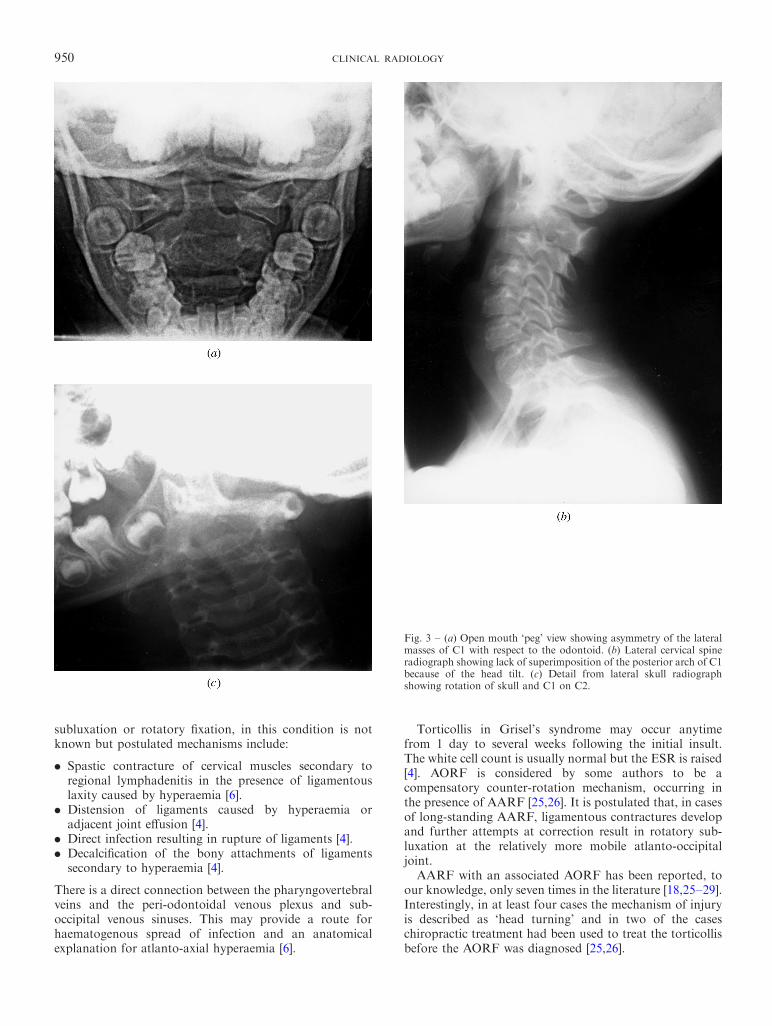
Fig. 3 ± (a) Open mouth `peg' view showing asymmetry of the lateralmasses of C1 with respect to the odontoid. (b) Lateral cervical spineradiograph showing lack of superimposition of the posterior arch of C1because of the head tilt. (c) Detail from lateral skull radiographshowing rotation of skull and C1 on C2.
PICTORIAL REVIEW 951
CLINICAL FEATURES
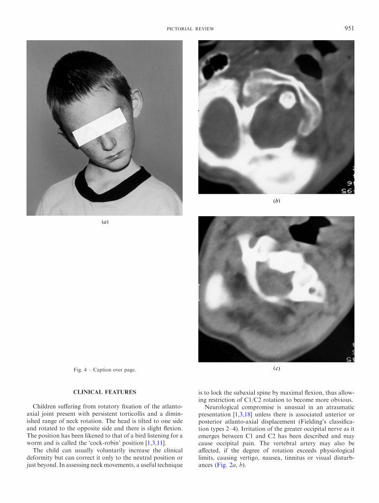
Children su�ering from rotatory ®xation of the atlanto-axial joint present with persistent torticollis and a dimin-ished range of neck rotation. The head is tilted to one sideand rotated to the opposite side and there is slight ¯exion.The position has been likened to that of a bird listening for aworm and is called the `cock-robin' position [1,3,11].The child can usually voluntarily increase the clinical
deformity but can correct it only to the neutral position orjust beyond. In assessing neckmovements, a useful technique
is to lock the subaxial spine by maximal ¯exion, thus allow-ing restriction of C1/C2 rotation to become more obvious.
Neurological compromise is unusual in an atraumaticpresentation [1,3,18] unless there is associated anterior orposterior atlanto-axial displacement (Fielding's classi®ca-tion types 2±4). Irritation of the greater occipital nerve as itemerges between C1 and C2 has been described and maycause occipital pain. The vertebral artery may also bea�ected, if the degree of rotation exceeds physiologicallimits, causing vertigo, nausea, tinnitus or visual disturb-ances (Fig. 2a, b).
Fig. 4 ± Caption over page.
952 CLINICAL RADIOLOGY
Cervical pain is produced when attempts are made tocorrect the deformity [1,3]. A distinguishing feature di�er-entiating benign torticollis and AARF is the side of sterno-cleidomastoid (SCM) spasm. In benign torticollis, the SCMis contracted on the side opposite the deformity, i.e. poten-tially causing the deformity. In AARF, the SCM is con-tracted on the side of the deformity, i.e. trying to reduce it[1±3,22]. In long-standing cases plagiocephaly and ¯atteningof the face may develop on the side of the tilt [1,6].Examination sometimes reveals a bulge in the retropharynx
representing the anterior arch of the atlas [4]. The spinousprocess of the axis may be prominent and palpable awayfrom the midline and towards the direction of head rotation[2,4]. Local anaesthetic injection into the atlanto-axial jointrelieves the pain but not the deformity [11].
DIAGNOSIS
Diagnosis is di�cult and often delayed. AARF should besuspected if a torticollis fails to resolve within 5±7 days,
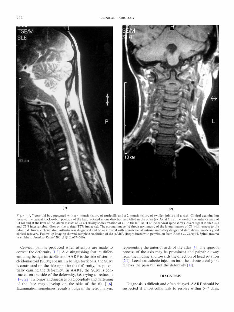
Fig. 4 ± A 7-year-old boy presented with a 4-month history of torticollis and a 2-month history of swollen joints and a rash. Clinical examinationrevealed the typical `cock-robin' position of the head, rotated in one direction and tilted in the other (a). Axial CT at the level of the anterior arch ofC1 (b) and at the level of the lateral masses of C1 (c) clearly shows rotation of C1 to the left. MRI of the cervical spine shows loss of signal in the C2/3and C3/4 intervertebral discs on the sagittal T2W image (d). The coronal image (e) shows asymmetry of the lateral masses of C1 with respect to theodontoid. Juvenile rheumatoid arthritis was diagnosed and he was treated with non-steroidal anti-in¯ammatory drugs and steroids and made a goodclinical recovery. Follow-up imaging showed complete resolution of the AARF. (Reproduced with permission from Roche C, Carty H. Spinal traumain children. Paediatr Radiol 2001;31(10):677±700).

PICTORIAL REVIEW 953
particularly if preceded by minor trauma or an upperrespiratory tract infection [1,2,5,30], and especially if thereis sternomastoid spasm on the side to which the head isrotated.
The diagnosis is established by demonstrating ®xation ofthe atlas on the axis. A dynamic test is required to provethat the atlas and axis no longer rotate independently ofeach other but as a unit [1,2,5,18].
PLAIN RADIOGRAPHY
The `cock-robin' posture makes radiographic positioningdi�cult. The complex anatomy of the atlanto-axial region,coupled with problems of positioning, make the radio-graphs di�cult to interpret [2,6,31].
The open-mouth `peg' or `odontoid' view shows asym-metry of the lateral masses with respect to the odontoid[3,11,18,22,24] (Fig. 3). The lateral mass that has rotatedforwards appears wider and closer to the midline. The facetjoint may be obscured because of apparent overlapping[1,6,13,32]. In practice, the odontoid view is often extremelydi�cult to obtain in these patients.
The lateral cervical spine radiograph is di�cult tointerpret because of head tilt. The atlas may be obscuredby the skull and appearances may simulate occipito-atlantalassimilation. Because the skull and the atlas usuallymaintain a normal relationship in this condition, a lateralskull radiograph will often show the relative positions of theatlas and axis more clearly [6]. Depending on the degree of
Fig. 5 ± A 7-year-old girl presented with a 7-month history oftorticollis which developed after an episode of minor trauma ± she felta `click' in her neck while lying in the bath. CT con®rms that C1 isrotated on C2. The atlanto-dens interval was 4 mm. By Fielding'sclassi®cation [1] this is AARF type 1. She was treated with halotraction but this failed to resolve the deformity and surgical fusion ofC1 and C2 was performed.
Fig. 6 ± An 8-year-old boy with a history of Marfan's syndrome presented with a 4-month history of torticollis which had developed 1 week aftertonsillectomy. Axial CT at the level of the anterior arch of C1 (a) and at the level of the body of C2 (b) shows the odontoid lying eccentrically betweenthe lateral masses of C1. The anterior arch lies 5 3 mm anterior to the odontoid, however, making this a type 1 AARF. Halo traction resulted ingood clinical resolution of the deformity but follow-up CT showed a persistently abnormal relationship between C1 and C2.
954 CLINICAL RADIOLOGY
rotation one lateral mass of the atlas may be seen anteriorto the odontoid on the lateral cervical radiograph [6,24](Fig. 3). Because of head tilt, the right and left sides of theposterior arch of the atlas fail to superimpose [6] (Fig. 3).
These radiographic appearances are not pathognomonicfor AARF, as similar appearances are found in normalchildren who voluntarily rotate their heads and in thosewith torticollis from any cause [6,18,24]. Dynamic tests arerecommended to establish that the deformity is ®xed.Cineradiography in the lateral position or peg views withthe head rotated 158 to either side in turn have beenrecommended for this purpose [11,18,22,24]. Co-operationis often di�cult to obtain and the resulting radiographs areoften suboptimal. There is an appreciable radiation dose[6,7]. CT is often easier to perform and interpret.
Flexion lateral cervical spine views have also beenadvocated to detect any associated anterior subluxationwhich would alter the classi®cation of the ®xation (afterFielding) and a�ect prognosis and subsequent manage-ment. Again, this can be easily assessed using CT.
COMPUTED TOMOGRAPHY
Because of the di�culties in obtaining and interpretingplain radiographs, CT is essential for imaging thiscondition.
Axial CT through the upper cervical spine demonstratesthe rotated position of the atlas on the axis (Figs 4±7).Associated forward (Fielding types 2±3) or backward(Fielding type 4) displacement of the atlas may beappreciated [2,3,18,27]. In post-traumatic cases fracturesundetectable at plain radiography may be revealed. Sagittaland coronal reformatting and 3-D reconstructions arehelpful (Figs 4±7). It is essential to also check the alignmentbetween C1 and the occiput to exclude an associatedAORF. Physiologically, less than 58 of rotation occurs atthe normal atlanto-occipital articulation [13,20]. Malalign-ment of the axes of C1 and the occiput indicates AORF [27](Fig. 8).
When there is associated anterior or posterior displace-ment of the atlas on the axis (Fielding types 2±4), static or
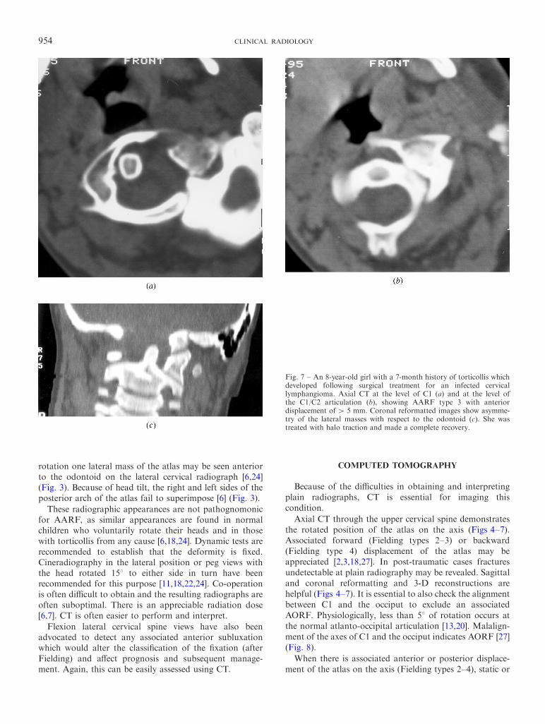
Fig. 7 ± An 8-year-old girl with a 7-month history of torticollis whichdeveloped following surgical treatment for an infected cervicallymphangioma. Axial CT at the level of C1 (a) and at the level ofthe C1/C2 articulation (b), showing AARF type 3 with anteriordisplacement of4 5 mm. Coronal reformatted images show asymme-try of the lateral masses with respect to the odontoid (c). She wastreated with halo traction and made a complete recovery.
PICTORIAL REVIEW 955
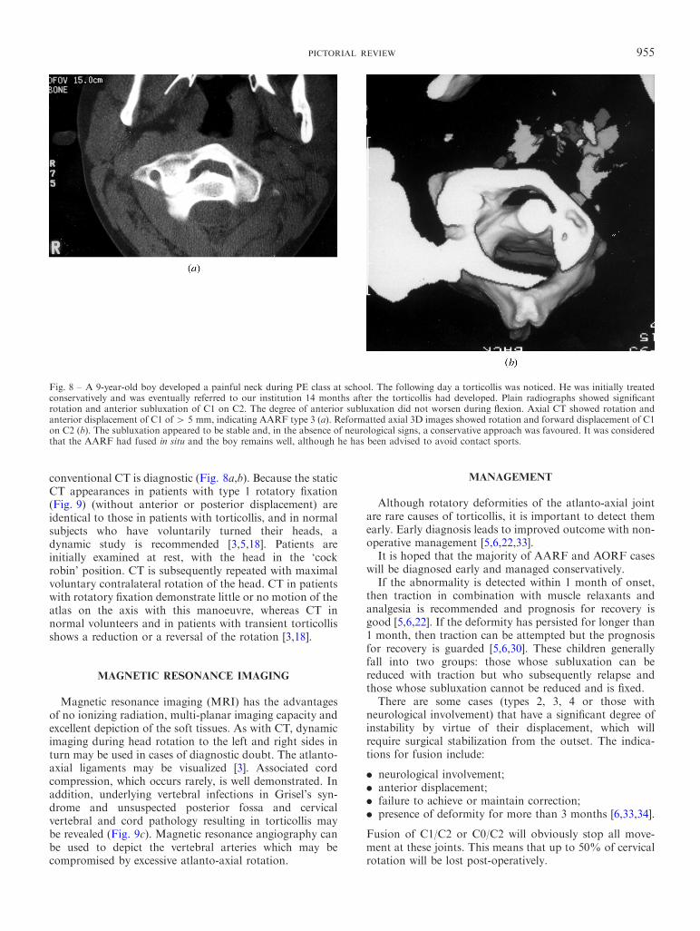
conventional CT is diagnostic (Fig. 8a,b). Because the staticCT appearances in patients with type 1 rotatory ®xation(Fig. 9) (without anterior or posterior displacement) areidentical to those in patients with torticollis, and in normalsubjects who have voluntarily turned their heads, adynamic study is recommended [3,5,18]. Patients areinitially examined at rest, with the head in the `cockrobin' position. CT is subsequently repeated with maximalvoluntary contralateral rotation of the head. CT in patientswith rotatory ®xation demonstrate little or no motion of theatlas on the axis with this manoeuvre, whereas CT innormal volunteers and in patients with transient torticollisshows a reduction or a reversal of the rotation [3,18].
MAGNETIC RESONANCE IMAGING
Magnetic resonance imaging (MRI) has the advantagesof no ionizing radiation, multi-planar imaging capacity andexcellent depiction of the soft tissues. As with CT, dynamicimaging during head rotation to the left and right sides inturn may be used in cases of diagnostic doubt. The atlanto-axial ligaments may be visualized [3]. Associated cordcompression, which occurs rarely, is well demonstrated. Inaddition, underlying vertebral infections in Grisel's syn-drome and unsuspected posterior fossa and cervicalvertebral and cord pathology resulting in torticollis maybe revealed (Fig. 9c). Magnetic resonance angiography canbe used to depict the vertebral arteries which may becompromised by excessive atlanto-axial rotation.
MANAGEMENT
Although rotatory deformities of the atlanto-axial jointare rare causes of torticollis, it is important to detect themearly. Early diagnosis leads to improved outcome with non-operative management [5,6,22,33].
It is hoped that the majority of AARF and AORF caseswill be diagnosed early and managed conservatively.
If the abnormality is detected within 1 month of onset,then traction in combination with muscle relaxants andanalgesia is recommended and prognosis for recovery isgood [5,6,22]. If the deformity has persisted for longer than1 month, then traction can be attempted but the prognosisfor recovery is guarded [5,6,30]. These children generallyfall into two groups: those whose subluxation can bereduced with traction but who subsequently relapse andthose whose subluxation cannot be reduced and is ®xed.
There are some cases (types 2, 3, 4 or those withneurological involvement) that have a signi®cant degree ofinstability by virtue of their displacement, which willrequire surgical stabilization from the outset. The indica-tions for fusion include:
. neurological involvement;
. anterior displacement;
. failure to achieve or maintain correction;
. presence of deformity for more than 3 months [6,33,34].
Fusion of C1/C2 or C0/C2 will obviously stop all move-ment at these joints. This means that up to 50% of cervicalrotation will be lost post-operatively.
Fig. 8 ± A 9-year-old boy developed a painful neck during PE class at school. The following day a torticollis was noticed. He was initially treatedconservatively and was eventually referred to our institution 14 months after the torticollis had developed. Plain radiographs showed signi®cantrotation and anterior subluxation of C1 on C2. The degree of anterior subluxation did not worsen during ¯exion. Axial CT showed rotation andanterior displacement of C1 of4 5 mm, indicating AARF type 3 (a). Reformatted axial 3D images showed rotation and forward displacement of C1on C2 (b). The subluxation appeared to be stable and, in the absence of neurological signs, a conservative approach was favoured. It was consideredthat the AARF had fused in situ and the boy remains well, although he has been advised to avoid contact sports.
956 CLINICAL RADIOLOGY
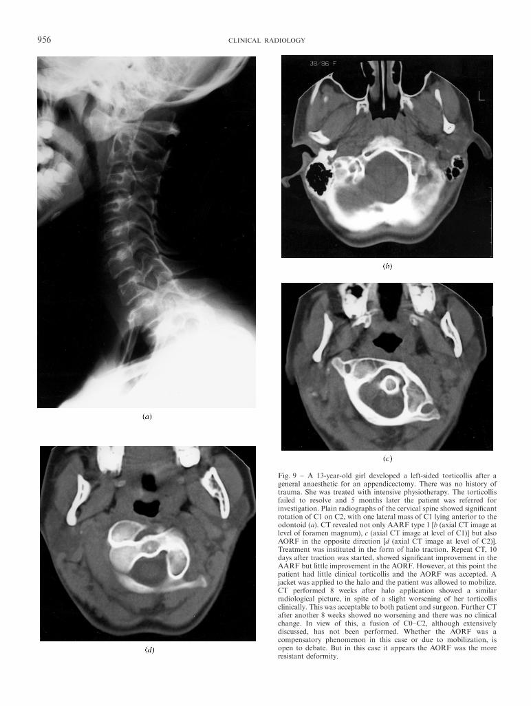
Fig. 9 ± A 13-year-old girl developed a left-sided torticollis after ageneral anaesthetic for an appendicectomy. There was no history oftrauma. She was treated with intensive physiotherapy. The torticollisfailed to resolve and 5 months later the patient was referred forinvestigation. Plain radiographs of the cervical spine showed signi®cantrotation of C1 on C2, with one lateral mass of C1 lying anterior to theodontoid (a). CT revealed not only AARF type 1 [b (axial CT image atlevel of foramen magnum), c (axial CT image at level of C1)] but alsoAORF in the opposite direction [d (axial CT image at level of C2)].Treatment was instituted in the form of halo traction. Repeat CT, 10days after traction was started, showed signi®cant improvement in theAARF but little improvement in the AORF. However, at this point thepatient had little clinical torticollis and the AORF was accepted. Ajacket was applied to the halo and the patient was allowed to mobilize.CT performed 8 weeks after halo application showed a similarradiological picture, in spite of a slight worsening of her torticollisclinically. This was acceptable to both patient and surgeon. Further CTafter another 8 weeks showed no worsening and there was no clinicalchange. In view of this, a fusion of C0±C2, although extensivelydiscussed, has not been performed. Whether the AORF was acompensatory phenomenon in this case or due to mobilization, isopen to debate. But in this case it appears the AORF was the moreresistant deformity.
PICTORIAL REVIEW 957
Management of AARF revolves around the timing ofdiagnosis (Table 1).There has been speculation that spontaneous improve-
ment in an AARF type 1 may be due to an attemptedcompensatory AORF [25,26]In some cases the symptoms and signs of deformity
resolve even though the radiological appearancesremain unchanged [6]. In these children, where there is
®xation with an intact transverse ligament and no anteriordisplacement there may be no need for operative interven-tion [3].
The aims of treatment in Grisel's syndrome also includebacteriological cure of the infection as well as stabilizationof the atlanto-axial joint [4].
AARF with associated AORF has been described onlyseven times in the literature [18,25±29], so there is little
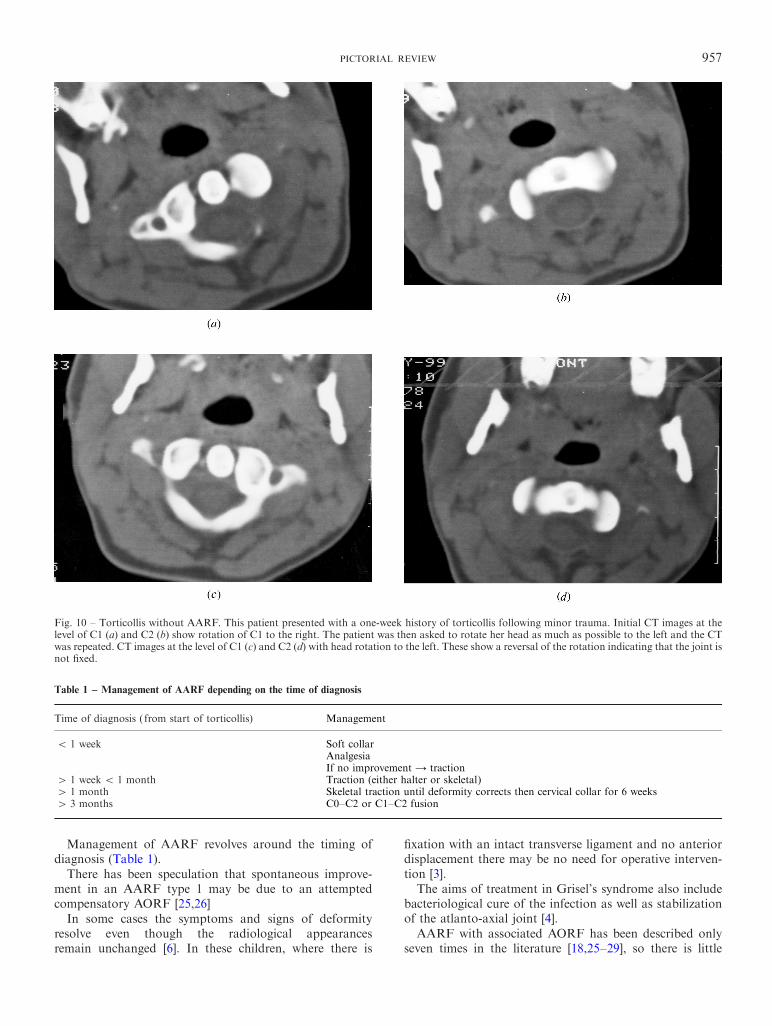
Fig. 10 ± Torticollis without AARF. This patient presented with a one-week history of torticollis following minor trauma. Initial CT images at thelevel of C1 (a) and C2 (b) show rotation of C1 to the right. The patient was then asked to rotate her head as much as possible to the left and the CTwas repeated. CT images at the level of C1 (c) and C2 (d) with head rotation to the left. These show a reversal of the rotation indicating that the joint isnot ®xed.
Table 1 ± Management of AARF depending on the time of diagnosis
Time of diagnosis ( from start of torticollis) Management
5 1 week Soft collarAnalgesiaIf no improvement 4 traction
4 1 week5 1 month Traction (either halter or skeletal)4 1 month Skeletal traction until deformity corrects then cervical collar for 6 weeks4 3 months C0±C2 or C1±C2 fusion
958 CLINICAL RADIOLOGY
experience of its management. Most of the reported caseshave undergone operative surgical stabilization.
CONCLUSION
An acquired persistent torticollis in a child should arousesuspicion of a rotatory disorder of the atlanto-axial joint,especially if there is a history of trivial trauma or of anupper respiratory tract infection. Early diagnosis leads tobetter outcome. AARF is, however, a rare cause oftorticollis and, because most cases of torticollis settlespontaneously, a conservative approach to investigationand management is appropriate. In general, children whopresent with atraumatic torticollis should be treatedconservatively with cervical collar and anti-in¯ammatorymedication for 1 week. Those children whose torticollis failsto resolve after 1 week require aggressive investigation by`dynamic' CT to assess whether the joint is ®xed. If,however, there is a history of signi®cant trauma thenimmediate radiological assessment is advised [30].This approach will avoid over-investigation and over-
treatment yet will still detect AARF early enough to achievea good outcome.
REFERENCES
1 Fielding JW, Hawkins RJ. Atlanto-axial rotatory ®xation. J BoneJoint Surg 1977;59-A:37±44.
2 Lukele M. Atlanto-axial rotatory ®xation. S Afr Med J 1986;12:1549±1552.
3 Maheshwaran S, Sgouros S, Jeyapalan K, Chapman S, Chardy J,Flint G. Imaging of childhood torticollis due to atlantoaxialrotatory ®xation. Childs Nerv Syst 1995;11:667±671.
4 Mathern G, Batzdorf U. Grisel's syndrome. Clin Orthop Rel Res1989;244:131±146.
5 Phillips WA, Hensinger RN. The management of rotatory atlanto-axial subluxation in children. J Bone Joint Surg 1989;71-A:664±668.
6 Weinstein SL. Atlantoaxial rotatory displacement. The PediatricSpine, 1. New York: Raven Press, 1994: 408±420.
7 Muniz A, Belfer RA. Atlantoaxial rotatory subluxation in children.Pediatr Emerg Care 1999;15:25±29.
8 Bland JH. Rheumatoid subluxations of the cervical spine. JRheumatol 1990;17:134±137.
9 Willauschus WG, Kladny B, Beyer WF, Gluckert K, Arnold H,Scheithauer R. Lesions of the alar ligaments. Spine 1995;20:2493±2498.
10 Daniels DL, Williams AL, Haughton VM. Computed tomographyof the articulations and ligaments at the occipito-atlantoaxialregion. Radiology 1983;146:709±716.
11 Wortzman G, Dewar F. Rotary ®xation of the atlantoaxial joint:rotational atlantoaxial subluxation. Radiology 1968;90:479±487.
12 Iai H, Goto S, Yamagata M, et al. Three-dimensional motion ofthe upper cervical spine in rheumatoid arthritis. Spine 1993;19:272±276.
13 Penning L. Normal movements of the cervical spine. Am JRoentgenol 1978;130:317±326.
14 Penning L, Wilmink JT. Rotation of the cervical spine: a CT studyin normal subjects. Spine 1987;12:732±738.
15 Dvorak J, Panjabi M, Gerber M, Wichmann W. CT-functionaldiagnostics of rotatory instability of the upper cervical spine. 1. Anexperimental study on cadavers. Spine 1987;12:197±205.
16 Dvorak J, Hayek J, Zehnder R. CT-functional diagnostics ofrotatory instability of the upper cervical spine. 2. An evaluation onhealthy adults and patients with suspected instability. Spine1987;12:726±731.
17 Dumas J-L, Thoreux P, Attali P, Goldlust D, Chevrel JP.3-dimensional CT analysis of atlantoaxial rotation: results in thenormal subject. Surg Radiol Anat 1994;16:199±204.
18 Kowalski HM, Cohen WA, Cooper P, Wisho� JH. Pitfalls in theCT diagnosis of atlantoaxial rotatory subluxation. Am J Neuror-adiol 1987;8:697±702.
19 Mazarra JT, Fielding JW. E�ect of C1-C2 rotation on canal size.Clin Orthop Rel Res 1988;237:115±119.
20 White AA, Panjabi MM. Kinematics of the spine. In: White AA,Panjabi MM, eds. Clinical Biomechanics of the Spine. Philadelphia,PA: JB Lippincott, 1978: 87±125.
21 Keats TE. The spine. In: Atlas of Roentgenographic Measurement,6th edn. St Louis, MO: Mosby-Year Book, 1990: 173±242.
22 Johnson D, Fergusson G. Early diagnosis of atlantoaxial rotatory®xation. J Bone Joint Surg 1986;68-B:698±701.
23 Leibner ED, Kaplan L, Sagiv S, Floman Y. Delayed closedreduction of rotatory atlantoaxial dislocation: case report andliterature review. J Trauma 1998;44:731±734.
24 Kawabe N, Hirotani M, Tanaka O. Pathomechanism of atlanto-axial rotatory ®xation in children. J Pediatr Orthop 1989;9:569±574.
25 Clark CR, Kathol MH, Walsh T, El-Khoury GY. Atlantoaxialrotatory ®xation with compensatory counter occipitoatlantalsubluxation. Spine 1986;11:1048±1050.
26 Bouillot P, Fuentes S, Dufour H, Manera L, Grisoli F. Imagingfeatures in combined atlantoaxial and occipitoatlantal rotatorysubluxation: a rare entity. J Neurosurg (Spine 2) 1999;90:258±260.
27 Fielding JW, Stillwell WT, Chynn KY, Spyropoulos EC. Use ofcomputed tomography for the diagnosis of atlanto-axial rotatory®xation. J Bone Joint Surg 1978;60-A:1102±1104.
28 Altongy JF, Fielding JW. Combined atlanto-axial and occipito-atlantal rotatory subluxation: a case report. J Bone Joint Surg1990;72:923±926.
29 Washington ER. Non-traumatic atlantooccipital and atlantoaxialdislocation. J Bone Joint Surg 1959;41:341±344.
30 Schwarz N. The fate of missed atlanto-axial rotatory subluxation inchildren. Acta Orthop Trauma Surg 1998;117:288±289.
31 El-Khoury GY, Clark CR, Gravett AW. Acute traumatic rotatoryatlanto-axial dislocation in children. J Bone Joint Surg 1984;66-A:774±777.
32 Hohl M, Baker HR. The atlanto-axial joint: roentgenographic andanatomical study of normal and abnormal motion. J Bone JointSurg 1964;46-A:1739±1752.
33 Subach BR, McLaughlin MR, Albright AL, Pollack IF. Currentmanagement of Pediatric atlantoaxial rotatory subluxation. Spine1998;23:2174±2179.
34 Fielding JW, Hawkins RJ, Ratzan SA. Spine Fusion for atlanto-axial instability. J Bone Joint Surg 1976;58-A:400±407.
35 Casey ATH, O'Brien M, Kumar V, Hayward RD, Crockard HA.Don't twist my child's head o�: iatrogenic cervical dislocation.BMJ 1995;311:1212±1213.