A novel protein–mineral interface
7
articles Organisms have developed sophisticated mechanisms for the uptake of Fe 3+ and other metal ions from the environment. Metals in mineral form are used as sources of energy by geobacteria 1 , which are critical in the control of mineral growth and phase trans- formations 2 . The recognition of minerals by microbial proteins is of current interest because microbes can affect the chemistry and distribution of nearly all elements in the periodic table 3,4 . Little structural information is currently available about the interactions between proteins and metal clusters during the initial stages of protein-mediated mineralization and mineral acquisition by bact- eria. Here we report the surprising discovery that an iron-binding protein in the transferrin superfamily can recognize a variety of metal clusters in its iron-binding cleft, a finding that has potential implications for our understanding of metal uptake mechanisms. Many pathogenic microorganisms require iron for virulence 5 . However, iron acquisition is a major problem for them in aerobic environments because aquated Fe 3+ ions are highly acidic and readily form hydroxide- and oxide-bridged polymers and insolu- ble FeO(OH) (rust) at neutral or alkaline pH 6 . Therefore, patho- genic Gram-negative bacteria, such as Neisseria gonorrhoeae, Neisseria meningitidis and Haemophilus influenzae, synthesize the iron-binding protein Fbp to capture iron presented at the outer membrane and transport it across the periplasm. Fbp, a single polypeptide chain of 309 amino acids, shows the same poly- peptide topology as the two lobes of serum transferrin and lacto- ferrin despite a lack of significant sequence similarity 7,8 . The Fe 3+ -binding site is in an interdomain cleft — a ‘Venus fly-trap’ — that is open in the apo protein and closed when Fe 3+ is bound. Fbp has been reported to bind one Fe 3+ strongly but reversibly 9 , as does each lobe of transferrin 10 . We sought to determine whether other metals bind strongly to Fbp from N. gonorrhoeae (nFbp) as a possible basis for the design of novel metalloantibiotics. Multinuclear metal binding The group four metal ions Ti 4+ , Zr 4+ and Hf 4+ , similar to Fe 3+ , are highly acidic and, therefore, expected to bind strongly to transferrin 11 . They have a high affinity for oxygen ligands — for example, hydroxide, oxide, phosphate and phenolate — and can be found along with Fe 3+ in the same geological minerals 12 . Ti 4+ has antibacterial properties 13 and binds strongly to human trans- ferrin 14 . There is interest in the binding of Hf 4+ to transferrin because of its chemical similarity to Pu 4+ ; injected Hf 4+ is almost totally bound to transferrin in animal serum 15 . The molar ratio of protein:Fe:phosphate (P) in isolated recombinant holo-nFbp overexpressed in the Escherichia coli periplasm is 1:1:1 (Table 1). Addition of an equimolar amount of Hf 4+ (as the mononuclear complex [Hf(NTA) 2 ] 2– ) to holo-nFbp led to displacement of 47% of the bound Fe (monitored by the intensity decrease of the tyrosinate-to-Fe 3+ charge-transfer band at 481 nm in 100 mM NaCl, 4 mM phosphate and 25 mM NaHCO 3 , pH 7.4, at 310 K). To fully load nFbp with Hf, apo nFbp was reacted with a 50-fold molar excess of the mono- nuclear chelated Hf 4+ complex. Unexpectedly, the Hf–nFbp product contained 4.0 Hf and 1.4 P per mol protein (Table 1). The presence of bound phosphate was confirmed by 31 P-NMR spectroscopy (new peaks shifted downfield from free phosphate by 2–3 p.p.m.; data not shown). Other metal ions, including Fe 3+ itself, yielded similar binding patterns. Treatment of apo nFbp with excess chelated Fe 3+ in the presence of phosphate gave rise to 2–3 mol Fe per mol protein (Table 1). Attempts to remove the excess bound Fe 3+ by treatment with phosphate (FePO 4 is insolu- ble) did not restore the 1:1 Fe:P ratio (Table 1). Treatment of native mono-ferric holo-nFbp with a large excess of chelated Fe 3+ did not lead to additional binding of Fe, although the bound phosphate was readily removed. The pathway for multinuclear loading, therefore, does not seem to involve the mononuclear form of the protein 7 , which is isolated from bacteria. Conformational heterogeneity in crystals We crystallized and solved the three-dimensional structure of Hf–nFbp, containing 4.8 Hf 4+ per mol protein, at 1.7 Å resolu- tion by molecular replacement (Table 2). Hemihedral twinning A novel protein–mineral interface Dmitriy Alexeev 1 , Haizhong Zhu 2 , Maolin Guo 2,3 , Weiqing Zhong 2,4 , Dominic J.B. Hunter 2 , Weiping Yang 2,4 , Dominic J. Campopiano 2 and Peter J. Sadler 2 Published online 24 February 2003; corrected 10 March 2003 (details online); doi:10.1038/nsb903 Transferrins transport Fe 3+ and other metal ions in mononuclear-binding sites. We present the first evidence that a member of the transferrin superfamily is able to recognize multi-nuclear oxo-metal clusters, small mineral fragments that are the most abundant forms of many metals in the environment. We show that the ferric ion–binding protein from Neisseria gonorrhoeae (nFbp) readily binds clusters of Fe 3+ , Ti 4+ , Zr 4+ or Hf 4+ in solution. The 1.7 Å resolution crystal structure of Hf–nFbp reveals three distinct types of clusters in an open, positively charged cleft between two hinged protein domains. A di-tyrosyl cluster nucleation motif (Tyr195-Tyr196) is situated at the bottom of this cleft and binds either a trinuclear oxo-Hf cluster, which is capped by phosphate, or a pentanuclear cluster, which in turn can be capped with phosphate. This first high-resolution structure of a protein–mineral interface suggests a novel metal-uptake mechanism and provides a model for protein-mediated mineralization/dissimilation, which plays a critical role in geochemical processes. 1 Institute of Cell and Molecular Biology, Michael Swann Building, University of Edinburgh, Mayfield Road, Edinburgh EH9 3JR, U.K. 2 School of Chemistry, University of Edinburgh, West Mains Road, Edinburgh EH9 3JJ, UK. 3 Current address: Department of Molecular Biology and Biochemistry, University of California, Irvine, California 92697, USA. 4 Current address: School of Pharmacy, Second Military Medicine University, Shanghai 200433, China. Correspondence should be addressed to P.J.S. e-mail: [email protected] nature structural biology • volume 10 number 4 • april 2003 297 © 2003 Nature Publishing Group http://www.nature.com/naturestructuralbiology
Transcript of A novel protein–mineral interface
articles
Organisms have developed sophisticated mechanisms for theuptake of Fe3+ and other metal ions from the environment. Metalsin mineral form are used as sources of energy by geobacteria1,which are critical in the control of mineral growth and phase trans-formations2. The recognition of minerals by microbial proteins isof current interest because microbes can affect the chemistry anddistribution of nearly all elements in the periodic table3,4. Littlestructural information is currently available about the interactionsbetween proteins and metal clusters during the initial stages ofprotein-mediated mineralization and mineral acquisition by bact-eria. Here we report the surprising discovery that an iron-bindingprotein in the transferrin superfamily can recognize a variety ofmetal clusters in its iron-binding cleft, a finding that has potentialimplications for our understanding of metal uptake mechanisms.
Many pathogenic microorganisms require iron for virulence5.However, iron acquisition is a major problem for them in aerobicenvironments because aquated Fe3+ ions are highly acidic andreadily form hydroxide- and oxide-bridged polymers and insolu-ble FeO(OH) (rust) at neutral or alkaline pH6. Therefore, patho-genic Gram-negative bacteria, such as Neisseria gonorrhoeae,Neisseria meningitidis and Haemophilus influenzae, synthesize theiron-binding protein Fbp to capture iron presented at the outermembrane and transport it across the periplasm. Fbp, a singlepolypeptide chain of 309 amino acids, shows the same poly-peptide topology as the two lobes of serum transferrin and lacto-ferrin despite a lack of significant sequence similarity7,8. TheFe3+-binding site is in an interdomain cleft — a ‘Venus fly-trap’— that is open in the apo protein and closed when Fe3+ is bound.Fbp has been reported to bind one Fe3+ strongly but reversibly9, asdoes each lobe of transferrin10. We sought to determine whetherother metals bind strongly to Fbp from N. gonorrhoeae (nFbp) asa possible basis for the design of novel metalloantibiotics.
Multinuclear metal bindingThe group four metal ions Ti4+, Zr4+ and Hf4+, similar to Fe3+, are highly acidic and, therefore, expected to bind strongly to
transferrin11. They have a high affinity for oxygen ligands — forexample, hydroxide, oxide, phosphate and phenolate — and canbe found along with Fe3+ in the same geological minerals12. Ti4+
has antibacterial properties13 and binds strongly to human trans-ferrin14. There is interest in the binding of Hf4+ to transferrinbecause of its chemical similarity to Pu4+; injected Hf4+ is almosttotally bound to transferrin in animal serum15.
The molar ratio of protein:Fe:phosphate (P) in isolatedrecombinant holo-nFbp overexpressed in the Escherichia coliperiplasm is 1:1:1 (Table 1). Addition of an equimolar amount ofHf4+ (as the mononuclear complex [Hf(NTA)2]2–) to holo-nFbpled to displacement of 47% of the bound Fe (monitored by theintensity decrease of the tyrosinate-to-Fe3+ charge-transfer bandat 481 nm in 100 mM NaCl, 4 mM phosphate and 25 mMNaHCO3, pH 7.4, at 310 K). To fully load nFbp with Hf, aponFbp was reacted with a 50-fold molar excess of the mono-nuclear chelated Hf4+ complex. Unexpectedly, the Hf–nFbpproduct contained 4.0 Hf and 1.4 P per mol protein (Table 1).The presence of bound phosphate was confirmed by 31P-NMRspectroscopy (new peaks shifted downfield from free phosphateby 2–3 p.p.m.; data not shown). Other metal ions, including Fe3+
itself, yielded similar binding patterns. Treatment of apo nFbpwith excess chelated Fe3+ in the presence of phosphate gave riseto 2–3 mol Fe per mol protein (Table 1). Attempts to remove theexcess bound Fe3+ by treatment with phosphate (FePO4 is insolu-ble) did not restore the 1:1 Fe:P ratio (Table 1). Treatment ofnative mono-ferric holo-nFbp with a large excess of chelatedFe3+ did not lead to additional binding of Fe, although the boundphosphate was readily removed. The pathway for multinuclearloading, therefore, does not seem to involve the mononuclearform of the protein7, which is isolated from bacteria.
Conformational heterogeneity in crystalsWe crystallized and solved the three-dimensional structure ofHf–nFbp, containing 4.8 Hf4+ per mol protein, at 1.7 Å resolu-tion by molecular replacement (Table 2). Hemihedral twinning
A novel protein–mineral interfaceDmitriy Alexeev1, Haizhong Zhu2, Maolin Guo2,3, Weiqing Zhong2,4, Dominic J.B. Hunter2, Weiping Yang2,4, Dominic J. Campopiano2 and Peter J. Sadler2
Published online 24 February 2003; corrected 10 March 2003 (details online); doi:10.1038/nsb903
Transferrins transport Fe3+ and other metal ions in mononuclear-binding sites. We present the first evidence thata member of the transferrin superfamily is able to recognize multi-nuclear oxo-metal clusters, small mineralfragments that are the most abundant forms of many metals in the environment. We show that the ferricion–binding protein from Neisseria gonorrhoeae (nFbp) readily binds clusters of Fe3+, Ti4+, Zr4+ or Hf4+ in solution.The 1.7 Å resolution crystal structure of Hf–nFbp reveals three distinct types of clusters in an open, positivelycharged cleft between two hinged protein domains. A di-tyrosyl cluster nucleation motif (Tyr195-Tyr196) issituated at the bottom of this cleft and binds either a trinuclear oxo-Hf cluster, which is capped by phosphate, ora pentanuclear cluster, which in turn can be capped with phosphate. This first high-resolution structure of aprotein–mineral interface suggests a novel metal-uptake mechanism and provides a model for protein-mediatedmineralization/dissimilation, which plays a critical role in geochemical processes.
1Institute of Cell and Molecular Biology, Michael Swann Building, University of Edinburgh, Mayfield Road, Edinburgh EH9 3JR, U.K. 2School of Chemistry, University ofEdinburgh, West Mains Road, Edinburgh EH9 3JJ, UK. 3Current address: Department of Molecular Biology and Biochemistry, University of California, Irvine, California92697, USA. 4Current address: School of Pharmacy, Second Military Medicine University, Shanghai 200433, China.
Correspondence should be addressed to P.J.S. e-mail: [email protected]
nature structural biology • volume 10 number 4 • april 2003 297
©20
03 N
atu
re P
ub
lish
ing
Gro
up
h
ttp
://w
ww
.nat
ure
.co
m/n
atu
rest
ruct
ura
lbio
log
y
articles
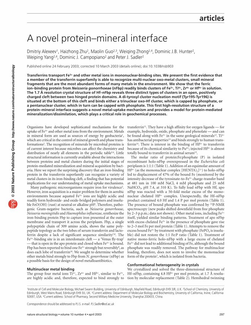
and the complex arrangement of nine protein molecules (labeledA–I) in the asymmetric unit (space group P32, Fig. 1) presentedsevere problems (see Methods) but the structure was finallysolved to Rcryst = 16.5% and Rfree = 26.4%.
The well-known fold of Fbp comprises two hinged α/βdomains. A single Fe3+ ion is octahedrally coordinated to His9and Glu57 from the N-terminal domain, di-tyrosyl Tyr195-Tyr196 from the hinge area, a water molecule and phosphate asan obligatory synergistic anion bound to the C-terminaldomain7,8. In the apo protein, the domains are open and mono-Fe3+ binding is accompanied by a hinge closure of ∼26°. Wefound a variety of Hf4+ ion clusters bound in the metal-bindingpocket. However, the interdomain hinge remained open and theside chains of His9 and Glu57, previously identified as Fe3+ lig-
298 nature structural biology • volume 10 number 4 • april 2003
Table 1 Analysis of nFbp samples
Sample Treatment1 Mol element per mol proteinFe P Other
Holo-native As isolated2 1.04 ± 0.03 0.98 ± 0.04Apo/Fe 50 × [Fe(NTA)2]3–; 10 mM Tris, pH 7.8, 12 h 2.4 ± 0.2 0.05 ± 0.08Apo/Fe/Pi
3 10 × [Fe(NTA)2]3–; 10 mM Tris, pH 7.8, 1.8 ± 0.1 1.1 ± 0.1then 10× phosphate, 1.5 h
Holo/Fe 10 × [Fe(NTA)2]3–; 10 mM Tris, pH 8.0, 4 h 1.10 ± 0.03 0.20 ± 0.01Apo/Ti4 100 × [Cp2TiCl2] (in 10% DMSO, 100 mM NaCl); – – Ti 3.4 ± 0.1
10 mM Tris, pH 8.0, 5 hApo/Zr5 6 × [Zr(NTA)2]2–; 10 mM HEPES, pH 7.4, 0.07 ± 0.01 1.10 ± 0.10 Zr 3.65 ± 0.40
10 mM phosphate, 10 mM HCO3–, 24 h
Apo/Hf 50 × [Hf(NTA)2]2–; 100 mM NaCl, 4 mM phosphate, – 1.39 ± 0.04 Hf 4.03 ± 0.1025 mM HCO3
–, pH 7.4, 3 h
1Representative experiments listed. Molar excess (for example, 10×) is relative to protein concentration. Cluster binding was found over a wide rangeof conditions (salt concentrations, buffers, pH). Apo Fbp was incubated with metal ions at 310 K, and the unbound metal was removed by gel filtra-tion (for example, PD-10 column) and/or by ultrafiltration. NTA = nitrilotriacetate.2Extensive washing with 10 mM Tris-HCl, pH 7.8, led to loss of phosphate (Fe = 1.10 ± 0.02 and P = 0.1 ± 0.03).3Tyr → Fe3+ charge-transfer band shifted from 481 nm for native holo-nFbp to 464 nm.4Ti4+ loading gives a tyrosinate-to-Ti4+ charge-transfer band at 323 nm (similar to Ti4+ human serum transferrin)40. Cp = cyclopentadienyl.5Crystalline mononuclear K2[Zr(NTA)2]•2H2O was used.
a
b
C↑↑G Arg22 F↓↑G Lys244 G↑↑C Arg48B↑↑H Arg22 D↓↓I Arg22 H↑↑B Arg48A↑↓I Lys244 E↓↑H Lys244 I↓↓D Arg48
c
ands in closed mono-Fe–nFbp, are swung away from the metal-binding site.
The hinge angles of the nine molecules in the crystal vary.They are distinctively wider for the group of three molecules G,H and I by 5.6° on average, with the maximum difference of 8.4°between molecules D and H. The crystal environment of themetal-binding pocket for this group is also distinct (Fig. 1), as isthe structure of the metal cluster (described below). Theobserved structural diversity of the Hf4+ clusters seems to belinked to variations in the protein conformation and in the crys-tal environment.
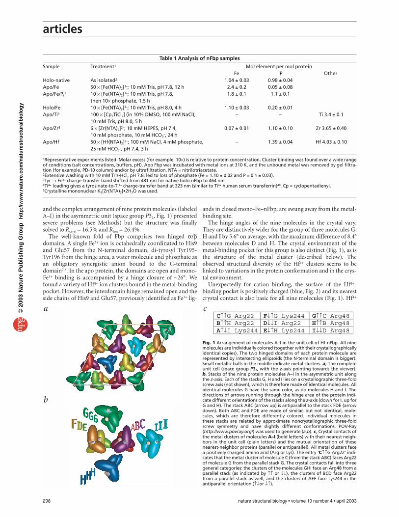
Unexpectedly for cation binding, the surface of the Hf4+-binding pocket is positively charged (blue, Fig. 2) and its nearestcrystal contact is also basic for all nine molecules (Fig. 1). Hf4+
Fig. 1 Arrangement of molecules A–I in the unit cell of Hf-nFbp. All ninemolecules are individually colored (together with their crystallographicallyidentical copies). The two hinged domains of each protein molecule arerepresented by intersecting ellipsoids (the N-terminal domain is bigger).Small metallic balls in the middle indicate metal clusters. a, The completeunit cell (space group P32, with the z-axis pointing towards the viewer). b, Stacks of the nine protein molecules A–I in the asymmetric unit alongthe z-axis. Each of the stacks G, H and I lies on a crystallographic three-foldscrew axis (not shown), which is therefore made of identical molecules. Allidentical molecules G have the same color, as do molecules H and I. Thedirections of arrows running through the hinge area of the protein indi-cate different orientations of the stacks along the z-axis (down for I, up forG and H). The stack ABC (arrow up) is antiparallel to the stack FDE (arrowdown). Both ABC and FDE are made of similar, but not identical, mole-cules, which are therefore differently colored. Individual molecules inthese stacks are related by approximate noncrystallographic three-foldscrew symmetry and have slightly different conformations. POV-Ray(http://www.povray.org/) was used to generate (a,b). c, Crystal contacts ofthe metal clusters of molecules A–I (bold letters) with their nearest neigh-bors in the unit cell (plain letters) and the mutual orientation of thesenearest-neighbor proteins (parallel or antiparallel). All metal clusters facea positively charged amino acid (Arg or Lys). The entry ‘C↑↑G Arg22’ indi-cates that the metal cluster of molecule C (from the stack ABC) faces Arg22of molecule G from the parallel stack G. The crystal contacts fall into threegeneral categories: the clusters of the molecules GHI face an Arg48 from aparallel stack (as indicated by ↑↑ or ↓↓), the clusters of BCD face Arg22from a parallel stack as well, and the clusters of AEF face Lys244 in theantiparallel orientation (↑↓or ↓↑).
©20
03 N
atu
re P
ub
lish
ing
Gro
up
h
ttp
://w
ww
.nat
ure
.co
m/n
atu
rest
ruct
ura
lbio
log
y
articles
ions are not in direct contact with any of the protein atoms,except for the phenolate oxygens of Tyr195 and Tyr196, to whichthey are bonded. Negatively charged oxygen atoms shield Hf4+
ions and bridge them in a manner similar to the structure of themineral HfO2
16 (Fig. 3). Clusters in the protein mirror the struc-tural heterogeneity of the mineral, which can adopt a variety ofcrystal forms, usually with coordination numbers of six, seven oreight. These observations suggest that Fbp might be able toacquire mineral oxo-metal clusters in vivo.
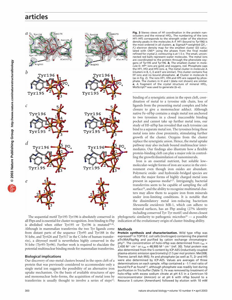
Trinuclear clusterThe cleft of molecule D is the most closed and contains thesmallest cluster of only three metals — Hf1, Hf2 and Hf3 (elec-tron density, Fig. 3a). Trinuclear cores of Hf4+ ions are commonin inorganic complexes containing alkoxide, aryloxide, hydrox-ide and oxide ligands17,18. All three metal atoms are well orderedin the crystal. Hf1 is coordinated to Tyr196, Hf2 to Tyr195, andHf3 to Hf1 and Hf2 through the bridging oxygens (Fig. 3b). Thistrinuclear cluster is capped by a phosphate, which faces outwardfrom the binding pocket. Each metal has pentagonal bipyra-midal coordination to seven oxygens. The five equatorial oxy-gens are oxides (or hydroxides), and the axial oxygens are fromtyrosinate (or water for Hf3) and phosphate. The Hf…Hf dis-tances range from 3.6 to 3.8 Å, and the Hf…O distances from 1.9to 3.0 Å, with the Hf-tyrosinate bonds being the shortest. TheTyr195-Tyr196 O…O distances of 4.03–4.48 Å are significantlylonger than in holo-Fe–nFbp (3.01 Å)7, where both tyrosines arebound to a single Fe3+. The overall calculated charge on the firstcoordination shell is –7 (if all four bridging oxygens are oxide).The net positive surface charge on the metal-binding cleft(Fig. 2) may facilitate formation of the protein-cluster complex.An example of this is the assembly of the molybdenum-iron(MoFe) protein of nitrogenase, which involves insertion of anegatively charged MoFe cofactor cluster into a positivelycharged funnel in the apo protein19.
Pentanuclear clustersAll molecules in the group A–F, except D, contain similar clustersof five Hf4+ ions. In these, the bound phosphate present in thetrinuclear cluster (Fig. 3b) is replaced by an oxo-metal ion (Hf4,Fig. 3c). As in cluster D, the Hf1, Hf2 and Hf3 ions are fullyordered. The additional two ions Hf4 and Hf5 point into the sol-vent and are disordered. High electron density peaks (>10 σ)and strong anomalous signals (>3 σ) confirm that they are Hfions, and their high temperature factors (B-factor = 95 Å2) indi-cate that they are mobile. Hf5 forms an oxygen-bridged trianglewith Hf1 and Hf4, similar to the triangle Hf1–Hf2–Hf3 of clusterD but without the central oxygen. Both Hf4 and Hf5 are octahe-drally coordinated to six oxygens.
Molecules A–C have similar hinge angles and crystal contacts(Fig. 1) and contain similar metal clusters. The clusters of themolecules E and F differ from the clusters in A–C by an evenhigher mobility/disorder of Hf5. The clusters G–I are similar andcontain five metal ions and an additional phosphate (Fig. 3d).Unlike cluster D, the phosphate bridges Hf1 to Hf4 and Hf5,which are mobile in A, B, C, E and F. The phosphate-bound ions
nature structural biology • volume 10 number 4 • april 2003 299
Hf4 and HF5 become well ordered (B-factor = 42 Å2) in thegroup G–I. The other pair of ions, Hf2 and Hf3, which is wellordered in A–F, becomes mobile (B-factor = 92 Å2) in G–I,although Hf2 is bonded to Tyr195 and Hf3 is the most buried inthe binding pocket. The phosphate-capped ordered clusterHf1–Hf4–Hf5 is identical to that of Hf1–Hf2–Hf3 in D, but situ-ated further away from the bottom of the metal binding pocket.Positional disorder seems to be associated with phosphate bind-ing and not with the tyrosyl ligands. The size of the clusters cor-relates with the hinge-angle opening: the most closed moleculeD contains the smallest cluster and the larger clusters are foundin the more open molecules G–I.
Clusters in mammalian transferrin?Hf–nFbp is the first observation of multinuclear binding to theopen form of Fbp. Open transferrin conformations are knownwith a single bound Fe3+ ion20–22. Domain closure was notobserved for mammalian transferrin treated with Hf(NTA) insolution23. When we treated human serum apo transferrin with a30-fold molar excess of [Hf(NTA)2]2– under similar conditionsas those used for Hf–nFbp preparations, we found a boundHf:protein:phosphate ratio of 3.5:1:1.1 (data not shown), similarto Fbp. This suggests that at least the N-lobe, which is wide openin the apo protein, binds a Hf cluster. The C-lobe may not beable to open wide enough to bind metal clusters, and this may berelevant to the functional difference between the two lobes.
Cluster binding seems to be more facile than mononuclearbinding, which requires a conformational transition plus a syn-ergistic helper anion (phosphate or carbonate). Helper anionsare redundant for cluster binding: oxygens of the oxo clusterstake the place of the synergistic anion in the positively chargedbinding site. The cleft of the N-lobe of transferrin can open some20° wider24 than Fbp and can contain more phosphate-bindingsites22, potential anchors for oxo-metal clusters. This, togetherwith the high affinity of Fbp for multimetal clusters, suggeststhat the N-lobe of transferrins could also bind metal clusters.
Fig. 2 Metal nanocluster in the positively charged protein-bindingpocket of molecule G. a, The positively charged molecular surfacearound the metal pocket (calculated by GRASP37), as indicated in blue.b, Lys and Arg residues in the vicinity of the Hf ions (gold spheres) in thecluster binding pocket. Bridging oxygens and the phosphate are omit-ted for clarity (Fig. 3d for these details). Arg101 is completely conservedin all Fbp sequences.
a
b
©20
03 N
atu
re P
ub
lish
ing
Gro
up
h
ttp
://w
ww
.nat
ure
.co
m/n
atu
rest
ruct
ura
lbio
log
y
articles
The sequential motif Tyr195-Tyr196 is absolutely conserved inall Fbps and is essential for cluster recognition. Iron binding to Fbpis abolished when either Tyr195 or Tyr196 is mutated25,26.Although in mammalian transferrins the two Tyr ligands comefrom distant parts of the sequence (Tyr95 and Tyr188 in the N-lobe, and Tyr426 and Tyr517 in the C-lobe of human transfer-rin), a dityrosyl motif is nevertheless highly conserved in the N-lobe (Tyr95-Tyr96). Further work is required to elucidate thepotential multinuclear binding mode for mammalian transferrins.
Biological implicationsOur discovery of oxo-metal clusters bound in the open cleft of aprotein that was previously considered to accommodate only asingle metal ion suggests the possibility of an alternative ironuptake mechanism. On the basis of available structures of apoand mononuclear holo-forms, the acquisition of metal ions bytransferrins is usually thought to involve a series of steps22:
300 nature structural biology • volume 10 number 4 • april 2003
binding of a synergistic anion in the open cleft, coor-dination of metal to a tyrosine side chain, loss of ligands from the presenting metal complex and lobeclosure to give a mononuclear adduct. Althoughnative Fe–nFbp contains a single metal ion anchoredto two tyrosines in a closed inaccessible binding pocket and cannot take up further metal ions, ourstudy of Hf–nFbp has revealed that each tyrosine canbind to a separate metal ion. The tyrosines bring thesemetal ions into close proximity, stimulating furthergrowth of the cluster. Oxygens from the clusterreplace the synergistic anion. Hence, the metal uptakepathway may also include bound multinuclear inter-mediates. Our findings also illustrate how a flexibleprotein-binding cleft can play a major role in control-ling the growth/dissimilation of nanominerals.
Iron is an essential nutrient, but soluble low–molecular-weight forms of iron are scarce in the envi-ronment even though iron oxides are abundant.Polymeric oxide- and hydroxide-bridged species areoften the major forms of highly charged metal ionspresent in aqueous media6,27. Intriguingly, bacterialtransferrins seem to be capable of sampling the cellsurface28, and the ability to recognize multimetal clus-ters may allow them to acquire iron from mineralsunder iron-limiting conditions. It is notable that the dissimilatory metal ion–reducing bacteriumShewanella oneidensis MR-1, which can adhere tomineral surfaces, has an Fbp analog (27% identityincluding conserved Tyr-Tyr motif) and shows closest
phylogenetic similarity to pathogenic microbes29 — a possibleindication of the evolutionary origin of cluster-binding ability.
MethodsProtein synthesis and characterization. Wild type nFbp wasexpressed30 in TOP10 E. coli cells (Invitrogen) containing the plasmidpTrc99A/Fbp/Ng and purified by cation exchange chromatogra-phy31. The concentration of holo-nFbp was determined from ε481 =2,430 M–1 cm–1 or ε280 = 48,900 M–1 cm–1 (ref. 30). Total protein wasalso determined from the S content by ICP-AES (inductively coupledplasma-atomic emission spectrometry) (1 S per mol protein: Met308;Thermo Jarrell Ash IRIS). Fe and phosphate (as well as Ti, Zr and Hf)were also determined by ICP-AES. Values are averages of threedeterminations on each sample. nFbp contained a ∼1:1 mol ratio ofbound Fe:P as found7,9, although phosphate was readily lost duringpurification in Tris buffer (Table 1). Fe was removed by treatment ofholo-nFbp with excess sodium citrate at pH 4.5 in a Centricon-10microconcentrator (Amicon) or at pH 6 with nFbp bound to aResource S column (Amersham) followed by elution with 10 mM
a
b
c
d
e
Fig. 3 Stereo views of Hf coordination in the protein nan-oclusters and the mineral HfO2. The numbering of the ionsHf1–Hf5 corresponds to the strength order of the electrondensity peaks in the molecules A–F. Hf1 (bound to Tyr196) isthe most ordered in all clusters. a, SigmaA38-weighted (2Fo –Fc) electron density map for the smallest cluster (D) calcu-lated with CNS34 using the phases from the final modelrefined for crystal 2; contouring is at 1.5 σ. The small, uncon-nected red balls represent water molecules. The metal ionsare coordinated to the protein through the phenolate oxy-gens of Tyr195 and Tyr196. b, The smallest cluster in mole-cule D. Hf4+ ions are gold; and oxygens, red. Phosphate capsthe Hf1, Hf2 and Hf3 ions. c, The metal cluster in molecule A(clusters in B, C, E and F are similar). This cluster contains fiveHf ions and no bound phosphate. d, Cluster in molecule G(as in Fig. 2). The ions Hf1, Hf4 and Hf5 are capped by phos-phate. The clusters in H and I (data not shown) are similar. e, A fragment of the crystal structure of mineral HfO2.MolScript39 was used to generate (b–e).
©20
03 N
atu
re P
ub
lish
ing
Gro
up
h
ttp
://w
ww
.nat
ure
.co
m/n
atu
rest
ruct
ura
lbio
log
y
articles
Tris-HCl buffer and a 0–1 M NaCl gradient. Apo nFbp concentrationswere determined at ε280 = 44,200 M–1 cm–1 (ref. 30). Metal-loadedand apo proteins were purified by ultrafiltration three times usingCentricon YM-30 ultrafilters (Amicon). In some cases, a previousstep of passage through a PD-10 (Amersham) column was used as toobtain an initial separation.
Crystallography. Hf–nFbp was prepared by reacting apo nFbpwith 4 mol equivalent of [Hf(NTA)2]2– (using the crystalline mono-nuclear complex Na2[Hf(NTA)2]•2H2O) for 3 h at 310 K in 100 mMNaCl, 4 mM phosphate and 25 mM NaHCO3, pH 7.4. Small molecules(<30 kDa) were removed by ultrafiltration using 0.1 M KCl. Reactionwith either 50 or 100 mol equiv of [Hf(NTA)2]2– gave similar results.Crystals were grown by the hanging drop technique at 290 K in∼20% (w/v) PEG 4000, 0.2 M KCl and 0.4 M imidazole/malate buffer,pH 7.7. The crystals were flash-frozen in liquid N2, and the X-raydata were collected on stations 14.1 and 9.6 (SRS, DaresburyLaboratory) using an ADSC Quantum4 CCD detector, and processedand scaled with HKL2000 (ref. 32). Crystals belong to the spacegroup P32 (a = b = 148.1 Å and c = 115.8 Å) and were ∼50% twinned.All except one data set (crystal 1, Table 2) could also be processed in
nature structural biology • volume 10 number 4 • april 2003 301
the space group P3212. This crystal 1 wasused for structure determination, and ahigher resolution data set from crystal 2(Table 2) was used for final refinement. Thestructure was solved by a combination ofmolecular replacement and SAD using CCP4suite33 and CNS34. Both AMoRe35 and MOLREP33 produced the same mutual dispo-sition of six protein molecules that obeytwo-fold noncrystallographic rotation axesparallel to the twinning axis of the crystal(search model PDB entry 1D9Y). Threeremaining molecules were found by manualanalysis of the anomalous differences (origi-nating from Hf4+) combined with the phasesfrom the molecular replacement model.Subsequent twinned rigid body refinementusing CNS reduced Rcryst to 36% and Rfree to38%. The positions of all Hf4+ ions were con-fidently revealed by the difference Fouriermap generated with coefficients (Fo – Fc) andthe phases calculated from the final proteinmodel. Anomalous difference Fourier mapsconfirmed that these peaks of electron density belong to Hf ions (f′ = 9 e– at a wave-length of 0.87 Å). We performed nine cyclesof manual rebuilding with O36 followed by
positional refinement, B-factor refinement, twinning fractionrefinement and automatic water building with CNS. We used bondangles and lengths from inorganic HfO2 in the Chemical DatabaseService (http://www.cds.dl.ac.uk/cds/) as the targets for refinement.The final model is the result of crystal 2 refinement (Table 2).
Coordinates. Coordinates have been deposited in the Protein DataBank (accession code 1O7T).
AcknowledgmentsWe thank The Wellcome Trust (Edinburgh Protein Interaction Centre, fellowshipsfor D.J.B.H. and W.Z., and International Research Development Award for W.Z.),Darwin Trust (fellowship for D.A.) and CVCP (ORS Awards for H.Z. and M.G.) fortheir support for this work, and G.G. Dodson, E.I. Stiefel, A.J. Thomson andR.J.P. Williams for their helpful comments on this script.
Competing interests statementThe authors declare that they have no competing financial interests.
Received 4 September, 2002; accepted 6 January, 2003.
Table 2 Crystallographic data and refinement statistics1
Parameter Crystal 1 Crystal 2Wavelength (Å) 0.87 0.87Resolution 20.0–1.7 (1.72–1.76) 30.0–1.6 (1.63–1.60)Refined twinning fraction (%) 19.9 43.8Completeness (%)2 98.2 (52.0) 100.0 (100.0)I / σ2 4.4 (1.0) 7.4 (1.8)Rsym (%)2,3 6.5 (45.1) 8.0 (58.4)Rcryst (%)4 – 16.5Rfree (%)5 – 26.4R.m.s. deviations
Bonds (Å) – 0.006Angles (°) – 1.24
Average B-factorsProtein (Å2) – 31.3Water (Å2) – 33.5
1The data from the more asymmetric crystal 1 were used to solve the structure, and better datafrom crystal 2 were used for refinement.2Values in parentheses are for the highest resolution shell.3Rsym = Σ|Ih – <Ih>| / Ih, where <Ih> is the average intensity over symmetry equivalent reflections.4Rcryst = Σ|Fo – Fc| / ΣFo, where summation is over the data used for refinement.5Rfree was calculated using 7% of data excluded from refinement.
©20
03 N
atu
re P
ub
lish
ing
Gro
up
h
ttp
://w
ww
.nat
ure
.co
m/n
atu
rest
ruct
ura
lbio
log
y
articles
1. Pennisi, E. Geobiologists: as diverse as the bugs they study. Science 296,1058–1060 (2002).
2. Mann, S. Biomineralization: Principles and Concepts in Bioinorganic MaterialsChemistry (Oxford University Press, New York; 2001).
3. Newmann, D.K. & Banfield, J.F. Geomicrobiology: how molecular-scaleinteractions underpin biogeochemical systems. Science 296, 1071–1077 (2002).
4. Butler, A. Acquisition and utilization of transition metal ions by marineorganisms. Science 281, 207–210 (1998).
5. Ratledge, C. & Dover, L.G. Iron metabolism in pathogenic bacteria. Annu. Rev.Microbiol. 54, 881–941 (2000).
6. Crichton, R.R. Inorganic Biochemistry of Iron Metabolism (Ellis Horwood, NewYork; 1991).
7. Bruns, C.M. et al. Fe+3-binding protein reveals convergent evolution within asuperfamily. Nat. Struct. Biol. 4, 919–924 (1997).
8. Bruns, C.M. et al. Crystallographic and biochemical analyses of the metal-freeHaemophilus influenzae Fe3+-binding protein Biochemistry 40, 15631–15637 (2001).
9. Taboy, C.H., Vaughan, K.G., Mietzner, T.A., Aisen, P. & Crumbliss, A.L. Fe3+
Coordination and redox properties of a bacterial transferrin. J. Biol. Chem. 276,2719–2724 (2001).
10. Aisen, P. Iron metabolism: an evolutionary perspective. in Iron Metabolism inHealth and Disease (eds. Brock, J. H., Halliday, J.W., Pippard, M.J. & Powell, L.W.)1–30 (W.B. Saunders Co., London; 1994).
11. Sun, H., Cox, M.C., Li, H. & Sadler, P.J. Rationalisation of metal binding totransferrin: prediction of metal-protein stability constants. Struct. Bonding 88,71–102 (1997).
12. Yang, X.J. & Pin, C. A novel solid-phase extraction scheme for the groupseparation of high field strength elements (Nb, Ta, Zr, Hf) from Al-, Ti-, and Fe-rich geological materials. Sep. Sci. Technol. 36, 571–585 (2001).
13. Schwietert, C.W. & McCue, J.P. Coordination compounds in medicinal chemistry.Coord. Chem. Rev. 184, 67–89 (1999).
14. Guo, M., Sun, H., McArdle, H.J., Gambling, L. & Sadler, P.J. Ti(IV) uptake andrelease by human serum transferrin and recognition of Ti(IV)-transferrin bycancer cells: understanding the mechanism of action of the anticancer drugtitanocene dichloride. Biochemistry 39, 10023–10033 (2000).
15. Then, G.M., Appel, H., Duffield, J., Taylor, D.M. & Thies, W.-G. In vivo and in vitrostudies of hafnium-binding to rat serum transferrin. J. Inorg. Biochem. 27,255–270 (1986).
16. Ruh, R. & Corfield, P.W.R. Crystal structure of monoclinic hafnia and comparisonwith monoclinic zirconia. J. Am. Ceram. Soc. 53, 126–129 (1970).
17. Evans, W.J., Ansari, M.A. & Ziller, J.W. Isolation and structural characterization ofthe polymetallic zirconium alkoxide complexes, Zr3O(OCH2CMe3)9Cl,Zr3O(OCMe3)9(OH), and Na4Zr6O2(OEt)24. Polyhedron 17, 869–877 (1998).
18. Starikova, Z.A. et al. Structural study of zirconium and hafnium oxoalkoxides.Polyhedron 18, 941–947 (1999).
19. Schmid, B. et al. Structure of a cofactor-deficient nitrogenase MoFe protein.Science 296, 352–356 (2002).
20. Mizutani, K., Yamashita, H., Kurokawa, H., Mikami, B. & Hirose, M. Alternativestructural state of transferrin. The crystallographic analysis of iron-loaded butdomain-opened ovotransferrin N-lobe. J. Biol. Chem. 247, 10190–10194 (1999).
302 nature structural biology • volume 10 number 4 • april 2003
21. Khan, J.A., Kumar, P., Srinivasan, A. & Singh, T.P. Protein intermediate trapped bythe simultaneous crystallization process. Crystal structure of an iron-saturatedintermediate in the Fe3+ binding pathway of camel lactoferrin at 2.7 Åresolution. J. Biol. Chem. 276, 36817–36823 (2001).
22. Kuser, P. et al. The mechanism of iron uptake by transferrins: the X-ray structuresof the 18 kDa NII domain fragment of duck ovotransferrin and itsnitrilotriacetate complex. Acta Crystallogr. D 58, 777–783 (2002).
23. Grossmann, J.G. et al. Metal-induced conformational changes in transferrins. J.Mol. Biol. 229, 585–590 (1993).
24. Jeffrey, P.D. et al. Ligand-induced conformational change in transferrins: crystalstructure of the open form of the N-terminal half-molecule of humantransferrin. Biochemistry 37, 13978–13986 (1998).
25. Nowalk, A.J., Vaughan, K.G., Day, B.W., Tencza, S.B. & Mietzner, T.A. Metal-dependent conformers of the periplasmic ferric ion binding protein.Biochemistry 36, 13054–13059 (1997).
26. Guo, M. Proteins and nucleic acids as targets for titanium(IV). PhD thesis, Univ.Edinburgh (2000).
27. Frausto da Silva, J.J.R. & Williams, R.J.P. The principles of the uptake and chemicalspeciation of the elements in biology. in The Biological Chemistry of theElements. Chapter 2, 23–70 (Oxford University Press, Oxford; 1991).
28. Ferreiros, C., Criado, M.T. & Gomez, J.A. The Neisserial 37 kDa ferric bindingprotein (FbpA). Comp. Biochem. Physiol. Biochem. Mol. Biol. 123, 1–7 (1999).
29. Heidelberg, J.F. et al. Genome sequence of the dissimilatory metal ion-reducingbacterium Shewanella oneidensis. Nat. Biotechnol. 20, 1118–1123 (2002).
30. Guo, M. et al. Synergistic anion and metal binding to the ferric-ion bindingprotein from Neisseria gonorrheae (nFBP). J. Biol. Chem. 278, 2490–2502 (2003).
31. Nowalk, A., Tencza, S. & Mietzner, T. Coordination of iron by the ferric iron-binding protein of pathogenic Neisseria is homologous to the transferrins.Biochemistry 33, 12769–12775 (1994).
32. Otwinowski, Z. & Minor, W. Processing of X-ray diffraction data collected inoscillation mode. Methods Enzymol. 276, 307–326 (1997).
33. Collaborative Computational Project, Number 4. The CCP4 suite: programs forprotein crystallography. Acta Crystallogr. D 50, 760–763 (1994).
34. Brunger, A.T. et al. Crystallography & NMR system: a new software suite formacromolecular structure determination. Acta Crystallogr. D 54, 905–921 (1998).
35. Navaza, J. & Saludjian, P. AMoRe: an automated molecular replacement programpackage. Methods Enzymol. 276, 581–594 (1997).
36. Jones, T.A. & Kjeldgaard, M. O Version 5.9, The Manual (Uppsala University,Uppsala; 1994).
37. Nicholls, A., Sharp, K.A. & Honig, B. Protein folding and association: insightsfrom the interfacial and thermodynamic properties of hydrocarbons. Proteins11, 281–296 (1991).
38. Read, R.J. Improved Fourier coefficients for maps using phases from partialstructures with errors. Acta Crystallogr. A 42, 140-149 (1986).
39. Kraulis, P.J. MOLSCRIPT: a program to produce both detailed and schematic plotsof protein structures. J. Appl. Crystallogr. 24, 945–949 (1991).
40. Sun, H., Li, H., Weir, R.A. & Sadler, P.J. The first specific Ti(IV)-protein complex:potential relevance to anticancer activity of titanocenes. Angew. Chem. Int. Ed.Engl. 37, 1577–1579 (1998).
©20
03 N
atu
re P
ub
lish
ing
Gro
up
h
ttp
://w
ww
.nat
ure
.co
m/n
atu
rest
ruct
ura
lbio
log
y
erratum
A novel protein–mineral interfaceDmitriy Alexeev, Haizhong Zhu, Maolin Guo, Weiqing Zhong, Dominic J.B. Hunter, Weiping Yang,Dominic J. Campopiano and Peter J. Sadler
Nature Structural Biology; doi: 10.1038/nsb903
In the version of this article initially published online, the institution affiliations were assigned incorrectly because of a mistake thatoccurred during production. The correct affiliations for all authors are as follows: Dmitriy Alexeev1, Haizhong Zhu2, Maolin Guo2,3,Weiqing Zhong2,4, Dominic J.B. Hunter2, Weiping Yang2,3, Dominic J. Campopiano2 and Peter J. Sadler2. All of the footnotes (corrected) are as follows: 1Institute of Cell and Molecular Biology, Michael Swann Building, University of Edinburgh, MayfieldRoad, Edinburgh EH9 3JR, UK; 2School of Chemistry, University of Edinburgh, West Mains Road, Edinburgh EH9 3JJ, UK;3Current address: Department of Molecular Biology and Biochemistry, University of California, Irvine, California 92697, USA; and4Current address: School of Pharmacy, Second Military Medicine University, Shanghai 200433, China. We apologize for any inconvenience this may have caused. This mistake has been corrected in the HTML and print version of the article.