A novel communication role for CYP17A1 in the progression of castration-resistant prostate cancer
10
The Prostate 69:928 ^ 937 (2009) A Novel Communication Role for CYP17A1 in the Progression of Castration-Resistant Prostate Cancer Jennifer A. Locke, 1 Ladan Fazli, 1 Hans Adomat, 1 Jil Smyl, 1 Kristin Weins, 1 Amy A. Lubik, 1,2 Dale B. Hales, 3 Colleen C. Nelson, 1,2 Martin E. Gleave, 1 and Emma S. Tomlinson Guns 1 * 1 Department of Urologic Sciences,University of British Columbia,The Prostate Centre at Vancouver General Hospital, Vancouver, British Columbia,Canada 2 Institute of Health and Biomedical Innovation,Queensland University of Technology, Brisbane,Queensland, Australia 3 Department of Physiology & Biophysics,University of Illinois College of Medicine at Chicago,Chicago, Illinois BACKGROUND. CYP17A1 is currently a target for total androgen blockade in advanced prostate cancer (CaP) patients. After castration, or removal of testicular androgens, CYP17A1 can act as a rate-limiting enzyme in androgen synthesis from cholesterol or other adrenal precursors within the tumor microenvironment ultimately contributing to disease progression. Herein we provide evidence that CYP17A1 could also be a mediator of cell-to-cell communication within the CaP tumor microenvironment. METHODS. CYP17A1 expression was evaluated by immunohistochemical analysis of human tumor sections and Western blot analysis of CaP patients’ serum and exosome isolates. CYP17A1 activity assays were conducted in human serum (and positive control human liver and kidney microsomes) using progesterone as a precursor and an LC-MS endpoint. RESULTS. These studies revealed that the expression pattern of CYP17A1 is typical of a secretory protein as it is localized to the luminal pole of the cells in exocrine secretory mode. CYP17A1 is expressed in human serum and in fact is elevated in the serum of CaP patients as compared to healthy controls. Serum CYP17A1 activity could not be confirmed, however, verification of CYP17A1 expression in exosomes suggests a role in cell-to-cell communication within the tumor microenvironment. CONCLUSIONS. CYP17A1 is a crucial enzyme for de novo androgen synthesis within the tumor microenvironment after removal of testicular androgens by castration. We provide evidence for a novel role for CYP17A1 in serum and further reiterate the importance of targeting this enzyme in CaP progression. Prostate 69: 928 – 937, 2009. # 2009 Wiley-Liss, Inc. KEY WORDS: CYP17A1; prostate cancer; serum; exosomes INTRODUCTION CYP17A1 is a member of the cytochrome P450 enzyme family which collectively function in steroid hydroxylation and carbon–carbon bond cleavage re- actions [1]. CYP17A1 is responsible for the conversion of C-21 steroids such as pregnenolone, progesterone and pregnan-3a-ol-20-one to their 17a-hydroxylated derivatives and further cleavage at the C17–20 bond to produce C-19 steroids: dehydroepiandrosterone (DHEA), androstenedione, and androsterone (Fig. 1). In humans, the activity of CYP17A1 is regulated by endogenous levels of P450 oxidoreductase (micro- somal electron transport chain), NADPH supply and co-localization with helper enzyme, cytochrome b5 (CYB5A1) [2 – 5]. P450 oxidoreductase and NADPH are *Correspondence to: Emma S. Tomlinson Guns, The Prostate Centre at Vancouver General Hospital, 2660 Oak Street, Vancouver, British Columbia, Canada V6H 3Z6. E-mail: [email protected] Received 25 November 2008; Accepted 20 January 2009 DOI 10.1002/pros.20940 Published online 6 March 2009 in Wiley InterScience (www.interscience.wiley.com). ȣ 2009 Wiley-Liss, Inc.
Transcript of A novel communication role for CYP17A1 in the progression of castration-resistant prostate cancer

The Prostate 69:928 ^ 937 (2009)
ANovel Communication Role for CYP17A1intheProgression of Castration-Resistant Prostate Cancer
Jennifer A. Locke,1 Ladan Fazli,1 Hans Adomat,1 Jil Smyl,1 Kristin Weins,1
Amy A. Lubik,1,2 Dale B. Hales,3 Colleen C. Nelson,1,2 Martin E. Gleave,1
and Emma S. Tomlinson Guns1*1DepartmentofUrologic Sciences,Universityof British Columbia,The Prostate Centreat VancouverGeneralHospital,
Vancouver, British Columbia,Canada2Institute ofHealthandBiomedical Innovation,QueenslandUniversityof Technology,Brisbane,Queensland, Australia
3Departmentof Physiology&Biophysics,Universityof Illinois College ofMedicineat Chicago,Chicago, Illinois
BACKGROUND. CYP17A1 is currently a target for total androgen blockade in advancedprostate cancer (CaP) patients. After castration, or removal of testicular androgens, CYP17A1can act as a rate-limiting enzyme in androgen synthesis from cholesterol or other adrenalprecursors within the tumor microenvironment ultimately contributing to disease progression.Herein we provide evidence that CYP17A1 could also be a mediator of cell-to-cellcommunication within the CaP tumor microenvironment.METHODS. CYP17A1 expression was evaluated by immunohistochemical analysis of humantumor sections and Western blot analysis of CaP patients’ serum and exosome isolates.CYP17A1 activity assays were conducted in human serum (and positive control human liverand kidney microsomes) using progesterone as a precursor and an LC-MS endpoint.RESULTS. These studies revealed that the expression pattern of CYP17A1 is typical ofa secretory protein as it is localized to the luminal pole of the cells in exocrine secretory mode.CYP17A1 is expressed in human serum and in fact is elevated in the serum of CaP patients ascompared to healthy controls. Serum CYP17A1 activity could not be confirmed, however,verification of CYP17A1 expression in exosomes suggests a role in cell-to-cell communicationwithin the tumor microenvironment.CONCLUSIONS. CYP17A1 is a crucial enzyme for de novo androgen synthesis within thetumor microenvironment after removal of testicular androgens by castration. We provideevidence for a novel role for CYP17A1 in serum and further reiterate the importance of targetingthis enzyme in CaP progression. Prostate 69: 928–937, 2009. # 2009 Wiley-Liss, Inc.
KEY WORDS: CYP17A1; prostate cancer; serum; exosomes
INTRODUCTION
CYP17A1 is a member of the cytochrome P450enzyme family which collectively function in steroidhydroxylation and carbon–carbon bond cleavage re-actions [1]. CYP17A1 is responsible for the conversionof C-21 steroids such as pregnenolone, progesteroneand pregnan-3a-ol-20-one to their 17a-hydroxylatedderivatives and further cleavage at the C17–20 bondto produce C-19 steroids: dehydroepiandrosterone(DHEA), androstenedione, and androsterone (Fig. 1).In humans, the activity of CYP17A1 is regulated by
endogenous levels of P450 oxidoreductase (micro-somal electron transport chain), NADPH supply andco-localization with helper enzyme, cytochrome b5(CYB5A1) [2–5]. P450 oxidoreductase and NADPH are
*Correspondence to: Emma S. Tomlinson Guns, The Prostate Centreat Vancouver General Hospital, 2660 Oak Street, Vancouver, BritishColumbia, Canada V6H 3Z6. E-mail: [email protected] 25 November 2008; Accepted 20 January 2009DOI 10.1002/pros.20940Published online 6 March 2009 in Wiley InterScience(www.interscience.wiley.com).
) 2009 Wiley-Liss, Inc.
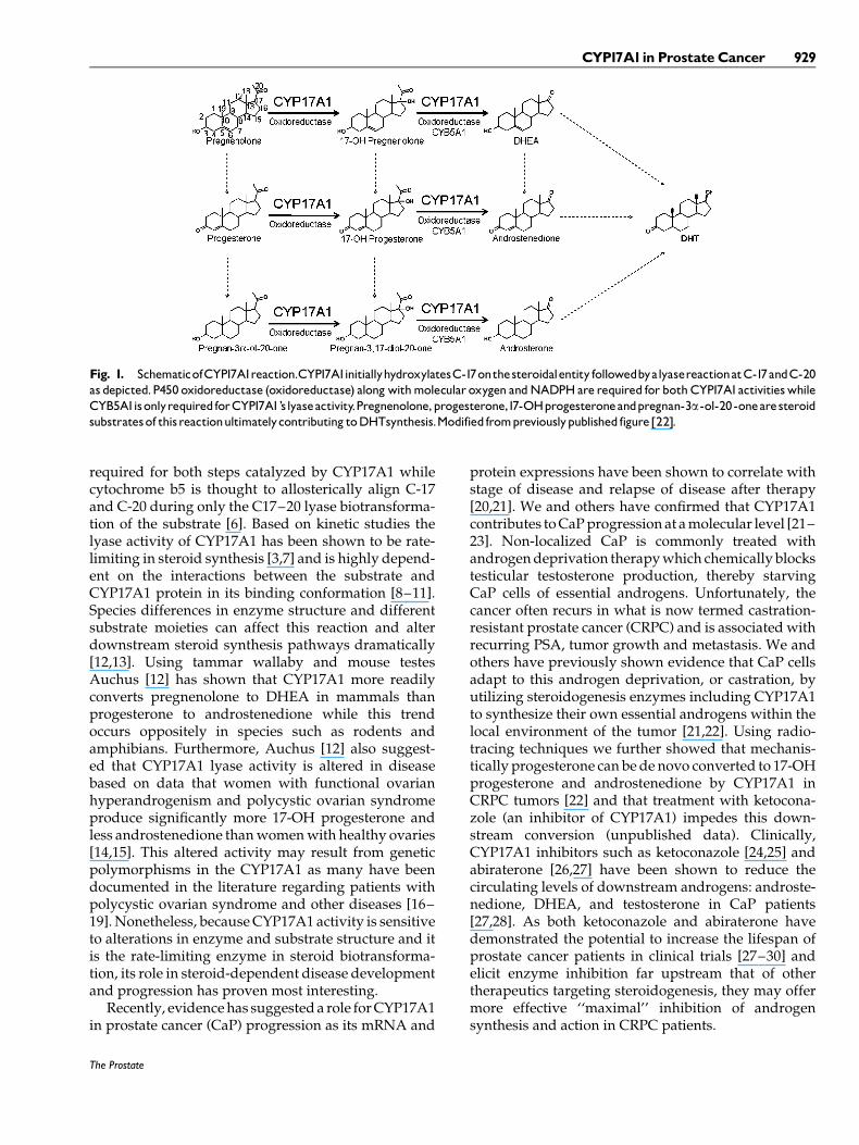
required for both steps catalyzed by CYP17A1 whilecytochrome b5 is thought to allosterically align C-17and C-20 during only the C17–20 lyase biotransforma-tion of the substrate [6]. Based on kinetic studies thelyase activity of CYP17A1 has been shown to be rate-limiting in steroid synthesis [3,7] and is highly depend-ent on the interactions between the substrate andCYP17A1 protein in its binding conformation [8–11].Species differences in enzyme structure and differentsubstrate moieties can affect this reaction and alterdownstream steroid synthesis pathways dramatically[12,13]. Using tammar wallaby and mouse testesAuchus [12] has shown that CYP17A1 more readilyconverts pregnenolone to DHEA in mammals thanprogesterone to androstenedione while this trendoccurs oppositely in species such as rodents andamphibians. Furthermore, Auchus [12] also suggest-ed that CYP17A1 lyase activity is altered in diseasebased on data that women with functional ovarianhyperandrogenism and polycystic ovarian syndromeproduce significantly more 17-OH progesterone andless androstenedione than women with healthy ovaries[14,15]. This altered activity may result from geneticpolymorphisms in the CYP17A1 as many have beendocumented in the literature regarding patients withpolycystic ovarian syndrome and other diseases [16–19]. Nonetheless, because CYP17A1 activity is sensitiveto alterations in enzyme and substrate structure and itis the rate-limiting enzyme in steroid biotransforma-tion, its role in steroid-dependent disease developmentand progression has proven most interesting.
Recently, evidence has suggested a role for CYP17A1in prostate cancer (CaP) progression as its mRNA and
protein expressions have been shown to correlate withstage of disease and relapse of disease after therapy[20,21]. We and others have confirmed that CYP17A1contributes to CaP progression at a molecular level [21–23]. Non-localized CaP is commonly treated withandrogen deprivation therapy which chemically blockstesticular testosterone production, thereby starvingCaP cells of essential androgens. Unfortunately, thecancer often recurs in what is now termed castration-resistant prostate cancer (CRPC) and is associated withrecurring PSA, tumor growth and metastasis. We andothers have previously shown evidence that CaP cellsadapt to this androgen deprivation, or castration, byutilizing steroidogenesis enzymes including CYP17A1to synthesize their own essential androgens within thelocal environment of the tumor [21,22]. Using radio-tracing techniques we further showed that mechanis-tically progesterone can be de novo converted to 17-OHprogesterone and androstenedione by CYP17A1 inCRPC tumors [22] and that treatment with ketocona-zole (an inhibitor of CYP17A1) impedes this down-stream conversion (unpublished data). Clinically,CYP17A1 inhibitors such as ketoconazole [24,25] andabiraterone [26,27] have been shown to reduce thecirculating levels of downstream androgens: androste-nedione, DHEA, and testosterone in CaP patients[27,28]. As both ketoconazole and abiraterone havedemonstrated the potential to increase the lifespan ofprostate cancer patients in clinical trials [27–30] andelicit enzyme inhibition far upstream that of othertherapeutics targeting steroidogenesis, they may offermore effective ‘‘maximal’’ inhibition of androgensynthesis and action in CRPC patients.
The Prostate
Fig. 1. SchematicofCYP17A1reaction.CYP17A1initiallyhydroxylatesC-17onthesteroidalentity followedbyalyasereactionatC-17andC-20as depicted.P450 oxidoreductase (oxidoreductase) alongwithmolecular oxygen andNADPHarerequired for bothCYP17A1activitieswhileCYB5A1isonlyrequiredforCYP17A1’slyaseactivity.Pregnenolone,progesterone,17-OHprogesteroneandpregnan-3a-ol-20-onearesteroidsubstratesof thisreactionultimatelycontributing toDHTsynthesis.Modifiedfrompreviouslypublishedfigure [22].
CYP17A1in Prostate Cancer 929
In our attempt to profile CYP17A1’s role in CRPCdisease progression we have discovered that CYP17A1may not only be a central enzyme in androgen synthesisbut that it could also play another role in cellularcommunication between CRPC cells within the tumor.Upon immunohistochemical analysis of CaP tumorsections we observe that CYP17A1 localization isorientated in a pattern typical of a secretory protein.Furthermore, detection of the enzyme in human serumas well as further verification that protein levels ofCYP17A1 are elevated in CaP patients as comparedto healthy controls suggests a functional role of thisenzyme in the circulating serum. Although the activityof CYP17A1 was not determined in human serum ascompared to positive control human liver and kidneymicrosomes, further delineation of CYP17A1’s expres-sion within human serum exosomes suggests a rolein cell-to-cell communication during progressionto CRPC. It is hypothesized herein that secretion ofCYP17A1 into serum within exosomes may offer anovel mechanism for the CaP cell to communicate withother cells perhaps triggering de novo steroidogenesisin CaP progression to castration-resistance.
MATERIALS ANDMETHODS
Materials
Steroid standards. Stock solutions of testosterone-16,16,17-d3 (deuterated testosterone) (CDN Isotopes,Pointe-Claire, Quebec, Canada), 4-androstene-3,17-dione (Sigma, Oakville, Ontario, Canada), 4-pregnen-17-ol-3,20-dione (Steraloids, Inc., Newport, Rhode Island),5a-androstan-17b-ol-3-one (Sigma), dihydrotestoster-one (DHT) (Sigma), 5b-pregnan-3a-27-diol-20-one(Steraloids, Inc.), 5b-pregnan-3,20-dione (Steraloids,Inc.), androsterone (Aldrich, Oakville, Ontario, Canada),pregnenolone (Sigma), progesterone (Sigma), andtestosterone (Sigma) were prepared in 100% methanolas mass spectrometry standards.
Human serum samples. Serum was obtained from36 prostate cancer patients, 24 of whom were under-going neoadjuvant hormone therapy (NHT>6 months)and 12 hormone naı̈ve patients. Inclusion criteriarequired participants to have biopsy confirmed pro-state cancer with no evidence of bone metastases.Samples were also obtained from healthy age matchedcontrols (n¼ 10). The men undergoing NHT werebeing treated for one of three indications: (1) localizedprostate cancer managed conservatively; (2) bio-chemical recurrence following surgery or radiation;(3) management of locally advanced disease. Hormone
naı̈ve patients were men with prostate cancer pre-viously treated with definitive therapy (radiation orsurgery) or those monitoring localized disease withactive surveillance (3 months or more post-diagnosis).Aged matched controls comprise of healthy menwithout prostate cancer who were asked to participatethrough a peer nomination process. Participants wereasked to nominate a peer (usually a sibling, friend orneighbor) who is similar in age (within 5 years). Allserum samples were taken as a fasting morning sample,between the hours of 7:30 and 10:30 am. Sera was thenseparated by centrifugation at 1,500g and analyzedimmediately or stored at �808C prior to analysis.Approval was obtained from The University of BritishColumbia Clinical Research Ethics Board and patientsconsented for their serum to be used for the purpose ofthis research.
Human microsomes. Pooled human liver and kidneymicrosomes were used for Western blotting andCYP17A1 activity assays (Celsis In Vitro Technologies,Chicago, IL).
Serum and H295 cell exosomes. Exosomes wereisolated from fresh human serum and H295 cells asdescribed by Taylor and Gercel-Taylor [31]. Briefly,serum and cell lysates was centrifuged at 2,000g for10 min and the supernatant was transferred to a newtube for further centrifugation at 21,000g for 20 minusing a swinging bucket Ti 40 rotor (Beckman Coulter,Mississauga, Ontario). The supernatant from this spinwas further centrifuged at 200,000g for 1 hr. and theremaining exosome containing pellet was reconstitutedin PBS. Exosome isolates were stored at �808C untilfurther analysis. Validation of exosome isolation wasdone by Western blot analysis for exosome markersheat shock protein-70 (HSP70) [32,33], glyceraldehyde3-phosphate dehydrogenase (GAPDH) [34] and Fasligand (FAS) [31].
Methods
Immunohistochemistry. Immunohistochemical stain-ing was conducted on tissues from radical prostatec-tomy patients (from 2000 to 2006) obtained from theVancouver General Hospital. Benign and cancer siteswere identified and marked in donor paraffin blocksusing matching H&E reference slides. Canceroustissues were analyzed by Ventana autostainer modelDiscover XTTM (Vantana Medical System, Tuscan,Arizona) using enzyme labeled biotin streptavidinsystem and solvent resistant DAB Map kit. CYP17A1rabbit polyclonal antibody was prepared in house byDr. Hales [35].
The Prostate
930 Locke et al.
Western blot analysis of CYP17A1. Protein concen-trations were assessed using a Bicinchoninic Acid Kit(Sigma) and 15 mg of each sample was loaded into eachlane. Relative enzyme levels were analyzed using thefollowing antibodies at the indicated dilutions: rabbitpolyclonal CYP17A1 (1:1,500) and P450 oxidoreductase(1:1,000) were prepared in house by Dr. Hales [35], goatmonoclonal CYB5A1 (1:1,000 Abcam, Cambridge,MA), mouse monoclonal HSP70 (1:1,000 Santa CruzBiotechnology, Inc., Santa Cruz, CA), rabbit polyclonalGAPDH (1:1,000 Abcam) and mouse monoclonal FAS(1:200 Santa Cruz Biotechnology, Inc.). Protein bandswere visualized and quantified using the Li-corBiosciences Odyssey Infrared Imaging system (Lin-coln, Nebraska). Since there are currently no knowncontrols for protein loading with human serum wecompensated for this by conducting repeat analyses bytwo independent researchers who replicated proteindetermination and Western blot analysis for CYP17A1in ten of the same human serum samples. The resultsfrom this experiment were reproducible and thusprovide confidence in the overall quantitation ofCYP17A1 protein expression by Western blot in eachsample. Each condition was conducted in triplicate andWestern blot analysis was conducted three times foreach sample.
CYP17A1 activity assay. Steroid standards: proges-terone, 17-OH progesterone or pregnan-3a-ol-20-onewere added (10 ml of 10 mg/ml methanolic stocksolution) to individual pre-rinsed glass vials and drieddown using a CentrivapTM centrifugal evaporationsystem (358C for 30 min). Steroids were reconstitutedin 500 ml of fresh human serum and an NADPHregenerating system added to initiate enzymaticactivity (BD Biosciences, San Jose, CA: 1.3 mM NADPþ,3.3 mM glucose-6-phosphate, 0.4 U/ml glucose-6-phosphate dehydrogenase, and 3.3 mM magnesiumchloride). Samples were further incubated at 378Cfor 0 and 2 hr time periods. Activity reactions werequenched with 50 ml of 100 ng/ml deuterated testoster-one (internal standard) and 465 ml of acetonitrile.Samples were vortexed several times and spun downat 1,000 rpm for 10 min. Five hundred microliters ofsupernatant was removed and dried down using aCentrivapTM centrifugal evaporation system (358Cfor 30 min). Samples were reconstituted in 50 ml ofmethanol with vortexing and sonication for 30 min,prior to dilution with 50 ml of water. Samples were thenclarified by centrifugation at 13,500g for 10 min prior toHPLC analysis. One milligram of human liver andkidney microsomes were used as positive CYP activitycontrols while PBS was used in all blank analyses. Foreach condition the activity assay was conducted intriplicate.
Steroid analysis by liquid chromatography-massspectrometry (LC-MS). A Waters 2695 SeparationsModule coupled to a Waters Quattro Micro was usedfor LCMS analysis. All MS data were collected in ESIþmode. Capillary voltage was set at 3 kV, source anddesolvation temperatures were 120 and 3508C respec-tively with N2 desolvation flow at 450 L/hr and Arcollision gas adjusted for 3.5e�3 mBars for fragmenta-tion when required. The instrument was operated atunit resolution to obtain MS scan and Fragment Ionscan spectra and with reduced high mass resolutionsettings to enhance MRM sensitivity. Cone voltage(CV) and collision energy (CE) settings were optimizedfor each standard. Chromatographic separations werecarried out using a Waters Exterra 2.1 mm� 50 mm,3.5 mm C18 column equilibrated with 20:80 ACN/H2O,ramped to 80:20 ACN/H2O from 0.5 to 8.0 min, furtherto 95:5 from 8.0 to 9.0 min and returned to 80:20 ACN/H2O from 10.0 to 10.5 min with a total run time of15 min. Flow rate was 0.3 ml/min, column temperature358C and 0.05% formic acid was present throughoutthe run. MS scan data for metabolite identificationwas collected using both 22 and 35 V for CV.
RESULTS
CYP17A1is expressedinhuman CaP tissues andappears tobe a secretoryprotein
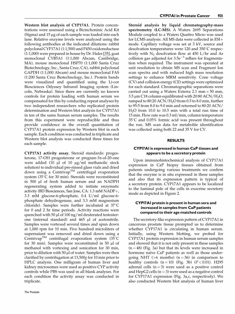
Upon immunohistochemical analysis of CYP17A1expression in CaP biopsy tissues obtained frompatients undergoing various treatments we confirmthat the enzyme is in situ expressed in these samplesand also that its expression pattern is typical ofa secretory protein. CYP17A1 appears to be localizedto the luminal pole of the cells in exocrine secretorymode as depicted in Figure 2.
CYP17A1protein is present inhuman sera andincreased in samples from CaPpatientscomparedto their agematchedcontrols
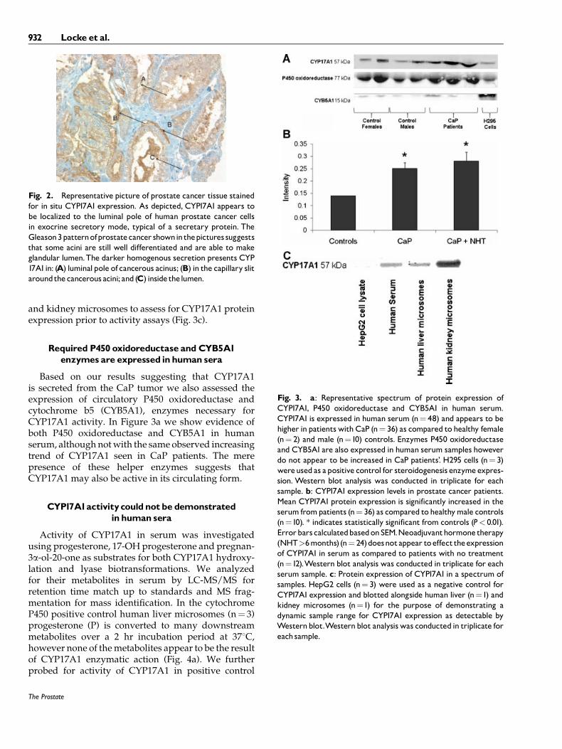
The secretory-like expression pattern of CYP17A1 incancerous prostate tissue prompted us to determinewhether CYP17A1 is circulating in human serum.Initially, using Western blotting, we probed forCYP17A1 protein expression in human serum samplesand showed that it is not only present in these samples(n¼ 48) (Fig. 3a) but that its levels were increased inhormone naı̈ve CaP patients as well as those under-going NHT (>6 months) (n¼ 36) in comparison tohealthy controls (n¼ 10) (Fig. 3b) (P< 0.01). H295adrenal cells (n¼ 3) were used as a positive controland HepG2 cells (n¼ 3) were used as a negative controlfor CYP17A1 expression (Fig. 3a,c, respectively). Wealso conducted Western blot analysis of human liver
The Prostate
CYP17A1in Prostate Cancer 931
and kidney microsomes to assess for CYP17A1 proteinexpression prior to activity assays (Fig. 3c).
Required P450 oxidoreductase and CYB5A1enzymes are expressedinhuman sera
Based on our results suggesting that CYP17A1is secreted from the CaP tumor we also assessed theexpression of circulatory P450 oxidoreductase andcytochrome b5 (CYB5A1), enzymes necessary forCYP17A1 activity. In Figure 3a we show evidence ofboth P450 oxidoreductase and CYB5A1 in humanserum, although not with the same observed increasingtrend of CYP17A1 seen in CaP patients. The merepresence of these helper enzymes suggests thatCYP17A1 may also be active in its circulating form.
CYP17A1activitycouldnotbe demonstratedinhuman sera
Activity of CYP17A1 in serum was investigatedusing progesterone, 17-OH progesterone and pregnan-3a-ol-20-one as substrates for both CYP17A1 hydroxy-lation and lyase biotransformations. We analyzedfor their metabolites in serum by LC-MS/MS forretention time match up to standards and MS frag-mentation for mass identification. In the cytochromeP450 positive control human liver microsomes (n¼ 3)progesterone (P) is converted to many downstreammetabolites over a 2 hr incubation period at 378C,however none of the metabolites appear to be the resultof CYP17A1 enzymatic action (Fig. 4a). We furtherprobed for activity of CYP17A1 in positive control
The Prostate
Fig. 2. Representative picture of prostate cancer tissue stainedfor in situ CYP17A1 expression. As depicted, CYP17A1 appears tobe localized to the luminal pole of human prostate cancer cellsin exocrine secretory mode, typical of a secretary protein. TheGleason3patternofprostatecancer showninthepictures suggeststhat some acini are still well differentiated and are able to makeglandular lumen.The darker homogenous secretion presents CYP17A1in: (A) luminalpole of cancerous acinus; (B) in the capillary slitaroundthecancerous acini; and (C) inside thelumen.
Fig. 3. a: Representative spectrum of protein expression ofCYP17A1, P450 oxidoreductase and CYB5A1 in human serum.CYP17A1 is expressed in human serum (n¼ 48) and appears to behigher in patientswith CaP (n¼ 36) as compared to healthy female(n¼ 2) and male (n¼10) controls. Enzymes P450 oxidoreductaseand CYB5A1are also expressed in human serum samples howeverdo not appear to be increased in CaP patients’. H295 cells (n¼ 3)wereusedas apositive control for steroidogenesis enzyme expres-sion. Western blot analysis was conducted in triplicate for eachsample. b: CYP17A1 expression levels in prostate cancer patients.Mean CYP17A1protein expression is significantly increased in theserum frompatients (n¼ 36) as compared to healthymale controls(n¼10). * indicates statistically significant from controls (P< 0.01).ErrorbarscalculatedbasedonSEM.Neoadjuvanthormone therapy(NHT>6months) (n¼ 24)doesnotappear toeffect theexpressionof CYP17A1 in serum as compared to patients with no treatment(n¼12).Western blot analysis was conducted in triplicate for eachserum sample. c: Protein expression of CYP17A1 in a spectrum ofsamples. HepG2 cells (n¼ 3) were used as a negative control forCYP17A1expression and blotted alongside human liver (n¼1) andkidney microsomes (n¼1) for the purpose of demonstrating adynamic sample range for CYP17A1 expression as detectable byWesternblot.Westernblot analysiswas conducted in triplicate foreach sample.
932 Locke et al.
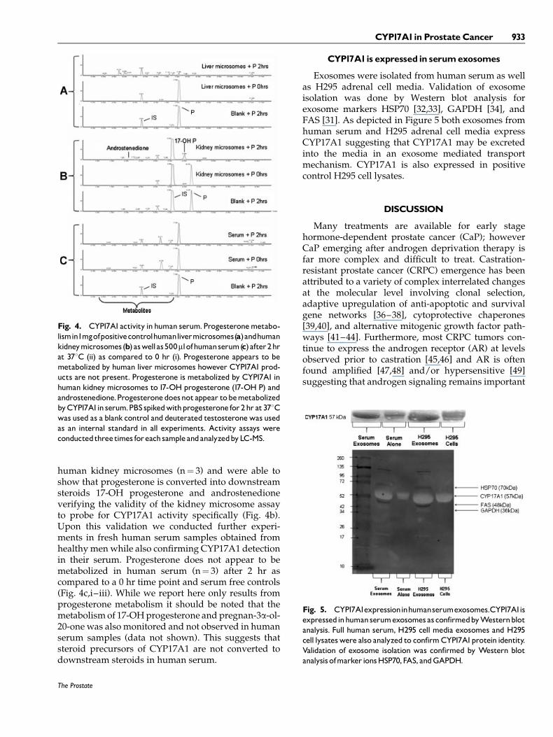
human kidney microsomes (n¼ 3) and were able toshow that progesterone is converted into downstreamsteroids 17-OH progesterone and androstenedioneverifying the validity of the kidney microsome assayto probe for CYP17A1 activity specifically (Fig. 4b).Upon this validation we conducted further experi-ments in fresh human serum samples obtained fromhealthy men while also confirming CYP17A1 detectionin their serum. Progesterone does not appear to bemetabolized in human serum (n¼ 3) after 2 hr ascompared to a 0 hr time point and serum free controls(Fig. 4c,i–iii). While we report here only results fromprogesterone metabolism it should be noted that themetabolism of 17-OH progesterone and pregnan-3a-ol-20-one was also monitored and not observed in humanserum samples (data not shown). This suggests thatsteroid precursors of CYP17A1 are not converted todownstream steroids in human serum.
CYP17A1is expressedin serumexosomes
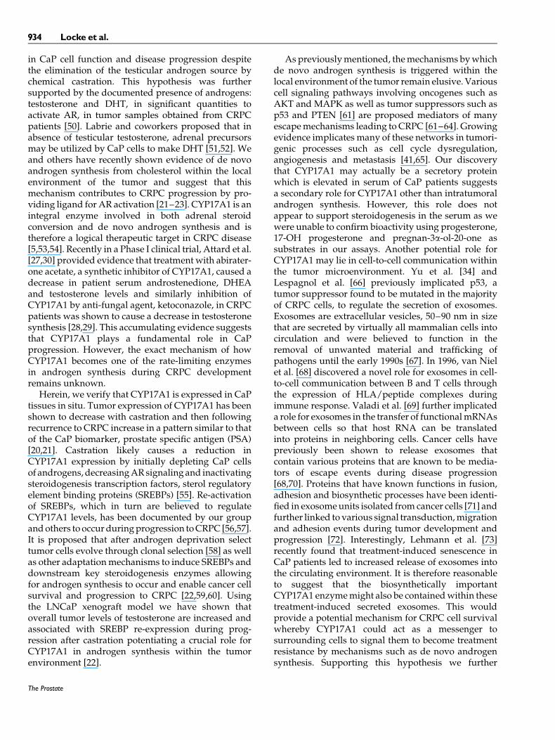
Exosomes were isolated from human serum as wellas H295 adrenal cell media. Validation of exosomeisolation was done by Western blot analysis forexosome markers HSP70 [32,33], GAPDH [34], andFAS [31]. As depicted in Figure 5 both exosomes fromhuman serum and H295 adrenal cell media expressCYP17A1 suggesting that CYP17A1 may be excretedinto the media in an exosome mediated transportmechanism. CYP17A1 is also expressed in positivecontrol H295 cell lysates.
DISCUSSION
Many treatments are available for early stagehormone-dependent prostate cancer (CaP); howeverCaP emerging after androgen deprivation therapy isfar more complex and difficult to treat. Castration-resistant prostate cancer (CRPC) emergence has beenattributed to a variety of complex interrelated changesat the molecular level involving clonal selection,adaptive upregulation of anti-apoptotic and survivalgene networks [36–38], cytoprotective chaperones[39,40], and alternative mitogenic growth factor path-ways [41–44]. Furthermore, most CRPC tumors con-tinue to express the androgen receptor (AR) at levelsobserved prior to castration [45,46] and AR is oftenfound amplified [47,48] and/or hypersensitive [49]suggesting that androgen signaling remains important
The Prostate
Fig. 4. CYP17A1activity in human serum.Progesteronemetabo-lismin1mgofpositivecontrolhumanlivermicrosomes(a)andhumankidneymicrosomes(b)aswellas500mlofhumanserum(c)after2hrat 378C (ii) as compared to 0 hr (i). Progesterone appears to bemetabolized by human liver microsomes however CYP17A1prod-ucts are not present. Progesterone is metabolized by CYP17A1 inhuman kidney microsomes to 17-OH progesterone (17-OH P) andandrostenedione.Progesteronedoesnotappear tobemetabolizedbyCYP17A1in serum.PBSspikedwithprogesterone for2hr at378Cwas used as a blank control and deuterated testosteronewas usedas an internal standard in all experiments. Activity assays wereconductedthree times foreach sampleandanalyzedbyLC-MS.
Fig. 5. CYP17A1expressioninhumanserumexosomes.CYP17A1isexpressedinhumanserumexosomes as confirmedbyWesternblotanalysis. Full human serum, H295 cell media exosomes and H295cell lysateswere also analyzed to confirmCYP17A1protein identity.Validation of exosome isolation was confirmed by Western blotanalysis ofmarker ionsHSP70,FAS, andGAPDH.
CYP17A1in Prostate Cancer 933
in CaP cell function and disease progression despitethe elimination of the testicular androgen source bychemical castration. This hypothesis was furthersupported by the documented presence of androgens:testosterone and DHT, in significant quantities toactivate AR, in tumor samples obtained from CRPCpatients [50]. Labrie and coworkers proposed that inabsence of testicular testosterone, adrenal precursorsmay be utilized by CaP cells to make DHT [51,52]. Weand others have recently shown evidence of de novoandrogen synthesis from cholesterol within the localenvironment of the tumor and suggest that thismechanism contributes to CRPC progression by pro-viding ligand for AR activation [21–23]. CYP17A1 is anintegral enzyme involved in both adrenal steroidconversion and de novo androgen synthesis and istherefore a logical therapeutic target in CRPC disease[5,53,54]. Recently in a Phase I clinical trial, Attard et al.[27,30] provided evidence that treatment with abirater-one acetate, a synthetic inhibitor of CYP17A1, caused adecrease in patient serum androstenedione, DHEAand testosterone levels and similarly inhibition ofCYP17A1 by anti-fungal agent, ketoconazole, in CRPCpatients was shown to cause a decrease in testosteronesynthesis [28,29]. This accumulating evidence suggeststhat CYP17A1 plays a fundamental role in CaPprogression. However, the exact mechanism of howCYP17A1 becomes one of the rate-limiting enzymesin androgen synthesis during CRPC developmentremains unknown.
Herein, we verify that CYP17A1 is expressed in CaPtissues in situ. Tumor expression of CYP17A1 has beenshown to decrease with castration and then followingrecurrence to CRPC increase in a pattern similar to thatof the CaP biomarker, prostate specific antigen (PSA)[20,21]. Castration likely causes a reduction inCYP17A1 expression by initially depleting CaP cellsof androgens, decreasing AR signaling and inactivatingsteroidogenesis transcription factors, sterol regulatoryelement binding proteins (SREBPs) [55]. Re-activationof SREBPs, which in turn are believed to regulateCYP17A1 levels, has been documented by our groupand others to occur during progression to CRPC [56,57].It is proposed that after androgen deprivation selecttumor cells evolve through clonal selection [58] as wellas other adaptation mechanisms to induce SREBPs anddownstream key steroidogenesis enzymes allowingfor androgen synthesis to occur and enable cancer cellsurvival and progression to CRPC [22,59,60]. Usingthe LNCaP xenograft model we have shown thatoverall tumor levels of testosterone are increased andassociated with SREBP re-expression during prog-ression after castration potentiating a crucial role forCYP17A1 in androgen synthesis within the tumorenvironment [22].
As previously mentioned, the mechanisms by whichde novo androgen synthesis is triggered within thelocal environment of the tumor remain elusive. Variouscell signaling pathways involving oncogenes such asAKT and MAPK as well as tumor suppressors such asp53 and PTEN [61] are proposed mediators of manyescape mechanisms leading to CRPC [61–64]. Growingevidence implicates many of these networks in tumori-genic processes such as cell cycle dysregulation,angiogenesis and metastasis [41,65]. Our discoverythat CYP17A1 may actually be a secretory proteinwhich is elevated in serum of CaP patients suggestsa secondary role for CYP17A1 other than intratumoralandrogen synthesis. However, this role does notappear to support steroidogenesis in the serum as wewere unable to confirm bioactivity using progesterone,17-OH progesterone and pregnan-3a-ol-20-one assubstrates in our assays. Another potential role forCYP17A1 may lie in cell-to-cell communication withinthe tumor microenvironment. Yu et al. [34] andLespagnol et al. [66] previously implicated p53, atumor suppressor found to be mutated in the majorityof CRPC cells, to regulate the secretion of exosomes.Exosomes are extracellular vesicles, 50–90 nm in sizethat are secreted by virtually all mammalian cells intocirculation and were believed to function in theremoval of unwanted material and trafficking ofpathogens until the early 1990s [67]. In 1996, van Nielet al. [68] discovered a novel role for exosomes in cell-to-cell communication between B and T cells throughthe expression of HLA/peptide complexes duringimmune response. Valadi et al. [69] further implicateda role for exosomes in the transfer of functional mRNAsbetween cells so that host RNA can be translatedinto proteins in neighboring cells. Cancer cells havepreviously been shown to release exosomes thatcontain various proteins that are known to be media-tors of escape events during disease progression[68,70]. Proteins that have known functions in fusion,adhesion and biosynthetic processes have been identi-fied in exosome units isolated from cancer cells [71] andfurther linked to various signal transduction, migrationand adhesion events during tumor development andprogression [72]. Interestingly, Lehmann et al. [73]recently found that treatment-induced senescence inCaP patients led to increased release of exosomes intothe circulating environment. It is therefore reasonableto suggest that the biosynthetically importantCYP17A1 enzyme might also be contained within thesetreatment-induced secreted exosomes. This wouldprovide a potential mechanism for CRPC cell survivalwhereby CYP17A1 could act as a messenger tosurrounding cells to signal them to become treatmentresistance by mechanisms such as de novo androgensynthesis. Supporting this hypothesis we further
The Prostate
934 Locke et al.
confirmed CYP17A1 expression in exosomes isolatedfrom human serum and that its protein expressionis elevated in patients’ serum as compared to healthycontrols. The employment of NHT of greater than6 months did not statistically increase CYP17A1 proteinexpression in CaP patients. However, as CRPC does notusually emerge until >9 months NHT [74] and we didnot have information regarding patients’ duration ofNHT after 6 months, it is difficult to make furtherclaims about how NHT affects CYP17A1 proteinsecretion. As we were unable to show activity withinthis environment this may be accounted for by Ieroet al.’s [70] observation that soluble factors andenzymes released in exosomes are likely to act locallyor in the immediate vicinity of their releasing site andunlikely to exhibit biological activity further away fromthe parent cell suggesting that CYP17A1 may act withinthe local environment of the tumor and not at otherdistant sites where we obtained our serum samplesfrom. Nonetheless, the presence of essential enzymesP450 oxidoreductase and cytochrome b5 in serumfurther indicates a functional role for CYP17A1 inneighboring cells through exosome transported units.
In summary, we report exciting and novel findingsof CYP17A1, P450 oxidoreductase and cytochrome b5presence in human serum and suggest that althoughCYP17A1 does not appear to be active in this environ-ment it may have another important role. Afterandrogen deprivation therapy levels of CYP17A1 inthe tumor decrease during progression to CRPC,however tumor cells often regain the ability to induceCYP17A1 expression leading to androgen synthesis.We show evidence that CYP17A1 may be secreted fromthe CaP cell as part of an exosome unit and proposethat it is used to initiate cell-to-cell communicationfor global androgen synthesis within the CRPC tumor.This evidence further rationalizes the clinical use ofwell-characterized and novel inhibitors of CYP17A1 intargeting CRPC progression.
REFERENCES
1. Bartsch W, Klein H, Schiemann U, Bauer HW, Voigt KD.Enzymes of androgen formation and degradation in the humanprostate. Ann NY Acad Sci 1990;595:53–66.
2. Miller WL, Auchus RJ, Geller DH. The regulation of 17,20 lyaseactivity. Steroids 1997;62:133–142.
3. Pandey AV, Miller WL. Regulation of 17,20 lyase activity bycytochrome b5 and by serine phosphorylation of P450c17. J BiolChem 2005;280:13265–13271.
4. Dharia S, Slane A, Jian M, Conner M, Conley AJ, Parker CR Jr.Colocalization of P450c17 and cytochrome b5 in androgen-synthesizing tissues of the human. Biol Reprod 2004;71:83–88.
5. Payne AH, Hales DB. Overview of steroidogenic enzymes in thepathway from cholesterol to active steroid hormones. EndocrRev 2004;25:947–970.
6. Akhtar MK, Kelly SL, Kaderbhai MA. Cytochrome b(5)modulation of 17{alpha} hydroxylase and 17-20 lyase (CYP17)activities in steroidogenesis. J Endocrinol 2005;187:267–274.
7. Tagashira H, Kominami S, Takemori S. Kinetic studies ofcytochrome P-45017 alpha, lyase dependent androstenedioneformation from progesterone. Biochemistry 1995;34:10939–10945.
8. Gupta MK, Guryev OL, Auchus RJ. 5alpha-reduced C21 steroidsare substrates for human cytochrome P450c17. Arch BiochemBiophys 2003;418:151–160.
9. Auchus RJ, Worthy K, Geller DH, Miller WL. Probing structuraland functional domains of human P450c17. Endocr Res 2000;26:695–703.
10. Mathieu AP, Auchus RJ, LeHoux JG. Comparison of the hamsterand human adrenal P450c17 (17 alpha-hydroxylase/17,20-lyase)using site-directed mutagenesis and molecular modeling.J Steroid Biochem Mol Biol 2002;80:99–107.
11. Auchus RJ, Miller WL. Molecular modeling of human P450c17(17alpha-hydroxylase/17,20-lyase): Insights into reactionmechanisms and effects of mutations. Mol Endocrinol 1999;13:1169–1182.
12. Auchus RJ. The backdoor pathway to dihydrotestosterone.Trends Endocrinol Metab 2004;15:432–438.
13. Bhangoo A, Aisenberg J, Chartoffe A, Ten S, Wallerstein RJ,Wolf R, Auchus RJ. Novel mutation in cytochrome P450c17causes complete combined 17alpha-hydroxylase/17,20-lyasedeficiency. J Pediatr Endocrinol Metab 2008;21:185–190.
14. Rosenfield RL, Barnes RB, Ehrmann DA. Studies of the nature of17-hydroxyprogesterone hyperresonsiveness to gonadotropin-releasing hormone agonist challenge in functional ovarianhyperandrogenism. J Clin Endocrinol Metab 1994;79:1686–1692.
15. Ghayee HK, Auchus RJ. Basic concepts and recent developmentsin human steroid hormone biosynthesis. Rev Endocr MetabDisord 2007;8:289–300.
16. Park JM, Lee EJ, Ramakrishna S, Cha DH, Baek KH. Associationstudy for single nucleotide polymorphisms in the CYP17A1 geneand polycystic ovary syndrome. Int J Mol Med 2008;22:249–254.
17. Onen IH, Ekmekci A, Eroglu M, Polat F, Biri H. The association of5alpha-reductase II (SRD5A2) and 17 hydroxylase (CYP17) genepolymorphisms with prostate cancer patients in the Turkishpopulation. DNA Cell Biol 2007;26:100–107.
18. Kakinuma H, Tsuchiya N, Habuchi T, Ohyama C, Matsuura S,Wang L, Nakamura A, Kato T. Serum sex steroid hormone levelsand polymorphisms of CYP17 and SRD5 A2: Implication forprostate cancer risk. Prostate Cancer Prostatic Dis 2004;7:333–337.
19. Madigan MP, Gao YT, Deng J, Pfeiffer RM, Chang BL, Zheng S,Meyers DA, Stanczyk FZ, Xu J, Hsing AW. CYP17 poly-morphisms in relation to risks of prostate cancer and benignprostatic hyperplasia: A population-based study in China. Int JCancer 2003;107:271–275.
20. Stigliano A, Gandini O, Cerquetti L, Gazzaniga P, Misiti S, MontiS, Gradilone A, Falasca P, Poggi M, Brunetti E, Agliano AM,Toscano V. Increased metastatic lymph node 64 and CYP17expression are associated with high stage prostate cancer.J Endocrinol 2007;194:55–61.
21. Montgomery RB, Mostaghel EA, Vessella R, Hess DL, KalhornTF, Higano CS, True LD, Nelson PS. Maintenance of intra-tumoral androgens in metastatic prostate cancer: A mechanismfor castration-resistant tumor growth. Cancer Res 2008;68:4447–4454.
The Prostate
CYP17A1in Prostate Cancer 935

22. Locke JA, Guns ES, Lubik AA, Adomat HH, Hendy SC, WoodCA, Ettinger SL, Gleave ME, Nelson CC. Androgen levelsincrease by intratumoral de novo steroidogenesis duringprogression of castration-resistant prostate cancer. Cancer Res2008;68:6407–6415.
23. Mostaghel EA, Nelson PS. Intracrine androgen metabolism inprostate cancer progression: Mechanisms of castration resist-ance and therapeutic implications. Best Pract Res Clin Endo-crinol Metab 2008;22:243–258.
24. Ayub M, Levell MJ. Inhibition of human adrenal steroidogenicenzymes in vitro by imidazole drugs including ketoconazole.J Steroid Biochem 1989;32:515–524.
25. Rajfer J, Sikka SC, Rivera F, Handelsman DJ. Mechanism ofinhibition of human testicular steroidogenesis by oral ketoco-nazole. J Clin Endocrinol Metab 1986;63:1193–1198.
26. Madan RA, Arlen PM. Abiraterone. Cougar biotechnology.IDrugs 2006;9:49–55.
27. Attard G, Reid AH, Yap TA, Raynaud F, Dowsett M, Settatree S,Barrett M, Parker C, Martins V, Folkerd E, Clark J, Cooper CS,Kaye SB, Dearnaley D, Lee G, de Bono JS. Phase, I clinical trial of aselective inhibitor of CYP17, abiraterone acetate, confirms thatcastration-resistant prostate cancer commonly remains hormonedriven. J Clin Oncol 2008;26:4563–4571.
28. Small EJ, Halabi S, Dawson NA, Stadler WM, Rini BI, Picus J,Gable P, Torti FM, Kaplan E, Vogelzang NJ. Antiandrogenwithdrawal alone or in combination with ketoconazole inandrogen-independent prostate cancer patients: A phase III trial(CALGB 9583). J Clin Oncol 2004;22:1025–1033.
29. Small EJ, Baron AD, Fippin L, Apodaca D. Ketoconazole retainsactivity in advanced prostate cancer patients with progressiondespite flutamide withdrawal. J Urol 1997;157:1204–1207.
30. Attard G, Belldegrun AS, de Bono JS. Selective blockade ofandrogenic steroid synthesis by novel lyase inhibitors as atherapeutic strategy for treating metastatic prostate cancer. BJUInt 2005;96:1241–1246.
31. Taylor DD, Gercel-Taylor C. Tumour-derived exosomes andtheir role in cancer-associated T-cell signalling defects. Br JCancer 2005;92:305–311.
32. Lancaster GI, Febbraio MA. Exosome-dependent trafficking ofHSP70: A novel secretory pathway for cellular stress proteins. JBiol Chem 2005;280:23349–23355.
33. Johnstone RM. Revisiting the road to the discovery of exosomes.Blood Cells Mol Dis 2005;34:214–219.
34. Yu X, Harris SL, Levine AJ. The regulation of exosome secretion:A novel function of the p53 protein. Cancer Res 2006;66:4795–4801.
35. Hales DB, Sha LL, Payne AH. Testosterone inhibits cAMP-induced de Novo synthesis of Leydig cell cytochrome P-450(17alpha) by an androgen receptor-mediated mechanism. J BiolChem 1987;262:11200–11206.
36. Gimenez-Bonafe P, Fedoruk MN, Whitmore TG, Akbari M,Ralph JL, Ettinger S, Gleave ME, Nelson CC. YB-1 is upregulatedduring prostate cancer tumor progression and increasesP-glycoprotein activity. Prostate 2004;59:337–349.
37. Fedoruk Matthew N, Gimenez-Bonafe P, Guns Emma S, MayerLawrence D, Nelson Colleen C. P-glycoprotein increases theefflux of the androgen dihydrotestosterone and reduces andro-gen responsive gene activity in prostate tumor cells. Prostate2004;59:77–90.
38. Gleave M, Tolcher A, Miyake H, Nelson C, Brown B, Beraldi E,Goldie J. Progression to androgen independence is delayed byadjuvant treatment with antisense Bcl-2 oligodeoxynucleotides
after castration in the LNCaP prostate tumor model. Clin CancerRes 1999;5:2891–2898.
39. Rocchi P, So A, Kojima S, Signaevsky M, Beraldi E, Fazli L,Hurtado-Coll A, Yamanaka K, Gleave M. Heat shock protein 27increases after androgen ablation and plays a cytoprotective rolein hormone-refractory prostate cancer. Cancer Res 2004;64:6595–6602.
40. Rocchi P, Beraldi E, Ettinger S, Fazli L, Vessella RL, Nelson C,Gleave M. Increased Hsp27 after androgen ablation facilitatesandrogen-independent progression in prostate cancer via signaltransducers and activators of transcription 3-mediated suppres-sion of apoptosis. Cancer Res 2005;65:11083–11093.
41. Culig Z. Androgen receptor cross-talk with cell signallingpathways. Growth Factors 2004;22:179–184.
42. Craft N, Shostak Y, Carey M, Sawyers CL. A mechanism forhormone-independent prostate cancer through modulation ofandrogen receptor signaling by the HER-2/neu tyrosine kinase.Nat Med 1999;5:280–285.
43. Miyake H, Nelson C, Rennie PS, Gleave ME. Overexpression ofinsulin-like growth factor binding protein-5 helps accelerateprogression to androgen-independence in the human prostateLNCaP tumor model through activation of phosphatidylinositol3’-kinase pathway. Endocrinology 2000;141:2257–2265.
44. So A, Gleave M, Hurtado-Col A, Nelson C. Mechanisms of thedevelopment of androgen independence in prostate cancer.World J Urol 2005;23:1–9.
45. Mohler JL, Gregory CW, Ford OH III, Kim D, Weaver CM,Petrusz P, Wilson EM, French FS. The androgen axis in recurrentprostate cancer. Clin Cancer Res 2004;10:440–448.
46. Van der Kwast V, Schalken TH, Ruizevend J, de Winter JA.Androgen receptors in endocrine therapy-resistant humanprostate cancer. Int J Cancer 1991;48:189–193.
47. Visakorpi T, Hyytinen E, Koivisto P, Tanner M, Keinanen R,Palmberg C, Palotie A, Tammela T, Isola J, Kallioniemi O. In vivoamplification of the androgen receptor gene and progression ofhuman prostate cancer. Nat Genet 1995;9:401–406.
48. Koivisto P, Kononen J, Palmberg C, Tammela T, HyytinenE, Isola J, Trapman J, Cleutjens K, Noordzij A, VisakorpiT, Kallioniemi OP. Androgen receptor gene amplification:A possible molecular mechanism for androgen deprivationtherapy failure in prostate cancer. Cancer Res 1997;57:314–319.
49. Gregory CW, Johnson RT Jr, Mohler JL, French FS, Wilson EM.Androgen receptor stabilization in recurrent prostate cancer isassociated with hypersensitivity to low androgen. Cancer Res2001;61:2892–2898.
50. Titus MA, Schell MJ, Lih FB, Tomer KB, Mohler JL. Testosteroneand dihydrotestosterone tissue levels in recurrent prostatecancer. Clin Cancer Res 2005;11:4653–4657.
51. Labrie F. Adrenal androgens and intracrinology. Semin ReprodMed 2004;22:299–309.
52. Suzuki K, Nishiyama T, Hara N, Yamana K, Takahashi K, LabrieF. Importance of the intracrine metabolism of adrenal androgensin androgen-dependent prostate cancer. Prostate Cancer Pro-static Dis 2007;10:301–306.
53. Attard G, Sarker D, Reid A, Molife R, Parker C, de Bono JS.Improving the outcome of patients with castration-resistantprostate cancer through rational drug development. Br J Cancer2006;95:767–774.
54. Yap TA, Carden CP, Attard G, de Bono JS. Targeting CYP17:established and novel approaches in prostate cancer. Curr OpinPharmacol 2008;8:449–457.
The Prostate
936 Locke et al.

55. Heemers HV, Verhoeven G, Swinnen JV. Androgen activation ofthe sterol regulatory element-binding protein pathway: Currentinsights. Mol Endocrinol 2006;20:2265–2277.
56. Ettinger SL, Sobel R, Whitmore TG, Akbari M, Bradley DR,Gleave ME, Nelson CC. Dysregulation of sterol responseelement-binding proteins and downstream effectors in prostatecancer during progression to androgen independence. CancerRes 2004;64:2212–2221.
57. Ozbay T, Rowan A, Leon A, Patel P, Sewer MB. Cyclic adenosine5’-monophosphate-dependent sphingosine-1-phosphate bio-synthesis induces human CYP17 gene transcription by activat-ing cleavage of sterol regulatory element binding protein 1.Endocrinology 2006;147:1427–1437.
58. Isaacs JT. The biology of hormone refractory prostate cancer.Why does it develop? Urol Clin North Am 1999;26:263–273.
59. Stanbrough M, Bubley GJ, Ross K, Golub TR, Rubin MA,Penning TM, Febbo PG, Balk SP. Increased expression of genesconverting adrenal androgens to testosterone in androgen-independent prostate cancer. Cancer Res 2006;66:2815–2825.
60. Mostaghel EA, Page ST, Lin DW, Fazli L, Coleman IM, True LD,Knudsen B, Hess DL, Nelson CC, Matsumoto AM, Bremner WJ,Gleave ME, Nelson PS. Intraprostatic androgens and androgen-regulated gene expression persist after testosterone suppression:Therapeutic implications for castration-resistant prostate cancer.Cancer Res 2007;67:5033–5041.
61. Shen MM, Abate-Shen C. Pten inactivation and the emergence ofandrogen-independent prostate cancer. Cancer Res 2007;67:6535–6538.
62. Wang Y, Kreisberg JI, Ghosh PM. Cross-talk between theandrogen receptor and the phosphatidylinositol 3-kinase/Aktpathway in prostate cancer. Curr Cancer Drug Targets 2007;7:591–604.
63. Ghosh PM, Malik S, Bedolla R, Kreisberg JI. Akt in prostatecancer: Possible role in androgen-independence. Curr DrugMetab 2003;4:487–496.
64. Li L, Ross AH. Why is PTEN an important tumor suppressor?J Cell Biochem 2007;102:1368–1374.
65. Lee JT, Lehmann BD, Terrian DM, Chappell WH, Stivala F, LibraM, Martelli AM, Steelman LS, McCubrey JA. Targeting prostatecancer based on signal transduction and cell cycle pathways.Cell Cycle 2008;7:1745–1762.
66. Lespagnol A, Duflaut D, Beekman C, Blanc L, Fiucci G, MarineJC, Vidal M, Amson R, Telerman A. Exosome secretion,including the DNA damage-induced p53-dependent secretorypathway, is severely compromised in TSAP6/Steap3-null mice.Cell Death Differ 2008;15:1723–1733.
67. Schorey JS, Bhatnagar S. Exosome function: From tumorimmunology to pathogen biology. Traffic 2008;9:871–881.
68. van Niel G, Porto-Carreiro I, Simoes S, Raposo G. Exosomes: Acommon pathway for a specialized function. J Biochem 2006;140:13–21.
69. Valadi H, Ekstrom K, Bossios A, Sjostrand M, Lee JJ, Lotvall JO.Exosome-mediated transfer of mRNAs and microRNAs is anovel mechanism of genetic exchange between cells. Nat CellBiol 2007;9:654–659.
70. Iero M, Valenti R, Huber V, Filipazzi P, Parmiani G, Fais S,Rivoltini L. Tumour-released exosomes and their implications incancer immunity. Cell Death Differ 2008;15:80–88.
71. Fevrier B, Raposo G. Exosomes: Endosomal-derived vesiclesshipping extracellular messages. Curr Opin Cell Biol 2004;16:415–421.
72. Hegmans JP, Bard MP, Hemmes A, Luider TM, KleijmeerMJ, Prins JB, Zitvogel L, Burgers SA, Hoogsteden HC,Lambrecht BN. Proteomic analysis of exosomes secretedby human mesothelioma cells. Am J Pathol 2004;164:1807–1815.
73. Lehmann BD, Paine MS, Brooks AM, McCubrey JA, Renegar RH,Wang R, Terrian DM. Senescence-associated exosome releasefrom human prostate cancer cells. Cancer Res 2008;68:7864–7871.
74. Paterson RF, Gleave ME, Jones EC, Zubovits JT, Goldenberg SL,Sullivan LD. Immunohistochemical analysis of radical prosta-tectomy specimens after 8 months of neoadjuvant hormonaltherapy. Mol Urol 1999;3:277–286.
The Prostate
CYP17A1in Prostate Cancer 937