Phase II Study of Dasatinib in Patients with Metastatic Castration-Resistant Prostate Cancer

Volume 13 Number 2 February 2011 pp. 108–119 108
www.neoplasia.com
Lycopene Enhances Docetaxel’sEffect in Castration-ResistantProstate Cancer Associatedwith Insulin-like GrowthFactor I Receptor Levels1
Yaxiong Tang*, Basmina Parmakhtiar†,Anne R. Simoneau*, Jun Xie*, John Fruehauf†,Michael Lilly† and Xiaolin Zi*,§,‡
*Department of Urology, University of California, Irvine,Orange, CA, USA; †Department of Medicine, Universityof California, Irvine, Orange, CA, USA; ‡Department ofPharmaceutical Sciences, University of California, Irvine,Orange, CA, USA; §Chao Family Comprehensive CancerCenter, University of California, Irvine, Orange, CA, USA
AbstractDocetaxel is currently the most effective drug for the treatment of castration-resistant prostate cancer (CRPC), butit only extends life by an average of 2 months. Lycopene, an antioxidant phytochemical, has antitumor activityagainst prostate cancer (PCa) in several models and is generally safe. We present data on the interaction betweendocetaxel and lycopene in CRPC models. The growth-inhibitory effect of lycopene on PCa cell lines was positivelyassociated with insulin-like growth factor I receptor (IGF-IR) levels. In addition, lycopene treatment enhanced thegrowth-inhibitory effect of docetaxel more effectively on DU145 cells with IGF-IR high expression than on those PCacell lines with IGF-IR low expression. In a DU145 xenograft tumor model, docetaxel plus lycopene caused tumorregression, with a 38% increase in antitumor efficacy (P = .047) when compared with docetaxel alone. Lycopeneinhibited IGF-IR activation through inhibiting IGF-I stimulation and by increasing the expression and secretion ofIGF-BP3. Downstream effects include inhibition of AKT kinase activity and survivin expression, followed by apoptosis.Together, the enhancement of docetaxel’s antitumor efficacy by lycopene supplementation justifies further clinicalinvestigation of lycopene and docetaxel combination for CRPC patients. CRPC patients with IGF-IR–overexpressingtumors may be most likely to benefit from this combination.
Neoplasia (2011) 13, 108–119
Address all correspondence to: Xiaolin Zi, Department of Urology, University of California,Irvine, 101 The City Dr S, Bldg 55 Rm. 302, Orange, CA 92868. E-mail: [email protected] work was supported by National Cancer Institute (grant CA122558 to X.Z.).Received 31 July 2010; Revised 16 October 2010; Accepted 18 October 2010
Copyright © 2011 Neoplasia Press, Inc. All rights reserved 1522-8002/11/$25.00DOI 10.1593/neo.101092
IntroductionMost advanced prostate cancer (PCa) patients respond well to initialtreatment with androgen-deprivation therapy (ADT) [1,2]. However,nearly all patients eventually develop resistance to ADT and diseaseprogression [1,2]. Docetaxel-based chemotherapy has recently shownsignificant but short-lived survival benefit and has become the majorremaining treatment option for castration-resistant PCa (CRPC) [3].However, with the demonstrated survival benefit of approximately2 months, median survival of 18 to 20 months and a response rate ofonly approximately 50%, the docetaxel-based chemotherapy remainspalliative for men with CRPC. Therefore, there is a need for investiga-tion and development of novel agents that can add to and improve thedocetaxel-based therapy.
PCa is a biologically complex and highly heterogeneous disease [4–6].The complex interactions of nutrients with genetic and hormonal effec-tors not only contribute to the development of distinct clinical picturesof PCa among different geographical regions [4–6] but also make PCa
an especially amenable target for preventive and therapeutic interven-tion through nutritional combinations. Lycopene is a plant carotenoidnaturally present in tomatoes and tomato products [7]. Lycopeneaccumulates in prostate tissue and functions as a potent antioxidant inin vitro systems [7–10]. In a large prospective cohort study, Giovannucciet al. [11] reported that consumption of lycopene-rich foods was asso-ciated with a 30% to 40% reduction in the PCa risk and that the pre-ventive effects of lycopene-rich foods aremore pronounced in subgroupswith aggressive disease, older men, or men without a family history ofPCa. Moreover, lycopene was shown to inhibit both IL-6 signaling and

Neoplasia Vol. 13, No. 2, 2011 Lycopene Supplementation Enhances Docetaxel’s Effect in Prostate Cancer Tang et al. 109
the IGF-I pathways that participate in resistance to ADT and chemo-therapy (e.g., docetaxel) [12–20]. Therefore, lycopene could be anattractive agent in augmenting docetaxel-based chemotherapy for treat-ment of men with CRPC.Alternatively, lycopene could potentially function as a potent anti-
oxidant in PCa per se to inhibit oxidative stresses and DNA damage[21–24], antagonizing the therapeutic effects of docetaxel [21]. De-spite the recommendations from oncology societies not to take supple-ments during chemotherapy [21], many patients do mix conventionaland alternative treatments. Therefore, there is an urgent clinical needto address whether the concurrent use of lycopene supplements withdocetaxel-based chemotherapy would antagonize or enhance anti-tumor efficacy.In this study, we demonstrate that lycopene enhances the effect of
docetaxel on the growth of CRPC cell lines both in vitro and in vivo.PCa cell lines expressing high insulin-like growth factor I receptor(IGF-IR) levels are more sensitive to the growth-inhibitory effect oflycopene compared with those cell lines with low IGF-IR. Our datasuggest that one potential mechanism of lycopene’s action is inac-tivation of IGF-IR by inhibiting IGF-I stimulation and by increasingthe expression and secretion of IGF-BP3. Downstream effects includeinhibition of AKT kinase activity and survivin expression, followedby apoptosis.
Materials and Methods
Cell Lines, Plasmid, Stable Transfection,Compounds, and ReagentsThe LNCaP, LAPC-4, DU145, 22Rv1, and PC-3 cell lines were
obtained from ATCC (Manassas, VA) and cultured in RPMI 1640medium with 10% FBS.The PCDNA3.1, containing a full-length 4.7-kb fragment of IGF-IR,
was described previously in detail [25]. LNCaP cells were stably trans-fected with pcDNA3.1(+)/IGF-IR using FuGENE 6 (Roche, Indiana-polis, IN). Transfected cells were selected with G418 (800 μg/ml)starting at 48 hours after transfection, and all of the stable transfectantswere pooled to avoid cloning artifacts. Pooled stable clones of LNCaPcells expressing IGF-IR were maintained in RPMI containing 10% FBSand 500 μg/ml G418.Tetrahydrofuran (THF) containing 0.025% butylated hydroxy-
toluene was purchased from Sigma (St Louis, MO). Pure all-translycopene was purchased from Wako Chemicals, Inc (Irvine, CA)and dissolved in THF to a concentration of 15 mM. This stock solu-tion was prepared with minimal exposure to air and light and stored at−80°C. LycoVit 10% cold water dispersible (CWD) was from BASFCorporation (Shreveport, LA), which contains microencapsulatedsynthetic lycopene. There is 11.45% total lycopene with 77% all-trans and 23% total cis-lycopene, and less than 2% vitamin E in thisproduct. Docetaxel injection solution was obtained from the UCIMedical Center Pharmacy. Picropodophyllin (PPP), a potent and se-lective IGF-IR kinase inhibitor, was purchased from Biaffin GmbH&CoKG (Kassel, Germany). Thymidine, 3-(4, 5-dimethylthiazol-2-yl)-2,5-diphenyltetrazolium bromide (MTT), propidium iodide (PI), andother chemicals were from Sigma.Antibodies for AKT, phospho-AKT, and survivin were from Cell Sig-
naling Technology, Inc (Beverly, MA). Antibodies against IGF-IRβand β-actin were from Santa Cruz Biotechnology (Santa Cruz, CA).IGF-BP3 antibody was purchased from Upstate Biotechnology (LakePlacid, NY). Ki67 antibody was from Novus Biologicals (Littleton,
CO). DeadEnd Colorimetric Terminal Deoxynucleotidyl TransferasedUTP Nick End Labeling (TUNEL) System was from Promega(Madison, WI).
MTT AssayThe MTTassay was performed as previously described [26]. Dose-
response curves for growth inhibition were generated as a percentageof vehicle-treated control. Immediately before the experiment, THF-lycopene aliquots from the stock solution were added to culture me-dium to achieve a final concentration of 0.05, 0.5, 1, 5, 10, 50, and100 μM. The final maximum concentration of THF in the culturemedium was 0.1%, which did not affect cell viability as indicated bycomparison with control medium. PPP was dissolved in ethanol andadded to the culture medium to achieve a final concentration of 7.8,15.6, 31.25, 62.5, 125, 250, and 500 nM. Cells were treated for 3 days.MTTwas added to a final concentration of 1 mg/ml. The reaction mix-ture was incubated for 3 hours at 37°C, and the absorbance was mea-sured at 570 nm.
DNA Histogram AnalysisCells were treated with vehicle controls (0.1% DMSO, 0.1% THF,
or 0.1% DMSO + 0.1% THF), 1 μM lycopene, 10 μM lycopene,1 nM docetaxel, 31.25 nM PPP, 1 or 10 μM lycopene plus 1 nMdocetaxel, or 31.25 nM PPP plus 1 nM docetaxel for 24 hours. Afterthe stated treatments, cells were stained with PI in PBS. All analyses ofcells were done using appropriate scatter gates to exclude cellular debrisand aggregated cells. Ten thousand events were collected for each sam-ple stained PI.
Western Blot Analysis and ImmunoprecipitationClarified protein lysates (20-80 μg) were denatured and resolved
by 8% to 16% SDS-PAGE. Proteins were transferred to nitrocellulosemembranes, probed with antibodies, and visualized by an enhancedchemiluminescence detection system.
Total protein (500 μg) was precleared with protein A-agarose andthen precipitated with 2 μg of anti–IGF-IRβ or IgG antibody over-night at 4°C. Agarose beads were washed four times with lysis bufferand resuspended in SDS-PAGE 2× sample buffer. Proteins were elutedby boiling the beads and subjected to immunoblot analysis of anti-phosphotyrosine or anti–IGF-IRβ.
In Vivo Tumor ModelNCR-nu/nu (nude) mice were obtained from Taconic Farms
(Germantown, NY). DU145 cells were concentrated to 1 × 10⁶ per100 μl of PBS and injected subcutaneously into the right flank of eachmouse. Once xenografts started growing, their sizes were measuredtwice a week. The tumor volume was calculated by the formula:0.5236L1(L2)
2, where L1 is the long axis and L2 is the short axis ofthe tumor. Once mice bearing the DU145 tumors (eight per group)developed a tumor size of approximately 200 mm3, they were randomlyassigned to four different treatment groups including vehicle control(water), 15 mg/kg LycoVit 10% CWD daily, 10 mg/kg docetaxelweekly for three doses, and 15 mg/kg lycopene daily plus three weeklydoses of docetaxel. Lycopene was given as LycoVit 10% CWD sus-pended in cold water by gavage. Docetaxel was given by intraperitonealinjection. At the end of the experiment, tumors were excised andweighed, blood was collected, and all were stored at −80°C until addi-tional analysis.

110 Lycopene Supplementation Enhances Docetaxel’s Effect in Prostate Cancer Tang et al. Neoplasia Vol. 13, No. 2, 2011
To assess if these treatments affected survival or tumor growth delayof mice bearing DU145 tumors, these mice were administered vehi-cle control (water), lycopene (15 mg/kg per day) alone, docetaxel (5 or10 mg/kg per week for three doses) alone or combination of both andfollowed up until the tumor volume reached 1500 mm3. All mice thatdied prematurely or were killed had necropsy done on them to ensurethat there were no pathologic disease other than tumor-related disease.
Immunohistochemical Staining for Ki67 and SurvivinTumor tissues were harvested and fixed in 10% phosphate-buffered
formalin, paraffin-embedded, and sectioned. Antigen retrieval wasdone using 10 mM sodium citrate (pH 6.0) at 95°C for 15 minutes.Sections were incubated with mouse monoclonal anti-Ki67 antibody(1:500 dilution) or antisurvivin (1:50) in PBS for 2 hours at room tem-perature in a humidity chamber followed by overnight incubation at4°C. Slides were then incubated with a biotinylated secondary anti-body. Slides were counterstained with Harris hematoxylin and photo-graphed using a light microscope. Negative control samples wereexposed to a secondary antibody with a similar IgG isotype to the pri-mary antibody. Proliferating cells were quantified by counting Ki67-positive cells (brown stained) and total number of cells at five arbitrarilyselected fields at ×400 magnification.
TUNEL Staining for Apoptotic CellsApoptotic cells were detected using the DeadEnd Colorimetric
TUNEL system following the manufacturer’s protocol. The extentof apoptosis was evaluated by counting the TUNEL-positive cells(brown-stained) as well as the total number of cells in five randomlyselected fields at ×400 magnification.
StatisticsComparisons of cell cycle population, proliferation and apoptosis
indices, cell viability between treatment and control were conductedusing Student t test or one-way ANOVA followed by Bonferroni t test.For tumor growth experiments, repeated-measures ANOVA was usedto examine the differences in tumor sizes among treatments, timepoints, and treatment-time interactions. Additional posttests weredone to examine the differences in tumor sizes between control andtreatments at each time point by using conservative Bonferroni method.To correlate cell growth-inhibitory effect of levels and IGF-IR proteinexpression on Western blots, Pearson correlation coefficient was calcu-lated from their IC50 values and from densitometry measurements ofprotein bands. Log-rank test was used to analyze the survival of tumor-bearing mice between control and treatment groups. All statistical testswere two-sided. P < .05 was considered statistically significant.
Results
Lycopene Enhances the Growth-Inhibitory Effect of Docetaxelon PCa Cell Lines
To examine whether lycopene can interfere with or enhance theanti-PCa activity of docetaxel, we have treated 22Rv1, LNCaP,LAPC-4, DU145, and PC-3 cells with 1 nMdocetaxel, 1μM lycopene(a pharmacologically achievable concentration) [7], or combination ofboth for 3 days. One nanomolar of docetaxel inhibited the cell growthof 22Rv1, LNCaP, LAPC-4, PC-3, and DU145 by approximately54%, 35%, 19%, 27%, and 0%, respectively, whereas 1 μM lycopenereduced their growth by 10% (22Rv1), 5% (LNCaP), 19% (LAPC-4),0% (PC-3), and 24% (DU145) (Figure 1A). When these cells were
treated with the combination of docetaxel and lycopene, the cell den-sities of DU145, PC-3, 22Rv1, LNCaP, and LAPC-4 were further re-duced by 78%, 21%, 20%, 21%, and 0%, respectively, compared withdocetaxel alone (Figure 1A). The growth-inhibitory effect of the com-bination on DU145, PC-3, 22Rv1, and LNCaP cells was greater thanthe effects of docetaxel treatment alone (ANOVA test, P values < .05;Figure 1A). Notably, the growth-inhibitory effect of the combinationon DU145 cells with higher IGF-IR expression is the most pro-nounced among the tested cell lines. These results suggest a synergisticand/or additive effect of docetaxel and lycopene on cell growth de-pending on cell types.
Cell cycle analysis further revealed that lycopene and docetaxelcombination resulted in a significant increase in the pre-G1 populationof DU145 cells compared with either lycopene or docetaxel treatmentalone (Figure 1, B and C ; P values < .05, ANOVA test). This resultsuggests that lycopene may increase DU145 cell sensitivity to docetaxel-mediated apoptosis. Its effects in other cell lines were less marked (datanot shown).
The Inhibitory Effect of Lycopene on the Growth of PCaCell Lines Is Associated with the Levels of IGF-IR
Figure 2A show that DU145 cells with higher levels of IGF-IR wereapproximately three- to seven-fold more sensitive to the inhibitoryeffect of lycopene on cell viability than other tested cell lines. Table 1shows that the IC50 values of lycopene for DU145, PC-3, LNCaP,22Rv1, and LAPC-4 cells were estimated to be 5.1, 15, 36 16, and50 μM, respectively, and their levels of IGF-IR (Figure 2B) were esti-mated by densitometry to be 9.3 (DU145), 4.1 (PC-3), 2.0 (LNCaP),1.0 (22Rv1), and 0.8 (LAPC-4), respectively. There was a trend thatcell lines with more IGF-IR expression are more sensitive to lyco-pene treatment (Pearson correlation coefficient, −0.58; Student t test,P values < .05). However, in addition to difference in IGF-IR levels,these cell lines have other distinct characteristics (e.g., androgen re-ceptor, p53, and PTEN status) that may influence their response tolycopene or docetaxel [27]. To avoid these confounding factors, weestablished a pair of isogenic cell lines: parental LNCaP and LNCaPstably expressing high levels of IGF-IR (LNCaP/IGF-IR) (Figure 2C).Figure 2D shows that LNCaP/IGF-IR cells were approximately 400-foldmore sensitive to the effect of lycopene on reduction of cell viability thanparental LNCaP cells (IC50 values for LNCaPand LNCaP/IGF-IRwere36 and 0.08 μM, respectively). This result suggests that IGF-IR maybe a critical factor for the growth-inhibitory effect of lycopene.
PPP, a Selective IGF-IR Kinase Inhibitor, Also Potentiatesthe Growth-Inhibitory Effect of Docetaxel on DU145 andLNCaP Overexpressing IGF-IR Cell Lines
We next examine whether IGF-IR expression was related to theeffectiveness of PPP, a selective IGF-IR kinase inhibitor, on PCa cellgrowth, and whether PPP could enhance the efficacy of docetaxel onPCa cell growth. Figure 3A shows that LNCaP overexpressing IGF-IRwere approximately seven-fold times more sensitive to PPP treatmentfor its growth-inhibitory effect than the parental LNCaP cell line. Thisresult suggested that inhibition of IGF-IR activation by an IGF-IRkinase inhibitor also, at least in part, required IGF-IR expression forits capacity to reduce PCa cell growth.
Figure 3B shows that 31.25 nM PPP in combination with 1 nMdocetaxel for the treatment of DU145 and LNCaP/IGF-IR cell linesincreased the absolute reduction of cell growth by approximately65% to 58%, respectively, compared with docetaxel treatment alone

Figure 1. Lycopene enhances the effect of docetaxel on reduction of cell viabilities and induction of apoptosis. (A) A total of 5× 10⁴ 22Rv1,LNCaP, LAPC-4, DU145, and PC-3 cellswere plated in 24-well culture plates. After 24 hours, themediumwas changed to freshmediumandtreated with vehicle controls (0.1% DMSO alone, 0.1% THF alone, or 0.1% DMSO plus 0.1% THF), 1 nM docetaxel (blank bars), 1 μMlycopene (strip bars), and 1 nM docetaxel plus 1 μM lycopene (solid bars). The medium was changed daily, after 72 hours of treatments;the number of viable cells was measured by the MTT assay and expressed relative to their vehicle controls. Each point is the mean of fourindependent plates. Bars, ±SE. (B and C) DU145 cells were stained by PI and pre-G1, and the cell cycle populations were analyzed by flowcytometry. Con, Lyco1, Lyco10, and Doc1 denote vehicle control, 1 μM lycopene, 10 μM lycopene, and 1 nM docetaxel, respectively. Arepresentative photograph of cell cycle population distribution for each treatment is presented. Each bar represents the mean ± SE fromthree independent experiments.
Neoplasia Vol. 13, No. 2, 2011 Lycopene Supplementation Enhances Docetaxel’s Effect in Prostate Cancer Tang et al. 111

Figure 2. The inhibitory effect of lycopene on the viability of PCa cell lines is dependent on their IGF-IR levels. (A) A total of 5 × 10⁴ 22Rv1,LNCaP, LAPC-4, DU145, and PC-3 cells were plated in 24-well plates. After 24 hours, the medium was changed to fresh medium andtreated with vehicle control (0.1% THF) and different concentrations of lycopene (1-100 μM). The medium was changed daily. After72 hours of treatments, cell viability was measured by MTT assay. Viable cell number relative to their vehicle controls for different treat-ments is calculated. Each point is themean±SE of four independent experiments. Each samplewas counted in duplicate. (B and C) IGF-IRexpression was analyzed by immunoblot analysis followed by densitometry. (D) LNCaP and LNCaP/IGF-R cells were treated similarly asdescribed in A with 0.05, 0.1, 0.5, 1, 5, 10, and 50 μM lycopene for 72 hours. Cell viability is determined byMTT assays. The percentage ofcell viabilities relative to their vehicle controls for different treatments is calculated. Each point is the mean ± SE of four independentexperiments. Each sample was counted in duplicate.
112 Lycopene Supplementation Enhances Docetaxel’s Effect in Prostate Cancer Tang et al. Neoplasia Vol. 13, No. 2, 2011
(P values < .01, Student t test). Furthermore, cell cycle analysis ofpre-G1 population showed that lycopene significantly potentiated theapoptotic effect of docetaxel in DU145 cells (Figure 3,C andD). Theseresults confirm that inhibition of IGF-IR activity can enhance the anti-PCa activities of docetaxel.
Lycopene Inhibits IGF-I–Induced IGF-IR Activation andIncreases IGF-BP3 Expression and Secretion, Leading toInhibition of Downstream Signaling Events
Because the inhibitory effect of lycopene on the growth of PCa celllines is associated with cellular IGF-IR levels, we examined whether
lycopene inhibited IGF-I–induced IGF-IR activation. Figure 4A(panels a and b) shows that pretreatment of serum-starvedDU145 cellswith lycopene or PPP for 2 hours attenuated IGF-I–induced IGF-IRphosphorylation without changing IGF-IR protein levels, suggestingthat lycopene may directly interfere with IGF-I activation of IGF-IRor IGF-IR kinase activity. In addition, Figure 4A (panels c and d ) showsthat treatment of DU145 cells with lycopene for 24 hours resulted in adose-dependent increase of IGF-BP3 protein expression and secretion.The increased IGF-BP3 levels seen after lycopene treatment are alsoexpected to block IGF-I activation of IGF-IR through autocrine andparacrine mechanisms.
Figure 4A (panels e and f ) shows that lycopene treatment causeda dose-dependent decrease of active AKT (as indicated by its phos-phorylation) without affecting total AKT protein levels. The inhibitoryeffect of lycopene on AKTactivation is more pronounced inDU145 thanin LNCaP cells. Compared with DU145 cells, LNCaP cell line has ahigher level of AKT phosporylation owing to its loss of PTEN [25],although it expresses less IGF-IR. It is possible that the downstream
Table 1. The Relationship between IGF-IR Levels and the IC50 Values of Lycopene in PCa Cell Lines.
LNCaP/IGF-IR
DU145 PC-3 LNCaP 22RV1 LAPC-4IC50 values of lycopene
0.08 5.1 15 36 16 50 IGF-IR levels estimated 77.19 9.3 4.1 2.0 1.0 0.8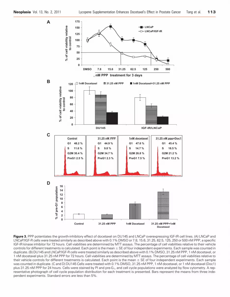
Figure 3. PPP potentiates the growth-inhibitory effect of docetaxel on DU145 and LNCaP overexpressing IGF-IR cell lines. (A) LNCaP andLNCaP/IGF-R cells were treated similarly as described above with 0.1%DMSO or 7.8, 15.6, 31.25, 62.5, 125, 250 or 500 nMPPP, a specificIGF-IR kinase inhibitor for 72 hours. Cell viabilities are determined by MTT assays. The percentage of cell viabilities relative to their vehiclecontrols for different treatments is calculated. Each point is themean± SE of four independent experiments. Each sample was counted induplicate. (B) DU145 and LNCaP/IGF-R cells were treated similarly as described abovewith 0.1%DMSO, 31.25 nMPPP, 1 nMdocetaxel, or1 nM docetaxel plus 31.25 nM PPP for 72 hours. Cell viabilities are determined byMTT assays. The percentage of cell viabilities relative totheir vehicle controls for different treatments is calculated. Each point is the mean ± SE of four independent experiments. Each samplewas counted in duplicate. (C and D) DU145 Cells were treated with 0.1%DMSO, 31.25 nM PPP, 1 nM docetaxel, or 1 nM docetaxel (Doc1)plus 31.25 nM PPP for 24 hours. Cells were stained by PI and pre-G1, and cell cycle populations were analyzed by flow cytometry. A rep-resentative photograph of cell cycle population distribution for each treatment is presented. Bars represent the means from three inde-pendent experiments. Standard errors are less than 5%.
Neoplasia Vol. 13, No. 2, 2011 Lycopene Supplementation Enhances Docetaxel’s Effect in Prostate Cancer Tang et al. 113

114 Lycopene Supplementation Enhances Docetaxel’s Effect in Prostate Cancer Tang et al. Neoplasia Vol. 13, No. 2, 2011
events of the IGF-IR signaling, AKT and survivin, will be more sub-jected to the IGF-IR regulation in DU145 cells than in LNCaP cells.Indeed, compared with their vehicle controls, 1 μM lycopene decreasedthe levels of phospho-AKT in LNCaPandDU145 cells by approximately20% and 60%, respectively. In addition, Figure 4A (panels g and h)shows that LNCaP cells overexpressing IGF-IR have increased protein
Figure 4. The effect of lycopene on IGF-IR and its mediated AKT/survserum-starved for 36 hours. During the last 2 hours of serum starvatiadded into the medium. After these treatments, cells were incubatewere prepared, IGF-IR was immunoprecipitated using anti–IGF-IRβphosphotyrosine (panel a) or anti–IGF-IRβ (panel b). Lanes 1, 2, 3, 4, 51 μMlycopene, 1μMlycopene+100 ng/ml IGF-I, 10μMlycopene+100docetaxel + 100 ng/ml IGF-I, respectively. Panel c: DU145 cells in the sfor 24 hours. IGF-BP3 expression in protein lysates was determined bytreated by lycopene at indicated concentrations for 48 hours. IGF-BP3conditioned medium. (B) LNCaP and DU145 cells were treated with 0Phospho-AKT and total AKT in the treated DU145, LNCaP, and LNCaPdensitometry measurement ratio relative to vehicle control. (C) DU 1450.1% DMSO + 0.1% THF), 1 μM lycopene (lyco1), 10 μM lycopene (lPPP, 1 or 10 μM lycopene plus 1 nM docetaxel, or 15.6 nM PPP plus 1trols. (D) Graphical presentation of the proposed mechanisms of lycenhancement of resistance to docetaxel-induced apoptosis.
levels of active AKTand antiapoptotic protein survivin compared withthe parental LNCaP cell line.
Figure 4C ( panels i and j) shows that docetaxel alone slightly increasesthe protein expression of survivin, whereas docetaxel in combinationwith lycopene or the IGF-IR kinase inhibitor, PPP, seems to synergis-tically downregulate the protein expression of survivin in DU145 cells.
ivin pathway in PCa cells. (A) DU145 cells at 70% confluency wereon, lycopene, PPP, and docetaxel at indicated concentrations wered with PBS or 100 ng/ml IGF-I for 15 minutes at 37°C. Cell lysatesantibody, and Western blot analyses were performed using anti-, 6, and 7 denote vehicle control, vehicle control + 100 ng/ml IGF-I,ng/ml IGF-I, 50 nMPPP+100ng/ml IGF-I, and 1μMlycopene+1nMerummediumwere treated by lycopene at indicated concentrationsWestern blot analysis. Panel d: DU145 in serum-free mediumweresecretion was determined byWestern blot analysis of concentrated.1% THF (vehicle control), 0.5, 1, or 10 μM lycopene for 24 hours./IGF-IR cells were determined by Western blot analysis. DR denotescells were treatedwith vehicle controls (0.1%DMSO, 0.1% THF, oryco10), 0.5 nM docetaxel (doc0.5), 1 nM docetaxel (doc1), 15.6 nMnM docetaxel. DRs are densitometry ratios relative to vehicle con-opene in inhibiting the IGF-IR–medicated survival pathway for the

Neoplasia Vol. 13, No. 2, 2011 Lycopene Supplementation Enhances Docetaxel’s Effect in Prostate Cancer Tang et al. 115
Taken together, these results suggest that lycopene inhibits IGF-IRactivation leading to inactivation of the AKT/survivin pathway.
Lycopene Supplement Enhances the Antitumor Efficacy ofDocetaxel and Prolongs the Survival of Tumor-BearingMice in a Xenograft ModelFigure 5A shows that docetaxel and docetaxel-plus-lycopene treat-
ments result in a significant decrease in the growth rate of DU145 tu-mors compared with vehicle control or 15mg/kg lycopene supplementalone (ANOVA, P values < .05). The inhibitory effect of lycopene sup-plement plus docetaxel on tumor growth is more pronounced than thatof docetaxel alone treatment (ANOVA, P values < .05). The tumorweights of DU145 cells in control, docetaxel, lycopene, and docetaxel-plus-lycopene groups recorded at the end of the treatment were 0.85 ±0.36, 0.39 ± 0.24, 0.84 ± 0.28, and 0.15 ± 0.15 g (mean tumor weight ±SD), respectively. Lycopene significantly enhanced the antitumor ef-ficacy of docetaxel by approximately 38% (Student t test, P = .042;Figure 5, B and C ).The efficacy of lycopene supplement (15 mg/kg per day), docetaxel
(5 or 10 mg/kg per week for three doses), or combination of each wasfurther assessed using growth delay, measured as the time from the startof tumor cell implantation for either tumor in amouse to reach a volumeof approximately 1500mm3 or when a mouse died spontaneously. Ad-ministration of a higher dose of docetaxel (10mg/kg per week) or com-bination of lycopene supplement with a lower (5 mg/kg per week)or higher (10 mg/kg per week) dose of docetaxel prolonged the sur-vival time ofmice (Figure 5D), compared with control, lycopene alone,or low-dose docetaxel alone. The median times for tumors to reach1500 mm3 in the control, lycopene supplement, and lower-dose doce-taxel groups are 41.5 ± 6, 43 ± 7, and 45 ± 3 days, respectively. Themedian times for tumors to reach 1500mm3 in the higher dose of doce-taxel and combination of lycopene supplements with the high or lowdose of docetaxel groups are 62 ± 8, 64 ± 8, and 70 ± 4 days, respec-tively, which demonstrated significantly delayed tumor growth by anaverage of 21 to 32 days relative to the controls (Figure 5D; log-ranktests, P values < .001). The efficacy of lycopene supplement in combi-nation with the lower dose of docetaxel (5 mg/kg per week) to delaytumor growth and prolonging the survival of tumor-bearing mice wasalmost equal to that of the higher dose of docetaxel (10 mg/kg perweek) alone. These results confirm that lycopene supplementation en-hanced the antitumor efficacy of docetaxel even at its suboptimal dose.
Lycopene Supplement and Docetaxel Combined TreatmentInduced a Significant Tumor Morphology AlternationResembling of Mitotic Catastrophe with Apoptosis,as well as Decreased Cell Proliferation andSurvivin Expression in Tumor TissuesHistologic analysis of hematoxylin and eosin–stained tumor sec-
tions from DU145 xenograft-bearing mice treated with lycopenesupplementation plus or minus docetaxel demonstrated a dramaticchange in tissue and cell morphology when compared with vehiclecontrol, lycopene supplement, or docetaxel alone (Figure 6A). Thesechanges include low cell density and multinucleated cells with con-densed chromatin staining and pyknosis, indicating mitotic catastropheand apoptosis.To examine whether these treatments affected cell proliferation and
apoptosis in in vivo situations, tumor xenograft tissue sections wereanalyzed by immunohistochemistry for Ki67, a marker for cell prolif-eration, and TUNEL, a marker for apoptotic response (Figure 6B). As
shown in Figure 6C , compared with controls, xenograft samples fromdocetaxel alone and lycopene supplement-plus-docetaxel groupsshowed a marked increase in number of TUNEL-positive cells. Quan-titative evaluation of apoptosis showed that docetaxel (10 mg/kg perweek) alone and docetaxel-plus-lycopene supplement (15 mg/kg perday) led 12.4% ± 3.2% and 24.6% ± 4.3% apoptotic cells, respec-tively, compared with control or lycopene supplement alone showing4.3% ± 1.2% or 5% ± 2.1% apoptotic cells (P values < .01). Docetaxelplus lycopene supplement resulted in an increase in apoptotic cellsapproximately 98% or 392% more than that of either docetaxel orlycopene supplement alone (P values < .05). The quantification ofKi67-positive cells in tumor sections showed no statistically significantdifference among the four different treatment groups (P values < .05).Together, these results suggest that the enhancement of the antitumorefficacy of docetaxel by lycopene supplement may be through mecha-nisms of apoptosis induction.
These tumor sections were further stained with antisurvivin anti-body. Figure 4G shows that tumor sections from mice treated withlycopene alone or lycopene supplement in combination with docetaxeldemonstrated significantly less staining of survivin protein, whereasdocetaxel alone seemed to slightly increase the staining of survivin intumor sections.
DiscussionLycopene has been suggested as a promising nutritional component forthe prevention and treatment of PCa [11,15]. As a result, many menwith PCa increase their intake of lycopene through dietary supplements[28], although there are no large-scale trials to provide evidence-basedclinical practice guidelines [29–32]. In addition, docetaxel has recentlybecome the first-line chemotherapeutic regimen for CRPC patients. Be-cause lycopene is a potent antioxidant, there is a concern whether lyco-pene could protect tumor cells as well as the healthy cells from oxidativedamage generated by docetaxel-based chemotherapy and therefore at-tenuate the antitumor efficacy of docetaxel [21]. In this study, our dataclearly demonstrate that lycopene supplementation can significantly en-hance the antitumor efficacy of docetaxel in the regression of establishedtumors in a CRPC xenograft model. This result should justify furtherclinical investigation of potential benefits of lycopene and docetaxelcombinations for treatment of CRPC patients.
Several in vitro cell culture studies [33] have suggested that androgen-induced increase in reactive oxygen species (ROS) levels in prostateepithelial cells may play a key role in prostate cancer occurrence, recur-rence, and progression. Therefore, agents that prevent the productionand chronic accumulation of ROS might be useful in the treatment ofprostate cancer. Venkateswaran et al. [34,35] reported that combina-tions of antioxidants (i.e., lycopene, vitamin E, and selenium) resultedin a significant reduction in both prostate cancer and liver metastasis inthe Lady transgenic mice, although the authors in this article did notexamine whether the anti–prostate cancer effects of these antioxidantswere due to their antioxidant properties. However, the authors didshow that a combination of vitamin E and selenium did not effectivelyinhibit prostate cancer in the Lady transgenic mice. Consistently,Limpens et al. [36] described that a lycopene and vitamin E combina-tion reduced tumor growth and prostate-specific antigen plasma levels inan orthotopic mouse model of human prostate cancer. Taken together,these studies suggested that lycopene is necessary for the inhibitory ef-fect of this combination on prostate cancer and that nonantioxidantmechanisms are also involved in their antineoplastic actions. Becausethere are concerns about the use of antioxidants during chemotherapy

Figure 5. Lycopene supplementation improves the antitumor efficacy of docetaxel in the DU145 tumor xenograft model. DU145 cells(1 × 10⁶) were injected into the right flank of NCR-nu/nu (nude) mice. Once tumors started growing, their sizes were measured twiceweekly in two dimensions, throughout the study. The tumor volume was calculated by the formula: 0.5236L1(L2)
2, where L1 is the longdiameter and L2 is the short diameter. Tumor volume (mm3) is represented as the mean of eight mice in each group. When tumorvolume reached 200 mm3 measured by caliper, mice bearing DU145 tumors were randomly divided into four or six different groupsas indicated. Control group of mice received water by gavage. Lycopene was given daily via gavage for the whole period, and docetaxelwas intraperitoneally injected into mice bearing DU145 tumors weekly for three doses. (A) Tumor growth curve, points, and mean tumorvolumes. Bars, SE. (B) Mean wet weights of tumors from different groups of treatments. At the termination of the study, tumors wereexcised from each mouse in different groups and weighed. Bars, SE. (C) Photographs of tumors from different groups of treatments.(D) Tumor growth curve for tumor volumes to reach 1500 mm3, points, mean tumor volumes. Bars, SE.
116 Lycopene Supplementation Enhances Docetaxel’s Effect in Prostate Cancer Tang et al. Neoplasia Vol. 13, No. 2, 2011
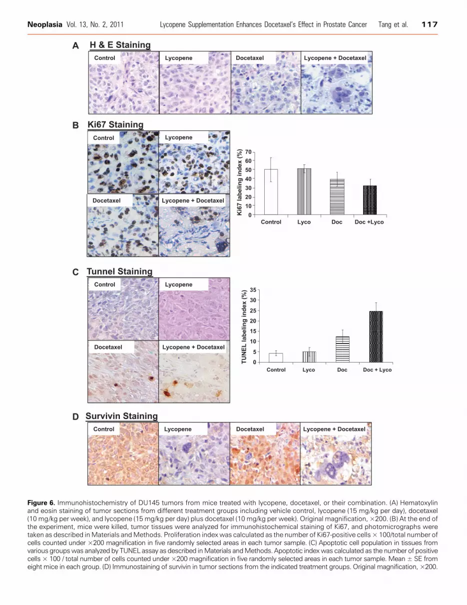
Figure 6. Immunohistochemistry of DU145 tumors from mice treated with lycopene, docetaxel, or their combination. (A) Hematoxylinand eosin staining of tumor sections from different treatment groups including vehicle control, lycopene (15 mg/kg per day), docetaxel(10 mg/kg per week), and lycopene (15 mg/kg per day) plus docetaxel (10 mg/kg per week). Original magnification, ×200. (B) At the end ofthe experiment, mice were killed, tumor tissues were analyzed for immunohistochemical staining of Ki67, and photomicrographs weretaken as described inMaterials andMethods. Proliferation indexwas calculated as the number of Ki67-positive cells × 100/total number ofcells counted under ×200 magnification in five randomly selected areas in each tumor sample. (C) Apoptotic cell population in tissues fromvarious groupswas analyzed by TUNEL assay as described inMaterials andMethods. Apoptotic indexwas calculated as the number of positivecells × 100 / total number of cells counted under ×200 magnification in five randomly selected areas in each tumor sample. Mean ± SE fromeight mice in each group. (D) Immunostaining of survivin in tumor sections from the indicated treatment groups. Original magnification, ×200.
Neoplasia Vol. 13, No. 2, 2011 Lycopene Supplementation Enhances Docetaxel’s Effect in Prostate Cancer Tang et al. 117
118 Lycopene Supplementation Enhances Docetaxel’s Effect in Prostate Cancer Tang et al. Neoplasia Vol. 13, No. 2, 2011
[21], the ability of lycopene to inhibit cancer growth through non-antioxidantmechanismsmay provide justification for combination ther-apies involving this agent and cytotoxic drugs like docetaxel.
IGF-IR not only plays a role in prostate carcinogenesis [37,38] but alsois elevated in metastatic PCa and is associated with resistance to androgenwithdraw [17,18,39]. In addition, maintaining IGF-responsivenessenables PCa survival and growth and is partially acquired throughandrogen-regulated IGF-IR expression [28]. These results suggest thatIGF-IR is a critical target for prevention and treatment of CRPC. Ly-copene has been shown to inhibit IGF-I signaling by down-regulationof IGF-I expression and up-regulation of IGF-BPs [13,14,40,41]. Inthis study, we showed that lycopene exhibited more potent inhibitoryeffects on the growth of DU145 cells with higher expression levels ofIGF-IR than those PCa cell lines (e.g., LNCaP, 22Rv1, LAPC-4, PC-3)with lower expression levels of IGF-IR. Moreover, transfected LNCaPcells stably expressing high levels of IGF-IR were approximately 400-fold more sensitive to lycopene than parental LNCaP cells (IC50 forlycopene in LNCaP/IGF-IR was 0.08 μM vs an IC50 of 36 μM forparental LNCaP cells). Intriguingly, although lycopene has been reportedto be accumulated in LNCaP cells to levels that are 4.5 times higherthan in DU145 cells [42], the growth-inhibitory effects of lycopene onDU145 cells is approximately seven times more pronounced than thaton LNCaP cells (IC50 for DU145 vs LNCaP are 5.1 vs 36 μM). Thisevidence indicates that IGF-IR levels may be more relevant to thegrowth-inhibitory effect of lycopene on PCa cell lines than cellularconcentrations of lycopene. We hypothesized that lycopene existingin extracellular or membrane compartments may also play a role onIGF-IR activation. Our results confirmed that lycopene increased theexpression and secretion of IGF-BP3, which sequesters the IGF-IRligand. We also showed for the first time that lycopene treatment foronly 2 hours can inhibit IGF-I–induced IGF-IR phosphorylation inDU145 cells. This result suggests a direct effect of lycopene on IGF-IRactivation. We also showed for the first time that lycopene can inhibitIGF-I–induced IGF-IR phosphorylation in DU145 cells. Further studiesare in progress to determine whether lycopene can directly bind to IGF-Ior IGF-IR and then inhibit IGF-IR kinase activity.
The general mechanism of IGF-IR activation is to enhance survivaland protect cells from apoptosis [16]. In addition, the IGF-IR interactswith, and influences, various signaling molecules such as ER, AR,EGFR,HER-2, and the DNA damage response pathway [16]. Together,these properties of IGF-IR activation provide mechanisms throughwhich it can cause resistance to multiple anticancer therapies includingchemotherapy, radiotherapy, and targeted therapies [16,25]. There-fore, in general, anticancer therapies targeting the IGF axis are promis-ing approaches for enhancing the efficacy of many conventional cancertherapies, including docetaxel-based chemotherapy [43]. We showhere that the synergistic growth-inhibitory effect of lycopene and doce-taxel was only found on those PCa cancer cell lines with higher IGF-IRlevels. This result provides a clue that CRPC patients with high levelsof IGF-IR activity may be most likely to benefit from combined ther-apy with lycopene and docetaxel.
Multiple studies have demonstrated that docetaxel causes cell deaththrough mitotic catastrophe as well as through caspase-dependent and-independent effects [44–46]. Our study showed that lycopene signifi-cantly enhanced the apoptotic effect of docetaxel in PCa both in vitroin cell cultures and in vivo in a xenograft model. The histologic analysisof tumor tissue sections revealed that the combination of lycopene anddocetaxel resulted in a significant increase in the number of tumor cellswith abnormal DNA condensation and multinucleation, consistent
with mitotic catastrophe. Further study showed that the docetaxeland lycopene combination could synergistically downregulate survivinexpression both in vitro and in vivo. Survivin is an antiapoptotic pro-tein and plays a role in cell cycle regulation during mitosis [47]. Sur-vivin inhibition, alone or in combination with the other therapies, hasbeen shown to induce or enhance apoptosis and mitotic catastrophein tumor cells [47]. In addition, survivin is regulated by IGFs and isassociated with the resistance to castration and progression in PCa[47–49].On the basis of evidences described,we can argue that survivin,serving as a critical downstream event for the combined effects of lyco-pene and docetaxel, may be a useful biomarker for predicting the out-come or response of this combined therapy in clinical studies.
In summary, these data provide a rationale for the clinical investi-gation of the efficacy and safety profile of lycopene in combinationwith docetaxel in CRPC patients. In particular, combining lycopenewith docetaxel may provide clinical benefit for men with metastaticCRPC, for whom morbidity and mortality remain high despite wideuse of docetaxel chemotherapy. Themechanism of this combined ther-apy seems to involve the IGF-IR/survivin pathway and is mediated bymitotic catastrophe and apoptosis.
References[1] Hsing AW and Devesa SS (2001). Trends and patterns of prostate cancer: what
do they suggest? Epidemiol Rev 23, 3–13.[2] Feldman BJ and Feldman D (2001). The development of androgen-independent
prostate cancer. Nat Rev Cancer 1, 34–45.[3] Dagher R, Li N, Abraham S, Rahman A, Sridhara R, and Pazdur R (2004).
Approval summary: docetaxel in combination with prednisone for the treatmentof androgen-independent hormone-refractory prostate cancer. Clin Cancer Res10, 8147–8151.
[4] Haenszel W and Kurihara M (1968). Studies of Japanese migrants. I. Mortalityfrom cancer and other diseases among Japanese in the United States. J Natl CancerInst 40, 43–68.
[5] Crawford ED (2003). Epidemiology of prostate cancer. Urology 62(6 suppl 1),3–12.
[6] Culig Z (2004). Androgen receptor cross-talk with cell signaling pathways.Growth Factors 22, 179–184.
[7] Clinton SK (1998). Lycopene: chemistry, biology, and implications for humanhealth and disease. Nutr Rev 56(2 pt 1), 35–51.
[8] Campbell JK, Engelmann NJ, Lila MA, and Erdman JW (2007). Phytoene,phytofluene, and lycopene from tomato powder differentially accumulate in tissuesof male Fisher 344 rats. Nutr Res 27, 794–801.
[9] Wertz K, Siler U, and Goralczyk R (2004). Lycopene: modes of action to promoteprostate health. Arch Biochem Biophys 430, 127–134.
[10] Erdman JW Jr, Ford NA, and Lindshield BL (2009). Are the health attributesof lycopene related to its antioxidant function? Arch Biochem Biophys 483,229–235.
[11] Giovannucci E, Ascherio A, Rimm EB, Stampfer MJ, Colditz GA, and WillettWC (1995). Intake of carotenoids and retinol in relation to risk of prostatecancer. J Natl Cancer Inst 87, 1767–1776.
[12] Siler U, Barella L, Spitzer V, Schnorr J, Lein M, Goralczyk R, and Wertz K(2004). Lycopene and vitamin E interfere with autocrine/paracrine loops inthe Dunning prostate cancer model. FASEB J 18, 1019–1021.
[13] Liu X, Allen JD, Arnold JT, and Blackman MR (2008). Lycopene inhibits IGF-Isignal transduction and growth in normal prostate epithelial cells by decreas-ing DHT-modulated IGF-I production in co-cultured reactive stromal cells.Carcinogenesis 29, 816–823.
[14] Liu C, Lian F, SmithDE, Russell RM, andWang XD (2003). Lycopene supplemen-tation inhibits lung squamous metaplasia and induces apoptosis via up-regulatinginsulin-like growth factor–binding protein 3 in cigarette smoke-exposed ferrets.Cancer Res 63, 3138–3144.
[15] van Breemen RB and Pajkovic N (2008). Multitargeted therapy of cancer bylycopene. Cancer Lett 269, 339–351.
[16] Casa AJ, Dearth RK, Litzenburger BC, Lee AV, and Cui X (2008). The type Iinsulin-like growth factor receptor pathway: a key player in cancer therapeuticresistance. Front Biosci 13, 3273–3287.
Neoplasia Vol. 13, No. 2, 2011 Lycopene Supplementation Enhances Docetaxel’s Effect in Prostate Cancer Tang et al. 119
[17] Krueckl SL, Sikes RA, Edlund NM, Bell RH, Hurtado-Coll A, Fazli L, GleaveME, and Cox ME (2004). Increased insulin-like growth factor I receptor ex-pression and signaling are components of androgen-independent progression in alineage-derived prostate cancer progression model. Cancer Res 64, 8620–8629.
[18] Plymate SR, Haugk K, Coleman I,Woodke L, Vessella R, Nelson P, MontgomeryRB, Ludwig DL, andWu JD (2007). An antibody targeting the type I insulin-likegrowth factor receptor enhances the castration-induced response in androgen-dependent prostate cancer. Clin Cancer Res 13, 6429–6439.
[19] Domingo-Domenech J, Oliva C, Rovira A, Codony-Servat J, Bosch M, Filella X,Montagut C, Tapia M, Campás C, Dang L, et al. (2006). Interleukin 6, a nuclearfactor-κB target, predicts resistance to docetaxel in hormone-independent prostatecancer and nuclear factor-κB inhibition by PS-1145 enhances docetaxel antitumoractivity. Clin Cancer Res 12, 5578–5586.
[20] Feng S, Tang Q, Sun M, Chun JY, Evans CP, and Gao AC (2009). Interleukin-6increases prostate cancer cells resistance to bicalutamide via TIF2. Mol CancerTher 8, 665–671.
[21] Lawenda BD, Kelly KM, Ladas EJ, Sagar SM, Vickers A, and Blumberg JB(2008). Should supplemental antioxidant administration be avoided during che-motherapy and radiation therapy? J Natl Cancer Inst 100, 773–783.
[22] Rehman A, Bourne LC, Halliwell B, and Rice-Evans CA (1999). Tomato con-sumption modulates oxidative DNA damage in humans. Biochem Biophys ResCommun 262, 828–831.
[23] Porrini M and Riso P (2000). Lymphocyte lycopene concentration and DNAprotection from oxidative damage is increased in women after a short period oftomato consumption. J Nutr 130, 189–192.
[24] Hsiao G,Wang Y, TzuNH, Fong TH, ShenMY, Lin KH, ChouDS, and Sheu JR(2005). Inhibitory effects of lycopene on in vitro platelet activation and in vivoprevention of thrombus formation. J Lab Clin Med 146(4), 216–226.
[25] Lu Y, Zi X, Zhao Y, Mascarenhas D, and Pollak M (2001). Insulin-like growthfactor-I receptor signaling and resistance to trastuzumab (Herceptin). J Natl CancerInst 93, 1852–1857.
[26] Zi X and Simoneau AR (2005). Flavokawain A, a novel chalcone from kavaextract, induces apoptosis in bladder cancer cells by involvement of Bax protein–dependent and mitochondria-dependent apoptotic pathway and suppresses tumorgrowth in mice. Cancer Res 65, 3479–3486.
[27] Sobel RE and Sadar MD (2005). Cell lines used in prostate cancer research: acompendium of old and new lines-part 1. J Urol 173, 342–359.
[28] Zimmerman RA and Thompson IM Jr (2002). Prevalence of complementarymedicine in urologic practice. A review of recent studies with emphasis on useamong prostate cancer patients. Urol Clin North Am 29, 1–9.
[29] KucukO, Sarkar FH, SakrW,Djuric Z, PollakMN,Khachik F, Li YW,BanerjeeM,Grignon D, Bertram JS, et al. (2001). Phase II randomized clinical trial of lyco-pene supplementation before radical prostatectomy. Cancer Epidemiol BiomarkersPrev 10, 861–868.
[30] Clark PE,HallMC,BordenLS Jr,Miller AA,Hu JJ, LeeWR, StindtD,D’AgostinoRJr, Lovato J, Harmon M, et al. (2006). Phase I-II prospective dose-escalating trialof lycopene in patients with biochemical relapse of prostate cancer after definitivelocal therapy. Urology 67, 1257–1261.
[31] Ansari MS and Gupta NP (2003). A comparison of lycopene and orchidectomyvs orchidectomy alone in the management of advanced prostate cancer. BJU Int 92,375–378.
[32] Schwenke C, Ubrig B, Thürmann P, Eggersmann C, and Roth S (2009). Lycopenefor advanced hormone refractory prostate cancer: a prospective, open phase II pilotstudy. J Urol 181, 1098–1103.
[33] Mehraein-Ghomi F, Lee E, ChurchDR, ThompsonTA, BasuHS, andWildingG
(2008). JunDmediates androgen-induced oxidative stress in androgen dependentLNCaP human prostate cancer cells. Prostate 68, 924–934.
[34] Venkateswaran V, Fleshner NE, Sugar LM, and Klotz LH (2004). Antioxidantsblock prostate cancer in Lady transgenic mice. Cancer Res 64, 5891–5896.
[35] Venkateswaran V, Klotz LH, Ramani M, Sugar LM, Jacob LE, Nam RK, andFleshner NE (2009). A combination of micronutrients is beneficial in reducingthe incidence of prostate cancer and increasing survival in the Lady transgenicmodel. Cancer Prev Res (Phila) 2, 473–483.
[36] Limpens J, Schröder FH, de Ridder CM, Bolder CA, WildhagenMF, Obermüller-Jevic UC, Krämer K, and van Weerden WM (2006). Combined lycopene andvitamin E treatment suppresses the growth of PC-346C human prostate cancercells in nude mice. J Nutr 136, 1287–1293.
[37] DiGiovanni J, Kiguchi K, Frijhoff A, Wilker E, Bol DK, Beltrán L, Moats S,Ramirez A, Jorcano J, and Conti C (2000). Deregulated expression of insulin-like growth factor 1 in prostate epithelium leads to neoplasia in transgenic mice.Proc Natl Acad Sci USA 97, 3455–3460.
[38] Hellawell GO, Turner GD, Davies DR, Poulsom R, Brewster SF, and MacaulayVM (2002). Expression of the type 1 insulin-like growth factor receptor is up-regulated in primary prostate cancer and commonly persists in metastatic dis-ease. Cancer Res 62, 2942–2950.
[39] Genua M, Pandini G, Sisci D, Castoria G, Maggiolini M, Vigneri R, and BelfioreA (2009). Role of cyclic AMP response element-binding protein in insulin-likegrowth factor-I receptor up-regulation by sex steroids in prostate cancer cells.Cancer Res 69, 7270–7277.
[40] Riso P, Brusamolino A, Martinetti A, and Porrini M (2006). Effect of a tomatodrink intervention on insulin-like growth factor (IGF)-1 serum levels in healthysubjects. Nutr Cancer 55, 157–162.
[41] Graydon R, Gilchrist SE, Young IS, Obermüller-Jevic U, Hasselwander O, andWoodside JV (2007). Effect of lycopene supplementation on insulin-like growthfactor-1 and insulin-like growth factor binding protein-3: a double-blind, placebo-controlled trial. Eur J Clin Nutr 61, 1196–1200.
[42] Liu A, Pajkovic N, Pang Y, Zhu D, Calamini B, Mesecar AL, and van BreemenRB (2006). Absorption and subcellular localization of lycopene in human pros-tate cancer cells. Mol Cancer Ther 5, 2879–2885.
[43] de Bono JS, Attard G, Adjei A, Pollak MN, Fong PC, Haluska P, Roberts L,Melvin C, Repollet M, Chianese D, et al. (2007). Potential applications for circu-lating tumor cells expressing the insulin-like growth factor-I receptor. Clin CancerRes 13, 3611–3616.
[44] Fabbri F, Amadori D, Carloni S, Brigliadori G, Tesei A, Ulivi P, Rosetti M, VanniniI, Arienti C, Zoli W, et al. (2008). Mitotic catastrophe and apoptosis induced bydocetaxel in hormone-refractory prostate cancer cells. J Cell Physiol 217, 494–501.
[45] Mediavilla-Varela M, Pacheco FJ, Almaguel F, Perez J, Sahakian E, Daniels TR,Leoh LS, Padilla A, Wall NR, Lilly MB, et al. (2009). Docetaxel-induced pros-tate cancer cell death involves concomitant activation of caspase and lysosomalpathways and is attenuated by LEDGF/p75. Mol Cancer 8, 68.
[46] Castedo M, Perfettini JL, Roumier T, Andreau K, Medema R, and Kroemer G(2004). Cell death bymitotic catastrophe: a molecular definition.Oncogene 23(16),2825–2837.
[47] Zhang M, Latham D, Delaney M, and Chakravarti A (2005). Survivin mediatesresistance to antiandrogen therapy in prostate cancer. Oncogene 24, 2474–2482.
[48] Vaira V, Lee C, Goel H, Bosari S, Languino L, and Altieri D (2007). Regulationof survivin expression by IGF-1/mTOR signaling. Oncogene 26, 2678–2684.
[49] Shariat S, Lotan Y, Saboorian H, Khoddami SM, Roehrborn CG, Slawin KM,and Ashfaq R (2005). Survivin expression is associated with features of biolog-ically aggressive prostate carcinoma. Cancer 100, 751–757.
Copyright © 2022 FDOKUMEN