On the anionic homo‐ and copolymerization of ethyl‐ and butyl‐2‐cyanoacrylates

Ag
Ma
b
c
d
a
ARRAA
KBHPPP
1
acbeiosgp[sEoto
CT
0d
Analytica Chimica Acta 693 (2011) 114–120
Contents lists available at ScienceDirect
Analytica Chimica Acta
journa l homepage: www.e lsev ier .com/ locate /aca
novel biosensing mechanism based on a poly(N-butyl benzimidazole)-modifiedold electrode for the detection of hydrogen peroxide
u-Yi Huaa,b,∗, Hsiao-Chien Chena,b, Rung-Ywan Tsai c, Yu-Chen Lina,b, Leeyih Wangd
Green Technology Research Center, Department of Chemical and Materials Engineering, Chang Gung University, Tao-Yuan 33302, Taiwan, ROCBiosensor Group, Biomedical Engineering Research Center, Chang Gung University, Tao-Yuan 33302, Taiwan, ROCElectronics and Optoelectronics Research Laboratories, Industrial Technology Research Institute, Hsinchu 31040, Taiwan, ROCCenter for Condensed Matter Science, Institute of Polymer Science and Technology, National Taiwan University, Taipei 10617, Taiwan, ROC
r t i c l e i n f o
rticle history:eceived 18 December 2010eceived in revised form 7 March 2011ccepted 8 March 2011vailable online 16 March 2011
eywords:
a b s t r a c t
A novel mechanism to detect hydrogen peroxide (H2O2) using a poly(N-butyl benzimidazole) (PBBI)-modified gold (PBBI/Au) electrode is proposed. Synthetic PBBI was oxidized using a mixture of aceticacid (AcOH) and H2O2 to form PBBI N-oxide (PBBINO). The structure of PBBINO was verified by Fouriertransform infrared spectroscopy (FT-IR) and the degree of oxidation was measured by X-ray photoelec-tron spectroscopy (XPS). Moreover, the oxide could be reduced electrochemically back to PBBI. Based onthis reaction, a novel enzyme-free PBBI/Au electrode was developed to detect H2O2 in the presence of
iosensorydrogen peroxideeracetic acidoly(N-butyl benzimidazole)oly(N-butyl benzimidazole) N-oxide
AcOH electrochemically. The biosensor detected H2O2 linearly over concentrations ranging from 25 �Mto 10 mM with a detection limit of 6.25 �M in phosphate buffer solution (PBS) mixed with AcOH at pH 6.4.In addition, at an applied potential of −0.5 V, the sensor characteristics could be tuned using AcOH overa pH range of 3.7–6.4. The sensitivity of the probe could be enhanced from 35.1 to 419.4 �A mM−1 cm−2
by modifying the surface morphology of the PBBI/Au electrode from a smooth plane to a granular, three-dimensional configuration. Furthermore, it was not influenced by interfering compounds and showed
high thermal stability.. Introduction
H2O2, a chemical used widely in the food, pharmaceutical, paper,nd chemical industries, is a reactive oxygen species that can causeancer when found in excess in the human body [1]. It is also ay-product of the reaction catalyzed by large number of oxidasenzymes [2,3]. The detection of H2O2 thus is important in biomed-cal and environmental applications. In past decades, a numberf technologies such as titrimetry, fluorescence, and absorbancepectra [4,5] have been developed to detect H2O2. Recently, a neweneration of sensors based on electrochemical technology hasroven simple to use and highly sensitive for real-time detection6,7]. Most of the electrochemical methods are based on enzymesuch as horseradish peroxidase, hemoglobin, or myoglobin [8–10].
nzymatic electrodes usually are prepared by immobilizing a coatf enzymes mixed with organic or inorganic materials on the elec-rode surface. Although such biosensors are highly sensitive, lossf content and degradation lead to decreased enzyme activity over∗ Corresponding author at: Green Technology Research Center, Department ofhemical and Materials Engineering, Chang Gung University, Tao-Yuan 33302,aiwan, ROC. Tel.: +886 3 2118800x5289; fax: +886 3 2118668.
E-mail address: [email protected] (M.-Y. Hua).
003-2670/$ – see front matter © 2011 Elsevier B.V. All rights reserved.oi:10.1016/j.aca.2011.03.020
© 2011 Elsevier B.V. All rights reserved.
time. The development of enzyme-free biosensors to overcome thislimitation is of great interest. The simplest alternative method isto electrocatalyze H2O2 directly at working potentials of 0.5–0.7 V[11–13]. Unfortunately, at this relatively high potential, the selec-tivity is easily influenced by the presence of interfering species[14].
Recently, electrodes modified with inorganic andorganic–inorganic materials have been studied widely owingto their selectivity, stability, and the efficiency with which theytransfer electrons; such modifications include metallic particles(Ag, Au, Co, and CuS nanoparticles) [15–17], metallic oxides(Cu2O, CuO, TiO2, and MnO2) [18–21], transition metals (Prussianblue and metalloporphyrins) [22,23], and biological complexesincorporating inorganic components [24]. Such polymer-modifiedelectrodes, either with immobilized enzymes or with hybridorganic–inorganic compounds, are widely used because of theirhigh build and ease of preparation [25,26]. However, only a fewstudies have used polymers without enzymes to detect H2O2 atlower potentials [27,28].
Polybenzimidazole (PBI) is a heterocyclic aromatic poly-mer with exceptional chemical resistance, excellent mechanicalstrength, and thermal stability [29,30]. After doping with acid, itis an excellent proton conductor and is used in proton exchangemembranes in fuel cells [31,32]. Furthermore, its fluorescence char-

himic
ahuiunf
tmqPbwralor
2
2
dSSw(e
2
epiwtaepoAA
2
2
tp(Spffssra
0oi
M.-Y. Hua et al. / Analytica C
cteristics – the result of benzimidazole’s conjugated structure –ave been investigated and reported widely [33]. PBI is poorly sol-ble in many solvents because of its rigid backbone and interchain
nteractions via hydrogen bonds. Alkylation of PBI improves its sol-bility without sacrificing its original properties [34]. However,either PBI nor its derivatives have been used to modify electrodes
or biosensor applications.Here, we demonstrate that PBBI prepared by the butyl alkyla-
ion of PBI with 1-iodobutane can be oxidized by peracetic acid (aixture of AcOH and H2O2; AcOOH) to form PBBINO, which subse-
uently can be reduced electrochemically. Based on this reaction, aBBI/Au electrode was developed for use as an enzyme-free H2O2iosensor. This biosensor showed superior performance comparedith traditional ones, including long-term detection capability,
apid response time, and high specificity. Furthermore, preparingthree-dimensional PBBI/Au (3D-PBBI/Au) electrode with a granu-
ated surface enhanced the sensitivity of the probe to 12 times thatf a PBBI/Au electrode with a smooth surface. We also propose theedox mechanism by which the PBBI/Au electrode detects H2O2.
. Experimental
.1. Chemicals
Isophthaloyl dichloride (98%) was purchased from Acros, 3,3′-iaminobenzidine tetrahydrochloride hydrate (>97%) was fromigma–Aldrich, polyphosphoric acid and H2O2 (30%) were fromhowa, dimethyl sulfoxide (DMSO) was from Tedia, sodium hydrideas from Fluka, 1-iodobutane (99%) was from Lancaster, and AcOH
99.8%) was from Scharlau. Deionized (DI) water was used in allxperiments.
.2. Instrumentation and measurements
FT-IR spectra were obtained using a Bruker-Tensor 27 spectrom-ter at a spectral resolution of 8 cm−1. XPS measurements wereerformed using a VG Scientific ESCALAB 250 system. Electrochem-
cal measurements were performed on a CHI 660A electrochemicalorkstation (CH Instruments, USA) with a three-electrode sys-
em consisting of the PBBI/Au electrode, a bare Au electrode,nd an Ag/AgCl electrode as the working, counter, and referencelectrodes, respectively. All electrochemical measurements wereerformed in 40 mL of 0.2 M PBS at 25 ◦C and at pH 7.0 (unless statedtherwise). The pH values of the electrolytes were adjusted withcOH to 7.0 (without AcOH), 6.4 (43.8 mM AcOH), 4.6 (216.0 mMcOH) or 3.7 (833.4 mM AcOH).
.3. Procedures
.3.1. Preparation of PBI, PBBI, and PBBINOPBI was synthesized by condensation of 3,3′-diaminobenzidine
etrahydrochloride hydrate and isophthaloyl dichloride inolyphosphoric acid [35]. To produce PBBI, 1 mmol of PBI0.3 g) was dissolved in 50 mL of DMSO and stirred at 80 ◦C for 1 h.odium hydride (0.1 g; 4.2 mmol) was added and the solution wasurged with nitrogen to form a deep red solution. After stirringor 2 h, 0.55 g (3 mmol) of 1-iodobutane was added and reactedor 24 h at 120 ◦C. The solution was added to 500 mL of DI water,tirred vigorously, and the precipitate was separated from theolution by filtration. The filtered solid was redissolved in DMSO,ecrystallized sequentially three times with acetone, and dried in
vacuum aspirator to obtain the PBBI.AcOOH solutions were prepared by mixing 0.2 mmol AcOH with.1, 0.2, or 0.4 mmol H2O2. PBBI (0.2 mmol) was dissolved in 10 mLf DMSO and added to the AcOOH solutions. The reactions werencubated in a 43-kHz ultrasonic bath at 40 ◦C for 0.5 h in a dark
a Acta 693 (2011) 114–120 115
room. The purification procedure was as described above for PBBI,producing PBBINO1, PBBINO2, and PBBINO3, with molar ratios ofPBBI:AcOH:H2O2 of 1:1:0.5, 1:1:1 and 1:1:2, respectively.
2.3.2. Preparation of the PBBI/Au and 3D-PBBI/Au electrodesTo produce the PBBI/Au electrode, 2 �L of a solution contain-
ing 0.23 g PBBI in 10 mL of DMSO were dropped onto an Au diskelectrode (0.196 cm2) and dried in a vacuum aspirator at 50 ◦C for5 h. The 3D-PBBI/Au electrode was similarly prepared by dissolving0.23 g PBBI in 10 mL of DMSO and then dispersing it with stirringinto a poor solvent consisting of 250 mL DI water and 150 mL ace-tone. After high-speed centrifugation (5500 rpm), the precipitatewas washed by DI water three times. Finally, the precipitate wasdispersed into 40 mL DI water and 4 �L of the solution with 1%DMSO was dropped onto an Au disk electrode (0.196 cm2) and driedin a vacuum aspirator at 50 ◦C for 5 h.
3. Results and discussion
3.1. Characterization of PBBI and PBBINO
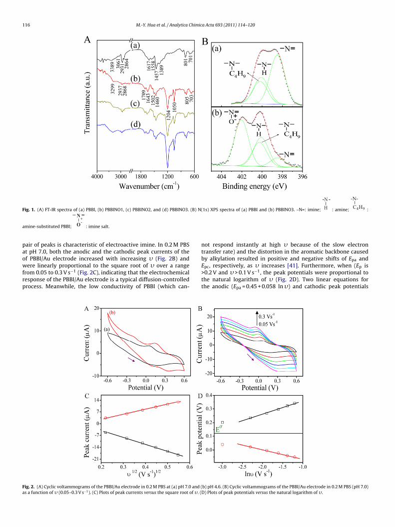
FT-IR spectroscopy detected the characteristic absorption bandsof PBBI (Fig. 1A), including the stretching vibrations (�) of N–H(�N–H) at 3389 cm−1, unsaturated �C–H (3061 cm−1), asymmetricand symmetric saturated �C–H (2931 and 2864 cm−1, respectively),�C N/C C (1617 cm−1), the in-plane deformation of benzimidazole(1558 cm−1), and C–H bending (1457 and 1389 cm−1). Two otherpeaks appearing at 801 and 701 cm−1 were indicative of hetero-cyclic ring vibrations [36]. After reacting with AcOOH, three newpeaks attributable to the resonance structure of PBBINO1 appearedat 1709 cm−1 (�C=N+ ), 1204 cm−1 (�N+–O− ), and 1050 cm−1 (ben-zene ring in-plane C–H bending) (Fig. 1A) [37]. Furthermore, theintensity of the latter two peaks increased with increasing con-centrations of H2O2 at a constant concentration of AcOH (Fig. 1A).However, oxidation did not occur in the absence of AcOH. This isconsistent with the results seen when pyridine is added to a mix-ture of AcOH and H2O2 to produce pyridine N-oxide [38], indicatingthat PBBINO is formed through the oxidation of PBBI by AcOOH (amixture of H2O2 and AcOH).
The N(1s) XPS spectrum of PBBI can be deconvoluted into threepeaks (Fig. 1B): imine at 398.4 eV, amine at 400.2 eV, and a peak at400.3 eV attributable to the amine substitution by butyl alkylation[39]. The ratio of the peak areas was 1:0.55:0.45; i.e., butyl alkyla-tion accounted for 45%, whereas 55% remained as native amine. TheN(1s) spectrum of PBBINO3 (Fig. 1B) was deconvoluted into fourpeaks: in addition to the previous three peaks, a new characteristicpeak formed at 402.0 eV, indicating formation of the N-oxide struc-ture (imine salt) [40]. The ratio of the peak areas (from lowest tohighest binding energy) was 8.60:27.24:22.60:41.56. Thus, the cor-responding ratios of amine N (native amine plus butyl amine):imineN:imine salt was 1.00:0.17:0.83; i.e., the total area of imine N andimine salt was equal to that of amine N. Consistent with the resultsof the FT-IR studies, the XPS spectra suggest that the reactive siteoccurring at the imine of PBBI can be oxidized by AcOOH to forman imine salt, which in PBBINO3 accounts for 41.5% of the total areaattributable to N.
3.2. Electrochemistry of the PBBI/Au electrode
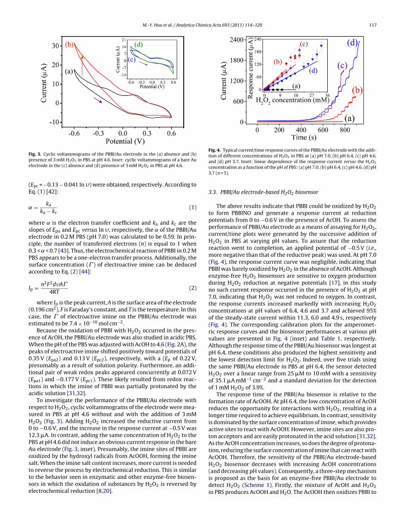
Cyclic voltammograms of the PBBI/Au electrode showed one
pair of redox peaks between −0.6 and 0.6 V at a potential scanrate (�) of 0.05 V s−1 in PBS at pH 7.0 (Fig. 2A). The potentials ofthe anodic (Epa) and cathodic (Epc) peaks were 0.20 and 0.04 V,respectively, with a peak-to-peak separation ((Ep) of 0.16 V and aformal potential (the average of Epa and Epc; E0′) of 0.12 V. This
116 M.-Y. Hua et al. / Analytica Chimica Acta 693 (2011) 114–120
Fig. 1. (A) FT-IR spectra of (a) PBBI, (b) PBBINO1, (c) PBBINO2, and (d) PBBINO3. (B) N(1s) XPS spectra of (a) PBBI and (b) PBBINO3. –N=: imine;
-N -H : amine;
-N-C4H9 :
a
paowfrp
Fa
mine-substituted PBBI;
N
O+-
: imine salt.
air of peaks is characteristic of electroactive imine. In 0.2 M PBSt pH 7.0, both the anodic and the cathodic peak currents of thef PBBI/Au electrode increased with increasing � (Fig. 2B) and
ere linearly proportional to the square root of � over a rangerom 0.05 to 0.3 V s−1 (Fig. 2C), indicating that the electrochemicalesponse of the PBBI/Au electrode is a typical diffusion-controlledrocess. Meanwhile, the low conductivity of PBBI (which can-
ig. 2. (A) Cyclic voltammograms of the PBBI/Au electrode in 0.2 M PBS at (a) pH 7.0 and (s a function of � (0.05–0.3 V s−1). (C) Plots of peak currents versus the square root of �. (
not respond instantly at high � because of the slow electrontransfer rate) and the distortion in the aromatic backbone causedby alkylation resulted in positive and negative shifts of Epa and
Epc, respectively, as � increases [41]. Furthermore, when (Ep is>0.2 V and � > 0.1 V s−1, the peak potentials were proportional tothe natural logarithm of � (Fig. 2D). Two linear equations forthe anodic (Epa = 0.45 + 0.058 ln �) and cathodic peak potentialsb) pH 4.6. (B) Cyclic voltammograms of the PBBI/Au electrode in 0.2 M PBS (pH 7.0)D) Plots of peak potentials versus the natural logarithm of �.
M.-Y. Hua et al. / Analytica Chimica Acta 693 (2011) 114–120 117
Fpe
(E
˛
wsec0Psa
I
(ce
eWp0pt(ta
rsH01PAosttse
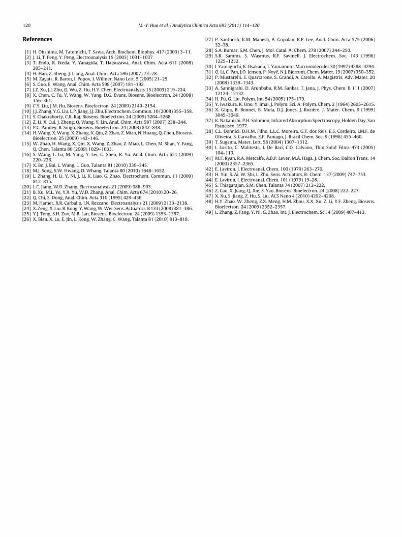
Fig. 4. Typical current/time response curves of the PBBI/Au electrode with the addi-tion of different concentrations of H2O2 in PBS at (a) pH 7.0, (b) pH 6.4, (c) pH 4.6,
ig. 3. Cyclic voltammograms of the PBBI/Au electrode in the (a) absence and (b)resence of 3 mM H2O2 in PBS at pH 4.6. Inset: cyclic voltammograms of a bare Aulectrode in the (c) absence and (d) presence of 3 mM H2O2 in PBS at pH 4.6.
Epc = −0.13 − 0.041 ln �) were obtained, respectively. According toq. (1) [42]:
= ka
ka − kc(1)
here ˛ is the electron transfer coefficient and ka and kc are thelopes of Epa and Epc versus ln �, respectively, the ˛ of the PBBI/Aulectrode in 0.2 M PBS (pH 7.0) was calculated to be 0.59. In prin-iple, the number of transferred electrons (n) is equal to 1 when.3 < ˛ < 0.7 [43]. Thus, the electrochemical reaction of PBBI in 0.2 MBS appears to be a one-electron transfer process. Additionally, theurface concentration (� ) of electroactive imine can be deducedccording to Eq. (2) [44]:
p = n2F2d�A�
4RT(2)
where Ip is the peak current, A is the surface area of the electrode0.196 cm2), F is Faraday’s constant, and T is the temperature. In thisase, the � of electroactive imine on the PBBI/Au electrode wasstimated to be 7.4 × 10⁻¹⁰ mol cm−2.
Because the oxidation of PBBI with H2O2 occurred in the pres-nce of AcOH, the PBBI/Au electrode was also studied in acidic PBS.hen the pH of the PBS was adjusted with AcOH to 4.6 (Fig. 2A), the
eaks of electroactive imine shifted positively toward potentials of.35 V (Epa2) and 0.13 V (Epc2), respectively, with a (Ep of 0.22 V,resumably as a result of solution polarity. Furthermore, an addi-ional pair of weak redox peaks appeared concurrently at 0.072 VEpa1) and −0.177 V (Epc1). These likely resulted from redox reac-ions in which the imine of PBBI was partially protonated by thecidic solution [31,32].
To investigate the performance of the PBBI/Au electrode withespect to H2O2, cyclic voltammograms of the electrode were mea-ured in PBS at pH 4.6 without and with the addition of 3 mM2O2 (Fig. 3). Adding H2O2 increased the reductive current fromto −0.6 V, and the increase in the response current at −0.5 V was2.3 �A. In contrast, adding the same concentration of H2O2 to theBS at pH 4.6 did not induce an obvious current response in the bareu electrode (Fig. 3, inset). Presumably, the imine sites of PBBI arexidized by the hydroxyl radicals from AcOOH, forming the iminealt. When the imine salt content increases, more current is needed
o reverse the process by electrochemical reduction. This is similaro the behavior seen in enzymatic and other enzyme-free biosen-ors in which the oxidation of substances by H2O2 is reversed bylectrochemical reduction [8,20].and (d) pH 3.7. Inset: linear dependence of the response current versus the H2O2
concentration as a function of the pH of PBS: (a) pH 7.0, (b) pH 6.4, (c) pH 4.6, (d) pH3.7 (n = 5).
3.3. PBBI/Au electrode-based H2O2 biosensor
The above results indicate that PBBI could be oxidized by H2O2to form PBBINO and generate a response current at reductionpotentials from 0 to −0.6 V in the presence of AcOH. To assess theperformance of PBBI/Au electrode as a means of assaying for H2O2,current/time plots were generated by the successive addition ofH2O2 in PBS at varying pH values. To assure that the reductionreaction went to completion, an applied potential of −0.5 V (i.e.,more negative than that of the reductive peak) was used. At pH 7.0(Fig. 4), the response current curve was negligible, indicating thatPBBI was barely oxidized by H2O2 in the absence of AcOH. Althoughenzyme-free H2O2 biosensors are sensitive to oxygen productionduring H2O2 reduction at negative potentials [17], in this studyno such current response occurred in the presence of H2O2 at pH7.0, indicating that H2O2 was not reduced to oxygen. In contrast,the response currents increased markedly with increasing H2O2concentrations at pH values of 6.4, 4.6 and 3.7 and achieved 95%of the steady-state current within 11.3, 6.0 and 4.9 s, respectively(Fig. 4). The corresponding calibration plots for the amperomet-ric response curves and the biosensor performances at various pHvalues are presented in Fig. 4 (inset) and Table 1, respectively.Although the response time of the PBBI/Au biosensor was longest atpH 6.4, these conditions also produced the highest sensitivity andthe lowest detection limit for H2O2. Indeed, over five trials usingthe same PBBI/Au electrode in PBS at pH 6.4, the sensor detectedH2O2 over a linear range from 25 �M to 10 mM with a sensitivityof 35.1 �A mM−1 cm−2 and a standard deviation for the detectionof 1 mM H2O2 of 3.9%.
The response time of the PBBI/Au biosensor is relative to theformation rate of AcOOH. At pH 6.4, the low concentration of AcOHreduces the opportunity for interactions with H2O2, resulting in alonger time required to achieve equilibrium. In contrast, sensitivityis dominated by the surface concentration of imine, which providesactive sites to react with AcOOH. However, imine sites are also pro-ton acceptors and are easily protonated in the acid solution [31,32].As the AcOH concentration increases, so does the degree of protona-tion, reducing the surface concentration of imine that can react withAcOOH. Therefore, the sensitivity of the PBBI/Au electrode-basedH2O2 biosensor decreases with increasing AcOH concentrations
(and decreasing pH values). Consequently, a three-step mechanismis proposed as the basis for an enzyme-free PBBI/Au electrode todetect H2O2 (Scheme 1). Firstly, the mixture of AcOH and H2O2in PBS produces AcOOH and H2O. The AcOOH then oxidizes PBBI to
118 M.-Y. Hua et al. / Analytica Chimica Acta 693 (2011) 114–120
Table 1Characteristics of the PBBI/Au electrode-based H2O2 biosensor in 0.2 M PBS at various pH values.
pH Sensor characteristic
Response time (s) Sensitivity (�A mM−1 cm−2) Detection range (mM) Detection limit (�M) R
3.7 <4.9 21.7 0.2–22.5 75.00 0.9974.6 <6.0 33.2 0.075–15.0 12.50 0.9986.4 <11.3 35.1 0.025–10.0 6.25 0.9997.0 – – – – –
–: no response detected.
or usin
fbrc
3
u
Fp
Scheme 1. Redox mechanism of the H2O2 biosens
orm PBBINO and AcOH. Finally, the PBBINO reverts to PBBI and H2Oy accepting two electrons and two protons by electrochemicaleduction. No PBBI is consumed at any point in the redox reaction,onfirming the catalysis nature of the PBBI/Au electrode.
.4. 3D-PBBI/Au electrode-based H2O2 biosensor
The above results demonstrate that a method to detect H2O2sing a PBBI/Au electrode is feasible and accurate. However, the
ig. 5. (A) Scanning electron micrographs of (a) PBBI/Au electrode and (b) 3D-PBBI/Au eleH 7.0 and (b) pH 6.4. Inset: linear dependence of the response current versus the H2O2 c
g the PBBI/Au electrode as the working electrode.
solubility of the PBBI polymer in DMSO resulted in a planar mor-phology on the PBBI/Au electrode surface (Fig. 5A), limiting theeffective surface area for detection. Therefore, a PBBI/Au electrodewith a granulated surface and porous structure (Fig. 5A) was pre-
pared by dispersing the PBBI in water before it was drop-coatedonto the Au electrode. Current/time plots of the 3D-PBBI/Au elec-trode showed a rapid response, reaching 95% of the steady-statecurrent in ∼6.3 s (Fig. 5B), presumably because AcOOH could diffusethrough the granulated surface freely. As with the PBBI/Au elec-ctrode. (B) Typical current/time response curves of the 3D-PBBI/Au electrode at (a)oncentration (n = 5).

M.-Y. Hua et al. / Analytica Chimica Acta 693 (2011) 114–120 119
Table 2Comparison of the performance of various H2O2 biosensors.
Modified electrode Detection range Sensitivity (�A mM−1 cm−2) Response time (s) Ref.
Gel/HRP/TTF–TCN/MWCNTs/Au 5 �M–1.05 mM 383.0 6.0 46MWCNTs/Ag/Au 50 �M–17 mM 20.1 5.0 47Hb/SA/MWCNTs/GC 40 �M–0.2 mM 224.6 10.0 48MnO2/Nf/GC 10 �M–0.15 mM – 5.0 49Cu O/Nf/GC 5 �M–1.5 mM 717.1 3.0 19
19.4
H hane;G
t(s(Pt
3b
fnai
FP36
2
3D-PBBI/Au 12.5 �M–5.0 mM 4
RP: horseradish peroxidase; TTF–TCN: tetrathiafulvalene–tetracyanoquinodimetC: glassy carbon; Nf: Nafion.
rode, no response current was observed in the absence of AcOHFig. 5B). The linear range of the 3D-PBBI/Au electrode-based H2O2ensor was 12.5 �M–5.0 mM with a slope of 419.4 �A mM−1 cm−2
Fig. 5B, inset) and a detection limit of 6.25 �M. Relative to theBBI/Au electrode, the 3D electrode showed higher sensitivity (∼12imes greater), a more rapid response and a lower detection limit.
.5. Selectivity and stability of the 3D-PBBI/Au electrode H2O2iosensor
Ascorbic acid (AA) and uric acid (UA) are often used to testor interference in H2O2 or glucose biosensors because they occuraturally in the body [14,15]. AA and UA are oxidized directlyt potentials of 0.25 and 0.54 V, respectively [45]. Thus, the abil-ty to detect H2O2 at negative potentials would avoid the effects
ig. 6. (A) Influence of interfering substances on the response currents of the 3D-BBI/Au electrode after the addition of 1 mM H2O2 in PBS at pH 6.4. (B) Variation ofD-PBBI/Au electrode response currents in the presence of 1 mM H2O2 in PBS at pH.4 versus storage time at 50 ◦C.
6.3 This work
MWCNTs: multiwalled carbon nanotubes; Hb: hemoglobin; SA: sodium alginate;
of such interfering compounds. Indeed, at an applied potential of−0.5 V, neither AA nor UA at concentrations of 0.1–1 mM interferednoticeably with the response of the 3D-PBBI/Au electrode to H2O2(Fig. 6A). Similarly, injecting 40 �L of PBS saturated with oxygenproduced no obvious variation in the response current, indicatingthat the 3D-PBBI/Au electrode is specific for H2O2.
The long-term stability of the 3D-PBBI/Au electrode was investi-gated by storing it at 50 ◦C in the atmosphere. The electrode showedvery good stability, with a deviation after 10 days under such con-ditions of ≤5.9% from its initial response current (Fig. 6B). Thisexcellent stability was attributed to the intrinsic thermal stabilityof PBBI.
The performance of the 3D-PBBI/Au electrode was comparableto that of other H2O2 biosensors based on modified Au or glassycarbon electrodes (Table 2). Enzyme-based biosensors are limitedby the number of active sites on each, and possibly by stereo-hindrance when the enzymes are immobilized [46–49]. Also, toavoid loss of activity, enzyme-based sensors must be stored atlower temperatures to avoid enzyme inactivation. Enzyme-freeH2O2 sensors using metal and metallic oxide particles can over-come such problems and show high stability. They also have rapidresponse times and lower detection ranges. However, because suchmaterials tend to be poorly adhesive, they must be hybridized witha polymer or electrodeposited directly onto the electrode surface.The polymer PBBI adheres readily to the Au surface and is eas-ily deposited by dropping, dipping or spin coating. Furthermore,there are two reactive imine sites for each repeating unit along thePBBI polymer chain. Finally, altering the morphology of the probeto enhance the surface area can produce a PBBI-modified probewith a short detection time, high sensitivity and good thermalstability.
4. Conclusions
This work uses an electrode material with imine residues thatcan be oxidized to form N-oxide in the presence of AcOH and H2O2.This novel mechanism forms the basis for an enzyme-free PBBI/Auelectrode to detect H2O2. Adding H2O2 in PBS that contains AcOHproduces AcOOH. PBBI is then oxidized by AcOOH to form PBBINOand AcOH. The PBBINO subsequently reverts to PBBI and waterby electrochemical reduction, producing a detectable signal. Theintrinsic properties of the polymer make the sensor based on theabove mechanism highly selective and stable. Furthermore, thenanoscale environment provided on the surface of the 3D-PBBI/Auelectrode greatly enhances the sensitivity (419.4 �A mM−1 cm−2)and lowers the detection limit to 6.25 �M.
Acknowledgements
We thank the National Science Council of the Republic of China,Chang Gung University, and Chang Gung Memorial Hospital forfinancial aid: NSC 98-3114-E-182-001-CC2, NSC 94-2216-E-182-001, and CGURPD280091.
1 himic
R
[[[[[
[
[
[[[
[[[[[[[
[
[[
[[[
[
[[[
[
[
[[
[
[[[[45] S. Thiagarajan, S.M. Chen, Talanta 74 (2007) 212–222.
20 M.-Y. Hua et al. / Analytica C
eferences
[1] H. Ohshima, M. Tatemichi, T. Sawa, Arch. Biochem. Biophys. 417 (2003) 3–11.[2] J. Li, T. Peng, Y. Peng, Electroanalysis 15 (2003) 1031–1037.[3] T. Endo, R. Ikeda, Y. Yanagida, T. Hatsuzawa, Anal. Chim. Acta 611 (2008)
205–211.[4] H. Han, Z. Sheng, J. Liang, Anal. Chim. Acta 596 (2007) 73–78.[5] M. Zayats, R. Baron, I. Popov, I. Willner, Nano Lett. 5 (2005) 21–25.[6] S. Guo, E. Wang, Anal. Chim. Acta 598 (2007) 181–192.[7] J.Z. Xu, J.J. Zhu, Q. Wu, Z. Hu, H.Y. Chen, Electroanalysis 15 (2003) 219–224.[8] X. Chen, C. Fu, Y. Wang, W. Yang, D.G. Evans, Biosens. Bioelectron. 24 (2008)
356–361.[9] C.Y. Liu, J.M. Hu, Biosens. Bioelectron. 24 (2009) 2149–2154.10] J.J. Zhang, Y.G. Liu, L.P. Jiang, J.J. Zhu, Electrochem. Commun. 10 (2008) 355–358.11] S. Chakraborty, C.R. Raj, Biosens. Bioelectron. 24 (2009) 3264–3268.12] Z. Li, X. Cui, J. Zheng, Q. Wang, Y. Lin, Anal. Chim. Acta 597 (2007) 238–244.13] P.C. Pandey, B. Singh, Biosens. Bioelectron. 24 (2008) 842–848.14] H. Wang, X. Wang, X. Zhang, X. Qin, Z. Zhao, Z. Miao, N. Huang, Q. Chen, Biosens.
Bioelectron. 25 (2009) 142–146.15] W. Zhao, H. Wang, X. Qin, X. Wang, Z. Zhao, Z. Miao, L. Chen, M. Shan, Y. Fang,
Q. Chen, Talanta 80 (2009) 1029–1033.16] S. Wang, L. Lu, M. Yang, Y. Lei, G. Shen, R. Yu, Anal. Chim. Acta 651 (2009)
220–226.17] X. Bo, J. Bai, L. Wang, L. Guo, Talanta 81 (2010) 339–345.18] M.J. Song, S.W. Hwang, D. Whang, Talanta 80 (2010) 1648–1652.19] L. Zhang, H. Li, Y. Ni, J. Li, K. Liao, G. Zhao, Electrochem. Commun. 11 (2009)
812–815.20] L.C. Jiang, W.D. Zhang, Electroanalysis 21 (2009) 988–993.
21] B. Xu, M.L. Ye, Y.X. Yu, W.D. Zhang, Anal. Chim. Acta 674 (2010) 20–26.22] Q. Chi, S. Dong, Anal. Chim. Acta 310 (1995) 429–436.23] M. Hamer, R.R. Carballo, I.N. Rezzano, Electroanalysis 21 (2009) 2133–2138.24] X. Zeng, X. Liu, B. Kong, Y. Wang, W. Wei, Sens. Actuators, B 133 (2008) 381–386.25] Y.J. Teng, S.H. Zuo, M.B. Lan, Biosens. Bioelectron. 24 (2009) 1353–1357.26] X. Bian, X. Lu, E. Jin, L. Kong, W. Zhang, C. Wang, Talanta 81 (2010) 813–818.[[[
[
a Acta 693 (2011) 114–120
27] P. Santhosh, K.M. Manesh, A. Gopalan, K.P. Lee, Anal. Chim. Acta 575 (2006)32–38.
28] S.A. Kumar, S.M. Chen, J. Mol. Catal. A: Chem. 278 (2007) 244–250.29] S.R. Samms, S. Wasmus, R.F. Savinell, J. Electrochem. Soc. 143 (1996)
1225–1232.30] I. Yamaguchi, K. Osakada, T. Yamamoto, Macromolecules 30 (1997) 4288–4294.31] Q. Li, C. Pan, J.O. Jensen, P. Noyé, N.J. Bjerrum, Chem. Mater. 19 (2007) 350–352.32] P. Mustarelli, E. Quartarone, S. Grandi, A. Carollo, A. Magistris, Adv. Mater. 20
(2008) 1339–1343.33] A. Sannigrahi, D. Arunbabu, R.M. Sankar, T. Jana, J. Phys. Chem. B 111 (2007)
12124–12132.34] H. Pu, G. Liu, Polym. Int. 54 (2005) 175–179.35] Y. Iwakura, K. Uno, Y. Imai, J. Polym. Sci. A: Polym. Chem. 2 (1964) 2605–2615.36] X. Glipa, B. Bonnet, B. Mula, D.J. Jones, J. Rozière, J. Mater. Chem. 9 (1999)
3045–3049.37] K. Nakanishi, P.H. Solomon, Infrared Absorption Spectroscopy, Holden Day, San
Francisco, 1977.38] C.L. Donnici, D.H.M. Filho, L.L.C. Moreira, G.T. dos Reis, E.S. Cordeiro, I.M.F. de
Oliveira, S. Carvalho, E.P. Paniago, J. Brazil Chem. Soc. 9 (1998) 455–460.39] T. Sugama, Mater. Lett. 58 (2004) 1307–1312.40] I. Losito, C. Malitesta, I. De Bari, C.D. Calvano, Thin Solid Films 473 (2005)
104–113.41] M.F. Ryan, R.A. Metcalfe, A.B.P. Lever, M.A. Haga, J. Chem. Soc. Dalton Trans. 14
(2000) 2357–2365.42] E. Laviron, J. Electroanal. Chem. 100 (1979) 263–270.43] H. Yin, S. Ai, W. Shi, L. Zhu, Sens. Actuators, B: Chem. 137 (2009) 747–753.44] E. Laviron, J. Electroanal. Chem. 101 (1979) 19–28.
46] Z. Cao, X. Jiang, Q. Xie, S. Yao, Biosens. Bioelectron. 24 (2008) 222–227.47] X. Xu, S. Jiang, Z. Hu, S. Liu, ACS Nano 4 (2010) 4292–4298.48] H.Y. Zhao, W. Zheng, Z.X. Meng, H.M. Zhou, X.X. Xu, Z. Li, Y.F. Zheng, Biosens.
Bioelectron. 24 (2009) 2352–2357.49] L. Zhang, Z. Fang, Y. Ni, G. Zhao, Int. J. Electrochem. Sci. 4 (2009) 407–413.
Copyright © 2022 FDOKUMEN


![1-[2-(3,4-Dichlorobenzyloxy)-2-phenylethyl]-1 H -benzimidazole](https://static.fdokumen.com/doc/165x107/63152ee185333559270d05af/1-2-34-dichlorobenzyloxy-2-phenylethyl-1-h-benzimidazole.jpg)


![Lower Rim Substituted p-tert -Butyl-Calix[4]arene. Part 15. Pb(II)-Ion-Selective Electrodes Based on p-tert -Butyl-calix[4]arene Thioamides](https://static.fdokumen.com/doc/165x107/6342a72ff9c0d1681b0ad302/lower-rim-substituted-p-tert-butyl-calix4arene-part-15-pbii-ion-selective.jpg)