A new species of Trypanosyllis (Polychaeta: Syllidae) from the Levantine coast of Turkey (eastern...
12
A new species of Trypanosyllis (Polychaeta: Syllidae) from Brazil, with a redescription of Brazilian material of Trypanosyllis zebra joA ˜ o miguel de matos nogueira and marcelo veronesi fukuda Laborato ´rio de Poliquetologia, Departamento de Zoologia, Instituto de Biocie ˆncias, Universidade de Sa ˜o Paulo, R. do Mata ˜o, travessa 14, n. 101, 05508-900, Sa ˜o Paulo, SP, Brazil A new species of Trypanosyllis was found in a collection of polychaetes living on algae, sponges, ascidians and sabelariid reefs at the intertidal zone of a rocky shore, at Praia do Guarau ´, south-eastern Brazil. Trypanosyllis aurantiacus sp. nov., is charac- terized by having an orange body in life, with dark red antennae and cirri throughout, falcigers with short, sub-bidentate blades, and parapodia with thick, distally sharp, protruding aciculae, two to three aciculae on each anterior parapodium, two aciculae on midbody segments, single acicula per parapodium on posteriormost chaetigers. Trypanosyllis aurantiacus sp. nov., is compared with the most similar congeners and a redescription of Trypanosyllis zebra, based on Brazilian speci- mens collected from similar environments at nearby beaches, is given. Keywords: new species, polychaetes, Brazil, Trypanosyllis zebra Submitted 12 November 2007; accepted 22 February 2008; first published online 24 June 2008 INTRODUCTION Trypanosyllis Clapare `de, 1864 is an easily recognizable genus of syllid polychaetes, characterized by the flattened, ribbon-like shape of the body, frequently conspicuously coloured, with one to two transverse red to dark bars per segment and anten- nae, peristomial and all dorsal cirri ranging from red to purple. The genus counts on more than 20 species currently known, living on hard substrates, such as rocks, stony corals and algae, and has been reported worldwide (San Martı ´n, 2003). The type-species, Trypanosyllis zebra (Grube, 1860) has been recorded throughout the Atlantic, and San Martı ´n (2003) suggested it may have a cosmopolitan distribution, as some species described from the Caribbean, Red Sea and Pacific Ocean are possible synonyms, or represent a mix of sibling species. In Brazil, T. zebra has been recorded from the states of Bahia and Pernambuco (Rullier & Amoureux, 1979), in the north-eastern region, and Rio de Janeiro (Attolini, 1997) and Sa ˜o Paulo (Morgado & Amaral, 1985; Duarte & Nalesso, 1996; Nogueira, 2000), in the south-eastern region. In addition, T. zebra has been a common species in our collec- tions of polychaetes living on algae, sponges, ascidians and sabelariid reefs at the intertidal zone of rocky shores along the coast of the state of Sa ˜o Paulo. Trypanosyllis vittigera Ehlers, 1887 was recently recorded from the states of Bahia and Rio de Janeiro (Paiva, 2006), but this species is a synonym of T. zebra (Aguado et al., 2008). More recently, in a collection from Praia do Guarau ´, the southernmost beach of the state of Sa ˜o Paulo we have sampled until present, a different, new to science species of Trypanosyllis was found. This species differs from T. zebra in body pigmentation, in having sub-bidentate falcigers and fewer rows of proventricular muscle cells. In the present paper we describe this new species and provide a redescription of T. zebra based on our specimens. This redescription is given not only for a matter of compari- son with the new species described herein, but especially because T. zebra has been reported in many localities distant from the type-locality, often with no descriptions, and since its holotype, originally deposited at the Museum fu ¨r Naturkunde of Berlin, was destroyed during World War II (anonymous referee, personal communication), it is difficult to evaluate whether this is really a cosmopolitan species, or another case of a complex of sibling species. Future molecular studies could be of help to elucidate this issue. MATERIALS AND METHODS The material for the present study came from two indepen- dent projects in which we are engaged. The first one is the project ‘Benthic Marine Biodiversity in the State of Sa ˜o Paulo’ and our participation is restricted to the identification of material previously collected. The second one is the project ‘Biodiversity of Intertidal Polychaetes (Annelida: Polychaeta) on Rocky Shores off the State of Sa ˜o Paulo’, which is being conducted by the Laborato ´rio de Poliquetologia, IB–USP. The material from the project ‘Benthic Marine Biodiversity in the State of Sa ˜o Paulo’ was received already sorted to family and preserved in 70% ethanol. Details on the collection of that material are available at the website http://www.ib.unicamp. br/projbiota/bentos_marinho/index.htm (Amaral, 2001). Corresponding author: J.M. de Matos Nogueira Email: [email protected]; [email protected] 913 Journal of the Marine Biological Association of the United Kingdom, 2008, 88(5), 913–924. #2008 Marine Biological Association of the United Kingdom doi:10.1017/S0025315408001707 Printed in the United Kingdom
Transcript of A new species of Trypanosyllis (Polychaeta: Syllidae) from the Levantine coast of Turkey (eastern...

A new species of Trypanosyllis (Polychaeta:Syllidae) from Brazil, with a redescription ofBrazilian material of Trypanosyllis zebra
joAo miguel de matos nogueira and marcelo veronesi fukuda
Laboratorio de Poliquetologia, Departamento de Zoologia, Instituto de Biociencias, Universidade de Sao Paulo, R. do Matao, travessa14, n. 101, 05508-900, Sao Paulo, SP, Brazil
A new species of Trypanosyllis was found in a collection of polychaetes living on algae, sponges, ascidians and sabelariid reefsat the intertidal zone of a rocky shore, at Praia do Guarau, south-eastern Brazil. Trypanosyllis aurantiacus sp. nov., is charac-terized by having an orange body in life, with dark red antennae and cirri throughout, falcigers with short, sub-bidentateblades, and parapodia with thick, distally sharp, protruding aciculae, two to three aciculae on each anterior parapodium,two aciculae on midbody segments, single acicula per parapodium on posteriormost chaetigers. Trypanosyllis aurantiacussp. nov., is compared with the most similar congeners and a redescription of Trypanosyllis zebra, based on Brazilian speci-mens collected from similar environments at nearby beaches, is given.
Keywords: new species, polychaetes, Brazil, Trypanosyllis zebra
Submitted 12 November 2007; accepted 22 February 2008; first published online 24 June 2008
I N T R O D U C T I O N
TrypanosyllisClaparede, 1864 is an easily recognizable genus ofsyllid polychaetes, characterized by the flattened, ribbon-likeshape of the body, frequently conspicuously coloured, withone to two transverse red to dark bars per segment and anten-nae, peristomial and all dorsal cirri ranging from red to purple.
The genus counts on more than 20 species currentlyknown, living on hard substrates, such as rocks, stony coralsand algae, and has been reported worldwide (San Martın,2003). The type-species, Trypanosyllis zebra (Grube, 1860)has been recorded throughout the Atlantic, and San Martın(2003) suggested it may have a cosmopolitan distribution, assome species described from the Caribbean, Red Sea andPacific Ocean are possible synonyms, or represent a mix ofsibling species.
In Brazil, T. zebra has been recorded from the states ofBahia and Pernambuco (Rullier & Amoureux, 1979), in thenorth-eastern region, and Rio de Janeiro (Attolini, 1997)and Sao Paulo (Morgado & Amaral, 1985; Duarte &Nalesso, 1996; Nogueira, 2000), in the south-eastern region.In addition, T. zebra has been a common species in our collec-tions of polychaetes living on algae, sponges, ascidians andsabelariid reefs at the intertidal zone of rocky shores alongthe coast of the state of Sao Paulo. Trypanosyllis vittigeraEhlers, 1887 was recently recorded from the states of Bahiaand Rio de Janeiro (Paiva, 2006), but this species is asynonym of T. zebra (Aguado et al., 2008).
More recently, in a collection from Praia do Guarau, thesouthernmost beach of the state of Sao Paulo we have
sampled until present, a different, new to science species ofTrypanosyllis was found. This species differs from T. zebrain body pigmentation, in having sub-bidentate falcigers andfewer rows of proventricular muscle cells.
In the present paper we describe this new species andprovide a redescription of T. zebra based on our specimens.This redescription is given not only for a matter of compari-son with the new species described herein, but especiallybecause T. zebra has been reported in many localitiesdistant from the type-locality, often with no descriptions,and since its holotype, originally deposited at the Museumfur Naturkunde of Berlin, was destroyed during World War II(anonymous referee, personal communication), it is difficultto evaluate whether this is really a cosmopolitan species, oranother case of a complex of sibling species. Future molecularstudies could be of help to elucidate this issue.
M A T E R I A L S A N D M E T H O D S
The material for the present study came from two indepen-dent projects in which we are engaged. The first one is theproject ‘Benthic Marine Biodiversity in the State of SaoPaulo’ and our participation is restricted to the identificationof material previously collected. The second one is theproject ‘Biodiversity of Intertidal Polychaetes (Annelida:Polychaeta) on Rocky Shores off the State of Sao Paulo’,which is being conducted by the Laboratorio dePoliquetologia, IB–USP.
The material from the project ‘Benthic Marine Biodiversityin the State of Sao Paulo’ was received already sorted to familyand preserved in 70% ethanol. Details on the collection of thatmaterial are available at the website http://www.ib.unicamp.br/projbiota/bentos_marinho/index.htm (Amaral, 2001).
Corresponding author:J.M. de Matos NogueiraEmail: [email protected]; [email protected]
913
Journal of the Marine Biological Association of the United Kingdom, 2008, 88(5), 913–924. #2008 Marine Biological Association of the United Kingdomdoi:10.1017/S0025315408001707 Printed in the United Kingdom
For the project ‘Biodiversity of Intertidal Polychaetes(Annelida: Polychaeta) on Rocky Shores off the State of SaoPaulo’, collections were made at low tide, on rocky shoresoff the cities of Ubatuba (Praia do Felix, Praia do PerequeMirim and Praia de Domingas Dias), Caraguatatuba (Praiade Martim de Sa), Sao Sebastiao (Praia de Sao Francisco,Praia da Baleia, Barra do Una, Praia do Araca, Praia Preta,Praia de Barequecaba, Praia de Guaeca and Praia deToque-toque Grande), Guaruja (Praia Branca and Praia dePernambuco), Santos (Ilha das Palmas), Sao Vicente (IlhaPorchat and Praia das Vacas), Itanhaem (Praia do Sonho)and Peruıbe (Praia do Guarau).
The rocky shores were sampled by scraping the rocks toextract small amounts of tufts of algae, colonies of sponges,small pieces of sabelariid reefs and ascidians. The materialwas studied alive under a stereomicroscope, polychaeteswere sorted, relaxed in menthol solution, fixed in 4% formal-dehyde, then washed and stored in 70% ethanol.
Further analysis under stereo- and light microscopes weremade from specimens and detached parapodia permanentlymounted on slides in glycerine jelly, in the case ofTrypanosyllis zebra, or polyvinyl-lactophenol (PVLP), in thecase of T. aurantiacus sp. nov. For the examination underSEM, one specimen of T. zebra and two specimens of T. aur-antiacus sp. nov., were critical point dried and covered with25 nm of gold. Trypanosyllis zebra was examined at theLaboratorio de Microscopia Eletronica, Instituto de Biologia,Universidade Estadual de Campinas (UNICAMP), andT. aurantiacus sp. nov. was examined at Laboratorio deMicroscopia Eletronica, Museu de Zoologia, Universidadede Sao Paulo (MZUSP).
Photographs under light microscope were taken with anOlympus C-7070 digital camera attached to an OlympusBX51 microscope and edited with Adobe Photoshop CSsoftware.
Type material is deposited at the MZUSP, Brazil, andthe Zoological Museum of the University of Copenhagen(ZMUC), Denmark. Specimens observed under SEM are depos-ited in the collection of the Laboratorio de Poliquetologia,IB–USP, but were not allocated collection numbers.
SYSTEMATICSFamily SYLLIDAE Grube, 1850
Subfamily SYLLINAE Grube, 1850Genus Trypanosyllis Claparede, 1864
T Y P E - S P E C I E S
Syllis zebra Grube, 1860, designated by Claparede (1864).
D I A G N O S I S
Medium to large sized syllines, up to 13 cm long, with nearly500 segments, with flattened, ribbon-like body. Prostomiumwith three antennae, two pairs of eyes, sometimes with onepair of anterior eyespots, and one pair of short, oval, comple-tely separated palps. Antennae, peristomial cirri, dorsal cirrithroughout and anal cirri moniliform. Peristomium usuallydorsally reduced, with two pairs of peristomial cirri.Parapodia with compound chaetae, sometimes secondarilysimple due to fusion between shaft and blade; dorsal and/or
ventral simple chaetae present sometimes, on posteriormostparapodia. Pharynx with an anterior trepan, a central, largertooth may also be present. Reproduction by buddingTetraglene-type stolons.
R E M A R K S
Trypanosyllis was split into four subgenera based on the pre-sence of a central pharyngeal tooth in addition to the trepan,on the presence of long cirrophores on dorsal cirri, and on themorphology of chaetae, if compound or secondarily simple(Imajima & Hartman, 1964).
According to this classification, Trypanosyllis(Trypanosyllis) Claparede, 1864 has a pharynx armed with aterminal trepan, usually with 10 small teeth, and a subdistalmid-dorsal tooth, and chaetae as compound falcigers only.Trypanosyllis (Trypanedenta) Imajima & Hartman, 1964 issimilar to Trypanosyllis (Trypanosyllis), but lacks a mid-dorsalpharyngeal tooth. Trypanosyllis (Trypanobia) Imajima &Hartman, 1964 also lacks a mid-dorsal pharyngeal toothand, in addition, it has dorsal cirri throughout with long cir-rophores and all chaetae secondarily simple. Finally,Trypanosyllis (Trypanoseta) Imajima & Hartman, 1964 wascharacterized by having a cylindrical body, not dorsoventrallydepressed as found in the other three subgenera, possessing atrepan with 10 teeth and a mid-dorsal tooth in the pharynx,and only simple chaetae throughout.
This classification was questioned by San Martın (1984),who stated that the presence of a mid-dorsal pharyngealtooth is frequently dependent on the ontogenetic status ofthe animal, with such tooth present in juveniles, but not inadults. In regards to Trypanobia, San Martın (1984) suggestedit should be raised to genus level. Except for Kudenov & Harris(1995), subsequent authors (Uebelacker, 1984; Nogueira,2000; San Martın, 2003) have followed San Martın’s sugges-tion and the division of Trypanosyllis into subgenera has notbeen adopted. Furthermore, Imajima (1966) raisedT. (Trypanoseta) to the category of genus, naming itGeminosyllis Imajima, 1966.
Trypanosyllis zebra (Grube, 1860)(Figure 1)
Syllis zebra Grube, 1860: 86, pl. 3, figure 7Trypanosyllis zebra. Fauvel, 1923: 269, figure 101a–e; Day,1967: 256, figure 12.6a–b; Gardiner, 1975: 138, figure 12f–h;Rullier & Amoureux, 1979: 162; San Martın, 1984: 277, lams64–65; 2003: 311, figures 171–173; Nunez, 1990: 357, figure
109; Nogueira, 2000: 91, figure 22a–f.Trypanosyllis taeniaformis. Westheide, 1974: 231: abb. 16;
Morgado & Amaral, 1985: 222.Trypanosyllis (Trypanedenta) taeniaformis. Imajima, 1966:
239, figure 45; Ben-Eliahu, 1977: 48.
M A T E R I A L E X A M I N E D
‘Biodiversity of Intertidal Polychaetes (Annelida: Polychaeta)on Rocky Shores off the State of Sao Paulo’. Ubatuba–Praiado Felix (23823
0
S 448580
W): 9 spec, 4 November 2002(MZUSP 596); Praia de Pereque-Mirim (23829
0
S 458060
W):1 spec, 5 January 2003 (ZMUC Pol-1948); Praia de
914 jo ~Ao miguel de matos nogueira and marcelo veronesi fukuda

Domingas Dias (238300
S 458080
W): 1 spec, 22 July 2002;1 spec, 2 November 2002. Sao Sebastiao–Praia de SaoFrancisco (23844
0
S 458240
W): 1 spec, 19 April 2003 (MZUSP597); Praia do Araca (23849
0
S 458240
W): 1 spec, 3 November2002; 1 spec, 25 September 2003 (MZUSP 600); Praia Preta(23849
0
S 458250
W): 4 specs, 18 April 2003 (ZMUCPol-1949); 3 specs, 18 July 2003 (MZUSP 599); Praia deGuaeca (23849
0
S 458280
W): 1 spec, 17 July 2003 (MZUSP598). Guaruja–Praia Pernambuco (23858
0
S 468100
W): 2specs, 22 June 2005 (ZMUC Pol-1952). Santos–Ilha dasPalmas (24800
0
S 468190
W): 3 specs, 6 March 2004 (ZMUCPol-1951). Sao Vicente–Ilha Porchat (23859
0
S 468220
W): 2specs, 15 June 2003 (ZMUC Pol-1950); 1 spec, 9 December2003 (MZUSP 601).
‘Project Benthic Marine Biodiversity in the State of SaoPaulo’. Ubatuba–Ilha dos Porcos Pequena (23823
0
S44856
0
W): 4 specs, 18 October 2001 (ZMUC Pol-1947).
Caraguatatuba–Ponta do Cambiri (238370
S 458230
W):1 spec, 9 May 2001; 3 specs, 17 October 2001 (MZUSP595). Sao Sebastiao–Praia da Baleia (23846
0
S 458390
W):4 specs, 8 April 2001 (MZUSP 594); Praia de Toque–ToqueGrande (23850
0
S 458300
W): 2 specs, 10 April 2001 (ZMUCPol-1946).
C O M P A R A T I V E M A T E R I A LE X A M I N E D
Sao Sebastiao–Ilha dos Alcatrazes, Baıa do Oratorio (248060S45842
0
W): 5 species, 4 December 1996, in colonies of the stonycoral Mussismilia hispida (Verrill, 1868). Santos–Laje deSantos (24805
0
S 468170
W): 5 species, 17 March 1996, in colo-nies of the stony coral Mussismilia hispida.
Fig. 1. Trypanosyllis zebra (A) MZUSP 597; (B–K) discarded specimen. (A) Live specimen, anterior end, dorsal view; (B) anterior end, dorsal view; (C) midbodysegments, ventral view; (D) midbody segments, dorsal view; (E) midbody parapodia; (F) falcigers, midbody parapodium; (G) superior falciger, anteriorparapodium; (H) inferior falciger, anterior parapodium; (I) dorsal simple chaeta; (J) superior falciger, midbody parapodium; (K) inferior falcigers, posteriorparapodium. Scale bars: A, 500 mm; B, 150 mm; C, E, 100 mm; D, 400 mm; F, G, 10 mm; H, J, K, 5 mm; I, 4 mm.
a new species of trypanosyllis from brazil 915

Table 1. Variation among the specimens of Trypanosyllis zebra examined for the present study (‘?’ was attributed when the condition of the specimen did not allow to see that character; ‘– ’ was attributed when thatparticular structure was absent in that specimen). Specimens 1–3: Laje de Santos (24805
0
S 468170
W), 17 March 1996; specimen 3 budding female epitokous; specimen 4: Praia da Baleia (238460
S 458390
W), 8 April 2001;specimen 5: Ponta do Cambirı (23837
0
S 458230
W), 9 May 2001; specimen 6 (MZUSP 596): Praia do Felix (238230
S 448580
W), 4 November 2002; specimen 7 (MZUSP 596): Praia do Felix (238230
S 448580
W), 4 November2002; specimen 8: Praia Domingas Dias (23830
0
S 458080
W), 22 July 2002.
Specimen 1 Specimen 2 Specimen 3 Specimen 4 Specimen 5 Specimen 6 Specimen 7 Specimen 8
Total length � width at proventricle(mm); number of chaetigers
22.8 � 0.9; 167 32.2 � 0.9; 161(incomplete)
22.7 � 1.12; 142Epitokous (C):3.85 � 1.12; 24
11.6 � 0.94; 116 11.4 � 0.75; 77(incomplete)
11.75 � 0.88; 101 7.4 � 0.49; 85 6.38 � 0.48; 70
Length of pharynx (number ofchaetigers)
14 8 11.5 6 9.5 13 8 7
Pharyngeal central tooth Absent Absent Present Present Absent Absent Absent AbsentLength of proventricle (chaetigers) �
no. of rows of muscle cells9; ? 10; ? 11; 36 12; 40 9; 40–43 9; ? 6; 38 7.5; 38
Dorsal and ventral simple chaetaebeginning from, respectively(chaetiger)
164 (?), – – , – – , – 112, 93 – , – 95, 88 – , – 69, 59
Number of articles of central � lateralantennae
21 � 19 25 � 18 ? � ? 12 � 10 – � – 22 � 14 15 � 11 15 � 11
Number of articles of dorsal � ventralperistomial cirri
20 � 12 29 � 17 25 � ~11 18 � 9 – � – 29 � 13 17 � ? 16 � 10
Number of articles of dorsal cirri onchaetiger 1, long cirri on midbody,short cirri on midbody, long cirrion posterior body, short cirri onposterior body, and anal cirri
19, 19–23, 12–14,12–16, 8–9, 17
48, 21–24, 13–17,18, 10, –
–, 17–35, 12–20,17–20,12–15(epitokous: 9),–
25, 17, 11,10, 5–8, 14
–, 14–16, 9–12,14–16, 9–12, –
?, 16–18, 10–12,16–18, 10–12,12
13, 17,12, 10, 9, 13
22, 10–12,6–8, 7–9, 5–6, 11
Number of falcigers per parapodiumon anterior, midbody and posteriorchaetigers
10, 7–8, 6–8 11, 9, 6–8 10–12, 10, 8–10(epitokous:6–8)
9–11, 5–7, 4–6 9–11, 8–9, 7 9, 7, 5 8–10, 7–8, 4–7 7–8, 6–8, 4–5
Length of blades of falcigers onanterior, midbody and posteriorchaetigers (mm)
37–27, 35–25,27–17
45–32, 35–22,32–22
45–25, 35–22,32–25(epitokous:30–22)
40–27, 35–25, 22–17 35–22, 32–22,27–20
37–25, 32–20,25–20
30–17, 22–17,20–15
27–28, 25–15, 18–12
Number of posterior achaetoussegments
7 – – 13 – 10 3 9
916
jo
~Ao
mig
ue
ld
em
at
os
no
gu
eir
aa
nd
ma
rc
el
ov
er
on
es
if
uk
ud
a

D E S C R I P T I O N
Brazilian specimens with up to 170 chaetigers and measuringup to 32 mm in length and 1.12 mm in width, at proventricu-lar level. Large specimens with characteristic pigmentation, astwo transverse dark bars per segment, and antennae, peristo-mial cirri, dorsal cirri throughout and anal cirri dark red(Figure 1A). Prostomium small, dorsally bilobed, with twopairs of eyes in trapezoidal arrangement; palps shorter thanprostomium, kidney-shaped; lateral antennae with 14–19articles, originating frontally, at anterior border of prosto-mium, central antenna longer, with 21–25 articles, originatingdorsally on prostomium, close to anterior border(Figure 1A,B; Table 1). Peristomium dorsally reduced;dorsal pair of peristomial cirri with up to 29 articles, ventralpair with 9–17 articles (Table 1). Ventrally, segmentsthroughout with two transverse ciliated bands each, oneband at midlength of segment, other band less conspicuous,
close to posterior border of segment (Figure 1C); dorsally, seg-ments with one conspicuous ciliated band at midlength, con-tinuing to parapodial lobes (Figure 1B,D&E). Dorsal cirrithroughout long, distally pointed, those on segment 1 slightlylonger than following cirri, usually with ~25 articles, but speci-mens with dorsal cirri on chaetiger 1 with up to 48 articleswere studied (Figure 1A–E; Table 1); from chaetiger 10onwards, dorsal cirri alternating long and short, long cirriwith 14–35 articles until midbody, 6–20 articles on posteriorsegments, short cirri with 7–20 articles on anterior andmidbody parapodia (Figure 1A,B,D&E), 5–15 articles on pos-terior segments (Table 1). Ventral cirri short, digitiform, notexceeding length of parapodial lobes (Figure 1C). Anteriorparapodia with 7–11 compound chaetae each, 7–10 chaetaeper parapodium at midbody, 4–8 chaetae per parapodiumon posterior chaetigers (Table 1). Compound chaetae asbidentate falcigers, with teeth about same size and roundedspace in between; on anterior segments, blades with
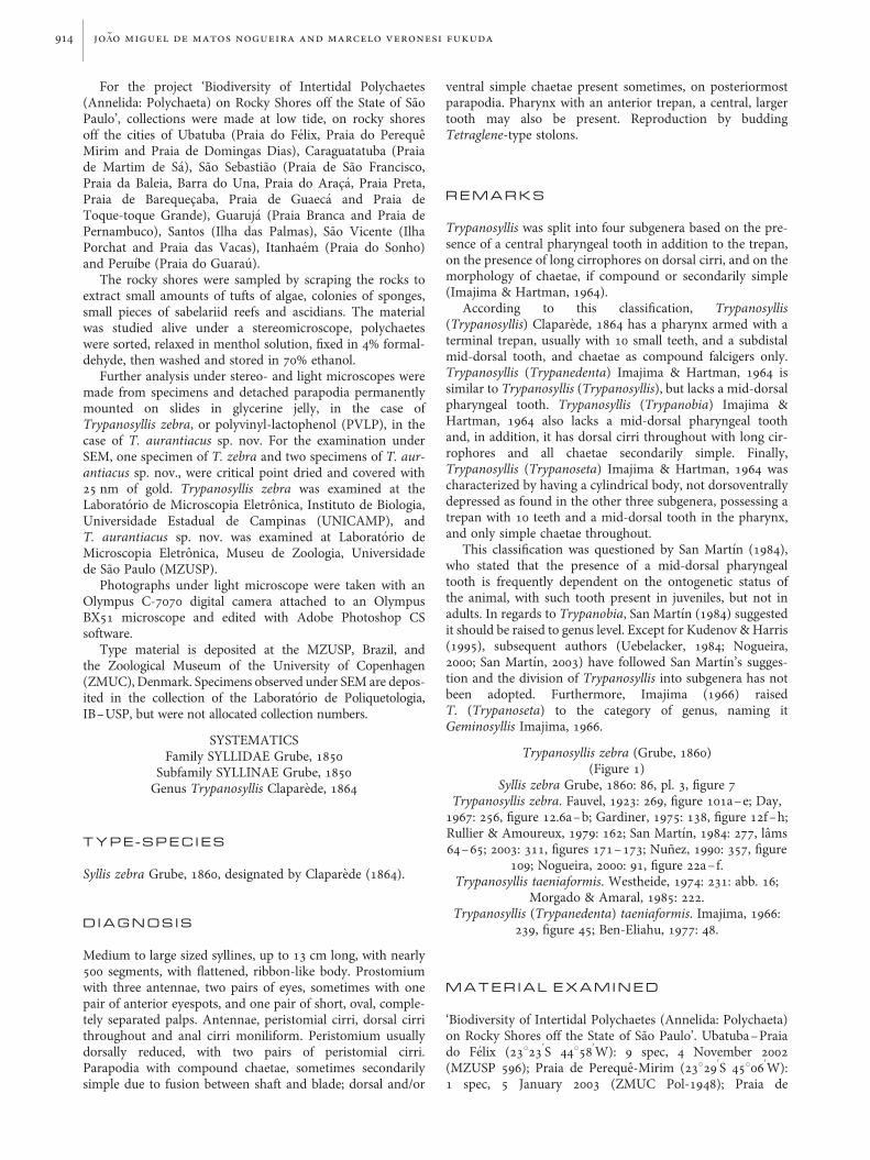
Fig. 2. Trypanosyllis aurantiacus sp. nov. (A–H) holotype (MZUSP 589); (I) paratype 2 (MZUSP 591). (A) Anterior end, dorsal view; (B) falcigers, anteriorparapodium; (C) falcigers, midbody parapodium; (D) falcigers, posterior parapodium; (E) ventral simple chaeta; (F) aciculae, anterior parapodium; (G)aciculae, midbody parapodium; (H) acicula, posterior parapodium; (I) posterior end, dorsal view. Scale bars: A, 500 mm; B–D, F–H, 10 mm; E, 5 mm; I, 150 mm.
a new species of trypanosyllis from brazil 917

dorsoventral gradation in length, measuring ~45–22 mm(Figure 1F–H; Table 1); blades shorter from midbody, withless conspicuous dorsoventral gradation in length, measuring~35–22 mm onmidbody parapodia (Figure 1J; Table 1), ~32–12 mm on posterior parapodia (Figure 1K; Table 1). Dorsaland ventral simple chaetae only present on posteriormostchaetigers, dorsal simple chaetae thin, distally bidentate(Figure 1I), beginning posteriorly to beginning of ventralsimple chaetae; ventral simple chaetae sigmoid, about asthick as shafts of falcigers, distally bidentate, with distaltooth larger than subdistal tooth, similar to blades of falcigers.Anterior parapodia with up to 4 aciculae each, aciculae thick,one of them slightly irregularly curved distally, remaining aci-culae straight, distally sharp; 2–3 aciculae on each midbodyparapodium, 1–2 aciculae on posterior parapodia; longest1–2 aciculae on each parapodium sharper, protruding fromparapodial lobe. Body terminating by one pair of anal cirriwith 13–17 articles (Table 1). Pharynx extending for 6–14
segments, with anterior trepan with about 10 sharp teeth,and, sometimes, a large, triangular, central tooth (Table 1);proventricle extending through 6–12 segments, with ~40rows of muscle cells (Table 1).
R E M A R K S
Trypanosyllis zebra is known to occur throughout the Atlantic,from the English Channel to South Africa and also in theMediterranean. According to San Martın (2003), it is possiblya cosmopolitan species. However, very few descriptions ofspecimens from different localities are available in the litera-ture, making it difficult to make comparisons between speci-mens from different parts of the world and evaluate whetherthis is a single species or a complex of sibling species. Inaddition, the holotype is lost (anonymous referee, personalcommunication).
Fig. 3. Trypanosyllis aurantiacus sp. nov. (A, C) holotype (MZUSP 589); (B) paratype 5 (ZMUC Pol 1943); (D–I) slide mounted detached parapodia, paratype 4(MZUSP 593). (A) Anterior end, dorsal view, slide mounted specimen, arrows point to beginning and ending of proventricle; (B) anterior end, dorsal view, recentlypreserved specimen; (C) posterior end, dorsal view, slide mounted specimen; (D, E), falcigers, anterior parapodium; (F) aciculae, midbody parapodium; (G)midbody parapodia; (H) chaetae and aciculae, midbody parapodium; (I) ventral simple chaetae and inferior falcigers, posterior parapodia. Scale bars: A–C,250 mm; D, 20 mm; E, I, 10 mm; F, 7 mm; G, 50 mm.
918 jo ~Ao miguel de matos nogueira and marcelo veronesi fukuda

Our material matches the description by San Martın (2003)and therefore we consider the Brazilian specimens as belong-ing to the same species as those from Spain.
Trypanosyllis aurantiacus sp. nov.(Figures 2–6)
M A T E R I A L E X A M I N E D
‘Biodiversity of Intertidal Polychaetes (Annelida: Polychaeta)on Rocky Shores off the State of Sao Paulo’. Peruıbe–Praiado Guarau (24822
0
S 478010
W): 23 specs, 5 March 2007.
Type series.Holotype and 4 paratypes deposited at MZUSP, allslide mounted specimens (holotype: MZUSP 589; paratypes:MZUSP 590–593), 3 paratypes deposited at ZMUC (ZMUCPol 1943–1945). Holotype (MZUSP 589): 113 chaetigers,
10 mm long, 0.6 mm wide; paratype 1 (MZUSP 590): 119chaetigers, 9.48 mm long, 0.65 mm wide; paratype 2(MZUSP 591): 66 chaetigers, 5.17 mm long, 0.45 mm wide;paratype 3 (MZUSP 592): 85 chaetigers, 7.25 mm long �0.57 mm wide; paratype 4 (MZUSP 593): 88 chaetigers;~11 mm long, 0.7 mm wide, posteriorly budding stolons;paratype 5 (ZMUC Pol 1943): 71 chaetigers (plus posteriorachaetous zone of ~10 segments), ~8.2 mm long, 0.5 mmwide; paratype 6 (ZMUC Pol 1944): ~110 chaetigers,~14 mm long, 0.9 mm wide, posteriorly budding stolons;paratype 7 (ZMUC Pol 1945): ~100 chaetigers, ~13 mmlong, 0.7 mm wide, posteriorly budding stolons.
D E S C R I P T I O N
Holotype, with 113 chaetigers, measuring 10 mm in length,0.6 mm in width, at proventricular level. In life, body
Fig. 4. Trypanosyllis aurantiacus sp. nov. (A–E) specimen 1; (F–G) specimen 2. (A) Anterior end, dorsal view; (B) detail, anterior end, dorsal view; (C) anteriorend, lateral view (pharynx everted); (D) anterior end, ventral view (pharynx everted); (E) detail, pharynx opening; (F) anterior end, ventral view; (G) anterior end,antero-lateral view. Scale bars: A, 90 mm; B, 30 mm; C, 150 mm; D, E & G, 60 mm; F, 150 mm.
a new species of trypanosyllis from brazil 919

yellow to orange, sometimes with two transverse dark redbars per segment, quickly fading after preservation; anten-nae, peristomial cirri, dorsal cirri throughout and anal cirridark red, due to inclusions arranged as one pair perarticle, darker on longer cirri from midbody (Figures 2A &3A, B, G). Prostomium small, dorsally bilobed, with twopairs of eyes in trapezoidal arrangement, quickly fadingafter preservation, especially in slide mounted specimens;palps shorter than prostomium, kidney-shaped; lateralantennae with ~14 articles, originating frontally, at anteriorborder of prostomium, central antenna longer, with 12–19articles, originating dorsally on prostomium, close toanterior border (Figure 4A–C, F & G; Table 2); nuchalorgans as one posterior row of cilia around each prostomiallobe (Figure 4A,B). Peristomium dorsally reduced; dorsalpair of peristomial cirri with 17–24 articles, ventral pairwith 7–15 articles (Figures 3A, B & 4A, C, F, G; Table 2).In atokous forms, body ventrally smooth, except on
parapodial lobes, which have one row of cilia arranged inirregularly distributed bunches (Figures 4F,G & 5B,C,E),with two transverse ciliated bands per segment dorsally,one near anterior border of segment, other at midlength(Figures 4A,B & 5A,D); cirrophores with an additional rowof cilia posteriorly (Figure 5D). Dorsal cirri throughoutlong, distally pointed, with conspicuous cirrophores, thoseon segment 1 longer than following cirri, with 17–27articles; until beginning of proventricle, dorsal cirri, with~20 articles; from proventricular level, dorsal cirri alternatinglong and short, long cirri with 13–17 articles, short cirri with7–11 articles (Figures 2A; 3A, B, G, 4A, C, G & 5A–E;Table 2). Ventral cirri short, digitiform, slightly exceedingparapodial lobes (Figures 4C,D,F,G & 5B,C,E). Anteriorparapodia with 3–8 compound chaetae each, 3–6 chaetaeper parapodium at midbody, 1–5 chaetae per parapodiumon posterior chaetigers (Table 2). Compound chaetae asbidentate to sub-bidentate falcigers, shafts of unequal
Fig. 5. Trypanosyllis aurantiacus sp. nov. (A–G) specimen 2; (H, I) specimen 1. (A) Midbody segments, dorsal view; (B) midbody segments, ventral view;(C) midbody parapodium, ventral view; (D) cilliation on midbody segments, dorsal view; (E) midbody parapodia, lateral view; (F) falcigers, anteriorparapodium; (G) detail, same parapodium; (H) falciger, midbody parapodium; (I) falciger, posterior parapodium. Scale bars: A, B, 200 mm; C, 50 mm; D,60 mm; E, 90 mm; F, 20 mm; G, 9 mm; H, I, 6 mm.
920 jo ~Ao miguel de matos nogueira and marcelo veronesi fukuda

lengths and thicknesses, with few subdistal spines, bladeswith sub-distal tooth resembling an enlarged spine, withshort spines on cutting edge; blades slightly diminishing inlength towards posterior end, measuring ~30–16 mm onanterior chaetigers, ~30–15 mm on midbody parapodia,~20–10 mm on posterior chaetigers (Figures 2B–D, 3D, E,H, I & 5F–I; Table 2); nearly on all chaetigers, falcigers information present inside parapodial lobes, not protrudingyet (Figure 3D,G). Dorsal simple chaetae absent in all speci-mens examined; ventral simple chaetae present in somespecimens including the holotype, chaetae sigmoid, slightlythinner than shafts of falcigers, sub-bidentate, sub-distaltooth resembling an enlarged spine (Figures 2E & 3I;Table 2). Anterior body with up to 3 aciculae per parapo-dium, midbody parapodia with 2 aciculae each; aciculaethick, distally sharp, tips protruding from parapodial lobes,one of them slightly curved distally; posterior parapodiawith single acicula, with same morphology as on anteriorand midbody chaetigers (Figures 2F–H & 3F–H). Severalspecimens with up to 8 achaetous segments in the posteriorend, followed by large unsegmented zone with chaetigers information (Figures 2I & 3C). Body terminating by one pair ofanal cirri, with ~14 articles (Figures 2I & 3C; Table 2).Pharynx extending for 9–13 chaetigers, with 10 roundedpapillae at anterior end and trepan with 10 small, triangularteeth, central tooth absent (Figure 4D,E); proventriclethrough 7–8 chaetigers, with ~28 rows of muscle cells(Figures 2A & 3A; Table 2).
B I O L O G Y
Six of the specimens examined were budding Tetraglenestolons from the posterior part of the body. Atokous formsbudding up to 9 epitokous simultaneously were observed.Epitokous forms conspicuously ciliated, with one irregularrow of cillia dorsally, arranged in circular bunches(Figure 6B,D), two continuous rows of cilia ventrally, one ofwhich near anterior border of segment, the other at midlength(Figure 6A,C&E). Epitokous forms with 1–3 falcigers perparapodium, notopodial capillary chaetae absent in all speci-mens examined.
R E M A R K S
This species is particularly tricky because several of its charac-ters fade very quickly after preservation, making it difficult toidentify from preserved specimens. Not only body pigmenta-tion, except for dorsal cirri, disappears in preserved material,but also the eyes fade and disappear in a matter of days in slidemounted specimens, the same happening with proventricularrows muscle cells (Figure 3A,B). Therefore, the colour of thespecimens is useful for the identification of this species, butonly on live or freshly collected material; on the other hand,the absence of pigmentation on preserved material, especiallyon slide mounted specimens, is also characteristic, as the
Fig. 6. Trypanosyllis aurantiacus sp. nov. (specimen 2). (A) posterior end, lateral view—three epitokous specimens budding from atokous specimen; (B) largerepitokous specimen, dorsal view; (C) same epitokous specimen, ventral view; (D) same epitokous specimen, detail, dorsal view; (E) same epitokous specimen,posterior end, ventral view. Scale bars: A, 300 mm; B,C, 200 mm; D, 60 mm; E, 40 mm.
a new species of trypanosyllis from brazil 921

species of Trypanosyllis usually have conspicuous dorsalpigmentation.
Trypanosyllis aurantiacus sp. nov., is part of a small groupof species of Trypanosyllis with falcigers with bidentate blades,with subdistal tooth distinctly shorter than distal one, con-dition sometimes referred to as ‘sub-bidentate’. To thisgroup also belong T. aeolis Langerhans, 1879, T. parazebraHartmann-Schroder, 1965, T. savagei Perkins, 1980,Trypanosyllis sp. B sensu Uebelacker, 1984, Trypanosyllis(Trypanosyllis) sp. B sensu Kudenov & Harris, 1995, andT. sanmartini Cinar, 2007.
Trypanosyllis aurantiacus sp. nov., differs from T. aeolis byhaving smaller body, by the absence of rounded papillaeforming two transverse lines on the dorsum of eachsegment, by the presence of ventral simple chaetae, by theabsence of a thinner acicula on parapodial lobes and byhaving a longer pharynx, extending through 9–12 segments,instead of only through 4 segments as in T. aeolis (SanMartın, 2003).
Trypanosyllis parazebra possesses pigmentation as a singletransverse brown bar per segment dorsally, antennae and cirrithroughout with fewer articles (central antenna: 10 articles;lateral antennae: 8–9 articles; peristomial dorsal cirri: 9–11articles; peristomial ventral cirri: 7 articles; dorsal cirri ofanterior parapodia: 9–11 articles; dorsal cirri of posteriorparapodia: 6–7 articles), peristomium not reduced dorsallyas in T. aurantiacus sp. nov., just slightly shorter than follow-ing chaetigers, aciculae straight, needle-like, and anteriorparapodia with 2 aciculae each at most. Finally, T. parazebrahas a large central tooth in the pharynx and smaller proven-tricle, extending through ~4 segments, with 17 rows ofmuscle cells (Hartmann-Schroder, 1965).
Trypanosyllis savagei is a smaller species, less than 5 mmlong and with 80 chaetigers at most. It also differs from ournew species by possessing antennae, tentacular cirri anddorsal cirri throughout with 4–8 articles only, blades of falci-gers ~12 mm long throughout, with smooth shafts, shorteranal cirri, with 3 articles each, and smaller proventricle, occu-pying 3–4 segments, with ~17 rows of muscle cells (Perkins,1980).
Trypanosyllis sp. B sensu Uebelacker, 1984 also has anten-nae, tentacular cirri and dorsal cirri throughout shorter thanT. aurantiacus sp. nov., with 8 articles at most.Furthermore, Trypanosyllis sp. B sensu Uebelacker differsfrom T. aurantiacus sp. nov., by having shorter proventricle,extending through 4–5 segments, with ~21 muscle cellrows, and shorter pharynx, extending for 3–4 segments(Uebelacker, 1984).
Trypanosyllis sp. B sensu Kudenov & Harris, 1995, is distin-guished from T. aurantiacus sp. nov., by possessing a tuft ofcilia on the last article of antennae, peristomial and dorsalcirri, at least on anteriormost chaetigers, according to thefigure provided by Kudenov & Harris (1995: figure 1.30A).Furthermore, Trypanosyllis sp. B sensu Kudenov & Harris,1995 has the superiormost falcigers with nearly smoothblades, becoming progressively more conspicuously spinu-lated inferiorly, blades with somewhat stronger subdistaltooth, according to the figures provided (Kudenov & Harris,1995: figure 1.30F–H), and aciculae with hook-like subdistalcollar (Kudenov & Harris 1995: figure 1.30J–K).
Finally, a recently described species from Turkey, easternMediterranean, is the most similar species to T. aurantiacussp. nov. Trypanosyllis sanmartini presents pattern ofT
able2.
Variation
amon
gtheslidemou
nted
specim
ensof
thetype-seriesof
Trypanosyllisa
uran
tiacus
sp.nov.(‘?’w
asattributed
whenthecond
itionof
thespecim
endidno
tallowto
seethatcharacter;‘–’w
asattributed
whenthat
particular
structurewas
absent
inthat
specim
en).
HolotypeMZUSP
589
Paratype1MZUSP
590
Paratype2MZUSP
591
Paratype3MZUSP
592
Totallength�
width
atproventricle(m
m);nu
mberof
chaetigers
10.06�
0.6;113
9.48�
0.65;119
5.17�
0.45;66
7.25�
0.57;85
Length
ofph
arynx(num
berof
chaetigers)
10.5
129
11Length
ofproventricle(chaetigers);n
umberof
rowsmusclecells
7;28
9;28
–29
6;29
6;?
Ventralsimplechaeta
beginn
ingfrom
(chaetiger)
9494
4856
Num
berof
articles
ofcentral�
lateralantennae
12�
13–14
16�
1514�
1019�
13Num
berof
articles
ofdo
rsal�
ventralp
eristomialcirri
24�
1122�
917
–19�
722�
9Num
berof
articles
ofdo
rsalcirrion
chaetiger1,long
dorsalcirri
througho
ut,sho
rtdo
rsalcirrithrou
ghou
t,andanalcirri
27,13–15,8
–11,1
521,1
4–17,7
–9,14
17,13–15,7
–9,12
26,1
5–17,7
–9,
–
Num
berof
falcigersperparapo
dium
onanterior,m
idbody,and
posteriorchaetigers
4–6,4,1–3
6–8,5–6,2–5
3–5,3–4,1–3
5–7,5–6,1–3
Length
ofblades
offalcigerson
anterior,m
idbody,and
posterior
chaetigers
(mm)
26–19,30–23,20–18
30–22,28–17,20–18
23–16,20–15,1
5–10
25–16,25–15,20–12
Num
berof
posteriorachaetou
ssegm
ents
86
75
922 jo ~Ao miguel de matos nogueira and marcelo veronesi fukuda

pigmentation, distribution of chaetae throughout, mor-phology of pharynx and proventricle, and stolonization verysimilar to T. aurantiacus sp. nov. However, it differs from T.aurantiacus sp. nov., in having each anterior segment with 2rows of papillae on dorsum, more articles on antennae andcirri throughout (central antenna: 28 articles; lateral antennae:20 articles; peristomial dorsal cirri: 30 articles; peristomialventral cirri: 20 articles; anterior dorsal cirri: 18–38 articles;posterior dorsal cirri: 12–26 articles), cirrophores with papil-lae but without cilia, longer blades of falcigers, measuring~40–27.5 mm on anterior body, ~42.5–30 mm on midbody,and ~40–25 mm on posterior chaetigers, and pharynx andproventricle densely pigmented (Cinar, 2007). As theBrazilian specimens studied for this paper were adults,many of them budding Tetraglene stolons, differences in sizecannot be attributed to different ontogenetic stages. Inaddition to the geographical distribution, we consider thosedifferences enough to describe T. aurantiacus sp. nov., as anew species.
E T Y M O L O G Y
The specific name aurantiacus is attributed to this species dueto the colour of specimens in life (aurantiacus ¼ Latin for‘orange colour’).
A C K N O W L E D G E M E N T S
This study was funded by FAPESP—Fundacao de Amparo aPesquisa no Estado de Sao Paulo (proc. 04/02774-4). Inaddition, J.M.M.N. receives a productivity fellowship fromCNPq—Conselho Nacional de Desenvolvimento Cientıfico eTecnologico and M.V.F. receives a PhD fellowship fromFAPESP (proc. 07/53040-9). We are grateful to CecıliaAmaral, coordinator of the project ‘BIOTA/FAPESP/BenthicMarine Biodiversity’, for all of her support; to the team ofthe project ‘BIOTA/FAPESP/Benthic Marine Biodiversity’,for collecting part of the material; to Nilcea AparecidaVeronesi, for providing accommodation in Praia Grande.We are also grateful to Professor Sergio Antonio Vanin,from Departamento de Zoologia, IB–USP, for the help withLatin adjectives; to Enio Mattos and Eduardo Mattos, fromDepartamento de Zoologia, IB–USP, for preparing the speci-mens for the study under SEM; and to Lara Guimaraes, fromLaboratorio de Microscopia Eletronica, MZUSP, and AdrianeSprogis and Antonia Lima, from Laboratorio de MicroscopiaEletronica, IB–UNICAMP for the SEMs included in thepresent paper.
R E F E R E N C E S
Aguado M.T., San Martın G. and ten Hove H.A. (2008) Syllidae(Annelida: Polychaeta) from Indonesia collected by the Siboga(1899–1900) and Snelius II (1984) expeditions. Zootaxa 1673, 1–48.
Amaral A.C.Z. (2001) Benthic marine biodiversity on the State of SaoPaulo. Available at: http://www.ib.unicamp.br/projbiota/bentos_marinho/index.htm Acessed 22 January 2008.
Attolini F.S. (1997) Composicao e distribuicao dos anelıdeos poliquetas naplataforma continental da regiao da Baıa de Campos, RJ, Brasil. Msc
dissertation, Instituto Oceanografico, Universidade de Sao Paulo, SaoPaulo, Brazil.
Ben-Eliahu M.N. (1977) Polychaete cryptofauna from rims of similarintertidial vermetid reefs on the Mediterranean coast of Israel and inthe Gulf of Elat: Syllinae and Eusyllinae (Polychaeta errantia:Syllidae). Israel Journal of Zoology 26, 1–58.
Cinar M.E. (2007) A new species of Trypanosyllis (Polychaeta: Syllidae)from the Levantine coast of Turkey (eastern Mediterranean). Journalof the Marine Biological Association of the United Kingdom 87,451–457.
Claparede E. (1864) Glanures zootomiques parmi les annelides dePor-Vendres (Pyrenees Orientales). Memoires de la Societe dePhysique et d’Histoire Naturelle de Geneve 17, 463–600.
Day J.H. (1967) A monograph on the polychaeta of southern Africa. Part. 1.Errantia. London: Trustees of the British Museum (Natural History).458pp.
Duarte L.F.L. and Nalesso R.C. (1996) The sponge Zygomycale parishii(Bowerbank) and its endobiotic fauna. Estuarine, Coastal and ShelfScience 42, 139–151.
Fauvel P. (1923) Polychetes errantes. In Faune de France, vol. 5. Paris: LeChevallier. 488pp.
Gardiner S.L. (1975) Errant polychaete annelids from North Carolina.Journal of the Elisha Mitchell Scientific Society 91, 77–220.
Grube A.E. (1860) Beschreibung neuer oder wenig bekannter Anneliden.Funter Beitrag. Archiv fur Naturgeschichte 26, 71–118.
Hartmann-Schroder G. (1965) Zur Kenntnis des Sublitorals der chile-nischen Kusten, unter besonder Berucksichtigung der Polychaetenund Ostracoden. Teil 2: Die Polychaeten des Sublitorals.Mitteilungen aus dem Hamburgischen Zoologischen Museum undInstitut 62, 59–305.
Imajima M. (1966) The Syllidae (polychaetous annelids) from Japan IV.Syllinae (1). Publications of the Seto Marine Biological Laboratory14, 219–252.
Imajima M. and Hartman O. (1964) The polychaetous annelids of Japan.Part I. Allan Hancock Foundation Publications, Occasional Papers no.26, 237 pp.
Kudenov J. and Harris L. (1995) Family Syllidae Grube, 1850. In Blake J.,Hilbig B. and Scott P.H. (eds.) Taxonomic atlas of the benthic fauna ofSanta Maria Basin and Western Santa Barbara Channel, volume 5.Santa Barbara: Santa Barbara Museum of Natural History, pp. 1–97.
Morgado E.H. and Amaral A.C.Z. (1985) Anelıdeos poliquetos associa-dos ao briozoario Schizoporella unicornis (Johnston). V. Syllidae.Revista Brasileira de Zoologia 3, 219–227.
Nogueira J.M.M. (2000) Anelıdeos poliquetas associados ao coralMussismilia hispida (Verrill, 1868) em ilhas do litoral do Estado deSao Paulo. Phyllodocida, Amphinomida, Eunicida, Spionida,Terebellida, Sabellida. PhD thesis, Instituto de Biociencias,Universidade de Sao Paulo, Sao Paulo, Brazil.
Nunez J. (1990) Anelidos Poliquetos de Canarias: Estudio sistematico de losordenes Phyllodocida, Amphinomida y Eunicida. PhD thesis,Departamento de Biologıa Animal (Zoologıa), Universidad de LaLaguna, La Laguna, Spain.
Paiva P.C. (2006) Filo Annelida. Classe Polychaeta. In Lavrado H.P. andIgnacio B.L. (eds.) Biodiversidade bentonica da regiao central da ZonaEconomica Exclusiva Brasileira. Rio de Janeiro: Museu Nacional (SerieLivros no. 18), pp. 261–298.
Perkins T. (1980) Syllidae (Polychaeta), principally from Florida, withdescriptions of a new genus and twenty-one new species.Proceedings of the Biological Society of Washington 93, 1080–1172.
a new species of trypanosyllis from brazil 923

Rullier F. and Amoureux I. (1979) Annelides Polychetes. Annales del’Institut Oceanographique 55 (supplement), 145–206.
San Martın G. (1984) Estudio biogeografico, faunıstico y sistematico delos poliquetos de la famılia sılidos (Syllidae: Polychaeta) en Baleares.Editorial de la Universidad Complutense de Madrid, no. 187, 529 pp.
San Martın G. (2003) Annelida, Polychaeta II: Syllidae. In Ramos M.A.et al. Fauna Iberica, volume 21. Madrid: Museo Nacional deCiencias Naturales, CSIC, 554 pp.
Uebelacker J.M. (1984) Family Syllidae Grube, 1850. In Uebelacker J.M.and Johnson P.G. (eds.) Taxonomic guide to the polychaetes of thenorthern Gulf of Mexico, volume 4. Mobile: Barry A. Vittor &Associates, Inc, pp. 30-1–30-151.
and
Westheide W. (1974) Interstitielle Fauna von Galapagos. XI. Pisionidae,Hesionidae, Pilargidae, Syllidae (Polychaeta). Mikrofauna desMeersbodens 44, 195–338.
Correspondence should be addressed to:Joao Miguel de Matos NogueiraLaboratorio de Poliquetologia, Departamento de ZoologiaInstituto de Biociencias, Universidade de Sao PauloR. do Matao, travessa 14, n. 101, 05508-900, Sao Paulo, SPBrazilemail: [email protected]; [email protected]
924 jo ~Ao miguel de matos nogueira and marcelo veronesi fukuda