Shortcomings of short hairpin RNA-based transgenic RNA interference in mouse oocytes

A New Noncoding RNA Arranges Bacterial Chromosome Organization
Zhong Qian,a Mirjana Macvanin,a Emilios K. Dimitriadis,b Ximiao He,c Victor Zhurkin,d Sankar Adhyaa
Laboratory of Molecular Biology, National Cancer Institute,a Biomedical Engineering and Physical Science, National Institute of Biomedical Imaging and Bioengineering,b
Laboratory of Metabolism, National Cancer Institute,c and Laboratory of Cell Biology, National Cancer Institute,d National Institutes of Health, Bethesda, Maryland, USA
ABSTRACT Repeated extragenic palindromes (REPs) in the enterobacterial genomes are usually composed of individual palin-dromic units separated by linker sequences. A total of 355 annotated REPs are distributed along the Escherichia coli genome.RNA sequence (RNAseq) analysis showed that almost 80% of the REPs in E. coli are transcribed. The DNA sequence of REP325
showed that it is a cluster of six repeats, each with two palindromic units capable of forming cruciform structures in supercoiledDNA. Here, we report that components of the REP325 element and at least one of its RNA products play a role in bacterial nucle-oid DNA condensation. These RNA not only are present in the purified nucleoid but bind to the bacterial nucleoid-associatedHU protein as revealed by RNA IP followed by microarray analysis (RIP-Chip) assays. Deletion of REP325 resulted in a dramaticincrease of the nucleoid size as observed using transmission electron microscopy (TEM), and expression of one of the REP325
RNAs, nucleoid-associated noncoding RNA 4 (naRNA4), from a plasmid restored the wild-type condensed structure. Indepen-dently, chromosome conformation capture (3C) analysis demonstrated physical connections among various REP elementsaround the chromosome. These connections are dependent in some way upon the presence of HU and the REP325 element; dele-tion of HU genes and/or the REP325 element removed the connections. Finally, naRNA4 together with HU condensed DNA invitro by connecting REP325 or other DNA sequences that contain cruciform structures in a pairwise manner as observed byatomic force microscopy (AFM). On the basis of our results, we propose molecular models to explain connections of remote cru-ciform structures mediated by HU and naRNA4.
IMPORTANCE Nucleoid organization in bacteria is being studied extensively, and several models have been proposed. However,the molecular nature of the structural organization is not well understood. Here we characterized the role of a novel nucleoid-associated noncoding RNA, naRNA4, in nucleoid structures both in vivo and in vitro. We propose models to explain hownaRNA4 together with nucleoid-associated protein HU connects remote DNA elements for nucleoid condensation. We presentthe first evidence of a noncoding RNA together with a nucleoid-associated protein directly condensing nucleoid DNA.
Received 8 July 2015 Accepted 3 August 2015 Published 25 August 2015
Citation Qian Z, Macvanin M, Dimitriadis EK, He X, Zhurkin V, Adhya S. 2015. A new noncoding RNA arranges bacterial chromosome organization. mBio 6(4):e00998-15. doi:10.1128/mBio.00998-15.
Editor Bonnie Bassler, Princeton University
Copyright © 2015 Qian et al. This is an open-access article distributed under the terms of the Creative Commons Attribution-Noncommercial-ShareAlike 3.0 Unported license,which permits unrestricted noncommercial use, distribution, and reproduction in any medium, provided the original author and source are credited.
Address correspondence toSankar Adhya, [email protected].
This article is a direct contribution from a Fellow of the American Academy of Microbiology.
Noncoding RNAs (ncRNAs) present in both prokaryotic andeukaryotic cells do not function as mRNA, tRNA, or rRNA
(1). Although many ncRNAs of different sizes and different func-tions have been widely reported (2–6), new ncRNAs with newfunctions are still being discovered. Recently, we discovered anovel ncRNA, transcribed from a specific repeated extragenic pal-indromic element, REP325, in the chromosome of Escherichia coliby RNA IP followed by microarray analysis (RIP-Chip) assays ofthe nucleoid-associated HU protein (7). In this paper, we termedit as nucleoid-associated ncRNA (naRNA). REP elements in theenterobacterial genomes, first reported 30 years ago, contain in-dividual palindromes separated by linkers (8–10). The functionsof the REPs have been speculated to be related to transcriptiontermination signals, binding sites for proteins, cleavage sites forDNA gyrase, and, possibly, manipulation of nucleoid structures(11–14). In this study, we investigated potential functions of theREP325 element, which is located between genes yjdM (phnA) andyjdN (phnB), and its RNA products. REP325 contains six homolo-
gous units (Fig. 1A). Each repeat is composed of two palindromiccruciform-generating motifs, Y and Z2, connected by a shortlinker, l. Cells deleted for the REP325 segment and/or hup genesencoding the nucleoid-associated HU protein showed a decon-densed nucleoid structure, suggesting that these two factors par-ticipate in nucleoid condensation (7). RNA sequencing (RNAseq)analysis and nucleoid RNA tiling array clearly showed the exis-tence of RNA species transcribed from each unit of REP325, namednaRNA1 to naRNA6 (Fig. 1A). Multialignment of DNA sequencesof the 6 repeats in REP325 showed high homology (Fig. 1B). EachnaRNA contains two potential hairpins, corresponding to Y andZ2 motifs (Fig. 1C). It is unknown whether these six RNAs aretranscribed independently or are the result of processing of alarger RNA transcribed from a common promoter (the promoterof the upstream gene, yjdM). One of these RNAs is naRNA4,which binds two dimeric forms of HU, HU�� and HU��. In thisreport, we show that the expression of naRNA4 from a plasmidrestores the decondensed morphology of the nucleoid caused by
RESEARCH ARTICLE crossmark
July/August 2015 Volume 6 Issue 4 e00998-15 ® mbio.asm.org 1
m
bio.asm.org
on Septem
ber 9, 2015 - Published by
mbio.asm
.orgD
ownloaded from

REP325 deletion. We also show intersegmental connections in vivobetween different remote cruciform-containing DNA structureslike those present in REP elements; the connections are affected bydeletion of hup genes encoding HU subunits and/or of REP325. Wealso demonstrate the connections between cruciforms in DNA invitro in the presence of naRNA4 and HU. We propose that theseconnections are a major part of the cellular nucleoid architectureand help its condensation.
RESULTSThe existence of nucleoid-associated RNA: tiling array andRNAseq analysis. Ohniwa et al. showed that the E. coli nucleoid isa 40-nm-thick fibrous structure as observed by atomic force mi-croscopy (AFM); the fibers assume 10-nm-thick structures in cellsdevoid of the nucleoid-associated protein (NAP) HU (15). Theyalso showed the existence of 10-nm-thick nucleoid fibers afterRNase treatment, suggesting a role of some RNA and HU in nu-cleoid architecture. Pettijohn and Hecht also suggested that theE. coli nucleoid contains RNAs which are important for the struc-tural integrity of the nucleoid (16). We isolated RNA from theE. coli nucleoid, which was purified from cells cultured in minimalmedium, and identified structural elements by the use of a DNAtiling array. It is clear that the nucleoid RNA contains fragments ofrRNAs, tRNAs, a few mRNAs, and many ncRNAs, many of whichare also present among HU binding RNAs (7). It is noticeable thatmore than 30 ncRNAs are transcribed from the REP elements (seeText S1 in the supplemental material).
In order to analyze the transcription profile of the REP ele-ments in E. coli, we analyzed RNAseq data that was previously
published and deposited in the NCBI Sequence Read Archive (17).According to the gene annotation of E. coli MG1655, there are atotal of 355 REP elements, 152 of which are transcribed in cellscultured in defined minimal medium (see Text S3 andText S4 in the supplemental material). In our previous study, twooverlapping RNA reads, which were part of transcripts fromREP325 and identified in RIP-Chip assays of HU protein, wereassumed to originate from a single RNA, named nc5 RNA (7).Exploring the RNAseq data with unique alignments revealed RNAsequences matched to six repeated structures (Y-l-Z2) in theREP325 (see Fig. S1A in the supplemental material). It appears togenerate six RNA species, now named naRNA1 to naRNA6(Fig. 1A). The DNA repeats are connected by five unknown motifswith identical sequences (named U motifs), which are also tran-scribed. We have not determined if the entire REP325 segment iscotranscribed and processed into six naRNAs. But the abundanceof matched readings suggests a potential direction of transcriptionof the entire REP325 from the promoter of the upstream yjdM(phnA) gene to the position upstream of the yjdN (phnB) gene.Furthermore, sequence analysis of the unmatched gaps for theREP325 segment showed high sequence similarity between the re-peats. Multisequence alignments of DNA sequences encoding thenaRNA1 gene to the naRNA6 gene indicated that these six repeatsshare high sequence homology and that the sequence of thenaRNA4 gene is identical to that of the naRNA2 gene (Fig. 1B).Multiple-sequence alignments also showed that the entire REP325
is transcribed (see Fig. S1B). RNA secondary structure analysisconfirmed that RNAs from the repeats have similar structures and
FIG 1 Description of the REP325 element. (A) Genetic map of the REP325 element. REP325, located in the intergenic region of yjdM and yjdN, is composed of 6highly homologous repeats separated by 5 unknown spacers with exactly the same DNA sequence (U in blue). The transcripts from the 6 repeats are namednaRNA1 to naRNA6, as shown. Each unit is 77 bp long and contains two different palindromes (Y and Z2 in diamonds) separated by a constant linker (l in black).Each naRNA contains a Z2 motif and a Y motif, connected by a linker (l). (B) Multisequence alignment analysis of the 6 repeats in REP325. Each repeat containsa Y motif and a Z2 motif connected by the linker l (boxed). Analysis was done by ClustalW. The corresponding RNA products are marked on the left. (C) Typicalsecondary RNA structure of an naRNA (naRNA4) as predicted by Mfold with modifications.
Qian et al.
2 ® mbio.asm.org July/August 2015 Volume 6 Issue 4 e00998-15
m
bio.asm.org
on Septem
ber 9, 2015 - Published by
mbio.asm
.orgD
ownloaded from

contain the Y and Z2 potential hairpins. The typical structure ofnaRNA4 is shown in Fig. 1C.
Decondensation of nucleoid in vivo: TEM analysis. Trans-mission electron microscopy (TEM) observations previouslyshowed that deletion mutants of HU genes (�hupA �hupB strain)and/or of the REP325 element (�REP325 gene strain) decondensedthe E. coli nucleoid in both growing and nongrowing cells com-pared to a compacted nucleoid observed in the wild-type strainunder similar conditions, suggesting that HU and part or all ofREP325 DNA and/or its RNA product affect nucleoid architecture(7). We extended the TEM observations further by investigatingthe details of REP325 participation in the nucleoid structure. Wefirst confirmed that in growing cells, compared to wild-type re-sults, deletion of HU genes or of REP325 decondensed the nucleoidsize (Fig. 2A; nucleoids are outlined in red). Moreover, the REP325
deletion strain carrying a plasmid vector showed no change indecondensed nucleoid morphology. But expression of naRNA4from the plasmid reproducibly condensed the nucleoid. We ob-served some overcondensation that was most likely due to over-expression of naRNA4. The expression of an unrelated RNA fromthe same plasmid had no effect on the decondensed nucleoid. Wealso tested the effect of expression of derivatives of naRNA4 con-taining only a Z2 or Y motif in the same REP325-deleted cells. Theexpression of RNA containing only the Z2 or Y motif alone, unlikethat of the intact naRNA4, did not restore the nucleoid morphol-
ogy to wild type, although we do not know anything about therelative stability of naRNA4 or its truncated derivatives under theconditions of the experiments. TEM analysis performed with non-growing cells in the same set of strains gave identical results; onlythe presence of an intact naRNA4 caused nucleoid condensation(Fig. 2B). We note that, in the absence of any simple way to quan-tify the nucleoid volume in TEM observations, we estimated thesize of the nucleoid in two-dimensional (2-D) analysis of the thinsections (shown by red outlines in Fig. 2). Although these obser-vations show the involvement of HU and naRNA4 in nucleoidcondensation, a direct participation of any part of the REP325 ele-ment in the process is not apparent. If DNA is involved in thecondensation, as seems very likely, other DNA sequences homol-ogous to REP325 may fulfill the same role. In summary, this is firstdemonstration of a direct involvement of an ncRNA in DNA con-densation at the molecular level.
Intersegmental chromosomal interactions in vivo: 3C anal-ysis. We hypothesized that one plausible mechanism of nucleoidstructural organization is that of facilitating contacts between REPelements around the chromosome by naRNA4 and the nucleoidprotein HU. To test the idea, we employed the chromosome con-formation capture (3C) approach, the use of which has been es-tablished in studies of distal intrachromosomal interactions invivo in both eukaryotes and prokaryotes (18–20). We designedprimers for 23 randomly selected REP segments, including
FIG 2 TEM analysis of nucleoid morphology of cells in log phase (A) or stationary phase (B). E. coli MG1655 is used as the wild-type (WT) strain. Deletions ofHU genes were constructed by replacement of hupA and hupB by ampicillin (Amp) and chloramphenicol (Cm) antibiotic cassettes. The �REP325 mutant wasconstructed by replacement of the wild-type allele by a sac-Cat cassette. The construction of the plasmids is described in Materials and Methods. The plasmidswere transformed into the �REP325 strain, resulting in the following mutant strains: the �REP325/pNM12 mutant, the �REP325/pnaR4 mutant, the �REP325/pconRNA mutant, the �REP325/pYRNA mutant, and the �REP325/pZ2RNA mutant. Nucleoids are identified in the EM pictures by red outlines. Scale bars,500 nm. The apparent differences in cell sizes are the result of angular variations that occurred during cell slicing. Only representative TEM images are shown.
Noncoding RNA and Nucleoid
July/August 2015 Volume 6 Issue 4 e00998-15 ® mbio.asm.org 3
m
bio.asm.org
on Septem
ber 9, 2015 - Published by
mbio.asm
.orgD
ownloaded from
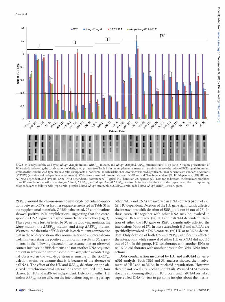
REP325, around the chromosome to investigate potential connec-tions between REP sites (primer sequences are listed in Table S1 inthe supplemental material). Of 253 pairs tested, 27 combinationsshowed positive PCR amplifications, suggesting that the corre-sponding DNA segments may be connected to each other (Fig. 3).These pairs were further tested by 3C in the following mutants: the�hup mutant, the �REP325 mutant, and �hup �REP325 mutant.We measured the ratio of PCR signals in each mutant compared tothat in the wild-type strain after normalization to an internal con-trol. In interpreting the positive amplification results in 3C exper-iments in the following discussion, we assume that an observedcontact involves the REP elements and not another DNA sequencepresent nearby in the chromosome. Similarly, when a contact sig-nal observed in the wild-type strain is missing in the �REP325
deletion strain, we assume that it is because of the absence ofnaRNA4. The effect of the HU and REP325 deletions on the ob-served intrachromosomal interactions were grouped into fourclasses. (i) HU and naRNA4 independent. Deletion of either HUand/or REP325 has no effect on the interactions suggesting perhaps
other NAPs and RNAs are involved in DNA contacts (4 out of 27).(ii) HU dependent. Deletion of the HU gene significantly affectedthe interactions while deletion of REP325 did not (6 out of 27). Inthese cases, HU together with other RNA may be involved inbringing DNA contacts. (iii) HU and naRNA4 dependent. Dele-tion of either the HU gene or REP325 significantly affected theinteractions (4 out of 27). In these cases, both HU and naRNA4 arespecifically involved in DNA contacts. (iv) HU or naRNA4 depen-dent. Only deletion of both HU and REP325 significantly affectedthe interactions while removal of either HU or RNA4 did not (13out of 27). In this group, HU collaborates with another RNA ornaRNA4 collaborates with another protein for DNA-DNA inter-actions.
DNA condensation mediated by HU and naRNA4 in vitro:AFM analysis. Both TEM and 3C analyses showed the involve-ment of HU and naRNA4 in nucleoid organization. However,they did not reveal any mechanistic details. We used AFM to mon-itor any condensing effects of HU protein and naRNA4 on nakedsupercoiled DNA in vitro to get some insights about the mecha-
FIG 3 3C analysis of the wild-type, �hupA �hupB mutant, �REP325 mutant, and �hupA �hupB �REP325 mutant strains. (Top panel) Graphic presentation of3C x-axis data showing the combinations of designated primers (see Table S1 in the supplemental material). y-axis data show the ratios of PCR signals in mutantstrains to those in the wild-type strain. A ratio change of 0.4 (horizontal solid black line) or lower is considered significant. Error bars indicate standard deviations(STDEV) (n � 4 sets of independent experiments). 3C data were grouped into four classes: (I) HU and naRNA4 independent, (II) HU dependent, (III) HU andnaRNA4 dependent, and (IV) HU or naRNA4 dependent. (Bottom panel) Typical PCR bands on 2% agarose gel. From top to bottom, the bands are amplifiedfrom 3C samples of the wild-type, �hupA �hupB, �REP325, and �hupA �hupB �REP325 strains. As indicated at the top of the upper panel, the correspondingcolor codes are as follows: wild-type strain, purple; �hupA �hupB strain, blue; �REP325 strain, red; �hupA �hupB �REP325 strain, green.
Qian et al.
4 ® mbio.asm.org July/August 2015 Volume 6 Issue 4 e00998-15
m
bio.asm.org
on Septem
ber 9, 2015 - Published by
mbio.asm
.orgD
ownloaded from

nism(s) of their action. A plasmid containing one REP325
(pQZ080) was used as the template for AFM (Fig. 4). The additionof naRNA4 or of different HU dimers to the plasmid did notnoticeably change its supercoiled morphology. The absence of anyeffect of HU in this experiment is consistent with previous reports(15, 21). However, the presence of either HU�� or HU�� to-gether with naRNA4 dramatically condensed the DNA, appar-ently because of the presence of multiple intersegmental contacts,thus demonstrating that naRNA4 collaborates with HU in con-densing DNA (Fig. 4A). Note that either HU�� or HU�� dimerworks but not HU�� dimer (��, ��, and ��). Since the plasmidDNA contained only one REP325 element, the multiple contactsvery likely involve either nonspecific DNA binding or some othersequences in the plasmid that allow DNA contacts. The REP325
palindromic repeats generate cruciform structures in a super-coiled state. We believe that several transcription terminators (22)that are present in the plasmid and which also generate cruciformstructures perform the role of REP325. Consistent with this idea,the addition of HU and naRNA4 to the parental plasmid (pSA508)containing no authentic REP element but several transcriptionterminators also resulted in DNA condensation (Fig. 4B). To in-vestigate the features of naRNA4 required in DNA condensation,
we first tested whether an intact naRNA4 is needed to condenseDNA. We performed AFM analysis with three 77-nucleotide (nt)RNAs: a nonspecific control RNA, an RNA containing only the Ymotif, and an RNA containing only the Z2 motif (see Table S2 inthe supplemental material). Compared to naRNA4, none of theseRNAs condensed DNA (Fig. 4B), which is in agreement with theTEM data (Fig. 2). We also asked whether the exact sequences oronly hairpin features of Z2 and Y motifs are involved. We synthe-sized a 77-nt-long RNA whose sequence was the exact comple-ment of naRNA4. This anti-naRNA4 molecule has an RNA se-quence completely different from that of naRNA4 but shouldcontain two hairpin structures. Figure 4B shows that the anti-naRNA4 also condensed DNA in the presence of HU protein,indicating that it is the secondary structure of naRNA4 and not thesequence of the RNA itself that is important in DNA condensa-tion.
Because of the large sizes of pQZ080 and pSA508 (3.9 kb and3.5 kb, respectively), we could not discern the precise organizationof the DNA contact points involved in the observed DNA conden-sation. To simplify the condensed DNA structure, we tested anumber of minicircle DNAs which contain 0, 1, or 2 potentialcondensation sites (cruciform structures originated from REP325
FIG 4 AFM images of plasmid DNA with HU and naRNA4. The components added for reactions are indicated below each AFM image. (A) AFM images oflarge-plasmid DNA containing the cloned REP325 element (pQZ080) in the original pSA508 plasmid vector (19) in the presence of different HU and naRNA4molecules. (B) AFM images of the DNA vector plasmid (pSA508) without the REP325 clone in the presence and absence of HU and naRNA4. (C) Minicircle DNA.(Top panel) Schematic representation of each minicircle DNA used. A red dot indicates the insertion site of one REP325 repeat; a green dot indicates the rpoCtranscription terminator that contains a palindrome. Both of the DNA elements are proven binding sites of HU (21). The lengths of expected sizes of the DNAloops are shown below each DNA molecule representation. (Bottom panel) AFM images of minicircle DNA in different combinations of HU and naRNA4. A redarrow indicates the looping complex. Only representative AFM images are shown.
Noncoding RNA and Nucleoid
July/August 2015 Volume 6 Issue 4 e00998-15 ® mbio.asm.org 5
m
bio.asm.org
on Septem
ber 9, 2015 - Published by
mbio.asm
.orgD
ownloaded from

or one of the transcription terminators, rpoC, present in the pa-rental plasmid, pSA508) at marked positions: mini103, mini104,mini105, mini106, mini107, and mini120 (Fig. 4C; see also Fig. S2in the supplemental material). Any looping generated using themarked cruciform sites can be discerned by measuring the size ofthe DNA loops between contact points. As elaborated below, con-sistent with our proposal, HU- and RNA4-mediated DNA con-densation in vitro requires only a cruciform structure in DNA andnot other parts of the multipalindromic unit and the presence ofboth HU and naRNA4 on minicircle DNAs (Fig. 4C). “Figure 8”structures, which are caused by either random crossover of DNAor specific bridging of DNA due to the presence of HU andnaRNA4, were observed in all minicircle DNAs. To determinewhether the looping was random or specific, we measured eachloop from the figure 8 structures in the minicircle DNAs in thepresence of naRNA4 and HU protein. We observed that the fre-quency of figure 8 structures with the expected loop sizes resultingfrom interactions between any two marked cruciform structureswas significantly higher in all minicircle DNAs except in mini103,which has no cruciform structure. For example, we found 79 fig-ure 8 structures in mini103 in the presence of naRNA4 and HU,and none was found with an expected loop size. In mini106,mini107, and mini120 containing two cruciforms, the ratios of thenumbers of observed figure 8 structures with expected loop sizesto the total numbers of counted structures were 24/49, 48/101, and28/53, respectively. These results suggest that HU and naRNA4form bridges between two cruciforms present in a minicircleDNA.
For mini104 and mini105 plasmids, which contain one rpoCtranscription terminator of pSA508 and one REP325 repeat, re-spectively, we observed only random figure 8 structures and, oc-casionally, two or more minicircle DNAs bridged together by acomplex core (red arrows in Fig. S2 in the supplemental material),suggesting that two cruciform structures present in different DNAmolecules can connect.
DISCUSSION
It has become clear that the organization of the chromosome inE. coli is not random. The chromosome is not merely a disorderedaggregate of randomly coiled DNA. Instead, it is a dynamic butspatially organized defined entity that undergoes strictly con-trolled and reproducible changes when they are needed (23).Structural models of elements such as “macro domains,” “super-coiled topological loops,” “filaments,” and “remote connections”are suggested to represent structural constituents of chromo-somes from observations using different approaches (19, 24–27).A number of NAPs, such as the HU, Fis, IHF, H-NS, and SMCproteins, modulate chromosome structure. We focused on HU,which binds to DNA nonspecifically but prefers distorted DNAstructures such as nicks, gaps, bends, and cruciforms (28, 29). Dueto its high abundance and growth-phase-dependent subunit com-positions (HU��, HU��, and HU��), HU is believed to modu-late chromosome structure in accordance with the growth phaseof the cell (30). We confirmed that HU binds to naRNA4 and toseveral other RNAs by electrophoretic mobility shift assay(EMSA) (see Fig. S3 in the supplemental material), but not allHU-RNA bindings could help DNA condensation both in vivo(Fig. 2) and in vitro but bound to those which contained twohairpin structures (Fig. 4). Thus, an HU-naRNA4 interaction maybe somewhat unusual and specific; the presence of at least two
hairpin motifs, such as Z2 and Y, in the RNA is needed for DNAcondensation. We conclude that two cruciform structures inDNA, not yet completely defined, interact with each other in apairwise fashion for DNA condensation, which needs both HUand naRNA4. We propose four mechanisms for interactions be-tween two DNA cruciforms mediated by HU and naRNA4(Fig. 5). (i) For DNA-naRNA-HU-naRNA-DNA interactions,each cruciform structure binds to one hairpin of naRNA and twoDNA-bound RNAs are bridged together by an HU dimer usingthe other hairpins of the two naRNAs (Fig. 5A). (ii) For DNA-naRNA(HU)-DNA interactions, the model is similar to model i,but the stoichiometry of HU and naRNA in the complex is 1:1. HUbinding to naRNA makes the latter amenable to interaction withtwo cruciforms (Fig. 5B). (iii) For DNA-HU-naRNA-HU-DNAinteractions, HU binds to cruciform DNA; two bound-HU dimersare then connected by a molecule of naRNA through the twohairpins (Fig. 5C). (iv) For DNA-HU(naRNA)-DNA interactions,the model is similar to model iii, but the stoichiometry of HU andnaRNA in the complex is 1:1. naRNA binding to HU makes thelatter potent for interactions with two cruciform structures. Wenote here that in models ii and iv, it is possible that the roles ofnaRNA and HU, respectively, could be only catalytic and that theyare not involved in the complex. At this stage, we are unable toprefer one model to the others except that a specific interactionbetween HU and a DNA cruciform structure has been previouslyestablished (31, 32), which would support models iii and iv.Cross-linking of the condensed DNA complexes followed by frag-mentation and chemical identification of the products may distin-guish between the different models.
MATERIALS AND METHODSConstruction of strains and plasmids. Wild-type E. coli MG1655 and the�hupA �hupB mutant were previously described (7). The �REP325 strainwas constructed by mini-� recombineering, in which REP325 was replacedby a Cat-SacB cassette (33, 34). Plasmid pQZ080 was constructed with theinsertion of REP325 into pSA508 (35) at SacI to BamHI sites. MinicircleDNA was purified according to the method of Choy and Adhya (35).Plasmid pNM12 was from Nadim Majdalani (NIH, USA). DNA frag-ments encoding the naRNA4 gene and Con, Y-Con, and Con-Z2 geneswere amplified using chemically synthesized single-stranded DNAs(ssDNAs) as templates and inserted into pNM12 at MscI to HindIII sites.All recombinant plasmids and pNM12 were transformed into the�REP325 strain.
Validation of expression of REP325 by analysis of RNAseq data. RawRNAseq data for E. coli MG1655 obtained from the NCBI Sequence ReadArchive (accession no. SRP006793) (17) were mapped onto the E. coligenome using Novoalign software and allowing up to two mismatchesbetween a 36-nt read and the genome sequence. Two different strategies,using unique map reads and total map reads, were applied, and the uniquemap read results and total map read results were preserved separately,with multiple alignments of up to 50 different locations in the genome.The alignment files (sorted by bam format) were used for visualization intracks in the genome browser at the University of Southern California,Santa Cruz (UCSC) (36).
Synthesis of RNA used in AFM analysis. A series of complementaryssDNAs that contain a T7 promoter sequence (5=-TAATACGACTCACTATAGGGAGA-3=) followed by experimental sequences and their com-plements, listed in Table S2 in the supplemental material, were chemicallysynthesized. The double-stranded DNAs (dsDNAs) were obtained by an-nealing the appropriate complementary ssDNAs (7). Synthesis and puri-fication of RNAs were completed by the use of an AmpliScribe T7-Flashtranscription kit according to the manufacturer’s instructions (Epicentre,Madison, WI). The quality and quantity of RNAs were determined by the
Qian et al.
6 ® mbio.asm.org July/August 2015 Volume 6 Issue 4 e00998-15
m
bio.asm.org
on Septem
ber 9, 2015 - Published by
mbio.asm
.orgD
ownloaded from

use of an agarose gel and a NanoDrop spectrophotometer (Thermo Sci-entific, Wilmington, DE), respectively. For RNAs used in the gel shiftassay, [�-32P]UTP was used instead of the unlabeled UTP provided in thekit.
Electrophoretic mobility shift assay. Electrophoretic mobility shiftassays in gels were done as described before (37, 38) with modifications.Radioactively labeled RNAs were incubated with increasing amounts (0 to1.6 mM) of HU protein in binding buffer containing 20 mM Tris-HCl(pH 7.5), 0.2 M NaCl, and 10% glycerol at 37°C for 20 min. The mixtureswere separated by the use of 8% prerun native polyacrylamide gels and 1�Tris-borate-EDTA (TBE) buffer. Gels were finally exposed to X-film at�80°C.
TEM analysis of nucleoid structure in E. coli. Strains used in TEManalysis were inoculated from plates with appropriate antibiotics intoM63 minimal medium with 0.2% fructose, 0.05% Casamino Acids, andproper antibiotics and incubated at 37°C overnight. The cultures werediluted into fresh medium as mentioned above with 0.1% arabinose andgrown to log or stationary phase for harvest. One milliliter of fresh cul-tures was mixed with an equal volume of Fixation buffer (8% formalde-hyde– 4% glutaraldehyde– 0.2 M cacodylate buffer or 2� phosphate-buffered saline [PBS]) and kept at room temperature for 2 h. The fixed cellsolutions were stored at 4°C until TEM analyses were performed. Cellswere spun down to form a small pellet and then processed for EM analysisof thin sections. Briefly, the pellet was postfixed in 1% osmium tetroxide(Electron Microscopy Sciences, Ft. Washington, PA)– 0.1 M cacodylatebuffer for 1 h at room temperature, stained in 0.5% uranyl acetate– 0.1 Macetate buffer for 1 h, and then dehydrated in a series of ethanol (35%,50%, 75%, 95%, and 100%) and propylene oxide (100%) solutions. Thepellets were infiltrated into 100% propylene oxide and epoxy resin (1:1)overnight and embedded in pure resin the following day. The epoxy resinwas cured in a 55°C oven for 48 h, and 70-to-80-nm-thick sections weremade and mounted on copper grids (300 mesh) and stained using uranylacetate followed by lead citrate. The cells were examined and imaged usinga model H7600 TEM (Hitachi, Tokyo, Japan) operated at 80 kV. Images
were captured by a bottom-mounted charge-coupled-device (CCD) cam-era (Gatan, Pleasanton, CA).
3C analysis of intrachromosomal interactions. 3C analysis was car-ried out as previously described (19). Primers are listed in Table S1 in thesupplemental material. After PCR, the products were separated by elec-trophoresis on a 2% agarose gel. Each amplified band of the images wasquantitatively measured by the use of 1-D gel analysis software (UVPBioimaging Systems). The fold change of interaction frequency for eachprimer pair was determined as the ratio of the 3C products of a givenmutant to those of the wild-type strain after normalization to the internalcontrol. Each frequency value represents the average of the results of fourindependent experiments.
AFM analysis of DNA condensation in vitro. Sample preparationsand AFM analysis were performed as reported previously (19) with somemodifications. After the binding step, samples were not directly deliveredto AFM analysis. Formaldehyde was added to reach a final concentrationof 1%, and the reaction mixture was incubated for 15 min at room tem-perature, followed by quenching with glycine at a final concentration of0.125 M for 5 min at room temperature.
Images were preprocessed using the instrument image processing soft-ware and then exported for further analysis with the NIH ImageJ imageprocessing software package. The lengths of DNA loops observed inminicircle DNA were measured by tracing.
SUPPLEMENTAL MATERIALSupplemental material for this article may be found at http://mbio.asm.org/lookup/suppl/doi:10.1128/mBio.00998-15/-/DCSupplemental.
Text S1, DOC file, 0.1 MB.Text S2, XLSX file, 0.04 MB.Text S3, XLSX file, 0.02 MB.Text S4, XLSX file, 0.02 MB.Figure S1, PDF file, 0.2 MB.Figure S2, PDF file, 0.1 MB.Figure S3, PDF file, 0.05 MB.
FIG 5 Models of HU- and naRNA-mediated DNA condensation. (A) DNA-naRNA-HU-naRNA-DNA model. Cruciform DNA structures connect with ahairpin in naRNA. Two DNA-bound RNAs are then bridged together by an HU dimer. (B) DNA-naRNA(HU)-DNA data are presented similarly to the modeldata presented in panel A, but the stoichiometry of HU and naRNA in the complex is 1:1. HU facilitates the binding of an RNA molecule to two cruciformstructures. (C) DNA-HU-naRNA-HU-DNA:HU binds to cruciform DNA structures; two bound-HU dimers are then connected by a molecule of naRNA. HownaRNA interacts with DNA cruciform, if such an interaction occurs, is unknown, but HU interaction with cruciform-generating DNA has been established (31,32). (D) DNA-HU(naRNA)-DNA data are presented similarly to the model data presented in panel C, but the stoichiometry of HU and naRNA is 1:1. naRNAbinding to HU facilitates one molecule of HU binding to two DNA cruciform structures.
Noncoding RNA and Nucleoid
July/August 2015 Volume 6 Issue 4 e00998-15 ® mbio.asm.org 7
m
bio.asm.org
on Septem
ber 9, 2015 - Published by
mbio.asm
.orgD
ownloaded from

Table S1, PDF file, 0.05 MB.Table S2, PDF file, 0.05 MB.
ACKNOWLEDGMENTS
This research was supported by the Intramural Research Program of theNational Institutes of Health, National Cancer Institute, Center for Can-cer Research.
We thank our colleagues Dale Lewis, Amlan Dhar, Phuoc Le, SangmiLee, and Andrei Trostel for assistance.
REFERENCES1. Hüttenhofer A, Vogel J. 2006. Experimental approaches to identify non-
coding RNAs. Nucleic Acids Res 34:635– 646. http://dx.doi.org/10.1093/nar/gkj469.
2. Gripenland J, Netterling S, Loh E, Tiensuu T, Toledo-Arana A, Johans-son J. 2010. RNAs: regulators of bacterial virulence. Nat Rev Microbiol8:857– 866. http://dx.doi.org/10.1038/nrmicro2457.
3. Han BW, Chen YQ. 2013. Potential pathological and functional linksbetween Long noncoding RNAs and hematopoiesis. Sci Signal 6:. http://dx.doi.org/10.1016/j.ijmm.2013.04.002.10.1126/scisignal.2004099.
4. Hoe CH, Raabe CA, Rozhdestvensky TS, Tang TH. 2013. BacterialsRNAs: regulation in stress. Int J Med Microbiol 303:217–229. http://dx.doi.org/10.1016/j.ijmm.2013.04.002.
5. Tomizawa J, Som T. 1984. Control of ColE1 plasmid replication: enhance-ment of binding of RNA I to the primer transcript by the Rom protein. Cell38:871–878. http://dx.doi.org/10.1016/0092-8674(84)90282-4.
6. Battesti A, Majdalani N, Gottesman S. 2011. The RpoS-mediated generalstress response in Escherichia coli. Annu Rev Microbiol 65:189 –213.http://dx.doi.org/10.1146/annurev-micro-090110-102946.
7. Macvanin M, Edgar R, Cui F, Trostel A, Zhurkin V, Adhya S. 2012.Noncoding RNAs binding to the nucleoid protein HU in Escherichia coli.J Bacteriol 194:6046 – 6055. http://dx.doi.org/10.1128/JB.00961-12.
8. Gilson E, Saurin W, Perrin D, Bachellier S, Hofnung M. 1991. TheBIME family of bacterial highly repetitive sequences. Res Microbiol 142:217–222. http://dx.doi.org/10.1016/0923-2508(91)90033-7.
9. Gilson E, Saurin W, Perrin D, Bachellier S, Hofnung M. 1991. Palindromicunits are part of a new bacterial interspersed mosaic element (BIME). NucleicAcids Res 19:1375–1383. http://dx.doi.org/10.1093/nar/19.7.1375.
10. Gilson E, Clément JM, Brutlag D, Hofnung M. 1984. A family of dis-persed repetitive extragenic palindromic DNA sequences in E. coli. EMBOJ 3:1417–1421.
11. Boccard F, Prentki P. 1993. Specific interaction of IHF with RIBs, a classof bacterial repetitive DNA elements located at the 3= end of transcriptionunits. EMBO J 12:5019 –5027.
12. Espéli O, Boccard F. 1997. In vivo cleavage of Escherichia coli BIME-2repeats by DNA gyrase: genetic characterization of the target and identi-fication of the cut site. Mol Microbiol 26:767–777. http://dx.doi.org/10.1046/j.1365-2958.1997.6121983.x.
13. Espéli O, Moulin L, Boccard F. 2001. Transcription attenuation associ-ated with bacterial repetitive extragenic BIME elements. J Mol Biol 314:375–386. http://dx.doi.org/10.1006/jmbi.2001.5150.
14. Gilson E, Perrin D, Hofnung M. 1990. DNA polymerase I and a proteincomplex bind specifically to E. coli palindromic unit highly repetitiveDNA: implications for bacterial chromosome organization. Nucleic AcidsRes 18:3941–3952. http://dx.doi.org/10.1093/nar/18.13.3941.
15. Ohniwa RL, Muchaku H, Saito S, Wada C, Morikawa K. 2013. Atomicforce microscopy analysis of the role of major DNA-binding proteins inorganization of the nucleoid in Escherichia coli. PLoS One 8:e72954.http://dx.doi.org/10.1371/journal.pone.0072954.
16. Pettijohn DE, Hecht R. 1974. RNA molecules bound to the folded bacte-rial genome stabilize DNA folds and segregate domains of supercoiling.Cold Spring Harb Symp Quant Biol 38:31– 41. http://dx.doi.org/10.1101/SQB.1974.038.01.006.
17. Raghavan R, Groisman EA, Ochman H. 2011. Genome-wide detection ofnovel regulatory RNAs in E. coli. Genome Res 21:1487–1497. http://dx.doi.org/10.1101/gr.119370.110.
18. Dekker J, Rippe K, Dekker M, Kleckner N. 2002. Capturing chromo-some conformation. Science 295:1306 –1311. http://dx.doi.org/10.1126/science.1067799.
19. Qian Z, Dimitriadis EK, Edgar R, Eswaramoorthy P, Adhya S. 2012.
Galactose repressor mediated intersegmental chromosomal connectionsin Escherichia coli. Proc Natl Acad Sci U S A 109:11336 –11341. http://dx.doi.org/10.1073/pnas.1208595109.
20. Umbarger MA, Toro E, Wright MA, Porreca GJ, Baù D, Hong SH, FeroMJ, Zhu LJ, Marti-Renom MA, McAdams HH, Shapiro L, Dekker J,Church GM. 2011. The three-dimensional architecture of a bacterial ge-nome and its alteration by genetic perturbation. Mol Cell 44:252–264.http://dx.doi.org/10.1016/j.molcel.2011.09.010.
21. Balandina A, Kamashev D, Rouviere-Yaniv J. 2002. The bacterialhistone-like protein HU specifically recognizes similar structures in allnucleic acids. DNA, RNA, and their hybrids. J Biol Chem 277:27622–27628. http://dx.doi.org/10.1074/jbc.M201978200.
22. Lewis DE, Adhya S. 2002. In vitro repression of the gal promoters by GalRand HU depends on the proper helical phasing of the two operators. J BiolChem 277:2498 –2504. http://dx.doi.org/10.1074/jbc.M108456200.
23. Robinow C, Kellenberger E. 1994. The bacterial nucleoid revisited. Mi-crobiol Rev 58:211–232.
24. Deng S, Stein RA, Higgins NP. 2005. Organization of supercoil domainsand their reorganization by transcription. Mol Microbiol 57:1511–1521.http://dx.doi.org/10.1111/j.1365-2958.2005.04796.x.
25. Espeli O, Mercier R, Boccard F. 2008. DNA dynamics vary according tomacrodomain topography in the E. coli chromosome. Mol Microbiol 68:1418 –1427. http://dx.doi.org/10.1111/j.1365-2958.2008.06239.x.
26. Wiggins PA, Cheveralls KC, Martin JS, Lintner R, Kondev J. 2010.Strong intranucleoid interactions organize the Escherichia coli chromo-some into a nucleoid filament. Proc Natl Acad Sci U S A 107:4991– 4995.http://dx.doi.org/10.1073/pnas.0912062107.
27. Wang W, Li GW, Chen C, Xie XS, Zhuang X. 2011. Chromosomeorganization by a nucleoid-associated protein in live bacteria. Science 333:1445–1449. http://dx.doi.org/10.1126/science.1204697.
28. Castaing B, Zelwer C, Laval J, Boiteux S. 1995. HU protein of Escherichiacoli binds specifically to DNA that contains single-strand breaks or gaps. JB i o l C h e m 2 7 0 : 1 0 2 9 1 – 1 0 2 9 6 . h t t p : / / d x . d o i . o r g / 1 0 . 1 0 7 4 /jbc.270.17.10291.
29. Kamashev D, Balandina A, Rouviere-Yaniv J. 1999. The binding motifrecognized by HU on both nicked and cruciform DNA. EMBO J 18:5434 –5444. http://dx.doi.org/10.1093/emboj/18.19.5434.
30. Claret L, Rouviere-Yaniv J. 1997. Variation in HU composition duringgrowth of Escherichia coli: the heterodimer is required for long term sur-vival. J Mol Biol 273:93–104. http://dx.doi.org/10.1006/jmbi.1997.1310.
31. Aki T, Adhya S. 1997. Repressor induced site-specific binding of HU fortranscriptional regulation. EMBO J 16:3666 –3674. http://dx.doi.org/10.1093/emboj/16.12.3666.
32. Vitoc CI, Mukerji I. 2011. HU binding to a DNA four-way junctionprobed by forster resonance energy transfer. Biochemistry 50:1432–1441.http://dx.doi.org/10.1021/bi1007589.
33. Lee EC, Yu D, Martinez de Velasco J, Tessarollo L, Swing DA, CourtDL, Jenkins NA, Copeland NG. 2001. A highly efficient Escherichiacoli-based chromosome engineering system adapted for recombinogenictargeting and subcloning of BAC DNA. Genomics 73:56 – 65. http://dx.doi.org/10.1006/geno.2000.6451.
34. Court DL, Swaminathan S, Yu D, Wilson H, Baker T, Bubunenko M,Sawitzke J, Sharan SK. 2003. Mini-lambda: a tractable system for chro-mosome and BAC engineering. Gene 315:63– 69. http://dx.doi.org/10.1016/S0378-1119(03)00728-5.
35. Choy HE, Adhya S. 1993. RNA polymerase idling and clearance in galpromoters: use of supercoiled minicircle DNA template made in vivo.Proc Natl Acad Sci U S A 90:472– 476. http://dx.doi.org/10.1073/pnas.90.2.472.
36. Raney BJ, Dreszer TR, Barber GP, Clawson H, Fujita PA, Wang T,Nguyen N, Paten B, Zweig AS, Karolchik D, Kent WJ. 2013. Track datahubs enable visualization of user-defined genome-wide annotations onthe UCSC genome browser. Bioinformatics 30:1003–1005.
37. Qian Z, Meng B, Wang Q, Wang Z, Zhou C, Tu S, Lin L, Ma Y, Liu S.2009. Systematic characterization of a novel gal operon in Thermoanaero-bacter tengcongensis. Microbiology 155:1717–1725. http://dx.doi.org/10.1099/mic.0.025536-0.
38. Qian Z, Wang Q, Tong W, Zhou C, Liu S. 2010. Regulation of galactosemetabolism through the HisK:GalR two-component system in Thermo-anaerobacter tengcongensis. J Bacteriol 192:4311– 4316. http://dx.doi.org/10.1128/JB.00402-10.
Qian et al.
8 ® mbio.asm.org July/August 2015 Volume 6 Issue 4 e00998-15
m
bio.asm.org
on Septem
ber 9, 2015 - Published by
mbio.asm
.orgD
ownloaded from
Copyright © 2022 FDOKUMEN