A Muscle Stent for Damage Repair
13
A Muscle Stent for Damage Repair Submitted in partial fulfillment of course requirements for Molecular Biology and Micro/Nano-scale Engineering MCEN 4228-002/5228-003 and MCDB 4100-002/6440-002 by Jeffrey Kimes Nicholas Jaouen Richard Koerner Takashi Kondo James Nabity University of Colorado Boulder, CO 30 Apr 2005
Transcript of A Muscle Stent for Damage Repair

A Muscle Stent for Damage Repair
Submitted in partial fulfillment of course requirements for
Molecular Biology and Micro/Nano-scale Engineering MCEN 4228-002/5228-003 and MCDB 4100-002/6440-002
by
Jeffrey Kimes Nicholas Jaouen Richard Koerner Takashi Kondo James Nabity
University of Colorado
Boulder, CO
30 Apr 2005

1
2. Specific Aims Since the late 1800s there have been no significant advances in muscle wound healing techniques; surgeons still suture the wound together and let nature take its course. Although research continues into the muscle healing process, no practical alternatives to suturing have been offered. Thus, we propose to create a thin, surgically implantable stent (or polymer scaffold) with impregnated growth factors. The stent will be attached to the damaged tissue to promote new muscle tissue growth through the use of growth factors time-released during the healing process. The time release, based on the degradation of the stent itself, will allow the desired growth factors to be administered directly into the damaged tissue as it proceeds through the three phases of the healing process. In our initial research we will:
• demonstrate polymer synthesis with growth factors, • design and fabricate thin film stents, • test material biodegradability and the uniformity of growth factor loading, and • show the steady time-release of growth factor(s) to achieve the desired dosage in cultured
muscle tissue. 3. Background and Significance Accidental injuries are common and frequently result in severe trauma to muscles, whether occurring at work, at home, on the road, or in competitive sports. The type of damage can vary with severity of the injury as shown in Figure 1; so, in our proposal we specifically address the need to repair lacerated muscles. After injury, the damaged tissue is immediately sutured back together at the epimysium (Figure 2), but the natural repair process occurs slowly as released growth factors regenerate the damaged muscle tissue often resulting in reduced functionality and disfigurement. The regeneration of damaged muscle tissue occurs in three basic phases: destruction, repair, and remodeling (see Figure 3). The destructive phase is characterized by necrosis of muscle tissue, degeneration, and an inflammatory response. The repair phase includes phagocytosis of the damaged tissue, regeneration of muscle, and formation of a connective-tissue scar. Finally, in the remodeling phase the regenerated muscle matures and contracts while reorganizing the scar tissue. This process is largely mediated by resident muscle-specific stem cells called satellite cells, but often brings incomplete restoration of the functional capacity of the injured muscle. Current research shows that added growth factors can significantly improve the healing process (Menetry, et al. (2000)). Based on this data, a combination of growth factors delivered in a time-released fashion would facilitate the rapid healing and restoration of full muscle function. Specifically, MGF, b-FGF, and NGF are the most likely candidates. Our choice of b-FGF is one based on the potency of its stimulation (final concentration of 1 ng/ml stimulated 3.5 times as much muscle regeneration as the control), although there is the possibility that it may over stimulate the fibrosis which follows muscle damage and create excessive scarring. NGF is a valuable growth factor in this scenario because it would not only stimulate further muscle regeneration, but also promote the reinnervation of the nerves. Depending on the final delivery mechanism, this will be a factor released later in the healing process towards the remodeling phase.
2
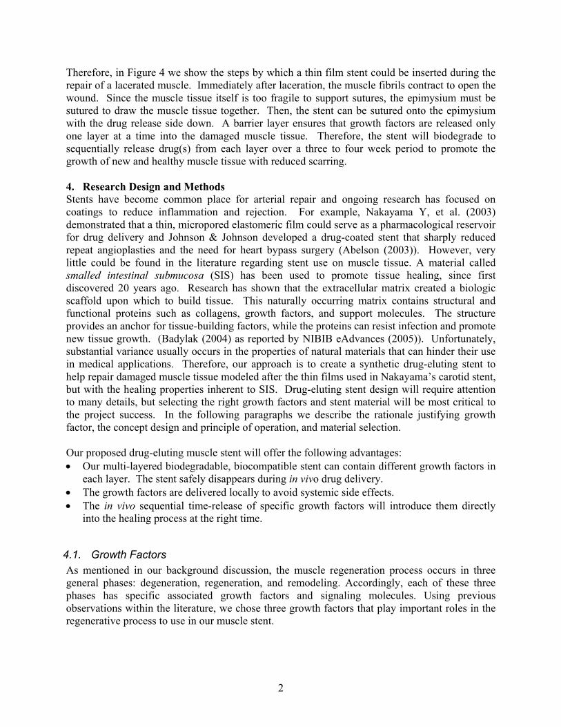
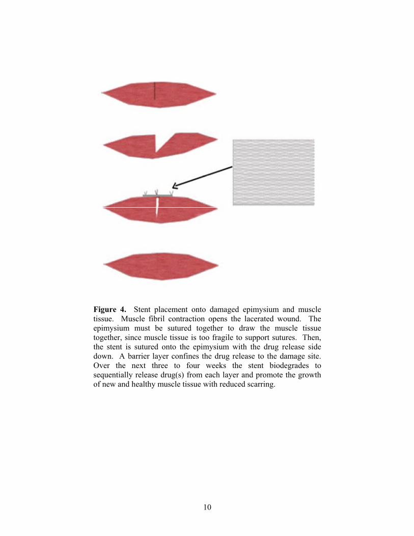
Therefore, in Figure 4 we show the steps by which a thin film stent could be inserted during the repair of a lacerated muscle. Immediately after laceration, the muscle fibrils contract to open the wound. Since the muscle tissue itself is too fragile to support sutures, the epimysium must be sutured to draw the muscle tissue together. Then, the stent can be sutured onto the epimysium with the drug release side down. A barrier layer ensures that growth factors are released only one layer at a time into the damaged muscle tissue. Therefore, the stent will biodegrade to sequentially release drug(s) from each layer over a three to four week period to promote the growth of new and healthy muscle tissue with reduced scarring. 4. Research Design and Methods Stents have become common place for arterial repair and ongoing research has focused on coatings to reduce inflammation and rejection. For example, Nakayama Y, et al. (2003) demonstrated that a thin, micropored elastomeric film could serve as a pharmacological reservoir for drug delivery and Johnson & Johnson developed a drug-coated stent that sharply reduced repeat angioplasties and the need for heart bypass surgery (Abelson (2003)). However, very little could be found in the literature regarding stent use on muscle tissue. A material called smalled intestinal submucosa (SIS) has been used to promote tissue healing, since first discovered 20 years ago. Research has shown that the extracellular matrix created a biologic scaffold upon which to build tissue. This naturally occurring matrix contains structural and functional proteins such as collagens, growth factors, and support molecules. The structure provides an anchor for tissue-building factors, while the proteins can resist infection and promote new tissue growth. (Badylak (2004) as reported by NIBIB eAdvances (2005)). Unfortunately, substantial variance usually occurs in the properties of natural materials that can hinder their use in medical applications. Therefore, our approach is to create a synthetic drug-eluting stent to help repair damaged muscle tissue modeled after the thin films used in Nakayama’s carotid stent, but with the healing properties inherent to SIS. Drug-eluting stent design will require attention to many details, but selecting the right growth factors and stent material will be most critical to the project success. In the following paragraphs we describe the rationale justifying growth factor, the concept design and principle of operation, and material selection. Our proposed drug-eluting muscle stent will offer the following advantages: • Our multi-layered biodegradable, biocompatible stent can contain different growth factors in
each layer. The stent safely disappears during in vivo drug delivery. • The growth factors are delivered locally to avoid systemic side effects. • The in vivo sequential time-release of specific growth factors will introduce them directly
into the healing process at the right time.
4.1. Growth Factors As mentioned in our background discussion, the muscle regeneration process occurs in three general phases: degeneration, regeneration, and remodeling. Accordingly, each of these three phases has specific associated growth factors and signaling molecules. Using previous observations within the literature, we chose three growth factors that play important roles in the regenerative process to use in our muscle stent.

3
bFGF The degenerative phase of muscle regeneration is largely characterized by the infiltration of phagocytic blood cells such as monocytes and neutrophils, which serve to clear the damaged site and induce an inflammatory response. While the inflammatory response includes the production of tissue-damaging free radicals and cytotoxic substances, it also induces the release of several signaling molecules. One major effect of this inflammatory response in muscle tissue is the activation of satellite cells (Tidball (2004)). Though many factors, both indigenous to the muscle tissue and secreted by phagocytes, have proven to have a role in activating satellite cells, basic fibroblast growth factor has been shown to be one of the most potent and essential early signals (Charge and Rudnicki (2004)). In damaged muscle tissue in vivo bFGF shows up within 8 hours in the extracellular space, peaks at 24 hours, and then slowly decreases over a period of about a week (Anderson, et al. (1995)). Furthermore, direct injection of bFGF into damaged muscle tissue was shown to increase muscle regeneration by 3.5 times (Menetrey, et al. (2000)). In addition, fibroblasts have been observed to enter the damaged site shortly following the infiltration by phagocytes and begin to reform the extracellular matrix and structural framework. Given that bFGF is also a potent stimulator of fibroblasts, the administration of this growth factor could also potentially accelerate the reformation of the overall structure. However, this growth factor is also known to inhibit muscle cell differentiation in vitro. Thus, while it strongly stimulates satellite cell activation and proliferation, another signal later on provides the cue to differentiate and fuse into myotubes. IGF-1 Following the inflammatory response and initial activation of satellite cells, the satellite cells continue to proliferate, migrate to the site of injury, and begin to form new myotubes or fuse with damaged myotubes. Insulin-like growth factor 1 (IGF-1) has been shown in several in vitro studies to be a potent stimulator of both satellite cell proliferation as well as their differentiation and fusion into functional myotubes (Charge and Rudnicki (2004), Coleman, et al. (1995), and Florini JR, et al. (1991)). Kinetically in vivo its expression begins after two days and persists for up to a week, then slowly decreases over a week (Jennische and Hansson (1987) and Jennische (1989)), and direct injection into damaged muscle has also been shown to significantly augment muscle regeneration (Menetrey, et al. (2000)). Thus, IGF-1’s role is a more general pro-regenerative one that persists throughout the healing process. NGF The remodeling process reestablishes a fully functional, contracting muscle, removes scar tissue, and reinnervates the muscle. The reestablishment of functional muscle is a process generated mostly through intercellular communication between reformed or newly formed myotubes. The reinnervation process, however, can often be left incomplete (Barton and Morris (2003)). Nerve growth factor (NGF) is a well-established promoter of axonal growth and thus may help to facilitate the reinnervation process. Furthermore, NGF also stimulates muscle maintenance and, to a degree, regeneration. This is most obviously demonstrated by denervation experiments or injections of anti-NGF antibodies both of which result in muscle atrophy (Baron, et al. (1994) and Vinores and Guroff (1980)). Therefore, the presence of NGF would help bring about a homeostatic survival at the end of the regenerative process.

4
4.2. Drug-eluting Muscle Stent Material A drug-eluting muscle stent can stimulate and enhance new growth to repair damaged muscles. The stent provides a physical scaffold that will be attached to the damaged muscle to direct new growth. Our proposed stent will be made up of multiple layers with each layer containing a different drug to promote rapid and healthy muscle tissue growth. The multi-functional stent sketched in Figure 5 will be made up of two active drug carrying layers and an outer barrier layer that confines the drug release to the muscle tissue needing repair. Spin-coating or spray deposition processes are used to build up thin layers of material. The stent should be fabricated using biocompatible, biodegradable, micro machineable, drug-eluting and highly elastic materials. Fortunately, there are many biodegradable materials that eliminate the need for later removal. The decomposition products are readily processed and expelled from the body. The stent will also be made from biocompatible materials to minimize the immunological response that causes fibrous capsules analogous to scar tissue to grow. In addition, we must also be able to load the desired growth factors into the material. Lithography, laser ablation or chemical processes can be used to pattern the thin films. Further, it is desirable that the drug release rate and dosage be controlled for up to three weeks. Several promising copolymers and polyurethanes have been researched for stent applications (Wang, et al. (2002), Gunatillake and Adhikari (2003), Gross and Kalra (2002), and Zeng, et al. (2004)). In particular, synthetic polymers can be fabricated in thin films and designed with the desired growth factors to promote healing. Of these we chose the copolymer poly glycerol sebacate (PGS) investigated by Wang, et al. (2002), since it triggers little immunological response by the host tissue (see Figure 6). The inflammation zone was not very thick and reduced with time. Also, the fibrous capsule growth that can block our drug infusion into the muscle tissue was minimal. Finally, after a 60 day trial with PGS implants, “[they] were completely absorbed without granulation or formation of scar tissues, and the implantation site was restored to its normal histologic architecture” (Wang, Ameer, et al. (2002)). In the event of unforeseen complications with PGS, polyglycolic acid and polylactic acid are biodegradable materials shown to provide satisfactory results by Nakayama, et al. (2003) and others. There are two primary approaches to infuse drugs into the stent material: one is to create micropores that become reservoirs to hold a gelatinous drug compound and the second is to covalently bind the drugs to the copolymer. Nakayama, et al. (2003) patterned stent films using laser and UV photolithography micromachining processes. Spaans et al. (2000) added salt crystals to polyurethanes to create porous structures with 70 to 80% void fraction. However, membranes must be coated on both sides of the stent to control the diffusion rate of drugs from the gelatin into the muscle tissue. Therefore, we propose to rely on material biodegradation to release the attached growth factors at the desired rate. The rate of material degradation passively controls the drug release rate, since the polymer binds the drugs until degradation (Figure 7). In research conducted on PGS, “The degradation of PGS is dominated by surface erosion, as indicated by linear mass loss with time, preservation of implant geometry, better retention of mechanical strength, absence of surface cracks, and minimal water uptake”. (Wang, et al (2002)) Thus, we can design the stent degradation rate and size (i.e. exposed surface area) to deliver the proper drug dosage to the damaged muscle tissue.

5
Mann, et al. (2001) synthesized polyethylene glycol derivatives with degradable peptides in their backbone, which suggests that the growth hormones may be coupled to the hydroxyl groups that are attached to the PGS backbone. This is very easy to achieve since the residues of growth factors already contain –OH groups. For example, Tyrosine, the 114th residue of bFGF, has -OH group attached to its benzene ring. However, significant research into the polymerization process will be required to introduce the growth factors without degrading copolymer properties or denaturing the growth factors. PGS is synthesized by polycondensation of 0.1 mol each of glycerol and sebacic acid at 120ºC under argon for 24 h. Then, the pressure is reduced from 1 torr to 40 mtorr over a 5 h period, where it remains for 48 h. (Wang, et al (2002)). Unfortunately, the proposed growth factors will not survive above 90ºC for more than three (3) hours. This makes manufacturing PGS with growth factors a challenge. We describe two possible solutions below: 1) Add the growth factors during the cooling process once the temperature has fallen to about 90-95ºC. This allows the growth factors to be mixed directly into the polymer. This is the simplest and easiest way to add the desired growth factors into the PGS, and there is no need to alter the process described in the literature. 2) Employ high pressure synthesis; rather than reduce the pressure from 1 torr to 40 mtorr, we can instead reduce the temperature from 120ºC to below 90ºC over the 5 hour period and maintain a constant 1 torr pressure. Once cooled, the growth factors can be added to the reaction mixture and then held there for 48 hours. Unfortunately, we will likely need to re-optimize the PGS synthesis process to obtain an acceptable product. Further investigation will be needed to choose the best method to produce the PGS with growth hormones. 5. Expected Results and Impact on the Field Our initial results will establish the feasibility of our concept to promote healthy muscle tissue repair. Once the synthesis process to impregnate growth factors has been developed, we will design and fabricate thin film stents. These stents will be laboratory tested to determine the initial drug loading, time release behavior and stent degradation. We will dice the fabricated stent material into multiple samples. The total concentration of each growth factor in each sample will be measured to determine the uniformity of drug loading. The stent will be placed onto cultured muscle tissue to determine the actual lifetime of the PGS biodegradable material. It will also show if the growth factor release rates and layer thicknesses are sufficient to sustain the desired dosages throughout the regeneration process (about three (3) weeks in total). Animal trials will not be undertaken until the next phase. If successful, a drug-eluting muscle stent would have global ramifications, since severe muscle injury would no longer need to cripple millions of patients each year. After 100 years, it is time to embrace a new technology that society has at its fingertips. 6. Acknowledgements We thank Prof. Michael Stowell and Dr. Richard M. Jaouen for the many insightful discussions, comments and suggestions that proved helpful to our development of the muscle stent concept.
6
7. References Abelson R (2003), “Johnson & Johnson’s new stent may dominate angioplasties,” The New York Times, Jan 3, 2003. Anderson JE, Mitchell CM, McGeachie JK and Grounds MD (1995), “The time course of basic fibroblast growth factor expression in crush-injured skeletal muscles of SJL/J and BALB/c mice,” Experimental Cell Research, 216: 324-334. Badylak SF (2004), “Xenogenic extracellular matrix as a scaffold for tissue reconstruction,” Transplant Immunology 12:367-377. Baron P, Scarpini E and Meola G (1994), “Expression of the low-affinity NGF receptor during human muscle development regeneration, and in tissue culture,” Muscle Nerve, 17: 276-284. Charge S and Rudnicki M (2004), “Cellular and molecular regulation of muscle regeneration,” Physiology Reviews, 84: 209-238. Coleman ME, Demayo F, Yin KC, Lee HM, Geske R, Montgomery C and Schwartz RJ (1995), “Myogenic vector expression of insulin-like growth factor I stimulates muscle cell differentiation and myotube hypertrophy in transgenic mice,” Journal of Biological Chemistry, 270: 12109-12116. Florini JR, Ewton DZ and Roof SL (1991), “Insulin-like growth factor-I stimulates terminal myogenic differentiation by induction of myogenic gene expression,” Molecular Endocrinology, 5: 718-724. Goldspink G (2004), “Age-related loss of skeletal muscle function; impairment of gene expression,” Journal of Musculoskeletal and Neuronal Interactions, 4: 143-147. Gross RA and Kalra B (2002), “Biodegradable polymers for the environment,” Science Magazine Vol 297. Gunatillake PA and Adhikari R (2003), “Biodegradable synthetic polymers for tissue engineering,” European Cells and Materials Vol. 5, pp. 1-16. Jennische E and Hansson HA (1987), “Regenerating skeletal muscle cells express insulin-like growth factor 1,” Acta Physiol Scandinavia, 130: 327-332. Jennische E (1989), “Sequential immunohistochemical expression of IGF-1 and the transferring receptor in regenerating rat muscle in vivo,” Acta Endocrinology of Copenhagen, 121: 733-738. Mann BK, Gobin AS, Tsai AT, Schmedlen RH and West JL (2001), “Smooth muscle cell growth in photopolymerized hydrogels with cell adhesive and proteolytically degradable domains: synthetic ECM analogs for tissue engineering,” Biomaterials 22: 3045-3051.
7
Menetry J, Kasemkijwattana C, Day CS, Bosch P, Vogt M, Fu FH, Moreland MS and Huard J (2000), “Growth factors improve muscle healing in vivo,” Journal of Bone and Joint Surgery, 82: 131-137. Nakayama Y, Nishi S, Ueda-Ishibashi H and Matsuda T (2003), “Fabrication of micropored elastomeric film-covered stents and acute-phase performances,” J Biomed Mater Res A, Jan 1;64(1):52-61. Nakayama Y, Nishi S and Ishibashi-Ueda H (2003), “Fabrication of drug-eluting covered stents with micropores and differential coating of heparin and FK506,” Cardiovascular Radiation Medicine 4: 77-82. Spaans CJ, Belgraver VW, Rienstra O, De Groot JH, Veth RPH and Pennings AJ (2000), “Solvent-free fabrication of micro-porous polyurethane amide and polyurethane-urea scaffolds for repair and replacement of the knee-joint meniscus,” Biomaterials 21: 2453-2460. Tidball J (2005) “Inflammatory processes in muscle injury and repair,” American Journal of Physiology. 288: R345-R353. Vinores S and Guroff G (1980), “Nerve growth factor: mechanisms of action,” Annual Review in Biophysical Bioengineering, 9: 223-257. Wang Y, Ameer GA, Sheppard BJ and Langer R (2002), "A tough biodegradable elastomer," Nature Biotechnology 20, pp. 602-606. http://biotech.nature.com. Wang Y, et al. (2002), "In vivo degradation characteristics of poly (glycerol sebacate)," Journal of Biomedical Materials Research Vol 66A, pp.192-197. <http://interscience.willey.com> Zeng F, Liu J and Allen C (2004), “Synthesis and characterization of biodegradable poly(ethylene glycol)-block-poly(5-benzyloxy-trimethylene carbonate) copolymers for drug delivery,” American Chemical Society. “Bioengineered Tissue Scaffold Promotes Wound Healing,” NIBIB eAdvances, Jan 28, 2005. http://www.nibib1.nih.gov/eAdvances/021805.html.

8
8. Figures
Figure 1. Severe Muscle Damage (left: rectum, and right: shotgun blast to forearm). Photos courtesy of Dr. Richard M. Jaouen.
Figure 2. Surgical repair: the epimysium is sutured together to bring the muscle tissue back together.

9
Figure 3. Natural muscle repair process. Degeneration. The damaged muscle tissue (black tear) releases its intracellular contents (orange), which attract phagocytic cells (grey). These cells signal the activation of satellite cells which reside on the edges of the muscle tissue (light blue). Regeneration. These satellite cells then proliferate and migrate to the site of damage where they differentiate and fuse to form myotubes (bright red). Remodeling. The muscle becomes fully functional and reinnervated by nerves (black) and resumes normal function.
10
Figure 4. Stent placement onto damaged epimysium and muscle tissue. Muscle fibril contraction opens the lacerated wound. The epimysium must be sutured together to draw the muscle tissue together, since muscle tissue is too fragile to support sutures. Then, the stent is sutured onto the epimysium with the drug release side down. A barrier layer confines the drug release to the damage site. Over the next three to four weeks the stent biodegrades to sequentially release drug(s) from each layer and promote the growth of new and healthy muscle tissue with reduced scarring.

11
BarrierIGF-1 + NGFbFGF + IGF-1
Figure 5. Exploded view of a tri-layered thin film stent. Each layer contains specific growth factor(s) with the outermost layer a slower biodegradable barrier, so that the stent surface degradation sequentially progresses from the innermost bFGF layer to the outermost NGF.
PGS inflammatory zone
PGS fibrous capsule growth
Figure 6. Inflammatory response of PGS compared to another polymer that was not considered, PGLA, adapted from Wang, Ameer, et al. (2002). The PGS inflammatory zone steadily decreases with time and more importantly, the fibrous capsule growth that could inhibit growth factor dispersion to the muscle tissue does not express during the first month. Also the fibrous capsule was small even at 35 days when compared to the six by six by three millimeter implant.

12
Figure 7. The growth factors are released as the biodegradable polymer decomposes.
Figure 8. The chemical reaction for PGS. On the right are the chemical structures for glycerol and sebacic acid which react to produce PGS (Wang, Ameer, et al. (2002)).