Neural Circuit Mechanisms Underlying the Exacerbation of ...

Raghunath et al. BMC Research Notes (2015) 8:170 DOI 10.1186/s13104-015-1128-6
RESEARCH ARTICLE Open Access
A molecular systems approach to modellinghuman skin pigmentation: identifying underlyingpathways and critical componentsArathi Raghunath1†, Awanti Sambarey2†, Neha Sharma2, Usha Mahadevan1 and Nagasuma Chandra2*
Abstract
Background: Ultraviolet radiations (UV) serve as an environmental stress for human skin, and result in melanogenesis,with the pigment melanin having protective effects against UV induced damage. This involves a dynamic and complexregulation of various biological processes that results in the expression of melanin in the outer most layers of theepidermis, where it can exert its protective effect. A comprehensive understanding of the underlying cross talkamong different signalling molecules and cell types is only possible through a systems perspective. Increasingincidences of both melanoma and non-melanoma skin cancers necessitate the need to better comprehend UVmediated effects on skin pigmentation at a systems level, so as to ultimately evolve knowledge-based strategiesfor efficient protection and prevention of skin diseases.
Methods: A network model for UV-mediated skin pigmentation in the epidermis was constructed and subjectedto shortest path analysis. Virtual knock-outs were carried out to identify essential signalling components.
Results: We describe a network model for UV-mediated skin pigmentation in the epidermis. The model consistsof 265 components (nodes) and 429 directed interactions among them, capturing the manner in which one componentinfluences the other and channels information. Through shortest path analysis, we identify novel signallingpathways relevant to pigmentation. Virtual knock-outs or perturbations of specific nodes in the network haveled to the identification of alternate modes of signalling as well as enabled determining essential nodes in theprocess.
Conclusions: The model presented provides a comprehensive picture of UV mediated signalling manifesting inhuman skin pigmentation. A systems perspective helps provide a holistic purview of interconnections and complexity inthe processes leading to pigmentation. The model described here is extensive yet amenable to expansion as new data isgathered. Through this study, we provide a list of important proteins essential for pigmentation which can befurther explored to better understand normal pigmentation as well as its pathologies including vitiligo andmelanoma, and enable therapeutic intervention.
Keywords: Systems perspective, Skin, UV, Melanin, Networks
* Correspondence: [email protected]†Equal contributors2Department of Biochemistry, Indian Institute of Science, Bangalore 560012,IndiaFull list of author information is available at the end of the article
© 2015 Raghunath et al.; licensee BioMed Central. This is an Open Access article distributed under the terms of the CreativeCommons Attribution License (http://creativecommons.org/licenses/by/4.0), which permits unrestricted use, distribution, andreproduction in any medium, provided the original work is properly credited. The Creative Commons Public DomainDedication waiver (http://creativecommons.org/publicdomain/zero/1.0/) applies to the data made available in this article,unless otherwise stated.

Raghunath et al. BMC Research Notes (2015) 8:170 Page 2 of 12
BackgroundSkin pigmentation is a phenotype evolved and retained overgenerations, primarily as protection against harmful Ultra-violet (UV) radiations [1]. Melanin, a light-absorbing poly-mer with photochemical properties imparts colour tohuman skin and other tissues such as hair and iris, and pro-vides protection against UV induced damage. Variations inskin pigmentation depend on the level and the type of mel-anin expressed. The two major forms of melanin expressedin humans are eumelanin and pheomelanin, impartingblack or brown and red haired or freckled phenotypes re-spectively. Melanin is synthesized in melanocytes in special-ized organelles called melanosomes [2]. Transfer of thepigment to keratinocytes that constitute the upper layers ofthe epidermis is essential for phenotypic manifestation ofskin pigmentation. The dendrites on melanocytes aid in thetransfer of melanosomes to keratinocytes where melano-some uptake occurs through phagocytosis [2].Exposure to UV radiation triggers signalling cascades in
keratinocytes, which in turn induce autocrine signalling aswell as paracrine signalling with neighbouring melanocytesresulting in melanogenesis, and thereby subsequent pro-cesses leading to skin pigmentation. While UV radiationstrigger pigmentation, they are also capable of causing skincancer due to the generation of free radicals and DNAdamage, which is actively prevented by the ‘epidermal mel-anin unit’ - the association of one melanocyte with severalsurrounding epidermal keratinocytes [3]. UV exposure canalso lead to the depletion of folic acid, though a certainamount of exposure is essential for Vitamin D synthesis [4].Hence, a balanced exposure to UV radiation is critical, andis maintained by the regulation of melanin synthesis. Con-stitutive skin pigmentation is determined by the geneticconstitution of the individuals while facultative pigmenta-tion is governed by environmental factors [5]. Oxidationand re-distribution of melanin in skin induces some mar-ginal protection but delayed and sustained tanning re-sponse to UV includes melanin synthesis and transport tokeratinocytes [6].The process of UV mediated skin pigmentation is com-
plex and dynamic, involving various cell types and extensivecross-talk between them as well as several intracellular sig-nalling cascades. While several studies over the past fewdecades have provided insights into melanogenesis, mela-nosome formation and pigmentation [7-9] their focus hasbeen primarily on individual reactions and molecularchanges. The World Health Organization reports inci-dences of between 2 and 3 million non-melanoma skincancers and more than 100,000 melanoma skin cancersglobally each year [10]. These statistics highlight the needfor further studies in this area, and a wholistic understand-ing of pigmentation is thus necessary.To this end, a network approach is useful as networks
have emerged as an important tool to study biological
phenomena [11]. The ‘omics’ era has resulted in the gen-eration of enormous biological data, from individualgene and protein sequences and structures to well-defined physical and functional interactions and elabor-ate signalling cascades. By integrating this informationof communication between various molecular compo-nents in the form of an interaction network, biologicalprocesses can be represented and analysed at a systemslevel, wherein the contributions and effects of individualcomponents and interactions on the entire system canbe studied to generate insights which cannot be achievedby reductionist approaches alone. Extensive research inthe field of skin pigmentation has resulted in vast accu-mulation of data, but studies that integrate and analysethis data are limited [12-14].In this study, we curate data available from literature
and construct a comprehensive network model of UVmediated pigmentation in both keratinocytes and mela-nocytes, in an attempt to provide mechanistic insightsinto signalling cascades underlying skin pigmentation.Further, we study the effects of knock-outs of certaincomponents on these signalling pathways, leading to theidentification of essential genes as well as highlight keydestinations and possible alternative signalling routes inthe process of pigmentation.
ResultsA comprehensive network model of human pigmentationWe have constructed a network model of human skin pig-mentation based on extensive data available from literature.The model includes various components of (a) melaninbiosynthesis, (b) melanosome biogenesis, (c) dendrite for-mation and (d) uptake of melanosomes by keratinocytes.Such a comprehensive systems-level picture of skin pig-mentation has the potential to address several questions,particularly the critical nature of various factors involved inpigmentation and its response to UV radiation.Several experimental studies have been referred to for
network construction as enlisted in Additional file 1.However, for clarity, a brief description of the model andits components is given below.Melanin, the photosensitive polymer that imparts skin
pigmentation is synthesised and stored in melanosomes -organelles specific to melanocytes [7]. Melanin is syn-thesised in the body from the amino acid tyrosine,through a series of biochemical reactions primarily involv-ing the enzymes tyrosinase, DOPAchrome tautomerase andtyrosinase-related protein 1 (TYRP1). Tyrosine is firstoxidized to DOPA and subsequently to DOPAquinoneby the enzyme tyrosinase. DOPAquinone can be enzymati-cally converted to either eumelanin or pheomelanin. Dur-ing eumelanin synthesis, DOPAquinone is converted toleucoDOPA and subsequently to DOPAchrome by auto-oxidation. DOPAchrome tautomerase, in the presence of

Raghunath et al. BMC Research Notes (2015) 8:170 Page 3 of 12
dihydroxyindole-2-carboxylic acid oxidase, converts DOPA-chrome to 5,6-dihydroxy indole. Tyrosinase acts on 5,6-hy-droxy indole finally resulting in eumelanin synthesis. Analternate biosynthesis pathway leading to pheomelanin in-volves conversion of DOPAquinone to cysteinylDOPA inpresence of glutathione/cysteine, followed by oxidativepolymerization of the benzothiazinylalanine intermediates,resulting in pheomelanin synthesis [15].Though melanin biosynthesis is a central process in
skin pigmentation, multiple associated pathways are ne-cessary for successful manifestation of the phenotype. Inresponse to the necessary UV stimulus, melanocytesform dendrites, and through them transport melanincarrying melanosomes to the peripheral keratinocyteswhere the melanosomes are subjected to phagocytosis,resulting in the expression of melanin in keratinocytes,ultimately leading to skin pigmentation [7].Here, we present an extensive systems model of pigmen-
tation that includes interactions and pathway networks in-volved in all the major biological processes discussed aboveleading to the pigmentation of skin, beginning with mela-nosome formation and melanogenesis and culminating inits expression in keratinocytes leading to skin pigmentation.Figure 1 provides a schematic representation of the com-plex network of the various processes leading to humanskin pigmentation. The model is depicted in the form of astatic map that is compartmentalised to depict several sig-nalling events and flow of signal information in keratino-cytes and melanocytes on exposure to UV. As can beobserved, the model captures several signalling cascadestriggered in keratinocytes on initial exposure such as lipidperoxidation, MAPK signalling, TNF-receptor mediatedskin inflammation and other processes which result in me-lanogenesis in melanocytes via extensive cross-talk. Themodel also contains a detailed molecular description of thesynthesis of melanin in melanosomes, followed by dendrito-genesis which enables melanosome phagocytosis by kerati-nocytes, where melanin finally manifests itself in the formof the skin pigment. The model also includes additionalpathways stimulated by UV radiation like skin ageing,cell proliferation and differentiation and DNA damaged-minimised by the process of DNA repair, mediated bymelanin. While the model provides a comprehensive de-scription of skin pigmentation, given the vastness of re-search in this field and increasing data availability, it lendsitself to incorporation of additional data as and when it be-comes available.Based on the signalling events described in the model,
265 participating molecules were identified, including189 proteins, 33 small molecules/compounds, 23 com-plexes, 18 biological processes and 2 environmental fac-tors. The model thus incorporates multiple levels ofdetail. For these 265 molecules or nodes, interactionswere extracted as described in Methods, and a network
was constructed comprising 265 nodes and 429 edges.The edges were assigned direction by extensive literaturecuration, highlighting the functional nature of eachinteraction. 142 nodes were involved in keratinocyte sig-nalling, while 113 nodes contributed to signalling withinmelanocytes, and these were ascribed the suffix ‘kerat’and ‘melan’ respectively. The complete list of all nodesand edges in the network along with the nature and dir-ection of their interactions are provided in Additionalfile 1. A sample network description has been providedin Table 1. This network was visualized in Cytoscapev3.1.0 and several network properties were computedbased solely on network topology. As the network wasdirected, both in-degree and out-degree distributionswere computed describing their connectivity and certain‘hub’ nodes which are central to the network were thusidentified. All network properties of degree distributions,identified hubs, node centrality, edge centrality and radi-ality computed are described in Additional file 2.
Identifying biochemical routes in the network amongdefined sources and targetsThe constructed network provides a basis for understand-ing communication and signalling within the system. Asystems level view of pigmentation provides insights intohow information flows via signalling cascades both atinter-and intra cellular levels, and encompasses cross talkamong different molecules and cell types. One approachtowards understanding network dynamics and communi-cation is through the analysis of shortest paths betweenpairs of nodes, as demonstrated in several studies in differ-ent biological conditions [16,17]. A total of 33,779 all-vs-all paths for the 265 nodes were computed. For a moredefinitive analysis of pathways of interest in the process ofpigmentation, specific source and destination nodes werechosen and paths from these sources to destinations wereinspected in detail. The initiating nodes or sources in-cluded key receptors and triggers such as UV radiation. 20source nodes were identified, based on their likelihood ofinitiating signalling events. All sources culminated in bio-logical end processes such as melanosome formation, mela-nogenesis, dendrite formation and melanosome phagocytosis,among others. A total of 9 destinations were thus shortlisted.Table 2 enlists the selected source and target nodes in thenetwork. Between these 20 sources and 9 destinationnodes, 157 paths were computed which were furtheranalyzed. The source to target (S2T) sub-network had127 nodes and 150 edges, and the resulting networkwas completely interconnected, with no independentinteractions. This indicates a well-knit network withextensive signalling crosstalk. The 127 nodes were seento participate in various biological processes in both ker-atinocytes and melanocytes including cell differentiation,proliferation, survival, cell cycle arrest, apoptosis, melanosome
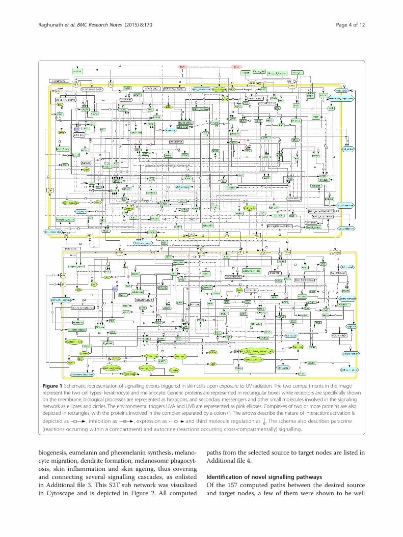
Figure 1 Schematic representation of signalling events triggered in skin cells upon exposure to UV radiation. The two compartments in the imagerepresent the two cell types- keratinocyte and melanocyte. Generic proteins are represented in rectangular boxes while receptors are specifically shownon the membrane; biological processes are represented as hexagons, and secondary messengers and other small molecules involved in the signalingnetwork as ellipses and circles. The environmental triggers UVA and UVB are represented as pink ellipses. Complexes of two or more proteins are alsodepicted in rectangles, with the proteins involved in the complex separated by a colon (:). The arrows describe the nature of interaction: activation is
depicted as , inhibition as , expression as and third molecule regulation as . The schema also describes paracrine
(reactions occurring within a compartment) and autocrine (reactions occurring cross-compartmentally) signalling.
Raghunath et al. BMC Research Notes (2015) 8:170 Page 4 of 12
biogenesis, eumelanin and pheomelanin synthesis, melano-cyte migration, dendrite formation, melanosome phagocyt-osis, skin inflammation and skin ageing, thus coveringand connecting several signalling cascades, as enlistedin Additional file 3. This S2T sub network was visualizedin Cytoscape and is depicted in Figure 2. All computed
paths from the selected source to target nodes are listed inAdditional file 4.
Identification of novel signalling pathwaysOf the 157 computed paths between the desired sourceand target nodes, a few of them were shown to be well

Table 2 Nodes selected as source and target for shortestpath analysis
Source nodes Target nodes
MET_melan Eumelanin_melan
EDNRB_melan Pheomelanin_melan
EGFR_kerat Dendrite_formation_melan
PTGER1_melan Melanosome_phagocytosis_kerat
PTGER2_kerat Apoptosis_melan
PTGER3_melan Cell_proliferation_melan
PTGER4_kerat Melanosome_biogenesis
FAS_kerat Cell_survival_melan
FZD3_melan Cell_cycle_arrest_melan
CSF2RA_melan
IL1R1_kerat
IL6R_kerat
KIT_melan
MC1R_kerat
MC1R_melan
F2RL1_kerat
TNFRSF1A_kerat
NTRK1_melan
UVA
UVB
Table 1 A sample Protein-Protein interaction networkconstructed in this study
Node A Node B Interaction typeA- > B
UVA Lipid_Peroxidation_kerat induces
Lipid_Peroxidation_kerat 4HNE_kerat increases level
4HNE_kerat DNA_Damage_kerat induces
DNA_Damage_kerat TP53_kerat activates
TP53_kerat ACTH_kerat increasesexpression
TP53_kerat alpha_MSH_kerat increasesexpression
ACTH_kerat MC1R_melan activates
alpha_MSH_kerat MC1R_melan activates
MC1R_melan ADCY4_melan activates
ADCY4_melan cAMP_melan increases level
CREB1_melan PTGS2_melan increasesexpression
PGE2_kerat PTGER3_melan activates
PGE2_kerat PTGER1_melan activates
PTGER3_melan PLC_melan activates
PTGER1_melan PLC_melan activates
PLC_melan DAG_melan increases level
DAG_melan PRKCB_melan activates
PTGS2_melan PGE2_melan Increases level
PRKCB_melan TYR_melan activates
TYR_melan Eumelanin_melan Increases level
The complete network can be found in Additional file 1.
Raghunath et al. BMC Research Notes (2015) 8:170 Page 5 of 12
characterised in literature, thus serving to validate ouranalysis. Signalling through Protein Kinase A (PKA) cul-minating in melanogenesis has been well established.Also, reports of melanocyte stimulating hormone(alphaMSH/MC1R) mediated melanogenesis involvingPKA/CREB signalling have been elucidated in a few indi-vidual studies [18,19]. Through our study, we identifiedthe same pathway and traced it to MSH as the source.Such a study helps us to systematically relate individualgenes to particular processes.Another example pathway identified in our network
describes the stem cell factor (KITLG) mediated activa-tion of MITF, a primary transcription factor that regu-lates expression of melanogenic proteins.Several novel signalling cascades showing possible
paracrine signalling between keratinocytes and melano-cytes were predicted by the network. As an example,some of the predicted UV mediated paths that lead tomelanin synthesis, dendrite formation, phagocytosis andmelanocyte proliferation are described in Table 3. Cer-tain observations from literature are also provided which
could possibly validate the existence of such biologicalroutes.
Perturbation networks: identifying ‘essential nodes’Interaction networks help identify important nodesbased on their connections and describe the topologicalarchitecture of a system. In order to assess the relativeimportance of a node, the following criteria was used: a)node degree, leading to the identification of hubs in thenetwork (b) frequency of occurrence of a given node inall shortest paths – the more frequently a node occurs,the greater is its importance in the system as it is an im-portant mediator in signalling from several sources totheir respective destinations.Based on these criteria, 33 nodes were shortlisted as
most important or significant in the network. While thisresult is significant in itself, our objective was to identifythe functional effects of these molecules in the process ofpigmentation. These nodes were then knocked-out and 33different perturbation networks were constructed, witheach network specific to a perturbation wherein a selectednode and its connecting edges were removed. Shortestpaths from the same 20 sources to the 9 destinations werecomputed for each perturbed network and analyzed toidentify all plausible paths leading to a similar end process.

Figure 2 Directed network of paths from selected source to target nodes (S2T). Arrows indicate direction of interaction. Paths from source totarget nodes can be traced. Nodes identified as ‘essential’ post perturbation analysis are highlighted in green.
Raghunath et al. BMC Research Notes (2015) 8:170 Page 6 of 12
Perturbation studies served to address the followingquestions: (a) does a signal from a given source reach itsdestination if the node mediating its pathway is perturbed?and (b) do alternate paths exist from a particular source toa destination that can be utilised upon perturbation? Whilesome perturbations resulted in the complete lack of signal-ling from a given source to a target, several others resultedin signalling via alternate routes. Nodes whose absence re-sulted in abrogation of pathways were considered as ‘essen-tial nodes’. In certain cases, two or more nodes are seen tobe essential for a given pathway. Table 4 shortlists these es-sential nodes, rank listed in order of the number of path-ways they disrupt, and these nodes are also highlighted inFigure 2. A few examples of alternate routes upon perturb-ation are provided in Table 5, and a few others illustrated inFigure 3. Studies from literature supporting the possi-bility of such alternate paths have also been cited, andnone of these studies overlap with the literature re-ferred to while building the model, thereby providinggreater credibility to our findings. All perturbation re-sults describing essential nodes as well as alternatepaths have been provided in Additional file 5.
New insights from the modelData from the network shows the influence of signallingfrom multiple receptors in processes leading to pigmen-tation. Significant contributors to skin pigmentationhave been identified by the model, with several receptorsseen to be playing a key role. Receptors EGFR, F2RL1,FAS, IL1R1, IL6R, PTGER2, PTGER4 and TNFRSF1A inkeratinocytes, and CSF2RA, EDNRB, FZD3, KIT, MET,
NTRK1, PTGER1 and PTGER3 in melanocytes are ob-served to be important for pigmentation. MC1R signal-ling is seen to be important in both keratinocytes andmelanocytes. Some of the paths in the model corrobor-ate with knockout and siRNA data that emphasize theneed for certain proteins for the manifestation of theskin pigmentation phenotype and their role in variousskin pathologies. The model also highlights paracrinesignalling in cells of the epidermis wherein factors se-creted from keratinocytes in response to UV have a keyrole to play in adult skin pigmentation by regulating sig-nalling in melanocytes. The model to a large extent en-compasses signalling involving various physiologicalfactors that are known to play a role in adult skin pig-mentation such as MC1R, CREB, MITF, PAX3, SOX10,LEF1/TCF, F2RL1, SCF, HGF and GM-CSF [15].
DiscussionHuman skin responds to UV radiation exposure by increas-ing the expression of melanin in skin, a pigment that pro-tects it from UV induced damage [6]. Melanin has a role toplay in various pigmentation disorders such as albinism[20], vitiligo [21] as well as in melanoma [22] and non-melanoma skin cancers [23]. A detailed understanding ofthe process of skin pigmentation is thus vital for under-standing the molecular pathology of these disease condi-tions. Melanin synthesis takes place in response to UVinduced cues from neighbouring cells such keratinocytesand dermal fibroblasts [24], and exerts its protective effectswhen transported and expressed in keratinocytes [25]. Mul-tiple roles are suggested for the protective effect of melanin

Table 3 Example predicted pathways from source to targets
Biologicalprocess
UV mediated melanogenesis [Tyrosinase activation]-paracrine effect
Predictedpathway
UV- > Singlet_oxygen_kerat- > Ceramide_kerat- > PLA2_kerat- > Arachidonic_Acid_kerat- > PGE2_kerat- > PTGER3_melan- > PLC_melan- >DAG_melan- > PRKCB_melan- > TYR_melan- > Eumelanin_melan/ Pheomelanin_melan
Validation PGE2 acts as a ligand for PTGER3 receptor. COX2 is the enzyme involved in the conversion of arachidonic acid to PGE2. UV induced COX2and increased PGE2 production in keratinocytes [42-45].
Predictedpathway
UV- > Singlet_oxygen_kerat- > Ceramide_kerat- > PRKCZ_kerat- > NFKB1_kerat- > PTGS2_kerat
Validation Antioxidants like Astaxanthin are shown to inhibit UV induced PGE2 production possibly by down regulating COX2 expression [46].
Biologicalprocess
UV mediated melanogenesis [Tyrosinase expression]-paracrine effect
Predictedpathway
UV- > Singlet_oxygen_kerat- > Ceramide_kerat- > PRKCZ_kerat- > NFKB1_kerat- > KITLG_kerat - > KIT_melan- > PIK3CA_melan- >PDPK1_melan- > AKT1_melan- > CREB1_melan- >MITF_melan- > TYR_melan- > Eumelanin_melan/ Pheomelanin_melan
Validation KIT mediated regulation of MITF activity is shown to involve PI3K/AKT signaling [47].
Predictedpathway
UVA- > Singlet_oxygen_kerat- > Ceramide_kerat- > PRKCZ_kerat- > NFKB1_kerat- > NOS2_kerat- > Nitric_oxide_kerat- >GUCY1A2_melan- > cGMP_melan- > PRKG1_melan- > CREB1_melan- > MITF_melan- > TYR_melan- > melanogenesis
Validation C-phycocyanin, a phycobiliprotein from spirulina that has antioxidant function inhibits melanogenesis by decreasing activity ofCREB and suppressing tyrosinase expression [48]. Compound, MHY498 inhibited sodium nitroprusside (SNP, a NO donor)-inducedNO generation, and suppressed tyrosinase expression, MITF stimulation and melanin synthesis through cGMP-mediated signalingpathway in B16F10 melanoma cells [49].
Biologicalprocess
UV mediated alpha MSH production
Predictedpathway
UVA- > Lipid_Peroxidation_kerat- > 4HNE_kerat- > DNA_Damage_kerat- > TP53_kerat- > alpha_MSH_kerat
Validation UV releases singlet oxygen upon lipid peroxidation [50]. N-acetylcysteine with antioxidant properties can suppress alphaMSH andACTH production in response to UV [51]. AlphaMSH is synthesised in keratinocytes in a p53 dependent manner [52].
Biologicalprocess
UV mediated melanocyte dendrite formation-paracrine effect
Predictedpathway
UV- > Singlet_oxygen_kerat- > Ceramide_kerat- > PRKCZ_kerat- > NFKB1_kerat- > KITLG_kerat- > KIT_melan- > PIK3CA_melan- >RAC1_melan- > RAC1:PARD6A:CDC42_melan- > PRKCZ_melan- > Dendrite_formation_melan
Validation UVB exposure increased tree branch-like dendrites and activated Rac1 in a time-dependent manner in B16 melanoma cells [53]. Transientlyexpressed Rac1 activated mutants induces the formation of dendrite-like structures in human melanoma cell lines [34].
Biologicalprocess
UV mediated NFKB1 activation and secretion of paracrine factors
Predictedpathway
UV- > Singlet_oxygen_kerat- > Ceramide_kerat- > PRKCZ_kerat- > NFKB1_kerat- > EDN1_kerat
Predictedpathway
UV- > Singlet_oxygen_kerat- > Ceramide_kerat- > PRKCZ_kerat- > NFKB1_kerat- > CSF2_kerat
Predictedpathway
UV- > Singlet_oxygen_kerat- > Ceramide_kerat- > PRKCZ_kerat- > NFKB1_kerat- > KITLG_kerat
Validation UV is known activate NFkappaB in keratinocytes [54,55].
Biologicalprocess
UV mediated melanocyte proliferation-paracrine effect
Predictedpathway
UVA- > Singlet_oxygen_kerat- > Ceramide_kerat- > PRKCZ_kerat- > NFKB1_kerat- > CSF2_kerat- > CSF2RA_melan- >CSF2RA:JAK1_melan- > STAT3_melan- > CCND1_melan- > CDK4:CCND1_melan- > Cell_proliferation_melan
Validation Korean Red Ginseng extract or its saponin (KRGE or SKRG) decreased the expression of CSF2 in keratinocytes induced by UVBirradiation and also decreased proliferation of melanocytes [56].
Biologicalprocess
UV induced melanosome phagocytosis
Predictedpathway
UVA- > Singlet_oxygen_kerat- > Ceramide_kerat- > PRKCZ_kerat- > NFKB1_kerat- > F2RL1_kerat- > RHOA_kerat- >Melanosome_phagocytosis_kerat
Raghunath et al. BMC Research Notes (2015) 8:170 Page 7 of 12

Table 3 Example predicted pathways from source to targets (Continued)
Validation In a co-culture system model constructed using the primary human melanocytes and keratinocytes, increased melanosome transfer andalso upregulation of F2RL1 protein in the keratinocytes is observed when treated with low concentrations of H(2)O(2) [57]. Madecassoside(MA), a pentacyclic triterpene significantly inhibited UVR-induced melanin synthesis and melanosome transfer in a co culture system ofkeratinocytes and melanocytes and also inhibited F2RL1 expression in keratinocytes [58].
Validation refers to observations from literature that could possibly support the existence of such pathways.
Raghunath et al. BMC Research Notes (2015) 8:170 Page 8 of 12
in UV induced damage [26]. Melanin can filter UV radi-ation and scavenge reactive oxygen species produced asa result of UV exposure. The supra-nuclear cap of mel-anin protects cells from DNA damage. It is reported toinhibit production of cyclobutane pyrimidine dimersand 6,4-photoproducts, both of which are mutagenic[27]. Patients with albinism have been shown to bemore susceptible to various skin cancers [28].Given the fact that manifestation of the phenotype in-
volves a complex cross-talk across different cell types thatare present in different strata of the skin through a well reg-ulated sequence of events, it becomes imperative to studythe process from a systems perspective. The availability ofdata through several reductionist experimental studies overthe years has greatly accelerated advances in systems’ ana-lyses of pigmentation [29]. A recent study integrated func-tional genomics and protein-protein interaction networksto identify novel components that impact melanogenesis,focusing largely on the endothelin receptor (EDNRB) medi-ated signalling pathway [30]. Our study also supports theimportance of ENDRB mediated signalling in melanocytesin the process of melanogenesis.Our model describes a well annotated, curated and di-
rected interaction network among 265 components thatcould have a direct or indirect role in influencing skin
Table 4 Essential nodes - Nodes are ranked according tothe number of source- > target pathways they areessential in
Node No: of source- > target pathwaysit is essential in
RAC1_melan 34
MITF_melan 29
NFKB1_kerat 25
TYR_melan 18
PLC_melan 14
PTGS2_kerat 9
CASP8_kerat 9
F2RL1_kerat 8
AKT1_melan 7
ADCY4_melan 7
CREB1_melan 4
PRKCD_kerat 2
PTGER1_melan 1
MC1R_melan 1
pigmentation. This is the first study reporting a directedinteraction network of human skin pigmentation, highlight-ing the functional nature of interactions among differentcomponents, as well as the direction in which a signal hasto flow to be biologically meaningful. Of the 265 nodes, 183have a role to play in regulating more than one biologicalprocess influencing skin pigmentation. This reflects thecomplexity as well as the connectivity within the systemwhere a single node can influence multiple biologicalprocesses. The model suggests that multiple cues fromneighbouring cells can trigger similar processes in me-lanocytes, highlighting the various possibilities of mul-tiple alternate paths a system could take leading to acommon end process. The importance of certain nodeshas been illuminated, some of which are well known,such as the indispensability of tyrosinase in melanogenesis[31] and the role of MITF in melanosome formation [32].We also report a list of nodes that are essential for certainprocesses, the absence of which leads to abrogation of sig-nalling through the required pathway. The model helps inbroadening the perspective of known data by suggesting anextended signalling pathway or potential alternate paths inthe absence of a node. F2RL1 is known to play an import-ant role in melanosome transfer [33]. The model suggeststhat NFKB could be the transcription factor regulating theexpression of F2RL1 in keratinocytes and hence NFKBcould also have a key role to play in melanosome transfer.RAC1 is involved in MSH mediated dendrite formation onmelanocytes [34]. The current model predicts the possibil-ity of an alternate path involving paracrine signalling ofKITLG from keratinocytes acting on the c-kit receptor onmelanocytes leading to RAC1 activation and subsequentdendrite formation. The model reiterates the communica-tion between keratinocytes and melanocytes.These observations highlight the need to study bio-
logical processes in the context of the system as a wholeinstead of individual entities, as varied factors both ex-ternal and internal have a role to play in their regulation.Perturbation studies with individual source-destinationpairs enable the identification of components that areessential for a biological process or its phenotypic mani-festation. It also hints at the possible dispensable natureof certain proteins and redundancy in the network bysuggesting alternate signalling routes.The vastness and constant addition of data in this
field impose a practical limitation on model complete-ness. Though the network is extensive, it can be further

Table 5 Alternate paths taken up post perturbation
Biological processperturbed gene
UV induced melanin synthesis PTGER3_melan
Control Path UV- > Singlet_oxygen_kerat- > Ceramide_kerat- > PLA2_kerat- > Arachidonic_Acid_kerat- > PGE2_kerat- >PTGER3_melan- > PLC_melan- > DAG_melan- > PRKCB_melan- > TYR_melan- > Melanogensis
Alternate Path UV- > Singlet_oxygen_kerat- > Ceramide_kerat- > PRKCZ_kerat- > NFKB1_kerat- > EDN1_kerat- > EDNRB_melan- >PLC_melan- > DAG_melan- > PRKCB_melan- > TYR_melan- > Eumelanin_melan
Validation Addition of EDN1 induced increase in tyrosinase activity in cultured human melanocytes [59].
Biological ProcessPerturbed gene
UV induced melanin synthesis
Tyr_melan
Control Path UV- > Singlet_oxygen_kerat- > Ceramide_kerat- > PLA2_kerat- > Arachidonic_Acid_kerat- > PGE2_kerat- >PTGER3_melan- > PLC_melan- > DAG_melan- > PRKCB_melan- > TYR_melan- > Melanogensis
Alternate Path None
Validation Tyrosinase mutations lead to albinism or hypopigmentation (MGI)
Biological ProcessPerturbed gene
UV induced dendrite formation
KIT_melan
Control Path UV- > Singlet_oxygen_kerat- > Ceramide_kerat- > PRKCZ_kerat- > NFKB1_kerat- > KITLG_kerat- > KIT_melan- >PIK3CA_melan- > RAC1_melan- > RAC1:PARD6A:CDC42_melan- > PRKCZ_melan- > Dendrite_formation_melan
Alternate Path UVB- > Lipid_Peroxidation_kerat- > 4HNE_kerat- > DNA_Damage_kerat- > TP53_kerat- > ACTH_kerat- > MC1R_melan- >ADCY4_melan- > cAMP_melan- > RAP1A_melan- > RAC1_melan- > RAC1:PARD6A:CDC42_melan- > PRKCZ_melan- >Dendrite_formation_melan
Validation Highly dendritic melanocytes are stimulated by injection of alpha-MSH in newborn mice. Formation and translocationof melanosomes to dendrites is triggered by alpha-MSH [60].
Biological ProcessPerturbed gene
UV induced melanosme phagocytosis
F2RL1_kerat
Control Path UV- > Singlet_oxygen_kerat- > Ceramide_kerat- > PRKCZ_kerat- > NFKB1_kerat- > F2RL1_kerat- > RHOA_kerat- >Melanosome_phagocytosis_kerat
Alternate Path None
Validation RWJ-50353, a serine protease inhibitor, led to reduced pigment deposition in melanocytes and de-pigmentation.Immature melanosomes accumulate inside melanocytes and there is abnormal dendrite dynamics in RWJ-50353-treated epidermal equivalents [61].
Biological ProcessPerturbed gene
UV induced melanocyte proliferation
NFKB1_kerat
Control Path UV- > Singlet_oxygen_kerat - > Ceramide_kerat- > PRKCZ_kerat- > NFKB1_kerat- > CSF2_kerat- > CSF2RA_melan- >CSF2RA:JAK1_melan- > STAT3_melan- > CCND1_melan- > CDK4:CCND1_melan- > Cell_proliferation_melan
Alternate Path UVB- > Lipid_Peroxidation_kerat- > 4HNE_kerat- > DNA_Damage_kerat- > TP53_kerat- > ACTH_kerat- > MC1R_melan- >ADCY4_melan- > cAMP_melan- > PRKACA_melan- > CREB1_melan- > MITF_melan- > CDK4_melan- >CDK4:CCND1_melan- > Cell_proliferation_melan
Validation In T-oligos pre-treated and UV light-irradiated keratinocytes, NFκB binding to the transcriptional co-activator p300 decreasedrelative to control whereas the amount of p53 binding to p300 was strikingly increased demonstrating activation of p53 andrepression of NFkB upon DNA damage in keratinocytes which could serve as an alternate path when NFkB is perturbed [62].
Validation refers to observations from literature that could possibly support the existence of such pathways.
Raghunath et al. BMC Research Notes (2015) 8:170 Page 9 of 12
extended with the availability of more information. Themodel can also be expanded to include factors fromdermal fibroblasts, which are now being reported as in-creasingly important in the manifestation of pigmenta-tion [35].Aberrant melanin expression can lead to multiple
pathological conditions like albinism - which results in acomplete loss of pigmentation, vitiligo - wherein thereoccur hypo-pigmented patches of skin, sunburn and skindamage due to insufficient protection of the skin fromUV radiation, or melanoma, one of the most aggressive
forms of human skin cancer. Skin pigmentation has alsobeen a significant field of interest for the cosmetic in-dustry which attempts to address issues of lentigines/agespots and improving the skin tone. Use of anti-oxidantsfor correcting pigmentation disorders [36,37] can bereadily elucidated from the model as UV induced oxida-tive stress is one of the primary steps triggering signal-ling leading to regulation of skin pigmentation. Thus, afoundation of cellular signalling has been providedwhich can be used to study conditions of disease andprovide a basis for identifying therapeutic targets.

Figure 3 Effect of perturbation on UV mediated dendrite formation and melanogenesis: Alternate paths taken post node knockouts. The perturbed nodesare highlighted in green. (a) & (b) pathway of UV mediated dendrite formation in melanocytes before and after PIK3CA_melan knockout (c) &(d) UV mediated melanogenesis before and after PLC_melan knockout.
Raghunath et al. BMC Research Notes (2015) 8:170 Page 10 of 12
ConclusionsThrough this study we have presented a systems perspec-tive to the process of UV mediated human skin pigmenta-tion, identifying underlying pathways in the process. Acomprehensive, well annotated directed interaction net-work of various molecules participating in the manifest-ation of skin pigmentation in the epidermis has beenconstructed. The model has provided a detailed descriptionof signalling pathways triggered in epidermal cells, andsequential signalling in keratinocytes and melanocytes.Several plausible routes of biological signalling in thenetwork have been identified, and the importance ofcertain components and their effects on signalling path-ways has been ascertained. A comprehensive understandingof the molecular events leading to skin pigmentation canthus provide valuable inputs for identifying potential targetsfor therapeutic consideration as well as a rational basis
for design/improvement of commercial products. The ap-proach adopted can be applied to various biological systemsand serves as a powerful tool to analyse cross-talk amongvarious biological components.
MethodsBuilding a network modelThrough an exhaustive literature survey, different processesin skin pigmentation including melanin biosynthesis, mela-nosome formation in melanocytes followed by transfer ofmelanin carrying melanosomes to keratinocytes via den-drites and phagocytosis in keratinocytes were identified. Foreach of these processes, important participating moleculesincluding proteins, secondary messengers and small mole-cules were identified. A model describing these processeswas built as a static map using the software Cell Designer(version 4.4) [38]. Based on this data, an interaction

Raghunath et al. BMC Research Notes (2015) 8:170 Page 11 of 12
network was constructed for these molecules that partici-pate in different stages leading to pigmentation. In the net-work, individual proteins and in some cases metabolitesform the nodes and interactions among the nodes form theedges. The network included protein-protein, protein-smallmolecule and protein-biological process interactions. Allprotein-protein interactions were high confidence interac-tions extracted manually, or from the STRING databaseand NetPro™ [39]. The protein-small molecule interactionsand protein-biological process interactions were curatedfrom literature [approximately 100 articles from PubMed].The interactions included in the network pertained pri-marily to human data but data from mouse/rat specieswas also used to fill in certain missing links. The net-work included molecular interactions influencing pig-mentation in response to UV radiation in two primaryepidermal cell types- keratinocytes and melanocytes.Interactions in the network were assigned direction,based on information available in literature and KEGGpathways. Directions were dependent on the functionalnature of the interaction, for e.g. ‘phosphorylation’ or ‘acti-vation’. All nodes were named according to their com-partment, with the suffix kerat or melan ascribed tothem (for e.g. NFKB_kerat), depending on their presence inkeratinocytes or melanocytes. This compartment-specificinteraction information was manually curated throughliterature.
Network analysisThe constructed network was visualized in Cytoscapev.3.1.0 [40]. Network properties of node and edge centrality,degree distributions, radiality, and shortest path lengthswere computed using the plugin NetworkAnalyzer. Forcomputing shortest paths, Dijkstra’s algorithm implementedin Matlab using the Matlab Boost Graph Library [41] wasused. Perturbation analysis for selected nodes was carriedout by eliminating the chosen nodes and their correspond-ing edges specific to each perturbation.
Additional files
Additional file 1: Directed interaction network of 265 nodes and429 edges. The functional 563 nature of interaction is also described.Sources from literature describing each interaction have been provided.
Additional file 2: Network properties of the interaction network.Computed network properties of degree, centrality and betweennesshave been provided.
Additional file 3: 127 nodes in the S2T sub-network annotatedaccording to the biological processes they participate in orinfluence.
Additional file 4: Paths from source to target nodes in the S2Tsub-network. 157 computed shortest paths from 20 source nodes to 9destination nodes have been enlisted.
Additional file 5: Paths computed post perturbation analysis andidentification of essential nodes. Effects of perturbation of 33 nodes
on source to target paths with possible alternate routes are provided.Essential nodes for each path have been enlisted.
Competing interestsThe authors declare that they have no competing interests.
Authors’ contributionsBoth AR and AS contributed equally to this work. AR and NS curatedrelevant data from literature and compiled the model. AR and ASconstructed the interaction network and performed network analysisincluding shortest path analysis and perturbation studies. AR and AS wrotethe manuscript. UM and NSC conceptualised and supervised the project. Allauthors read and approved of the manuscript.
AcknowledgementsWe thank the Department of Biotechnology (DBT), Government of India forfinancial support. Facilities used at the Department of Biochemistry, IndianInstitute of Science are duly acknowledged. We thank Deepesh Nagarajan forhis help with Figure 1.
Author details1Molecular Connections Private Limited, Bangalore 560004, India.2Department of Biochemistry, Indian Institute of Science, Bangalore 560012,India.
Received: 8 December 2014 Accepted: 17 April 2015
References1. Rees JL, Harding RM. Understanding the evolution of human pigmentation:
recent contributions from population genetics. J Invest Dermatol.2011;132(3 Pt 2):846–53.
2. Seiberg M. Keratinocyte-melanocyte interactions during melanosome transfer.Pigment Cell Res. 2001;14(4):236–42.
3. Gilchrest BA, Park HY, Eller MS, Yaar M. Mechanisms of ultraviolet light-inducedpigmentation. Photochem Photobiol. 1996;63(1):1–10.
4. Jablonski NG, Chaplin G. The evolution of human skin coloration. J HumEvol. 2000;39(1):57–106.
5. Yamaguchi Y, Brenner M, Hearing VJ. The regulation of skin pigmentation.J Biol Chem. 2007;282(38):27557–61.
6. Brenner M, Hearing VJ. The protective role of melanin against UV damagein human skin. Photochem Photobiol. 2008;84(3):539–49.
7. Costin GE, Hearing VJ. Human skin pigmentation: melanocytes modulateskin color in response to stress. FASEB J. 2007;21(4):976–94.
8. Miyamura Y, Coelho SG, Wolber R, Miller SA, Wakamatsu K, Zmudzka BZ,et al. Regulation of human skin pigmentation and responses to ultravioletradiation. Pigment Cell Res. 2007;20(1):2–13.
9. Videira IF, Moura DF, Magina S. Mechanisms regulating melanogenesis. AnBras Dermatol. 2013;88(1):76–83.
10. WHO [http://www.who.int/uv/faq/skincancer/en/index1.html].11. Barabasi A-L, Oltvai ZN. Network biology: understanding the cell’s functional
organization. Nat Rev Genet. 2004;5(2):101–13.12. Pawelek JM, Chakraborty AK, Osber MP, Orlow SJ, Min KK, Rosenzweig KE,
et al. Molecular cascades in UV-induced melanogenesis: a central role formelanotropins? Pigment Cell Res. 1992;5(5 Pt 2):348–56.
13. D’Orazio J, Jarrett S, Amaro-Ortiz A, Scott T. UV radiation and the skin. IntJ Mol Sci. 2013;14(6):12222–48.
14. Maddodi N, Jayanthy A, Setaluri V. Shining light on skin pigmentation: thedarker and the brighter side of effects of UV radiation. PhotochemPhotobiol. 2012;88(5):1075–82.
15. Yamaguchi Y, Hearing VJ. Physiological factors that regulate skinpigmentation. Biofactors. 2009;35(2):193–9.
16. Ghosh S, Baloni P, Mukherjee S, Anand P, Chandra N. A multi-level multi-scaleapproach to study essential genes in Mycobacterium tuberculosis. BMC Syst Biol.2013;7(1):132.
17. Verkhedkar KD, Raman K, Chandra NR, Vishveshwara S. Metabolome basedreaction graphs of M. tuberculosis and M. Leprae: a comparative networkanalysis. PLoS One. 2007;2(9):e881.

Raghunath et al. BMC Research Notes (2015) 8:170 Page 12 of 12
18. Abdel-Malek Z, Scott MC, Suzuki I, Tada A, Im S, Lamoreux L, et al. Themelanocortin-1 receptor is a key regulator of human cutaneous pigmentation.Pigment Cell Res. 2000;13 Suppl 8:156–62.
19. Busca R, Ballotti R. Cyclic AMP a key messenger in the regulation of skinpigmentation. Pigment Cell Res. 2000;13(2):60–9.
20. Spritz RA. Molecular genetics of oculocutaneous albinism. Hum Mol Genet.1994;3:1469–75.
21. Roy S. Melanin, melanogenesis, and vitiligo. Fortschr Chem Org Naturst.2007;88:131–85.
22. Dadachova E, Casadevall A. Melanin as a potential target for radionuclidetherapy of metastatic melanoma. Future Oncol. 2005;1(4):541–9.
23. Assefa Z, Van Laethem A, Garmyn M, Agostinis P. Ultraviolet radiation-inducedapoptosis in keratinocytes: on the role of cytosolic factors. Biochim Biophys Acta.2005;1755(2):90–106.
24. Archambault M, Yaar M, Gilchrest BA. Keratinocytes and fibroblasts in ahuman skin equivalent model enhance melanocyte survival and melaninsynthesis after ultraviolet irradiation. J Invest Dermatol. 1995;104(5):859–67.
25. Boissy RE. Melanosome transfer to and translocation in the keratinocyte. ExpDermatol. 2003;12 Suppl 2:5–12.
26. Takeuchi S, Zhang W, Wakamatsu K, Ito S, Hearing VJ, Kraemer KH, et al.Melanin acts as a potent UVB photosensitizer to cause an atypical mode ofcell death in murine skin. Proc Natl Acad Sci U S A. 2004;101(42):15076–81.
27. Kobayashi N, Muramatsu T, Yamashina Y, Shirai T, Ohnishi T, Mori T. Melaninreduces ultraviolet-induced DNA damage formation and killing rate incultured human melanoma cells. J Invest Dermatol. 1993;101(5):685–9.
28. Perry PK, Silverberg NB. Cutaneous malignancy in albinism. Cutis.2001;67(5):427–30.
29. Baxter LL, Loftus SK, Pavan WJ. Networks and pathways in pigmentation,health, and disease. Wiley Interdiscip Rev Syst Biol Med. 2009;1(3):359–71.
30. Ho H, Milenkovic T, Memisevic V, Aruri J, Przulj N, Ganesan A. Proteininteraction network topology uncovers melanogenesis regulatory networkcomponents within functional genomics datasets. BMC Syst Biol.2010;4(1):84.
31. Hearing VJ, Jimenez M. Mammalian tyrosinase–the critical regulatory controlpoint in melanocyte pigmentation. Int J Biochem. 1987;19(12):1141–7.
32. Vetrini F, Auricchio A, Du J, Angeletti B, Fisher DE, Ballabio A, et al. Themicrophthalmia transcription factor (Mitf) controls expression of the ocularalbinism type 1 gene: link between melanin synthesis and melanosomebiogenesis. Mol Cell Biol. 2004;24(15):6550–9.
33. Seiberg M, Paine C, Sharlow E, Andrade-Gordon P, Costanzo M, Eisinger M,et al. The protease-activated receptor 2 regulates pigmentation viakeratinocyte-melanocyte interactions. Exp Cell Res. 2000;254(1):25–32.
34. Scott GA, Cassidy L. Rac1 mediates dendrite formation in response tomelanocyte stimulating hormone and ultraviolet light in a murinemelanoma model. J Invest Dermatol. 1998;111(2):243–50.
35. Zhang K, Liu G-H, Yi F, Montserrat N, Hishida T, Esteban C, et al. Directconversion of human fibroblasts into retinal pigment epithelium-like cellsby defined factors. Protein Cell. 2014;5(1):48–58.
36. Di Nuzzo S, Masotti A. Depigmentation therapy in vitiligo universalis withcryotherapy and 4-hydroxyanisole. Clin Exp Dermatol. 2010;35(2):215–6.
37. Dell’Anna ML, Mastrofrancesco A, Sala R, Venturini M, Ottaviani M, VidolinAP, et al. Antioxidants and narrow band-UVB in the treatment of vitiligo: adouble-blind placebo controlled trial. Clin Exp Dermatol. 2007;32(6):631–6.
38. Funahashi A, Tanimura N, Morohashi M, Kitano H. Cell designer: a processdiagram editor for gene-regulatory and biochemical networks. BIOSILICO.2003;1:159–62.
39. NetPro [http://www.molecularconnections.com/home/home/products/netPro].
40. Shannon P, Markiel A, Ozier O, Baliga NS, Wang JT, Ramage D, et al.Cytoscape: a software environment for integrated models of biomolecularinteraction networks. Genome Res. 2003;13(11):2498–504.
41. Matlab BGL [http://www.stanford.edu/~dgleich/programs/matlab_bgl].42. Buckman SY, Gresham A, Hale P, Hruza G, Anast J, Masferrer J, et al. COX-2
expression is induced by UVB exposure in human skin: implications for thedevelopment of skin cancer. Carcinogenesis. 1998;19(5):723–9.
43. Kim JY, Shin JY, Kim MR, Hann SK, Oh SH. siRNA-mediated knock-down ofCOX-2 in melanocytes suppresses melanogenesis. Exp Dermatol.2012;21(6):420–5.
44. Kamijo M, Nishiyama C, Takagi A, Nakano N, Hara M, Ikeda S, et al.Cyclooxygenase-2 inhibition restores ultraviolet B-induced downregulationof ATP2A2/SERCA2 in keratinocytes: possible therapeutic approach of
cyclooxygenase-2 inhibition for treatment of Darier disease. Br J Dermatol.2012;166(5):1017–22.
45. Smith KA, Tong X, Abu-Yousif AO, Mikulec CC, Gottardi CJ, Fischer SM, et al. UVBradiation-induced beta-catenin signaling is enhanced by COX-2 expression inkeratinocytes. Mol Carcinog. 2012;51(9):734–45.
46. Terazawa S, Nakajima H, Shingo M, Niwano T, Imokawa G. Astaxanthinattenuates the UVB-induced secretion of prostaglandin E2 and interleukin-8in human keratinocytes by interrupting MSK1 phosphorylation in a ROSdepletion-independent manner. Exp Dermatol. 2012;21 Suppl 1:11–7.
47. Phung B, Sun J, Schepsky A, Steingrimsson E, Ronnstrand L. C-KIT signalingdepends on microphthalmia-associated transcription factor for effects oncell proliferation. PLoS One. 2011;6(8), e24064.
48. Wu LC, Lin YY, Yang SY, Weng YT, Tsai YT. Antimelanogenic effect ofc-phycocyanin through modulation of tyrosinase expression by upregulation ofERK and downregulation of p38 MAPK signaling pathways. J Biomed Sci.2011;18:74.
49. Kim SH, Choi YJ, Moon KM, Lee HJ, Woo Y, Chung KW, et al. The inhibitoryeffect of a synthetic compound, (Z)-5-(2,4-dihydroxybenzylidene)thiazolidine-2,4-dione (MHY498), on nitric oxide-induced melanogenesis.Bioorg Med Chem Lett. 2013;23(15):4332–5.
50. Kvam E, Tyrrell RM. Induction of oxidative DNA base damage in human skincells by UV and near visible radiation. Carcinogenesis. 1997;18(12):2379–84.
51. Chakraborty AK, Funasaka Y, Slominski A, Ermak G, Hwang J, Pawelek JM,et al. Production and release of proopiomelanocortin (POMC) derivedpeptides by human melanocytes and keratinocytes in culture: regulation byultraviolet B. Biochim Biophys Acta. 1996;1313(2):130–8.
52. Fabrikant J, Touloei K, Brown SM. A review and update on melanocytestimulating hormone therapy: afamelanotide. J Drugs Dermatol.2013;12(7):775–9.
53. Wang WQ, Wu JF, Xiao XQ, Xiao Q, Wang J, Zuo FG. Narrow-band UVBradiation promotes dendrite formation by activating Rac1 in B16 melanomacells. Mol Clin Oncol. 2013;1(5):858–62.
54. Wu CS, Lan CC, Kuo HY, Chai CY, Chen WT, Chen GS. Differential regulationof nuclear factor-kappa B subunits on epidermal keratinocytes by ultravioletB and tacrolimus. Kaohsiung J Med Sci. 2012;28(11):577–85.
55. Ahmad I, Jimenez H, Yaacob NS, Yusuf N. Tualang honey protectskeratinocytes from ultraviolet radiation-induced inflammation and DNAdamage. Photochem Photobiol. 2012;88(5):1198–204.
56. Oh CT, Park JI, Jung YR, Joo YA, Shin DH, Cho HJ, et al. Inhibitory effect of KoreanRed Ginseng on melanocyte proliferation and its possible implication in GM-CSFmediated signaling. J Ginseng Res. 2013;37(4):389–400.
57. Tang L, Li J, Lin X, Wu W, Kang K, Fu W. Oxidation levels differentiallyimpact melanocytes: low versus high concentration of hydrogen peroxidepromotes melanin synthesis and melanosome transfer. Dermatology.2012;224(2):145–53.
58. Jung E, Lee JA, Shin S, Roh KB, Kim JH, Park D. Madecassoside inhibitsmelanin synthesis by blocking ultraviolet-induced inflammation. Molecules.2013;18(12):15724–36.
59. Imokawa G, Kobayashi T, Miyagishi M, Higashi K, Yada Y. The role ofendothelin-1 in epidermal hyperpigmentation and signaling mechanisms ofmitogenesis and melanogenesis. Pigment Cell Res. 1997;10(4):218–28.
60. Hirobe T. Stimulation of dendritogenesis in the epidermal melanocytes ofnewborn mice by melanocyte-stimulating hormone. J Cell Sci.1978;33:371–83.
61. Seiberg M, Paine C, Sharlow E, Andrade-Gordon P, Costanzo M, Eisinger M,et al. Inhibition of melanosome transfer results in skin lightening. J InvestDermatol. 2000;115(2):162–7.
62. Marwaha V, Chen YH, Helms E, Arad S, Inoue H, Bord E, et al. T-oligotreatment decreases constitutive and UVB-induced COX-2 levelsthrough p53- and NFkappaB-dependent repression of the COX-2promoter. J Biol Chem. 2005;280(37):32379–88.
Copyright © 2022 FDOKUMEN