Gliosarcoma Stem Cells Undergo Glial and Mesenchymal Differentiation In Vivo
1996, 70(11):7992. J. Virol.
VolskyM Shahabuddin, G Bentsman, B Volsky, I Rodriguez and D J human glial cells.immunodeficiency virus type 1 expression in A mechanism of restricted human
http://jvi.asm.org/content/70/11/7992Updated information and services can be found at:
These include:
CONTENT ALERTS more»cite this article),
Receive: RSS Feeds, eTOCs, free email alerts (when new articles
http://journals.asm.org/site/misc/reprints.xhtmlInformation about commercial reprint orders: http://journals.asm.org/site/subscriptions/To subscribe to to another ASM Journal go to:
on Novem
ber 4, 2014 by guesthttp://jvi.asm
.org/D
ownloaded from
on N
ovember 4, 2014 by guest
http://jvi.asm.org/
Dow
nloaded from
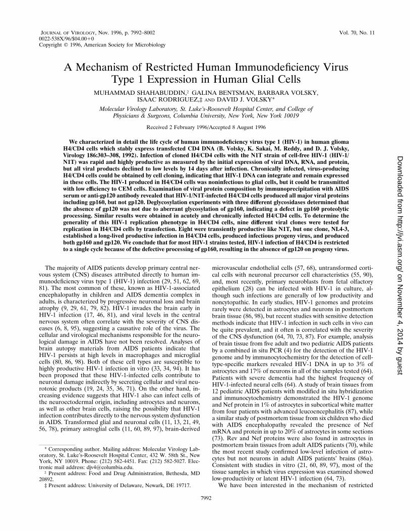
JOURNAL OF VIROLOGY, Nov. 1996, p. 7992–8002 Vol. 70, No. 110022-538X/96/$04.0010Copyright q 1996, American Society for Microbiology
A Mechanism of Restricted Human Immunodeficiency VirusType 1 Expression in Human Glial Cells
MUHAMMAD SHAHABUDDIN,† GALINA BENTSMAN, BARBARA VOLSKY,ISAAC RODRIGUEZ,‡ AND DAVID J. VOLSKY*
Molecular Virology Laboratory, St. Luke’s-Roosevelt Hospital Center, and College ofPhysicians & Surgeons, Columbia University, New York, New York 10019
Received 2 February 1996/Accepted 8 August 1996
We characterized in detail the life cycle of human immunodeficiency virus type 1 (HIV-1) in human gliomaH4/CD4 cells which stably express transfected CD4 DNA (B. Volsky, K. Sakai, M. Reddy, and D. J. Volsky,Virology 186:303–308, 1992). Infection of cloned H4/CD4 cells with the N1T strain of cell-free HIV-1 (HIV-1/N1T) was rapid and highly productive as measured by the initial expression of viral DNA, RNA, and protein,but all viral products declined to low levels by 14 days after infection. Chronically infected, virus-producingH4/CD4 cells could be obtained by cell cloning, indicating that HIV-1 DNA can integrate and remain expressedin these cells. The HIV-1 produced in H4/CD4 cells was noninfectious to glial cells, but it could be transmittedwith low efficiency to CEM cells. Examination of viral protein composition by immunoprecipitation with AIDSserum or anti-gp120 antibody revealed that HIV-1/N1T-infected H4/CD4 cells produced all major viral proteinsincluding gp160, but not gp120. Deglycosylation experiments with three different glycosidases determined thatthe absence of gp120 was not due to aberrant glycosylation of gp160, indicating a defect in gp160 proteolyticprocessing. Similar results were obtained in acutely and chronically infected H4/CD4 cells. To determine thegenerality of this HIV-1 replication phenotype in H4/CD4 cells, nine different viral clones were tested forreplication in H4/CD4 cells by transfection. Eight were transiently productive like N1T, but one clone, NL4-3,established a long-lived productive infection in H4/CD4 cells, produced infectious progeny virus, and producedboth gp160 and gp120. We conclude that for most HIV-1 strains tested, HIV-1 infection of H4/CD4 is restrictedto a single cycle because of the defective processing of gp160, resulting in the absence of gp120 on progeny virus.
The majority of AIDS patients develop primary central ner-vous system (CNS) diseases attributed directly to human im-munodeficiency virus type 1 (HIV-1) infection (29, 51, 62, 69,81). The most common of these, known as HIV-1-associatedencephalopathy in children and AIDS dementia complex inadults, is characterized by progressive neuronal loss and brainatrophy (9, 29, 61, 79, 82). HIV-1 invades the brain early inHIV-1 infection (17, 46, 81), and viral levels in the centralnervous system often correlate with the severity of CNS dis-eases (6, 8, 95), suggesting a causative role of the virus. Thecellular and virological mechanisms responsible for the neuro-logical damage in AIDS have not been resolved. Analyses ofbrain autopsy materials from AIDS patients indicate thatHIV-1 persists at high levels in macrophages and microglialcells (80, 86, 98). Both of these cell types are susceptible tohighly productive HIV-1 infection in vitro (33, 34, 94). It hasbeen proposed that these HIV-1-infected cells contribute toneuronal damage indirectly by secreting cellular and viral neu-rotoxic products (19, 24, 35, 36, 71). On the other hand, in-creasing evidence suggests that HIV-1 also can infect cells ofthe neuroectodermal origin, including astrocytes and neurons,as well as other brain cells, raising the possibility that HIV-1infection contributes directly to the nervous system dysfunctionin AIDS. Transformed glial and neuronal cells (11, 13, 21, 49,56, 78), primary astroglial cells (11, 60, 89, 97), brain-derived
microvascular endothelial cells (57, 68), untransformed corti-cal cells with neuronal precursor cell characteristics (55, 90),and, most recently, primary neuroblasts from fetal olfactoryepithelium (28) can be infected with HIV-1 in culture, al-though such infections are generally of low productivity andnoncytopathic. In early studies, HIV-1 genomes and proteinsrarely were detected in astrocytes and neurons in postmortembrain tissue (86, 98), but recent studies with sensitive detectionmethods indicate that HIV-1 infection in such cells in vivo canbe quite prevalent, and it often is correlated with the severityof the CNS dysfunction (64, 70, 73, 87). For example, analysisof brain tissue from five adult and two pediatric AIDS patientsby a combined in situ PCR (4) for the detection of the HIV-1genome and by immunocytochemistry for the detection of cell-type-specific markers revealed HIV-1 DNA in up to 3% ofastrocytes and 17% of neurons in all of the samples tested (64).Patients with severe dementia had the highest frequency ofHIV-1-infected neural cells (64). A study of brain tissues from12 pediatric AIDS patients with modified in situ hybridizationand immunocytochemistry demonstrated the HIV-1 genomeand Nef protein in 1% of astrocytes in subcortical white matterfrom four patients with advanced leucoencephalitis (87), whilea similar study of postmortem tissue from six children who diedwith AIDS encephalopathy revealed the presence of NefmRNA and protein in up to 20% of astrocytes in some sections(73). Rev and Nef proteins were also found in astrocytes inpostmortem brain tissues from adult AIDS patients (70), whilethe most recent study confirmed low-level infection of astro-cytes but not neurons in adult AIDS patients’ brains (86a).Consistent with studies in vitro (21, 60, 89, 97), most of thetissue samples in which virus expression was examined showedlow-productivity or latent HIV-1 infection (64, 73).We have been interested in the mechanism of restricted
* Corresponding author. Mailing address: Molecular Virology Lab-oratory, St. Luke’s-Roosevelt Hospital Center, 432 W. 58th St., NewYork, NY 10019. Phone: (212) 582-4451. Fax: (212) 582-5027. Elec-tronic mail address: [email protected].† Present address: Food and Drug Administration, Bethesda, MD
20892.‡ Present address: University of Delaware, Newark, DE 19717.
7992
on Novem
ber 4, 2014 by guesthttp://jvi.asm
.org/D
ownloaded from
HIV-1 replication in human glial cells in vitro as a model fordirect interaction of HIV-1 with nervous system-derived cells.In general, the pattern of HIV-1 infection of these cells in vitrodiffers radically from that in T lymphocytes (93). Cultured glialcells and primary astrocytes generally do not express surfaceCD4 (10), and HIV-1 infects these cells by a CD4-independentroute (15, 16, 38, 56). Galactosylceramide serves as an HIV-1receptor on some glial cells (5, 37). HIV-1 has been shown tofuse with (56) and enter (39, 56) CD4-negative glial cells invitro, but viral expression in these cells is about 3 orders ofmagnitude less efficient than that in T cells, and no cytopathiceffects are observed (39, 48, 56, 91). Transfection of viral DNAinto glial cells permitted a more robust HIV-1 expression thanin cells exposed to cell-free virus (21, 77, 85, 89), indicating thatthe CD4-independent mode of virus entry is one limiting factorin HIV-1 infection in these cells. However, both stable andtransient HIV-1 DNA transfectants appeared to extinguishvirus expression over time (21, 78, 88, 89), suggesting thatadditional negative regulatory mechanisms after HIV-1 entrycontribute to the restricted infection in these cells. The dor-mant HIV-1 genome in such long-term astrocytic cultures canbe induced by agents known to activate long terminal repeat-mediated gene expression, such as sodium butyrate (77) ortumor necrosis factor alpha and interleukin 1b (88, 89), indi-cating that these cells restrict HIV-1 expression at the level oflong terminal repeat-mediated transcription. Another level ofcellular regulation of HIV-1 expression has been described inthe chronically infected TH4-7-5 glial cell line derived fromHIV-1-infected 85HG-66 glioma cells (7). These cells exhibit acell-determined block of HIV-1 Rev/RRE function (45) whichcontributes to the observed low levels of viral structural pro-teins and elevated levels of Nef (63).The inefficient HIV-1 infection of glial cells in vitro and in
vivo hinders studies of the regulation of the HIV-1 life cycle inthese cells. In an attempt to circumvent this problem, we trans-fected human glioma H4 cells (HTB148) with human CD4cDNA (53) and selected cells which stably express high levelsof human CD4 (92). Coculture of stable transfectants, termedH4/CD4, with HIV-1-positive T cells permitted efficient virusreplication. Surprisingly, HIV-1 expression in H4/CD4 cellswas transient and declined to low levels typical of CD4-nega-tive glial cells within 2 to 3 weeks after infection (92). In thisreport, we present detailed characterization of the restrictedHIV-1 life cycle in these CD4-positive glial cells. Using mo-lecular clones of HIV-1 and subclones of H4/CD4 cells whichpermit efficient infection with cell-free HIV-1, we show thatthese cells support efficient but transient synthesis of HIV-1DNA and RNA and permit viral DNA integration. Examina-tion of viral protein production revealed a defect in the cleav-age of the envelope precursor glycoprotein gp160. Of ninevirus strains examined by transfection into H4/CD4 cells, eightshowed the efficient, but transient replication phenotype. Weconclude that infection of glial cells can be restricted by theproduction of progeny virus carrying only immature forms ofthe viral envelope glycoprotein.
MATERIALS AND METHODS
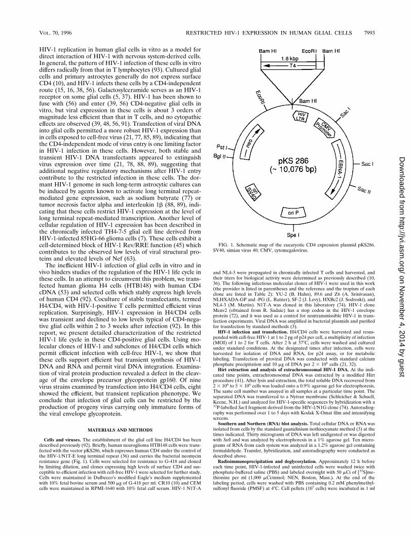
Cells and viruses. The establishment of the glial cell line H4/CD4 has beendescribed previously (92). Briefly, human neuroglioma HTB148 cells were trans-fected with the vector pKS286, which expresses human CD4 under the control ofthe HIV-1/N1T-E long terminal repeat (36) and carries the bacterial neomycinresistance gene (Fig. 1). Cells were selected for resistance to G-418 and clonedby limiting dilution, and clones expressing high levels of surface CD4 and sus-ceptible to efficient infection with cell-free HIV-1 were selected for further study.Cells were maintained in Dulbecco’s modified Eagle’s medium supplementedwith 10% fetal bovine serum and 500 mg of G-418 per ml. CR10 (10) and CEMcells were maintained in RPMI-1640 with 10% fetal calf serum. HIV-1 N1T-A
and NL4-3 were propagated in chronically infected T cells and harvested, andtheir titers for biological activity were determined as previously described (10,36). The following infectious molecular clones of HIV-1 were used in this work(the provider is listed in parentheses and the reference and the tropism of eachclone are listed in Table 2): YU-2 (B. Hahn), 89.6 and Z6 (A. Srinivasan),NLHXADA-GP and -PG (L. Ratner), SF-2 (J. Levy), HXBc2 (J. Sodroski), andNL4-3 (M. Martin). N1T-A was cloned in this laboratory (74). HIV-1 cloneMenv2 (obtained from R. Sadaie) has a stop codon in the HIV-1 envelopeprotein (72), and it was used as a control for nontransmissible HIV-1 in trans-fection experiments. Viral DNA was amplified in bacterial plasmids and purifiedfor transfection by standard methods (3).HIV-1 infection and transfection. H4/CD4 cells were harvested and resus-
pended with cell-free HIV-1 at 1 to 2 pg of p24 per cell, a multiplicity of infection(MOI) of 1 to 2 for T cells. After 2 h at 378C, cells were washed and culturedunder standard conditions. At the designated times after infection, cells wereharvested for isolation of DNA and RNA, for p24 assay, or for metaboliclabeling. Transfection of proviral DNA was conducted with standard calciumphosphate precipitation and 10 mg of DNA per 2 3 106 cells (21, 32).Hirt extraction and analysis of extrachromosomal HIV-1 DNA. At the indi-
cated time points, extrachromosomal DNA was extracted by a modified Hirtprocedure (41). After lysis and extraction, the total soluble DNA recovered from2 3 106 to 5 3 106 cells was loaded onto a 0.9% agarose gel for electrophoresis.The same cell number was assayed in all samples at a particular time point. Theseparated DNA was transferred to a Nytran membrane (Schleicher & Schuell,Keene, N.H.) and analyzed for HIV-1-specific sequences by hybridization with a32P-labelled SacI fragment derived from the HIV-1/N1G clone (74). Autoradiog-raphy was performed over 1 to 5 days with Kodak X-Omat film and intensifyingscreens.Southern and Northern (RNA) blot analysis. Total cellular DNA or RNA was
isolated from cells by the standard guanidinium isothiocyanate method (3) at thetimes indicated. Thirty micrograms of DNA was left undigested or was digestedwith SstI and was analyzed by electrophoresis in a 1% agarose gel. Ten micro-grams of RNA from each system was analyzed in a 1.2% agarose gel containingformaldehyde. Transfer, hybridization, and autoradiography were conducted asdescribed above.Radioimmunoprecipitation and deglycosylation. Approximately 12 h before
each time point, HIV-1-infected and uninfected cells were washed twice withphosphate-buffered saline (PBS) and labeled overnight with 50 mCi of [35S]me-thionine per ml (1,000 mCi/mmol; NEN, Boston, Mass.). At the end of thelabeling period, cells were washed with PBS containing 0.2 mM phenylmethyl-sulfonyl fluoride (PMSF) at 48C. Cell pellets (107 cells) were incubated in 1 ml
FIG. 1. Schematic map of the eucaryotic CD4 expression plasmid pKS286.SV40, simian virus 40; CMV, cytomegalovirus.
VOL. 70, 1996 RESTRICTED HIV-1 EXPRESSION IN HUMAN GLIAL CELLS 7993
on Novem
ber 4, 2014 by guesthttp://jvi.asm
.org/D
ownloaded from
of immunoprecipitation buffer for 30 min at 48C and centrifuged at 3,000 3 g for20 min to remove nuclei. Immunoprecipitation buffer contained 0.02 M Tris-HCl(pH 3.0), 0.12 M NaCl, 0.2 mM PMSF, 5 mg of aprotinin per ml, 0.2 mM EGTA,0.2 mM sodium fluoride, 0.2% sodium deoxycholate, and 0.5% Nonidet P-40(NP-40). Cell lysates were preabsorbed with protein G-Sepharose beads (Phar-macia, Inc., Piscataway, N.J.) for 2 h at 48C. Lysates were then incubated at 48Cfor 2 h with 100 ml of protein G-Sepharose and 5 ml of either pooled serum fromAIDS patients, sheep anti-human T-cell leukemia virus 3B gp120 antiserum(catalog number 288; AIDS Research and Reference Reagent Program) (seeFig. 9A), or goat anti-gp160 and -gp120 antiserum (catalog number 191, AIDSResearch and Reference Reagent Program) (Fig. 9B). Conjugated beads werewashed twice with a buffer containing 20 mM Tris-HCl (pH 8.0), 140 mM NaCl,and 0.2% NP-40 and once with 50 mM Tris-HCl (pH 6.8). Washed beads weresuspended in 100 mM sodium phosphate (pH 7.0)–0.1% sodium dodecyl sulfate(SDS)–1% b-mercaptoethanol and boiled for 3 min. Electrophoresis was per-formed in 8% or 10% polyacrylamide gels with a 4.5% stacking gel. Gels werefixed, dried, and autoradiographed with Kodak X-Omat film. Deglycosylationanalyses were performed on immunoprecipitated proteins by two procedures. Inthe first experiment with glycopeptidase F (PNGase F) (Boehringer MannheimBiochemicals, Indianapolis, Ind.), eluted proteins were adjusted to 1% NP-40–50mM EDTA–0.5-mg/ml pepstatin–0.2-mg/ml leupeptin–1 mM PMSF. Sampleswere incubated at 378C for 3 h in the presence or absence of 1 U of PNGase F.Samples were then resuspended in 23 polyacrylamide gel electrophoresis(PAGE) buffer (2.5% SDS, 80 mM Tris-HCl [pH 6.8], 5% b-mercaptoethanol,0.008% bromophenol blue), incubated for 30 min at 378C and 5 min at 1008C,and electrophoresed as described above. For comparison of three glycosidases,we used a procedure described by Jones et al. (42). Immunoprecipitates wereboiled for 3 min in the presence of 0.15% SDS and b-mercaptoethanol andresuspended in a 35 mM phosphate buffer (pH 6.0) containing 2.5 mM CaCl2, 1mM PMSF, and 2% NP-40, the latter added to quench SDS. The samples werecentrifuged, and supernatants were divided into aliquots and incubated at 378Cfor 16 h with no further additions or after addition of PNGase F, neuraminidase(Sigma, St. Louis, Mo.), or endoglycosidase H (Endo H1; New England Biolabs,Beverly, Mass.) at final doses, respectively, of 0.5, 0.1, and 20 U. Reactions werestopped by addition of concentrated reduced loading buffer, and samples wereboiled for 3 min and electrophoresed as described above.Sequencing of N1T-A envelope. Overlapping fragments from the N1T-A en-
velope gene were cloned into M13mp18 and M13mp19 vectors and sequenced inboth directions by the dideoxyribonucleotide chain termination method (76).Reactions were catalyzed with modified T7 DNA polymerase (Sequenase;United States Biochemical Corp., Cleveland, Ohio) according to the manufac-turer’s instructions.Other analytical assays. The levels of HIV-1 p24 core antigen were deter-
mined in culture supernatants or cell lysates with the Coulter HIV Ag kit(Hialeah, Fla.) according to the manufacturer’s instructions. An indirect immu-nofluorescence (IF) assay for HIV-1 antigens was conducted with pooled AIDSsera as previously described (10). The IF assay for glial fibrillary acidic protein(GFAP) was conducted as previously described (21) with rabbit anti-humanGFAP (Dako, Santa Barbara, Calif.).
RESULTS
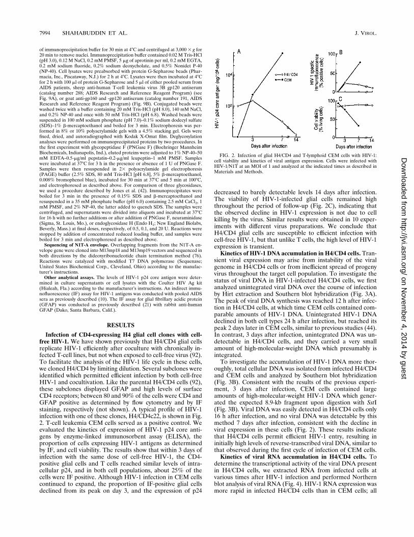
Infection of CD4-expressing H4 glial cell clones with cell-free HIV-1. We have shown previously that H4/CD4 glial cellsreplicate HIV-1 efficiently after coculture with chronically in-fected T-cell lines, but not when exposed to cell-free virus (92).To facilitate the analysis of the HIV-1 life cycle in these cells,we cloned H4/CD4 by limiting dilution. Several subclones wereidentified which permitted efficient infection by both cell-freeHIV-1 and cocultivation. Like the parental H4/CD4 cells (92),these subclones displayed GFAP and high levels of surfaceCD4 receptors; between 80 and 90% of the cells were CD4 andGFAP positive as determined by flow cytometry and by IFstaining, respectively (not shown). A typical profile of HIV-1infection with one of these clones, H4/CD4c22, is shown in Fig.2. T-cell leukemia CEM cells served as a positive control. Weevaluated the kinetics of expression of HIV-1 p24 core anti-gens by enzyme-linked immunosorbent assay (ELISA), theproportion of cells expressing HIV-1 antigens as determinedby IF, and cell viability. The results show that within 3 days ofinfection with the same dose of cell-free HIV-1, the CD4-positive glial cells and T cells reached similar levels of intra-cellular p24, and in both cell populations, about 25% of thecells were IF positive. Although HIV-1 infection in CEM cellscontinued to expand, the proportion of IF-positive glial cellsdeclined from its peak on day 3, and the expression of p24
decreased to barely detectable levels 14 days after infection.The viability of HIV-1-infected glial cells remained highthroughout the period of follow-up (Fig. 2C), indicating thatthe observed decline in HIV-1 expression is not due to cellkilling by the virus. Similar results were obtained in 10 exper-iments with different virus preparations. We conclude thatH4/CD4 glial cells are susceptible to efficient infection withcell-free HIV-1, but that unlike T cells, the high level of HIV-1expression is transient.Kinetics of HIV-1 DNA accumulation in H4/CD4 cells. Tran-
sient viral expression may arise from instability of the viralgenome in H4/CD4 cells or from inefficient spread of progenyvirus throughout the target cell population. To investigate thestatus of viral DNA in HIV-1-infected H4/CD4 cells, we firstanalyzed unintegrated viral DNA over the course of infectionby Hirt extraction and Southern blot hybridization (Fig. 3A).The peak of viral DNA synthesis was reached 12 h after infec-tion in H4/CD4 cells, at which time CEM cells contained com-parable amounts of HIV-1 DNA. Unintegrated HIV-1 DNAdeclined in both cell types 24 h after infection, but reached itspeak 2 days later in CEM cells, similar to previous studies (44).In contrast, 3 days after infection, unintegrated DNA was un-detectable in H4/CD4 cells, and they carried a very smallamount of high-molecular-weight DNA which presumably isintegrated.To investigate the accumulation of HIV-1 DNA more thor-
oughly, total cellular DNA was isolated from infected H4/CD4and CEM cells and analyzed by Southern blot hybridization(Fig. 3B). Consistent with the results of the previous experi-ment, 3 days after infection, CEM cells contained largeamounts of high-molecular-weight HIV-1 DNA which gener-ated the expected 8.9-kb fragment upon digestion with SstI(Fig. 3B). Viral DNA was easily detected in H4/CD4 cells only16 h after infection, and no viral DNA was detectable by thismethod 7 days after infection, consistent with the decline inviral expression in these cells (Fig. 2). These results indicatethat H4/CD4 cells permit efficient HIV-1 entry, resulting ininitially high levels of reverse-transcribed viral DNA, similar tothat observed during the first cycle of infection of CEM cells.Kinetics of viral RNA accumulation in H4/CD4 cells. To
determine the transcriptional activity of the viral DNA presentin H4/CD4 cells, we extracted RNA from infected cells atvarious times after HIV-1 infection and performed Northernblot analysis of viral RNA (Fig. 4). HIV-1 RNA expression wasmore rapid in infected H4/CD4 cells than in CEM cells; all
FIG. 2. Infection of glial H4/CD4 and T-lymphoid CEM cells with HIV-1:cell viability and kinetics of viral antigen expression. Cells were infected withHIV-1/N1T at an MOI of 1 and analyzed at the indicated times as described inMaterials and Methods.
7994 SHAHABUDDIN ET AL. J. VIROL.
on Novem
ber 4, 2014 by guesthttp://jvi.asm
.org/D
ownloaded from
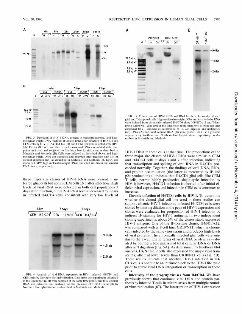
three major size classes of HIV-1 RNA were present in in-fected glial cells but not in CEM cells 16 h after infection. Highlevels of viral RNA were detected in both cell populations 3days after infection, but HIV-1 RNA levels decreased by 7 daysin infected H4/CD4 cells, consistent with very low levels of
HIV-1 DNA in these cells at that time. The proportions of thethree major size classes of HIV-1 RNA were similar in CEMand H4/CD4 cells at days 3 and 7 after infection, indicatingthat transcription and splicing of viral RNA in H4/CD4 pro-ceeded normally. Together, the findings of viral DNA, RNA,and protein accumulation (the latter as measured by IF andp24 production) all indicate that H4/CD4 glial cells, like CEMT cells, permit highly productive single-cycle infection byHIV-1; however, H4/CD4 infection is aborted after initial ef-ficient viral expression, and infection in CEM cells continues toincrease.Chronic infection of H4/CD4 cells by HIV-1. To determine
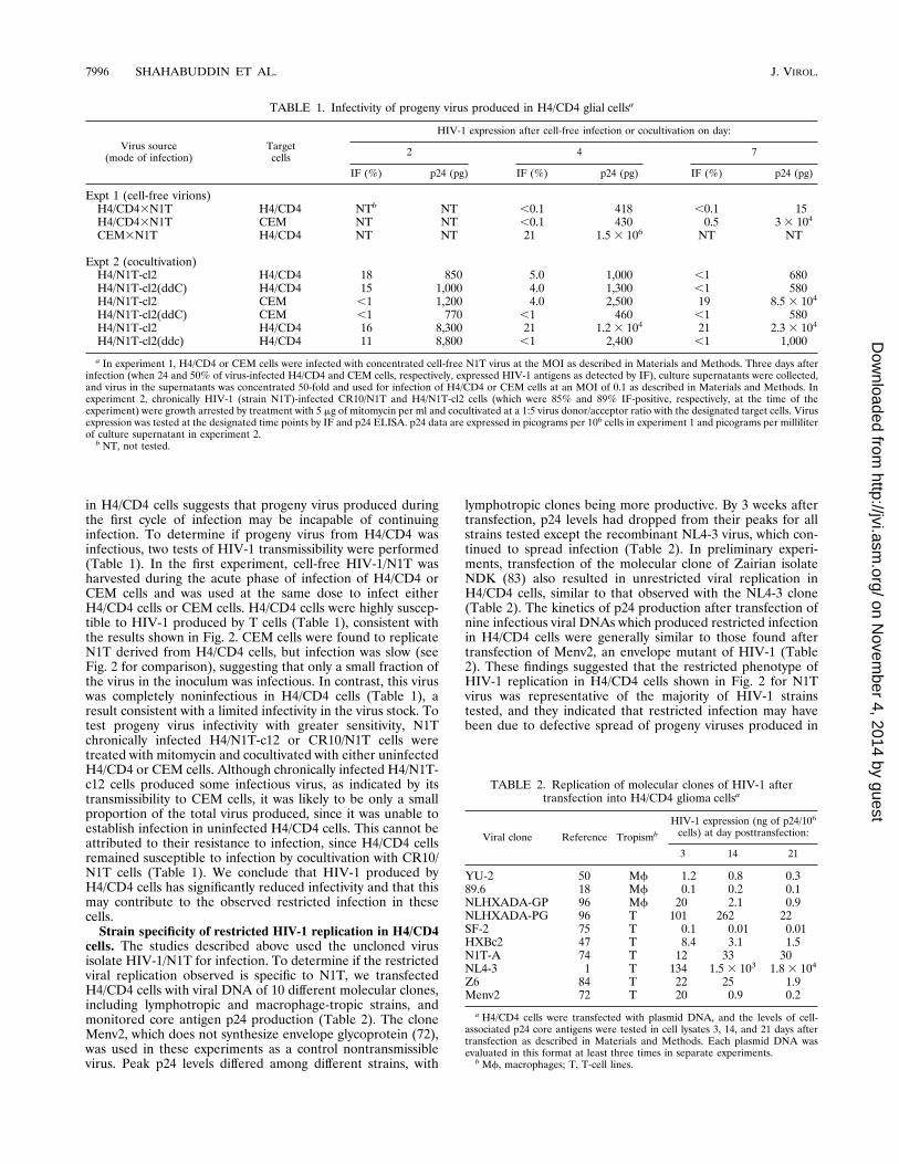
whether the cloned glial cell line used in these studies cansupport chronic HIV-1 infection, infected H4/CD4 cells werecloned by limiting dilution at the peak of HIV-1 expression andclones were evaluated for progression of HIV-1 infection byindirect IF staining for HIV-1 antigens. In two independentcloning experiments, about 5% of the clones stably expressedHIV-1 antigens. One of the IF-positive clones, H4/N1T-c12,was compared with a T-cell line, CR10/N1T, which is chroni-cally infected by the same virus strain and produces high levelsof viral proteins. The chronically infected glial cells were sim-ilar to the T-cell line in terms of viral DNA burden, as evalu-ated by Southern blot analysis of total cellular DNA or DNAafter SstI digestion (Fig. 5A). As determined by Northern blotanalysis, H4/N1T-c12 cells also expressed the major viral tran-scripts, albeit at lower levels than CR10/N1T cells (Fig. 5B).These results indicate that abortive HIV-1 infection in H4/CD4 cells is not due to an intrinsic block to the HIV-1 life cycleprior to stable viral DNA integration or transcription in thesecells.Infectivity of the progeny viruses from H4/CD4. We have
previously shown that continued viral DNA and protein syn-thesis by infected T cells in culture arises from multiple roundsof virus replication (67). The interruption of HIV-1 expression
FIG. 3. Detection of HIV-1 DNA present in extrachromosomal and high-molecular-weight DNA fractions at various times after infection of H4/CD4 andCEM cells by HIV-1. (A) H4/CD4 (H) and CEM (C) were infected with HIV-1/N1T at an MOI of 1, and then extrachromosomal DNA was isolated at the timepoints indicated and subjected to Southern blot hybridization as described inMaterials and Methods. (B) Cells were infected as described above, and high-molecular-weight DNA was extracted and analyzed after digestion with SstI orwithout digestion (un) as described in Materials and Methods. M, DNA sizemarkers; HMW, high-molecular-weight DNA; Lin. and Circ., linear and circularDNA forms, respectively.
FIG. 4. Analysis of viral RNA expression in HIV-1-infected H4/CD4 andCEM cells by Northern blot hybridization. Cells from the experiment describedin the legend to Fig. 3B were sampled at the same time points, and total cellularRNA was extracted and analyzed for the presence of HIV-1 transcripts byNorthern blot hybridization as described in Materials and Methods.
FIG. 5. Comparison of HIV-1 DNA and RNA levels in chronically infectedglial and T-lymphoid cells. High-molecular-weight DNA and total cellular RNAwere isolated from chronically infected H4/CD4 clone H4/N1T-c12 and T-lym-phoid CR10/N1T cells (10) at the time when more than 80% of both cell linesexpressed HIV-1 antigens as determined by IF. SstI-digested and undigested(un) DNA (A) and total cellular RNA (B) were probed for HIV-1 genomicsequences by Southern and Northern blot hybridization, respectively, as de-scribed in Materials and Methods.
VOL. 70, 1996 RESTRICTED HIV-1 EXPRESSION IN HUMAN GLIAL CELLS 7995
on Novem
ber 4, 2014 by guesthttp://jvi.asm
.org/D
ownloaded from
in H4/CD4 cells suggests that progeny virus produced duringthe first cycle of infection may be incapable of continuinginfection. To determine if progeny virus from H4/CD4 wasinfectious, two tests of HIV-1 transmissibility were performed(Table 1). In the first experiment, cell-free HIV-1/N1T washarvested during the acute phase of infection of H4/CD4 orCEM cells and was used at the same dose to infect eitherH4/CD4 cells or CEM cells. H4/CD4 cells were highly suscep-tible to HIV-1 produced by T cells (Table 1), consistent withthe results shown in Fig. 2. CEM cells were found to replicateN1T derived from H4/CD4 cells, but infection was slow (seeFig. 2 for comparison), suggesting that only a small fraction ofthe virus in the inoculum was infectious. In contrast, this viruswas completely noninfectious in H4/CD4 cells (Table 1), aresult consistent with a limited infectivity in the virus stock. Totest progeny virus infectivity with greater sensitivity, N1Tchronically infected H4/N1T-c12 or CR10/N1T cells weretreated with mitomycin and cocultivated with either uninfectedH4/CD4 or CEM cells. Although chronically infected H4/N1T-c12 cells produced some infectious virus, as indicated by itstransmissibility to CEM cells, it was likely to be only a smallproportion of the total virus produced, since it was unable toestablish infection in uninfected H4/CD4 cells. This cannot beattributed to their resistance to infection, since H4/CD4 cellsremained susceptible to infection by cocultivation with CR10/N1T cells (Table 1). We conclude that HIV-1 produced byH4/CD4 cells has significantly reduced infectivity and that thismay contribute to the observed restricted infection in thesecells.Strain specificity of restricted HIV-1 replication in H4/CD4
cells. The studies described above used the uncloned virusisolate HIV-1/N1T for infection. To determine if the restrictedviral replication observed is specific to N1T, we transfectedH4/CD4 cells with viral DNA of 10 different molecular clones,including lymphotropic and macrophage-tropic strains, andmonitored core antigen p24 production (Table 2). The cloneMenv2, which does not synthesize envelope glycoprotein (72),was used in these experiments as a control nontransmissiblevirus. Peak p24 levels differed among different strains, with
lymphotropic clones being more productive. By 3 weeks aftertransfection, p24 levels had dropped from their peaks for allstrains tested except the recombinant NL4-3 virus, which con-tinued to spread infection (Table 2). In preliminary experi-ments, transfection of the molecular clone of Zairian isolateNDK (83) also resulted in unrestricted viral replication inH4/CD4 cells, similar to that observed with the NL4-3 clone(Table 2). The kinetics of p24 production after transfection ofnine infectious viral DNAs which produced restricted infectionin H4/CD4 cells were generally similar to those found aftertransfection of Menv2, an envelope mutant of HIV-1 (Table2). These findings suggested that the restricted phenotype ofHIV-1 replication in H4/CD4 cells shown in Fig. 2 for N1Tvirus was representative of the majority of HIV-1 strainstested, and they indicated that restricted infection may havebeen due to defective spread of progeny viruses produced in
TABLE 1. Infectivity of progeny virus produced in H4/CD4 glial cellsa
Virus source(mode of infection)
Targetcells
HIV-1 expression after cell-free infection or cocultivation on day:
2 4 7
IF (%) p24 (pg) IF (%) p24 (pg) IF (%) p24 (pg)
Expt 1 (cell-free virions)H4/CD43N1T H4/CD4 NTb NT ,0.1 418 ,0.1 15H4/CD43N1T CEM NT NT ,0.1 430 0.5 3 3 104
CEM3N1T H4/CD4 NT NT 21 1.5 3 106 NT NT
Expt 2 (cocultivation)H4/N1T-cl2 H4/CD4 18 850 5.0 1,000 ,1 680H4/N1T-cl2(ddC) H4/CD4 15 1,000 4.0 1,300 ,1 580H4/N1T-cl2 CEM ,1 1,200 4.0 2,500 19 8.5 3 104
H4/N1T-cl2(ddC) CEM ,1 770 ,1 460 ,1 580H4/N1T-cl2 H4/CD4 16 8,300 21 1.2 3 104 21 2.3 3 104
H4/N1T-cl2(ddc) H4/CD4 11 8,800 ,1 2,400 ,1 1,000
a In experiment 1, H4/CD4 or CEM cells were infected with concentrated cell-free N1T virus at the MOI as described in Materials and Methods. Three days afterinfection (when 24 and 50% of virus-infected H4/CD4 and CEM cells, respectively, expressed HIV-1 antigens as detected by IF), culture supernatants were collected,and virus in the supernatants was concentrated 50-fold and used for infection of H4/CD4 or CEM cells at an MOI of 0.1 as described in Materials and Methods. Inexperiment 2, chronically HIV-1 (strain N1T)-infected CR10/N1T and H4/N1T-cl2 cells (which were 85% and 89% IF-positive, respectively, at the time of theexperiment) were growth arrested by treatment with 5 mg of mitomycin per ml and cocultivated at a 1:5 virus donor/acceptor ratio with the designated target cells. Virusexpression was tested at the designated time points by IF and p24 ELISA. p24 data are expressed in picograms per 106 cells in experiment 1 and picograms per milliliterof culture supernatant in experiment 2.b NT, not tested.
TABLE 2. Replication of molecular clones of HIV-1 aftertransfection into H4/CD4 glioma cellsa
Viral clone Reference TropismbHIV-1 expression (ng of p24/106
cells) at day posttransfection:
3 14 21
YU-2 50 Mf 1.2 0.8 0.389.6 18 Mf 0.1 0.2 0.1NLHXADA-GP 96 Mf 20 2.1 0.9NLHXADA-PG 96 T 101 262 22SF-2 75 T 0.1 0.01 0.01HXBc2 47 T 8.4 3.1 1.5N1T-A 74 T 12 33 30NL4-3 1 T 134 1.53 103 1.8 3 104
Z6 84 T 22 25 1.9Menv2 72 T 20 0.9 0.2
a H4/CD4 cells were transfected with plasmid DNA, and the levels of cell-associated p24 core antigens were tested in cell lysates 3, 14, and 21 days aftertransfection as described in Materials and Methods. Each plasmid DNA wasevaluated in this format at least three times in separate experiments.bMf, macrophages; T, T-cell lines.
7996 SHAHABUDDIN ET AL. J. VIROL.
on Novem
ber 4, 2014 by guesthttp://jvi.asm
.org/D
ownloaded from
these cells. These results also suggested that NL4-3 (and NDK)differ from other HIV-1 strains in the progress of infection inH4/CD4 cells. Since two of the clones which replicated tran-siently in H4/CD4 cells, 89.6 and SF-2, have intact open read-ing frames for all of the auxiliary genes of HIV-1 (41, 59, 66),the data shown in Table 2 also indicate that restricted HIV-1infection in H4/CD4 cannot be attributed to nonfunctionalVpu, Vpr, Vif, or Nef.Expression of HIV-1 proteins in H4/CD4 cells. To determine
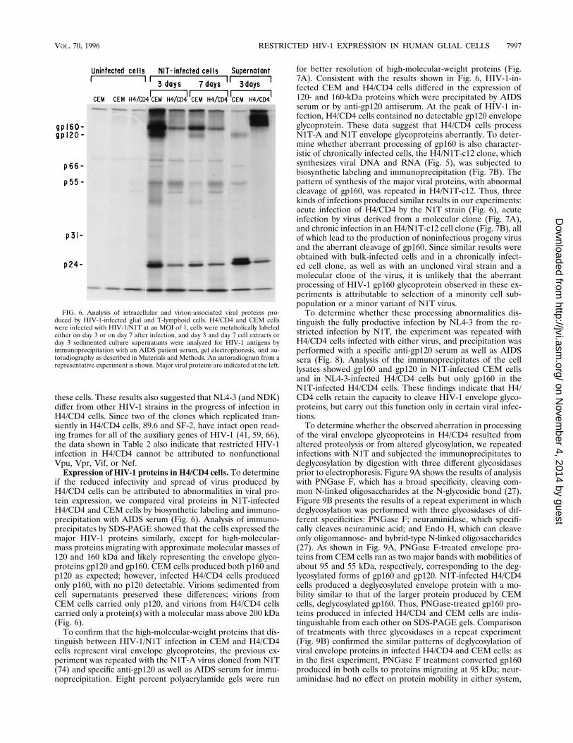
if the reduced infectivity and spread of virus produced byH4/CD4 cells can be attributed to abnormalities in viral pro-tein expression, we compared viral proteins in N1T-infectedH4/CD4 and CEM cells by biosynthetic labeling and immuno-precipitation with AIDS serum (Fig. 6). Analysis of immuno-precipitates by SDS-PAGE showed that the cells expressed themajor HIV-1 proteins similarly, except for high-molecular-mass proteins migrating with approximate molecular masses of120 and 160 kDa and likely representing the envelope glyco-proteins gp120 and gp160. CEM cells produced both p160 andp120 as expected; however, infected H4/CD4 cells producedonly p160, with no p120 detectable. Virions sedimented fromcell supernatants preserved these differences; virions fromCEM cells carried only p120, and virions from H4/CD4 cellscarried only a protein(s) with a molecular mass above 200 kDa(Fig. 6).To confirm that the high-molecular-weight proteins that dis-
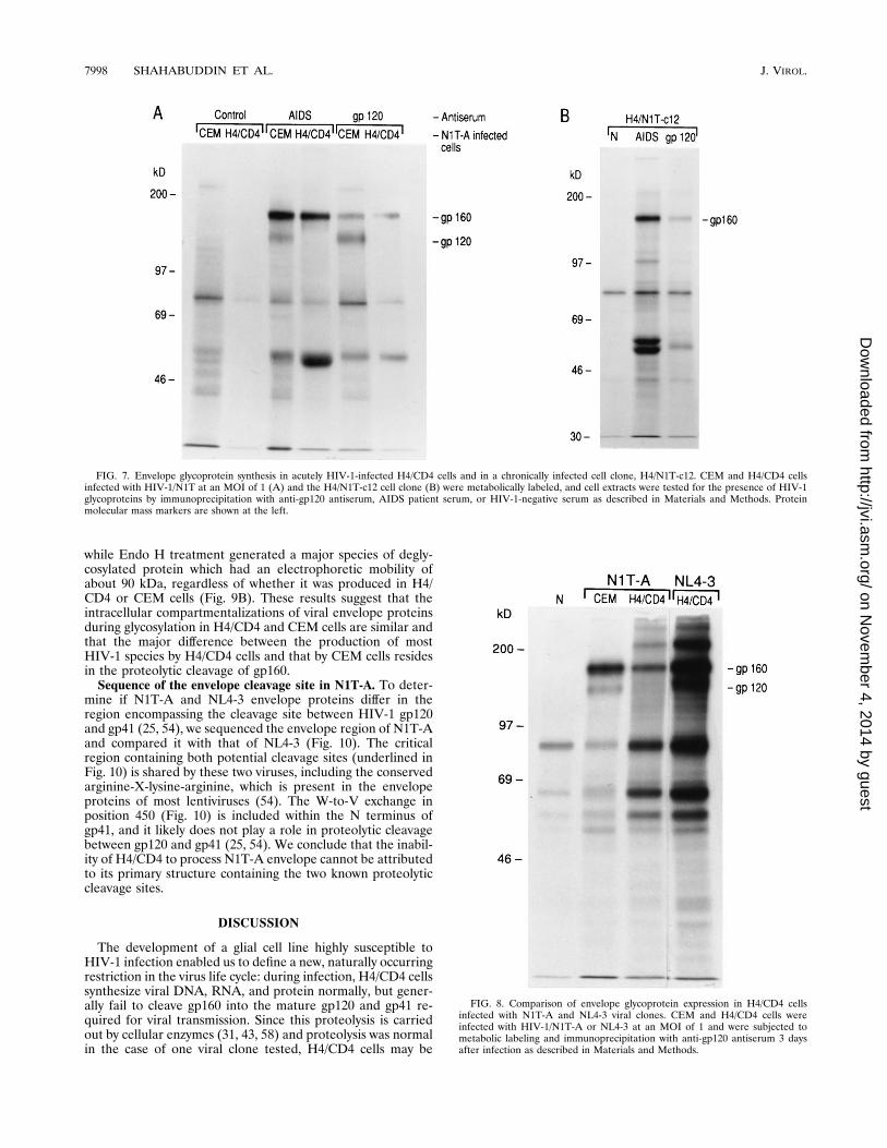
tinguish between HIV-1/N1T infection in CEM and H4/CD4cells represent viral envelope glycoproteins, the previous ex-periment was repeated with the N1T-A virus cloned from N1T(74) and specific anti-gp120 as well as AIDS serum for immu-noprecipitation. Eight percent polyacrylamide gels were run
for better resolution of high-molecular-weight proteins (Fig.7A). Consistent with the results shown in Fig. 6, HIV-1-in-fected CEM and H4/CD4 cells differed in the expression of120- and 160-kDa proteins which were precipitated by AIDSserum or by anti-gp120 antiserum. At the peak of HIV-1 in-fection, H4/CD4 cells contained no detectable gp120 envelopeglycoprotein. These data suggest that H4/CD4 cells processN1T-A and N1T envelope glycoproteins aberrantly. To deter-mine whether aberrant processing of gp160 is also character-istic of chronically infected cells, the H4/N1T-c12 clone, whichsynthesizes viral DNA and RNA (Fig. 5), was subjected tobiosynthetic labeling and immunoprecipitation (Fig. 7B). Thepattern of synthesis of the major viral proteins, with abnormalcleavage of gp160, was repeated in H4/N1T-c12. Thus, threekinds of infections produced similar results in our experiments:acute infection of H4/CD4 by the N1T strain (Fig. 6), acuteinfection by virus derived from a molecular clone (Fig. 7A),and chronic infection in an H4/N1T-c12 cell clone (Fig. 7B), allof which lead to the production of noninfectious progeny virusand the aberrant cleavage of gp160. Since similar results wereobtained with bulk-infected cells and in a chronically infect-ed cell clone, as well as with an uncloned viral strain and amolecular clone of the virus, it is unlikely that the aberrantprocessing of HIV-1 gp160 glycoprotein observed in these ex-periments is attributable to selection of a minority cell sub-population or a minor variant of N1T virus.To determine whether these processing abnormalities dis-
tinguish the fully productive infection by NL4-3 from the re-stricted infection by N1T, the experiment was repeated withH4/CD4 cells infected with either virus, and precipitation wasperformed with a specific anti-gp120 serum as well as AIDSsera (Fig. 8). Analysis of the immunoprecipitates of the celllysates showed gp160 and gp120 in N1T-infected CEM cellsand in NL4-3-infected H4/CD4 cells but only gp160 in theN1T-infected H4/CD4 cells. These findings indicate that H4/CD4 cells retain the capacity to cleave HIV-1 envelope glyco-proteins, but carry out this function only in certain viral infec-tions.To determine whether the observed aberration in processing
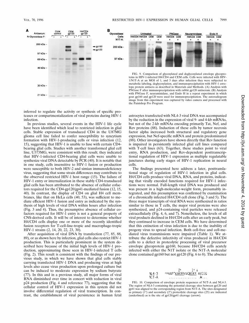
of the viral envelope glycoproteins in H4/CD4 resulted fromaltered proteolysis or from altered glycosylation, we repeatedinfections with N1T and subjected the immunoprecipitates todeglycosylation by digestion with three different glycosidasesprior to electrophoresis. Figure 9A shows the results of analysiswith PNGase F, which has a broad specificity, cleaving com-mon N-linked oligosaccharides at the N-glycosidic bond (27).Figure 9B presents the results of a repeat experiment in whichdeglycosylation was performed with three glycosidases of dif-ferent specificities: PNGase F; neuraminidase, which specifi-cally cleaves neuraminic acid; and Endo H, which can cleaveonly oligomannose- and hybrid-type N-linked oligosaccharides(27). As shown in Fig. 9A, PNGase F-treated envelope pro-teins from CEM cells ran as two major bands with mobilities ofabout 95 and 55 kDa, respectively, corresponding to the deg-lycosylated forms of gp160 and gp120. N1T-infected H4/CD4cells produced a deglycosylated envelope protein with a mo-bility similar to that of the larger protein produced by CEMcells, deglycosylated gp160. Thus, PNGase-treated gp160 pro-teins produced in infected H4/CD4 and CEM cells are indis-tinguishable from each other on SDS-PAGE gels. Comparisonof treatments with three glycosidases in a repeat experiment(Fig. 9B) confirmed the similar patterns of deglycosylation ofviral envelope proteins in infected H4/CD4 and CEM cells: asin the first experiment, PNGase F treatment converted gp160produced in both cells to proteins migrating at 95 kDa; neur-aminidase had no effect on protein mobility in either system,
FIG. 6. Analysis of intracellular and virion-associated viral proteins pro-duced by HIV-1-infected glial and T-lymphoid cells. H4/CD4 and CEM cellswere infected with HIV-1/N1T at an MOI of 1, cells were metabolically labeledeither on day 3 or on day 7 after infection, and day 3 and day 7 cell extracts orday 3 sedimented culture supernatants were analyzed for HIV-1 antigens byimmunoprecipitation with an AIDS patient serum, gel electrophoresis, and au-toradiography as described in Materials and Methods. An autoradiogram from arepresentative experiment is shown. Major viral proteins are indicated at the left.
VOL. 70, 1996 RESTRICTED HIV-1 EXPRESSION IN HUMAN GLIAL CELLS 7997
on Novem
ber 4, 2014 by guesthttp://jvi.asm
.org/D
ownloaded from
while Endo H treatment generated a major species of degly-cosylated protein which had an electrophoretic mobility ofabout 90 kDa, regardless of whether it was produced in H4/CD4 or CEM cells (Fig. 9B). These results suggest that theintracellular compartmentalizations of viral envelope proteinsduring glycosylation in H4/CD4 and CEM cells are similar andthat the major difference between the production of mostHIV-1 species by H4/CD4 cells and that by CEM cells residesin the proteolytic cleavage of gp160.Sequence of the envelope cleavage site in N1T-A. To deter-
mine if N1T-A and NL4-3 envelope proteins differ in theregion encompassing the cleavage site between HIV-1 gp120and gp41 (25, 54), we sequenced the envelope region of N1T-Aand compared it with that of NL4-3 (Fig. 10). The criticalregion containing both potential cleavage sites (underlined inFig. 10) is shared by these two viruses, including the conservedarginine-X-lysine-arginine, which is present in the envelopeproteins of most lentiviruses (54). The W-to-V exchange inposition 450 (Fig. 10) is included within the N terminus ofgp41, and it likely does not play a role in proteolytic cleavagebetween gp120 and gp41 (25, 54). We conclude that the inabil-ity of H4/CD4 to process N1T-A envelope cannot be attributedto its primary structure containing the two known proteolyticcleavage sites.
DISCUSSION
The development of a glial cell line highly susceptible toHIV-1 infection enabled us to define a new, naturally occurringrestriction in the virus life cycle: during infection, H4/CD4 cellssynthesize viral DNA, RNA, and protein normally, but gener-ally fail to cleave gp160 into the mature gp120 and gp41 re-quired for viral transmission. Since this proteolysis is carriedout by cellular enzymes (31, 43, 58) and proteolysis was normalin the case of one viral clone tested, H4/CD4 cells may be
FIG. 7. Envelope glycoprotein synthesis in acutely HIV-1-infected H4/CD4 cells and in a chronically infected cell clone, H4/N1T-c12. CEM and H4/CD4 cellsinfected with HIV-1/N1T at an MOI of 1 (A) and the H4/N1T-c12 cell clone (B) were metabolically labeled, and cell extracts were tested for the presence of HIV-1glycoproteins by immunoprecipitation with anti-gp120 antiserum, AIDS patient serum, or HIV-1-negative serum as described in Materials and Methods. Proteinmolecular mass markers are shown at the left.
FIG. 8. Comparison of envelope glycoprotein expression in H4/CD4 cellsinfected with N1T-A and NL4-3 viral clones. CEM and H4/CD4 cells wereinfected with HIV-1/N1T-A or NL4-3 at an MOI of 1 and were subjected tometabolic labeling and immunoprecipitation with anti-gp120 antiserum 3 daysafter infection as described in Materials and Methods.
7998 SHAHABUDDIN ET AL. J. VIROL.
on Novem
ber 4, 2014 by guesthttp://jvi.asm
.org/D
ownloaded from
inferred to regulate the activity or synthesis of specific pro-teases or compartmentalization of viral proteins during HIV-1infection.In previous studies, several events in the HIV-1 life cycle
have been identified which lead to restricted infection in glialcells. Stable expression of transduced CD4 in the U87MGglioma cell line failed to confer susceptibility to syncytiumformation with HIV-1-producing cells or virus infection (12,15), suggesting that HIV-1 is unable to fuse with certain CD4-bearing glial cells. Studies with another transformed glial cellline, U373MG, were consistent with this result; they indicatedthat HIV-1-infected CD4-bearing glial cells were unable tosynthesize viral DNA detectable by PCR (40). It is notable thatin one study, cells insensitive to HIV-1 fusion or productionwere susceptible to both HIV-2 and simian immunodeficiencyvirus, suggesting that some strain differences may contribute tothe observed restricted HIV-1 host range (15). The failure ofHIV-1 entry or internalization in these stably CD4-transducedglial cells has been attributed to the absence of cellular cofac-tors required for the CD4-gp120/gp41-mediated fusion (12, 15,40). In contrast, the display of CD4 on glial H4 cell mem-branes, like that on HeLa cells (40, 53), was sufficient to me-diate efficient HIV-1 fusion and entry as indicated by the syn-thesis of high levels of viral DNA within hours after infection(Fig. 3 and 4). Thus, the presumptive absence of cellular co-factors required for HIV-1 entry is not a general property ofCNS-derived cells. It will be of interest to determine whetherH4/CD4 cells display one or more of the recently identifiedfusion receptors for T-cell-line-tropic and macrophage-tropicHIV-1 strains (2, 14, 20, 22, 23, 30).After acquisition of viral DNA by transfection (77, 85, 88,
89), or as shown here by infection, glial cells also restrict HIV-1production. This is particularly prominent in the system de-scribed here because of the initial high levels of HIV-1 pro-duction, approximating those seen in HIV-1-infected T cells(Fig. 2). This result is consistent with the findings of our pre-vious study, in which we have shown that glial cells stablycarrying transfected HIV-1 DNA and producing virus at highlevels also cease virus production upon prolonged culture butcan be induced to moderate expression by sodium butyrate(77). In this and in a previous study, all major forms of viralRNA diminished over time in concert with reduction in viralp24 production (Fig. 4 and reference 77), suggesting that thecellular control of HIV-1 expression in this system did notinvolve differential regulation of viral transcription. In con-trast, the establishment of viral persistence in human fetal
astrocytes transfected with NL4-3 viral DNA was accompaniedby the reduction in the expression of viral 9- and 4-kb mRNAs,but not of the 2-kb mRNAs encoding primarily Tat, Nef, andRev proteins (88). Induction of these cells by tumor necrosisfactor alpha increased both structural and regulatory geneexpression, but Nef-specific mRNA and protein predominated(88). Other investigators have shown directly that Rev functionis impaired in persistently infected glial cell lines comparedwith T-cell lines (63). Together, these studies point to viralentry, RNA production, and Rev-dependent posttranscrip-tional regulation of HIV-1 expression as multiple regulatablejunctures during early stages of HIV-1 replication in neuralcells.The findings presented here address a later, posttransla-
tional stage of regulation of HIV-1 infection in glial cells.H4/CD4 cells produce viral DNA, RNA, and proteins, indicat-ing that virally encoded functions required for HIV-1 infec-tions were normal. Full-length viral DNA was produced andwas present in a high-molecular-weight form, presumably in-tegrated, and this presumption was confirmed by examinationof a chronically infected H4/CD4 cell clone (Fig. 3 and 5). Thethree major transcripts of viral RNA were synthesized in ratiossimilar to those in T cells, the major viral proteins were alsosynthesized, and p24-containing viral particles were releasedextracellularly (Fig. 4, 6, and 7). Nonetheless, the levels of allviral products declined in H4/CD4 cells after an early peak, butthey continued to increase in CEM cells. We have determinedthat this extinction of virus infection is due to the inability ofprogeny virus to spread infection. Both cell-free and cell-me-diated virus transmissions were impaired (Table 1). We at-tribute the defective infectivity of virus produced in H4/CD4cells to a defect in proteolytic processing of viral precursorenvelope glycoprotein gp160, because H4/CD4 cells acutelyinfected with either the N1T isolate or the N1T-A molecularclone contained gp160 but not gp120 (Fig. 6 to 8). The absence
FIG. 9. Comparison of glycosylated and deglycosylated envelope glycopro-teins in HIV-1-infected H4/CD4 and CEM cells. Cells were infected with HIV-1/N1T-A at an MOI of 1, and 3 days after infection they were subjected tometabolic labeling, deglycosylation, and immunoprecipitation with HIV-1 enve-lope protein antisera as described in Materials and Methods. (A) Analysis withPNGase F after immunoprecipitation with rabbit gp120 antiserum. (B) Analysiswith PNGase F, neuraminidase, and Endo H in a repeat experiment in whichgoat gp160 and gp120 were used for immunoprecipitation. The autoradiogramimage from this experiment was captured by video camera and processed withthe Paintshop Pro Program.
FIG. 10. Comparison of envelope protein sequences of N1T-A and NL4-3.The region of NL4-3 containing the potential cleavage sites between gp120 andgp41 was aligned to the corresponding region from N1T-A. The sites designatedas primary (18) and secondary (28) proteolytic cleavage sites (25) are indicated(underlined) as is the site of gp120/gp41 cleavage (arrow).
VOL. 70, 1996 RESTRICTED HIV-1 EXPRESSION IN HUMAN GLIAL CELLS 7999
on Novem
ber 4, 2014 by guesthttp://jvi.asm
.org/D
ownloaded from
of gp120 could not be attributed to defective glycosylation ofthe precursor envelope protein, because deglycosylation exper-iments with glycosidases of different specificities did not revealsignificant differences in electrophoretic mobilities of precur-sor envelope proteins made in H4/CD4 and CEM cells (Fig. 9).Blocking of gp160 cleavage by mutations in the proteolyticcleavage site (25, 54, 99, 100) or in T cells unable to processgp160 (26) resulted in the production of noninfectious HIV-1.Although there is controversy whether uncleaved gp160 can beincorporated into virions (25, 100), it is accepted that un-cleaved gp160 cannot mediate fusion with CD4-positive cells(25, 26, 99, 100). We conclude that the failure to cleave gp160limits HIV-1 infection in H4/CD4 cells to one or few rounds ofreplication, similar to the restricted viral spread observed aftertransfection of HIV-1 env-negative mutant DNA (Table 2).Since the N1T virus produced in H4/CD4 could eventually berescued by culture in T cells (Table 1), the blocking of gp160proteolytic processing in these glial cells is not absolute, and itmay differ in its extent for different viral clones (Table 2). It hasbeen reported that during infection of T lymphocytes, only asmall fraction of gp160 is cleaved to mature gp120 and gp41(99), and H4/CD4 may reduce this fraction still further, to alevel which does not permit spread of infection. Whatever themechanism, the defective gp160 processing during N1T infec-tion is a general feature of H4/CD4 cells, because it was ob-served both during acute infection of bulk cell populations andin a chronically infected H4/CD4 cell clone.The restricted HIV-1 infection in H4/CD4 cells was ob-
served with eight of nine infectious HIV-1 clones tested. Oneclone, NL4-3, was able to produce efficiently spreading infec-tion, and NL4-3-infected H4/CD4 cells expressed both gp160and gp120, suggesting that gp160 cleavage occurred normally(Table 1 and Fig. 8). In preliminary experiments, we found thatthe molecular clone of an African HIV-1 isolate, NDK (83),also exhibited an NL4-3-like pattern of replication in H4/CD4cells. This indicates that the absence of cleavage of gp160 inH4/CD4 is virus strain specific, and it is common to the ma-jority of the HIV-1 strains tested. It is puzzling that H4/CD4cells discriminate between highly homologous polypeptides forcleavage (Fig. 10). Even NLHXADA-PG, which is essentiallyNL4-3 with a homologous HXB-2 replacement and a smallheterologous ADA replacement (96), replicated only tran-siently in H4/CD4. The cellular proteases which catalyze thegp160 cleavage have not been fully identified (31, 43, 58, 65),and H4/CD4 cells may express a rare enzyme which can rec-ognize a presently unknown cleavage motif in NL4-3 (andNDK) envelope proteins. Alternatively, NL4-3 and NDK maydiffer from other HIV-1 clones tested here in the intracellulartrafficking of precursor envelope protein such that greater frac-tions of gp160 are processed rather than being degraded inlysosomes (99). The means to control the efficiency of envelopeprocessing is currently under investigation.We have described a novel, posttranslational mechanism
responsible for a virus strain-specific restriction of HIV-1 in-fection in CD4-expressing human glial cells in vitro, namely adefective proteolytic processing of precursor envelope glyco-proteins. A similar mechanism was found to be responsible forthe restricted infection of microglial cells by a highly neuro-virulent murine retrovirus, FrCasE (52). Our data add to thegrowing body of evidence indicating that human neural cellscontrol HIV-1 infection by several different mechanisms. It isunclear at present which specific mechanism(s) is responsiblefor the observed restricted HIV-1 infection of astrocytes (73,87) and possibly other neural cells (64) in vivo. Our attempts atbiochemical analysis of envelope glycoproteins in HIV-1-in-fected CD4-negative U-257 glial cells and primary astrocytes
were hampered by inefficient expression of viral proteins inthese cells (92a). Use of H4/CD4 cells, which as shown here arepermissive for efficient primary infection by HIV-1, will permitdetailed molecular studies of the control of HIV-1 proteinexpression in nervous system-derived cells and may lead to thedevelopment of novel ways to control HIV-1 replication.
ACKNOWLEDGMENTS
The CD4 expression plasmid pKS286 was constructed by K. Sakai.We are grateful to the following investigators for providing the HIV-1proviral DNAs used in this work: B. Hahn, J. Levy, M. Martin, L.Ratner, R. Sadaie, J. Sodroski, and A. Srinivasan. We thank the AIDSReference Reagent Repository for sheep gp120 and goat gp160 andgp120 antisera, P. Sova for help with image analysis, M. J. Potash forunrestricted comments, and L. Peters for typing the manuscript.This work was supported by grant NS PO1-31492 from the Public
Health Service.
REFERENCES
1. Adachi, A., H. E. Gendelman, S. Koenig, T. Folks, R. Willey, A. Rabson, andM. A. Martin. 1986. Production of acquired immunodeficiency syndrome-associated retrovirus in human and nonhuman cells transfected with aninfectious molecular clone. J. Virol. 59:284–291.
2. Alkhatib, G., C. Combadiere, C. C. Broder, Y. Feng, P. E. Kennedy, P. M.Murphy, and E. A. Bergert. 1996. CC CKR5: a RANTES, MIP-1a, MIP-1breceptor as a fusion cofactor for macrophage-tropic HIV-1. Science 272:1955–1958.
3. Ausubel, F. M., R. Brent, R. E. Kingston, D. D. Moore, J. G. Seidman, J. A.Smith, and K. Struhl. 1987. Current protocols in molecular biology. GreenePublishing Associates and Wiley Interscience, New York.
4. Bagasra, O., S. P. Hauptman, H. W. Lischer, M. Sachs, and R. J. Pomer-antz. 1992. Detection of human immunodeficiency virus type 1 provirus inmononuclear cells by in situ polymerase chain reaction. N. Engl. J. Med.326:1385–1391.
5. Bhat, S., S. L. Spitalnik, F. Gonzalez-Scarano, and D. H. Silberberg. 1991.Galactosyl ceramide or a derivative is an essential component of the neuralreceptor for human immunodeficiency virus type 1 envelope glycoproteingp120. Proc. Natl. Acad. Sci. USA 88:7131–7134.
6. Boni, J., B. S. Emmerich, S. L. Leib, O. D. Wiestler, J. Scupbach, and P.Kleihues. 1992. PCR identification of HIV-1 DNA sequences in brain tissueof patients with AIDS encephalopathy. Neurology 43:1813–1817.
7. Brack-Werner, R., A. Kleinschmidt, A. Ludvigsen, W. Mellert, M. Neu-mann, R. Hermann, M. C. L. Khim, A. Burny, N. Muller-Lantzsch, D.Stavrou, and V. Erfle. 1992. Infection of human brain cells by HIV-1:restricted virus production in chronically infected human glial cell lines.AIDS 6:273–285.
8. Bruce, J., M. Rosenblum, K. Cronin, and R. W. Price. 1995. AIDS dementiacomplex and HIV-1 brain infection: clinical-virological correlations. Ann.Neurol. 38:563–570.
9. Budka, H. 1991. Neuropathology of human immunodeficiency virus infec-tion. Brain Pathol. 1:163–175.
10. Casareale, D., M. Stevenson, K. Sakai, and D. J. Volsky. 1987. A humanT-cell line resistant to cytopathic effects of the human immunodeficiencyvirus (HIV). Virology 156:40–49.
11. Cheng-Mayer, C., J. T. Rutka, M. L. Rosenblum, T. McHugh, D. P. Stites,and J. A. Levy. 1987. Human immunodeficiency virus can productivelyinfect cultured human glial cells. Proc. Natl. Acad. Sci. USA 84:3526–3530.
12. Chesebro, B., R. Buller, J. Portis, and K. Wehrly. 1990. Failure of humanimmunodeficiency virus entry and infection in CD-4-positive human brainand skin cells. J. Virol. 64:215–221.
13. Chiodi, F., S. Fuerstenberg, M. Gidlund, B. Åsjo, and E. M. Fenyo. 1987.Infection of brain-derived cells with the human immunodeficiency virus.J. Virol. 61:1244–1247.
14. Choe, H., M. Farzan, Y. Sun, N. Sullivan, B. Rollins, P. D. Ponath, L. Wu,C. R. Mackay, G. LaRosa, W. Newman, N. Gerard, C. Gerard, and J.Sodroski. 1996. The b-chemokine receptors CCR3 and CCR5 facilitateinfection by primary HIV-1 isolates. Cell 85:1135–1148.
15. Clapham, P. R., D. Blanc, and R. A. Weiss. 1991. Specific cell surfacerequirements for the infection of CD4-positive cells by human immunode-ficiency virus type 1 and 2 and by simian immunodeficiency virus. Virology181:703–715.
16. Clapham, P. R., J. N. Weber, D. Whitby, K. McIntosh, A. G. Dalgleish, P. J.Maddon, K. C. Deen, R. W. Sweet, and R. A. Weiss. 1989. Soluble CD4blocks the infectivity of diverse strains of HIV SIV for T cells and mono-cytes but not for brain and muscle cells. Nature (London) 337:368–370.
17. Coburn, K. L., N. C. Moore, H. P. Katner, K. A. Tucker, W. S. Pritchard,and D. W. Duke. 1992. HIV and the brain: evidence of early involvementand progressive damage. Neuroreport 3:429–541.
8000 SHAHABUDDIN ET AL. J. VIROL.
on Novem
ber 4, 2014 by guesthttp://jvi.asm
.org/D
ownloaded from
18. Collman, R., J. W. Balliet, S. A. Gregory, H. Friedman, D. L. Kolson, N.Nathanson, and A. Srinivasan. 1992. An infectious molecular clone of anunusual macrophage-tropic and highly cytopathic strain of human immu-nodeficiency virus type 1. J. Virol. 66:7517–7521.
19. Dawson, V. L., T. M. Dawson, G. R. Uhl, and S. H. Snyder. 1993. Humanimmunodeficiency virus type 1 coat protein neurotoxicity mediated by nitricoxide in primary cortical cultures. Proc. Natl. Acad. Sci. USA 90:3256–3259.
20. Deng, H., R. Liu, W. Ellmeier, S. Choe, D. Unutmaz, M. Burkhart, P. DiMarzio, S. Marmon, R. E. Sutton, C. M. Hill, C. B. Davis, S. C. Peiper, T. J.Schall, D. R. Littman, and N. R. Landau. 1996. Identification of a majorco-receptor for primary isolates of HIV-1. Nature (London) 381:661–666.
21. Dewhurst, S., K. Sakai, J. Bresser, M. Stevenson, M. J. Evinger-Hodges,and D. J. Volsky. 1987. Persistent productive infection of human glial cellsby human immunodeficiency virus (HIV) and by infectious molecularclones of HIV. J. Virol. 61:3774–3782.
22. Doranz, B. J., J. Rucker, Y. Yi, R. J. Smyth, M. Samson, S. C. Peiper, M.Parmentier, R. G. Collman, and R. W. Doms. 1996. A dual-tropic primaryHIV-1 isolate that uses fusin and the b-chemokine receptors CKR-5,CKR-3, and CKR-2b as fusion cofactors. Cell 85:1149–1158.
23. Dragic, T., V. Litwin, G. P. Allaway, S. R. Martin, Y. Huang, K. A. Na-gashima, C. Cayanan, P. J. Maddon, R. A. Koup, J. P. Moore, and W. A.Paxton. 1996. HIV-1 entry into CD41 cells is mediated by the chemokinereceptors CC-CKR-5. Nature (London) 381:667–673.
24. Dreyer, E. B., P. K. Kaiser, J. T. Offermann, and S. A. Lipton. 1990. HIV-1coat protein neurotoxicity prevented by calcium channel antagonists. Sci-ence 248:364–367.
25. Dubay, J. W., S. R. Dubay, H.-J. Shin, and E. Hunter. 1995. Analysis of thecleavage site of the human immunodeficiency virus type 1 glycoprotein:requirement of precursor cleavage for glycoprotein incorporation. J. Virol.69:4675–4682.
26. Duensing, T. D., H. Fang, D. W. Dorward, and S. H. Pincus. 1995. Pro-cessing of the envelope glycoprotein gp160 in immunotoxin-resistant celllines chronically infected with human immunodeficiency virus type 1. J. Vi-rol. 69:7122–7131.
27. Dwek, R. A., C. J. Edge, D. J. Harvey, and M. R. Wormald. 1993. Analysisof glycoprotein-associated oligosaccharides. Annu. Rev. Biochem. 62:65–100.
28. Ensoli, F., A. Cafaro, V. Fiorelli, B. Vannelli, B. Ensoli, and C. J. Thiele.1995. HIV-1 infection of primary human neuroblasts. Virology 210:221–225.
29. Epstein, L. G., L. R. Sharer, V. V. Joshi, M. M. Fojas, M. R. Koenigsberger,and J. M. Oleske. 1985. Progressive encephalopathy in children with ac-quired immune deficiency syndrome. Ann. Neurol. 17:488–496.
30. Feng, Y., C. C. Broder, P. E. Kennedy, and E. A. Berger. 1996. HIV-1 entrycofactor: functional cDNA cloning of a seven-transmembrane, G protein-coupled receptor. Science 272:872–877.
31. Franzusoff, A., A. M. Volpe, D. Josse, S. Pichuantes, and J. R. Wolf. 1995.Biochemical and genetic definition of the cellular protease required forHIV-1 gp160 processing. J. Biol. Chem. 270:3154–3159.
32. Gartner, S., P. Markovits, D. M. Markovits, M. H. Kaplan, R. C. Gallo, andM. Popovic. 1986. The role of mononuclear phagocytes in HTLV-III/LAVinfection. Science 233:215–219.
33. Gendelman, H. E., J. M. Orenstein, M. A. Martin, C. Ferrua, M. Mitra, T.Phipps, L. Wahl, H. C. Lane, A. S. Fauci, I. S. Burke, D. Skillman, and M.Meltzer. 1988. Efficient isolation and propagation of human immunodefi-ciency virus on recombinant colony-stimulating factor 1-treated monocytes.J. Exp. Med. 167:1428–1434.
34. Genis, P., M. Jett, E. W. Berton, T. Boyle, H. A. Gelbard, K. Dzenko, R. W.Keane, L. Resnick, Y. Mizrachi, D. J. Volsky, L. G. Epstein, and H. E.Gendelman. 1992. Cytokines and arachidonic metabolites produced duringhuman immunodeficiency virus (HIV)-infected macrophage-astroglia inter-actions: implications for the neuropathogenesis of HIV disease. J. Exp.Med. 176:1703–1718.
35. Giulian, D., K. Vaca, and C. A. Noonan. 1990. Secretion of neurotoxins bymononuclear phagocytes infected with HIV-1. Science 250:1593–1596.
36. Golub, E. I., G. Li, and D. J. Volsky. 1990. Differences in the basal activityof the long terminal repeat determine different replicative capacities of twoclosely related human immunodeficiency virus type 1 isolates. J. Virol.64:3654–3660.
37. Harouse, J. M., S. Bhat, S. L. Spitalnik, M. Laughlin, K. Stefano, D. H.Silberg, and F. Gonzalez-Scarano. 1991. Inhibition of entry of HIV-1 neu-ral cell lines by antibodies against galactosyl ceramide. Science 253:320–322.
38. Harouse, J. M., C. Kunsch, H. T. Hartle, M. A. Laughlin, J. A. Hoxie, B.Wigdahl, and F. Gonzalez-Scarano. 1989. CD4-independent infection ofhuman neural cells by human immunodeficiency virus type 1. J. Virol.63:2527–2533.
39. Harouse, J. M., M. A. Laughlin, C. Pletcher, H. M. Friedman, and F.Gonzalez-Scarano. 1991. Entry of human immunodeficiency virus-1 intoglial cells proceeds via an alternate, efficient pathway. J. Leukocyte Biol.49:605–609.
40. Harrington, R. D., and A. P. Geballe. 1993. Cofactor requirement for
human immunodeficiency virus type 1 into a CD4-expressing human cellline. J. Virol. 67:5939–5947.
41. Hirt, B. 1967. Selective extraction of polyoma DNA from infected mousecell culture. J. Mol. Biol. 26:365–369.
42. Jones, W. M., B. Walcheck, and M. A. Jutila. 1996. Generation of a new gdT cell-specific monoclonal antibody (GD3.5). J. Immunol. 156:3772–3779.
43. Kido, H., K. Kamoshita, A. Fukutomi, and N. Katunuma. 1993. Processingprotease for gp160 human immunodeficiency virus type 1 envelope glyco-protein precursor in human T41 lymphocytes. J. Biol. Chem. 268:13406–13413.
44. Kim, S., R. Byrn, J. Groopman, and D. Baltimore. 1989. Temporal aspectsof DNA and RNA synthesis during human immunodeficiency virus infec-tion: evidence for differential gene expression. J. Virol. 63:3708–3713.
45. Kleinschmidt, A., M. Neumann, C. Moller, V. Erfle, and R. Brack-Werner.1994. Restricted expression of HIV-1 in human astrocytes: molecular basisfor viral persistence in the CNS. Res. Virol. 145:147–153.
46. Koenig, S., H. E. Gendelman, J. M. Orenstein, M. C. Dal Canto, G. H.Pezeshkpour, M. Yungbluth, F. Janotta, A. Aksamit, M. A. Martin, andA. S. Fauci. 1986. Detection of AIDS virus in macrophages in brain tissuefrom AIDS patients with encephalopathy. Science 233:1089–1093.
47. Kowalski, M., L. Bergeron, T. Dorfman, W. Haseltine, and J. Sodroski.1991. Attenuation of human immunodeficiency virus type 1 cytopathiceffect by a mutation affecting the transmembrane envelope glycoprotein.J. Virol. 65:281–291.
48. Kunsch, C., and B. Wigdahl. 1991. Maintenance of human immunodefi-ciency virus type-1 proviral DNA in human fetal dorsal root ganglia neuralcells following a nonproductive infection. J. Leukocyte Biol. 49:505–510.
49. Li, X. L., T. Moudgil, H. V. Vinters, and D. D. Ho. 1990. CD4-independent,productive infection of a neuronal cell line by human immunodeficiencyvirus type 1. J. Virol. 64:1383–1387.
50. Li, Y., J. C. Kappes, J. A. Conway, R. W. Price, G. M. Shaw, and B. H.Hahn. 1991. Molecular characterization of human immunodeficiency virustype 1 cloned directly from uncultured human brain tissue: identification ofreplication-competent and -defective viral genomes. J. Virol. 65:3973–3985.
51. Lipton, S. A., and H. E. Gendelman. 1995. Dementia associated with theacquired immunodeficiency syndrome. N. Engl. J. Med. 332:934–940.
52. Lynch, W. P., W. J. Brown, G. J. Spangrude, and J. L. Portis. 1994.Microglial infection by a neurovirulent murine retrovirus results in defec-tive processing of envelope protein and intracellular budding of virus par-ticles. J. Virol. 68:3401–3409.
53. Maddon, P. J., A. G. Dalgleish, J. S. McDougal, P. R. Clapman, R. A. Weiss,and R. Axel. 1986. The T4 gene encodes the AIDS virus receptor and isexpressed in the immune system and the brain. Cell 47:333–348.
54. McCune, J. M., L. B. Rabin, M. B. Feinberg, M. Leiberman, J. C. Kosek,G. R. Reyes, and I. L. Weissman. 1988. Endoproteolytic cleavage of gp160is required for the activation of human immunodeficiency virus. Cell 53:55–66.
55. Mizrachi, Y., I. Rodriguez, P. M. Sweetnam, A. Rubinstein, and D. J.Volsky. 1994. HIV type 1 infection of human cortical neuronal cells: en-hancement by select neuronal growth factors. AIDS Res. Hum. Retrovi-ruses 10:1593–1596.
56. Mizrachi, Y., M. Zeira, M. Shahabuddin, G. Li, F. Sinangil, and D. J.Volsky. 1991. Efficient binding, fusion and entry of HIV-1 into CD4 nega-tive neural cells: a mechanism for neuropathogenesis in AIDS. Bull. Inst.Pasteur 89:81–96.
57. Moses, A. V., F. E. Bloom, C. D. Pauza, and J. A. Nelson. 1993. Humanimmunodeficiency virus infection of human brain capillary endothelial cellsoccurs via a CD4/galactosylceramide-independent mechanism. Proc. Natl.Acad. Sci. USA 90:10474–10478.
58. Moulard, M., T. Achstetter, Y. Ikehara, and E. Baharaoui. 1994. T4-lym-phocyte endoprotease responsible for the proteolytic processing of HIV-1gp160, like Kex2p endoprotease, is a calcium-dependent enzyme. Biochimie76:251–256.
59. Myers, G., B. Korber, J. A. Berzofsky, T. F. Smith, and G. N. Pavlakis. 1995.Human retroviruses and AIDS. Los Alamos National Laboratory, LosAlamos, N.Mex.
60. Nath, A., V. Hartloper, M. Furer, and K. R. Fowke. 1995. Infection ofhuman fetal astrocytes with HIV-1: viral tropism and the role of cell to cellcontact in viral transmission. J. Neuropathol. Exp. Neurol. 54:320–330.
61. Navia, B. A., B. D. Jordan, and R. W. Price. 1986. The AIDS dementiacomplex. I. Clinical features. Ann. Neurol. 19:517–524.
62. Navia, B. A., and R. W. Price. 1987. The acquired immunodeficiency syn-drome dementia complex as the presenting or sole manifestation of humanimmunodeficiency virus type infection. Arch. Neurol. 44:65–69.
63. Neumann, M., B. K. Felber, A. Kleinschmidt, B. Froese, V. Erfle, G. N.Pavlakis, and R. Brack-Werner. 1995. Restriction of human immunodefi-ciency virus type 1 production in a human astrocytoma cell line is associatedwith a cellular block in Rev function. J. Virol. 69:2159–2167.
64. Nuovo, G. J., F. Gallery, P. MacConnell, and A. Braun. 1994. In situdetection of polymerase chain reaction-amplified HIV-1 nucleic acids andtumor necrosis factor-a RNA in the central nervous system. Am. J. Pathol.144:659–666.
VOL. 70, 1996 RESTRICTED HIV-1 EXPRESSION IN HUMAN GLIAL CELLS 8001
on Novem
ber 4, 2014 by guesthttp://jvi.asm
.org/D
ownloaded from
65. Ohnishi, Y., T. Shioda, K. Nakayama, S. Iwata, B. Gotoh, M. Hamaguchi,and Y. Nagai. 1994. A furin-defective cell line is able to process correctlythe gp160 of human immunodeficiency virus type 1. J. Virol. 68:4075–4079.
66. Pang, S., Y. Koyanagi, S. Miles, C. Wiley, H. V. Vinters, and I. S. Y. Chen.1990. High levels of unintegrated HIV-1 DNA in brain tissue of AIDSdementia patients. Nature (London) 343:85–89.
67. Pellegrino, M. G., G. Li, M. J. Potash, and D. J. Volsky. 1991. Contributionof multiple rounds of viral entry and reverse transcription to expression ofhuman immunodeficiency virus type 1. J. Biol. Chem. 266:1783–1788.
68. Poland, S. D., G. P. A. Rice, and G. A. Dekaban. 1995. HIV-1 infection ofhuman brain-derived microvascular endothelial cells in vitro. J. AcquiredImmune Defic. Syndr. Hum. Retroviruses 8:437–445.
69. Price, R. W., B. Brew, J. Sidtis, M. Rosenblum, A. C. Scheck, and P. Cleary.1988. The brain in AIDS: central nervous system HIV-1 infection and AIDSdementia complex. Science 239:586–592.
70. Ranki, A., M. Nyberg, V. Ovod, M. Haltia, I. Elovaara, R. Raininko, H.Haapasalo, and K. Krohn. 1995. Abundant expression of HIV Nef and Revproteins in brain astrocytes in vivo is associated with dementia. AIDS9:1001–1008.
71. Sabatier, J. M., E. Vives, K. Mabrouk, A. Benjouad, H. Rochat, A. Duval,B. Hue, and E. Bahraoui. 1991. Evidence for neurotoxic activity of tat fromhuman immunodeficiency virus type 1. J. Virol. 65:961–967.
72. Sadaie, M. R., V. S. Kalyanaraman, R. Mukopadhayaya, E. Tschachler,R. C. Gallo, and F. Wong-Staal. 1992. Biological characterization of non-infectious HIV-1 particles lacking the envelope protein. Virology 187:604–611.
73. Saito, Y., L. R. Sharer, L. G. Epstein, J. Michaels, M. Mintz, M. Louder, K.Golding, T. A. Cvetkovich, and B. M. Blumberg. 1994. Overexpression ofnef as a marker for restricted HIV-1 infection of astrocytes in postmortempediatric central nervous tissues. Neurology 44:474–481.
74. Sakai, K., S. Dewhurst, X. Ma, and D. J. Volsky. 1988. Differences incytopathogenicity and host cell range among infectious molecular clones ofhuman immunodeficiency virus type 1 simultaneously isolated from anindividual. J. Virol. 62:4078–4085.
75. Sanchez-Pescador, R., M. D. Power, P. J. Barr, K. S. Steimer, M. M.Stempien, S. L. Brown-Shimer, W. W. Gee, A. Renard, A. Randolph, J. A.Levy, D. Dina, and P. A. Luciw. 1985. Nucleotide sequence and expressionof an AIDS-associated retrovirus. Science 227:484–492.
76. Sanger, F., S. Nicklen, and A. R. Coulson. 1977. DNA sequencing withchain-terminating inhibitors. Proc. Natl. Acad. Sci. USA 74:5463–5467.
77. Shahabuddin, M., B. Volsky, H. Kim, K. Sakai, and D. J. Volsky. 1992.Regulated expression of human immunodeficiency virus type 1 in humanglial cells: induction of dormant virus. Pathobiology 60:195–205.
78. Shapshak, P., N. C. J. Sun, L. Resnick, J. T. Thornthwaite, P. Schiller, M.Yoshioka, A. Svenningsson, W. W. Tourtellotte, and D. T. Imagawa. 1991.HIV-1 propagates in human neuroblastoma cells. J. Acquired ImmuneDefic. Syndr. 4:228–237.
79. Sharer, L. R. 1992. Pathology of HIV-1 infection of the central nervoussystem. J. Neuropathol. Exp. Neurol. 51:3–11.
80. Sharer, L. R., E. S. Cho, and L. G. Epstein. 1985. Multinucleated giant cellsand HTLV-III in AIDS encephalopathy. Hum. Pathol. 16:760.
81. Shaw, G. M., M. E. Harper, and B. E. Hahn. 1985. HTLV-III infection inbrains of children and adults with AIDS encephalopathy. Science 1:177–182.
82. Spencer, D. C., and R. W. Price. 1992. Human immunodeficiency virus andthe central nervous system. Annu. Rev. Microbiol. 46:655–693.
83. Spire, B., J. Sire, V. Zachar, F. Rey, F. Barre-Sinoussi, F. Galibert, A.Hampe, and J. C. Chermann. 1989. Nucleotide sequence of HIV-1-NDK: ahighly cytopathic strain of the human immunodeficiency virus. Gene 81:275–284.
84. Srinivasan, A., R. Anand, D. York, P. Ranganathan, P. Feorino, G.Schochetman, J. Curran, V. S. Kalyanaraman, P. A. Luciw, and R.Sanchez-Pescador. 1987. Molecular characterization of human immunode-
ficiency virus from Zaire: nucleotide sequence analysis identifies conservedand variable domains in the envelope gene. Gene 52:71–82.
85. Srinivasan, A., D. Dorsett, D. York, C. Bohan, and R. Anand. 1988. Humanimmunodeficiency virus replication in human brain cells. Arch. Virol. 98:135–141.
86. Stoler, M. H., T. A. Eskin, S. Benn, R. C. Angerer, and L. M. Angerer. 1986.Human T-cell lymphotropic virus type III infection of the central nervoussystem. JAMA 256:2360–2364.
86a.Takahashi, K., S. L. Wesselingh, D. E. Griffin, J. C. McArthur, R. T.Johnson, and J. D. Glass. 1996. Localization of HIV-1 in human brain usingpolymerase chain reaction/in situ hybridization and immunocytochemistry.Ann. Neurol. 39:705–711.
87. Tornatore, C., R. Chandra, J. R. Berger, and E. O. Major. 1994. HIV-1infection of subcortical astrocytes in the pediatric central nervous system.Neurology 44:481–487.
88. Tornatore, C., K. Meyers, W. Atwood, K. Conant, and E. Major. 1994.Temporal patterns of human immunodeficiency virus type 1 transcripts inhuman fetal astrocytes. J. Virol. 68:93–102.
89. Tornatore, C., A. Nath, K. Amemiya, and E. O. Major. 1991. Persistenthuman immunodeficiency virus type 1 infection in human fetal glial cellsreactivated by T-cell factor(s) or by the cytokines tumor necrosis factoralpha and interleukin-1 beta. J. Virol. 65:6094–6100.
90. Truckenmiller, M. A., H. Kulaga, M. Cogiano, R. Wyatt, S. H. Snyder, W.Wyatt, and P. M. Sweetnam. 1993. Human cortical neuronal cell line: amodel for HIV-1 infection in an immature neuronal system. AIDS Res.Hum. Retroviruses 9:445–453.
91. Vesanen, M., T. Linna, and A. Vaheri. 1991. Persistent inapparent HIV-1infection of human neuroblastoma cells. Arch. Virol. 120:253–261.
92. Volsky, B., K. Sakai, M. M. Reddy, and D. J. Volsky. 1992. A system for thehigh efficiency replication of HIV-1 in neural cells and its application toanti-viral evaluation. Virology 186:303–308.
92a.Volsky, D. J., et al. Unpublished results.93. Volsky, D. J., M. Shahabuddin, and Y. Mizrachi. 1992. The role of human
immunodeficiency virus type 1 (HIV-1) in neurologic disorders of AIDS, p.527–589. R. P. Roos (ed.), Molecular neurovirology. Humana Press, To-towa, N.J.
94. Watkins, B. A., H. H. Dorn, W. B. Kelly, R. C. Armstrong, B. J. Potts, F.Michaels, C. V. Kufta, and M. Dubois-Dalcq. 1990. Specific tropism ofHIV-1 for microglial cells in primary human brain cultures. Science 249:549–553.
95. Weiser, B., N. Peress, D. La Neve, D. J. Eilbott, R. Seidman, and H. Burger.1990. Human immunodeficiency virus type 1 expression in the centralnervous system correlates directly with extent of disease. Proc. Natl. Acad.Sci. USA 87:3997–4001.
96. Westervelt, P., H. E. Gendelman, and L. Ratner. 1991. Identification of adeterminant within the human immunodeficiency virus surface envelopeglycoprotein critical for productive infection of primary monocytes. Proc.Natl. Acad. Sci. USA 88:3097–3101.
97. Wigdahl, B., R. A. Guyton, and P. S. Sarin. 1987. Human immunodeficiencyvirus infection of the developing human nervous system. Virology 159:440–445.
98. Wiley, C. A., R. D. Schrier, J. A. Nelson, P. W. Lampert, and M. B. A.Oldstone. 1986. Cellular localization of human immunodeficiency virusinfection within the brains of acquired immune deficiency syndrome pa-tients. Proc. Natl. Acad. Sci. USA 83:7089–7093.
99. Willey, R. L., J. S. Bonifacino, B. J. Potts, M. A. Martin, and R. D.Klausner. 1988. Biosynthesis, cleavage, and degradation of the humanimmunodeficiency virus type 1 envelope glycoprotein gp160. Proc. Natl.Acad. Sci. USA 85:9580–9584.
100. Willey, R. L., T. Klimkait, D. M. Frucht, J. S. Bonifacino, and M. A. Martin.1991. Mutations within the human immunodeficiency virus type 1 gp160envelope glycoprotein alter its intracellular transport and processing. Vi-rology 184:319–329.
8002 SHAHABUDDIN ET AL. J. VIROL.
on Novem
ber 4, 2014 by guesthttp://jvi.asm
.org/D
ownloaded from
Copyright © 2022 FDOKUMEN