IGF2 ligand receptor create a functional endogenous loop within preimplantation embryos

http://rsx.sagepub.com
Reproductive Sciences
DOI: 10.1177/1933719109337544 2009; 16; 947 originally published online Jun 22, 2009; Reproductive Sciences
Yingchun Wang, Yufen Xie, Dana Wygle, Hayley H. Shen, Elizabeth E. Puscheck and Daniel A. Rappolee Lethality Associated With Elevated Phosphorylated SAPK/JNK
A Major Effect of Simulated Microgravity on Several Stages of Preimplantation Mouse Development is
http://rsx.sagepub.com/cgi/content/abstract/16/10/947 The online version of this article can be found at:
Published by:
http://www.sagepublications.com
On behalf of:
Society for Gynecologic Investigation
can be found at:Reproductive Sciences Additional services and information for
http://rsx.sagepub.com/cgi/alerts Email Alerts:
http://rsx.sagepub.com/subscriptions Subscriptions:
http://www.sagepub.com/journalsReprints.navReprints:
http://www.sagepub.com/journalsPermissions.navPermissions:
http://rsx.sagepub.com/cgi/content/refs/16/10/947 Citations
by guest on September 22, 2009 http://rsx.sagepub.comDownloaded from
A Major Effect of Simulated Microgravity on Several Stages ofPreimplantation Mouse Development is Lethality Associated
With Elevated Phosphorylated SAPK/JNKYingchun Wang, MD, PhD, Yufen Xie, MD, Dana Wygle, MS,
Hayley H. Shen, PhD, Elizabeth E. Puscheck, MD, and Daniel A. Rappolee, PhD
We tested whether microgravity affects mouse development during a period when gravity cues chick and frog
embryo development. A rotating vessel developed *0.1% simulated microgravity (MGS) for embryos.
Microgravity simulation resulted in blocked cell accumulation in E2.5 embryos. E1.5 and E3.5 embryos
showed lesser effects. For E1.5/2.5 embryos, cell accumulation block was followed by lethality at 48 hours
after MGS. For E3.5 embryos, MGS blocked development without lethality but with apoptosis. E1.5-
3.5 embryos from the rotational control developed lesser effects than MGS embryos. Embryonic stress-
activated protein kinase (SAPK) was phosphorylated during MGS and mediated apoptosis. Increased
pSAPK suggested that lethality is due to cellular stress induced by MGS, unlike the dysfunctional devel-
opment after gravitational disorientation in frog and chick embryos. Thus, MGS causes lethality, a novel
phenotype not often observed in microgravity or MGS. Embryonic lethality at E2.5 and apoptosis at E3.5
are associated with SAPK function, suggesting that MGS causes a general stress response that immediately
affects many aspects of development. In addition, MGS and many aspects of In vitro fertilization/assisted
reproductive technologies (IVF/ART) produce nonphysiological, nonevolutionary stresses that are mediated
by SAPK, suggesting the primacy of this protein kinase in a wide range of mechanisms mediating negative
reproductive outcomes in IVF/ART and potentially in spaceflight.
KEY WORDS: Microgravity, preimplantation, signal transduction, stress activated protein
kinase (SAPK/JNK), trophoblast.
INTRODUCTION
Gravity is an essential cue for development of early
embryos in frog1-4 and chick.5 There is no evidence that
gravity is a cue for mammalian embryos, and mammalian
fertilization appears normal under conditions of simulated
microgravity.6 However, Kojima and colleagues found
that early postfertilization development was slowed by
microgravity simulation (MGS).
In spaceflight, frog and chick embryos compensate
for the lost gravity cues that are essential for proper early
postfertilization development on earth.7-9 In addition, sea
urchins have nearly normal fertilization and postfertiliza-
tion development in spaceflight.10-12 However,
From the Department of Obstetrics and Gynecology, C. S. Mott Center for
Human Growth and Development, Wayne State University School of
Medicine, Detroit, Michigan, (YW, YX, DW, EEP, DAR); YW is now with
Interdisciplinary Stem Cell Institute, University of Miami, Florida; Department
of Civil & Environmental Engineering Clarkson University Potsdam, New
York (HHS); Departments of Reproductive Sciences, Physiology, Wayne State
University School of Medicine (DAR); Karmanos Cancer Institute, Wayne
State University School of Medicine, Detroit, Michigan, (DAR); Institute for
Environmental Health and Safety, Wayne State University School of Medicine,
Detroit, Michigan, (DAR); and Department of Biological Sciences, University
of Windsor, Ontario, Canada (DAR).
Address correspondence to: Daniel A. Rappolee, C. S. Mott Center for Human
Growth and Development, Wayne State University School of Medicine, 275
East Hancock, Detroit, MI 48201. E-mail: [email protected].
Reproductive Sciences Vol. 16 No. 10 October 2009 947-959DOI. 10.1177/1933719109337544# 2009 The Author(s)
947 by guest on September 22, 2009 http://rsx.sagepub.comDownloaded from
mammalian embryonic development at the gravity-
sensitive period in chicks and frogs has not been studied
in space flight. Postgastrulation rats at E8.5 are the earliest
gestational age for mammals tested in space flight and
developed normally.13 Typically, aspects of gravitational
effects are studied in experimental models on earth using
MGS before more costly models in space flight are used.
Microgravity simulation moves the specimen
through orbits in a fluid-filled disk-shaped vessel in a
clinostat or rotating wall vessel that creates low-shear,
solid body movement of a fluid medium.14,15 When
rotating about its center axis parallel to the ground, the
specimen is placed inside liquid in the vessel and begins
to rotate. The specimen is slightly heavier than liquid,
hence it falls. It is then subject to forces from its weight
and the pressure and shear from the liquid. A micrograv-
ity condition is generated when these three forces nearly
balance and the specimen feels ‘‘weightless.’’
In humans, 70% of embryos are lost before birth,16
and most of this lost occurs soon after implantation at
4.5 days after fertilization (E4.5). In the mouse, the first
major peak period of lethality for single gene knockouts
is just after implantation, but it is likely that the lethality
is delayed by gene products stored in the egg and the cru-
cial period of development is before implantation.17,18
Large new programs of messenger RNA (mRNA) synth-
esis are crucial from onset of zygotic transcription (2-cell-
8-cell stage) and at the blastocyst stage.19,20 Cellular stress
can have large effects in diverting energy from new tran-
scription and translation.21-23 This makes the preimplanta-
tion period, exemplified by new global waves of zygotic
macromolecular synthesis20 very sensitive to cellular stress.
The implanting blastocyst is a fluid-filled sphere of
cells with an outer placental epithelium and an inner cell
mass (ICM) of embryonic cells. Compaction is the event
at the 8-cell stage (E2.0-E2.5), where the epithelium is
first formed. Compaction is such a crucial event that a
molecule responsible for epithelialization, ECadherin, is
a rare preimplantation null lethal.24 Other key develop-
mental events are the initiation of embryonic transcription
at the 2-cell stage (E1.5) and forming the fluid-filled
cavity (cavitation; E2.5-4.5), and hatching from the
outer acellular protein shell, the zona pellucida (E4.5).
It has become clear that patterning of the embryonic
mesoderm and ectoderm that initiates at E6.7 at the start
of mouse gastrulation25 is preceded by endoderm pattern-
ing initiating in the E3.5-E4.5 embryo.18,26-29 Thus, this
time period may also be important in pregastrulation pat-
terning in mammals, similar to the requirement for the
endodermal Nieuwkoop center required to pattern
Spemann’s organizer in frog. This patterning step occur-
ring in the E3.5-E4.5 blastocyst would be a target of
altered gravity.
Cellular stress induction has been observed in MGS
and in space flight. Stress-induced protein kinase
(SAPK)a/junC N-terminal kinase (JNK2) mRNA is
induced 3.4-fold during space flight (SAPK/JNK will
be referred to as SAPK here).30,31 Other stress-induced
genes such as tumor necrosis factor (TNF) receptor were
modulated, although a large number of shear stress and
heat shock regulated genes were not induced. SAPK is
a mediator of extracellular stressors such as hyperosmolar-
ity and cytokines such as TNFa, as well as by DNA
damage.32-37 SAPK phosphorylation at amino acid resi-
dues Thr183/Tyr185 is a marker for activation and is
causal for stress-induced apoptosis and cell cycle arrest in
somatic cells,38 and in embryos and their stem cells.39-42
SAPK1/2 isoforms are expressed throughout preimplanta-
tion development, in mouse and human placental cell lines
and in early gestation human placenta,43 and increase in
phosphorylated SAPK (pSAPK) is negatively proportional
to preimplantation development rate.44
MATERIALS AND METHODS
Reagents
Potassium simplex optimized medium supplemented
with amino acids (KSOMaa) was from Specialty Media
(Phillipsburg, NJ). All media and embryo-tested mineral
oil (Sigma) were equilibrated overnight at 37�C in 5%CO2 before embryo culture.
SAPK Antibodies and Inhibitors
The primary antibodies for SAPK Thr183/Tyr185
(CS9251) antibodies were from Cell signaling (Danvers,
MA). These antibodies have previously been used in
immunofluorescence and immunoblotting studies in
mouse embryos.40,41,44
We used SAPK inhibitors as we have described
previously.39,40,42 The specific inhibitors for SAPK,
LJNKI1, the protease-resistant DJNKI1, and the penetra-
tion control TAT-FITC,45-47 were from Alexis (San
Diego, CA). LJNKI1 and DJNKI1 are based on the pep-
tide sequence of IB1/JIP1 that binds SAPK and leads to its
inhibition in trophoblast stem cells and embryos.39,40,42,45
TAT is the delivery peptide derived from the HIV retro-
virus. Thus, DJNKl1 is TAT-JIP1. DJNKl1 or TAT-FITC
948 Reproductive Sciences Vol. 16, No. 10, October 2009 Wang et al
by guest on September 22, 2009 http://rsx.sagepub.comDownloaded from
were preloaded for 2 hours and used at 1 mmol/L as
previously described.42
DNTT dUTP Nick End Labeling (TUNEL)
Assays
The embryos were washed, fixed, permeabilized, and the
TUNEL assay (DeadEndTM Fluorometric TUNEL Sys-
tem, Promega, Madison, WI, USA) was used as previously
described.41 The fraction of TUNEL-positive cells was
quantitated in embryos by visually inspecting them using
the Z-axis control of an epifluorescent microscope (Leica
DM IRE2, Germany). The criteria for assigning a positive
status was the co-localization of TUNEL product around a
single DAPI-stained nucleus, above the background level
of TUNEL staining in normal, cultured embryos.
Collection of Mouse Embryos
Standard techniques were used for obtaining mouse
embryos.48 Female MF-1 mice were superovulated and
mated with C57BL/6J x SJL/J F1 males as previously
described.40,41 Noon of the day following coitus was con-
sidered day E0.5. Embryos were obtained at the following
stages: 2-cell stage (E1.5), 8-cell stage (E2.5), and morula-
early blastocyst (E3.5). The animal-use protocols were
approved by the Wayne State University Animal Investi-
gation Committee (AIC).
Embryo Culture and Evaluation
For each analysis, groups of 30 embryos were cultured at
37�C in an atmosphere of 5% CO2 in air for 24 to 48 hours
30 mL of media overlayed with mineral oil. Number of
embryos developed to the 4-cell to 8-cell (compaction),
morula, blastocyst, and hatched blastocyst stages were
monitored under microscope (Leica DM IRE2, Germany).
The criteria used for evaluating compaction, morula, blas-
tocyst formation, and hatching were done as previously
described.49
Indirect Immunocytochemistry and
Nuclear Staining
For immunocytochemical analysis, the blastocysts were
stained with rabbit polyclonal anti-phosphorylated
SAPK/JNK Thr183/Tyr185 (Cell Signaling Technol-
ogy, Inc, Beverly, MA) as previously described.40,41 The
antibodies used in embryos yielded bands of the correct
sizes with Western blot analysis.39-41,44 Nuclear counter-
staining was done with Hoechst 33258 (10 mg/mL).
Photomicrography was done with a Leica DM IRE2
epifluorescence microscope controlled electronically by
Spot-Advanced software (Vysis, Downers Grove, IL).
Photomicrographs were formatted using Adobe Photo-
shop 6.0 (San Jose, CA). FITC intensity measurement and
comparison was done with SimplePCI software (Compix
Inc, Imaging Systems, Cranberry Township, PA).
Microgravity Simulation
To decrease the net gravitational force on preimplanta-
tion mouse embryos, we used a Synthecon (Houston,
TX) rotating wall vessel apparatus (Rotary Cell Culture
System, RCCS, with 10 mL high-aspect-ratio vessel
(HARV) disposable vessels) designed by the National
Aeronautics and Space Administration (NASA) for simu-
lating microgravity for cultured cells.15,50 The detailed
structure of this bioreactor has been described.51 Briefly,
the HARV (Figure 1A) is a 9-cm diameter, 2-cm wide,
hollow, plastic, disk-shaped vessel, perfused through a
semipermeable membrane on one side of the disk, and
with a clear plastic wall on the other side of the disk.
Embryos placed in this apparatus without rotation pro-
gressed the same as in static controls (data not shown).
An experiment consisted of MGS, rotational control
(RC), and static culture (SC) control groups. For MGS,
embryos were treated using the Synthecon RCCS1 as
previously described41 except 30 mL microdroplets were
used instead of the 60 mL microdroplets used before. The
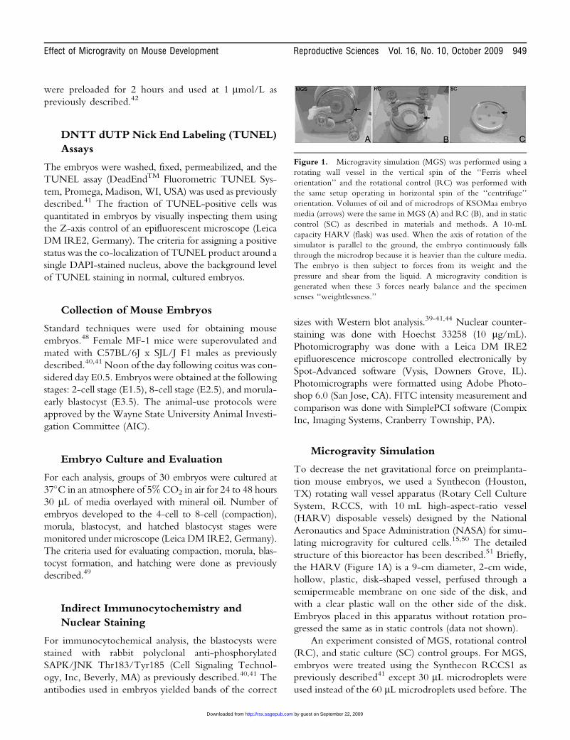
Figure 1. Microgravity simulation (MGS) was performed using a
rotating wall vessel in the vertical spin of the ‘‘Ferris wheel
orientation’’ and the rotational control (RC) was performed with
the same setup operating in horizontal spin of the ‘‘centrifuge’’
orientation. Volumes of oil and of microdrops of KSOMaa embryo
media (arrows) were the same in MGS (A) and RC (B), and in static
control (SC) as described in materials and methods. A 10-mL
capacity HARV (flask) was used. When the axis of rotation of the
simulator is parallel to the ground, the embryo continuously falls
through the microdrop because it is heavier than the culture media.
The embryo is then subject to forces from its weight and the
pressure and shear from the liquid. A microgravity condition is
generated when these 3 forces nearly balance and the specimen
senses ‘‘weightlessness.’’
Effect of Microgravity on Mouse Development Reproductive Sciences Vol. 16, No. 10, October 2009 949
by guest on September 22, 2009 http://rsx.sagepub.comDownloaded from

30 mL volume creates less resistance and travels in a near-
circular orbit with the oil in the filled 10 mL HARV.
However, only 1 speed and 1 level of MGS can be created
as there is 1 point of equilibrium between the denser aqu-
eous microdroplet and lighter oil. Thus, instead of per-
forming an MGS dose-response, we tested 3 stages of
preimplantation embryo using a single level of MGS.
After turning the bioreactor on, angular force is applied
from the rigid vessel wall to the oil and then the microdro-
plet containing the embryos and a low shear inertial mass
equilibrium is set up within 10 minutes. The bioreactor was
designed on the principle that the net force vector an object
is exposed to at any point in the vessel’s rotation is a com-
bination of force vectors created by the vessel’s rotation, the
pressure and viscosity of the fluids, and the gravitational
vector.14 If the speed of rotation and the size of microdro-
plets are adjusted correctly, the force vectors interact so that
the microdroplets were in a synchronous, near circular
orbit. There was also some rotation of the embryo-
containing aqueous microdroplet around its center. This
can be visually verified through the clear Teflon disk of the
HARV. A circular shape to the orbit is indicative of a net
force vector that has the same magnitude at all points in the
rotation, but whose direction is constantly changing.52
Because we have verified that the shape of the orbit is
approximately circular at 7.5 rotations per minute
(RPM), the net force that the embryos ‘‘feel’’ can be
approximated by the centripetal acceleration. The centri-
petal acceleration vector is directed toward the center of
the bioreactor and is the primary force responsible for
keeping the embryos in a circular orbit. The minimum
and maximum centripetal accelerations, based on the
minimum and maximum possible radius of the circular
orbit, can be calculated. (v ¼ velocity, a ¼ acceleration,
m ¼ meters, s ¼ second, T ¼ period of 1 revolution of
HARV, r ¼ radius of orbit).
Minimum Orbital
Velocity ¼ vmin
Maximum Orbital
Velocity ¼ vmax
V ¼ 2�r min / T V ¼ 2�r max / T
r min ¼ 0.025 m r max ¼ 0.045 m
T ¼ 8 s T ¼ 8s
v min ¼ 0.0196 m/s v max ¼ 0.0353m/s
r min and r max ¼ the minimum and maximum radius of an orbit
inside the HARV
Minimum centripetal
acceleration ¼ a min
Maximum centripetal
acceleration ¼ a max
a min ¼ v min2 / r min ¼ a max ¼ v max2 /r max ¼a min ¼ 0.01962/0.025 a max ¼ 0.03532/0.045
a min ¼ m/s2 ¼ 0.00157g a max ¼ m/s2 ¼ 0.00283 g
So the simulated microgravity we got in this study is
approximately 1.6 � 10–3g to 2.8 � 10–3g.
Because of a higher angular velocity of oil at the outer
tangent of the microdroplet (near the outer rim of the
HARV) compared with the inner tangent (nearer the
center of the HARV) of the microdroplet, a counter-
clockwise rotation of the droplet imparts a small shear
on the embryo less than an approximate 10–2 dynes/cm2
estimate. This is *1% of the shear stress level for embryos
generated in our previous experiments.41
Embryo Culture and Evaluation
We used 2 kinds of control groups: RC group and SC
group. Rotational control is a motion control for turbu-
lence, shear forces, and vibrations with a gravity vector at
1G. Static culture is a culture control for incubator/media
conditions at 1g without motion. After 24 hours of cul-
ture or rotation, embryos were examined under micro-
scope (Leica DM IRE2, Germany) for number of
embryos developed to the 4-cell to 8-cell (compaction),
morula, blastocyst, and hatched blastocyst stages. After the
24-hour stimulus period, embryos isolated on E1.5 and
E2.5 were subjected to normal culture for another 24
to 48 hours and the developmental morphology was con-
tinuously monitored. Embryos isolated on E3.5 were
subjected to indirect immunocytochemistry (ICC) with
antiphosphorylated SAPK to determine the stress they
had been exposed to during the past 24 hours of culture
under different conditions on a molecular level. Quanti-
tative immunofluorescence was performed by analyzing
micrographs using the DNN module of Simple PCI soft-
ware (Cranberry Township, PA) Embryos isolated at E3.5
were also cultured for another 24 hours under normal con-
ditions and assayed for development using morphological
criteria and cell number. SAPK Thr183/Tyr185 levels are
similar when immunofluorescence or immunoblots are
quantitated.41,44 The criteria used for evaluating compac-
tion, morula, blastocyst formation, blastocyst size, and
hatching were described previously.41,53
Statistical Analysis
The data in this study are representative of 3 independent
biological experiments and indicated as mean + SD. Sta-
tistical significance of differences between different
treated samples were calculated by 1-way analysis of var-
iance (ANOVA; SPSS 10.0).
950 Reproductive Sciences Vol. 16, No. 10, October 2009 Wang et al
by guest on September 22, 2009 http://rsx.sagepub.comDownloaded from
RESULTS
To simulate microgravity, we used a rotating wall vessel
(RWV) designed by NASA, which has been used previ-
ously to test for effects of microgravity stimulation (MGS)
on development of the vestibular apparatus in Zebrafish
(Danio rerio) embryos.51 At 7.5 RPM, we estimated a
simulation of 1.6-2.8 � 10-3g on the embryos in a 30 mL
microdrop that moved with the oil filling the HARV
(Materials and Methods) when the rotation axis was hor-
izontal and the rotation was in the ‘‘Ferris wheel’’ orien-
tation (Figure 1A). In a previous study, we used this
orientation to develop 1.2 dynes/cm2 shear stress where
the microdrop was 60 mL and was stationary as the oil-
filled RWV rotated and imparted a large rotating force
to the microdrop and created arteriolar velocities on the
embryos.41 However, in this study, because the smaller
microdrop rotated with the oil in a solid body minimal
shear and maximal MGS developed. As a control for rota-
tional effects independent of MGS, the RWV was oper-
ated with a vertical axis of rotation in the ‘‘Centrifuge
orientation’’ (rotational control ¼ RC, Figure 1B). The
RC control and MGS aqueous microdrops both have a
very slow counterclockwise rotation due to higher velo-
city at the outer tangent of the drop compared with the
inner tangent. This would lead to less that 10-2 dynes/cm2
shear force which is about 1% of the shear stress we pre-
viously reported41 (Materials and Methods). To control
for the effects of movement of embryos in the MGS
and RC groups, embryos were cultured under standard
optimal static conditions in microdroplets under oil at
approximately 1 embryo/mL (static control ¼ SC).
Embryos were evaluated for embryo progression
through cleavage division from the 8-cell stage. Then
embryos were scored for the sequential developmental
events; compaction at the 8-cell stage at 2.5 days after ferti-
lization (E2.5), cavitation to form the blastocyst at E3.5, and
expansion and hatching of the late blastocyst stage (E4.5).
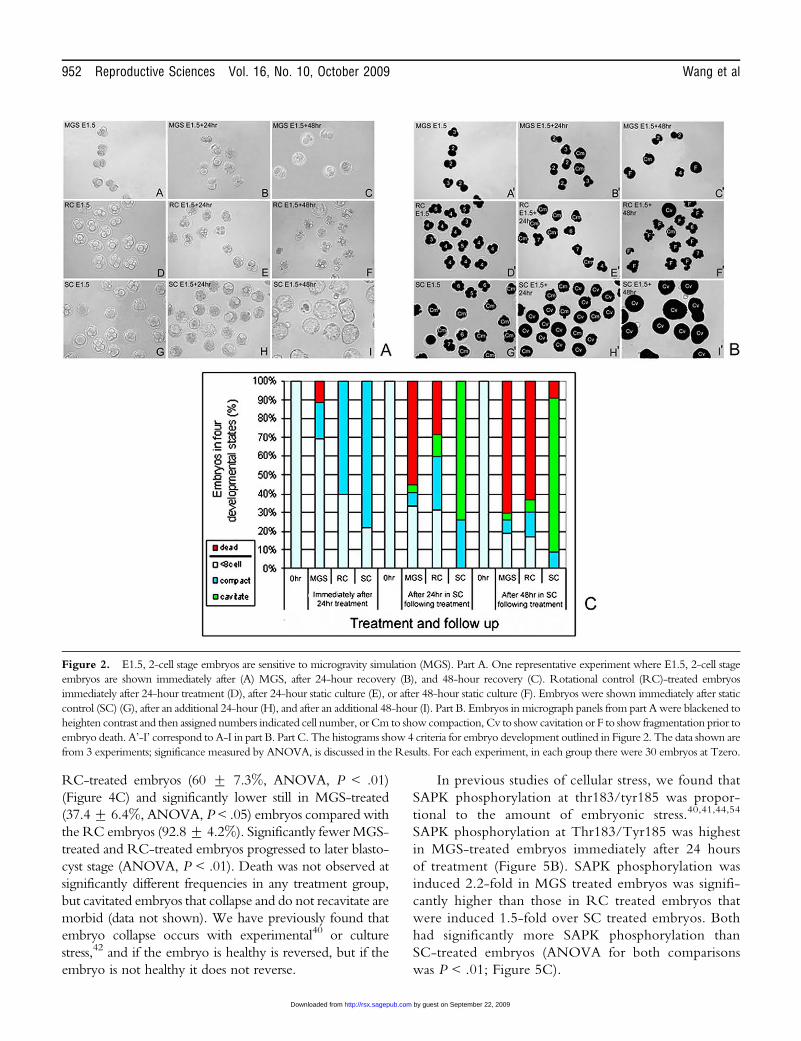
Embryos started 24 hours of treatment at the late
2-cell stage (E1.5). Immediately after 24 hours of treat-
ment, 78.3% + 6.5% of SC embryos had compacted, but
60.1% + 5.5% of RC-treated embryos and only
28.7% + 3.4% of MGS-treated embryos had compacted
(Figure 2). Thus, the largest effects at the end of culture
were on the MGS group. All treatment groups were cul-
tured for an additional 24 hours and 48 hours to observe
further accumulated effects of treatments. By 24 hours
following treatment, MGS-treated embryos were less fre-
quently compacted and cavitated (8.1% + 2.7% and
4.9% + 2.9%, respectively) compared with RC-treated
embryos (28.9% + 3.1% and 12.3% + 5.2%, respec-
tively), whereas the SC group had more cavitated
embryos (84% + 7.7%). Microgravity simulation–
treated embryos were more frequently fragmented and
dead (45.9% + 3.7%) compared with RC-treated
embryos (28.8% + 1.6%). But, by 48 hours after treat-
ment MGS- and RC-treated embryos had similar fre-
quencies of dead and fragmented embryos (70.4% +7.3% vs 63.5% + 10.7%, respectively).
Next we used 24 hours of treatment to test embryos at
the compacted, 8-cell stage (E2.5). Immediately after 24
hours of treatment, 40.8% + 4.1% of SC embryos had
cavitated and rest were in the compacted-morula stage, but
30.2% + 5.8% of RC-treated embryos and 90.1% +9.4% of MGS embryos had reversed development and
decompacted (Figure 3, note that small groups of embryos
(<10%) were late precompaction at the start of treatment).
Thus the largest effects at the end of culture were on the
MGS group as at E1.5, but these effects were more severe
and development was reversed. All treatment groups were
cultured for an additional 24 hours and 48 hours to observe
further accumulated effects of treatments. By 24 hours fol-
lowing treatment, MGS-treated embryos had fewer com-
pacted embryos (6.5% + 5.2%) and fragmented and dead
embryos had appeared (17.4% + 6.0%), and more RC
embryos had decompacted (38.1 + 3.8) but no fragmen-
ted or dead embryos were observed. But, by 48 hours all
embryos in both MGS and RC decompacted and this ulti-
mately leads to embryo death. As with the E1.5 embryos,
initial MGS treatment caused the highest immediate
effects, but by 48 hours it was clear that all RC embryos
had sustained sufficient cellular stress to cause the same fatal
outcome as for all MGS embryos.
Next, we treated E3.5 embryos for 24 hours and then
cultured an additional 24 to 48 hours after treatment in
SC. After 24 hours of treatment, SC embryos had twice
the cross-sectional area (Figure 4B) and were maximally
expanded. However RC-treated embryos only increased
by approximately 33% in the 24 hours after treatment
(although this was significant, ANOVA, P < .01), and
many cavitated embryos had collapsed and appeared
to be compacted embryos (Figure 4A). Microgravity
simulation–treated embryos did not significantly increase
in size by 24 hours after treatment (ANOVA, P ¼ .7) and
many had collapsed and appeared as compacted, not
cavitated embryos (Figure 4A). The RC- and MGS-
treated embryos expanded at a significantly decreased rate
compared with SC embryos (ANOVA, P < .05). The
fraction of embryos that progressed to expanded blastocyst
stage 24 hours after treatment was significantly lower in
Effect of Microgravity on Mouse Development Reproductive Sciences Vol. 16, No. 10, October 2009 951
by guest on September 22, 2009 http://rsx.sagepub.comDownloaded from
RC-treated embryos (60 + 7.3%, ANOVA, P < .01)
(Figure 4C) and significantly lower still in MGS-treated
(37.4+ 6.4%, ANOVA, P < .05) embryos compared with
the RC embryos (92.8 + 4.2%). Significantly fewer MGS-
treated and RC-treated embryos progressed to later blasto-
cyst stage (ANOVA, P < .01). Death was not observed at
significantly different frequencies in any treatment group,
but cavitated embryos that collapse and do not recavitate are
morbid (data not shown). We have previously found that
embryo collapse occurs with experimental40 or culture
stress,42 and if the embryo is healthy is reversed, but if the
embryo is not healthy it does not reverse.
In previous studies of cellular stress, we found that
SAPK phosphorylation at thr183/tyr185 was propor-
tional to the amount of embryonic stress.40,41,44,54
SAPK phosphorylation at Thr183/Tyr185 was highest
in MGS-treated embryos immediately after 24 hours
of treatment (Figure 5B). SAPK phosphorylation was
induced 2.2-fold in MGS treated embryos was signifi-
cantly higher than those in RC treated embryos that
were induced 1.5-fold over SC treated embryos. Both
had significantly more SAPK phosphorylation than
SC-treated embryos (ANOVA for both comparisons
was P < .01; Figure 5C).
Figure 2. E1.5, 2-cell stage embryos are sensitive to microgravity simulation (MGS). Part A. One representative experiment where E1.5, 2-cell stage
embryos are shown immediately after (A) MGS, after 24-hour recovery (B), and 48-hour recovery (C). Rotational control (RC)-treated embryos
immediately after 24-hour treatment (D), after 24-hour static culture (E), or after 48-hour static culture (F). Embryos were shown immediately after static
control (SC) (G), after an additional 24-hour (H), and after an additional 48-hour (I). Part B. Embryos in micrograph panels from part A were blackened to
heighten contrast and then assigned numbers indicated cell number, or Cm to show compaction, Cv to show cavitation or F to show fragmentation prior to
embryo death. A’-I’ correspond to A-I in part B. Part C. The histograms show 4 criteria for embryo development outlined in Figure 2. The data shown are
from 3 experiments; significance measured by ANOVA, is discussed in the Results. For each experiment, in each group there were 30 embryos at Tzero.
952 Reproductive Sciences Vol. 16, No. 10, October 2009 Wang et al
by guest on September 22, 2009 http://rsx.sagepub.comDownloaded from

Figure 3. E2.5 embryos were immediately arrested in development and died between 24 and 48 hours after microgravity simulation (MGS).
Part A. Embryos were blackened and assigned embryo evaluations as in Figure 2. Microgravity simulation–treated embryos immediately after 24
hours (A), 24 hours after MGS (B), and 48 hours after MGS (C). Rotational control (RC)-treated embryos immediately after treatment (D), 24
hours after RC (E), and 48 hours after RC (F). Static control (SC) treated embryos immediately after treatment (G), after and additional 24 hours
of SC (H), and after an additional 48 hours of SC (I). Part B. The histograms show 4 criteria for embryo development from data gathered from 3
experiments, significance measured by ANOVA is discussed in the Results. For each experiment, in each group there were 30 embryos at Tzero.
Figure 4. After 24 hours of microgravity simulation (MGS), E3.5 embryos were less developed than in rotational control (RC) or static
control (SC) treatments, and this led to morbidity after MGS. Part A. E3.5 embryos immediately after MGS treatment (A), or after an additional
24 hours (B). Embryos immediately after 24 hours of RC treatment (C), or 24 hours later (D). Embryos immediately after 24 hours of SC (E) and
24 hours later (F). Part B. Histograms show quantitated cross-sectional area of micrographed embryos from 3 experiments. (a) Shows a significant
difference of RC and MGS compared with SC (ANOVA, P < .01), (c) shows a significant difference for RC and MGS 24 hours after treatment
compared with SC embryos (ANOVA, P < .001). (b) Shows no significant difference between RC and MGS immediately after treatment
(ANOVA, P ¼ .3), but (d) shows a significant decrease in embryo development for MGS compared with RC 24 hours after treatment
(ANOVA, P < .01). Part C. Histograms show embryo development, reversal of development, and arrest in embryos from 3 experiments. Error
bars are standard deviations and (a) shows that RC is significantly less than SC and (b) and (c) show that MGS is significantly less than SC and
RC, respectively (ANOVA, P < .01). For each experiment, in each group there were 30 embryos at Tzero.
Effect of Microgravity on Mouse Development Reproductive Sciences Vol. 16, No. 10, October 2009 953
by guest on September 22, 2009 http://rsx.sagepub.comDownloaded from

To test for the role of SAPK in the effects of MGS,
we treated embryos for 24 hours of MGS with or without
SAPK inhibitor and then assayed for cell number and
apoptosis. SC embryos started at 32.4 + 6.4 cells at time
zero and were 84.7 + 9.9 cells after 24 hours of culture
(Figure 6A). However, MGS embryos had 37.2 + 11.4
cells, significantly less than SC embryos (ANOVA, P <
.001), and not significantly more than the cell number
of embryos before treatment (ANOVA, P¼ .8). Embryos
exposed to MGS but preloaded and cultured with 1uM
Figure 5. Microgravity simulation (MGS)-perturbed E3.5 embryos express higher levels of phosphorylated SAPK than rotational control (RC)
embryos which expresses a higher level than static control (SC). Part A. E3.5 embryos were subject to 24 hours of MGS (A), RC (B), or SC
(C), fixed, stained and developed using immunocytochemical means for phosphorylated SAPK thr183/tyr185 (SAPK phospho). (D) Shows
embryos after 24 hours of MGS, but with no antibody. Part B. Histogram is the immunofluorescence measurements of A-C for 3 experiments
that used 95 embryos (error bars are standard deviations). (a) Shows that phosphorylated SAPK is significantly higher in embryos treated with RC
or MGS than SC embryos, respectively (ANOVA, P < .01, and P < .005, respectively). (b) Shows that phosphorylated SAPK in embryos treated
with MGS is significantly higher than in RC treated embryos (ANOVA, P < .01).
954 Reproductive Sciences Vol. 16, No. 10, October 2009 Wang et al
by guest on September 22, 2009 http://rsx.sagepub.comDownloaded from
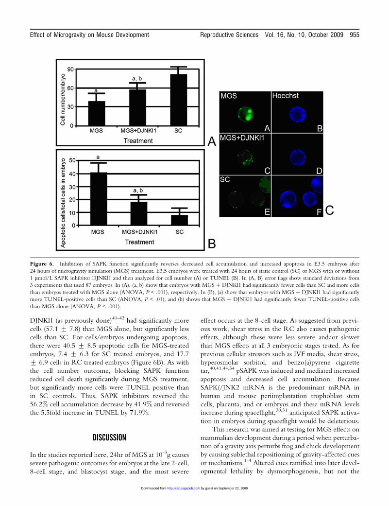
DJNKl1 (as previously done)40-42 had significantly more
cells (57.1 + 7.8) than MGS alone, but significantly less
cells than SC. For cells/embryos undergoing apoptosis,
there were 40.5 + 8.5 apoptotic cells for MGS-treated
embryos, 7.4 + 6.3 for SC treated embryos, and 17.7
+ 6.9 cells in RC treated embryos (Figure 6B). As with
the cell number outcome, blocking SAPK function
reduced cell death significantly during MGS treatment,
but significantly more cells were TUNEL positive than
in SC controls. Thus, SAPK inhibitors reversed the
56.2% cell accumulation decrease by 41.9% and reversed
the 5.5fold increase in TUNEL by 71.9%.
DISCUSSION
In the studies reported here, 24hr of MGS at 10-3g causes
severe pathogenic outcomes for embryos at the late 2-cell,
8-cell stage, and blastocyst stage, and the most severe
effect occurs at the 8-cell stage. As suggested from previ-
ous work, shear stress in the RC also causes pathogenic
effects, although these were less severe and/or slower
than MGS effects at all 3 embryonic stages tested. As for
previous cellular stressors such as IVF media, shear stress,
hyperosmolar sorbitol, and benzo(a)pyrene cigarette
tar,40,41,44,54 pSAPK was induced and mediated increased
apoptosis and decreased cell accumulation. Because
SAPK(/JNK2 mRNA is the predominant mRNA in
human and mouse periimplantation trophoblast stem
cells, placenta, and or embryos and these mRNA levels
increase during spaceflight,30,31 anticipated SAPK activa-
tion in embryos during spaceflight would be deleterious.
This research was aimed at testing for MGS effects on
mammalian development during a period when perturba-
tion of a gravity axis perturbs frog and chick development
by causing sublethal repositioning of gravity-affected cues
or mechanisms.1-4 Altered cues ramified into later devel-
opmental lethality by dysmorphogenesis, but not the
Figure 6. Inhibition of SAPK function significantly reverses decreased cell accumulation and increased apoptosis in E3.5 embryos after
24 hours of microgravity simulation (MGS) treatment. E3.5 embryos were treated with 24 hours of static control (SC) or MGS with or without
1 mmol/L SAPK inhibitor DJNKl1 and then analyzed for cell number (A) or TUNEL (B). In (A, B) error flags show standard deviations from
3 experiments that used 87 embryos. In (A), (a, b) show that embryos with MGS þ DJNKl1 had significantly fewer cells than SC and more cells
than embryos treated with MGS alone (ANOVA, P < .001), respectively. In (B), (a) show that embryos with MGS þ DJNKl1 had significantly
more TUNEL-positive cells than SC (ANOVA, P < .01), and (b) shows that MGS þ DJNKl1 had significantly fewer TUNEL-positive cells
than MGS alone (ANOVA, P < .001).
Effect of Microgravity on Mouse Development Reproductive Sciences Vol. 16, No. 10, October 2009 955
by guest on September 22, 2009 http://rsx.sagepub.comDownloaded from

immediate cessation of cell number increase and later
death reported here. Thus, MGS causes a more rapid,
severe, pathogenic response in mouse embryos, not the
slower developmental effects observed in frog and chick
embryogenesis.
A previous study reported that MGS during fertiliza-
tion had no effect on later mouse development.6 But, this
study reported that MGS deranged early cleavage divi-
sions and a substantial number of embryos were retarded
from fertilization to the morula stage. The phenotype
reported here was much more severe than in the previous
report. In the 2 periods traversed by embryos in the pre-
vious report and this one, 2-cell and 8-cell/morula stages,
there was a severe growth arrest, failure to compact,
decompaction, and ultimate death in most embryos
observed here, but not in the previous report. This could
be due to several reasons, difference in mouse strain,
media, and magnitude of MGS are possible differences.
In a separate study focused on the effects of shear
stress on the preimplantation embryo,41 the droplet size
was increased until it fell through the moving oil at a
velocity equal to the rotation of the oil. Thus the aqueous
drop was stationary and this maximized shear stress at
1.2 dynes/cm2 and minimized MGS to near zero.41 This
level of shear stress was over 100-fold greater than the
shear stress in RC here, so we anticipate that RC should
have minimal shear and effects due to shear. The MGS
here causes rapid cessation of cell accumulation at E1.5
and E2.5 immediately after stimulation but death did not
occur until 24 to 48 hours after stimulation. Similarly for
E3.5 embryos, MGS caused attenuation of embryo devel-
opment, but decreases in embryo development and
attenuation in cell accumulation became apparent in the
24 hours after stimulus. In contrast with the previous
study on shear stress, there was little embryo death here
whereas there was a rapid death that became irreversible
for E3.5 embryos by 12 hours of rotation and all embryos
were dead by the end of a 24 hours of shear stress.41 This
suggests that high shear stress can be potently lethal, with
a more rapid lethality than 1 mol/L high-dose hyperosmo-
lar stress, and 1/100th shear stress here contributes to
the RC-triggered lethality and partially to the MGS-
triggered lethality. These data together suggest that earlier
embryos, known to be more susceptible to stressful pertur-
bations, die due to a failure to maintain cellular homeosta-
sis, due to MGS with a lesser component of shear stress,
rather than suffer a developmental misprogramming.
This contention is supported by the increase in
pSAPK, a reaction at this stage of development to stress
triggered by MGS, and to a lesser extent rotation without
MGS. The nature of the mechanism of SAPK induction is
unclear. But, we have found that pSAPK is induced by
poor media such as M16 and Ham’s F-10þ bovine serum
albumin (BSA) with preimplantation development from
2-cell to blastocyst-stage negatively affected proportional
to pSAPK levels.44 It is unlikely that SAPK is part of any
essential normal preimplantation development as recently
reported for p38 mitogen-activated protein kinase
(p38MAPK).55 This is suggested by data showing that
SAPK inhibitors have no effect on development of
embryos cultured from the 2-cell to blastocyst-stage in
optimal media ([KSOMaa]42), but embryos cultured in
KSOMAaa with p38MAPK inhibitors are blocked at the
8-cell stage.55 Because the MGS experiments were per-
formed with optimal KSOMaa that induces little SAPK
phosphorylation, SAPK was induced by some aspect of
MGS. Because pSAPK increase is not a required develop-
mental event, it is most likely a cellular stress response.
Cellular stress responses have been reported for cells in
MGS and in space flight at the level of SAPK phosphor-
ylation and the induction of SAPKa/JNK2.30,31,56
There are a number of candidate processes, aside from
pSAPK, and mechanisms that have been identified that
may lead to cellular stress and death in MGS and space-
flight. Changes in cell cycle commitment, TUNEL/
apoptosis, growth factor sensitivity, signal transduction,
and cytoskeleton integrity are observed in MGS and
spaceflight.57-64 Phosphorylated SAPK elevation can be
involved with all of these mechanisms, and we have also
observed some actin cytoskeleton derangement (unpub-
lished data). A kinase related to SAPK, p38MAPK
regulates actin polymerization at the 8-cell stage and
inhibiting p38MAPK blocks embryo progression.55
P38MAPK is induced during stressful embryo culture and
by hyperosmolar stress.65,66 It has not been established how
mammalian cells sense gravity, or MGS perturbation, but
structural (cytoskeletal) components such as actin normally
sensing dense cellular bodies are candidates,67 as these have
already been established in the columella cells in the plant
root tip that are geotropic (and sense gravity).68
CONCLUSIONS
(1) MGS is most lethal at 8-cell, less at 2-cell (but still
lethal), and without lethality at E3.5 although apoptosis
is induced. (2) Cell cycle arrest occurs without lethality
during 24 hours of rotation during MGS. Cell cycle arrest
is more profound in MGS compared with RC when per-
turbation starts at the 2-cell stage. Decompaction, a
956 Reproductive Sciences Vol. 16, No. 10, October 2009 Wang et al
by guest on September 22, 2009 http://rsx.sagepub.comDownloaded from

reversal of development, is more severe for 8-cell
embryos for MGS than for RC, after 24 hours of MGS.
But, RC embryos finally also decompact and die between
24 hours and 48 hours after treatment. (3) The SAPK is
proportional to lethality and is probably involved. Also,
elevated pSAPK suggests that this lethality is not a devel-
opmental response at the embryonic or cell lineage level,
but part of a general homeostatic effect in response to
stress at the cellular level. (4) Lesser RC lethality is prob-
ably due to a low amount of shear stress that was at levels
about 1% of those previously reported.41
Future studies will include the study of TUNEL/apop-
tosis as a mediator of death and the causal role of SAPK in
the induction of TUNEL/apoptosis. An alternate apparatus
or experiments during spaceflight are important to establish
dose-dependent effects and validate MGS findings. We
have established that SAPK is causal in shear stress-
mediated lethality,41 and mediates hyperosmolarity-
induced lethality and cell cycle arrest.39 However, alternate
mechanisms, such as cytoskeletal disruption, are likely to
contribute to pathogenesis. Cytoskeleton disruptors that
lead to chronic SAPK activation for 12 hours in endothelial
cells are sufficient to induce apoptosis,69 suggesting an inter-
esting exacerbation by 2 stress inducers, MGS and shear
stress, operating via SAPK and cytoskeletal disruption.
In addition, it is important to understand how MGS
and other stressors affect other segments of preimplanta-
tion development. Essential processes in the zygote such
as genome activation are likely to be affected by MGS and
its associated stress. Implantation itself may be influenced
by cues mediated by gravity and affected by stress
responses generated by the alteration of gravity.
Finally, it is clear that SAPK is activated by many
physiological stimuli of embryos and their constituent
trophoblast stem cells.54 However, a growing list of non-
physiological stimuli also activate SAPK and its depen-
dent negative effects. These include embryo culture
media used in IVF, pipetting during handling, shear stress,
dioxin, benzo(a)pyrene,41,44,54,70 and now MGS. SAPK
may have been selected during evolution to mediate stress
responses to physiological stressors, but its common
response during nonevolutionary, nonphysiological
stimuli suggest that it will be important to analyze and
perhaps manage during any protocol in IVF/ART.
ACKNOWLEDGMENTS
We thank Mike Kruger for advice on statistical analysis.
We also thank Dr Josh Zimmerberg of NICHHD for the
use of two additional RCCS1 microgravity simulators and
additional HARVS. We are also indebted to Dr Richard
Tasca, Dr Horacio Croxatto, and Dr Anne McLaren for
helpful discussion and criticisms of the manuscript. This
research was supported by grants from NASA (NRA-
NAG2-1503) and the National Institute of Child Health
and Human Development, NIH, (R01-HD40972).
REFERENCES
1. Gerhart JC, Vincent JP, Scharf SR, Black SD, Gimlich RL,
Danilchik M. Localization and induction in early develop-
ment of Xenopus. Philos Trans R Soc Lond B Biol Sci.
1984;307(1132):319-330.
2. Vincent JP, Gerhart JC. Subcortical rotation in Xenopus eggs:
an early step in embryonic axis specification. Dev Biol.
1987;123(2):526-539.
3. Pitts GC. Effects of gravity on ontogeny in animals. Life Sci
Space Res. 1973;11:171-176.
4. Cooke J. Permanent distortion of positional system of Xeno-
pus embryo by brief early perturbation in gravity. Nature.
1986;319(6048):60-63.
5. Kochav S, Eyal-Giladi H. Bilateral symmetry in chick embryo
determination by gravity. Science. 1971;171(975):1027-1029.
6. Kojima Y, Sasaki S, Kubota Y, Ikeuchi T, Hayashi Y,
Kohri K. Effects of simulated microgravity on mammalian fer-
tilization and preimplantation embryonic development in
vitro. Fertil Steril. 2000;74(6):1142-1147.
7. Young RS, Tremor JW. Weightlessness and the developing
frog egg. Life Sci Space Res. 1968;6:87-93.
8. Ubbels GA, Berendsen W, Narraway J. Fertilization of frog
eggs on a Sounding Rocket in space. Adv Space Res. 1989;
9(11):187-197.
9. Suda T. Lessons from the space experiment SL-J/FMPT/L7:
the effect of microgravity on chicken embryogenesis and bone
formation. Bone. 1998;22(5 suppl):73S-78S.
10. Schatten H, Chakrabarti A, Taylor M, et al. Effects of space-
flight conditions on fertilization and embryogenesis in the sea
urchin Lytechinus pictus. Cell Biol Int. 1999;23(6):407-415.
11. Marthy HJ, Schatt P, Santella L. Fertilization of sea urchin
eggs in space and subsequent development under normal con-
ditions. Adv Space Res. 1994;14(8):197-208.
12. Steffen S, Fiser R, Simerly C, Schatten H, Schatten G. Micro-
gravity effects on sea urchin fertilization and development.
Adv Space Res. 1992;12(1):167-173.
13. Ronca AE, Alberts JR. Effects of prenatal spaceflight on
vestibular responses in neonatal rats. J Appl Physiol.
2000;89(6):2318-2324.
14. Kessler JO. Theory and experimental results on gravitational
effects on monocellular algae. Adv Space Res. 1992;12(1):33-42.
15. Klaus DM. Clinostats and bioreactors. Gravit Space Biol Bull.
2001;14(2):55-64.
Effect of Microgravity on Mouse Development Reproductive Sciences Vol. 16, No. 10, October 2009 957
by guest on September 22, 2009 http://rsx.sagepub.comDownloaded from
16. Cross JC, Werb Z, Fisher SJ. Implantation and the placenta:
key pieces of the development puzzle. Science. 1994;
266(5190):1508-1518.
17. Copp AJ. Death before birth: clues from gene knockouts and
mutations. Trends Genet. 1995;11(3):87-93.
18. Rappolee DA. It’s not just baby’s babble/Babel: recent prog-
ress in understanding the language of early mammalian devel-
opment: a minireview. Mol Reprod Dev. 1999;52(2):234-240.
19. Hamatani T, Carter MG, Sharov AA, Ko MS. Dynamics of
global gene expression changes during mouse preimplantation
development. Dev Cell. 2004;6(1):117-131.
20. Wang QT, Piotrowska K, Ciemerych MA, et al. A genome-
wide study of gene activity reveals developmental signaling
pathways in the preimplantation mouse embryo. Dev Cell.
2004;6(1):133-144.
21. Hardie DG. Minireview: the AMP-activated protein kinase
cascade: the key sensor of cellular energy status. Endocrinology.
2003;144(12):5179-5183.
22. Liu L, Cash TP, Jones RG, Keith B, Thompson CB,
Simon MC. Hypoxia-induced energy stress regulates mRNA
translation and cell growth. Mol Cell. 2006;21(4):521-531.
23. Weston CR, Davis RJ. The JNK signal transduction pathway.
Curr Opin Genet Dev. 2002;12(1):14-21.
24. Larue L, Ohsugi M, Hirchenhain J, Kemler R. E-cadherin
null mutant embryos fail to form a trophectoderm epithelium.
Proc Natl Acad Sci U S A. 1994;91(17):8263-8267.
25. Lawson KA, Meneses JJ, Pedersen RA. Clonal analysis of epi-
blast fate during germ layer formation in the mouse embryo.
Development. 1991;113(3):891-911.
26. Rossant J. Lineage development and polar asymmetries in the
peri-implantation mouse blastocyst. Semin Cell Dev Biol.
2004;15(5):573-581.
27. Lu CC, Brennan J, Robertson EJ. From fertilization to gastru-
lation: axis formation in the mouse embryo. Curr Opin Genet
Dev. 2001;11(4):384-392.
28. Thomas PQ, Brown A, Beddington RS. Hex: a homeobox
gene revealing peri-implantation asymmetry in the mouse
embryo and an early transient marker of endothelial cell pre-
cursors. Development. 1998;125(1):85-94.
29. Labelle-Dumais C, Jacob-Wagner M, Pare JF, Belanger L,
Dufort D. Nuclear receptor NR5A2 is required for proper
primitive streak morphogenesis. Dev Dyn. 2006;235(12):
3359-3369.
30. Hammond TG, Benes E, O’Reilly KC, et al. Mechanical cul-
ture conditions effect gene expression: gravity-induced
changes on the space shuttle. Physiol Genomics. 2000;3(3):
163-173.
31. Hammond TG, Lewis FC, Goodwin TJ, et al. Gene expres-
sion in space. Nat Med. 1999;5(4):359.
32. Davis RJ. Signal transduction by the JNK group of MAP
kinases. Cell. 2000;103(2):239-252.
33. Whitmarsh AJ, Davis RJ. Signal transduction by MAP kinases:
regulation by phosphorylation-dependent switches. Sci
STKE. 1999;1999(1):PE1.
34. Whitmarsh AJ, Davis RJ. Regulation of transcription factor
function by phosphorylation. Cell Mol Life Sci. 2000;57(8-9):
1172-1183.
35. Kyriakis JM, Avruch J. Sounding the alarm: protein kinase
cascades activated by stress and inflammation. J Biol Chem.
1996;271(40):24313-24316.
36. Kyriakis JM, Avruch J. Mammalian mitogen-activated protein
kinase signal transduction pathways activated by stress and
inflammation. Physiol Rev. 2001;81(2):807-869.
37. Rappolee DA. Signal transduction. In: Krawetz S, Womble D,
eds. Introduction to Bioinformatics. A Theoretical and Practical
Approach. 2nd ed. Totowa, NJ: Humana Press; 2003:55-71.
38. Woodgett JR, Avruch J, Kyriakis J. The stress activated pro-
tein kinase pathway. Cancer Surv. 1996;27:127-138.
39. Zhong W, Xie Y, Wang Y, et al. Use of hyperosmolar stress to
measure stress-activated protein kinase activation and function
in human HTR cells and mouse trophoblast stem cells. Reprod
Sci. 2007;14(6):534-547.
40. Xie Y, Zhong W, Wang Y, et al. Using hyperosmolar stress to
measure biologic and stress-activated protein kinase responses
in preimplantation embryos. Mol Hum Reprod. 2007;13(7):
473-481.
41. Xie Y, Wang F, Zhong W, Puscheck E, Shen H,
Rappolee DA. Shear stress induces preimplantation embryo
death that is delayed by the zona pellucida and associated with
stress-activated protein kinase-mediated apoptosis. Biol
Reprod. 2006;75(1):45-55.
42. Xie Y, Puscheck EE, Rappolee DA. Effects of SAPK/JNK
inhibitors on preimplantation mouse embryo development are
influenced greatly by the amount of stress induced by the
media. Mol Hum Reprod. 2006;12(4):217-224.
43. Zhong W, Sun T, Wang Q, et al. SAPK/JNK1, 2, but not
SAPK/JNK3 mRNA transcripts, are expressed in early gesta-
tion human placenta and mouse eggs, preimplantation
embryos, and trophoblast stem cells. Fertility and Sterility.
2004;82:1140-1148.
44. Wang Y, Puscheck EE, Wygle DL, et al. Increases in phos-
phorylation of SAPK/JNK and p38MAPK correlate nega-
tively with mouse embryo development after culture in
different media. Fertility and Sterility. 2005;83:1144-1154.
45. Bonny C, Oberson A, Negri S, Sauser C, Schorderet DF.
Cell-permeable peptide inhibitors of JNK: novel blockers of
beta-cell death. Diabetes. 2001;50(1):77-82.
46. Dickens M, Rogers JS, Cavanagh J, et al. A cytoplasmic inhi-
bitor of the JNK signal transduction pathway. Science.
1997;277(5326):693-696.
47. Bonny C, Nicod P, Waeber G. IB1, a JIP-1-related nuclear
protein present in insulin-secreting cells. J Biol Chem.
1998;273(4):1843-1846.
48. Hogan B, Beddington R, Constantini F, Lacy B. Manipulating
the mouse embryo, A laboratory manual. 3rd ed. Cold Spring
Harbor, New York: Cold Spring Harbor Laboratory; 2002.
49. Nagy A, Gertsenstein M, Vintersten K, Behringer RR.
Manipulating the mouse embryo. A laboratory manual. 3rd ed.
958 Reproductive Sciences Vol. 16, No. 10, October 2009 Wang et al
by guest on September 22, 2009 http://rsx.sagepub.comDownloaded from

Cold Spring Harbor, New York: Cold Spring Harbor Labora-
tory Press; 2003.
50. Klaus DM, Todd P, Schatz A. Functional weightlessness dur-
ing clinorotation of cell suspensions. Adv Space Res. 1998;
21(8-9):1315-1318.
51. Moorman SJ, Burress C, Cordova R, Slater J. Stimulus depen-
dence of the development of the zebrafish (Danio rerio)
vestibular system. J Neurobiol. 1999;38(2):247-258.
52. Halliday Ra. Fundamentals of Physics. vol 1, 2nd ed. New York
City, NY: Wiley and Sons; 1977.
53. Chai N, Patel Y, Jacobson K, McMahon J, McMahon A,
Rappolee DA. FGF is an essential regulator of the fifth cell
division in preimplantation mouse embryos. Dev Biol.
1998;198(1):105-115.
54. Xie Y, Liu J, Proteasa S, et al. Transient stress and stress
enzyme responses have practical impacts on parameters of
embryo development, from IVF to directed differentiation
of stem cells. Mol Reprod Dev. 2008;75(4):689-697.
55. Natale DR, Paliga AJ, Beier F, D’Souza SJ, Watson AJ. p38
MAPK signaling during murine preimplantation develop-
ment. Dev Biol. 2004;268(1):76-88.
56. Hughes-Fulford M. The role of signaling pathways in osteoblast
gravity perception. J Gravit Physiol. 2002;9(1):P257-P260.
57. de Groot RP, Rijken PJ, den Hertog J, et al. Nuclear
responses to protein kinase C signal transduction are sensitive
to gravity changes. Exp Cell Res. 1991;197(1):87-90.
58. d’Ascanio P, Balaban E, Pompeiano M, Centini C,
Pompeiano O. Fos and FRA protein expression in rat precer-
ebellar structures during the Neurolab Space Mission. Brain
Res Bull. 2003;62(3):203-221.
59. Boonstra J. Growth factor-induced signal transduction in
adherent mammalian cells is sensitive to gravity. Faseb J.
1999;13(suppl):S35-S42.
60. Hughes-Fulford M, Tjandrawinata R, Fitzgerald J, Gasuad K,
Gilbertson V. Effects of microgravity on osteoblast growth.
Gravit Space Biol Bull. 1998;11(2):51-60.
61. Komazaki S. Gravitational effects on apoptosis of presumptive
ectodermal cells of amphibian embryo. J Exp Zoolog Part A
Comp Exp Biol. 2004;301(3):204-211.
62. Lewis ML, Reynolds JL, Cubano LA, Hatton JP, Lawless BD,
Piepmeier EH. Spaceflight alters microtubules and increases
apoptosis in human lymphocytes (Jurkat). Faseb J. 1998;
12(11):1007-1018.
63. Ontiveros C, McCabe LR. Simulated microgravity suppresses
osteoblast phenotype, Runx2 levels and AP-1 transactivation.
J Cell Biochem. 2003;88(3):427-437.
64. Sato A, Hamazaki T, Oomura T, et al. Effects of microgravity
on c-fos gene expression in osteoblast-like MC3T3-E1 cells.
Adv Space Res. 1999;24(6):807-813.
65. Fong B, Watson PH, Watson AJ. Mouse preimplantation
embryo responses to culture medium osmolarity include
increased expression of CCM2 and p38 MAPK activation.
BMC Dev Biol. 2007;7:2.
66. Wang Y, Puscheck EE, Lewis JJ, Trostinskaia AB, Wang F,
Rappolee DA. Increases in phosphorylation of SAPK/JNK
and p38MAPK correlate negatively with mouse embryo
development after culture in different media. Fertil Steril.
2005;83(suppl 1):1144-1154.
67. Todd P, Klaus DM, Stodieck LS, et al. Cellular responses to
gravity: extracellular, intracellular and in-between. Adv Space
Res. 1998;21(8-9):1263-1268.
68. Staehelin LA, Zheng HQ, Yoder TL, Smith JD, Todd P.
Columella cells revisited: novel structures, novel properties,
and a novel gravisensing model. Gravit Space Biol Bull.
2000;13(2):95-100.
69. Hu YL, Li S, Shyy JY, Chien S. Sustained JNK activation
induces endothelial apoptosis: studies with colchicine and
shear stress. Am J Physiol. 1999;277(4 pt 2):H1593-H1599.
70. Xie Y, Wang F, Puscheck EE, Rappolee DA. Pipetting causes
shear stress and elevation of phosphorylated stress-activated
protein kinase/jun kinase in preimplantation embryos. Mol
Reprod Dev. 2007;74(10):1287-1294.
For reprints and permissions queries, please visit SAGE’s Web site at http://www.sagepub.com/journalsPermissions.nav
Effect of Microgravity on Mouse Development Reproductive Sciences Vol. 16, No. 10, October 2009 959
by guest on September 22, 2009 http://rsx.sagepub.comDownloaded from
Copyright © 2022 FDOKUMEN