A Literature Systematic R - MDPI
43
Citation: Inchingolo, F.; Hazballa, D.; Inchingolo, A.D.; Malcangi, G.; Marinelli, G.; Mancini, A.; Maggiore, M.E.; Bordea, I.R.; Scarano, A.; Farronato, M.; et al. Innovative Concepts and Recent Breakthrough for Engineered Graft and Constructs for Bone Regeneration: A Literature Systematic Review. Materials 2022, 15, 1120. https://doi.org/10.3390/ ma15031120 Academic Editor: Eugenio Velasco-Ortega Received: 14 December 2021 Accepted: 27 January 2022 Published: 31 January 2022 Publisher’s Note: MDPI stays neutral with regard to jurisdictional claims in published maps and institutional affil- iations. Copyright: © 2022 by the authors. Licensee MDPI, Basel, Switzerland. This article is an open access article distributed under the terms and conditions of the Creative Commons Attribution (CC BY) license (https:// creativecommons.org/licenses/by/ 4.0/). materials Review Innovative Concepts and Recent Breakthrough for Engineered Graft and Constructs for Bone Regeneration: A Literature Systematic Review Francesco Inchingolo 1, * ,† , Denisa Hazballa 1,2,† , Alessio Danilo Inchingolo 1,† , Giuseppina Malcangi 1,† , Grazia Marinelli 1 , Antonio Mancini 1 , Maria Elena Maggiore 1 , Ioana Roxana Bordea 3 , Antonio Scarano 4 , Marco Farronato 5 , Gianluca Martino Tartaglia 5 , Felice Lorusso 4, * ,‡ , Angelo Michele Inchingolo 1,‡ and Gianna Dipalma 1, * ,‡ 1 Department of Interdisciplinary Medicine, University of Medicine Aldo Moro, 70124 Bari, Italy; [email protected] (D.H.); [email protected] (A.D.I.); [email protected] (G.M.); [email protected] (G.M.); [email protected] (A.M.); [email protected] (M.E.M.); [email protected] (A.M.I.) 2 Kongresi Elbasanit, Rruga: Aqif Pasha, 3001 Elbasan, Albania 3 Department of Oral Rehabilitation, Faculty of Dentistry, Iuliu Hat , ieganu University of Medicine and Pharmacy, 400012 Cluj-Napoca, Romania; [email protected] 4 Department of Innovative Technologies in Medicine and Dentistry, University of Chieti-Pescara, 66100 Chieti, Italy; [email protected] 5 UOC Maxillo-Facial Surgery and Dentistry, Department of Biomedical, Surgical and Dental Sciences, School of Dentistry, Fondazione IRCCS Ca Granda, Ospedale Maggiore Policlinico, University of Milan, 20100 Milan, Italy; [email protected] (M.F.); [email protected] (G.M.T.) * Correspondence: [email protected] (F.I.); [email protected] (F.L.); [email protected] (G.D.); Tel.: +39-3312111104 (F.I.); +39-3282132586 (F.L.); +39-3396989939 (G.D.) † These authors contributed equally to this work as co-first authors. ‡ These authors contributed equally to this work as co-last authors. Abstract: Background: For decades, regenerative medicine and dentistry have been improved with new therapies and innovative clinical protocols. The aim of the present investigation was to evaluate through a critical review the recent innovations in the field of bone regeneration with a focus on the healing potentials and clinical protocols of bone substitutes combined with engineered constructs, growth factors and photobiomodulation applications. Methods: A Boolean systematic search was conducted by PubMed/Medline, PubMed/Central, Web of Science and Google scholar databases according to the PRISMA guidelines. Results: After the initial screening, a total of 304 papers were considered eligible for the qualitative synthesis. The articles included were categorized according to the main topics: alloplastic bone substitutes, autologous teeth derived substitutes, xenografts, platelet-derived concentrates, laser therapy, microbiota and bone metabolism and mesenchymal cells construct. Conclusions: The effectiveness of the present investigation showed that the use of biocompatible and bio-resorbable bone substitutes are related to the high-predictability of the bone regeneration protocols, while the oral microbiota and systemic health of the patient produce a clinical advantage for the long-term success of the regeneration procedures and implant-supported restorations. The use of growth factors is able to reduce the co-morbidity of the regenerative procedure ameliorating the post-operative healing phase. The LLLT is an adjuvant protocol to improve the soft and hard tissues response for bone regeneration treatment protocols. Keywords: autologous teeth bone substitute; platelet derivates growth factors; PRP; PRF; CGF; APAG; low level laser therapy; graft; bone substitutes; bio-ceramics; scaffold 1. Introduction In recent years, the bone regeneration procedure for implant-supported rehabilitations gained increased predictability due to innovative biomaterials, new generation bone grafts Materials 2022, 15, 1120. https://doi.org/10.3390/ma15031120 https://www.mdpi.com/journal/materials
-
Upload
khangminh22 -
Category
Documents
-
view
1 -
download
0
Transcript of A Literature Systematic R - MDPI

�����������������
Citation: Inchingolo, F.; Hazballa, D.;
Inchingolo, A.D.; Malcangi, G.;
Marinelli, G.; Mancini, A.; Maggiore,
M.E.; Bordea, I.R.; Scarano, A.;
Farronato, M.; et al. Innovative
Concepts and Recent Breakthrough
for Engineered Graft and Constructs
for Bone Regeneration: A Literature
Systematic Review. Materials 2022, 15,
1120. https://doi.org/10.3390/
ma15031120
Academic Editor:
Eugenio Velasco-Ortega
Received: 14 December 2021
Accepted: 27 January 2022
Published: 31 January 2022
Publisher’s Note: MDPI stays neutral
with regard to jurisdictional claims in
published maps and institutional affil-
iations.
Copyright: © 2022 by the authors.
Licensee MDPI, Basel, Switzerland.
This article is an open access article
distributed under the terms and
conditions of the Creative Commons
Attribution (CC BY) license (https://
creativecommons.org/licenses/by/
4.0/).
materials
Review
Innovative Concepts and Recent Breakthrough for EngineeredGraft and Constructs for Bone Regeneration: A LiteratureSystematic ReviewFrancesco Inchingolo 1,*,† , Denisa Hazballa 1,2,† , Alessio Danilo Inchingolo 1,† , Giuseppina Malcangi 1,† ,Grazia Marinelli 1 , Antonio Mancini 1 , Maria Elena Maggiore 1, Ioana Roxana Bordea 3 , Antonio Scarano 4 ,Marco Farronato 5 , Gianluca Martino Tartaglia 5, Felice Lorusso 4,*,‡ , Angelo Michele Inchingolo 1,‡
and Gianna Dipalma 1,*,‡
1 Department of Interdisciplinary Medicine, University of Medicine Aldo Moro, 70124 Bari, Italy;[email protected] (D.H.); [email protected] (A.D.I.); [email protected] (G.M.);[email protected] (G.M.); [email protected] (A.M.); [email protected] (M.E.M.);[email protected] (A.M.I.)
2 Kongresi Elbasanit, Rruga: Aqif Pasha, 3001 Elbasan, Albania3 Department of Oral Rehabilitation, Faculty of Dentistry, Iuliu Hat,ieganu University of Medicine and
Pharmacy, 400012 Cluj-Napoca, Romania; [email protected] Department of Innovative Technologies in Medicine and Dentistry, University of Chieti-Pescara,
66100 Chieti, Italy; [email protected] UOC Maxillo-Facial Surgery and Dentistry, Department of Biomedical, Surgical and Dental Sciences,
School of Dentistry, Fondazione IRCCS Ca Granda, Ospedale Maggiore Policlinico, University of Milan,20100 Milan, Italy; [email protected] (M.F.); [email protected] (G.M.T.)
* Correspondence: [email protected] (F.I.); [email protected] (F.L.);[email protected] (G.D.); Tel.: +39-3312111104 (F.I.); +39-3282132586 (F.L.); +39-3396989939 (G.D.)
† These authors contributed equally to this work as co-first authors.‡ These authors contributed equally to this work as co-last authors.
Abstract: Background: For decades, regenerative medicine and dentistry have been improved withnew therapies and innovative clinical protocols. The aim of the present investigation was to evaluatethrough a critical review the recent innovations in the field of bone regeneration with a focus on thehealing potentials and clinical protocols of bone substitutes combined with engineered constructs,growth factors and photobiomodulation applications. Methods: A Boolean systematic search wasconducted by PubMed/Medline, PubMed/Central, Web of Science and Google scholar databasesaccording to the PRISMA guidelines. Results: After the initial screening, a total of 304 papers wereconsidered eligible for the qualitative synthesis. The articles included were categorized accordingto the main topics: alloplastic bone substitutes, autologous teeth derived substitutes, xenografts,platelet-derived concentrates, laser therapy, microbiota and bone metabolism and mesenchymalcells construct. Conclusions: The effectiveness of the present investigation showed that the useof biocompatible and bio-resorbable bone substitutes are related to the high-predictability of thebone regeneration protocols, while the oral microbiota and systemic health of the patient produce aclinical advantage for the long-term success of the regeneration procedures and implant-supportedrestorations. The use of growth factors is able to reduce the co-morbidity of the regenerative procedureameliorating the post-operative healing phase. The LLLT is an adjuvant protocol to improve the softand hard tissues response for bone regeneration treatment protocols.
Keywords: autologous teeth bone substitute; platelet derivates growth factors; PRP; PRF; CGF;APAG; low level laser therapy; graft; bone substitutes; bio-ceramics; scaffold
1. Introduction
In recent years, the bone regeneration procedure for implant-supported rehabilitationsgained increased predictability due to innovative biomaterials, new generation bone grafts
Materials 2022, 15, 1120. https://doi.org/10.3390/ma15031120 https://www.mdpi.com/journal/materials

Materials 2022, 15, 1120 2 of 43
and substitutes and novel adjuvant therapies able to increase the osseointegration, thenew bone formation and substitution, promote the bone remodeling and decrease thepost-operative co-mobility and healing period. In fact, the objective of bone regenerationprotocols is determined by the long-term predictability and survival rate of the implantrehabilitation and the control of the patient’s general and local risks factors [1–11]. Interms of a general clinical evaluation of the patient, the presence of systemic disorders,neoplasms, chemotherapy and radiotherapies, the health of the gastrointestinal tract, sys-temic pH balance and the local release of metallic ions related to implant positioning coulddetermine a considerable influence on the immune/metabolic system [12,13]. Moreover,the autologous tooth-derived graft materialinistration of drugs acting on vascularizationand bone metabolism and the evidence of chronic infections could determine an increasedrisk of implant failure [14–16]. A recent review that evaluated a total of 120 clinical andradiographical prospective studies stated that the implant survival rate was 85% after1 year and 78% after 10 years with a success rate of 81%. Age-related degenerative andperiodontal diseases were visibly connected to a general degenerative metabolic state inconjunction with systemic dysbiosis involving the oral cavity and the intestinal compart-ment. These conditions represent the cause of an increased degree of loss and wear of bothimplants and prostheses. Moreover, the peri-implantitis could be found in patients withlow standards of both oral hygiene and a high degree of dysbiosis with a high bacterial loadand chronic inflammation, even in patients with a regular follow-up schedule. Dysbiosishas been associated with a pseudo-allergic state with alterations of immune responses andgenes polymorphisms (SNPs) that regulate the expression of immuno-modulating and pro-inflammatory responses: IL-10, IL-6 TNF, IFN IL-1 and VDR (vitamin D metabolism). Thesealterations have been mainly observed in patients with implant rehabilitations with at leastthree or more units and also in patients with metabolic, cardiovascular, renal deficiencyand diabetes comorbidities [17–20]. The need to develop new, more efficient materials andtechniques for functional and aesthetic rehabilitations remains a priority for researchersin the dental field [21–27]. A biomaterial that is used as a bone substitute should possesscertain qualitative criteria: biocompatibility, which represents the capability of providingosseointegration without causing inflammatory reactions [28–32], osteoconductivity, thenatural properties that allow cell activity, reproduction and amplification, and at last os-teoinductive properties, or to be capable of triggering the bio-chemical and modulatingprocesses, so stem cells can differentiate into osteoblasts, osteoclasts and osteocytes andinduce osteogenesis, which is related to the formation of a new bone matrix [1,33–35].Moreover, osteoinductive biomaterials should be able to recruit progenitor cells (MCS) tothe grafted site, induce the formation of osteoblasts by differentiating progenitor cells (MCS)in mature cells and eventually regenerate ectopic bone where there is no extraskeletal struc-ture [36]. The prevalence of implant failure and wear has been related to the presence ofinflammation and infections as a result of oral diseases, such as peri-implant and oral floradisorders/dysbiosis [37–47]. The biophysical and biochemical properties of biomaterialscould influence the cellular responses, including the macro-topography, pore geometry andstiffness, surface chemistry, rate of degradation and presence of biomolecules, influenceproliferation and differentiation to eventually give tissue regeneration [48,49]. Healing ofbone tissue occurs as a result of coupling activity between osteoclasts and osteoblasts andstem cells with immune cells. The inflammatory response of the host body is important foractivating biochemical signals that bring the immune cells to the implant region, althoughchronic inflammation may be the cause of the implant failure [50]. The materials usedin bone regeneration are divided into four main groups according to the origin of thebiomaterials: alloplastic grafts, autologous grafts, xenografts and allografts [51].
These bone substitutes are biocompatible and bioabsorbable, able to induce the forma-tion of new bone and maintain the bone volume. However, the biocompatibility and thebioabsorbable/preservation ratio of bone volume need to be improved, through research,to obtain increasingly predictable and favorable results for rapid bone regeneration [52].The aim of the present investigation was to evaluate through a critical systematic overview
Materials 2022, 15, 1120 3 of 43
of the recent innovations in the field of bone regeneration of alloplastic bone substitutes,peripheral mesenchymal cells constructs, growth factors and photobiomodulation appli-cations. The aim of the present review was to perform a systematic review of the recentinnovations in the field of bone regeneration with a focus on the healing potentials andclinical protocols of bone substitutes combined with engineered constructs, growth factorsand photobiomodulation applications.
2. Materials and Methods2.1. Article Search Methodology
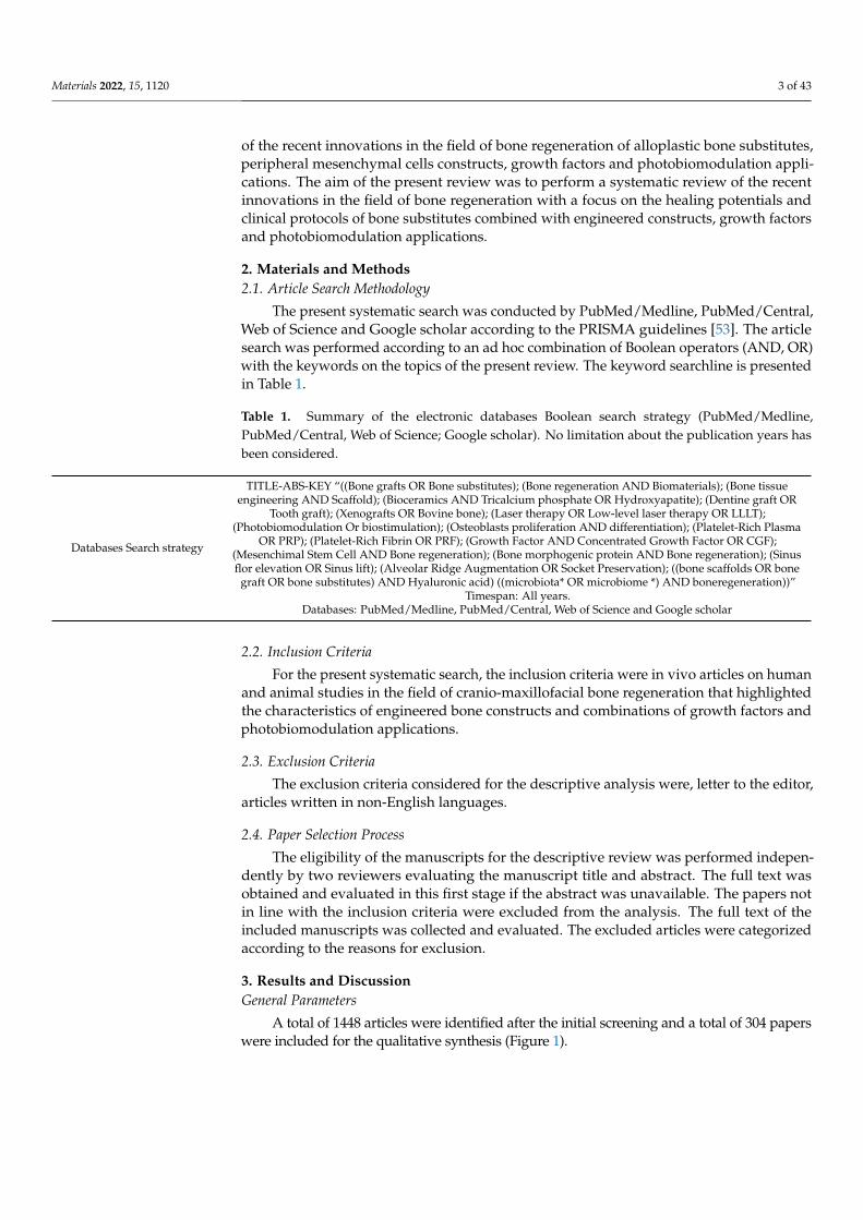
The present systematic search was conducted by PubMed/Medline, PubMed/Central,Web of Science and Google scholar according to the PRISMA guidelines [53]. The articlesearch was performed according to an ad hoc combination of Boolean operators (AND, OR)with the keywords on the topics of the present review. The keyword searchline is presentedin Table 1.
Table 1. Summary of the electronic databases Boolean search strategy (PubMed/Medline,PubMed/Central, Web of Science; Google scholar). No limitation about the publication years hasbeen considered.
Databases Search strategy
TITLE-ABS-KEY “((Bone grafts OR Bone substitutes); (Bone regeneration AND Biomaterials); (Bone tissueengineering AND Scaffold); (Bioceramics AND Tricalcium phosphate OR Hydroxyapatite); (Dentine graft OR
Tooth graft); (Xenografts OR Bovine bone); (Laser therapy OR Low-level laser therapy OR LLLT);(Photobiomodulation Or biostimulation); (Osteoblasts proliferation AND differentiation); (Platelet-Rich Plasma
OR PRP); (Platelet-Rich Fibrin OR PRF); (Growth Factor AND Concentrated Growth Factor OR CGF);(Mesenchimal Stem Cell AND Bone regeneration); (Bone morphogenic protein AND Bone regeneration); (Sinusflor elevation OR Sinus lift); (Alveolar Ridge Augmentation OR Socket Preservation); ((bone scaffolds OR bone
graft OR bone substitutes) AND Hyaluronic acid) ((microbiota* OR microbiome *) AND boneregeneration))”Timespan: All years.
Databases: PubMed/Medline, PubMed/Central, Web of Science and Google scholar
2.2. Inclusion Criteria
For the present systematic search, the inclusion criteria were in vivo articles on humanand animal studies in the field of cranio-maxillofacial bone regeneration that highlightedthe characteristics of engineered bone constructs and combinations of growth factors andphotobiomodulation applications.
2.3. Exclusion Criteria
The exclusion criteria considered for the descriptive analysis were, letter to the editor,articles written in non-English languages.
2.4. Paper Selection Process
The eligibility of the manuscripts for the descriptive review was performed indepen-dently by two reviewers evaluating the manuscript title and abstract. The full text wasobtained and evaluated in this first stage if the abstract was unavailable. The papers notin line with the inclusion criteria were excluded from the analysis. The full text of theincluded manuscripts was collected and evaluated. The excluded articles were categorizedaccording to the reasons for exclusion.
3. Results and DiscussionGeneral Parameters
A total of 1448 articles were identified after the initial screening and a total of 304 paperswere included for the qualitative synthesis (Figure 1).

Materials 2022, 15, 1120 4 of 43
Figure 1. PRISMA flowchart of the included articles.
4. Bone Substitutes and Graft4.1. Alloplastic Grafts
Alloplastic bone grafts are synthetic biomaterials with no risks of antigenic reac-tions or transmission of various diseases. Many materials have been proposed as bonegrafts containing calcium phosphate derivates with high biocompatibility. Calcium phos-phate is similar to the inorganic components of human bones [54]. Hydroxyapatite (HA)Ca10(PO4)6(OH)2 is present in the human body, bones and teeth as an inorganic mineral,but in addition to the inorganic matrix; a consistent component of the human bone is theorganic part that consists of type 1 proteins and collagen [55]. In a physiological environ-ment, HA is not soluble; therefore, the formation of new bone occurs only through thereabsorption of these materials. The osteoclast activity is not optimal against the biphasiccalcium phosphate (BCP) in 75% HA and 25% β-TCP. The solubility also influences theosteoclastic resorption pattern. The β-TCP has the highest solubility and HA the lowest,so the osteoclasts’ reabsorption of the biomaterial proceeded in the right proportion tothe solubility of the substances, but this is not entirely accurate [56]. Zwingenberger et al.reported similarly to Yamada et al. that HA with HA/β-TCP 75/25% mix is not completelyreabsorbed, but β-TCP has a higher solubility than HA/β-TPC 25/75% but HA/β-TPC25/75% had better osteoclastic resorption [56,57]. Ortiz-Puigpelat et al. reported thatHA/TCP 50% ratio is more appropriate in bone regeneration for the percentage of residualmaterial and new newly formed bone after 12–24 weeks [58]. The hydroxyapatite (HA) andcalcium tri-phosphate (TPC) are characterized by high compressive strength (statical forces)but are very fragile due to dynamic forces, so this feature limits their use in large bonedefects [59]. In operative clinical practice, these defects are correlated to various causes,such as bone tumors, severe trauma or drugs abuse with an alteration in bone metabolism.

Materials 2022, 15, 1120 5 of 43
In a pure form, HA and β-TCP have been widely used as biodegradable coatings. HAand β-TCP also prevent the formation of fibrotic tissue around the implants derived bythe body’s immune response. This fibrotic tissue can also lead to implant failure [60,61].The porosity of calcium phosphates is one of the factors that induce their poor initialmechanical property, but the porosity is able to influence the early fibro-vascularizationof the graft and replace it with new bone [62]. Moreover, the scaffold pore size is ableto affect the progression of osteogenesis. Small pores promote hypoxic conditions andinduce osteochondral formation before osteogenesis occurs. On the other hand, largerscaffold pores allow vascularization, directly evoking the onset of osteogenesis [63,64]. Theoptimal pore diameter of bone substitutes should be between 0.2 and 0.5 mm [65,66], whileGhayer and Weber reported an optimal pore range between 0.7 and 1.2 mm with excellentresults in healing cranial defects [67]. Another factor of these biomaterials for the newbone formation is the presence of micropores [68], while a submicron micropore structure<10 µm has been reported to accelerate the formation of new bone, and the quantity ofthese micropores improves the results [69]. Many studies are focused on compound formsor biphasic forms, thus improving the mechanical properties, solubility and integrationof these biomaterials [59,60,70–73]. De Tullio et al. used bone replacement materials, suchas calcium sulphate (CS) and nano-hydroxyapatite (NHA), in a comparative study toprevent a reduction in the alveolar ridge after tooth extraction [74]. After 5 months, thehistomorphometric analysis showed that these biomaterials led to bone formation, but thecombination of CS with NHA showed better results in the regeneration of new bone andin the number of residual graft particles [74]. Lin et al. reported on 51 patients that theHA/TCP+ collagen composite gave excellent results in preserving post-extraction alveolarbone [75]. Maji et al. in order to improve the mechanical properties, suggested gelatin-chitosan-β-TCP 30% with high porosity the GCT 30 scaffold. This scaffold has improvedthe mechanical properties showing high strength and compressive strength of the newbone up to 2.5 MPa, which is also the lower range of cancellous bone. The change in thepercentage of β-TCP changes the absorption of proteins, and GCT 30 also has the abilityto stimulate mesenchymal stem cells (MSCs) and transform them into osteoblasts, thusstimulating angiogenesis [76]. Afroze et al. proposed a different combination of HA withthe addition of a fMWCNT 0.5 wt% multi-walled carbon nanotube. The study was carried outin vitro with the aim of improving the mechanical properties of HA without modifying itsstructure. In fact, the HA, which contains carbon, seems to have greater similarity with theinorganic phase of the bone [77]. The fMWCNT prevents the HA from decomposing intoα-TCP and Ca2P2O7, and it promotes the porosity of the composite, which is essential forthe growth of cells. This combination of HA with 0.5 wt% low dose fMWCNT is favorablefor improving mechanical strength by approximately 317.5%, hardness by 172.4% andfracture strength by 237% [77]. This nanocomposite’s favorable application, as has beendemonstrated, is when the bone will be a load-bearing support for implants [77]. Bonematerials, such as β-TCP, have the ability to induce bone regeneration through the releaseof Ca ions [78]. Synthetic materials have high osteoconductive properties but no evidenceof osteoinductivity [79]. Recently, a new synthetic biomaterial named Osopia has beensuggested, in relation of its osteoinductive properties. This biomaterial is composed of thecombination of HA and β-TCP to obtain medium resorption properties. The structure ofthe Osopia is extended in macropores with a submicronic surface. The submicronic surfacestructure promotes osteogenesis by regulating the transforming growth factor (β-TGF)pathway and thus inducing the differentiation of mesenchymal stem cell (MSC) [80]. Thetopographic/chemical characteristic of this biomaterial drives self-induction and conse-quently the differentiation of osteoblasts [80]. Chen et al. confirmed the possibility thata biphasic HA/TCP can induce ectopic bone in non-osseous sites and that this abilityis closely related to the immune response that is caused by this material [81]. A newbio-scaffold called Compact Bio Bone Cells has recently been tested. Since this scaffold iscomposed of β-TCP, fibrin gel matrix and peripheral blood stem cells, it creates an imitationof the microenvironment of the bone tissue. The in vitro study showed that the Compact

Materials 2022, 15, 1120 6 of 43
Bio Bone Cells had a comparable biodegradability rate to the bone regeneration rate, so at7–12 days, cells resembling mature osteoblasts were highlighted. The presence of stem cellsinduces the vascularization of the region [82]. Even when stem cells are harvested from thepulp of the tooth, it is suggested to use them in bone regeneration. In combination withsynthetic scaffolds, stem cells originating from the dental pulp have given excellent resultsin tissue neoformation in a shorter time [83]. The combination of TCP with platelet-richplasma (PRF) has also been suggested in the case of fractures. This combination not onlyprovides bone regeneration (resorption occurs at the same time as bone regeneration), butthe presence of PRF aids in the healing of the positioning soft tissues [84].
The recent advances in reconstructive dentistry are oriented towards using bioactiveglasses due to the increased potential bioactivity and biocompatibility for bone regenerationprocedures [85]. Recent studies reported that the biocompatibility of these biomaterials iscorrelated with the silicate-derived percentage range between 45 and 52%, which representsan optimal equilibrium between the tissue tolerance and the osteogenesis capability ofthe graft [85–89]. The bioglasses-acting mechanism is produced by the local release ofions and the generation of amorphous calcium phosphates precipitates that are able togenerate a local change of the pH levels and osmotic flows [90–93]. Due to the new boneformation properties, the bioglasses are able to interact with the grafting environment andmodulate the activity of several osteogenetic factors, such as the alkaline phosphatase andbone morphogenetic proteins (BMP2).
4.2. Autologous Graft
The dental matrix has also been tested and used for a long time as a bone regenerationmaterial [94,95]. After extraction, the tooth is considered a useless material and is elimi-nated, and for this reason, the use of this material has a low cost. The dental matrix containsHA and type 1, type 3 and type 5 collagen, and proteins are very similar to alveolar boneas they are both derived from the cells of the neural crest [55,96–98]. The hydroxyapatitepresent in the dental matrix has the same composition of HA as that present in bone, and forthis reason, it is similar to the synthetic one [99]. The osteoconductive properties of tooth-derived materials are widely accepted by several authors [96,100,101]. In fact, the toothcan be considered the best natural scaffold. Comparing the dental matrix to HA/TCP, it isshown to be more biocompatible and bioactive [98,102]. Dentin is composed of inorganicmaterial (65%) and organic matrix and water (35%). The inorganic part, HA [3Ca3 (PO4)2Ca (OH)2], has crystals 10-times larger than those of bone (this is the biggest differencebetween these materials) and 300-times smaller than those of enamel [98,103]. The organicpart is composed of 90% type 1 collagen and 10% non-collagenic proteins including growthfactors (BMPs) and enzymes, such as alkalin and acid phosphatases and MMPs [98,103].The presence of growth factors (GFs), as well as mesenchymal cells, is also found in theapical part of the wisdom teeth [104] and in the dental pulp in a considerable amount [105].In 1965, Marshall R. Urist hypothesized the presence of proteins (BMP) in the bone matrix,and these proteins have the ability to induce ectopic bone [106,107]. BMPs play an impor-tant role in the differentiation of mesenchymal cells into osteoblasts [108], in addition tostimulating angiogenesis [109,110]. In 1991, Bessho et al. observed the presence of the bonemorphogenic protein (BMP) in the dentin matrix [111]. Many researchers have focused on(BMPs) to evaluate the effectiveness of this low-molecular-weight glycoprotein [98,108–115].In the BMP family, BMP-9 is considered the protein with the highest osteogenic potential.BMP-9 is capable of inducing the formation of new bone in critical defects by interveningin the differentiation of progenitor cells and in angiogenesis [110,112]. BMP-2 is also oneof the strongest stimulants among growth factors. Its use has led to results comparable tothose of autologous grafts in terms of bone volume and density and even reduced the riskof infection and the time of hospitalization [113]. It has been reported that bone repair couldbe produced by BMP-2, but this ability is dose-dependent [114], while a high dosage couldgive unwanted effects [115,116]. In the tooth matrix, the BMP is found in physiologicalquantities and therefore adequate for the patient. The discussion continues on how this

Materials 2022, 15, 1120 7 of 43
material will be used—mineralized or demineralized. The tooth crushing technique is atooth processing method that is proposed for use as an autologous bone graft. This tech-nique was initially applied to the newly extracted wisdom tooth. The tooth was crushedwith a bone mill and hammer without being made into a solution in order to retain thepulp. Six months after the surgery, radiological and clinical control showed that there wasnormal bone healing [117]. Other researchers also support the techniques with the toothnot transformed with acids, aiming to preserve the osteoinductive properties [118,119]. Onthe other hand, numerous studies evaluate the demineralized and granulated materialas a bone graft. Woong et al. state that the demineralized dentin acts as a carrier for theBMP-2 contained within the dental structure, thus combining the properties of the scaffoldwith the cells of the BMP-2 [120]. After being treated with acids, the dentin releases Ca2+
ions, thus creating a porous graft that acts as a perfect scaffold. The porous structure willcontribute not only to the circulation of proteins (BMP) but also to the positioning of thefibrovascular network, which will help osteogenesis [121]. Rijal et al. compared the miner-alized tooth matrix (MDM) as a bone graft with the demineralized tooth matrix (DDM).Human teeth were treated, and the experiment was performed on rats. DDM gave positiveresults, allowing the formation of new bone, thus demonstrating the regenerative abilitiesof this autologous material. This study did not confirm the same for MDM; in fact, themineralized tooth matrix fails to trigger the regeneration of new bone and takes longer tore-absorb. The authors express the need for a protocol for the demineralization of the toothso that this material can maintain its osteoinductive and osteoconductive properties [121].In a recent review of the dental graft by Gharupure and Bhatavadekar, they analyzedall the different protocols for using a tooth as a graft material and found that new bonewas created around the graft material, but the dental bone graft was not fully absorbedand replaced with new bone. The amount of residual non-resorbed dental bone graft isapproximately equal to that of bovine bone [122]. This study found a significant numberof complications, such as the separation of the mineralized block of the scaffold in somecases, causing the loss of primary stability to the implant, lack of osseointegration in othersand loss of marginal bone more than one millimeter after 5 years showed an increased riskof dehiscence. As many procedures are empirical and not really standardized, this leadsthe authors to conclude that a standardization is needed for the procedure of transformingthe tooth into microparticles that will be used as a graft [122]. During the demineralizationprocess, the dentinal tubules will turn into channels for the circulation and the release ofproteins as they will become wider. The protocol used for the demineralization of the toothis very important as it can also lead to the complete destruction of dentinal tubules [102].Further, the technique uses six liquids in addition to a partially controlled demineraliza-tion. With this patent method, the disinfection of the dental matrix is allowed, giving, inthe end, a graft of 0.4–0.8 mm, which is easy to handle and apply [98,123]. Minetti et al.evaluated the Tooth Transformer (TT), which performs the transformation of the tooth byalso disinfecting the dental matrix. This procedure lasts 25 min and can be performed inthe same session immediately after tooth avulsion. It is totally automatic, thus reducingthe risk of human error. Koga et al. state that if the dental matrix is partially demineralizedwith particles around 500–1000 µm, it has greater regeneration potential than when it hasbeen completely demineralized, as it preserves more growth factors that will intervene inosteogenesis [124]. Bono et al. Support the idea of using treated and demineralized teeth,confirming that the demineralization performed with TT increases the bioavailability ofBMP-2. The protocol used includes six solutions that demineralize and sterilize the crushedtooth [125]. It turns out that they do not damage the microstructure of dentin and organicmatrix, as has been stated in other studies [126,127], but they increase the availabilityof BMP-2 in the matrix [98,125]. Substances that determine too high a demineralization(substances that give a high demineralization) produce a graft material of dental originwith low osteogenic potential as they eliminate or decrease the proteins present in thetooth, including BMP-2 [98]. Bono et al. affirm that this slight demineralization of the toothlowers (reduces) the content of Ca and P compared to the non-demineralized matrix, but

Materials 2022, 15, 1120 8 of 43
at the same time, it increases the bioavailability of BMP-2 [125,128]. BMP-2 increases theactivity of ALP (alkaline phosphatase), which leads to an increase in osteodifferentiation.To induce significant ALP activity, a minimum concentration of 12.5 ng/mL of BMP-2is needed, and after treatment with TT, the concentration of BMP-2 in the dentin matrixis 22 ng/mL [98,128]. An evaluation through scanning electron microscopy has shownthat the density, roughness and homogeneity of the autologous dental graft is relativelysimilar to that of autogenous cortical bone, with a surface containing both organic andmineralized material [129]. The technique with the autologous tooth elaborated with theTooth Transformer has been tested in the alveolar socket preservation of the bone after theextraction of the teeth [98]. The authors state that the success of the implant after boneregeneration was 99.1%, and the time it took for bone healing to be ready for implant wasfour months. The demineralization and decontamination performed by the solution doesnot eliminate BMP-2 and collagen [130,131]. The same author continued his research byusing the Tooth Transformer on sinus lift with the same technique on a demineralized andgranulated tooth [98]. In this case, the implants were inserted after six months. In this study,no inflammatory reaction was observed around the graft, and the tooth material used asgraft was perfectly integrated with the host tissue. Unlike other autologous grafts, the toothmatrix does not lose bone volume with the passage of months [132]. In large bone defects,there is the problem of the amount of grafting that is needed in these cases. Umebayashiet al. have suggested the use of autologous bone when the quantity of teeth is limited,which can also be mixed with other biomaterials [133]. The matrix of the autologoustooth was partially demineralized (APDDM) and used in combination with particulatecancellous and medullary bone (PCBM) in bilateral sinus lift and in anterior maxillaryreconstruction. This combination showed the osteoinductive capacities of PBCM and theosteoconductivity of APDDM. The implant was performed after 3.5 months [133]. Anothersuggested combination was that with xenographic material (Bio-Oss), which gave positiveresults in bone regeneration, but compared with the dental matrix alone, this last oneforms a greater amount of bone [134]. In any case, the dental matrix can be combined withother biomaterials in large bone defects, which provides very positive results [133–136].From the physio-chemical side, the tooth can be considered a material similar to bone butwith a higher mineralization. A completely demineralized or a mineralized tooth did notbring good results in bone regeneration. In fact, the decalcification serves to make thedentin more accessible to collagenolytic enzymes and also for the fact that demineralizedsurfaces are the most adherent for osteoblasts [98]. The use of Tooth Transformer is highlymanageable and is totally automatic, with no risk of human error. The use of this machineoffers shredding at low speed that allows for homogenous particles and more material touse. The protocol used here is the one that offers a ratio between Ca and P (1.70) that iscloser to the natural ratio found in bone (1.67) and, moreover, preserves proteins in thedental matrix (Figure 2) [98].
Figure 2. Synthesis of the biomaterials for bone regeneration.

Materials 2022, 15, 1120 9 of 43
4.3. Xenografts Bone Substitutes
Xenotransplantation or bone derived from another species has been applied for manyyears as regenerative bone material. Their component is hydroxyapatite. Bone hydroxyap-atite (BHA) is obtained naturally and has a lower cost than synthetic hydroxyapatite. Thepresence of ions, such as Na+ and CO3, seems to influence the molecular structure withlower porosity and thicker microstructure [137]. It has been shown that for bovine bone, thebest results are obtained when it is used in particles with a diameter of 1.0–2.0 mm. Thesedimensions provide the best results in preserving bone volume compared to micro-sizedparticles [138]. The same result was accepted by Kon et al. for autologous bone, wherelarger particles (1.0–2.0 mm) give the best result in the formation of new bone and smallparticles tend to be absorbed more quickly [139]. Bovine-derived xenograft is one suchmaterial, which has been recognized as biocompatible with other animal and human or-ganisms [140]. This material acts as a scaffold and promotes osteoblastic activity [141]. Themicroporous structure of bovine bone, which is considered similar to that of cancellousbone, allows this material to have osteoconductive properties [142]. The researchers demon-strated bone healing when they examined the bovine bone, and it was found that the boneis compact and mineralized and surrounds the bovine graft particles. The colonization ofcapillaries and newly formed cells is appreciated in Haversian canals [140–142]. Piattelliet al. used an inorganic bovine bone Bio-Oss for a maxillary sinus lift. After six months,the histomorphometric analysis showed that 30% of the samples selected for biopsy thatwere removed with a 4 mm drill under saline irrigation to a depth of 10 mm and examinedby microscopy still contained Bio-Oss particles, but these particles were also observedafter four years. The authors’ autologous tooth-derived graft material behaves like a scaf-fold an implant but has a very slow resorption [143]. However, other authors think thatthe slow reabsorption of this material serves as a source of mineralization [144,145]. In2005, Sanchez et al. tested a combination of platelet-rich plasma (PRP) on dogs. In thisstudy, they stated that the use of PRP in these animals did not give significant changesin bone regeneration [146]. More recent research has continued to combine Bio-Oss withplatelet-rich fibrin PRF [147–149]. This combination is used in sinus lift, and the implantsite was ready in 106 days, less than four months after surgery. The authors declare 100%success in bone regeneration following the Choukroun protocol [147]. The PRF has adense and strong fibrin matrix, which is a great advantage over PRP. The PRP has alsofailed in other studies [150–152], but it should also be mentioned that the denominationof blood-derived products has often been confusing, but lately, everything has alreadybeen clarified, and their classification is made based on architecture of fibrin and cellularcontent [147,150,151,153–160]. Xenografts with CGF Growth Factor Concentrate is anothersuggested method. The authors used the Silfradent device to allow the CGF to penetrate thebone graft (to better allow fusion). Their technique in the augmentation of atrophic ridgesgave excellent results; in just four months, mineralized bone was evident and ready for theimplant procedure [161,162]. Continuing to try and improve osteoconductive properties,another study suggests combining Bio-Oss with BMP-2. To increase the building activity ofthis protein, rhBMP/Bio-Oss was coated with heparinized dopamine. The result showedthat new bone formation began four weeks after surgery, while in week eight, there wasa huge difference in the rate of bone formation between this study group and the groupthat was only used in the Bio-Oss graft [163]. Another combination tested is that betweenbovine bone Bio-Oss and autologous bone, but these two materials have differences in thetime of absorption and have often not given good results in bone regeneration [164]. Asxenografts are available in significant quantities, there are many studies focusing on thesematerials. A recent review on the use of bovine bone highlighted in the conclusions that58% of cases failed in osseointegration, while 83% of these continued to have pain even aftera long time. The main factor causing this complication is the body’s immune response tothis material [165]. Therefore, for xenografts, the main antigen is the epitop alpha Gal, andto inhibit this antigen, the xenograft should be decellularized, and only then can it be usedas a scaffold. However, the risk of an antigenic reaction will always be there, especially for

Materials 2022, 15, 1120 10 of 43
bovine bone [165]. A new combination of bovine bone with type 1 atelocollagen has beenrecommended to minimize the antigenic reaction. Indeed, this combination is regardedas a new generation of xenografts. Atelocollagen has a low risk of antigenic reaction andinhibits bacterial activity. Additionally, unlike most xenografts, this combination of bovinebone graft with atelocollagen is totally resorbable [80]. Despite the treatment that xenograftundergoes, in 2007, Terry et al. highlighted the first case of bovine spongiform H-typeencephalopathy related to bone reconstruction with bovine bone [166]. This means that therisk of transmitting diseases from one species to another exists.
5. Platelet Derivates and Growth Factors
From the peripheral blood, a platelet-rich autologous concentrate is collected, which isconsidered a “good healer” obtained in a non-invasive and rapid way [167], and when com-bined with other biomaterials, gives excellent results [168,169]. The first piastin concentratehas been called PRP and normally contains 0.5 × 10¹¹ platelets for each unit. PRP doesnot have good mechanical properties. Before being activated, PRP is in liquid form [170].This platelet concentrate releases growth factors and cytokines in a very short time andthen reabsorbs in about 12–14 days. PRP has given the best results in combination with anautogenous graft compared to other materials used in bone regeneration [171]. However,the PRP gel in periodontology and dentoalveolar surgery has lost its use with the discoveryof PRF, which has a lower cost and is easier to use [155]. The second-generation PRFencapsulates leukocytes, cytokines and, above all, growth factors in an autologous fibrinmatrix. This PRF has been produced in the absence of anticoagulants, and for this reason, itis considered 100% autologous material that facilitates tissue regeneration by eliminatingthe transmission of diseases through the blood [172]. The fibrin matrix will act as a 3Dscaffold in which cells and proteins will be captured. The cellular components of PRF areleukocytes, platelets, macrophages, granulocytes, neutrophils and erythrocytes. These cellsplay a fundamental role; in fact, platelets intervened in the first phase of healing in hemosta-sis with leukocytes, macrophages, granulocytes and neutrophils and participated in theanti-inflammatory phase. Platelets and macrophages release a large number of growthfactors within 7–14 days, including platelet-derived growth factor (PDGF), transform-ing growth factor-β1 (TGF-β1), insulin-like growth factor (IGF) and vascular endothelialgrowth factor (VEGF) [150,151,155–157]. These bioactive molecules are able to furtherpromote cell proliferation and bone remodeling [173,174]. The architecture of the fibrinmatrix influences the trapping/release of GFs [156]. The cellular content and the fibrinmatrix differentiate between the materials derived from the platelets because these arethe elements that are involved in the regeneration and healing of the tissues. Therefore,it is necessary to recommend the use of this method of peripheral blood derivatives PRFassociated with biomaterials [157]. When the use of PRF is included, the results indicate thatosseointegration and bone regeneration takes place in a short time—only 106 days [147].Currently, L-PRF as a component of the large PRF family has the potential to biostimulatebone regeneration and accelerate osseointegration [175]. The PRF does not induce new boneformation directly, but what all authors agree on is that PRF significantly improves earlyvascularization in bone grafts [147,150,151,155–157,176]. The PRF through the angiogenesispromoter (VEGF) induces the vascularization of the area and decreases the inflammatoryreaction around the graft since it contains anti-inflammatory cytokines (IL-4) [177]. PRFimproves soft tissue healing when in contact [178–180]. Indeed, some studies have come tothe conclusion that PRF promotes and improves soft tissue regeneration more than hardtissue and therefore consider PRF to have slight potential in the GBR technique (guidedbone regeneration). Excellent results have been obtained on soft tissues, even from theuse of a mixture of ozolipoilein, which, in only 3–5 days, can heal small ulcers caused byradiation therapy cycles. This blend is also antibacterial and antifungal [181]. Instead,PRF significantly contributes to tissue healing through its antibacterial properties, espe-cially against S. aureus and E. Coli. However, this is mostly expressed when a horizontalcentrifuge is used to produce PRF [182]. The use of PRF as a membrane has produced

Materials 2022, 15, 1120 11 of 43
effective results in both bone regeneration and soft tissue regeneration. This membranecan be obtained in a non-invasive way and is considered a protective barrier of newlyformed tissues. For the patient, this method is very comfortable [183,184]. The PRF canbe considered a very valid barrier membrane between the oral cavity and bone, with aneasy and predictable preparation protocol and a well-defined structure with impressivemechanical properties, as reported by several authors [147,150,151,155–160,185]. A PRFmembrane can also be used many hours after preparation as the release of PDGF continuesto be high, as long as the membrane is properly prepared in a PRF box and stored underphysiological conditions. The release of VEGF and β-TGF significantly increases in the firstfour hours, but then the amount of these growth factors does not show large differencesbetween blood derivatives because GFs are mainly derived from leukocytes [186]. A newgeneration of blood products called CGF from Silfradent is produced without anticoagu-lants. The MEDIFUGE MF200 centrifuge that produces CGF is from Silfradent, has a specialmulti-speed centrifugation program, creates a gradual sinusoid (RPM) at various speedsand offers a large, high-density fibrin matrix rich in GFs [187]. This CGF is composed ofthree layers, a dense fibrin network and blood cells trapped within it, especially in themost outer part [188]. The first layer is called PPP and is a network of plasma proteins inwhich cells without a nucleus are found. These cells seem to have the typical appearance oferythrocytes. The second layer of CGF is much denser than the first and third layers. It is afibrin network, in which cellular elements are trapped and collagen fibers are found whereother corpuscular elements are captured. The CGF layer consists of three fragments: theupper white part, the lower red part and the buffycoat at the interface between the whiteand red part [187]. The third layer of RBC red blood cells appears to have a wider networkof collagen fibers than the second layer, and coreless corpuscular elements, such as platelets,are found entangled in this network [188]. The CGF fibrin matrix is denser than PRF andhas GFs present in it, such as TGF-β1 and VEGF, which are important in cell proliferation.The presence of these growth factors was also identified in the RBC layer. The CD34+ cellsare trapped in the CGF matrix with a considerable number of them and have been found inboth levels (CGF-RBC). The CGF appears to be more promising in tissue regeneration, asit promotes osteogenic cell differentiation and proliferation. Therefore, with the CGF, theactivity of ALP [162,189] significantly increases. CGF also appears to be very promising inneural tissue regeneration. The controlled release of growth factors and cytokines from CGFinfluences cell differentiation and intervenes in the growth of neurons [190]. In conclusion,as a result, we have the PRP with a lower concentration of GFs compared to A-PRF orCGF. Advanced Platelet Fibrin (A-PRF) and Concentrated Growth Factors (CGFs) containTGF-β1, VEGF, PDGF-BB, IL-1β and IL-6. Masuki et al. said that the main source of PRFgrowth factors is in the exudate and has a very low percentage of GFs found in the fibrinnetwork [191]. However, Ehrenfestet al. suggest that exudate is not the most importantpart in platelet concentrates [159]. Kawaseet al. reported that even for PRP, which is in aliquid state, fibrin gel and thrombocyte aggregates, many more biological mechanisms willemerge [192,193]. This is also accepted from other studies [150,151,155–158]. An effectivemethod used for bone reconstruction is the penetration of CGF into the Xenograft blockswith the method that uses the Round Up device from Silfradent [161,187]. The same methodis also used with β-TCP, which shows that this combination significantly increases therelease of certain growth factors, such as BMP-2 and BMP-7. Scientific evidence showsthat calcium ions that are released by the dissolution of β-TCP influence the activation ofplatelets [150,151,155–160]. The Ca2+ ions stimulate the differentiation of osteoblasts andincrease the stability of BMP-2. The same study reveals that CGF used alone instead ofa combination increases the release of IGF-1 [194]. The release of growth factors is highin the first few days then starts to slow down. With the aim of overcoming one of themain limitations of PRF and its degradation, an in vitro study was conducted on Alb-PRF,an injectable biomaterial, produced by PRF in liquid form and PPP heated to 75 degreesCelsius in 10 min with a device called APAG [187,195–197]. Since PPP is composed of60% albumin, its denaturation creates an organized and dense protein structure without

Materials 2022, 15, 1120 12 of 43
cells. This combination with APAG has been shown in vitro to preserve the volume andprolong the reabsorption times, leading to that the conclusion that the release of growthfactors can continue much longer than in the traditional PRF [187,196,197]. The Alb-PRFpreserves its volume even after 21 days [198]; however, the Alb-PRF membrane loses theelasticity of the PRF membrane and becomes very fragile. To overcome this deficiency,the use of high-power laser pulses is suggested [199]. Laser irradiation is performed ona surface, leaving the inner part of the membrane intact. This method seems to stimulateand increase stability in the body and allows for better suturing/fixing of the membrane,thus optimizing the Alb-PRF [199]. Even with the albumin denatured with CGF, a solidand dense membrane of cells with nuclei is obtained. This Alb-CGF membrane is capableof releasing growth factors (VEGF, PDGF, FGF2) for seven days [195]. A comparative studywas conducted on the preservation of the alveoli after extraction (socket preservation)using only Alb-CGF and allograft covered with albumin. This study demonstrates thatradiographically and histologically, there was no significant difference between the twogroups tested; thus, Alb-CGF is considered a promising biomaterial in tissue regeneration.In fact, the authors conclude that Alb-CGF gives the same results as a bone graft withallografts and albumin (sticky bone). This material is believed to be capable of immediateand prolonged release of growth factors [200]. Other studies have reported the effectivenessof CGF and PRF in osteogenesis in patients with systemic diseases. Systematic diseases arethose that must be taken into consideration in particular for bone regeneration because, inthese diseases, there is the need for a personalized protocol according to the underlyingpathologies and the planning of the work to be carried out. The use of bisphosphonates inpatients with osteoporosis reduces the production of BMP-2, while CGF or treatment withresveratrol combined with CGF instead promotes the production of BMP-2, and they have apositive role on osteoblasts in patients treated with bisphosphonates [150,151,155–160,201].Bisphosphonates induce bone growth, but they alter bone turnover, decrease osteoclastsand significantly reduce vascularization in adult patients [202]. Moreover, in childrenand young patients, they are protected from this phenomenon [203]. Due to the apopto-sis of endothelial cells, bisphosphonates decrease the number of endothelial progenitorcells, so both vascularization and neo-vascularization are reduced [204]. On the otherhand, the use of CGF in combination with sodium orthosilicate has been shown in vitroto stimulate cell growth, proliferation and metabolic activity in several human cell lines,including fibroblasts, endothelial cells and osteoblasts [205], and that the use of growthfactors, such as epidermal growth factor (EGF), may partially neutralize the effects ofbisphosphonates on human oral keratinocytes and human umbilical cord endothelial cellsvia the EGFR/Akt/PI3K signaling pathway [206]. Diabetes is another very widespreadsystemic disease. Patients with diabetes have a noticeable reduction in the potential forhealing and vascularization of the affected tissues. Durmuslar et al. concluded that the useof PRF alone did not produce significant good results; only in combination with autologousbone does PRF induce bone formation in diabetic rats and significantly increase bonevolume compared to the group where only autologous bone was used [207–220].
6. Biostimulation and Laser–Graft Interactions
In daily practice, the use of Lasers, Light Amplification by Stimulated Emission ofRadiation (Table 2), is increasingly being applied. Its use leads to an operating field cleanerfrom blood and atraumatic work. Additionally, it significantly reduces pain and postoper-ative edema [221–225]. The use of lasers offers better aesthetic effects compared to othertechniques since it does not leave an obvious scar, giving the part of the body where theintervention is performed an excellent restitutio ad integrum [221–224]. Photodynamicsincludes a photochemical and non-thermal biological interaction. Photobiomodulation(PBM), also known as low-level laser therapy (LLLT) [226], is a safe and non-invasive clini-cal method that, in most cases, uses red 600–700 nm and infrared 770–1200 nm light, withthe aim of stimulating healing and reducing inflammation and pain [227]. The mechanismof action of the LLLT is based on the interaction between chromophores cells (especially the

Materials 2022, 15, 1120 13 of 43
enzyme of the mitochondrial respiratory chain; cytochrome C oxidase) and laser light. Theabsorption of light by these chromosensors generates more energy (ATP) and, in addition,causes the modulation of the calcium level and an increase in the production of nitric ox-ide [227]. The LLLT has been recommended by the Multinational Association of SupportiveCare in Cancer/International Society of OralOncology for the treatment of oral mucositis inadult cancer patients, but LLLT has been shown to reduce severe mucositis even in youngcancer patients [228]. The use of low-intensity lasers has also given excellent results inmany oral diseases [229]. In recurrent aphtosis and herpes simplex, it reduces pain andprovides complete wound healing, and when combined with antibiotics, improves thecondition of BRONJ/MRONJ patients by reducing clinical symptoms in hyposalivationor xerostomia. Therefore, LLLT is very effective in cell repair and increases secretion bystimulating the salivary glands. LLLT also reduces the symptoms of trigeminal neural-gia [230–235]. The use of LLLT has also spread widely in the field of periodontology andperi-implantology [236,237], but the effects that lead to the use of long-term laser on scalingand root planing (SRP), bacterial-subgingival reduction and sulcular debridement (lasercurettage) appear to have no significant advantages over other methods [238]. However,the use of the laser gives excellent results on the surfaces of implants that have been at-tacked by peri-implantitis and have part of the fixture exposed in the oral cavity. The useof the laser decreases the microroughness and porosity of the implants, thus giving a moreglossy surface, which prevents the adhesion of bacteria on smooth surfaces [225]. In theirstudy, Pereira et al. reported that the use of the LLLT did not lead to any change in boneregeneration compared to the control group, but there was a noticeable difference in theinterface between the implant and the bone, thus allowing for better osseointegration [239].Grassi et al. reported that the use of LLLT promotes proliferation and differentiation bysignificantly increasing cell adhesion on the implant surface [216]. The ability to promoteosseointegration and stimulate bone formation around the implant has also been confirmedby other authors in their research [240,241]. Other studies have also been developed to testthe effect on mechanical strength that occurs during the use of the LLLT laser [242,243].Maluf et al. tested two groups of mice: one group received laser therapy and the otherdid not. This study concluded that the group of mice that received laser irradiation hadgreater difficulties detaching the implant from the bone than the group that was not irra-diated. A torque machine was used to measure the torque needed to detach the implantfrom the bone. The result showed that the group treated with laser had a resistance of50% more than the other group without laser irradiation [242]. Other studies have usedthe LLLT laser to enhance osteoinductive properties in biomaterials that only possessosteoconduction [212,213,244–246]. For this purpose, the biphasic HA/TCP and bovinebone were tested as a scaffold. The groups to be tested were irradiated every 48 h for13 days of 40 s per sector and, after 60 days, with a second intervention the endosseous im-plants were inserted. The results showed significant differences between the control groupand the group tested 15 and 45 days after endosseous implantology. In this experiment,increased exposure of BMP-2 and osteocalcin was observed [213]. The authors concludedthat the LLLT promotes osseointegration with a high expression of BMP2 and osteocalcinrelated to osteoblastic activity and that the force required to remove the implants couldbe compared with that of the implants inserted in autologous bone [213]. Another studythat tested the use of LLLT on bovine bone (DBB) and biphasic HA/TCP led to the sameconclusion. After 90 days in the irradiated group, there was bone augmentation, with anincrease in the expression of BMP2, Osteocalcin (OCN), alkaline phosphates (ALP) andgenes [212]. The LLLT laser is described as a promising therapy for stimulating osteoblas-tic activity and differentiation. Furthermore, it has been observed that LLLT improvesthe osteoconductive properties of these materials while maintaining the volume of thebone graft [212]. Other studies have used photobiomodulation to repair bone defects withbovine bone grafts. The irradiation began on the day of the surgery, and then seven otherirradiations were performed every 48 h [244–246]. The use of LLLT in these studies led to asignificant increase in the deposition of collagen in the first healing phase, as well as greater

Materials 2022, 15, 1120 14 of 43
osteoblastic activity, rapid bone remediation with neo-formation of the Haversian systemand regeneration of the cortex [244–246]. Another study, which lasted 12 months, usedenamel matrix protein-derived (EMD) combined with laser LLLT in an intra-osseous defect.The radiation was performed after the surgery with a 4 J/cm2 diode laser for five minuteson each side of the defect in five days. The result showed that the use of LLLT improvedthe effect of EMD in the intra-bone defect [247]. The light-emitting diode (LED) is anotherpromising alternative for tissue biostimulation. Unlike the LLLT, which emits coherentlight, the LED has non-coherent light. The LED has a lower cost, is more manageable anddoes not carry the same risk to the eyes. The LED appears to have the same effects as the redlight laser on the stimulation for the growth of pre-osteoblasts [217], promotes osteogenicdifferentiation and inhibits cell proliferation [248]. The application of the LED togetherwith photosensitive substances has also led to excellent results in the osseointegration ofimplants inserted into bone diffusers and filled with biomaterials. After some time, thebone formed around the implants can be compared to autologous bone [249]. However,the use of LEDs is limited; for a small area when a high power density is required, italmost always requires the use of lasers [250]. Mergoni et al. conducted an in vitro 915 nmlaser study on human osteoblasts. The treatment did not provide improved effects on cellproliferation and differentiation compared to the non-irradiated group but induced theformation of bone nodules by a considerable amount [219]. Instead, Jawad et al. concludedthat decreasing the laser power increases osteoblastic cell differentiation and cell prolifera-tion increases as the laser power increases. Thus, the cell proliferation of osteoblasts usinga 940 n diode laser increased more when the irradiation power was 300 mW compared to100 mW, while the expression of ALP and osteocalcin was, respectively, higher with the ra-diation at 100 mW and 200 mW compared to 300 Mw [220]. Barbosa et al., In their research,made a comparison between red light lasers and infrared lasers. They claim that the use ofthe infrared wavelength laser gives better results compared to the red wavelength laser,thus concluding that the bone healing process depends on the time and wavelength [251].This result also agrees with the conclusions of Queiroga et al. [218] and Renno et al., whichalso confirm the fact that different cell lines respond differently to specific combinationsof wavelengths and doses [252]. Tani et al. tested biomodulation with three differentwavelengths: red light (635 nm), infrared light (808 nm) and LED (405 nm). The aim was toevaluate the differences in proliferation, adhesion and differentiation of pre-osteoblasticand human mesenchymal stomal (hMSC) cells [253]. Bone nodules (Ca2 deposition) wereobserved differently in both laser wavelengths from the radiation with LEDs. On humanmesenchymal stromal cells, the 635 nm wavelength is capable of promoting osteogenicproliferation, adhesion and differentiation, while on osteoblasts, this wavelength does nothave a significant effect on cell differentiation by questioning red light (the wavelengths) orthe parameters used for radiation (0.5–1–2 J/cm2). Moreover, this study proposes the diodelaser with a wavelength of 635 nm as an effective option to promote and stimulate boneregeneration [253]. On the other hand, Ghidini et al. concluded that red waves (645 nm)could penetrate to considerable depths [254]. All these data show that there is still a greatdeal of confusion on the working protocols for biomodulation. Many authors suggestthat the use of LLLT fundamentally improves and accelerates the healing processes ofnewly formed tissues [208–214,218–220]. These conclusions have already necessitated arecommended and studied protocol of a guideline with irradiations and the type of laserprescribed by the World Association for Laser Therapy (WALT) on biomodulation, as itspositive effects certainly cannot be denied [255].

Materials 2022, 15, 1120 15 of 43
Table 2. Summary of the laser protocols for biomaterials osseointegration enhancement.
Type of LLLT Type of Irradiation Groups Stydy Results Conclusion
Nagata et al. [208] InGaAIP (λ 660 nm) Power 35 mW/point Energy density4.9 J/cm2/point
1-LLLT alone2-(BMA) bone marrow aspirate
3-LLLT/BMA 4-control group with ablood clot
-Not suitable for proliferation ofosteoblasts cell-Proliferation and
differentiation was seen only for MSCpresent in BMA
The use of LLLT alone did not induceosteoblast proliferation but BMA/LLLT is
a promising combined therapy inbone regeneration
Garcia et al. [209] InGaAIP (λ 660 nm) Power 35 mW/point energy density4.9 J/cm2/point
1-control group with a blood clot2-dexamethasone with a blood clot3-dexamethasone+autologus bone
4-dexamethasone+LLLT 5-autologusbone+LLLT
-Dexamethasone group show less boneformation with a reduction in osteoblasts
-Group treated with AB/LLLTosteogenic potential
LLLT helped bone from the inhibitoryeffects of dexamethasone LLLT improve
bone healing in critical defects
Saygyn et al. [210] Diode laser (λ 685 nm) Power 25 mW/point energy density2 J/cm2/point
1-MSC single dose irradiated2-MSC double dose irradiated
3-control group
-Double dose group stimulate the releaseof IGFBP3, IGF-1 and bFGF-LLLTstimulate osteoblasts proliferation
LLLT improved wound healing andbone regeneration
Cunha et al. [211] GaAIAs (λ 780 nm) Power 100 mW/point energy density6 J/cm2/point
1-LLLT group2-autogenous bone
3-autogenous bone+LLLT4-inorganic bovine bone
5-inorganic bovine bone+LLLT6-contol group
-LLLT stimulates new bone formation Laser accelerated graft material particlesand bone healing
de Olivera et al. [212] GaAlAs (λ 808 nm) Power 100 mW/point energy density4 J/cm2/session
1-LLLT major group2-control major group (each major group
divided in threegroups-coagulum-inorganic bovine bone,
-HA/TPC)
-LLLT group shows osteogenic potential-expression of BMP2, Osteocalcin, ALP
and genes (Runx2, Jagged1)-Maintained the volume of biomaterials
-Osteoblastic differentiation
LLLT stimulated bone formation ingrafted area with
osteoconductive materials
de Olivera et al. [213] GaAlAs (λ 808 nm) Power 100 mW/point energy density4 J/cm2/session
1-deproteinized bovine bone (DBB)2-HA/TCP
3-LLLT+DBB4-LLLT+HA/TCP
-LLLT group osteogenic potential with theexpression of BMP2 and OCN-increase of
implant osteointegration
LLLT increased osteointegration in graftedarea with osteoconductive materials
Gerbi et al. [214] GaAlAs diode laser (λ 830 nm) Power 40 m/W/point energy density4 J/cm2/point
1-control group2-LLLT group
3-BMP+membrane4-BMP+membrane+LLLT group
-Osteogenic potentialLLLT combined with the use ofbiomaterials accelerated bone
regeneration process
Renno et al. [215] GaAlAs diode laser (λ 830) Power 30 m/W energy density10 J/cm2
1-MC3T3 grown on biosilicate+LLLT2-control group
-LLLT GROUP 13% decrease incell proliferation
LLLT group resulted in a reduction incell growth
Grassi et al. [216] Laser diode (λ 920 nm) Power 0.1 W energy density 3 J/cm21-Osteoblasts-like cells seeded on zirconia
or titanium surface+LLLT2-control group
-Osteogenic potential-cell proliferation-cell
differentiation-ALP expression-the mRNA of RUNX2 and OSTERIX
LLLT significantly increased cellularadhesion on implant surface

Materials 2022, 15, 1120 16 of 43
Table 2. Cont.
Type of LLLT Type of Irradiation Groups Stydy Results Conclusion
Pagin et al. [217]
Visible red (λ 660 nm)Infrared
(λ 780 nm) LED(λ 630 ± 10 nm)
Laser: power 1 W/cm2 energydensity 3 J and 5 J/cm2
LED: power 60 mW/cm2 energy3 J and 5 J/cm2
MC3T3 irradiated with red/infrared laserand LED
Red/infrared and LED-influenced ALP
-no effect on cell differentiation
Red/infrared laser and LED had similareffects et early periods of time on
stimulating pre-osteoblasts
Queiroga et al. [218] Red spectrum (λ 660 nm) infrared(λ 780 nm)
Power 40 mWenergy density 2 J/point
1-LILT 660 nm2-LILT 780 nm
3-Control group
-LILT with 780 nm newly formed bone-LILT with 660 nm no difference from
control group
LILT with 780 nm wavelength promotebone reparation
Mergoni et al. [219] Diode laser GaAs (λ 915 nm) Power 0.12 and 1.25 W/cm2 5.15 and45 J/cm2
-Osteoblasts isolated from mandibularcortical LLLT treated
-control group
-No osteoblast cell proliferation-no osteoblast cell differentiation
LLLT induced more bonenodules formation
Jawad et al. [220] Diode laser GaAlAs (λ 940 nm)
Power+energy100 mW/45.85 J/cm2
200 mW/91.79 J/cm2
3000 mW/137.57 J/cm2
-LLLT groups-Control group
-Cell proliferation-cell differentiation
-ALP and osteocalcin expression
LLLT improved bone formation bystimulating osteoblast cells

Materials 2022, 15, 1120 17 of 43
7. Microbiota in Tissue Repair Processes
Other factors come into play in bone and soft tissue metabolism. The crucial role ofthe intestinal microbiota in the regeneration process of our organisms is the subject of greatstudy and debate. Several authors agree in asserting that the intestine as a whole is theplace where basically any dysfunction begins, even before a symptom or pathological eventis diagnosed or even evolved. The balance/imbalance between the constituent parts of thegut is known as symbiosis/dysbiosis, which accurately reflects systemic health. A healthyintestine is made up of the balanced presence of four Phila bacteria: Bacteroideti, Firmicutes,Actinobacteria, Proteobacteria and Verrucomicrobia. Ibacteroideti, and firmicutes represent themajority of the microbiota [256,257]. Over the past two decades, one of the most significantscientific discoveries has been the confirmation of the Neuro-Endocrine-Immune natureof the intestine, which as a single apparatus tends to perform an activity believed to beexclusive to the central nervous system (CNS), so much so that nowadays, it refers to thegut–brain axis. In fact, the two systems share anatomically and physiologically importantmulti-functionalities and have neurochemical, endocrine and immune significance thatmakes the intestine an autonomous system called the enteric nervous system (ENS), whichis responsible for all gastrointestinal activities in full coordination with the CNS [258]. Afurther aspect that has been highlighted is the inter-communicative capacity of the twosystems through a very complex network outlining a common clinical-pathological picture.In fact, many important pathologies tend to afflict the two systems in a one-to-one andconstant way [259,260]. Within the intestine, the microbiome functions as a guarantor of thehomeostasis of all the other systems of the organism, capable of producing 50 to 100 mmol/Lper day of short-chain fatty acids (SCFA), such as acetic acid, propionic acid and butyricacid, which are the sources of energy for both the proper functioning of the complexcellular structure that makes up the intestinal epithelium and for the constant maintenanceof the skeletal system [261–264]. The importance of SCFA among acetic acid, for example,is essential against infections, in regulating blood pressure and against the depositionof sclerotic plaque in the arterial walls. Butyric acid, for its part, acts as an inhibitor ofinflammatory responses thanks to its immunomodulating properties, while propionic acidhas been found vital in the prevention of obesity and diabetes 2 [261–264]. Furthermore, theintestinal system performs a “prompt delivery” function of vitamins, such as folic acid andthe vitamin B2–12 complex, and at the same time, it allows the synthesis of vitamin K, whichis involved in many physiological, metabolic and immunological activities and plays afundamental role in the prohormone/vitamin D absorption mechanism, which is importantfor the homeostasis of the skeletal system and the formation of osteocytes, osteoblasts andosteoclasts [263,265]. The way in which this interaction takes place and the mechanismin which they carry out these processes reveals something that is revolutionizing theconventional thinking of contemporary medicine, for which we refer more and more oftento the term epigenetics [266].
Epigenetic events are highly dynamic and change in response to the availability,quality and quantity of environmental insults both external and internal to the oral cavity.In this perspective, there is a tendency to have a substantially different perspective in whichbacteria, viruses and fungi, which are also harmful and dangerous, are still indispensablefor life. The results of the experiments conducted on germ-free (GF) mice confirmed thatthe shortage of gut microbiota was to be indicated as a functional cause of the severedeficiency of the immune system. GF animals were shown to have low levels of naturalkiller cells (NK), dendritic cells (DC), α/β + and γ/δ + T cell populations, which play animportant role in defense and pathogenesis during inflammation and infection, particularlyagainst some types of malignant neoplasms. Furthermore, the above experiments haveunequivocally demonstrated that GF animals were particularly susceptible to frequentinfections substantially due to the decline of angiogenin-4 (Ang4), a powerful antimicrobialmolecule belonging to the class of microbiocidal proteins in Paneth cells [267,268].

Materials 2022, 15, 1120 18 of 43
Dysbiosis and Metabolic Disorders Related to the Bone Tissue Degenerative Processes
What is the connection between dysbiosis and the bone system? The conclusionsrevealed a different type of bacteria. In fact, patients suffering from osteopenia revealedan increase in the number of Firmicutes phyla with a reduced number of Bacteroidetescompared to patients in the control group; the genera Lachnoclostridium and Klebsiella,Gemmatimonadetes Chloroflexi and Synergistetes were detected to a high extent in both patientswith osteoporosis and in those with osteopenia but were absent or very low in the controlgroup; the control group showed a prevalence of Bacteroides, Faecalibacterium and Prevotella.Interestingly, Prevotella was in a constant presence in the osteoporosis group but very lowin the osteopenia group [269–272].
In patients suffering from Crohn’s disease and ulcerative colitis, recent studies haveconfirmed the coexisting presence of osteoporosis, and the cause can be found in a chronicinflammatory state. One of the mechanisms involved seems to be related to the immune-mediated bone metabolism involving the RANKL axis (an activator of the NF receptorkappa B ligand NFκB-RANK), the osteoprotegerin (OPG) and the basic activation of theimmune-receptor of the tyrosine (ITAM), all members belonging to the TNF super-familyand all factors sharing the same androgen signaling pathway [269–271]. The chronicoverexpression of pro-inflammatory interleukins determines inhibitory activity towardshormones, such as estrogen (E2), testosterone and progesterone, which play a key role inthe formation, production and stabilization of such bone tissue. The decreased intake ofthese hormones triggers a vicious cycle of worsening the equilibrium state of the immuneresponse and increasing the inflammatory quotient to almost block physiological boneturnover completely. This scenario is often found in women going through menopause andin men over 60 who present a physio-pathological picture that recalls the typical picture ofmany autoimmune syndromes and of different neoplastic forms [273–275]. The increase inphlogosis, the expression of inflammatory interleukins, such as interleukin 1α and 1β, IL-6,IL-11 and IL-17, also contributes to the decrease in the pH, which becomes peremptorilyacidic and also contributes to slowing down the differentiation of mesenchymal cells fromosteoblasts. In fact, it is well known that this process from MSCs to osteoblasts can onlyoccur in an alkaline physiological environment. An interesting fact is the relationshipbetween OPG and MSCs; both intervene in the regenerative phase by containing osteoclas-togenesis precisely through their blocking action on RANK via RANKL. This ultimatelyexplains how in a state of imbalance, determined both by a chronic inflammatory state andby the lack of male and female steroids, such as estrogen, testosterone and progesterone,the regenerative process of the bone tends to undergo a major arrest at the expense ofremodeling activity on osteoclasts [276–279]. It is now known that not only intestinaldysbiosis but also oral bacteria, particularly periodontal bacteria, influence osteoporosisand bone loss. The oral microbiota plays an important role in many systematic conditionsand vice versa [258,272,280–284]. Except for oral hygiene, which helps maintain the physio-logical state of saliva, a balanced diet and supplements of non-pathogenic microorganismsmust be used to keep the oral microbiota in the normal range [285]. These non-pathogenicmicroorganisms are named probiota, and by interacting with the intestinal microbiota, havegiven good results in various conditions and pathologies [259,265,286–290].
8. Stem Cells Therapies for Regenerative and Translational Medicine
The direct use of stem cells requires a studied, planned approach and, above all,one that takes into account the general condition of the patient. For doctors, orthope-dists and dentists, the use of pluripotent and multipotent stem cells, such as MSCs, con-stitute a superior quality method that exponentially improves the body’s regenerativecapacity and ability. In fact, stem cells (SCs) are generally able not only to differentiateinto new tissue but also coordinate, modulate and manage the entire chain of events ofboth repair and regenerative responses—just think of their role on M1 pro-inflammatorymacrophages [291–296]. The M1s under pressure from MSCs trigger the recruitment of in-terleukins and pro-inflammatory cytokines involved in the first phase of the repair process,
Materials 2022, 15, 1120 19 of 43
such as IL-1β, IL-2, IL-6, IL-17, TNFα and IFNγ, while M2 initiate recruitment in the secondphase of anti-inflammatory interleukins, such as IL-10, IL-4 and IL-13, prostaglandin E2,oncostatin and bone morphogenetic protein 2 (BMP-2). This mechanism is of crucial impor-tance, especially in the presence of the inhibitory effect of some autoimmune disorders onM2, in which the bone tissues are compromised and the regenerative process is completelyblocked [297–299]. Preparing the laboratory procedures for bone regenerative therapy forSCs requires much more than a simple study centrifuge. The SCs are isolated and culturedin a sophisticated sterile cell culture laboratory by cell biologists, usually for a periodranging from a few days to two weeks. SCs can be obtained from different sources andcan be both autologous and heterologous (from a donor). The main sources are peripheralblood (PB-SCs), bone marrow (BM-SCs), adipose tissue (AT-SCs), dental pulp (DP-SCs) orplacenta (Pl-SCs), amniotic fluid (FA-MSCs) and umbilical cords (UCB-SCs). The efficacy ofSCs from BM, AT, DP, UCB, Pl, FA or PB alone or in combination with bio-implants is a vastand expanding field of research and study. The information and data obtained are all inagreement in confirming the immuno-modulating and regenerative properties of SCs withevident improvements in anti-inflammatory, osteogenic and angiogenic activity. Part ofthis effect is surely due to the intrinsic ability of stem cells to secrete anti-inflammatory andangiogenic cytokines and growth factors, such as IL-10 and TGF-beta [297–300]. Peripheralblood has shown some advantages over other solutions for everything related to the useof PB-SCs for clinical purposes in vivo. From the peripheral blood, the cells are easy toharvest with a minimally invasive procedure in numbers and quantities readily availableand with a low risk for any immunological reaction or rejection. Furthermore, PB-SCsdo not require prolonged periods of in vitro culture, retain their multipotent capacity andremain substantially able to differentiate into different cellular phenotypes [297–299]. Thesecells were photographed with an electron microscope and then morphologically evaluated,and they were then tested and carefully characterized with the RT PCR method to identifythe expression of genes such as Oct4, Sox2, OCN, Nestin, Nanog and DMP, all highlyspecific genes of pluripotent and multipotent SCs [297–299]; confirmations in this sensewere then obtained with fluorescence analysis for the expression of genes such as TRA-1and CD-44; flow-cytometric analysis then confirmed the trend in which both adherentand non-adherent mononuclear cells showed positivity with a panel of multipotency andpluripotency markers, such as CD44, CD73, CD90, CD133, CD34, CD45, CD14, NestinandSSEA-3 and TRA-1, of NSCs. In addition, it was possible to perform a hormone quantifi-cation analysis from both female and male donors, and the results showed the presenceof 14 hormones in the extracellular matrix component, the major ones being testosterone,estradiol, progesterone and cortisol. The isolated adherent and suspended mononuclearcells were able to maintain their plastic properties during in vitro culture while maintain-ing their ability to proliferate and differentiate when exposed to the appropriate culturemedium [299]. The regenerative, restorative capacity of these SCs has been proven by mul-tiple studies, especially in the area of the osteo-skeletal system. Recently, a study exploreda new solution for osteo-regeneration at the level of the mandibular and maxillary boneswith a resolution composed of the combination of β-TCP granules, autologous peripheralblood fibrin (hPB) and autologous peripheral blood stem cells [82]. The isolated PB-SCswere directly transferred and inserted into the previously constructed fibrin and β-TCPscaffolds. The β-TCP granules with diameters of 1 mm and 1–2.5 mm were embedded in afibrin gel matrix and subsequently cultured with serum-free medium (SFM) for a period of7–10 days. The compound was named compact bio-bone (CBB) [82]. In vivo CBB inducedboth horizontal (Table 1) and vertical (Table 2) growth, but the most important aspect wasthe quality and compactness of the new bone tissue, which revealed a formation of solidand compact tissue very similar to the structure of the original endogenous bone. Thehistological analyses carried out then, in fact, confirmed the presence and formation of newlamellar bone in excellent condition with the presence of osteocytic islets in lacunae andclusters of active osteoblasts in the production phase of a new matrix [82]. Understandingthe tissue microenvironment in which you intend to perform an implant, a graft or simply

Materials 2022, 15, 1120 20 of 43
inserting supports to facilitate bone fusion has certainly conditioned a new way of doingRegenerative Medicine, supported by in-depth studies on both the choice of elements andon the selection of innovative biomaterials to be used in the operating room. However,the use and continuous experimentation with new materials and new solutions had todeal not only with the vision of the mechanical properties of the tissue to be recoveredor regenerated, be it bone, tendon or skin, but that of being fully compatible and similarwith the part in which it is inserted keeping the surrounding structures intact [17,60,301].An attempt was therefore made to broaden the overall vision by trying to identify themetabolic, genetic and decline conditions of the recipient because the failure of a specificimplant or regeneration procedure also includes a broader and more careful analysis ofthe conditions that led to the process of degradation or rupture of a specific structure,such as bone [60,104,302]. In fact, a careful phase of tests and clinical evaluations wasnecessary, which must have the individual as its center in its complexity, and this epistemo-logical attitude will therefore not only be focused on the quality of new and increasinglycompatible bio-materials and inductive fabrics but will have to ignite a new perspective,on the importance of the physiological-molecular processes involved in the disease stateintended as a progressive decline of the vital activities of the organism. Much remains tobe learned about the close relationship between the microenvironment of bone tissue, theprocess of degenerative states and the patient’s external environment in order to evaluate acompatible and individualized treatment [14].
9. Bone Substitutes and Teeth Graft
Few studies evaluated the use of the autologous tooth in bone regeneration, wherethere is little evidence on the use of this biomaterial. Based on the properties that thismaterial possesses and what it can offer in bone regeneration, there are great expectationsin this regard (Tables 3 and 4), but further studies are still needed [98,303].
Table 3. Summary of the biomaterials and graft for sinus augmentation procedure.
Sinus Augmentation Bone Substitutes
Minetti et al. 2019 [132] Demineralized and granulated tooth;Disinfected dental matrix
23 patients; 40 implants; ridge height 5.22 ± 2.04 mm increased to14.72 ± 2.83 mmbone healing. At six months 1 implant fail; 97.5%
survival rate
Lui et al. 2020 [304] Deproteinized bovine bone delay implant placement(two-stage) MSFE; resorbable membrane
20 patients; 36 implants placement; The loss of bone volume: test13.29 ± 8.56% vs. control 12.87 ± 5.15%; ISQ test group vs.
71.85 ± 5.59 increased to 80.42 ± 3.38 ISQ for the control group was72.46 ± 4.86 increased to 82.39 ± 1.57
Younes et al. 2019 [305] Deproteinized bovine boneAfter 4 months of implant placement
22 patients; 50 implants (2 weeks; 3 months; 2 years) Graft volumesamounted at 2 weeks 1418.26 mm3, at 3 months 1201.21 mm3 at 2 years
1130.13 mm3 graft volume stability of 79.7%.
Fouad et al. 2018 [306] Demineralized bovine bone with simultaneous implantplacement and collagen membrane
17 patients- 20 sinus lift; six months follow-up:Bone height was increase 8.59 ± 0.74 mm
Bone density was 375.59 ± 49.38ISQ values was 78.3 ± 5.08
Mazzocco et al. 2014 [307]An organic bovine bone with a bioresorbable collagen
membrane; simultaneous implant placement anddelayed with nine months placements
20 patients; 8–9 months later controlGraft volume amount:
immediately after procedure (T1): 1.432 ± 539 mm3
8–9 months later (T2): 1.287 ± 498 mm3
Graft volume contraction from T1 to T2 was 10%
Younes et al. 2016 [308] Bovine-derived bone and collagen membraneimplant placement after 4.6 ± 1.5 months
57 patients; 53 sinus lift; 105 implants placementImplant survival was 99% after 19 ± 9 months.
Bone height at the beginning was 3.87 ± 1.74 mmBone height at the moment of implantation and final control:
13.7 ± 2.12 mm and 13.11 ± 2.12 mmICC for marginal bone loss was 0.96 (p < 0.001)
Scarano et al. 2017 [309] Decellularized bovine compact bone, collagen membraneand implant placement after six months
4 patients; six months controlGraft bone volume
Immediately postoperative was 2106 mm3
After 6 months was 2053 mm3
Olaechea et al. 2019 [310] Biphasic HA/β-TCP-30/70% bony closed with acollagen membrane
10 patients; six month controlincrease in vertical bone height 8.03 ± 1.72 mm
mineralized tissue 34.93 ± 14.68%non-mineralized tissue 55.23 ± 11.03%
remnant biomaterial 9.82 ± 11.42%

Materials 2022, 15, 1120 21 of 43
Table 3. Cont.
Sinus Augmentation Bone Substitutes
Olaechea et al. 2016 [311] B-TCP and simultaneous implant placement
30 patients; 58 implants Bone volume decrease:immediately after surgery: 1206.9 ± 437 cm3
6 months after surgery: 912.6 ± 356 cm3
2.5 years after surgery: 662 ± 294 cm3
2.5 years after surgery, 41/58 implants were without bone around thetip of the implant
Oba et al. 2020 [312] B-TCP bone graft with immediate implant placement
23 patients; 30 implants placement; ≥3 years follow-upHeight of the augmented sinus floor:from 6.54 ± 1.51 to 3.11 ± 1.35 mm
Height of the bone above the implant apex:from 3.17 ± 0.97 to −0.25 ± 1.19 mm
Ohe et al. 2016 [313] Biphasic calcium phosphate (BCP), collagen membraneand implant placement in one stage
15 patients; 16 sinus liftBone graft volume decreased
to 1117.04 ± 686.74 mm3 from 1350.44 ± 562.56 mm3
Graft maintained 82.71% until post-op 6the average volume loss is 203.73 mm3 (about 0.20 cc)
Calcium phosphate creates new bone faster than bovine bone [314,315], but they arealso reabsorbed faster. The bovine bone preserves its volume for a long time by keeping theapex of the implant covered. The Calcium triphosphate (TCP) loses its volume, and the apexof the implant is exposed [307,310,311]. Yoko et al. believe that this loss of volume does notlead to any contraindication because this resorption is related to the length of the implantinside the sinus, and after three years, it is osteointegrated [311,312]. Oliveira et al., in acomparative study between TCP and inorganic bovine bone, stated that the implants havebetter primary stability in patients where only bovine bone was used [314]. For a differentresult, these materials can also be used combined together [316] or with blood derivativesPRF [160]; however, PRF improves the primary healing of soft tissues but does not givelong-term benefits on hard tissues [305,317]. The use of the BMP morphogenic proteinhas given very promising results [318]. Although BMPs have a strong osteoinductivepotential, used alone, they are very soluble and lose their properties. The combinationwith biomaterials serves as a carrier for BMPs. The BMP is found naturally in the dentalmatrix [98,111].
Table 4. Summary of the biomaterials and graftfor ridge preservation procedure. [T: Test; C; Control].
Ridge Preservation Bone Substitutes
Minetti et al. 2019 [131] Demineralized and granulated autologous tooth graft;collagen membrane
98 patients; 119 socket sites; 106 implantations; 4-month implantplacement; follow-up 9–45 months
The mesio-distal defect was 10.3 mm, buccal lateral/palatal 7.0 mm,and vertical 9.16 mm. After 4 months: bone volume was
41.47 ± 11.51%; residual graft was 16.60 ± 7.09%;vital bone was 21.89 ± 9.72%
Valdec et al. 2017 [319] Demineralized autologous tooth graft
4 patients; 3–4 months implant placement1 year after prosthetic procedure:
vertical dimension: a loss of 0.76 mmhorizontal dimension: a loss of 1.1 mm
Minetti et al. 2020 [320] Demineralized deciduous teeth material1 patient; 2 alveolar socket grafted; 2 years follow-up2 implantplacement after 4 months (3.1, and 4.1). Bone volume 47.22%.
residual graft volume 18.68%, vital bone 28.55%
Del Canto-Diaz et al. 2018 [321] Autologous dental materialCollagen membrane
6 patients; follow-up 8–16 weeks;Bone lost: Vertical from bottom to lingual crest
(VL): control group 1.77 mm, autologous tooth-derived graft materialgroup 0.42 mm. Height differences from lingual to buccal cortical
bone decrease (HL-BCB): control 2.22 mm AutologousTooth-Derived Graft Material 0.16 mm
Al Qabbani et al. 2018 [322] Lyophilized bovine bone and resorbable membrane
20 patients; followed up until 9 monthsComparisons within the groups showed a significant difference in
bone resorption between the two groups: 1.49 mm at 3 months in thegrafted group 1.84 mm at 9 months in the control group

Materials 2022, 15, 1120 22 of 43
Table 4. Cont.
Ridge Preservation Bone Substitutes
Fischer et al. 2018 [323]
T1-demineralized bovine bone/soft tissue punchT2-demineralized bovine bone
T3-demineralized bovine bone/collagen membraneT4-control group non treated
35 patients; 35 single-gap extraction sites; 6 months implantplacement and control.
Bone resorption at each group after 6 months:T1 −0.874 ± 0.713 mm T2 −0.968 ± 0.344 mmT3 −1.26 ± 0.942 mm T4 −2.15 ± 1.349 mm
Bovine bone/control group (>1 mm/<2 mm)
Pang et al. 2014 [324] Deproteinized bovine bone and collagen membrane,delay implant placement after 6 months
30 patients; 6 months Bone height: Test 1.54 (0.25) mmControl 3.26 (0.29) mm;
Bone width: Test 1.84 (0.35) mm Control 3.56 (0.28) mmBone volume Test 262.06 (33.08) mm Control 342.32 (36.41) mm
Naenni et al. 2018 [325]
T1-(PLGA): 60%/40% HA/ß-TCP andcollagen membrane
T2-biphasic calcium phosphate 60%/40% HA/ß-TCPand collagen membrane
T3-control group
16 dogs experiment; 62 extraction site Pre-extraction to sacrifice:Median buccal volume change: T1: −1.76 mm T2: −1.62 mm
T3: −2.42 mm Ridge width change: T1: −2.51 mm T2: −2.04 mmT3: −3.85 mm
Ikawa et al. 2016 [326] TG: β-TCP block (TCP, polyvinyl alcohol, distilled water)CG: no graft
6 dogs-animal experiment; Bone loss measurements:Coronal/middle horizontal width:TG: 3.2 ± 0.5 mm/3.6 ± 0.4 mmCG: 1.2 ± 0.3 mm/2.0 ± 0.6 mm
Amount of woven bone:TG: 62.4% ± 7.9% CG: 26.8% ± 5.3%Connective tissue and bone marrow:
TG: 10.7% ± 5.7%/4.1% ± 2.2%CG: 38.1% ± 6.2%/16.0% ± 6.9%
Mayer et al. 2016 [327]
test group (T)—composite BCS/BCP, (Biphasic calciumsulphate with β Tri-Calcium Phosphate
and Hydroxyapatite)control group (C)—no grafting material
36 patients; 40 extraction sockets; 29 follow-upHorizontal ridge width change 4 months(T) at −3 mm from crest: 0.03 ± 2.32 mm(C) at −3 mm from crest: 2.28 ± 2.36 mm(T) at −6 mm from crest: 0.035 ± 3.05 mm(C) at −6 mm from crest: 2.28 ± 2.43 mm
Vertical ridge change 4 months(T) 0.307 ± 2.01 mm (C) 0.14 ± 2.03 mm
Total bone/connective tissue/residual graft(T) 47.7 ± 10.6/36.3 ± 19.4/15.9 ± 11.4
(C) 52.6 ± 11.6/46.7 ± 10.6/NA
Baranes et al. 2019 [315] Biphasic calcium sulfate Composite biomaterial for small osseous defects andextraction sockets
In the case of tooth extraction, a loss of approximately 30–60% in horizontal width and10–20% in vertical length of the bone is expected after six months compared to the volumebefore tooth extraction [328]. Very few studies have been conducted on calcium triphos-phate (TCP) in humans. This material resorbs completely, and the resorption time is rapidand fails to retain volume. It is often suggested to use it in the form of composites with a col-lagen matrix [75,329,330] or with calcium sulphate, which has given good results [327,331].Compared to TCP, bovine bone preserves the volume longer because it has more graftresidues, but it has less vital bone [331]. A rare case of a human autopsy was carried outfive and a half years after the operation. The guided bone regeneration technique, GBR, wasapplied to the patient using biphasic calcium phosphate and autologous bone in contactwith the implant and then using Bio-Oss bovine bone. The result of the autopsy showsthat Bio-Oss had preserved the bone volume, but in the meantime, autologous bone withbiphasic calcium phosphate had lost bone volume. The vestibular bone was concave, andrecession of the mucosa was also seen [332]. Carmagnola et al. observed that the qualityand quantity of bone added with the bovine graft was sufficient for the preservation ofthe alveolus and that the implantology and regenerative procedure was performed cor-rectly [333]. However, the residues of the bovine graft remain for a long time without beingreabsorbed, and consequently, there remains the suspicion that the implant at the time ofcontact with the bovine bone residues has osseointegration problems [334]. The dentalmatrix has a porous microstructure that promotes cell adhesion, blood circulation and slowresorption that guarantees correct osteoconduction, thus ensuring that the bone volume ispreserved for longer [303]. Joshiet al. states that the autogenous tooth material has givengood results compared to TCP while maintaining the length and width of the alveolarridge after extractions [335]. In another analysis, the tooth material was found to be in closecontact with the implant surface [336], making the dental matrix a promising biomaterial

Materials 2022, 15, 1120 23 of 43
in preserving the alveolar ridge [98]. Kabir et al. removed the enamel and pulp from thetooth, making holes in it for use in critical defects. After four months, the new bone formedaround the dentin graph and inside the pores was identified. This demineralized dentinmicrostructure showed osteoconductive and osteoinductive properties [337]. Schwarz et al.used autogenous teeth in an animal study for lateral alveolar ridge growth. The teethwere cleaned of cement, and the crown was cut. After endodontic treatment, the canal wasfilled with Ca(OH)2 in one group and the tooth was not treated endodontically in the other.Root dentin was used en bloc to fill the bone defect. To compare the results, they used thebone block obtained in the retromolar region. The authors concluded that the presenceof dental pulp made no difference in bone regeneration compared to the endodonticallytreated group. The grafts are gradually reabsorbed to be replaced by homogenous boneintertwined with parallel fibers, and the dentinal block in this case has given good resultsfor the lateral augmentation of alveolar bone followed by the subsequent positioning ofthe implants [338]. In the continuation of this experiment, Becker et al. reported that thedental block as a bone graft has a greater exposure and a higher failure rate than autologousbone [339]. Pohl et al. compared the dental block with the dentin particles for the lateralgrowth of the alveolar ridge. The authors state that the tooth material can be considered agood alternative in bone regeneration, but the dentinal block compared with the dentinalparticles had distinct, clear bone margins and no macroscopic signs of bone remodeling,indicating very slow resorption [119]. Due to biocompatibility and bioactivity biomaterialsof synthetic origin are widely used, but their study must be expanded in their compoundform or with the combination of biomaterials or substances derived from macromolecules.Jack and Lemon state the need for these biologically derived substances [340]. For defectslarger than two centimeters (>2 cm), it is recommended the use of bioactive molecules,such as growth factors, peptides of the extracellular matrix (EMC), and small molecules,such as Parathyroidhormone, Nel-like molecule-1 and LIM mineralization protein-1 [341].Research has been developed for a long time to finalize and improve the mechanical prop-erties and osteoinductivity of alloplasts. The combination of HA conf MWCNT 0.5% wtappears to be an appropriate combination as it improves mechanical strength and hardness;however, it remains a bio-scaffold with osteoconductive properties even though it has beenclaimed that this combination gives a scaffold with magnetic properties that will help tobring the growth factors into the implant site [77]. There are authors who give importanceto osteoconductivity [342,343], but others think that poor osteoinductivity has often ledto lower expectations in bone regeneration [175,344]. However, the third generation ofbone grafting materials continues to develop more and more on the search for scaffoldscapable of inducing cellular responses (Tables 5 and 6) [60,345]. The use of human growthfactors and stem cells together with biomaterials is believed to improve the osteoinductiveproperties of the scaffold [346]. Harwood et al. support the idea that the lack of growthfactor secretion leads to delayed bone healing or even non-union with the host site [347].Compact Bio Bone Cell increased osteoinductivity using stem cells [82].
Table 5. Comparative summary of the main biomaterials’ properties.
Ideal/Autologous Graft TCP Dentine Matrix Bovine Bone
Biocompactibility ++ ++ ++ ++
Mechanicalproperties ++ + - ++ ++
Osteogenic ++ - - + - - -
Osteoconductivity ++ ++ ++ ++
Osteoinductivity ++ - - + - - -
Resorbtion Regular Fast resorbtion Slow resorbtion Slow resorbtion
However, these bio-scaffolds have a high preparation cost. Synthetic HA and TCPneed further studies to improve their mechanical and osteoinductive properties. The newalloplast osteoinductive Osopia seems to complete the three osteoinduction formation crite-ria, generating ectopic bone in the end. However, it is too early to discuss this biomaterial

Materials 2022, 15, 1120 24 of 43
as further studies are needed to prove and determine its properties [80]. Calcifications ofthe bovine and dental matrix are considered, from the physiological point of view, as themost acceptable processes of HA production [348]. Bovine bone has a high resistance in thescaffolding, and its porosity allows cell circulation and the formation of angiogenesis. Thereabsorption of this material remains very slow [143]. Xenografts do not have the ability torecruit or differentiate mesenchymal cells, so this biomaterial cannot produce ectopic bone.The increase in growth factors in this material has given positive results because xenograftsare devoid of cells and proteins. The application of GFs has been extensively studied,and some of these are involved in the bone regeneration process, such as the BMP bonemorphogenic protein [349]. The use of BMP-2 with other reconstructive materials recruitsMSCs to improve differentiation in mature osteoblasts. When the BMP is used correctly, theregeneration of complex bone defects is possible, and BMP also reduces healing times [115],but these growth factors that participate in particular in osteogenic differentiation and cellinteraction need to be explored in further studies because they are of great importancein bone regeneration [350]. Bracey et al. stated negative results using bovine bone asrestorative material [165]. Out of ten cases considered, four of them failed in a period of24 months. The etiology of this biomaterial is considered as a strong cause of the failures,and the immune response of the host body (anti-Gal antibody) led to the rejection of theimplant because it is not possible to eliminate 100% alpha-Galepitop antigens. This reviewsuggests using this biomaterial less and less [164]. The autologous graft remains the goldstandard in bone regeneration [351], but due to the morbidity it causes and the limitation ofthe amount that we can obtain, studies continue to look for a replacement material [352,353].Today, more and more attention is paid to autologous dentin grafts, which eliminate therisk of antigenicity [96]. An autologous tooth can be used by the same patient even after along time from its avulsion, and the dental element can be preserved without additionalliquids. Schmidt-Schultz and Schultz confirm that the tooth retains the inorganic matrixlike the organic one even after hundreds of years [354]. If the dental matrix is processed inthe right way, it has osteoinductive properties determined by the morphogenetic proteinsBMPs, which are contained in physiological quantities in the tooth. The tooth crushed withTooth Transformer (TT) seems to preserve these osteoinductive properties with a partialdemineralization of the dental matrix without damaging its structure, offering a graft in400–800 mm particles that are easy to handle during surgery [98,123,125,128,130–132,134].The demineralized dental matrix has a faster resorption time than mineralized dentin asbiodegradation is almost impossible for large particles with high crystalline. Small-sizedHA crystals show the best osteoconduction effects [98,129,130,132]. In terms of comparingbiomaterials, the healing time that each material takes is important, as it shows a great effectof new bone formation. Autogenic bone appears to be more effective in the first six months,but for a longer healing time, different materials achieve similar histomorphometric resultsbetween the various biomaterials. This means that when a surgeon has no time limit andcan therefore wait more than six months for loading, can choose several alternatives, whichgive similar results to the autogenous bone, but the use of GFs and MSC in combinationwith other biomaterials increase and improve the healing rate [355].
Table 6. Comparative summary of the healing characteristics of the bone substitutes (AB: autogenousbone; AP: alloplastic graft; XG: xenografts; DM: dentin matrix; NBR: New Bone Regeneration; RG:Residual Grafts; CT: Connective Tissue).
NBR RG% CT%
Healing time < 6 months
AB > AP AB > AP AB > AP
AB > XG AB < XG AB > XG
AB > DM AB < DM AB > DM
Healing time ≥ 6 months
AB > AP AB > AP AB > AP
AB ≈ XG AB < XG AB > XG
AB ≈ DM AB < DM AB ≈ DM

Materials 2022, 15, 1120 25 of 43
10. Hyaluronic Acid and Bone Regeneration
Hyaluronic acid (Ha) is a natural extracellular matrix element that could be a promis-ing component for the regenerative procedures for hard and soft tissues [356,357]. Inliterature, it was demonstrated that the Ha is able to improve cell adhesion, proliferation,and differentiation [358]. The treatment of the maxillary bone defects often requires agrafting approach with scaffold materials in order to restore the anatomy and the functionof the damaged soft and hard tissues. In addition to the scaffold biocompatibility, themechano-physical behavior, biodegradability and bioresorption represent a key factor forsuccessful osteointegration [359]. The hyaluronic acid-based scaffold could play a broadrole in improving the scaffold’s biological behavior and other implant materials [359]. Manydifferent combinations of Ha-based biomaterials have been studied, such as Ha–bioglasses,Ha–metals, Ha–inorganic (calcium phosphate, Ha–hydroxyapatite) and organic grafts(polycaprolactone and poly-L-lactic acid) [360]. The principal advantages connected to thehyaluronic acid compounds are increased homogeneous and stable morphology, enhancedmineralization processes and material degradation [358]. Moreover, the hyaluronic acid-based biomaterials are available in the form of colloids, an injectable polymeric scaffold.In colloid form, the Ha crosslinking gradation could affect the mechanical behavior, thearchitectural characteristics and the material’s stability [361–363]. The colloids form couldbe used as drug carriers by incorporating osteogenic molecules, such as TGF-β, bone mor-phogenetic proteins and angiogenetic factors, such as VEGF, FGF, PDGF and chemotacticcytokines [364].
11. Conclusions
Numerous medical requirements in bone regeneration have made it possible for awide range of materials. Studies continue in order to offer a product that meets the clinicalneeds of bone regeneration. To be used as bone substitutes, biomaterials must maintain thevolume for a sufficient time until they are completely replaced by bone tissue and resistover time. None of the materials fully meet the requirements for any bone defect. This mustbe taken into consideration, and depending on the procedure and surgical needs of thebone defects, the materials must be chosen. The choice will be made based on the surgicalproblem to be treated, the clinical condition of the patient who may have comorbidities orhormonal-metabolic problems and also the needs related to the necessary regeneration time,the size of the defect, the opportunities that each country offers through bone banks andthe economic and religious requirements of patients. An increasing amount of literatureis focused on the recent applications of non-transfusional hemocomponents to enhancebone repair capability and the local response to tissue repair. These novel techniquescould reduce the healing period of the alveolar bone defects. In particular, the use ofspecific growth factors and BMP in combination with different grafts could represent anadvantageous approach to improve the new bone formation for a wide range of defects.The advantages deriving from the use of LLLT and lasers could produce different resultsdepending on the frequency used and the type of regenerative intervention to be performed.
Author Contributions: Conceptualization, D.H., A.D.I., A.S., A.M.I., M.F., G.M.T. and F.L.; method-ology, A.D.I., I.R.B., A.S., D.H., F.L., M.E.M. and A.M.; software, I.R.B., A.D.I., A.M.I. and G.D.;validation, F.I., F.L., A.M.I., M.E.M., A.M., D.H., I.R.B. and A.S.; formal analysis, A.M.I., F.L., A.S.,G.D. and A.D.I.; investigation, G.M. (Giuseppina Malcangi), G.D., A.D.I., F.L., A.M.I., A.S. and F.I.;resources, A.M.I., G.D., A.D.I., F.I., I.R.B., G.M. (Grazia Marinelli) and G.M. (Giuseppina Malcangi);data curation, G.D., F.L., M.F., A.S., G.M. (Giuseppina Malcangi), G.M.T., M.F., F.I. and G.M. (GraziaMarinelli); writing—original draft preparation, A.D.I., A.M.I., G.D., A.S., G.M. (Giuseppina Malcangi),F.L. and F.I.; writing—review and editing, F.I., F.L., I.R.B., G.M. (Giuseppina Malcangi), A.S. andG.D.; visualization, F.L., A.S. and I.R.B.; supervision, G.M.T., I.R.B., F.I., A.D.I., A.S. and F.L.; projectadministration, F.I., G.D., G.M. (Giuseppina Malcangi), G.D., A.D.I. and A.M.I. All authors have readand agreed to the published version of the manuscript.
Funding: This research received no external funding.

Materials 2022, 15, 1120 26 of 43
Institutional Review Board Statement: Not applicable.
Informed Consent Statement: Not applicable.
Data Availability Statement: All experimental data to support the findings of this study are availableby contacting the corresponding author upon request. The authors have annotated the entire databuilding process and empirical techniques presented in the paper.
Acknowledgments: The authors declare no acknowledgement for the present research.
Conflicts of Interest: The authors declare no conflict of interest.
Abbreviations
3Ca3(PO4)2Ca(OH)2 hydroxyapatitepurum3D three-dimensionalADM autologous tooth-derived graft materialAlb-CGF autologous albumin gel and concentrated growth factorAlb-PRF autologous albumin gel and platelet-rich fibrinALP alkalin phosphataseAng4 angiogenin-4APAG activated plasma albumin gelAPDDM autogenous partially demineralized dentin matrixA-PRF advanced platelet-rich fibrinAT adipose tissueATP adenosine triphosphateAT-SCs adipose tissue-stem cellsBCP biphasic calcium phosphatebFGF basic fibroblast growth factorBHA bone hydroxyapatiteBM bone marrowBMA bone marrow aspirateBMPs bone morphogenetic proteinsBM-SCs bone marrow-stem cellsBRONJ bisphosphonate-related osteonecrosis of the jawCa calciumCa2+ calcium ionCa2P2O7 calcium pyrophosphateCa(OH)2 calcium hydroxideCBB compact bio-boneCD34 encoded gene in human and other speciesCG control groupCGF® concentrated growth factorCNS central nervous systemCO3(2−) carbonate ionCS calcium sulphateDBB deproteinized bovine boneDC dendritic cellsDDM demineralized dentin matrixDMP gene protein coding (dentin matrix acidic phosphoprotein)E2 estradiolEGF epidermal growth factor
EGFR/Akt/PI3Kepidermal growth factor receptor/protein kinase B/phosphatidylinositol-3-kinase
EMD enamel matrix protein derivedENS enteric nervous systemFA amniotic fluidFA-MSCs amniotic fluid-stem cellsfMWCNT functionalized multi-walled carbon nanotubeGBR guided bone regeneration

Materials 2022, 15, 1120 27 of 43
GCT 30 gelatin-chitosan-β-TCP 30%GF germ freeGF growth factorHA hydroxyapatite (Ca10(PO4)6(OH)2)Ha hyaluronic acidHA/TCP combination of hydroxyapatite with tricalcium phosphateHL-BCB vertical measurements from buccal cortical bonehMSC human mesenchymal stem cellsIFN interferonIFNγ interferon gammaIGF insulin-like growth factorIGFBP3 insulin-like growth factor-binding protein 3IL-1 interleukin-1ITAM immunoreceptor tyrosine-based activation motifLED light-emitting diodeLLLT low-level laser therapyL-PRF leukocyte platelet-rich fibrinMCS mesenchymal stem cellMDM mineralized dentin matrixMMPs matrix metalloproteinasesMRONJ medication-related osteonecrosis of the jawMSFE maxillary sinus floor elevationNa+ sodium ionNHA nano-hydroxyapatiteNK natural killerOct4 octamer-blinding transcription factor 4ONC osteocalcinOPG osteoprotegerinP phosphorusPB peripheral bloodPBM photobiomodulationPB-SCs peripheral-blood stem cellsPCBM particulate cancellous bone and marrowPDGF platelet-derived growth factorPDGF platelet-derived growth factorPDGF-BB platelet-derived growth factor-BBPl placentaPLGA poly(lactic-co-glycolic acid)Pl-SCs placenta-stem cellsPPP platelet-poor plasmaPRF® platelet-rich fibrinPRP® platelet-rich plasmaR.P.M revolution per minuteRANK receptor activator of nuclear factor kappa-BRANKL receptor activator of nuclear factor kappa-B ligandRBC red blood cells
rhBMP/Bio-Oss® bovine bone as a carrier of recombinant human bonemorphogenetic protein
RT PCR reverse transcription polymerase chain reactionSCFA short chain fatty acidsSCs stem cellsSEM scanning electron microscopeSFM serum-free mediaSox2 sry-box-containing gene 2SRP scaling and root planingSSEA-3 stage-specific embryonic antigen 3T α/β + e γ/δ + cell populationT1 tested group 1TCP tricalcium phosphateTG test group

Materials 2022, 15, 1120 28 of 43
TGF transforming growth factorTGF-β1 transforming growth factor-beta1TNF tumor necrosis factorTT® tooth transformerUCB umbilical cord bloodUCB-SCs umbilical cord bloodVDR vitamin D receptorVEGF vascular endothelial growth factorVL vertical line, tooth’s axisVL-BCB horizontal measurements from buccal cortical boneWALT world association for laser therapyα-TCP alfa-tricalcium phosphateβ-TCP beta-tricalcium phosphateβ-TGF beta-transforming factor
References1. Albrektsson, T.; Johansson, C. Osteoinduction, osteoconduction and osseointegration. Eur. Spine J. 2001, 10 (Suppl. 2), S96–S101.
[CrossRef] [PubMed]2. Adell, R.; Lekholm, U.; Rockler, B.; Brånemark, P.I. A 15-year study of osseointegrated implants in the treatment of the edentulous
jaw. Int. J. Oral. Surg. 1981, 10, 387–416. [CrossRef]3. Davies, J.E. Understanding Peri-Implant Endosseous Healing. J. Dent. Educ. 2003, 67, 932–949. [CrossRef] [PubMed]4. Brånemark, P.I. Osseointegration and its experimental background. J. Prosthet. Dent. 1983, 50, 399–410. [CrossRef]5. Tumedei, M.; Savadori, P.; Del Fabbro, M. Synthetic Blocks for Bone Regeneration: A Systematic Review and Meta-Analysis. Int.
J. Mol. Sci. 2019, 20, 4221. [CrossRef] [PubMed]6. Comuzzi, L.; Tumedei, M.; Piattelli, A.; Iezzi, G. Short vs. Standard Length Cone Morse Connection Implants: An In Vitro Pilot
Study in Low Density Polyurethane Foam. Symmetry 2019, 11, 1349. [CrossRef]7. Scarano, A.; Lorusso, F.; Orsini, T.; Morra, M.; Iviglia, G.; Valbonetti, L. Biomimetic Surfaces Coated with Covalently Immobilized
Collagen Type I: An X-Ray Photoelectron Spectroscopy, Atomic Force Microscopy, Micro-CT and Histomorphometrical Study inRabbits. Int. J. Mol. Sci. 2019, 20, 724. [CrossRef]
8. Scarano, A.; Lorusso, F.; Staiti, G.; Sinjari, B.; Tampieri, A.; Mortellaro, C. Sinus Augmentation with Biomimetic NanostructuredMatrix: Tomographic, Radiological, Histological and Histomorphometrical Results after 6 Months in Humans. Front. Physiol.2017, 8, 565. [CrossRef]
9. Anitua, E.; Sánchez, M.; Nurden, A.T.; Nurden, P.; Orive, G.; Andía, I. New insights into and novel applications for platelet-richfibrin therapies. Trends Biotechnol. 2006, 24, 227–234. [CrossRef]
10. Vermesan, D.; Prejbeanu, R.; Poenaru, D.V.; Petrescu, H.; Apostol, E.; Inchingolo, F.; Dipalma, G.; Abbinante, A.; Caprio, M.;Potenza, M.A.; et al. Do intramedullary implants improve survival in elderly patients with trochanteric fractures? A retrospectivestudy. Clin. Ter. 2015, 166, e140–e145. [CrossRef]
11. Scarano, A.; Inchingolo, F.; Leo, L.; Buggea, C.; Crisante, A.; Lucchina, A.G.; Scogna, G. Bacterial adherence to silk and expandedpolytatrafluorethilene sutures: An in vivo human study. J. Biol. Regul. Homeost. Agents 2021, 35, 205–210. [CrossRef] [PubMed]
12. Gehrke, S.; Mazón, P.; Del Fabbro, M.; Tumedei, M.; Júnior, J.A.; Pérez-Díaz, L.; De Aza, P. Histological and HistomorphometricAnalyses of Two Bovine Bone Blocks Implanted in Rabbit Calvaria. Symmetry 2019, 11, 641. [CrossRef]
13. Comuzzi, L.; Tumedei, M.; Pontes, A.E.; Piattelli, A.; Iezzi, G. Primary Stability of Dental Implants in Low-Density (10 and 20 pcf)Polyurethane Foam Blocks: Conical vs Cylindrical Implants. Int. J. Environ. Res. Public Health 2020, 17, 2617. [CrossRef] [PubMed]
14. Isacco, C.G.; Paduanelli, G.; Inchingolo, A.D.; Nguyen, K.C.D.; Inchingolo, A.M.; Pham, V.H.; Aityan, S.K.; Schiffman, M.; Tran,T.C.; Huynh, T.D.; et al. Bone decay and beyond: How can we approach it better. J. Biol. Regul. Homeost. Agents 2019, 33, 143–154.
15. Castellanos-Cosano, L.; Rodriguez-Perez, A.; Spinato, S.; Wainwright, M.; Machuca-Portillo, G.; Serrera-Figallo, M.-A.; Torres-Lagares, D. Descriptive retrospective study analyzing relevant factors related to dental implant failure. Med. Oral Patol. Oral YCir. Bucal 2019, 24, e726–e738. [CrossRef]
16. Isacco, C.G.; Inchingolo, A.D.; Cao, K.D.N.; Malcangi, G.; Paduanelli, G.; Hung, V.P.; Cong, T.T.; Bordea, I.R.; Scarano, A.; Laforgia,A.; et al. The bad relationship, osteo-decay and diabetes type 2 searching for a link: A literature review. J. Biol. Regul. Homeost.Agents 2021, 35, 253–269. [CrossRef]
17. Ballini, A.; Dipalma, G.; Isacco, C.G.; Boccellino, M.; Di Domenico, M.; Santacroce, L.; Nguyễn, K.C.D.; Scacco, S.; Calvani, M.;Boddi, A.; et al. Oral Microbiota and Immune System Crosstalk: A Translational Research. Biology 2020, 9, 131. [CrossRef]
18. Lorusso, F.; Postiglione, F.; Delvecchio, M.; Rapone, B.; Scarano, A. The impact of diabetes in implant oral rehabilitations: Abibliometric study and literature review. Acta Med. Mediterr. 2020, 36, 3333. [CrossRef]
19. Scarano, A.; Lorusso, F.; Ravera, L.; Mortellaro, C.; Piattelli, A. Bone Regeneration in Iliac Crestal Defects: An Experimental Studyon Sheep. BioMed Res. Int. 2016, 2016, 4086870. [CrossRef]

Materials 2022, 15, 1120 29 of 43
20. Scarano, A.; Carinci, F.; Lorusso, F.; Festa, F.; Bevilacqua, L.; de Oliveira, P.S.; Maglione, M. Ultrasonic vs Drill Implant SitePreparation: Post-Operative Pain Measurement Through VAS, Swelling and Crestal Bone Remodeling: A Randomized ClinicalStudy. Materials 2018, 11, 2516. [CrossRef]
21. Wisniewska, L.M.; Ehrenfest, D.M.D.; Galindo-Moreno, P.; Segovia, J.D.; Inchingolo, F.; Wang, H.-L.; Fernandes-Cruz, M. Molecu-lar, Cellular and Pharmaceutical Aspects of Biomaterials in Dentistry and Oral and Maxillofacial Surgery. An Internationalizationof Higher Education and Research Perspective. Curr. Pharm. Biotechnol. 2017, 18, 10–18. [CrossRef] [PubMed]
22. Abdulghani, S.; Mitchell, G.R. Biomaterials for In Situ Tissue Regeneration: A Review. Biomolecules 2019, 9, 750. [CrossRef][PubMed]
23. Inchingolo, F.; Paduanelli, G.; De Oliveira, L.A.; Inchingolo, A.M.; Georgakopoulos, P.I.; Inchingolo, A.D.; Malcangi, G.;Athanasiou, E.; Fotopoulou, E.; Tsantis, S.; et al. Computer-based quantification of an atraumatic sinus augmentation techniqueusing CBCT. J. Biol. Regul. Homeost. Agents 2019, 33, 31–39. [PubMed]
24. Inchingolo, F.; Ballini, A.; Cagiano, R.; Inchingolo, A.D.; Serafini, M.; De Benedittis, M.; Cortelazzi, R.; Tatullo, M.; Marrelli, M.;Inchingolo, A.M.; et al. Immediately loaded dental implants bioactivated with platelet-rich plasma (PRP) placed in maxillary andmandibular region. Clin. Ter. 2015, 166, e146–e152. [CrossRef]
25. Karakasli, K.; Erdur, E.A. The effect of platelet-rich fibrin (PRF) on maxillary incisor retraction rate. Angle Orthod. 2020, 91, 213–219.[CrossRef]
26. Inchingolo, A.D.; Inchingolo, A.M.; Bordea, I.R.; Xhajanka, E.; Romeo, D.M.; Romeo, M.; Zappone, C.M.F.; Malcangi, G.; Scarano,A.; Lorusso, F.; et al. The Effectiveness of Osseodensification Drilling Protocol for Implant Site Osteotomy: A Systematic Reviewof the Literature and Meta-Analysis. Materials 2021, 14, 1147. [CrossRef]
27. Datta, L.P.; Manchineella, S.; Govindaraju, T. Biomolecules-derived biomaterials. Biomaterials 2020, 230, 119633. [CrossRef]28. Munerato, M.S.; Biguetti, C.C.; da Silva, R.B.P.; da Silva, A.C.R.; Bacelar, A.C.Z.; da Silva, J.L.; Couto, M.C.R.; Duarte, M.A.H.;
Santiago-Junior, J.F.; Bossini, P.S.; et al. Inflammatory response and macrophage polarization using different physicochemicalbiomaterials for oral and maxillofacial reconstruction. Mater. Sci. Eng. C Mater. Biol. Appl. 2020, 107, 110229. [CrossRef]
29. Tumedei, M.; Piattelli, A.; Degidi, M.; Mangano, C.; Iezzi, G. A Narrative Review of the Histological and HistomorphometricalEvaluation of the Peri-Implant Bone in Loaded and Unloaded Dental Implants. A 30-Year Experience (1988–2018). Int. J. Environ.Res. Public Health 2020, 17, 2088. [CrossRef]
30. Fanali, S.; Tumedei, M.; Pignatelli, P.; Inchingolo, F.; Pennacchietti, P.; Pace, G.; Piattelli, A. Implant primary stability with anosteocondensation drilling protocol in different density polyurethane blocks. Comput. Methods Biomech. Biomed. Eng. 2020,24, 14–20. [CrossRef]
31. Fujiwara, S.; Kato, S.; Bengazi, F.; Velez, J.U.; Tumedei, M.; Kotsu, M.; Botticelli, D. Healing at implants installed in osteotomiesprepared either with a piezoelectric device or drills: An experimental study in dogs. Oral Maxillofac. Surg. 2020, 25, 65–73.[CrossRef] [PubMed]
32. Kotsu, M.; Urbizo Velez, J.; Bengazi, F.; Tumedei, M.; Fujiwara, S.; Kato, S.; Botticelli, D. Healing at implants installed from ~70- to<10-Ncm insertion torques: An experimental study in dogs. Oral Maxillofac. Surg. 2020, 25, 55–64. [CrossRef] [PubMed]
33. Scarano, A.; Lorusso, F.; Arcangelo, M.; D’Arcangelo, C.; Celletti, R.; de Oliveira, P.S. Lateral Sinus Floor Elevation Performedwith Trapezoidal and Modified Triangular Flap Designs: A Randomized Pilot Study of Post-Operative Pain Using ThermalInfrared Imaging. Int. J. Environ. Res. Public Health 2018, 15, 1277. [CrossRef] [PubMed]
34. Scarano, A.; de Oliveira, P.S.; Traini, T.; Lorusso, F. Sinus Membrane Elevation with Heterologous Cortical Lamina: A RandomizedStudy of a New Surgical Technique for Maxillary Sinus Floor Augmentation without Bone Graft. Materials 2018, 11, 1457.[CrossRef] [PubMed]
35. Scarano, A.; Lorusso, F.; Di Cerbo, A.; Lucchina, A.G.; Carinci, F. Eradication of hairy mouth after oncological resection of thetongue and floor mouth using a diode laser 808 nm. Postoperative pain assessment using thermal infrared imaging. Lasers Surg.Med. 2019, 51, 516–521. [CrossRef]
36. Miron, R.J.; Zhang, Y.F. Osteoinduction: A review of old concepts with new standards. J. Dent. Res. 2012, 91, 736–744. [CrossRef]37. Albrektsson, T.; Canullo, L.; Cochran, D.; Bruyn, H.D. “Peri-Implantitis”: A Complication of a Foreign Body or a Man-Made
“Disease”. Facts and Fiction. Clin. Implant. Dent. Relat. Res. 2016, 18, 840–849. [CrossRef]38. Karl, M.; Albrektsson, T. Clinical Performance of Dental Implants with a Moderately Rough (TiUnite) Surface: A Meta-Analysis
of Prospective Clinical Studies. Int. J. Oral Maxillofac. Implant. 2017, 32, 717–734. [CrossRef]39. Dewhirst, F.E.; Chen, T.; Izard, J.; Paster, B.J.; Tanner, A.C.R.; Yu, W.-H.; Lakshmanan, A.; Wade, W.G. The Human Oral
Microbiome. J. Bacteriol. 2010, 192, 5002–5017. [CrossRef]40. Scarano, A.; Crincoli, V.; Di Benedetto, A.; Cozzolino, V.; Lorusso, F.; Vulpiani, M.P.; Grano, M.; Kalemaj, Z.; Mori, G.; Grassi, F.R.
Bone Regeneration Induced by Bone Porcine Block with Bone Marrow Stromal Stem Cells in a Minipig Model of Mandibular“Critical Size” Defect. Stem Cells Int. 2017, 2017, 9082869. [CrossRef]
41. Scarano, A.; Valbonetti, L.; Marchetti, M.; Lorusso, F.; Ceccarelli, M. Soft Tissue Augmentation of the Face With AutologousPlatelet-Derived Growth Factors and Tricalcium Phosphate. Microtomography Evaluation of Mice. J. Craniofacial Surg. 2016,27, 1212–1214. [CrossRef] [PubMed]
42. Scarano, A.; Inchingolo, F.; Murmura, G.; Traini, T.; Piattelli, A.; Lorusso, F. Three-Dimensional Architecture and MechanicalProperties of Bovine Bone Mixed with Autologous Platelet Liquid, Blood, or Physiological Water: An In Vitro Study. Int. J. Mol.Sci. 2018, 19, 1230. [CrossRef] [PubMed]

Materials 2022, 15, 1120 30 of 43
43. Scarano, A.; Sinjari, B.; Murmura, G.; Lorusso, F. Neurosensory Disturbance of the Inferior Alveolar Nerve After 3025 ImplantPlacements. Implant Dent. 2017, 26, 735–743. [CrossRef] [PubMed]
44. Maglione, M.; Bevilacqua, L.; Dotto, F.; Costantinides, F.; Lorusso, F.; Scarano, A. Observational Study on the Preparation of theImplant Site with Piezosurgery vs. Drill: Comparison between the Two Methods in terms of Postoperative Pain, Surgical Times,and Operational Advantages. BioMed Res. Int. 2019, 2019, 8483658. [CrossRef]
45. Scarano, A.; Lorusso, F.; Brucoli, M.; Lucchina, A.G.; Carinci, F.; Mortellaro, C. Upper Eyelid Blepharoplasty With Voltaic ArcDermabrasion. J. Craniofacial Surg. 2018, 29, 2263–2266. [CrossRef]
46. Scarano, A.; Murmura, G.; Vantaggiato, G.; Lauritano, D.; Silvestre-Rangil, J.; DI Cerbo, A.; Lorusso, F. Delayed expansion ofatrophic mandible (deam): A case report. ORAL Implantol. 2017, 10, 190–196. [CrossRef]
47. Lorusso, F.; Conte, R.; Inchingolo, F.; Festa, F.; Scarano, A. Survival Rate of Zygomatic Implants for Fixed Oral MaxillaryRehabilitations: A Systematic Review and Meta-Analysis Comparing Outcomes between Zygomatic and Regular Implants. Dent.J. 2021, 9, 38. [CrossRef]
48. Place, E.S.; Evans, N.D.; Stevens, M.M. Complexity in biomaterials for tissue engineering. Nat. Mater. 2009, 8, 457–470. [CrossRef]49. Leipzig, N.D.; Shoichet, M.S. The effect of substrate stiffness on adult neural stem cell behavior. Biomaterials 2009, 30, 6867–6878.
[CrossRef]50. Lee, J.; Byun, H.; Perikamana, S.K.M.; Lee, S.; Shin, H. Current Advances in Immunomodulatory Biomaterials for Bone
Regeneration. Adv. Healthc. Mater. 2019, 8, 1801106. [CrossRef]51. Titsinides, S.; Agrogiannis, G.; Karatzas, T. Bone grafting materials in dentoalveolar reconstruction: A comprehensive review. Jpn.
Dent. Sci. Rev. 2019, 55, 26–32. [CrossRef] [PubMed]52. Yamada, M.; Egusa, H. Current bone substitutes for implant dentistry. J. Prosthodont. Res. 2018, 62, 152–161. [CrossRef] [PubMed]53. Hutton, B.; Salanti, G.; Caldwell, D.M.; Chaimani, A.; Schmid, C.H.; Cameron, C.; Ioannidis, J.P.A.; Straus, S.; Thorlund, K.;
Jansen, J.P.; et al. The PRISMA extension statement for reporting of systematic reviews incorporating network meta-analyses ofhealth care interventions: Checklist and explanations. Ann. Intern. Med. 2015, 162, 777–784. [CrossRef] [PubMed]
54. Giannoudis, P.V.; Dinopoulos, H.; Tsiridis, E. Bone substitutes: An update. Injury 2005, 36, S20–S27. [CrossRef]55. Habibah, T.U.; Salisbury, H.G. Hydroxyapatite Dental Material. In StatPearls; StatPearls Publishing: Treasure Island, FL,
USA, 2020.56. Yamada, S.; Heymann, D.; Bouler, J.M.; Daculsi, G. Osteoclastic resorption of calcium phosphate ceramics with different
hydroxyapatite/beta-tricalcium phosphate ratios. Biomaterials 1997, 18, 1037–1041. [CrossRef]57. Zwingenberger, S.; Nich, C.; Valladares, R.D.; Yao, Z.; Stiehler, M.; Goodman, S.B. Recommendations and considerations for the
use of biologics in orthopedic surgery. BioDrugs 2012, 26, 245–256. [CrossRef]58. Ortiz-Puigpelat, O.; Elnayef, B.; Satorres-Nieto, M.; Gargallo-Albiol, J.; Hernández-Alfaro, F. Comparison of Three Biphasic
Calcium Phosphate Block Substitutes: A Histologic and Histomorphometric Analysis in the Dog Mandible. Int. J. PeriodonticsRestor. Dent. 2019, 39, 315–323. [CrossRef] [PubMed]
59. Prakasam, M.; Locs, J.; Salma-Ancane, K.; Loca, D.; Largeteau, A.; Berzina-Cimdina, L. Biodegradable Materials and MetallicImplants-A Review. J. Funct. Biomater. 2017, 8, 44. [CrossRef]
60. Polo-Corrales, L.; Latorre-Esteves, M.; Ramirez-Vick, J.E. Scaffold Design for Bone Regeneration. J. Nanosci. Nanotechnol. 2014,14, 15–56. [CrossRef]
61. Saulacic, N.; Fujioka-Kobayashi, M.; Kimura, Y.; Bracher, A.I.; Zihlmann, C.; Lang, N.P. The effect of synthetic bone graftsubstitutes on bone formation in rabbit calvarial defects. J. Mater. Sci. Mater. Med. 2021, 32, 14. [CrossRef]
62. Bhatt, R.A.; Rozental, T.D. Bone Graft Substitutes. Hand Clin. 2012, 28, 457–468. [CrossRef] [PubMed]63. Hutmacher, D.W.; Schantz, J.T.; Lam, C.X.F.; Tan, K.C.; Lim, T.C. State of the art and future directions of scaffold-based bone
engineering from a biomaterials perspective. J. Tissue Eng. Regen. Med. 2007, 1, 245–260. [CrossRef] [PubMed]64. Nava, M.M.; Draghi, L.; Giordano, C.; Pietrabissa, R. The effect of scaffold pore size in cartilage tissue engineering. J. Appl.
Biomater. Funct. Mater. 2016, 14, e223–e229. [CrossRef] [PubMed]65. Murphy, C.M.; Haugh, M.G.; O’Brien, F.J. The effect of mean pore size on cell attachment, proliferation and migration in
collagen–glycosaminoglycan scaffolds for bone tissue engineering. Biomaterials 2010, 31, 461–466. [CrossRef] [PubMed]66. LeGeros, R.Z. Properties of Osteoconductive Biomaterials: Calcium Phosphates. Clin. Orthop. Relat. Res. 2002, 395, 81–98.
[CrossRef]67. Ghayor, C.; Weber, F.E. Osteoconductive Microarchitecture of Bone Substitutes for Bone Regeneration Revisited. Front. Physiol.
2018, 9, 960. [CrossRef]68. Polak, S.J.; Levengood, S.K.L.; Wheeler, M.B.; Maki, A.J.; Clark, S.G.; Johnson, A.J.W. Analysis of the roles of microporosity and
BMP-2 on multiple measures of bone regeneration and healing in calcium phosphate scaffolds. Acta Biomater. 2011, 7, 1760–1771.[CrossRef]
69. Kakuta, A.; Tanaka, T.; Chazono, M.; Komaki, H.; Kitasato, S.; Inagaki, N.; Akiyama, S.; Marumo, K. Effects of micro-porosity andlocal BMP-2 administration on bioresorption of β-TCP and new bone formation. Biomater. Res. 2019, 23, 12. [CrossRef]
70. Taktak, R.; Elghazel, A.; Bouaziz, J.; Charfi, S.; Keskes, H. Tricalcium phosphate-Fluorapatite as bone tissue engineering:Evaluation of bioactivity and biocompatibility. Mater. Sci. Eng. C Mater. Biol. Appl. 2018, 86, 121–128. [CrossRef]

Materials 2022, 15, 1120 31 of 43
71. Da Silva Brum, I.; de Carvalho, J.J.; da Silva Pires, J.L.; de Carvalho, M.A.A.; Dos Santos, L.B.F.; Elias, C.N. Nanosizedhydroxyapatite and β-tricalcium phosphate composite: Physico-chemical, cytotoxicity, morphological properties and in vivo trial.Sci. Rep. 2019, 9, 19602. [CrossRef]
72. Ebrahimi, M.; Botelho, M.G.; Dorozhkin, S.V. Biphasic calcium phosphates bioceramics (HA/TCP): Concept, physicochemicalproperties and the impact of standardization of study protocols in biomaterials research. Mater. Sci. Eng. C 2017, 71, 1293–1312.[CrossRef] [PubMed]
73. Fang, C.-H.; Lin, Y.-W.; Sun, J.-S.; Lin, F.-H. The chitosan/tri-calcium phosphate bio-composite bone cement promotes betterosteo-integration: An in vitro and in vivo study. J. Orthop. Surg. Res. 2019, 14, 162. [CrossRef] [PubMed]
74. De Tullio, I.; Caputi, S.; Perfetti, G.; Mavriqi, L.; Wismeijer, D.; Traini, T. A Human Clinical and Histomorphometrical Study onDifferent Resorbable and Non-Resorbable Bone Substitutes Used in Post-Extractive Sites. Preliminary Results. Materials 2019,12, 2408. [CrossRef] [PubMed]
75. Lin, H.K.; Pan, Y.H.; Salamanca, E.; Lin, Y.T.; Chang, W.J. Prevention of Bone Resorption by HA/β-TCP + Collagen Compositeafter Tooth Extraction: A Case Series. Int. J. Environ. Res. Public Health 2019, 16, 4616. [CrossRef]
76. Maji, K.; Dasgupta, S.; Pramanik, K.; Bissoyi, A. Preparation and characterization of gelatin-chitosan-nanoβ-TCP based scaffoldfor orthopaedic application. Mater. Sci. Eng. C Mater. Biol. Appl. 2018, 86, 83–94. [CrossRef]
77. Afroze, J.D.; Abden, M.J.; Islam, M.A. An efficient method to prepare magnetic hydroxyapatite-functionalized multi-walledcarbon nanotubes nanocomposite for bone defects. Mater. Sci. Eng. C Mater. Biol. Appl. 2018, 86, 95–102. [CrossRef]
78. Gao, P.; Zhang, H.; Liu, Y.; Fan, B.; Li, X.; Xiao, X.; Lan, P.; Li, M.; Geng, L.; Liu, D.; et al. Beta-tricalcium phosphate granulesimprove osteogenesis in vitro and establish innovative osteo-regenerators for bone tissue engineering in vivo. Sci. Rep. 2016,6, 23367. [CrossRef]
79. Bouler, J.M.; Pilet, P.; Gauthier, O.; Verron, E. Biphasic calcium phosphate ceramics for bone reconstruction: A review of biologicalresponse. Acta Biomater. 2017, 53, 1–12. [CrossRef]
80. Miron, R.J.; Zhang, Y. Next-Generation Biomaterials for Bone & Periodontal Regeneration. Available online: http://www.quintpub.com/display_detail.php3?psku=B7963 (accessed on 30 October 2020).
81. Chen, X.; Wang, M.; Chen, F.; Wang, J.; Li, X.; Liang, J.; Fan, Y.; Xiao, Y.; Zhang, X. Correlations between macrophage polarizationand osteoinduction of porous calcium phosphate ceramics. Acta Biomater. 2020, 103, 318–332. [CrossRef]
82. Isacco, C.G.; Nguyen, K.C.D.; Ballini, A.; Paduanelli, G.; Pham, V.H.; Aityan, S.K.; Schiffman, M.; Tran, T.C.; Huynh, T.D.;Filgueira, L.; et al. Innovative Scaffold Solution for Bone Regeneration Made of Beta-Tricalcium Phosphate Granules, AutologousFibrin Fold, and Peripheral Blood Stem Cells. In Regenerative Medicine and Plastic Surgery: Skin and Soft Tissue, Bone, Cartilage,Muscle, Tendon and Nerves; Duscher, D., Shiffman, M.A., Eds.; Springer International Publishing: Cham, Switzerland, 2019;pp. 167–179, ISBN 978-3-030-19962-3.
83. Lorusso, F.; Inchingolo, F.; Dipalma, G.; Postiglione, F.; Fulle, S.; Scarano, A. Synthetic Scaffold/Dental Pulp Stem Cell (DPSC)Tissue Engineering Constructs for Bone Defect Treatment: An Animal Studies Literature Review. Int. J. Mol. Sci. 2020, 21, 9765.[CrossRef]
84. Szponder, T.; Wessely-Szponder, J.; Sobczynska-Rak, A.; Zylinska, B.; Radzki, R.P.; Polkowska, I. Application of Platelet-richPlasma and Tricalcium Phosphate in the Treatment of Comminuted Fractures in Animals. In Vivo 2018, 32, 1449–1455. [CrossRef][PubMed]
85. Cannio, M.; Bellucci, D.; Roether, J.A.; Boccaccini, D.N.; Cannillo, V. Bioactive Glass Applications: A Literature Review of HumanClinical Trials. Materials 2021, 14, 5440. [CrossRef] [PubMed]
86. Rau, J.V.; Antoniac, I.; Cama, G.; Komlev, V.S.; Ravaglioli, A. Bioactive Materials for Bone Tissue Engineering. BioMed Res. Int.2016, 2016, 3741428. [CrossRef] [PubMed]
87. Eliaz, N.; Metoki, N. Calcium Phosphate Bioceramics: A Review of Their History, Structure, Properties, Coating Technologies andBiomedical Applications. Materials 2017, 10, 334. [CrossRef]
88. Jones, J.R. Review of bioactive glass: From Hench to hybrids. Acta Biomater. 2013, 9, 4457–4486. [CrossRef]89. Antoniac, I.V. (Ed.) Handbook of Bioceramics and Biocomposites; Springer International Publishing: Cham, Switzerland, 2016;
ISBN 978-3-319-12459-9.90. Maidaniuc, A.; Miculescu, F.; Voicu, S.I.; Andronescu, C.; Miculescu, M.; Matei, E.; Mocanu, A.C.; Pencea, I.; Csaki, I.; Machedon-
Pisu, T.; et al. Induced wettability and surface-volume correlation of composition for bovine bone derived hydroxyapatiteparticles. Appl. Surf. Sci. 2018, 438, 158–166. [CrossRef]
91. Miculescu, F.; Bojin, D.; Ciocan, L.T.; Antoniac, I.; Miculescu, M.; Miculescu, N. Experimental researches on biomaterial-tissueinterface interactions. J. Optoelectron. Adv. Mater. 2007, 9, 3303–3306.
92. Guazzo, R.; Gardin, C.; Bellin, G.; Sbricoli, L.; Ferroni, L.; Ludovichetti, F.S.; Piattelli, A.; Antoniac, I.; Bressan, E.; Zavan, B.Graphene-Based Nanomaterials for Tissue Engineering in the Dental Field. Nanomaterials 2018, 8, 349. [CrossRef]
93. Miculescu, F.; Stan, G.E.; Ciocan, L.T.; Miculescu, M.; Berbecaru, A.; Antoniac, I. Cortical Bone as Resource for ProducingBiomimetic Materials for Clinical Use. Dig. J. Nanomater. Biostruct. 2012, 7, 1667–1677.
94. Ravindran, S.; George, A. Dentin Matrix Proteins in Bone Tissue Engineering. Adv. Exp. Med. Biol. 2015, 881, 129–142. [CrossRef]95. Hazballa, D.; Inchingolo, A.D.; Inchingolo, A.M.; Malcangi, G.; Santacroce, L.; Minetti, E.; Di Venere, D.; Limongelli, L.; Bordea,
I.R.; Scarano, A.; et al. The effectiveness of autologous demineralized tooth graft for the bone ridge preservation: A systematicreview of the literature. J. Biol. Regul. Homeost. Agents 2021, 35, 293–294. [CrossRef]

Materials 2022, 15, 1120 32 of 43
96. Kim, Y.-K.; Lee, J.; Um, I.-W.; Kim, K.-W.; Murata, M.; Akazawa, T.; Mitsugi, M. Tooth-derived bone graft material. J. Korean Assoc.Oral Maxillofac. Surg. 2013, 39, 103–111. [CrossRef] [PubMed]
97. Ramalingam, S.; Sundar, C.; Jansen, J.A.; Alghamdi, H. Chapter 1-Alveolar bone science: Structural characteristics and pathologicalchanges. In Dental Implants and Bone Grafts; Alghamdi, H., Jansen, J., Eds.; Woodhead Publishing: Thorston, UK, 2020; pp. 1–22.ISBN 978-0-08-102478-2.
98. Minetti, E.; Carasco, A.; Carasco, M.; Giacometti, E.; Ho, K.L.H.; Palermo, A.; Savadori, P.; Taschieri, S. Il Dente Come Materiale DaInnesto, 1st ed.; EDRA: Wood Buffalo, AB, Canada, 2020; Volume UNICO, ISBN 978-88-214-5353-3.
99. Azariah, E.D.S.; Evangeline, A.; Chinnasami, R. Comparison of tooth derived hydroxyapatite with commercially pure hydroxyap-atite. Int. J. Oral Maxillofac. Surg. 2017, 46, 365. [CrossRef]
100. Min, B.; Song, J.S.; Kim, S.-O.; Kim, K.-M.; Park, W.S.; Lee, J.-H. Osteoconduction capacity of human deciduous and permanentteeth ash in a rat calvarial bone defect model. Cell Tissue Bank. 2015, 16, 361–369. [CrossRef]
101. Wang, F.; Xie, C.; Ren, N.; Bai, S.; Zhao, Y. Human Freeze-dried Dentin Matrix as a Biologically Active Scaffold for Tooth TissueEngineering. J. Endod. 2019, 45, 1321–1331. [CrossRef]
102. Li, R.; Guo, W.; Yang, B.; Guo, L.; Sheng, L.; Chen, G.; Li, Y.; Zou, Q.; Xie, D.; An, X.; et al. Human treated dentin matrix as anatural scaffold for complete human dentin tissue regeneration. Biomaterials 2011, 32, 4525–4538. [CrossRef]
103. Goldberg, M.; Kulkarni, A.B.; Young, M.; Boskey, A. Dentin structure composition and mineralization. Front. Biosci. 2011,E3, 711–735. [CrossRef]
104. Cantore, S.; Ballini, A.; De Vito, D.; Martelli, F.S.; Georgakopoulos, I.; Almasri, M.; Dibello, V.; Altini, V.; Farronato, G.; Dipalma,G.; et al. Characterization of human apical papilla-derived stem cells. J. Biol. Regul. Homeost. Agents 2017, 31, 901–910.
105. Campanella, V. Dental Stem Cells: Current research and future applications. Eur. J. Paediatr. Dent. 2018, 19, 257. [CrossRef]106. Urist, M.R. Bone: Formation by Autoinduction. Science 1965, 150, 893–899. [CrossRef]107. Urist, M.R.; Strates, B.S. Bone Morphogenetic Protein. J. Dent. Res. 1971, 50, 1392–1406. [CrossRef] [PubMed]108. Luu, H.H.; Song, W.-X.; Luo, X.; Manning, D.; Luo, J.; Deng, Z.-L.; Sharff, K.A.; Montag, A.G.; Haydon, R.C.; He, T.-C. Distinct
roles of bone morphogenetic proteins in osteogenic differentiation of mesenchymal stem cells. J. Orthop. Res. 2007, 25, 665–677.[CrossRef] [PubMed]
109. Chen, W.-C.; Chung, C.-H.; Lu, Y.-C.; Wu, M.-H.; Chou, P.-H.; Yen, J.-Y.; Lai, Y.-W.; Wang, G.-S.; Liu, S.-C.; Cheng, J.-K.; et al.BMP-2 induces angiogenesis by provoking integrin α6 expression in human endothelial progenitor cells. Biochem. Pharmacol.2018, 150, 256–266. [CrossRef] [PubMed]
110. Mostafa, S.; Pakvasa, M.; Coalson, E.; Zhu, A.; Alverdy, A.; Castillo, H.; Fan, J.; Li, A.; Feng, Y.; Wu, D.; et al. The wonders ofBMP9: From mesenchymal stem cell differentiation, angiogenesis, neurogenesis, tumorigenesis, and metabolism to regenerativemedicine. Genes Dis. 2019, 6, 201–223. [CrossRef]
111. Bessho, K.; Tanaka, N.; Matsumoto, J.; Tagawa, T.; Murata, M. Human dentin-matrix-derived bone morphogenetic protein. J. Dent.Res. 1991, 70, 171–175. [CrossRef]
112. Zhang, L.; Luo, Q.; Shu, Y.; Zeng, Z.; Huang, B.; Feng, Y.; Zhang, B.; Wang, X.; Lei, Y.; Ye, Z.; et al. Transcriptomic landscaperegulated by the 14 types of bone morphogenetic proteins (BMPs) in lineage commitment and differentiation of mesenchymalstem cells (MSCs). Genes Dis. 2019, 6, 258–275. [CrossRef]
113. Xiao, W.-L.; Jia, K.-N.; Yu, G.; Zhao, N. Outcomes of bone morphogenetic protein-2 and iliac cancellous bone transplantation onalveolar cleft bone grafting: A meta-analysis. J. Plast. Reconstr. Aesthet. Surg. 2020, 73, 1135–1142. [CrossRef]
114. Zara, J.N.; Siu, R.K.; Zhang, X.; Shen, J.; Ngo, R.; Lee, M.; Li, W.; Chiang, M.; Chung, J.; Kwak, J.; et al. High doses of bonemorphogenetic protein 2 induce structurally abnormal bone and inflammation in vivo. Tissue Eng. Part A 2011, 17, 1389–1399.[CrossRef]
115. James, A.W.; LaChaud, G.; Shen, J.; Asatrian, G.; Nguyen, V.; Zhang, X.; Ting, K.; Soo, C. A Review of the Clinical Side Effects ofBone Morphogenetic Protein-2. Tissue Eng. Part B Rev. 2016, 22, 284–297. [CrossRef]
116. Bal, Z.; Kushioka, J.; Kodama, J.; Kaito, T.; Yoshikawa, H.; Korkusuz, P.; Korkusuz, F. BMP and TGFβ use and release in boneregeneration. Turk. J. Med. Sci. 2020, 50, 1707–1722. [CrossRef]
117. Arabadzhiev, I.; Maurer, P.; Stevao, E. Particulated wisdom teeth as an autologous bone substitute for grafting/filling material inbone defects: Case Report. J. Clin. Exp. Dent. 2020, 12, e424–e428. [CrossRef] [PubMed]
118. Pohl, V.; Schuh, C.; Fischer, M.; Haas, R. A New Method Using Autogenous Impacted Third Molars for Sinus Augmentationto Enhance Implant Treatment: Case Series with Preliminary Results of an Open, Prospective Longitudinal Study. Int. J. OralMaxillofac. Implant. 2016, 31, 622–630. [CrossRef] [PubMed]
119. Pohl, V.; Pohl, S.; Sulzbacher, I.; Fuerhauser, R.; Mailath-Pokorny, G.; Haas, R. Alveolar Ridge Augmentation Using DystopicAutogenous Tooth: 2-Year Results of an Open Prospective Study. Int. J. Oral Maxillofac. Implant. 2017, 32, 870–879. [CrossRef][PubMed]
120. Um, I.W. Demineralized Dentin Matrix (DDM) As a Carrier for Recombinant Human Bone Morphogenetic Proteins (rhBMP-2).Adv. Exp. Med. Biol. 2018, 1077, 487–499. [CrossRef]
121. Rijal, G.; Shin, H.-I. Human tooth-derived biomaterial as a graft substitute for hard tissue regeneration. Regen. Med. 2017,12, 263–273. [CrossRef]
122. Gharpure, A.S.; Bhatavadekar, N.B. Clinical Efficacy of Tooth-Bone Graft: A Systematic Review and Risk of Bias Analysis ofRandomized Control Trials and Observational Studies. Implant Dent. 2018, 27, 119–134. [CrossRef]

Materials 2022, 15, 1120 33 of 43
123. Minetti, E.; Berardini, M.; Trisi, P. A New Tooth Processing Apparatus Allowing to Obtain Dentin Grafts for Bone Augmentation:The Tooth Transformer. Open Dent. J. 2019, 13, 6–14. [CrossRef]
124. Koga, T.; Minamizato, T.; Kawai, Y.; Miura, K.I.T.; Nakatani, Y.; Sumita, Y.; Asahina, I. Bone Regeneration Using Dentin MatrixDepends on the Degree of Demineralization and Particle Size. PLoS ONE 2016, 11, e0147235. [CrossRef]
125. Bono, N.; Tarsini, P.; Candiani, G. Demineralized dentin and enamel matrices as suitable substrates for bone regeneration. J. Appl.Biomater. Funct. Mater. 2017, 15, 236–243. [CrossRef]
126. Kim, Y.-K.; Lee, J.; Kim, K.-W.; Um, I.-W.; Murata, M.; Ito, K. Analysis of Organic Components and Osteoinductivity in AutogenousTooth Bone Graft Material. Maxillofac. Plast. Reconstr. Surg. 2013, 35, 353–359. [CrossRef]
127. Hedenbjörk-Lager, A.; Hamberg, K.; Pääkkönen, V.; Tjäderhane, L.; Ericson, D. Collagen degradation and preservation of MMP-8activity in human dentine matrix after demineralization. Arch. Oral Biol. 2016, 68, 66–72. [CrossRef] [PubMed]
128. Bono, N.; Tarsini, P.; Candiani, G. BMP-2 and type I collagen preservation in human deciduous teeth after demineralization.J. Appl. Biomater. Funct. Mater. 2019, 17, 2280800018784230. [CrossRef] [PubMed]
129. Kim, Y.-K.; Kim, S.-G.; Yun, P.-Y.; Yeo, I.-S.; Jin, S.-C.; Oh, J.-S.; Kim, H.-J.; Yu, S.-K.; Lee, S.-Y.; Kim, J.-S.; et al. Autogenous teethused for bone grafting: A comparison with traditional grafting materials. Oral Surg. Oral Med. Oral Pathol. Oral Radiol. 2014,117, e39–e45. [CrossRef] [PubMed]
130. Minetti, E.; Giacometti, E.; Gambardella, U.; Contessi, M.; Ballini, A.; Marenzi, G.; Celko, M.; Mastrangelo, F. Alveolar SocketPreservation with Different Autologous Graft Materials: Preliminary Results of a Multicenter Pilot Study in Human. Materials2020, 13, 1153. [CrossRef] [PubMed]
131. Minetti, E.; Palermo, A.; Ferrante, F.; Schmitz, J.H.; Ho, H.K.L.; Hann, S.N.D.; Giacometti, E.; Gambardella, U.; Contessi, M.;Celko, M.; et al. Autologous Tooth Graft after Endodontical Treated Used for Socket Preservation: A Multicenter Clinical Study.Appl. Sci. 2019, 9, 5396. [CrossRef]
132. Minetti, E.; Palermo, A.; Contessi, M.; Gambardella, U.; Schmitz, J.H.; Giacometti, E.; Celko, M.; Trisi, P. Autologous tooth graftfor maxillary sinus augmentation: A multicenter clinical study. Int. J. Growth Factors Stem Cells Dent. 2019, 2, 45. [CrossRef]
133. Umebayashi, M.; Ohba, S.; Kurogi, T.; Noda, S.; Asahina, I. Full Regeneration of Maxillary Alveolar Bone Using Autogenous Par-tially Demineralized Dentin Matrix and Particulate Cancellous Bone and Marrow for Implant-Supported Full Arch Rehabilitation.J. Oral Implant. 2020, 46, 122–127. [CrossRef]
134. Minetti, E.; Savadori, P.; Barlattani, A.; Franco, R.; Michele, M.; Gianfreda, F.; Bollero, P. Autologous tooth graft: A histologicalcomparison between dentin mixed with xenograft and dentin alone grafts in socket preservation. J. Biol. Regul. Homeost. Agents2019, 33, 189–197.
135. Liu, X.; Li, Q.; Wang, F.; Wang, Z. Maxillary sinus floor augmentation and dental implant placement using dentin matrix protein-1gene-modified bone marrow stromal cells mixed with deproteinized boving bone: A comparative study in beagles. Arch. OralBiol. 2016, 64, 102–108. [CrossRef]
136. Andrade, C.; Camino, J.; Nally, M.; Quirynen, M.; Martínez, B.; Pinto, N. Combining autologous particulate dentin, L-PRF, andfibrinogen to create a matrix for predictable ridge preservation: A pilot clinical study. Clin. Oral Investig. 2020, 24, 1151–1160.[CrossRef]
137. Rincón-López, J.A.; Hermann-Muñoz, J.A.; Giraldo-Betancur, A.L.; De Vizcaya-Ruiz, A.; Alvarado-Orozco, J.M.; Muñoz-Saldaña,J. Synthesis, Characterization and In Vitro Study of Synthetic and Bovine-Derived Hydroxyapatite Ceramics: A Comparison.Materials 2018, 11, 333. [CrossRef] [PubMed]
138. Testori, T.; Wallace, S.S.; Trisi, P.; Capelli, M.; Zuffetti, F.; Del Fabbro, M. Effect of xenograft (ABBM) particle size on vital boneformation following maxillary sinus augmentation: A multicenter, randomized, controlled, clinical histomorphometric trial. Int. J.Periodontics Restor. Dent. 2013, 33, 467–475. [CrossRef] [PubMed]
139. Kon, K.; Shiota, M.; Ozeki, M.; Kasugai, S. The Effect of Graft Bone Particle Size on Bone Augmentation in a Rabbit CranialVertical Augmentation Model: A Microcomputed Tomography Study. Int. J. Oral Maxillofac. Implant. 2014, 29, 402–406. [CrossRef][PubMed]
140. Berglundh, T.; Lindhe, J. Healing around implants placed in bone defects treated with Bio-OssR. An experimental study in thedog. Clin. Oral Implant. Res. 1997, 8, 117–124. [CrossRef]
141. Tapety, F.I.; Amizuka, N.; Uoshima, K.; Nomura, S.; Maeda, T. A histological evaluation of the involvement of Bio-Oss inosteoblastic differentiation and matrix synthesis. Clin. Oral Implant. Res. 2004, 15, 315–324. [CrossRef]
142. Orsini, G.; Traini, T.; Scarano, A.; Degidi, M.; Perrotti, V.; Piccirilli, M.; Piattelli, A. Maxillary sinus augmentation with Bio-Ossparticles: A light, scanning, and transmission electron microscopy study in man. J. Biomed. Mater. Res. Part B Appl. Biomater. 2005,74, 448–457. [CrossRef]
143. Piattelli, M.; Favero, G.A.; Scarano, A.; Orsini, G.; Piattelli, A. Bone reactions to anorganic bovine bone (Bio-Oss) used in sinusaugmentation procedures: A histologic long-term report of 20 cases in humans. Int. J. Oral Maxillofac. Implant. 1999, 14, 835–840.
144. Hofman, S.; Sidqui, M.; Abensur, D.; Valentini, P.; Missika, P. Effects of Laddec on the formation of calcified bone matrix in ratcalvariae cells culture. Biomaterials 1999, 20, 1155–1166. [CrossRef]
145. Artzi, Z.; Weinreb, M.; Givol, N.; Rohrer, M.D.; Nemcovsky, C.E.; Prasad, H.S.; Tal, H. Biomaterial Resorption Rate and HealingSite Morphology of Inorganic Bovine Bone and beta-Tricalcium Phosphate in the Canine: A 24-month Longitudinal HistologicStudy and Morphometric Analysis. Int. J. Oral Maxillofac. Implant. 2004, 19, 357–368.

Materials 2022, 15, 1120 34 of 43
146. Sánchez, A.; Eckert, S.; Sheridan, P.; Weaver, A. Influence of platelet-rich plasma added to xenogeneic bone grafts on bone mineraldensity associated with dental implants. Int. J. Oral Maxillofac. Implant. 2005, 20, 526–532.
147. Tatullo, M.; Marrelli, M.; Cassetta, M.; Pacifici, A.; Stefanelli, L.V.; Scacco, S.; Dipalma, G.; Pacifici, L.; Inchingolo, F. Platelet RichFibrin (P.R.F.) in reconstructive surgery of atrophied maxillary bones: Clinical and histological evaluations. Int. J. Med. Sci. 2012,9, 872–880. [CrossRef] [PubMed]
148. Do Lago, E.S.; Ferreira, S.; Garcia, I.R.; Okamoto, R.; Mariano, R.C. Improvement of bone repair with l-PRF and bovine bone incalvaria of rats. histometric and immunohistochemical study. Clin. Oral Investig. 2020, 24, 1637–1650. [CrossRef]
149. You, J.-S.; Kim, S.-G.; Oh, J.-S.; Kim, J.-S. Effects of Platelet-Derived Material (Platelet-Rich Fibrin) on Bone Regeneration. ImplantDent. 2019, 28, 244–255. [CrossRef] [PubMed]
150. Dohan Ehrenfest, D.M.; Del Corso, M.; Diss, A.; Mouhyi, J.; Charrier, J.-B. Three-dimensional architecture and cell composition ofa Choukroun’s platelet-rich fibrin clot and membrane. J. Periodontol. 2010, 81, 546–555. [CrossRef] [PubMed]
151. Simonpieri, A.; Del Corso, M.; Vervelle, A.; Jimbo, R.; Inchingolo, F.; Sammartino, G.; Dohan Ehrenfest, D.M. Current knowledgeand perspectives for the use of platelet-rich plasma (PRP) and platelet-rich fibrin (PRF) in oral and maxillofacial surgery part 2:Bone graft, implant and reconstructive surgery. Curr. Pharm. Biotechnol. 2012, 13, 1231–1256. [CrossRef] [PubMed]
152. He, L.; Lin, Y.; Hu, X.; Zhang, Y.; Wu, H. A comparative study of platelet-rich fibrin (PRF) and platelet-rich plasma (PRP) on theeffect of proliferation and differentiation of rat osteoblasts in vitro. Oral Surg. Oral Med. Oral Pathol. Oral Radiol. Endod. 2009,108, 707–713. [CrossRef] [PubMed]
153. Dohan Ehrenfest, D.M.; Rasmusson, L.; Albrektsson, T. Classification of platelet concentrates: From pure platelet-rich plasma(P-PRP) to leucocyte- and platelet-rich fibrin (L-PRF). Trends Biotechnol. 2009, 27, 158–167. [CrossRef]
154. Dohan Ehrenfest, D.M.; Bielecki, T.; Del Corso, M.; Inchingolo, F.; Sammartino, G. Shedding light in the controversial terminologyfor platelet-rich products: Platelet-rich plasma (PRP), platelet-rich fibrin (PRF), platelet-leukocyte gel (PLG), preparation rich ingrowth factors (PRGF), classification and commercialism. J. Biomed. Mater. Res. A 2010, 95, 1280–1282. [CrossRef]
155. Del Corso, M.; Vervelle, A.; Simonpieri, A.; Jimbo, R.; Inchingolo, F.; Sammartino, G.; Dohan Ehrenfest, D.M. Current knowledgeand perspectives for the use of platelet-rich plasma (PRP) and platelet-rich fibrin (PRF) in oral and maxillofacial surgery part 1:Periodontal and dentoalveolar surgery. Curr. Pharm. Biotechnol. 2012, 13, 1207–1230. [CrossRef]
156. Dohan Ehrenfest, D.M.; Bielecki, T.; Jimbo, R.; Barbé, G.; Del Corso, M.; Inchingolo, F.; Sammartino, G. Do the fibrin architectureand leukocyte content influence the growth factor release of platelet concentrates? An evidence-based answer comparing a pureplatelet-rich plasma (P-PRP) gel and a leukocyte- and platelet-rich fibrin (L-PRF). Curr. Pharm. Biotechnol. 2012, 13, 1145–1152.[CrossRef]
157. Dohan Ehrenfest, D.M.; Bielecki, T.; Mishra, A.; Borzini, P.; Inchingolo, F.; Sammartino, G.; Rasmusson, L.; Everts, P.A. In searchof a consensus terminology in the field of platelet concentrates for surgical use: Platelet-rich plasma (PRP), platelet-rich fibrin(PRF), fibrin gel polymerization and leukocytes. Curr. Pharm. Biotechnol. 2012, 13, 1131–1137. [CrossRef] [PubMed]
158. Inchingolo, F.; Cantore, S.; Dipalma, G.; Georgakopoulos, I.; Almasri, M.; Gheno, E.; Motta, A.; Marrelli, M.; Farronato, D.; Ballini,A.; et al. Platelet rich fibrin in the management of medication-related osteonecrosis of the jaw: A clinical and histopathologicalevaluation. J. Biol. Regul. Homeost. Agents 2017, 31, 811–816. [PubMed]
159. Ehrenfest, D.M.D.; Corso, M.D.; Inchingolo, F.; Charrier, J.-B. Selecting a relevant in vitro cell model for testing and comparingthe effects of a Choukroun’s platelet-rich fibrin (PRF) membrane and a platelet-rich plasma (PRP) gel: Tricks and traps. Oral Surg.Oral Med. Oral Pathol. Oral Radiol. Endod. 2010, 110, 409–411. [CrossRef] [PubMed]
160. Inchingolo, F.; Tatullo, M.; Marrelli, M.; Inchingolo, A.M.; Scacco, S.; Inchingolo, A.D.; Dipalma, G.; Vermesan, D.; Abbinante, A.;Cagiano, R. Trial with Platelet-Rich Fibrin and Bio-Oss used as grafting materials in the treatment of the severe maxillar boneatrophy: Clinical and radiological evaluations. Eur. Rev. Med. Pharmacol. Sci. 2010, 14, 1075–1084.
161. Gheno, E.; Palermo, A.; Rodella, L.F.; Buffoli, B. The effectiveness of the use of xenogeneic bone blocks mixed with autologousConcentrated Growth Factors (CGF) in bone regeneration techniques: A case series. J. Osseointegr. 2014, 6, 37–42. [CrossRef]
162. Rodella, L.F.; Favero, G.; Boninsegna, R.; Buffoli, B.; Labanca, M.; Scarì, G.; Sacco, L.; Batani, T.; Rezzani, R. Growth factors, CD34positive cells, and fibrin network analysis in concentrated growth factors fraction. Microsc. Res. Tech. 2011, 74, 772–777. [CrossRef]
163. Huh, J.B.; Yang, J.J.; Choi, K.H.; Bae, J.H.; Lee, J.Y.; Kim, S.E.; Shin, S.-W. Effect of rhBMP-2 immobilized anorganic bovine bonematrix on bone regeneration. Int. J. Mol. Sci. 2015, 16, 16034. [CrossRef]
164. Temmerman, A.; Cortellini, S.; Van Dessel, J.; De Greef, A.; Jacobs, R.; Dhondt, R.; Teughels, W.; Quirynen, M. Bovine-derivedxenograft in combination with autogenous bone chips versus xenograft alone for the augmentation of bony dehiscences aroundoral implants: A randomized, controlled, split-mouth clinical trial. J. Clin. Periodontol. 2020, 47, 110–119. [CrossRef]
165. Bracey, D.N.; Cignetti, N.E.; Jinnah, A.H.; Stone, A.V.; Gyr, B.M.; Whitlock, P.W.; Scott, A.T. Bone xenotransplantation: A reviewof the history, orthopedic clinical literature, and a single-center case series. Xenotransplantation 2020, 27, e12600. [CrossRef]
166. Terry, L.A.; Jenkins, R.; Thorne, L.; Everest, S.J.; Chaplin, M.J.; Davis, L.A.; Stack, M.J. First case of H-type bovine spongiformencephalopathy identified in Great Britain. Vet. Rec. 2007, 160, 873–874. [CrossRef]
167. Singh, G.; Kumari, B.; Grover, H.S.; Mahajan, A.; Jabeen, F.; Kumar, A. Application of Platelet Rich Fibrin, Platelet Rich Plasma,Plasma Rich Growth Factors in Different Fields of Dentistry: An Overview. J. Complementary Altern. Med. Res. 2020, 11, 12–23.[CrossRef]
Materials 2022, 15, 1120 35 of 43
168. Isobe, K.; Watanebe, T.; Kawabata, H.; Kitamura, Y.; Okudera, T.; Okudera, H.; Uematsu, K.; Okuda, K.; Nakata, K.; Tanaka, T.;et al. Mechanical and degradation properties of advanced platelet-rich fibrin (A-PRF), concentrated growth factors (CGF), andplatelet-poor plasma-derived fibrin (PPTF). Int. J. Implant Dent. 2017, 3, 17. [CrossRef] [PubMed]
169. Inchingolo, F.; Tatullo, M.; Marrelli, M.; Inchingolo, A.M.; Inchingolo, A.D.; Dipalma, G.; Flace, P.; Girolamo, F.; Tarullo, A.; Laino,L.; et al. Regenerative surgery performed with platelet-rich plasma used in sinus lift elevation before dental implant surgery: Anuseful aid in healing and regeneration of bone tissue. Eur. Rev. Med. Pharmacol. Sci. 2012, 16, 1222–1226. [PubMed]
170. Dohan Ehrenfest, D.M.; Andia, I.; Zumstein, M.A.; Zhang, C.-Q.; Pinto, N.R.; Bielecki, T. Classification of platelet concentrates(Platelet-Rich Plasma-PRP, Platelet-Rich Fibrin-PRF) for topical and infiltrative use in orthopedic and sports medicine: Currentconsensus, clinical implications and perspectives. Muscles Ligaments Tendons J. 2014, 4, 3–9. [CrossRef]
171. Rocha, C.A.; Arantes, R.V.N.; Cestari, T.M.; Santos, P.S.; Assis, G.F.; Taga, R. Maxillary sinus lift response to platelet-rich plasmaassociated with autogenous bone, ceramic biphasic HA/β-TCP (70:30), or deproteinized bovine bone. Int. J. Implant Dent. 2020,6, 79. [CrossRef]
172. Srivastava, S.; Kalburgi, V.; Jain, K.; Modi, S.; Gupta, A.; Rajendra, R.S. Platelet-Rich Fibrin: Its Role in Advanced SurgicalDentistry-A Review. Int. J. Drug Res. Dent. Sci. 2020, 2, 16–22. [CrossRef]
173. Choukroun, J.; Miron, R.J. Platelet Rich Fibrin: A Second-Generation Platelet Concentrate. In Platelet Rich Fibrin in RegenerativeDentistry: Biological Background and Clinical Indications; Miron, R.J., Choukroun, J., Eds.; John Wiley & Sons, Ltd: Oxford, UK, 2017;pp. 1–14. ISBN 978-1-119-40679-2.
174. Fujioka-Kobayashi, M.; Miron, R.J. Biological Components of Platelet Rich Fibrin: Growth Factor Release and Cellular Activity. InPlatelet Rich Fibrin in Regenerative Dentistry: Biological Background and Clinical Indications; Miron, R.J., Choukroun, J., Eds.; JohnWiley & Sons, Ltd: Oxford, UK, 2017; pp. 15–31. ISBN 978-1-119-40679-2.
175. Öncü, E.; Bayram, B.; Kantarcı, A.; Gülsever, S.; Alaaddinoglu, E.-E. Posıtıve effect of platelet rich fibrin on osseointegration. Med.Oral Patol. Oral Cir. Bucal. 2016, 21, e601–e607. [CrossRef]
176. Miron, R.J.; Pikos, M.A.; Zhang, Y.; Fretwurst, T. Guided Bone Regeneration with Platelet Rich Fibrin. In Platelet Rich Fibrin inRegenerative Dentistry: Biological Background and Clinical Indications; Miron, R.J., Choukroun, J., Eds.; John Wiley & Sons, Ltd.:Oxford, UK, 2017; pp. 159–183. ISBN 978-1-119-40679-2.
177. Dohan, D.M.; Choukroun, J.; Diss, A.; Dohan, S.L.; Dohan, A.J.J.; Mouhyi, J.; Gogly, B. Platelet-rich fibrin (PRF): A second-generation platelet concentrate. Part III: Leucocyte activation: A new feature for platelet concentrates? Oral Surg. Oral Med. OralPathol. Oral Radiol. Endod. 2006, 101, e51–e55. [CrossRef]
178. Barrientos, S.; Stojadinovic, O.; Golinko, M.S.; Brem, H.; Tomic-Canic, M. Growth factors and cytokines in wound healing. WoundRepair Regen. 2008, 16, 585–601. [CrossRef]
179. Pitzurra, L.; Jansen, I.D.C.; de Vries, T.J.; Hoogenkamp, M.A.; Loos, B.G. Effects of L-PRF and A-PRF+ on periodontal fibroblastsin in vitro wound healing experiments. J. Periodontal Res. 2020, 55, 287–295. [CrossRef]
180. Choukroun, J.; Diss, A.; Simonpieri, A.; Girard, M.-O.; Schoeffler, C.; Dohan, S.L.; Dohan, A.; Mouhyi, J.; Dohan, D.M. Platelet-richfibrin (PRF): A second-generation platelet concentrate. Part IV: Clinical effects on tissue healing. Oral Surg. Oral Med. Oral Pathol.Oral Radiol. Endod. 2006, 101, e56–e60. [CrossRef] [PubMed]
181. Inchingolo, F.; Tarullo, A.; Cagiano, R.; Resta, G.; Dipalma, G.; Inchingolo, A.M.; Tarullo, A.; Scacco, S.; Marrelli, M.; Corti, L.;et al. Successful use of a topical mixture with ozolipoile in the treatment of actinic ulcers. Clin. Cosmet. Investig. Dermatol. 2015,8, 147–150. [CrossRef] [PubMed]
182. Feng, M.; Wang, Y.; Zhang, P.; Zhao, Q.; Yu, S.; Shen, K.; Miron, R.J.; Zhang, Y. Antibacterial effects of platelet-rich fibrin producedby horizontal centrifugation. Int. J. Oral Sci. 2020, 12, 32. [CrossRef] [PubMed]
183. Toeroek, R.; Ehrenfest, D.M.D. The concept of Screw-Guided Bone Regeneration (S-GBR). Part 3: Fast Screw-Guided BoneRegeneration (FS-GBR) in the severely resorbed preimplant posterior mandible using allograft and Leukocyte-and Platelet-RichFibrin (L-PRF): A 4-year follow-up. POSEIDO J. 2013, 1, 93–100.
184. Balciunaite, A.; Rusilas, H.; Žilinskas, J. Use of platelet-rich fibrin versus connective tissue graft in treatment of gingival recessions:Literature review. Stomatologija 2020, 21, 58–64.
185. Crisci, A.; Conte, A.; Crisci, M.; Cardillo, F. Second Generation Platelet Concentrates-L-PRF (Fibrin Rich in Platelets andLeukocytes) and Its Derivatives (A-PRF, I-PRF)-: Morphological Characteristics to be Used in Modern Regenerative Surgery.Experimental Research. J. Clin. Haematol. 2020, 1, 90–102.
186. Ehrenfest, D.M.D. How to optimize the preparation of leukocyte- and platelet-rich fibrin (L-PRF, Choukroun’s technique) clotsand membranes: Introducing the PRF Box. Oral Surg. Oral Med. Oral Pathol. Oral Radiol. Endod. 2010, 110, 275–278. [CrossRef]
187. Silfradent Srl-Medifuge MF 200: A Special Blood Separator for Tissue and Bone Regeneration, Platelets + Concentrated GrowthFactors. Available online: https://www.infodent.com/showroom?op=rec&id=1879 (accessed on 20 December 2020).
188. Bernardi, S.; Mummolo, S.; Tecco, S.; Continenza, M.; Marzo, G. Histological Characterization of Sacco’s Concentrated GrowthFactors Membrane. Int. J. Morphol. 2017, 35, 114–119. [CrossRef]
189. Zhang, L.; Ai, H. Concentrated growth factor promotes proliferation, osteogenic differentiation, and angiogenic potential ofrabbit periosteum-derived cells in vitro. J. Orthop. Surg. Res. 2019, 14, 146. [CrossRef]
190. Borsani, E.; Buffoli, B.; Bonazza, V.; Brunelli, G.; Monini, L.; Inchingolo, F.; Ballini, A.; Rezzani, R.; Rodella, L.F. In vitro effects ofconcentrated growth factors (CGF) on human SH-SY5Y neuronal cells. Eur. Rev. Med. Pharmacol Sci. 2020, 24, 304–314. [CrossRef]

Materials 2022, 15, 1120 36 of 43
191. Masuki, H.; Okudera, T.; Watanebe, T.; Suzuki, M.; Nishiyama, K.; Okudera, H.; Nakata, K.; Uematsu, K.; Su, C.-Y.; Kawase,T. Growth factor and pro-inflammatory cytokine contents in platelet-rich plasma (PRP), plasma rich in growth factors (PRGF),advanced platelet-rich fibrin (A-PRF), and concentrated growth factors (CGF). Int. J. Implant Dent. 2016, 2, 19. [CrossRef][PubMed]
192. Kawase, T.; Okuda, K.; Wolff, L.F.; Yoshie, H. Platelet-Rich Plasma-Derived Fibrin Clot Formation Stimulates Collagen Synthesisin Periodontal Ligament and Osteoblastic Cells In Vitro. J. Periodontol. 2003, 74, 858–864. [CrossRef] [PubMed]
193. Kawase, T.; Okuda, K.; Saito, Y.; Yoshie, H. In Vitro Evidence That the Biological Effects of Platelet-Rich Plasma on PeriodontalLigament Cells Is Not Mediated Solely by Constituent Transforming-Growth Factor-β or Platelet-Derived Growth Factor.J. Periodontol. 2005, 76, 760–767. [CrossRef] [PubMed]
194. Bonazza, V.; Hajistilly, C.; Patel, D.; Patel, J.; Woo, R.; Cocchi, M.A.; Buffoli, B.; Lancini, D.; Gheno, E.; Rezzani, R.; et al.Growth Factors Release From Concentrated Growth Factors: Effect of β-Tricalcium Phosphate Addition. J. Craniofac. Surg. 2018,29, 2291–2295. [CrossRef] [PubMed]
195. Mourão, C.F.; Gheno, E.; Lourenço, E.; Barbosa, R.; Kurtzman, G.; Javid, K.; Mavropoulos, E.; Benedicenti, S.; Maia, M.; Machado,R.; et al. Characterization of a new membrane from concentrated growth factors associated with denaturized albumin (Alb-Cgf)for clinical applications: A preliminary study. Int. J. Growth Factors Stem Cells Dent. 2018, 1, 64–69. [CrossRef]
196. Fujioka-Kobayashi, M.; Schaller, B.; De Almeida Barros Mourão, C.F.; Zhang, Y.; Sculean, A.; Miron, R.J. Biological characterizationof an injectable platelet-rich fibrin mixture consisting of autologous albumin gel and liquid platelet-rich fibrin (Alb-PRF). Platelets2020, 32, 74–81. [CrossRef]
197. Palermo, A.; Ferrante, F.; Stanca, E.; Damiano, F.; Gnoni, A.; Batani, T.; Carluccio, M.A.; Demitri, C.; Siculella, L. Release of VEGFfrom Dental Implant Surface (IML® Implant) Coated with Concentrated Growth Factors (CGF) and the Liquid Phase of CGF(LPCGF): In Vitro Results and Future Expectations. Appl. Sci. 2019, 9, 2114. [CrossRef]
198. Gheno, E.; Carlos Fernando de Almeida Barros, C.; de Mello-Machado, R.C.; Lourenço, E.S.; Miron, R.J.; Catarino, K.F.F.; Alves,A.T.; Alves, G.G.; Calasans-Maia, M.D. In vivo evaluation of the biocompatibility and biodegradation of a new denatured plasmamembrane combined with liquid PRF (Alb-PRF). Platelets 2020, 32, 542–554. [CrossRef]
199. De Almeida Barros, M.C.F.; Javid, K. New and improved platelet-rich fibrin membranes. Int. J. Growth Factors Stem Cells Dent.2020, 3, 1. [CrossRef]
200. Ayoub, A.H.; Belal, S.M.; Ramadan, O. Comparative Histologic and Radiographic Evaluation of Alveolar Ridge Preservation inEsthetic Zone Using Concentrated Growth Factors Associated with Denaturated Albumin (Alb-CGF) and Albumin Coated BoneAllograft. EC Dent. Sci. 2019, 18, 2221–2231.
201. Borsani, E.; Bonazza, V.; Buffoli, B.; Nocini, P.F.; Albanese, M.; Zotti, F.; Inchingolo, F.; Rezzani, R.; Rodella, L.F. Beneficial Effectsof Concentrated Growth Factors and Resveratrol on Human Osteoblasts In Vitro Treated with Bisphosphonates. Biomed Res. Int.2018, 2018, 4597321. [CrossRef] [PubMed]
202. Yifat, M.; Hila, E.; Avraham, H.; Inchingolo, F.; Mortellaro, C.; Peleg, O.; Mijiritsky, E. Histologic and Radiographic Characteristicsof Bone Filler Under Bisphosphonates. J. Craniofacial Surg. 2019, 30, 1085–1088. [CrossRef] [PubMed]
203. Contaldo, M.; Luzzi, V.; Ierardo, G.; Raimondo, E.; Boccellino, M.; Ferati, K.; Bexheti-Ferati, A.; Inchingolo, F.; Di Domenico, M.;Serpico, R.; et al. Bisphosphonate-related osteonecrosis of the jaws and dental surgery procedures in children and young peoplewith osteogenesis imperfecta: A systematic review. J. Stomatol. Oral Maxillofac. Surg. 2020, 121, 556–562. [CrossRef] [PubMed]
204. Ziebart, T.; Pabst, A.; Klein, M.O.; Kämmerer, P.; Gauss, L.; Brüllmann, D.; Al-Nawas, B.; Walter, C. Bisphosphonates: Restrictionsfor vasculogenesis and angiogenesis: Inhibition of cell function of endothelial progenitor cells and mature endothelial cellsin vitro. Clin. Oral Investig. 2011, 15, 105–111. [CrossRef] [PubMed]
205. Bonazza, V.; Borsani, E.; Buffoli, B.; Parolini, S.; Inchingolo, F.; Rezzani, R.; Rodella, L.F. In vitro treatment with concentratedgrowth factors (CGF) and sodium orthosilicate positively affects cell renewal in three different human cell lines: An in vitroapproach for tissue regeneration. Cell Biol. Int. 2018, 42, 353–364. [CrossRef]
206. Wang, Q.; Liu, J.; Guo, T.; Liu, D.; Pan, J. Epidermal Growth Factor Reverses the Inhibitory Effects of the Bisphosphonate,Zoledronic Acid, on Human Oral Keratinocytes and Human Vascular Endothelial Cells In Vitro via the Epidermal Growth FactorReceptor (EGFR)/Akt/Phosphoinositide 3-Kinase (PI3K) Signaling Pathway. Med. Sci. Monit. 2019, 25, 700–710. [CrossRef]
207. Durmuslar, M.C.; Ballı, U.; Öngöz Dede, F.; Bozkurt Dogan, S.; Mısır, A.F.; Barıs, E.; Yılmaz, Z.; Çelik, H.H.; Vatansever, A.Evaluation of the effects of platelet-rich fibrin on bone regeneration in diabetic rabbits. J. Cranio-Maxillofac. Surg. 2016, 44, 126–133.[CrossRef]
208. Nagata, M.J.H.; Santinoni, C.S.; Pola, N.M.; de Campos, N.; Messora, M.R.; Bomfim, S.R.M.; Ervolino, E.; Fucini, S.E.; Faleiros,P.L.; Garcia, V.G.; et al. Bone marrow aspirate combined with low-level laser therapy: A new therapeutic approach to enhancebone healing. J. Photochem. Photobiol. B Biol. 2013, 121, 6–14. [CrossRef]
209. Garcia, V.G.; Sahyon, A.S.; Longo, M.; Fernandes, L.A.; Gualberto Junior, E.C.; Novaes, V.C.N.; Ervolino, E.; de Almeida, J.M.;Theodoro, L.H. Effect of LLLT on autogenous bone grafts in the repair of critical size defects in the calvaria of immunosuppressedrats. J. Cranio-Maxillofac. Surg. 2014, 42, 1196–1202. [CrossRef]
210. Saygun, I.; Nizam, N.; Ural, A.U.; Serdar, M.A.; Avcu, F.; Tözüm, T.F. Low-Level Laser Irradiation Affects the Release of BasicFibroblast Growth Factor (bFGF), Insulin-Like Growth Factor-I (IGF-I), and Receptor of IGF-I (IGFBP3) from Osteoblasts. Photomed.Laser Surg. 2012, 30, 149–154. [CrossRef]

Materials 2022, 15, 1120 37 of 43
211. Cunha, M.J.S.; Esper, L.A.; Sbrana, M.C.; de Oliveira, P.G.F.P.; do Valle, A.L.; de Almeida, A.L.P.F. Effect of Low-Level Laser onBone Defects Treated with Bovine or Autogenous Bone Grafts: In Vivo Study in Rat Calvaria. BioMed Res. Int. 2014, 2014, 104230.[CrossRef] [PubMed]
212. De Oliveira, G.J.P.L.; Aroni, M.A.T.; Medeiros, M.C.; Marcantonio, E.; Marcantonio, R.A.C. Effect of low-level laser therapyon the healing of sites grafted with coagulum, deproteinized bovine bone, and biphasic ceramic made of hydroxyapatite andβ-tricalcium phosphate. In vivo study in rats: Effect of lllt on bone grafted areas. Lasers Surg. Med. 2018, 50, 651–660. [CrossRef][PubMed]
213. De Oliveira, G.J.P.L.; Aroni, M.A.T.; Pinotti, F.E.; Marcantonio, E.; Marcantonio, R.A.C. Low-level laser therapy (LLLT) in sitesgrafted with osteoconductive bone substitutes improves osseointegration. Lasers Med. Sci. 2020, 35, 1519–1529. [CrossRef][PubMed]
214. De Martinez Gerbi, M.E.M.; Miranda, J.M.; de Arruda, J.A.A.; Moreno, L.M.M.; Carneiro, V.S.M.; Brasilino, N.C.; Menezes, R.F.;Brugnera Junior, A.; Pinheiro, A.L.B. Photobiomodulation Therapy in Bone Repair Associated with Bone Morphogenetic Proteinsand Guided Bone Regeneration: A Histomorphometric Study. Photomed. Laser Surg. 2018, 36, 581–588. [CrossRef]
215. Renno, A.C.M.; McDonnell, P.A.; Crovace, M.C.; Zanotto, E.D.; Laakso, L. Effect of 830 nm Laser Phototherapy on OsteoblastsGrown In Vitro on Biosilicate®Scaffolds. Photomed. Laser Surg. 2009, 28, 131–133. [CrossRef]
216. Grassi, F.R.; Ciccolella, F.; D’Apolito, G.; Papa, F.; Iuso, A.; Salzo, A.E.; Trentadue, R.; Nardi, G.M.; Scivetti, M.; De Matteo, M.;et al. Effect of low-level laser irradiation on osteoblast proliferation and bone formation. J. Biol. Regul. Homeost. Agents 2011,25, 603–614.
217. Pagin, M.T.; de Oliveira, F.A.; Oliveira, R.C.; Sant’Ana, A.C.P.; de Rezende, M.L.R.; Greghi, S.L.A.; Damante, C.A. Laser andlight-emitting diode effects on pre-osteoblast growth and differentiation. Lasers Med. Sci. 2014, 29, 55–59. [CrossRef]
218. Queiroga, A.S.; Sousa, F.B.; Araújo, J.M.S.; Santos, S.D.; Sousa, C.D.S.; Quintans, T.C.; Almeida, T.P.; Nonaka, C.F.W.; Batista, L.V.;Limeira Junior, F.A. Evaluation of bone repair in the femur of rats submitted to laser therapy in different wavelengths: An imagesegmentation method of analysis. Laser Phys. 2008, 18, 1087–1091. [CrossRef]
219. Mergoni, G.; Vescovi, P.; Belletti, S.; Uggeri, J.; Nammour, S.; Gatti, R. Effects of 915 nm laser irradiation on human osteoblasts: Apreliminary in vitro study. Lasers Med. Sci. 2018, 33, 1189–1195. [CrossRef]
220. Jawad, M.M.; Husein, A.; Azlina, A.; Alam, M.K.; Hassan, R.; Shaari, R. Effect of 940 nm low-level laser therapy on osteogenesisin vitro. J. Biomed. Opt 2013, 18, 128001. [CrossRef]
221. Inchingolo, F.; Tatullo, M.; Abenavoli, F.M.; Marrelli, M.; Inchingolo, A.D.; Inchingolo, A.M.; Dipalma, G. Comparison betweentraditional surgery, CO2 and Nd:Yag laser treatment for generalized gingival hyperplasia in Sturge-Weber syndrome: Aretrospective study: Generalized gingival hyperplasia. J. Investig. Clin. Dent. 2010, 1, 85–89. [CrossRef] [PubMed]
222. De Benedittis, M.; Petruzzi, M.; Pastore, L.; Inchingolo, F.; Serpico, R. Nd:YAG Laser for Gingivectomy in Sturge-Weber Syndrome.J. Oral Maxillofac. Surg. 2007, 65, 314–316. [CrossRef] [PubMed]
223. Scarano, A.; Petrini, M.; Inchingolo, F.; Lorusso, F.; Amuso, D. A new technique for the treatment of nasal telangiectasia usingatmospheric plasma (voltaic arc dermabrasion): Postoperative pain assessment by thermal infrared imaging. J. Cosmet. Dermatol.2020, 19, 2912–2918. [CrossRef] [PubMed]
224. Tarullo, A.; Laino, L.; Tarullo, A.; Inchingolo, F.; Flace, P.; Inchingolo, A.M.; Inchingolo, A.D.; Dipalma, G.; Podo Brunetti, S.;Cagiano, R. Use of a diode laser in an excisional biopsy of two spoonlike neoformations on the tongue tip. Acta Bio-Med. AteneiParm. 2011, 82, 63–68.
225. Scarano, A.; Lorusso, F.; Inchingolo, F.; Postiglione, F.; Petrini, M. The Effects of Erbium-Doped Yttrium Aluminum Garnet Laser(Er: YAG) Irradiation on Sandblasted and Acid-Etched (SLA) Titanium, an In Vitro Study. Materials 2020, 13, 4174. [CrossRef][PubMed]
226. Anders, J.J.; Lanzafame, R.J.; Arany, P.R. Low-Level Light/Laser Therapy Versus Photobiomodulation Therapy. Photomed. LaserSurg. 2015, 33, 183–184. [CrossRef]
227. Hamblin, M.R. Mechanisms and applications of the anti-inflammatory effects of photobiomodulation. AIMS Biophys. 2017,4, 337–361. [CrossRef]
228. He, M.; Zhang, B.; Shen, N.; Wu, N.; Sun, J. A systematic review and meta-analysis of the effect of low-level laser therapy (LLLT)on chemotherapy-induced oral mucositis in pediatric and young patients. Eur. J. Pediatr. 2018, 177, 7–17. [CrossRef]
229. Kalhori, K.A.M.; Vahdatinia, F.; Jamalpour, M.R.; Vescovi, P.; Fornaini, C.; Merigo, E.; Fekrazad, R. Photobiomodulation in OralMedicine. Photobiomodulation Photomed. Laser Surg. 2019, 37, 837–861. [CrossRef]
230. Vale, F.A.; Moreira, M.S.; de Almeida, F.C.S.; Ramalho, K.M. Low-level laser therapy in the treatment of recurrent aphthous ulcers:A systematic review. Sci. World J. 2015, 2015, 150412. [CrossRef]
231. Honarmand, M.; Farhadmollashahi, L.; Vosoughirahbar, E. Comparing the effect of diode laser against acyclovir cream for thetreatment of herpes labialis. J. Clin. Exp. Dent. 2017, 9, e729–e732. [CrossRef] [PubMed]
232. Wibawa, A.; Sucharitakul, J.; Dansirikul, R.; Pisarnturakit, P.; Pisarnturakit, P.; Bhuridej, P.; Arirachakaran, P. Low-LevelLaser Therapy to the Major Salivary Glands Increases Salivary Flow and MUC5B Protein Secretion in Diabetic Patients withHyposalivation: A Preliminary Study. MSK 2018, 22, 14–21. [CrossRef]
233. Falaki, F.; Nejat, A.H.; Dalirsani, Z. The Effect of Low-level Laser Therapy on Trigeminal Neuralgia: A Review of Literature.J. Dent. Res. Dent. Clin. Dent. Prospect. 2014, 8, 1–5. [CrossRef]

Materials 2022, 15, 1120 38 of 43
234. Weber, J.B.B.; Camilotti, R.S.; Ponte, M.E. Efficacy of laser therapy in the management of bisphosphonate-related osteonecrosis ofthe jaw (BRONJ): A systematic review. Lasers Med. Sci. 2016, 31, 1261–1272. [CrossRef]
235. Tenore, G.; Zimbalatti, A.; Rocchetti, F.; Graniero, F.; Gaglioti, D.; Mohsen, A.; Caputo, M.; Lollobrigida, M.; Lamazza, L.; DeBiase, A.; et al. Management of Medication-Related Osteonecrosis of the Jaw (MRONJ) Using Leukocyte- and Platelet-Rich Fibrin(L-PRF) and Photobiomodulation: A Retrospective Study. J. Clin. Med. 2020, 9, 3505. [CrossRef] [PubMed]
236. Gholami, L.; Asefi, S.; Hooshyarfard, A.; Sculean, A.; Romanos, G.E.; Aoki, A.; Fekrazad, R. Photobiomodulation in Periodontol-ogy and Implant Dentistry: Part 1. Photobiomodulation Photomed. Laser Surg. 2019, 37, 739–765. [CrossRef]
237. Gholami, L.; Asefi, S.; Hooshyarfard, A.; Sculean, A.; Romanos, G.E.; Aoki, A.; Fekrazad, R. Photobiomodulation in Periodontol-ogy and Implant Dentistry: Part 2. Photobiomodulation Photomed. Laser Surg. 2019, 37, 766–783. [CrossRef]
238. American Academy of Periodontology Statement on the Efficacy of Lasers in the Non-Surgical Treatment of InflammatoryPeriodontal Disease. J. Periodontol. 2011, 82, 513–514. [CrossRef]
239. Pereira, C.L.; Sallum, E.A.; Nociti, F.H.; Moreira, R.W.F. The effect of low-intensity laser therapy on bone healing around titaniumimplants: A histometric study in rabbits. Int. J. Oral Maxillofac. Implant. 2009, 24, 47–51.
240. Mayer, L.; Gomes, F.V.; Carlsson, L.; Gerhardt-Oliveira, M. Histologic and Resonance Frequency Analysis of Peri-Implant BoneHealing After Low-Level Laser Therapy: An In Vivo Study. Int. J. Oral Maxillofac. Implant. 2015, 30, 1028–1035. [CrossRef]
241. Jafarabadi, M.R.; Rouhi, G.; Kaka, G.; Sadraie, S.H.; Arum, J. The effects of photobiomodulation and low-amplitude high-frequencyvibration on bone healing process: A comparative study. Lasers Med. Sci. 2016, 31, 1827–1836. [CrossRef] [PubMed]
242. Maluf, A.P.; Maluf, R.P.; Brito, C.D.R.; França, F.M.G.; de Brito, R.B. Mechanical evaluation of the influence of low-level lasertherapy in secondary stability of implants in mice shinbones. Lasers Med. Sci. 2010, 25, 693–698. [CrossRef] [PubMed]
243. Boldrini, C.; de Almeida, J.M.; Fernandes, L.A.; Ribeiro, F.S.; Garcia, V.G.; Theodoro, L.H.; Pontes, A.E.F. Biomechanical effect ofone session of low-level laser on the bone-titanium implant interface. Lasers Med. Sci. 2013, 28, 349–352. [CrossRef] [PubMed]
244. Pinheiro, A.L.B.; Gerbi, M.E.M.; Ponzi, E.A.C.; Ramalho, L.M.P.; Marques, A.M.C.; Carvalho, C.M.; De Carneiro Santos, R.;Oliveira, P.C.; Nóia, M. Infrared Laser Light Further Improves Bone Healing When Associated with Bone Morphogenetic Proteinsand Guided Bone Regeneration: An in Vivo Study in a Rodent Model. Photomed. Laser Surg. 2008, 26, 167–174. [CrossRef][PubMed]
245. Gerbi, M.E.M.; Pinheiro, A.L.B.; Marzola, C.; Limeira Júnior, F.D.A.; Ramalho, L.M.P.; Ponzi, E.A.C.; Soares, A.O.; Carvalho,L.C.B.; Lima, H.V.; Gonçalves, T.O. Assessment of bone repair associated with the use of organic bovine bone and membraneirradiated at 830 nm. Photomed. Laser Surg. 2005, 23, 382–388. [CrossRef]
246. Márquez Martínez, M.E.; Pinheiro, A.L.B.; Ramalho, L.M.P. Effect of IR laser photobiomodulation on the repair of bone defectsgrafted with organic bovine bone. Lasers Med. Sci. 2008, 23, 313–317. [CrossRef]
247. Ozcelik, O.; Haytac, M.C.; Seydaoglu, G. Enamel matrix derivative and low-level laser therapy in the treatment of intra-bonydefects: A randomized placebo-controlled clinical trial: Emdogain with low-level laser. J. Clin. Periodontol. 2007, 35, 147–156.[CrossRef]
248. Zhu, T.; Wu, Y.; Zhou, X.; Yang, Y.; Wang, Y. Irradiation by blue light-emitting diode enhances osteogenic differentiation ingingival mesenchymal stem cells in vitro. Lasers Med. Sci. 2019, 34, 1473–1481. [CrossRef]
249. Faria, P.E.P.; Felipucci, D.N.B.; Simioni, A.R.; Primo, F.L.; Tedesco, A.C.; Salata, L.A. Effects of Photodynamic Process (PDP) inImplant Osseointegration: A Histologic and Histometric Study in Dogs: Photodynamic Process in Implant Osseointegration. Clin.Implant Dent. Relat. Res. 2015, 17, 879–890. [CrossRef]
250. Stübinger, S.; Klämpfl, F.; Schmidt, M.; Zeilhofer, H.-F. (Eds.) Lasers in Oral and Maxillofacial Surgery; Springer InternationalPublishing: Cham, Switzerland, 2020; ISBN 978-3-030-29603-2.
251. Barbosa, D.; de Souza, R.A.; Xavier, M.; da Silva, F.F.; Arisawa, E.Â.L.; Villaverde, A.B. Effects of low-level laser therapy (LLLT)on bone repair in rats: Optical densitometry analysis. Lasers Med. Sci. 2013, 28, 651–656. [CrossRef]
252. Renno, A.C.M.; McDonnell, P.A.; Parizotto, N.A.; Laakso, E.-L. The Effects of Laser Irradiation on Osteoblast and OsteosarcomaCell Proliferation and Differentiation in Vitro. Photomed. Laser Surg. 2007, 25, 275–280. [CrossRef] [PubMed]
253. Tani, A.; Chellini, F.; Giannelli, M.; Nosi, D.; Zecchi-Orlandini, S.; Sassoli, C. Red (635 nm), Near-Infrared (808 nm) and Violet-Blue(405 nm) Photobiomodulation Potentiality on Human Osteoblasts and Mesenchymal Stromal Cells: A Morphological andMolecular In Vitro Study. Int. J. Mol. Sci. 2018, 19, 1946. [CrossRef] [PubMed]
254. Ghidini, G.; Sala, A.; Giovannacci, I.; Veneri, F.; Greco Lucchina, A.; Sala, R.; Vescovi, P. Absorption and diffusion of a 645 nmdiode laser beam in the bone. An ex vivo study. J. Biol. Regul. Homeost. Agents 2019, 33, 137–141. [PubMed]
255. Bjordal, J.M. Low Level Laser Therapy (LLLT) and World Association for Laser Therapy (WALT) Dosage Recommendations.Photomed. Laser Surg. 2012, 30, 61–62. [CrossRef] [PubMed]
256. Pei, Z.; Bini, E.J.; Yang, L.; Zhou, M.; Francois, F.; Blaser, M.J. Bacterial biota in the human distal esophagus. Proc. Natl. Acad. Sci.USA 2004, 101, 4250–4255. [CrossRef] [PubMed]
257. Inchingolo, F.; Dipalma, G.; Cirulli, N.; Cantore, S.; Saini, R.S.; Altini, V.; Santacroce, L.; Ballini, A.; Saini, R. Microbiological resultsof improvement in periodontal condition by administration of oral probiotics. J. Biol. Regul. Homeost. Agents 2018, 32, 1323–1328.
258. Isacco, C.G.; Ballini, A.; De Vito, D.; Nguyen, K.C.D.; Cantore, S.; Bottalico, L.; Quagliuolo, L.; Boccellino, M.; Di Domenico, M.;Santacroce, L.; et al. Rebalance the oral microbiota as efficacy tool in endocrine, metabolic, and immune disorders. Endocr. Metab.Immune Disord. Drug Targets 2020, 21, 777–784. [CrossRef]

Materials 2022, 15, 1120 39 of 43
259. Ballini, A.; Gnoni, A.; De Vito, D.; Dipalma, G.; Cantore, S.; Isacco, C.G.; Saini, R.; Santacroce, L.; Topi, S.; Scarano, A.; et al.Effect of probiotics on the occurrence of nutrition absorption capacities in healthy children: A randomized double-blindedplacebo-controlled pilot study. Eur. Rev. Med. Pharmacol. Sci. 2019, 23, 8645–8657. [CrossRef]
260. Cantore, S.; Ballini, A.; De Vito, D.; Abbinante, A.; Altini, V.; Dipalma, G.; Inchingolo, F.; Saini, R. Clinical results of improvementin periodontal condition by administration of oral probiotics. J. Biol. Regul. Homeost. Agents 2018, 32, 1329–1334.
261. Larsbrink, J.; Rogers, T.E.; Hemsworth, G.R.; McKee, L.S.; Tauzin, A.S.; Spadiut, O.; Klinter, S.; Pudlo, N.A.; Urs, K.; Koropatkin,N.M.; et al. A discrete genetic locus confers xyloglucan metabolism in select human gut Bacteroidetes. Nature 2014, 506, 498–502.[CrossRef]
262. Morowitz, M.J.; Denef, V.J.; Costello, E.K.; Thomas, B.C.; Poroyko, V.; Relman, D.A.; Banfield, J.F. Strain-resolved communitygenomic analysis of gut microbial colonization in a premature infant. Proc. Natl. Acad. Sci. USA 2011, 108, 1128–1133. [CrossRef][PubMed]
263. Wang, B.; Yao, M.; Lv, L.; Ling, Z.; Li, L. The Human Microbiota in Health and Disease. Engineering 2017, 3, 71–82. [CrossRef]264. Campanella, V.; Syed, J.; Santacroce, L.; Saini, R.; Ballini, A.; Inchingolo, F. Oral probiotics influence oral and respiratory tract
infections in pediatric population: A randomized double-blinded placebo-controlled pilot study. Eur. Rev. Med. Pharmacol. Sci.2018, 22, 8034–8041. [CrossRef] [PubMed]
265. Signorini, L.; Ballini, A.; Arrigoni, R.; De Leonardis, F.; Saini, R.; Cantore, S.; De Vito, D.; Coscia, M.F.; Dipalma, G.; Santacroce,L.; et al. Evaluation of a nutraceutical product with probiotics, vitamin d, plus banaba leaf extracts (Lagerstroemia speciosa) inglycemic control. Endocr. Metab. Immune Disord. Drug Targets 2020, 21, 1356–1365. [CrossRef] [PubMed]
266. Round, J.L.; Mazmanian, S.K. The gut microbiota shapes intestinal immune responses during health and disease. Nat. Rev.Immunol. 2009, 9, 313–323. [CrossRef]
267. Reschner, A.; Hubert, P.; Delvenne, P.; Boniver, J.; Jacobs, N. Innate lymphocyte and dendritic cell cross-talk: A key factor in theregulation of the immune response. Clin. Exp. Immunol. 2008, 152, 219–226. [CrossRef]
268. Haag, L.-M.; Siegmund, B. Intestinal Microbiota and the Innate Immune System—A Crosstalk in Crohn’s Disease Pathogenesis.Front. Immunol. 2015, 6, 489. [CrossRef]
269. Wang, J.; Wang, Y.; Gao, W.; Wang, B.; Zhao, H.; Zeng, Y.; Ji, Y.; Hao, D. Diversity analysis of gut microbiota in osteoporosis andosteopenia patients. PeerJ 2017, 5, e3450. [CrossRef]
270. Boyce, B.F.; Xing, L. The RANKL/RANK/OPG pathway. Curr. Osteoporos. Rep. 2007, 5, 98–104. [CrossRef]271. Park, J.H.; Lee, N.K.; Lee, S.Y. Current Understanding of RANK Signaling in Osteoclast Differentiation and Maturation. Mol. Cells
2017, 40, 706–713. [CrossRef]272. Contaldo, M.; Itro, A.; Lajolo, C.; Gioco, G.; Inchingolo, F.; Serpico, R. Overview on Osteoporosis, Periodontitis and Oral Dysbiosis:
The Emerging Role of Oral Microbiota. Appl. Sci. 2020, 10, 6000. [CrossRef]273. Walsh, M.C.; Kim, N.; Kadono, Y.; Rho, J.; Lee, S.Y.; Lorenzo, J.; Choi, Y. Osteoimmunology: Interplay between the immune
system and bone metabolism. Annu. Rev. Immunol. 2006, 24, 33–63. [CrossRef] [PubMed]274. Bernstein, C.N.; Leslie, W.D.; Leboff, M.S. AGA technical review on osteoporosis in gastrointestinal diseases. Gastroenterology
2003, 124, 795–841. [CrossRef] [PubMed]275. Yokota, K.; Sato, K.; Miyazaki, T.; Kitaura, H.; Kayama, H.; Miyoshi, F.; Araki, Y.; Akiyama, Y.; Takeda, K.; Mimura, T. Combination
of Tumor Necrosis Factor α and Interleukin-6 Induces Mouse Osteoclast-like Cells With Bone Resorption Activity Both In Vitroand In Vivo. Arthritis Rheumatol. 2014, 66, 121–129. [CrossRef]
276. Zhang, J.; Fu, Q.; Ren, Z.; Wang, Y.; Wang, C.; Shen, T.; Wang, G.; Wu, L. Changes of serum cytokines-related Th1/Th2/Th17concentration in patients with postmenopausal osteoporosis. Gynecol. Endocrinol. 2015, 31, 183–190. [CrossRef]
277. Boron, D.; Agnieszka, S.-M.; Daniel, K.; Anna, B.; Adam, K. Polymorphism of interleukin-17 and its relation to mineral density ofbones in perimenopausal women. Eur. J. Med. Res. 2014, 19, 69. [CrossRef]
278. Vermesan, D.; Prejbeanu, R.; Haragus, H.; Poenaru, D.V.; Mioc, M.L.; Tatullo, M.; Abbinante, A.; Scacco, S.; Tarullo, A.; Inchingolo,F.; et al. Clinical relevance of altered bone immunopathology pathways around the elbow. Eur. Rev. Med. Pharmacol. Sci. 2014,18, 2846–2850.
279. Isacco, C.G.; Ballini, A.; Vito, D.D.; Inchingolo, A.M.; Cantore, S.; Paduanelli, G.; Nguyen, K.C.D.; Inchingolo, A.D.; Dipalma, G.;Inchingolo, F. Probiotics in Health and Immunity: A First Step toward Understanding the Importance of Microbiota System inTranslational Medicine. Prebiotics Probiotics Potential Benefits Nutr. Health 2019, 1–18. [CrossRef]
280. Santacroce, L.; Sardaro, N.; Topi, S.; Pettini, F.; Bottalico, L.; Cantore, S.; Cascella, G.; Del Prete, R.; Dipalma, G.; Inchingolo, F. Thepivotal role of oral microbiota in health and disease. J. Biol. Regul. Homeost. Agents 2020, 34, 733–737. [CrossRef]
281. Santacroce, L.; Charitos, I.A.; Ballini, A.; Inchingolo, F.; Luperto, P.; De Nitto, E.; Topi, S. The Human Respiratory System and itsMicrobiome at a Glimpse. Biology 2020, 9, 318. [CrossRef]
282. Inchingolo, A.D.; Di Cosola, M.; Inchingolo, A.M.; Greco Lucchina, A.; Malcangi, G.; Pettini, F.; Scarano, A.; Bordea, I.R.; Hazballa,D.; Lorusso, F.; et al. Correlation between occlusal trauma and oral microbiota: A microbiological investigation. J. Biol. Regul.Homeost. Agents 2021, 35, 295–302. [CrossRef] [PubMed]
283. Di Cosola, M.; Cazzolla, A.P.; Charitos, I.A.; Ballini, A.; Inchingolo, F.; Santacroce, L. Candida albicans and Oral Carcinogenesis.A Brief Review. J. Fungi 2021, 7, 476. [CrossRef] [PubMed]

Materials 2022, 15, 1120 40 of 43
284. Contaldo, M.; Fusco, A.; Stiuso, P.; Lama, S.; Gravina, A.G.; Itro, A.; Federico, A.; Itro, A.; Dipalma, G.; Inchingolo, F.; et al. OralMicrobiota and Salivary Levels of Oral Pathogens in Gastro-Intestinal Diseases: Current Knowledge and Exploratory Study.Microorganisms 2021, 9, 1064. [CrossRef] [PubMed]
285. Inchingolo, F.; Santacroce, L.; Cantore, S.; Ballini, A.; Del Prete, R.; Topi, S.; Saini, R.; Dipalma, G.; Arrigoni, R. Probiotics andEpiCor®in human health. J. Biol. Regul. Homeost. Agents 2019, 33, 1973–1979. [CrossRef] [PubMed]
286. Pacifici, L.; Santacroce, L.; Dipalma, G.; Haxhirexha, K.; Topi, S.; Cantore, S.; Altini, V.; Pacifici, A.; De Vito, D.; Dds, F.; et al.Gender medicine: The impact of probiotics on male patients Clinical Trial. La Clin. Ter. 2021, 171, 8–15. [CrossRef]
287. Ballini, A.; Signorini, L.; Inchingolo, A.D.; Saini, R.; Gnoni, A.; Scacco, S.; Cantore, S.; Dipalma, G.; Inchingolo, F.; Santacroce,L. Probiotics May Improve Serum Folate Availability in Pregnant Women: A Pilot Study. Open Access Maced. J. Med. Sci. 2020,8, 1124–1130. [CrossRef]
288. Ballini, A.; Santacroce, L.; Cantore, S.; Bottalico, L.; Dipalma, G.; Vito, D.D.; Saini, R.; Inchingolo, F. Probiotics Improve UrogenitalHealth in Women. Open Access Maced. J. Med. Sci. 2018, 6, 1845–1850. [CrossRef]
289. Signorini, L.; De Leonardis, F.; Santacroce, L.; Haxhirexha, K.; Topi, S.; Fumarola, L.; Dipalma, G.; Coscia, M.F.; Inchingolo, F.Probiotics may modulate the impact of aging on adults. J. Biol. Regul. Homeost. Agents 2020, 34, 1601–1606. [CrossRef]
290. Inchingolo, A.D.; Cazzolla, A.P.; Di Cosola, M.; Greco Lucchina, A.; Santacroce, L.; Charitos, I.A.; Topi, S.; Malcangi, G.; Hazballa,D.; Scarano, A.; et al. The integumentary system and its microbiota between health and disease. J. Biol. Regul. Homeost. Agents2021, 35, 303–321. [CrossRef]
291. Gargiulo, C.; van Pham, H.; Duy Huynh, T.; Vo Le Hoang, T.; Nguyen, K.; Shiffman, M.; Holterman, M.; Aityan, S.; Filgueira, L.;Agbo, J. Novel Therapeutic Strategy in the Treatment of Diabetes Type 2, the Use of Autologous Peripheral Blood Stem Cells in15 Patients: Is There Any Relation with the Incretin-GLP-1/GIP Axis? J. Adv. Med. Med. Res. 2017, 20, 1–16. [CrossRef]
292. Dohan Ehrenfest, D.M.; Del Corso, M.; Inchingolo, F.; Sammartino, G.; Charrier, J.-B. Platelet-rich plasma (PRP) and platelet-richfibrin (PRF) in human cell cultures: Growth factor release and contradictory results. Oral Surg. Oral Med. Oral Pathol. Oral Radiol.Endod. 2010, 110, 418–421. [CrossRef] [PubMed]
293. Ballini, A.; Cantore, S.; Farronato, D.; Cirulli, N.; Inchingolo, F.; Papa, F.; Malcangi, G.; Inchingolo, A.D.; Dipalma, G.; Sardaro,N.; et al. Periodontal Disease and Bone Pathogenesis: The Crosstalk between Cytokines and Porphyromonas Gingivalis. J. Biol.Regul. Homeost. Agents 2015, 29, 273–281. [PubMed]
294. Cantore, S.; Mirgaldi, R.; Ballini, A.; Coscia, M.F.; Scacco, S.; Papa, F.; Inchingolo, F.; Dipalma, G.; De Vito, D. Cytokine GenePolymorphisms Associate with Microbiogical Agents in Periodontal Disease: Our Experience. Int. J. Med. Sci. 2014, 11, 674–679.[CrossRef]
295. Tatullo, M.; Gargiulo, I.C.; Dipalma, G.; Ballini, A.; Inchingolo, A.M.; Paduanelli, G.; Nguyen, C.; Dẳng, K.; Inchingolo, A.D.;Makeeva, I.; et al. Stem cells and regenerative medicine. In Translational Systems Medicine and Oral Disease; Sonis, S.T., Villa, A.,Eds.; Academic Press: Cambridge, MA, USA, 2020; pp. 387–407. ISBN 978-0-12-813762-8.
296. Dang, Q.T.; Huynh, T.D.; Inchingolo, F.; Dipalma, G.; Inchingolo, A.D.; Cantore, S.; Paduanelli, G.; Nguyen, K.C.D.; Ballini,A.; Isacco, C.G.; et al. Human Chondrocytes from Human Adipose Tissue-Derived Mesenchymal Stem Cells Seeded on aDermal-Derived Collagen Matrix Sheet: Our Preliminary Results for a Ready to Go Biotechnological Cartilage Graft in ClinicalPractice. Stem Cells Int. 2021, 2021, e6664697. [CrossRef] [PubMed]
297. Wagner, W.; Wein, F.; Seckinger, A.; Frankhauser, M.; Wirkner, U.; Krause, U.; Blake, J.; Schwager, C.; Eckstein, V.; Ansorge, W.;et al. Comparative characteristics of mesenchymal stem cells from human bone marrow, adipose tissue, and umbilical cord blood.Exp. Hematol. 2005, 33, 1402–1416. [CrossRef] [PubMed]
298. Ballini, A.; Cantore, S.; Scacco, S.; Perillo, L.; Scarano, A.; Aityan, S.; Contaldo, M.; Cd Nguyen, K.; Santacroce, L.; Syed, J.; et al. Acomparative study on different stemness gene expression between dental pulp stem cells vs. dental bud stem cells. Eur. Rev. Med.Pharmacol. Sci. 2019, 23, 1626–1633. [CrossRef]
299. Gargiulo, C.; Pham, V.H.; Thuy Hai, N.; Nguyen, K.C.D.; Phuc, P.V.; Abe, K.; Flores, V.; Shiffman, M. Isolation and Characterizationof Multipotent and Pluripotent Stem Cells from Human Peripheral Blood. Stem Cell Discov. 2015, 05, 19–32. [CrossRef]
300. Scarano, A.; Inchingolo, F.; Rapone, B.; Lucchina, A.G.; Qorri, E.; Lorusso, F. Role of Autologous Platelet Gel (APG) in BoneHealing: A Rabbit Study. Appl. Sci. 2021, 11, 395. [CrossRef]
301. Inchingolo, F.; Martelli, F.S.; Gargiulo Isacco, C.; Borsani, E.; Cantore, S.; Corcioli, F.; Boddi, A.; Nguyễn, K.C.D.; De Vito,D.; Aityan, S.K.; et al. Chronic Periodontitis and Immunity, Towards the Implementation of a Personalized Medicine: ATranslational Research on Gene Single Nucleotide Polymorphisms (SNPs) Linked to Chronic Oral Dysbiosis in 96 CaucasianPatients. Biomedicines 2020, 8, 115. [CrossRef]
302. Gargiulo, C.; Hung, P.; Schifman, M. Innovative Concepts of Cell Therapy: Pluripotent and Multipotent Stem Cells and NewBio-Material Solution in Research and Clinical Application. Recent Patents Regen. Med. 2016, 5, 102–111. [CrossRef]
303. Calvo-Guirado, J.L.; Montilla, A.B.; De Aza, P.N.; Fernández-Domínguez, M.; Gehrke, S.A.; Cegarra-Del Pino, P.; Mahesh,L.; Pelegrine, A.A.; Aragoneses, J.M.; de Val, J.E.M.-S. Particulated, Extracted Human Teeth Characterization by SEM–EDXEvaluation as a Biomaterial for Socket Preservation: An In Vitro Study. Materials 2019, 12, 380. [CrossRef] [PubMed]
304. Liu, Y.; Wang, J.; Chen, F.; Feng, Y.; Xie, C.; Li, D. A reduced healing protocol for sinus floor elevation in a staged approachwith deproteinized bovine bone mineral alone: A randomized controlled clinical trial of a 5-month healing in comparison to the8-month healing. Clin. Implant. Dent. Relat. Res. 2020, 22, 281–291. [CrossRef] [PubMed]

Materials 2022, 15, 1120 41 of 43
305. Younes, F.; Cosyn, J.; Bruyckere, T.D.; Cleymaet, R.; Eghbali, A. A 2-year prospective case series on volumetric changes, PROMs,and clinical outcomes following sinus floor elevation using deproteinized bovine bone mineral as filling material. Clin. Implant.Dent. Relat. Res. 2019, 21, 301–309. [CrossRef] [PubMed]
306. Fouad, W.; Osman, A.; Atef, M.; Hakam, M. Guided maxillary sinus floor elevation using deproteinized bovine bone versusgraftless Schneiderian membrane elevation with simultaneous implant placement: Randomized clinical trial. Clin. Implant. Dent.Relat. Res. 2018, 20, 424–433. [CrossRef] [PubMed]
307. Mazzocco, F.; Lops, D.; Gobbato, L.; Lolato, A.; Romeo, E.; del Fabbro, M. Three-dimensional volume change of grafted bone inthe maxillary sinus. Int. J. Oral Maxillofac. Implant. 2014, 29, 178–184. [CrossRef]
308. Younes, F.; Eghbali, A.; De Troyer, S.; De Bruyckere, T.; Cleymaet, R.; Cosyn, J. Marginal and apical bone stability after stagedsinus floor augmentation using bone condensing implants with variable-thread design: A two-dimensional analysis. Int. J. OralMaxillofac. Surg. 2016, 45, 1135–1141. [CrossRef]
309. Scarano, A. Maxillary Sinus Augmentation with Decellularized Bovine Compact Particles: A Radiological, Clinical, and HistologicReport of 4 Cases. Available online: https://www.hindawi.com/journals/bmri/2017/2594670/ (accessed on 11 October 2020).
310. Olaechea, A.; Mendoza-Azpur, G.; O Valle, F.; Padial-Molina, M.; Martin-Morales, N.; Galindo-Moreno, P. Biphasic hydroxyapatiteand ß-tricalcium phosphate biomaterial behavior in a case series of maxillary sinus augmentation in humans. Clin. Oral Implant.Res. 2019, 30, 336–343. [CrossRef]
311. Okada, T.; Kanai, T.; Tachikawa, N.; Munakata, M.; Kasugai, S. Long-term radiographic assessment of maxillary sinus flooraugmentation using beta-tricalcium phosphate: Analysis by cone-beam computed tomography. Int. J. Implant Dent. 2016, 2, 8.[CrossRef]
312. Oba, Y.; Tachikawa, N.; Munakata, M.; Okada, T.; Kasugai, S. Evaluation of maxillary sinus floor augmentation with the crestalapproach and beta-tricalcium phosphate: A cone-beam computed tomography 3- to 9-year follow-up. Int. J. Implant Dent. 2020,6, 27. [CrossRef]
313. Ohe, J.-Y.; Kim, G.-T.; Lee, J.-W.; Al Nawas, B.; Jung, J.; Kwon, Y.-D. Volume stability of hydroxyapatite and β-tricalcium phosphatebiphasic bone graft material in maxillary sinus floor elevation: A radiographic study using 3D cone beam computed tomography.Clin. Oral Implant. Res. 2016, 27, 348–353. [CrossRef]
314. Oliveira, G.; Pignaton, T.B.; Ferreira, C.E.A.; Peruzzo, L.C.; Marcantonio, E. New bone formation comparison in sinuses graftedwith anorganic bovine bone and β-TCP. Clin. Oral Implant. Res. 2019, 30, 483. [CrossRef]
315. Baranes, D.; Kurtzman, G.M. Biphasic Calcium Sulfate as an Alternative Grafting Material in Various Dental Applications. J. OralImplantol. 2019, 45, 247–255. [CrossRef] [PubMed]
316. Haugen, H.J.; Lyngstadaas, S.P.; Rossi, F.; Perale, G. Bone grafts: Which is the ideal biomaterial? J. Clin. Periodontol. 2019,46, 92–102. [CrossRef] [PubMed]
317. Meng, Y.; Huang, X.; Wu, M.; Yang, X.; Liu, Y. The Effect of Autologous Platelet Concentrates on Maxillary Sinus Augmentation: AMeta-Analysis of Randomized Controlled Trials and Systematic Review. Available online: https://www.hindawi.com/journals/bmri/2020/7589072/ (accessed on 4 October 2020).
318. Ding, Y.; Wang, X. Long-Term Effects of Bone Morphogenetic Protein-2-Loaded Calcium Phosphate on Maxillary Sinus LiftSurgery for Delayed and Simultaneous Dental Implantation. J. Craniofac. Surg. 2018, 29, e58–e61. [CrossRef] [PubMed]
319. Valdec, S.; Pasic, P.; Soltermann, A.; Thoma, D.; Stadlinger, B.; Rücker, M. Alveolar ridge preservation with autologous particulateddentin—a case series. Int. J. Implant. Dent. 2017, 3, 12. [CrossRef]
320. Minetti, E.; Taschieri, S.; Corbella, S. Autologous Deciduous Tooth-Derived Material for Alveolar Ridge Preservation: A Clinicaland Histological Case Report. Available online: https://www.hindawi.com/journals/crid/2020/2936878/ (accessed on 11October 2020).
321. Del Canto-Diaz, A.; de Elio-Oliveros, J.; del Canto-Diaz, M.; Alobera-Gracia, M.; del Canto-Pingarron, M.; Martinez-Gonzalez,J. Use of autologous tooth-derived graft material in the post-extraction dental socket. Pilot study. Med. Oral 2018, 24, e53–e60.[CrossRef]
322. Al Qabbani, A.; Al Kawas, S.; Razak, N.H.A.; Al Bayatti, S.W.; Enezei, H.H.; Samsudin, A.R.; Ab Hamid, S.S. Three-DimensionalRadiological Assessment of Alveolar Bone Volume Preservation Using Bovine Bone Xenograft. J. Craniofacial Surg. 2018,29, e203–e209. [CrossRef]
323. Fischer, K.R.; Mühlemann, S.; Jung, R.E.; Friedmann, A.; Fickl, S. Dimensional Evaluation of Different Ridge PreservationTechniques with a Bovine Xenograft: A Randomized Controlled Clinical Trial. Int. J. Periodontics Restor. Dent. 2018, 38, 549–556.[CrossRef]
324. Pang, C.; Ding, Y.; Zhou, H.; Qin, R.; Hou, R.; Zhang, G.; Hu, K. Alveolar Ridge Preservation With Deproteinized Bovine BoneGraft and Collagen Membrane and Delayed Implants. J. Craniofacial Surg. 2014, 25, 1698–1702. [CrossRef]
325. Naenni, N.; Sapata, V.; Bienz, S.P.; Leventis, M.; Jung, R.E.; Hämmerle, C.H.F.; Thoma, D.S. Effect of flapless ridge preservationwith two different alloplastic materials in sockets with buccal dehiscence defects-volumetric and linear changes. Clin. OralInvestig. 2018, 22, 2187–2197. [CrossRef]
326. Ikawa, T.; Akizuki, T.; Matsuura, T.; Hoshi, S.; Ammar, S.A.; Kinoshita, A.; Oda, S.; Izumi, Y. Ridge Preservation After ToothExtraction With Buccal Bone Plate Deficiency Using Tunnel Structured β-Tricalcium Phosphate Blocks: A 2-Month HistologicPilot Study in Beagle Dogs. J. Periodontol. 2016, 87, 175–183. [CrossRef] [PubMed]

Materials 2022, 15, 1120 42 of 43
327. Mayer, Y.; Zigdon-Giladi, H.; Machtei, E.E. Ridge Preservation Using Composite Alloplastic Materials: A Randomized ControlClinical and Histological Study in Humans. Clin. Implant. Dent. Relat. Res. 2016, 18, 1163–1170. [CrossRef] [PubMed]
328. Tan, W.L.; Wong, T.L.T.; Wong, M.C.M.; Lang, N.P. A systematic review of post-extractional alveolar hard and soft tissuedimensional changes in humans. Clin. Oral Implant. Res. 2012, 23 (Suppl. 5), 1–21. [CrossRef] [PubMed]
329. Hojo, S.; Bamba, N.; Kojima, K.; Kodama, T. Examination of β-TCP/collagen composite in bone defects without periosteum indogs: A histological and cast model evaluation. Odontology 2020, 108, 578–587. [CrossRef]
330. Kato, E.; Lemler, J.; Sakurai, K.; Yamada, M. Biodegradation property of beta-tricalcium phosphate-collagen composite inaccordance with bone formation: A comparative study with Bio-Oss Collagen®in a rat critical-size defect model. Clin. Implant.Dent. Relat. Res. 2014, 16, 202–211. [CrossRef]
331. Machtei, E.E.; Mayer, Y.; Horwitz, J.; Zigdon-Giladi, H. Prospective randomized controlled clinical trial to compare hard tissuechanges following socket preservation using alloplasts, xenografts vs no grafting: Clinical and histological findings. Clin. ImplantDent. Relat. Res. 2019, 21, 14–20. [CrossRef]
332. Cha, J.-K.; Sanz, M.; Jung, U.-W. Human Autopsy Study of Peri-implant Dehiscence Defects with Guided Bone Regeneration: ACase Report. Int. J. Periodontics Restor. Dent. 2019, 39, 517–524. [CrossRef]
333. Carmagnola, D.; Adriaens, P.; Berglundh, T. Healing of human extraction sockets filled with Bio-Oss. Clin. Oral Implant. Res. 2003,14, 137–143. [CrossRef]
334. Guarnieri, R.; Belleggia, F.; DeVillier, P.; Testarelli, L. Histologic and Histomorphometric Analysis of Bone Regeneration withBovine Grafting Material after 24 Months of Healing. A Case Report. J. Funct. Biomater. 2018, 9, 48. [CrossRef]
335. Joshi, C.P.; Dani, N.H.; Khedkar, S.U. Alveolar ridge preservation using autogenous tooth graft versus beta-tricalcium phosphatealloplast: A randomized, controlled, prospective, clinical pilot study. J. Indian Soc. Periodontol. 2016, 20, 429–434. [CrossRef]
336. Schwarz, F.; Mihatovic, I.; Golubovic, V.; Becker, J. Dentointegration of a titanium implant: A case report. Oral Maxillofac. Surg.2013, 17, 235–241. [CrossRef] [PubMed]
337. Kabir, M.A.; Murata, M.; Akazawa, T.; Kusano, K.; Yamada, K.; Ito, M. Evaluation of perforated demineralized dentin scaffold onbone regeneration in critical-size sheep iliac defects. Clin. Oral Implant. Res. 2017, 28, e227–e235. [CrossRef] [PubMed]
338. Schwarz, F.; Golubovic, V.; Becker, K.; Mihatovic, I. Extracted tooth roots used for lateral alveolar ridge augmentation: Aproof-of-concept study. J. Clin. Periodontol. 2016, 43, 345–353. [CrossRef] [PubMed]
339. Becker, K.; Drescher, D.; Hönscheid, R.; Golubovic, V.; Mihatovic, I.; Schwarz, F. Biomechanical, micro-computed tomographicand immunohistochemical analysis of early osseous integration at titanium implants placed following lateral ridge augmentationusing extracted tooth roots. Clin. Oral Implant. Res. 2017, 28, 334–340. [CrossRef] [PubMed]
340. Lemons, J.E. Ceramics: Past, present, and future. Bone 1996, 19, S121–S128. [CrossRef]341. Ho-Shui-Ling, A.; Bolander, J.; Rustom, L.E.; Johnson, A.W.; Luyten, F.P.; Picart, C. Bone regeneration strategies: Engineered
scaffolds, bioactive molecules and stem cells current stage and future perspectives. Biomaterials 2018, 180, 143–162. [CrossRef][PubMed]
342. Ghouse, S.; Reznikov, N.; Boughton, O.R.; Babu, S.; Ng, K.C.G.; Blunn, G.; Cobb, J.P.; Stevens, M.M.; Jeffers, J.R.T. The Design andIn Vivo Testing of a Locally Stiffness-Matched Porous Scaffold. Appl. Mater. Today 2019, 15, 377–388. [CrossRef]
343. Schlichting, K.; Schell, H.; Kleemann, R.; Schill, A.; Weiler, A.; Duda, G.; Epari, D. Influence of Scaffold Stiffness on SubchondralBone and Subsequent Cartilage Regeneration in an Ovine Model of Osteochondral Defect Healing. Am. J. Sports Med. 2008,36, 2379–2391. [CrossRef]
344. Wang, W.; Yeung, K.W.K. Bone grafts and biomaterials substitutes for bone defect repair: A review. Bioact. Mater. 2017, 2, 224–247.[CrossRef]
345. Li, S.; Tallia, F.; Mohammed, A.A.; Stevens, M.M.; Jones, J.R. Scaffold channel size influences stem cell differentiation pathway in3-D printed silica hybrid scaffolds for cartilage regeneration. Biomater. Sci. 2020, 8, 4458–4466. [CrossRef]
346. Fiorillo, L.; Cervino, G.; Galindo-Moreno, P.; Herford, A.S.; Spagnuolo, G.; Cicciù, M. Growth Factors in Oral Tissue Engineering:New Perspectives and Current Therapeutic Options. Available online: https://www.hindawi.com/journals/bmri/2021/8840598/(accessed on 17 January 2021).
347. Harwood, P.J.; Ferguson, D.O. (ii) An update on fracture healing and non-union. Orthop. Trauma 2015, 29, 228–242. [CrossRef]348. Agbeboh, N.I.; Oladele, I.O.; Daramola, O.O.; Adediran, A.A.; Olasukanmi, O.O.; Tanimola, M.O. Environmentally sustainable
processes for the synthesis of hydroxyapatite. Heliyon 2020, 6, e03765. [CrossRef] [PubMed]349. Fisher, D.M.; Min-Leong Wong, J.; Crowley, C.S.; Khan, W. Preclinical and Clinical Studies on the Use of Growth Factors for Bone
Repair: A Systematic Review. Curr. Stem Cell Res. Ther. 2013, 8, 260–268. [CrossRef] [PubMed]350. Tóth, F.; Gáll, J.M.; Tozsér, J.; Hegedus, C. Effect of inducible bone morphogenetic protein 2 expression on the osteogenic
differentiation of dental pulp stem cells in vitro. Bone 2020, 132, 115214. [CrossRef] [PubMed]351. Schmitt, C.M.; Doering, H.; Schmidt, T.; Lutz, R.; Neukam, F.W.; Schlegel, K.A. Histological results after maxillary sinus
augmentation with Straumann®BoneCeramic, Bio-Oss®, Puros®, and autologous bone. A randomized controlled clinical trial.Clin. Oral Implant. Res. 2013, 24, 576–585. [CrossRef] [PubMed]
352. Schulhofer, S.D.; Oloff, L.M. Iliac crest donor site morbidity in foot and ankle surgery. J. Foot Ankle Surg. 1997, 36, 155–158.[CrossRef]

Materials 2022, 15, 1120 43 of 43
353. Danesh-Sani, S.A.; Engebretson, S.P.; Janal, M.N. Histomorphometric results of different grafting materials and effect of healingtime on bone maturation after sinus floor augmentation: A systematic review and meta-analysis. J. Periodont. Res. 2017,52, 301–312. [CrossRef]
354. Schmidt-Schultz, T.H.; Schultz, M. Intact growth factors are conserved in the extracellular matrix of ancient human bone andteeth: A storehouse for the study of human evolution in health and disease. Biol. Chem. 2005, 386, 767–776. [CrossRef]
355. Al-Moraissi, E.A.; Alkhutari, A.S.; Abotaleb, B.; Altairi, N.H.; Del Fabbro, M. Do osteoconductive bone substitutes result insimilar bone regeneration for maxillary sinus augmentation when compared to osteogenic and osteoinductive bone grafts? Asystematic review and frequentist network meta-analysis. Int. J. Oral Maxillofac. Surg. 2020, 49, 107–120. [CrossRef]
356. Zhai, P.; Peng, X.; Li, B.; Liu, Y.; Sun, H.; Li, X. The application of hyaluronic acid in bone regeneration. Int. J. Biol. Macromol. 2020,151, 1224–1239. [CrossRef]
357. Inchingolo, F.; Tatullo, M.; Abenavoli, F.M.; Marrelli, M.; Inchingolo, A.D.; Servili, A.; Inchingolo, A.M.; Dipalma, G. Ahypothetical correlation between hyaluronic acid gel and development of cutaneous metaplastic synovial cyst. Head Face Med.2010, 6, 13. [CrossRef] [PubMed]
358. Niklason, L.E. Understanding the Extracellular Matrix to Enhance Stem Cell-Based Tissue Regeneration. Cell Stem Cell 2018,22, 302–305. [CrossRef] [PubMed]
359. Huang, L.; Cheng, Y.Y.; Koo, P.L.; Lee, K.M.; Qin, L.; Cheng, J.C.Y.; Kumta, S.M. The effect of hyaluronan on osteoblast proliferationand differentiation in rat calvarial-derived cell cultures. J. Biomed. Mater. Res. 2003, 66A, 880–884. [CrossRef] [PubMed]
360. Kaczmarek, B.; Sionkowska, A.; Gołynska, M.; Polkowska, I.; Szponder, T.; Nehrbass, D.; Osyczka, A.M. In vivo study on scaffoldsbased on chitosan, collagen, and hyaluronic acid with hydroxyapatite. Int. J. Biol. Macromol. 2018, 118, 938–944. [CrossRef]
361. Scarano, A.; Puglia, F.; Cassese, R.; Mordente, I.; Amore, R.; Ferraro, G.; Sbarbati, A.; Lo Russo, F.; Lucchina, A.G.; Amuso, D.Hyaluronic acid fillers in lip augmentation procedure: A clinical and histological study. J. Biol. Regul. Homeost. Agents 2019,33, 103–108.
362. Scarano, A.; Amuso, D.; Amore, R.; Greco Lucchina, A.; Inchingolo, F.; Marchetti, M.; Lorusso, F. Malar augmentation withHyaluronic acid enriched with glycine and proline: A clinical evaluation. J. Biol. Regul. Homeost. Agents 2021, 35, 187–194.
363. Scarano, A.; Sbarbati, A.; Amore, R.; Iorio, E.L.; Ferraro, G.; Marchetti, M.; Amuso, D. The role of hyaluronic acid and amino acidagainst the aging of the human skin: A clinical and histological study. J. Cosmet. Dermatol. 2020, 20, 2296–2304. [CrossRef]
364. Kim, M.H.; Kim, B.S.; Park, H.; Lee, J.; Park, W.H. Injectable methylcellulose hydrogel containing calcium phosphate nanoparticlesfor bone regeneration. Int. J. Biol. Macromol. 2018, 109, 57–64. [CrossRef]