A high proportion of cells carrying trisomy 12 is associated with a worse outcome in patients with...
9
Original Research Article A high proportion of cells carrying trisomy 12 is associated with a worse outcome in patients with chronic lymphocytic leukemia Isabel González-Gascón y Marín 1 , María Hernández-Sánchez 2,3 , Ana-Eugenia Rodríguez-Vicente 2,3 , Carmen Sanzo 4 , Anna Aventín 5 , Anna Puiggros 6 , Rosa Collado 7 , Cecilia Heras 1 , Carolina Muñoz 1 , Julio Delgado 8 , Margarita Ortega 9 , María-Teresa González 10 , Isabel Marugán 11 , Ignacio de la Fuente 12 , Isabel Recio 13 , Francesc Bosch 9 , Blanca Espinet 6 , Marcos González 2,3 , Jesús-María Hernández-Rivas 2,3 , José-Ángel Hernández 1,14 * , On behalf of Grupo Español de Leucemia Linfática Crónica (GELLC) and Grupo Cooperativo Español de Citogenética Hematológica (GCECGH) 1 Servicio de Hematología, Hospital Universitario Infanta Leonor, Madrid, Spain 2 Servicio de Hematología, IBSAL-Hospital Universitario de Salamanca, Spain 3 Centro de Investigación del Cáncer-IBMCC, Universidad de Salamanca (USAL-CSIC), Spain 4 Hospital Central de Asturias, Oviedo, Spain 5 Hospital Santa Creu i Sant Pau, Barcelona, Spain 6 Hospital del Mar, Barcelona, Spain 7 Hospital General, Valencia, Spain 8 Hospital Clinic i Provincial, Spain 9 Hospital Vall d’Hebron, Barcelona, Spain 10 Fundación Pública Galega de Medicina Xenomica, Santiago de Compostela, Spain 11 Hospital Clínico Universitario, Valencia, Spain 12 Hospital Rio Hortega, Valladolid, Spain 13 Hospital Nuestra Señora de Sonsoles, Avila, Spain 14 Universidad Complutense, Madrid, Spain *Correspondence to: José-Ángel Hernández, Servicio de Hematología, Departamento de Medicina, Universidad Complutense de Madrid, Hospital Universitario Infanta Leonor, C/ Gran Vía del Este 80, 28031 Madrid, Spain. E-mail: jahernandezr@salud. madrid.org Received 23 November 2014 Revised 19 January 2015 Accepted 21 January 2015 Abstract The prognosis of chronic lymphocytic leukemia (CLL) patients displaying trisomy 12 (+12) remains unclear. In this study, we analyzed the influence of the proportion of cells with +12, and other clinical and biologic factors, in time to first therapy (TTFT) and overall sur- vival (OS), in 289 patients diagnosed with CLL carrying +12. Median OS was 129 months. One hundred seventy-four patients (60.2%) presented +12 in <60% of cells. TTFT and OS for this subgroup were longer than for the subgroup with +12 in ≥60% of cells, with a median TTFT of 49 months (CI95%, 39–58) vs 30 months (CI95%, 22–38) (P = 0.001); and a median OS of 159months (CI95%, 119–182), vs 96months (CI95%, 58–134) (P = 0.015). Other fac- tors associated with a shorter TTFT were: Binet stage, B symptoms, lymphadenopathy, splenomegaly, high lymphocyte count, 11q-, high β 2 microglobulin, and high LDH. In the mul- tivariate analysis, clinical stage, +12 in ≥60% of cells, high lymphocyte count, B symptoms, and 11q- in addition, resulted of significance in predicting shorter TTFT. Significant variables for OS were: Binet stage, lymphadenopathy, splenomegaly, high LDH, high β 2 microglobulin, 11q-, and CD38. In the multivariate analysis, only Binet stage, 11q-, and high β2microglobu- lin significantly predicted shorter OS. CLL with +12 entails a heterogeneous group with intermediate prognosis. However, a high proportion of cells carrying +12 separates a subgroup of patients with poor outcome. Copyright © 2015 John Wiley & Sons, Ltd. Keywords: chronic lymphocytic leukemia; +12; prognosis Introduction Chronic lymphocytic leukemia (CLL) is a heterogeneous disease, with survival times ranging from months to decades [1–3]. Clonal genomic aberrations can be identified in approximately 80% of CLL patients by fluo- rescent in situ hybridization (FISH), and they constitute one of the most important predictors of disease progression and survival. The most common recurrent chromosomal abnormalities include: 13q deletion (13q-); 11q deletion Hematological Oncology Hematol Oncol 2015 Published online in Wiley Online Library (wileyonlinelibrary.com) DOI: 10.1002/hon.2196 Copyright © 2015 John Wiley & Sons, Ltd.
-
Upload
independent -
Category
Documents
-
view
0 -
download
0
Transcript of A high proportion of cells carrying trisomy 12 is associated with a worse outcome in patients with...
Hematological OncologyHematol Oncol 2015Published online in Wiley Online Library(wileyonlinelibrary.com) DOI: 10.1002/hon.2196
Original Research Article
A high proportion of cells carrying trisomy 12 is associatedwith a worse outcome in patients with chroniclymphocytic leukemia
Isabel González-Gascón y Marín1, María Hernández-Sánchez2,3, Ana-Eugenia Rodríguez-Vicente2,3,Carmen Sanzo4, Anna Aventín5, Anna Puiggros6, Rosa Collado7, Cecilia Heras1, Carolina Muñoz1, Julio Delgado8,Margarita Ortega9, María-Teresa González10, Isabel Marugán11, Ignacio de la Fuente12, Isabel Recio13,Francesc Bosch9, Blanca Espinet6, Marcos González2,3, Jesús-María Hernández-Rivas2,3, José-Ángel Hernández1,14*,On behalf of Grupo Español de Leucemia Linfática Crónica (GELLC) and Grupo Cooperativo Español deCitogenética Hematológica (GCECGH)1Servicio de Hematología, Hospital Universitario Infanta Leonor, Madrid, Spain2Servicio de Hematología, IBSAL-Hospital Universitario de Salamanca, Spain3Centro de Investigación del Cáncer-IBMCC, Universidad de Salamanca (USAL-CSIC), Spain4Hospital Central de Asturias, Oviedo, Spain5Hospital Santa Creu i Sant Pau, Barcelona, Spain6Hospital del Mar, Barcelona, Spain7Hospital General, Valencia, Spain8Hospital Clinic i Provincial, Spain9Hospital Vall d’Hebron, Barcelona, Spain10Fundación Pública Galega de Medicina Xenomica, Santiago de Compostela, Spain11Hospital Clínico Universitario, Valencia, Spain12Hospital Rio Hortega, Valladolid, Spain13Hospital Nuestra Señora de Sonsoles, Avila, Spain14Universidad Complutense, Madrid, Spain
*Correspondence to:José-Ángel Hernández, Servicio deHematología, Departamento deMedicina, UniversidadComplutense de Madrid, HospitalUniversitario Infanta Leonor, C/Gran Vía del Este 80, 28031Madrid, Spain.E-mail: [email protected]
Received 23 November 2014Revised 19 January 2015Accepted 21 January 2015
Copyright © 2015 John Wiley & Son
AbstractThe prognosis of chronic lymphocytic leukemia (CLL) patients displaying trisomy 12 (+12)remains unclear. In this study, we analyzed the influence of the proportion of cells with+12, and other clinical and biologic factors, in time to first therapy (TTFT) and overall sur-vival (OS), in 289 patients diagnosed with CLL carrying +12. Median OS was 129months.One hundred seventy-four patients (60.2%) presented +12 in <60% of cells. TTFT and OSfor this subgroup were longer than for the subgroup with +12 in ≥60% of cells, with a medianTTFT of 49months (CI95%, 39–58) vs 30months (CI95%, 22–38) (P=0.001); and a medianOS of 159months (CI95%, 119–182), vs 96months (CI95%, 58–134) (P=0.015). Other fac-tors associated with a shorter TTFT were: Binet stage, B symptoms, lymphadenopathy,splenomegaly, high lymphocyte count, 11q-, high β2microglobulin, and high LDH. In themul-tivariate analysis, clinical stage, +12 in ≥60% of cells, high lymphocyte count, B symptoms,and 11q- in addition, resulted of significance in predicting shorter TTFT. Significant variablesfor OS were: Binet stage, lymphadenopathy, splenomegaly, high LDH, high β2microglobulin,11q-, and CD38. In the multivariate analysis, only Binet stage, 11q-, and high β2microglobu-lin significantly predicted shorter OS. CLL with +12 entails a heterogeneous group withintermediate prognosis. However, a high proportion of cells carrying +12 separates asubgroup of patients with poor outcome. Copyright © 2015 John Wiley & Sons, Ltd.
Keywords: chronic lymphocytic leukemia; +12; prognosis
Introduction
Chronic lymphocytic leukemia (CLL) is a heterogeneousdisease, with survival times ranging from months todecades [1–3]. Clonal genomic aberrations can be
s, Ltd.
identified in approximately 80% of CLL patients by fluo-rescent in situ hybridization (FISH), and they constituteone of the most important predictors of disease progressionand survival. The most common recurrent chromosomalabnormalities include: 13q deletion (13q-); 11q deletion
I González-Gascón y Marín et al.
(11q-); trisomy 12 (+12); and 17p deletion (17p-), definingfive prognostic categories with survival times ranging from32months in patients with 17p- to 133months in patientswith 13q- as the sole abnormality [4]. Recently, a newprognostic scoring system which separates CLL patientsinto four prognostic risk groups has been published:high-risk, harboring TP53 and/or BIRC3 abnormalities(10-year OS: 29%); intermediate-risk, harboring NOTCH1and/or SF3B1 mutations and/or del(11q22-q23) (10-yearOS: 37%); low-risk, harboring trisomy 12 or a normal ge-netics (10-year OS: 57%); and very low-risk, harboring del(13q) only, whose 10-year OS (69.3%) did not signifi-cantly differ from a matched general population [5].Trisomy 12 is the third most frequent cytogenetic aber-
ration in CLL, occurring in up to 15–20%. It often appearsas the unique cytogenetic alteration (40–70% of cases with+12), although it can be associated with other chromo-somal aberration [6].The critical genes involved in this aberration remain un-
known [7], and it has been associated with an atypical mor-phology or immunophenotype [8]. CLL patients with thisaberration have been classically considered to have an in-termediate prognosis [4], although evidence from prospec-tive trials suggests that overall survival is favorable despiteprogression-free survival may be shorter [7,9].CLL patients with +12 rarely show TP53 mutations
and rarely acquire these over time, finding that maypartly explain the benign course after treatment [5,10].However, the presence of NOTCH-1 mutations can beidentified in 30–40% of patients carrying +12, and itconfers a worse disease outcome in this subset ofpatients [11–14].Several studies demonstrated that the presence of a spe-
cific cytogenetic abnormality is not sufficient to identifyhomogeneous subgroups of patients. Thus, a high propor-tion of cells carrying 13q14-, or large deletions includingRB1 gene are associated with a worse outcome [15–18].Similar results have been observed regarding 11q-, with anegative prognostic impact when 11q- involves the major-ity of CLL clone [19], and regarding 17p-, with a worseclinical outcome when a higher percentage of deleted cellsis present [20]. Therefore, a multicenter analysis including289 patients diagnosed with CLL harboring +12 was car-ried out. We described and compared the clinical and bio-logical characteristics of these patients and found that ahigh number of +12 lymphocytes has a bad impact in theclinical outcome.
Patients and methods
Patients
An electronic database containing information from 2561patients diagnosed with CLL from 25 Spanish institutions
Copyright © 2015 John Wiley & Sons, Ltd.
was retrospectively screened. The diagnosis was based ac-cording to the World Health Organization Classificationof Tumors [21] and International Workshop on CLLguidelines [22]. All (289) cases carrying +12 detectedin the routine FISH analysis were selected. Clinical in-formation recorded at diagnosis included age, Binetstage, and physical examination. Analytical parametersincluded absolute white blood cell and lymphocytecounts, serum lactate dehydrogenase (LDH), and serumbeta2-microglobulin (B2M) concentrations. Prognosticfactors such as CD38, ZAP70 expression, and muta-tional status of immunoglobulin heavy chain (IGHV)were collected when available. The study was approvedby the local ethics committee, and all individuals pro-vided their informed consent.
FISH analysis
Interphase FISH was performed on peripheral bloodsamples at the time of diagnosis using commercially avail-able probes for the following regions: 11q22/ATM,12q13, 13q14, and 17p13/TP53 (Vysis/Abbott Co,Downers Grove, IL, USA). Methods for FISH analysisare described elsewhere [23]. Dual color FISH usingdifferently labeled control probes was performed, andsignal screening was carried out on at least 200 cellswith well delineated signals. Hybridization was repeatedin those slides with less than 80% cells showing twocontrol-probe signals. The sensitivity limit for thedetection of +12 and deletions were >5%, and >10%interphase cells with three signals and one signal,respectively.
Statistical analysis
Statistical analysis was performed using the SPSS 21.0software package (SPSS, Chicago, IL, USA). The cut-off point for percentage of +12 was selected by dividingthe variable into deciles and selecting the most efficientcut-point. The Fischer’s exact test and the Chi-squaredtest were used to determine the relationship betweencategorical variables. Quantitative variables were com-pared by using the Student t test and the Mann–Whitney U test.
OS was calculated from the time of diagnosis todeath or last follow-up visit. TTFT was calculated asthe interval between diagnosis and the start of first linetreatment. OS and TTFT were estimated by the Kaplan–Meier method and assessed by the log-rank test. Univar-iate and multivariate analyses were performed usingCox regression method. Statistical significance was de-fined as P< 0.05.
Hematol Oncol 2015
DOI: 10.1002/hon
Worse prognosis in CLL patients with a high number +12 cells
Results
Patient characteristics
FISH detected +12 in 355 patients (13.9%) of the 2561patients initially included in the study. The final analysiswas limited to 289 cases, after excluding monoclonalB-cell lymphocytosis, cases that acquired +12 as clonalevolution, or with inadequate follow-up. Median age was68 years old (range, 22–88 years). A hundred andseventy-eight patients were male (61.6%). At the time ofdiagnosis, most cases were classified as stage A (68.9%),while only 5.2% were in stage C according to Binet classi-fication [3]. Median white blood cell (WBC) count was19×109/L, and only 16.3% of patients presented with>30×109/L lymphocytes. Splenomegaly, hepatomegaly,and lymph node involvement were present in 15.5%,5.7%, and 54.1%, respectively. Regarding IGHV mutationstatus, 53.8% of the patients were classified as unmutated.CD38 and ZAP-70 were positive in 37.4%, and 55.8% ofthe patients, respectively. Multiple genetic abnormalitiesdetected by FISH were present in 56 cases including:11q- (3.9%), 13q- (17%), and 17p- (6.1%). Of the patients,78.2% were alive at the time of analysis, and medianfollow-up was 41months (range 1–197months).The impact of proportion of cells with +12 on progno-
sis was assessed by dividing cases into different cutoffvalues, and we found 60% as the best predictive cut-offvalue to divide patients with different clinical outcomes.A total of 174 patients (60.2%) presented the +12 in<60% of cells; whereas the remaining 115 patients(39.8%) carried +12 in ≥60% of cells. Cases with a higher(≥60%) proportion of cells with +12, presented with ahigher WBC (P=0.001), lymphocyte count (P= 0.006),LDH (P= 0.03), size of spleen (P= 0.001), and moreadvanced Binet stage (P=0.04) (Table 1).
Time to first therapy
In our cohort of 289 patients, 175 (60.6%) were treatedduring follow-up, and median TTFT was 42months(CI95%, 34–49months). Of the patients in the group withlower proportion of +12, 51.2% required treatment, com-pared with 75.7% of patients in the group with ≥60% ofcells showing +12 (P< 0.001).As shown in Figure 1, we found that median TTFT was
49months (CI95%, 39–58months) in cases with +12 in<60% of cells compared with 30months (CI95%,22–38months) in cases with ≥60% (P=0.001). Other sig-nificant prognostic variables for TTFT in the univariateanalysis were (Figure 1B–H): Binet stage (P<0.0001), Bsymptoms (P<0.0001), lymphadenopathy (P<0.0001),splenomegaly (P<0.0001), high lymphocyte count(P<0.0001), high B2M (P=0.02), and high LDH
Copyright © 2015 John Wiley & Sons, Ltd.
(P=0.01) (Table 2). In the multivariate analysis, a shorterTTFT was predicted by Binet stage (P=0.002), ≥60% ofcells with +12 (P=0.013), high lymphocyte count(P=0.04), B symptoms (P=0.009), and 11q- in additionto +12 (P=0.002) (Table 3).
Overall survival
At the time of analysis, 69 patients (21.8%) had died.Median OS was 129months (CI95%, 100–158months).A total of 16.7% of patients in the group with lower pro-portion of +12 died compared with 29.6% of patients inthe group with ≥60% of cells showing +12 (P=0.009)(Table 1).Regarding OS based on the percentage of cells with +12,
cases with less than 60% of cells with +12 had a signifi-cantly longer OS,159months (CI95%, 119–182 months)compared to patients with a higher proportion of cells car-rying +12, 96months (CI95%, 58–134 months) (P=0.015)(Figure 2A). Other variables that also showed a significantimpact in OS in the univariate analysis were (Figure 2B–G): advanced Binet stage (P<0.0001), lymphadenopathy(P=0.001), splenomegaly (P=0.001), high LDH (P=0.009),high (P<0.0001), and CD38 (P=0.04) (Table 4). Inthe multivariate analysis, only Binet stage (P=0.04),11q- in addition to +12 (P= 0.01), and high B2M(P= 0.03) resulted significant in predicting a shorter OS(Table 5).
Sole +12 compared with additional cytogeneticabnormalities
In our group of 289 patients harboring +12, 56 cases (19%)presented additional cytogenetic abnormalities distributedas detailed as follows: 13q- (n=34); 17p- (n=11); 11q-(n=5); 11q- and 13q- (n=3); 17p- and 13q- (n=2); and17p- and 11q- (n=1); (Supporting information Table S1).Distribution between the different cytogenetic subgroupsand the percentage of cells with +12 was similar, as shownin Table 1.TTFT was shorter in the group of cases with +12 and
11q- compared with cases with +12 as a unique aberration(23months [CI95%, 9–37] vs 44months [CI95%, 36–52][P=0.02]) (Supporting information Figure S1A). A shorterOS was observed when simultaneous +12 and 11q- werepresent (44months [CI95%, 26–62] vs 159months[CI95%, 92–226] [P=0.02]) (Supporting informationFigure S1B).We could not find any difference in TTFT or OS when
+12 was accompanied by 17p- or 13q-. It is remarkablethat most of the patients with 11q- (7/9) presented itin≥ than 25% of 11q deleted nuclei.
Hematol Oncol 2015
DOI: 10.1002/hon
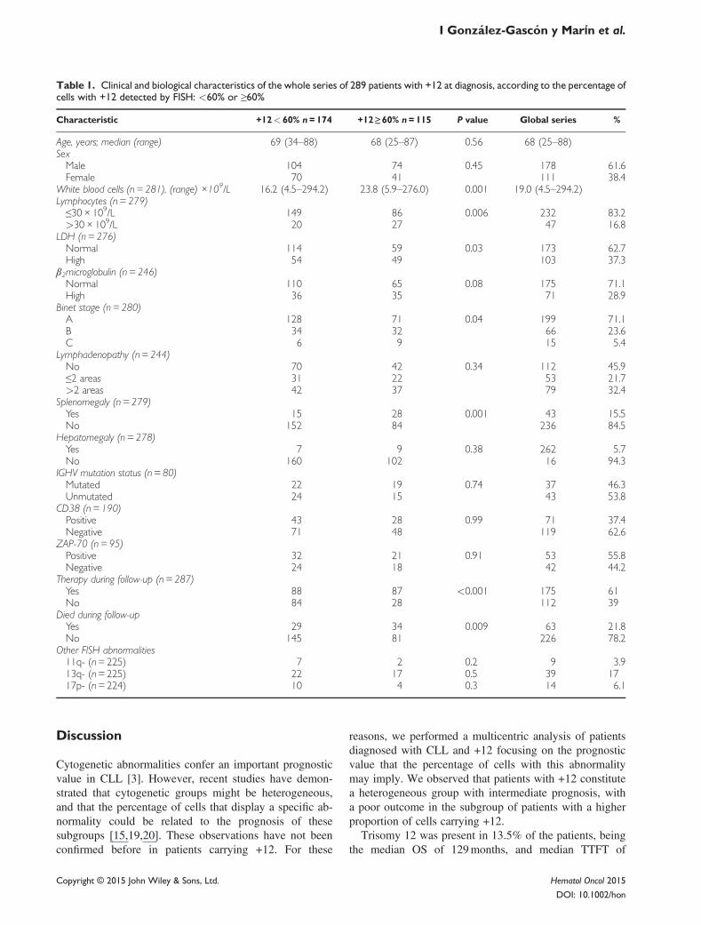
Table 1. Clinical and biological characteristics of the whole series of 289 patients with +12 at diagnosis, according to the percentage ofcells with +12 detected by FISH: <60% or ≥60%
Characteristic +12< 60% n = 174 +12 ≥ 60% n = 115 P value Global series %
Age, years; median (range) 69 (34–88) 68 (25–87) 0.56 68 (25–88)SexMale 104 74 0.45 178 61.6Female 70 41 111 38.4
White blood cells (n = 281), (range) ×109/L 16.2 (4.5–294.2) 23.8 (5.9–276.0) 0.001 19.0 (4.5–294.2)Lymphocytes (n = 279)≤30× 109/L 149 86 0.006 232 83.2>30× 109/L 20 27 47 16.8
LDH (n= 276)Normal 114 59 0.03 173 62.7High 54 49 103 37.3
β2microglobulin (n = 246)Normal 110 65 0.08 175 71.1High 36 35 71 28.9
Binet stage (n = 280)A 128 71 0.04 199 71.1B 34 32 66 23.6C 6 9 15 5.4
Lymphadenopathy (n = 244)No 70 42 0.34 112 45.9≤2 areas 31 22 53 21.7>2 areas 42 37 79 32.4
Splenomegaly (n = 279)Yes 15 28 0.001 43 15.5No 152 84 236 84.5
Hepatomegaly (n = 278)Yes 7 9 0.38 262 5.7No 160 102 16 94.3
IGHV mutation status (n = 80)Mutated 22 19 0.74 37 46.3Unmutated 24 15 43 53.8
CD38 (n = 190)Positive 43 28 0.99 71 37.4Negative 71 48 119 62.6
ZAP-70 (n = 95)Positive 32 21 0.91 53 55.8Negative 24 18 42 44.2
Therapy during follow-up (n = 287)Yes 88 87 <0.001 175 61No 84 28 112 39
Died during follow-upYes 29 34 0.009 63 21.8No 145 81 226 78.2
Other FISH abnormalities11q- (n=225) 7 2 0.2 9 3.913q- (n=225) 22 17 0.5 39 1717p- (n=224) 10 4 0.3 14 6.1
I González-Gascón y Marín et al.
Discussion
Cytogenetic abnormalities confer an important prognosticvalue in CLL [3]. However, recent studies have demon-strated that cytogenetic groups might be heterogeneous,and that the percentage of cells that display a specific ab-normality could be related to the prognosis of thesesubgroups [15,19,20]. These observations have not beenconfirmed before in patients carrying +12. For these
Copyright © 2015 John Wiley & Sons, Ltd.
reasons, we performed a multicentric analysis of patientsdiagnosed with CLL and +12 focusing on the prognosticvalue that the percentage of cells with this abnormalitymay imply. We observed that patients with +12 constitutea heterogeneous group with intermediate prognosis, witha poor outcome in the subgroup of patients with a higherproportion of cells carrying +12.
Trisomy 12 was present in 13.5% of the patients, beingthe median OS of 129months, and median TTFT of
Hematol Oncol 2015
DOI: 10.1002/hon
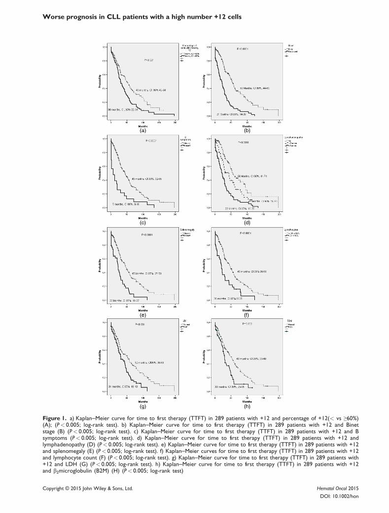
Figure 1. a) Kaplan–Meier curve for time to first therapy (TTFT) in 289 patients with +12 and percentage of +12(< vs ≥60%)(A); (P< 0.005; log-rank test). b) Kaplan–Meier curve for time to first therapy (TTFT) in 289 patients with +12 and Binetstage (B) (P< 0.005; log-rank test). c) Kaplan–Meier curve for time to first therapy (TTFT) in 289 patients with +12 and Bsymptoms (P< 0.005; log-rank test). d) Kaplan–Meier curve for time to first therapy (TTFT) in 289 patients with +12 andlymphadenopathy (D) (P< 0.005; log-rank test). e) Kaplan–Meier curve for time to first therapy (TTFT) in 289 patients with +12and splenomegaly (E) (P< 0.005; log-rank test). f) Kaplan–Meier curves for time to first therapy (TTFT) in 289 patients with +12and lymphocyte count (F) (P< 0.005; log-rank test). g) Kaplan–Meier curve for time to first therapy (TTFT) in 289 patients with+12 and LDH (G) (P< 0.005; log-rank test). h) Kaplan–Meier curve for time to first therapy (TTFT) in 289 patients with +12and β2microglobulin (B2M) (H) (P< 0.005; log-rank test)
Worse prognosis in CLL patients with a high number +12 cells
Copyright © 2015 John Wiley & Sons, Ltd. Hematol Oncol 2015
DOI: 10.1002/hon
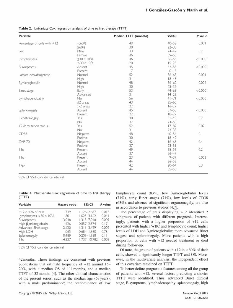
Table 2. Univariate Cox regression analysis of time to first therapy (TTFT)
Variable Median TTFT (months) 95%CI P value
Percentage of cells with +12 <60% 49 40–58 0.001≥60% 30 22–38
Sex Male 33 24–42 0.2Female 46 39–53
Lymphocytes ≤30× 109/L 46 36–56 <0.0001>30× 109/L 20 15–25
B symptoms Absent 45 32–55 <0.0001Present 7 0–18
Lactate dehydrogenase Normal 52 36–68 0.001High 31 18–43
β2microglobulin Normal 48 36–60 0.002High 30 25–35
Binet stage Early 53 44–63 <0.0001Advanced 21 14–28
Lymphadenopathy No 56 41–71 <0.0001≤2 areas 43 25–60>2 areas 22 16–27
Splenomegaly Absent 45 37–53 <0.0001Present 22 18–27
Hepatomegaly Yes 40 31–49 0.7No 37 24–50
IGHV mutation status Yes 52 17–87 0.07No 31 23–38
CD38 Negative 48 40–56 0.1Positive 30 18–42
ZAP-70 Negative 42 16–68 0.4Positive 37 23–51
13q- Present 49 38–59 0.2Absent 37 26–47
11q- Present 23 9–37 0.002Absent 44 36–52
17p- Present 42 20–64 0.3Absent 44 35–53
95% CI, 95% confidence interval.
Table 3. Multivariate Cox regression of time to first therapy(TTFT)
Variable Hazard ratio 95%CI P value
+12 ≥ 60% of cells 1.739 1.126–2.687 0.013Lymphocytes >30× 109/L 1.801 1.025–3.162 0.041B symptoms 3.038 1.315–7.018 0.009High β2microglobulin 1.426 0.857–2.374 0.17Advanced Binet stage 2.120 1.311–3.429 0.002High LDH 1.065 0.684–1.660 0.78Splenomegaly 0.489 0.201–1.188 0.1111q- 4.327 1.737–10.782 0.002
95% CI, 95% confidence interval
I González-Gascón y Marín et al.
42months. These findings are consistent with previouspublications that estimate frequency of +12 around 15–20%, with a median OS of 111months, and a medianTTFT of 32months [4]. The other clinical characteristicsof the present series, such as the median age (68 years),with a male predominance; the predominance of low
Copyright © 2015 John Wiley & Sons, Ltd.
lymphocyte count (83%), low β2microglobulin levels(71%), early Binet stages (71%), low levels of CD38(63%), and absence of significant organomegaly, are alsoin accordance to previous studies [4,7].
The percentage of cells displaying +12 identified 2subgroups of patients with different prognosis. Interest-ingly, patients with a higher proportion of +12 cellspresented with higher WBC and lymphocyte count; higherlevels of LDH and β2microglobulin; more advanced Binetstages; and splenomegaly. More patients with a highproportion of cells with +12 needed treatment or diedduring follow-up.
Of note, the group of patients with +12 in <60% of theircells, showed a significantly longer TTFT and OS. More-over, in the multivariate analysis, the independent effectof this covariate remained on TTFT.
To better define prognostic features among all the groupof patients with +12, several factors predicting a shorterTTFT were identified. Thus, advanced Binet clinicalstage, B symptoms, lymphadenopathy, splenomegaly, high
Hematol Oncol 2015
DOI: 10.1002/hon
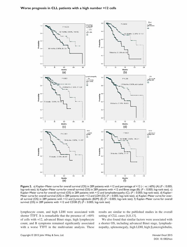
Figure 2. a) Kaplan–Meier curve for overall survival (OS) in 289 patients with +12 and percentage of +12 (< vs ≥ 60%) (A) (P< 0.005;log-rank test). b) Kaplan–Meier curve for overall survival (OS) in 289 patients with +12 and Binet stage (B); (P< 0.005; log-rank test). c)Kaplan–Meier curve for overall survival (OS) in 289 patients with +12 and lymphadenopathy (C); (P< 0.005; log-rank test). d) Kaplan–Meier curve for overall survival (OS) in 289 patients with +12 and LDH (D); (P< 0.005; log-rank test). e) Kaplan–Meier curve for over-all survival (OS) in 289 patients with +12 and β2microglobulin (B2M) (E) (P< 0.005; log-rank test). f) Kaplan–Meier curve for overallsurvival (OS) in 289 patients with +12 and CD38 (F) (P< 0.005; log-rank test)
Worse prognosis in CLL patients with a high number +12 cells
lymphocyte count, and high LDH were associated withshorter TTFT. It is remarkable that the presence of >60%of cells with +12, advanced Binet stage, high lymphocytecount, and B symptoms remained significantly associatedwith a worse TTFT in the multivariate analysis. These
Copyright © 2015 John Wiley & Sons, Ltd.
results are similar to the published studies in the overallsetting of CLL cases [4,8,13].We also found that similar factors were associated with
a shorter OS, including advanced Binet stage, lymphade-nopathy, splenomegaly, high LDH, high β2microglobulin,
Hematol Oncol 2015
DOI: 10.1002/hon

Table 4. Univariate Cox regression analysis of overall survival (OS)
Variable Median OS (months) 95%CI P value
Percentage ofcells with +12
<60% 159 100–148 0.015≥60% 96 58–134
Sex Male 129 82–176 0.9Female 129 Not reached
Lymphocytes ≤30× 109/L 129 102–156 0.05>30× 109/L 96 55–137
B symptoms Absent 129 118–140 0.3Present Not reached Not reached
Lactatedehydrogenase
Normal 197 Not reached 0.009High 81 60–101
β2microglobulin Normal 159 111–207 <0.0001High 76 45–107
Binet stage Early 159 131–187 <0.0001Advanced 79 48–109
Lymphadenopathy No 159 123–195 0.001>2 areas 79 51–106
Splenomegaly Absent 129 102–155 0.001Present 121 Not reached
Hepatomegaly Yes 129 101–157 0.2No Not reached Not reached
IGHV mutationstatus
Yes 159 Not reached 0.6No Not reached Not reached
CD38 Negative 159 104–214 0.04Positive 129 77–181
13q- Present 129 230–227 0.4Absent 159 80–237
11q- Present 44 26–61 0.018Absent 159 92–226
17p- Present 81 1.1–160 0.4Absent 159 91–226
95% CI, 95% confidence interval
Table 5. Multivariate Cox regression of overall survival (OS)
Variable Hazard ratio 95%CI P value
+12 ≥ 60% of cells 1.631 0.834–3.188 0.15High β2microglobulin 2.259 1.070–4.770 0.03Advanced Binet stage 1.717 1.023–2.881 0.04High LDH 1.451 0.685–3.074 0.3311q- 5.097 1.420–18.289 0.01
95% CI, 95% confidence interval
I González-Gascón y Marín et al.
and expression of CD38. However, only advance Binetstage and high β2microglobulin remained independentlysignificant in predicting OS.As speculated with other cytogenetic abnormalities, it
may be possible that the greatest number of losses in13q 11q, or 17p deletions or a high percentage of cellswith +12, translates genetic instability that makes theoutcome of these patients worse. In our series we foundthat 60% of cells with +12 was the better cut-off withclinical significance, after trying different thresholds.However, further studies preferably in a prospectivecontext need to be performed to validate this limit.
Copyright © 2015 John Wiley & Sons, Ltd.
We could not find predictive value regarding otherimportant prognostic factors such as IGHV mutation status,and expression of ZAP-70. Nevertheless, data were col-lected retrospectively, and these factors were not analyzedin the whole series of patients.
Coexistence of other cytogenetic abnormalities in addi-tion to +12 was rare in our cohort, and it is consistent withprevious publications [6,7,10]. Only 19% of patientspresented other cytogenetic abnormalities, being 13q- themost frequent followed by 17p- and 11q-. We analyzedTTFT and OS in the different cytogenetic abnormalities,and we found a significantly shorter TTFT and OS in thegroup of patients with 11q-. This covariate preserved itseffect in the multivariate analysis. We failed to observethese findings in the subgroup of patients with 17p-.Moreover, only a tendency to a longer OS in patients with13q- was observed. It is noteworthy that even though 11q-was only present in 9 patients, nearly all of them presentedit in≥ than 25% of 11q deleted nuclei, which has previ-ously been associated with a worse outcome [8]. However,larger studies with more patients are needed to ascertainthese findings, and address the relationship between +12and other cytogenetic abnormalities.
Hematol Oncol 2015
DOI: 10.1002/hon
Worse prognosis in CLL patients with a high number +12 cells
To summarize, our findings suggest that the percentageof cells carrying +12 influences the outcome of thesepatients. We demonstrated that a high proportion of cellswith +12 detected by FISH is associated with a short OSand TTFT. Our results suggest the need to consider thepercentage of cells with +12 as an important prognosticfactor, in future prognosis scales.
Conflict of interest
The authors declare that there is no conflict of interestsregarding the publication of this article.
Acknowledgements
We thank all the physicians from the Spanish institutions whocontributed clinical data. We are also grateful to Irene Rodríguez,Sara González, Teresa Prieto, M. Ángeles Ramos, AlmudenaMartín, Ana Díaz, Ana Simón, María del Pozo, Vanesa Gutiérrez,and Sandra Pujante from Centro de Investigación del Cáncer,Salamanca, for their technical assistance.
This work was partially supported by grants from the SpanishFondo de Investigaciones Sanitarias FIS 09/01543, PI12/00281,Proyectos de Investigación del SACYL 355/A/09, COST ActionEuGESMA (BM0801), Fundación Manuel Solórzano, ObraSocial Banca Cívica (Caja Burgos), Fundación Española deHematología y Hemoterapia (FEHH), and by a grant (RD12/0036/0069) from Red Temática de Investigación Cooperativa enCáncer (RTICC), Instituto de Salud Carlos III (ISCIII), SpanishMinistry of Economy and Competitiveness, and EuropeanRegional Development Fund (ERDF) “Una manera de hacerEuropa” and IRON-II collaborative network. The research lead-ing to these results has received funding from the EuropeanUnion Seventh Framework Programme [FP7/2007-2013] underGrant Agreement no. 306242-NGS-PTL. María Hernández-Sánchez is fully supported by an Ayuda Predoctoral de la Juntade Castilla y León from the Fondo Social Europeo.
References
1. Dighiero G, Hamblin TJ. Chronic lymphocytic leukaemia. Lan-cet 2008; 371: 1017–1029.
2. Rai KR, Sawitsky A, Cronkite EP, Chanana AD, Levy RN,Pasternack BS. Clinical staging of chronic lymphocytic leuke-mia. Blood 1975; 46: 219–234.
3. Binet JL, Auquier A, Dighiero G, et al. A new prognosticclassification of chronic lymphocytic leukemia derived from amultivariate survival analysis. Cancer 1981; 48: 198–206.
4. Döhner H, Stilgenbauer S, Benner A, et al. Genomic aberrationsand survival in chronic lymphocytic leukemia. N Engl J Med2000; 343: 1910–1916.
5. Rossi D, Rasi S, Spina V, et al. Integrated mutational andcytogenetic analysis identifies new prognostic subgroups inchronic lymphocytic leukemia. Blood 2013; 218: 1403–1412.
6. Puiggros A, Blanco G, Espinet B. Genetic abnormalities inchronic lymphocytic leukemia: where we are and where we go.BioMed Res Int 2014; 2014: 435983.
Copyright © 2015 John Wiley & Sons, Ltd.
7. Rodríguez-Vicente AE, Díaz MG, Hernández-Rivas JM. Chroniclymphocytic leukemia: a clinical and molecular heterogenousdisease. Cancer Genet 2013; 206: 49–62.
8. Matutes E, Oscier D, Garcia-Marco J, et al. +12 defines a groupof CLL with atypical morphology: correlation between cytoge-netic, clinical and laboratory features in 544 patients. Br JHaematol 1996; 92: 382–388.
9. Hallek M, Fischer K, Fingerle-Rowson G, et al. Addition ofrituximab to fludarabine and cyclophosphamide in patients withchronic lymphocytic leukaemia: a randomised, open-label, phase3 trial. Lancet 2010; 376: 1164–1174.
10. Zenz T, Vollmer D, Trbusek M, et al. TP53 mutation profile inchronic lymphocytic leukemia: evidence for a disease specificprofile from a comprehensive analysis of 268 mutations.Leukemia 2010; 24: 2072–2079.
11. Villamor N, Conde L, Martínez-Trillos A, et al. NOTCH1mutations identify a genetic subgroup of chronic lymphocyticleukemia patients with high risk of transformation and pooroutcome. Leukemia 2012; 27: 1100–1106.
12. Balatti V, Bottoni A, Palamarchuk A, et al. NOTCH1 mutationsin CLL associated with +12. Blood 2011; 119: 329–331.
13. Balatti V, Lerner S, Rizzotto L, et al. +12 CLLs progress throughNOTCH1 mutations. Leukemia 2013; 273: 740–743.
14. Weissmann S, Roller A, Jeromin S, et al. Prognostic impact andlandscape of NOTCH1mutations in chronic lymphocytic leukemia(CLL): a study on 852 patients. Leukemia 2013; 12: 2393–2396.
15. Hernández JA, Rodríguez AE, González M, et al. A high numberof losses in 13q14 chromosome band is associated with a worseoutcome and biological differences in patients with B-cellchronic lymphoid leukemia. Haematologica 2009; 94: 364–371.
16. Hernández JA, González M, Hernández JM. Chronic lympho-cytic leukemia. Med Clínica 2010; 135: 172–178.
17. Dal Bo M, Rossi FM, Rossi D, et al. 13q14 deletion size and num-ber of deleted cells both influence prognosis in chronic lymphocyticleukemia. Genes Chromosomes Cancer 2011; 50: 633–643.
18. Van Dyke DL, Shanafelt TD, Call TG, et al. A comprehensiveevaluation of the prognostic significance of 13q deletions inpatients with B-chronic lymphocytic leukaemia. Br J Haematol2010; 148: 544–550.
19. Marasca R, Maffei R, Martinelli S, et al. Clinical heterogeneityof de novo 11q deletion chronic lymphocytic leukaemia:prognostic relevance of extent of 11q deleted nuclei inside leuke-mic clone. Hematol Oncol 2013; 31: 348–355.
20. Tam CS, Shanafelt TD, Wierda WG, et al. De novo deletion17p13.1 chronic lymphocytic leukemia shows significant clinicalheterogeneity: the M. D. Anderson and Mayo Clinic experience.Blood 2009; 114: 957–964.
21. Harris NL, Jaffe ES, Diebold J, et al. World Health Organizationclassification of neoplastic diseases of the hematopoietic andlymphoid tissues: report of the Clinical Advisory Committeemeeting-Airlie House, Virginia, November 1997. J Clin OncolOff J Am Soc Clin Oncol 1999; 17: 3835–3849.
22. Hallek M, Cheson BD, Catovsky D, et al. Guidelines for thediagnosis and treatment of chronic lymphocytic leukemia: a re-port from the International Workshop on Chronic LymphocyticLeukemia updating the National Cancer Institute-WorkingGroup 1996 guidelines. Blood 2008; 111: 5446–5456.
23. González MB, Hernández JM, García JL, et al. The value offluorescence in situ hybridization for the detection of 11q inmultiple myeloma. Haematologica 2004; 89: 1213–1218.
Supporting information
Additional supporting information may be found in theonline version of this article at the publisher’s web site.
Hematol Oncol 2015
DOI: 10.1002/hon