A HEPATOPROTECTIVE TRIHERBAL DECOCTION WITH ...
267
CHAPTER ONE INTRODUCTION 1.1. Background of the Study The rate of use of herbal medicines are increasing rapidly worldwide, although the reasons for this development may vary for different people in different environments. The observed safety of herbal medicines and their affordability is a major consideration. Herbal medicines derived from locally available medicinal plants, have been used to manage many medical problems such as hypertension, malaria, arthritis, fatty liver disease, cancer etc. in different parts of Nigeria. The evaluation of the hepatoprotective activity of a Nigerian tri-herbal formulation (GOV) consisting of 50 % ethanolic extract of Gongronema latifolia Benth, Ocimum gratissimum Linn. and Vernonia amygdalina Del. by investigating its effect on the biochemical, antioxidant, anti-inflammatory, anti- proliferative indices in normal albino rats and HepG2 cell line, is the subject of this study. The liver is the primary site for drug metabolism and hepatotoxicity is one of the most frequently reported human adverse drug reactions. Adverse drug reactions (ADRs) present serious human health problems and cause reactions similar to those of acute viral hepatitis (Mumoli, et al., 2006). Potential hepatotoxicity of some of the first-line anti -tubercular and anti-retroviral drugs remains a problem (Shakya, et al., 2006). It has been observed that liver diseases present a leading cause of medical emergency in Nigeria resulting from poorly treated or untreated infective hepatitis, late presentation at hospitals for treatment, alcoholism and drug misuse (Adesunkanmi et al., 2002). Hepatoprotective drugs are frequently prescribed as part of treatment for tuberculosis in China (Ho, 2006) because the first line anti-tuberculosis drugs isoniazid (INH), rifampicin (RIF) and pyrazinamide (PZA) are also associated with toxic reactions in tissues, particularly in the liver, leading to hepatitis (Liu, et al., 2008). Drug-induced liver injury has become a leading cause of severe liver
-
Upload
khangminh22 -
Category
Documents
-
view
0 -
download
0
Transcript of A HEPATOPROTECTIVE TRIHERBAL DECOCTION WITH ...

CHAPTER ONE
INTRODUCTION
1.1. Background of the Study
The rate of use of herbal medicines are increasing rapidly worldwide, although the reasons for
this development may vary for different people in different environments. The observed safety of
herbal medicines and their affordability is a major consideration. Herbal medicines derived from
locally available medicinal plants, have been used to manage many medical problems such as
hypertension, malaria, arthritis, fatty liver disease, cancer etc. in different parts of Nigeria. The
evaluation of the hepatoprotective activity of a Nigerian tri-herbal formulation (GOV) consisting of 50
% ethanolic extract of Gongronema latifolia Benth, Ocimum gratissimum Linn. and Vernonia
amygdalina Del. by investigating its effect on the biochemical, antioxidant, anti-inflammatory, anti-
proliferative indices in normal albino rats and HepG2 cell line, is the subject of this study.
The liver is the primary site for drug metabolism and hepatotoxicity is one of the most
frequently reported human adverse drug reactions. Adverse drug reactions (ADRs) present serious
human health problems and cause reactions similar to those of acute viral hepatitis (Mumoli, et al.,
2006). Potential hepatotoxicity of some of the first-line anti -tubercular and anti-retroviral drugs
remains a problem (Shakya, et al., 2006). It has been observed that liver diseases present a leading
cause of medical emergency in Nigeria resulting from poorly treated or untreated infective hepatitis,
late presentation at hospitals for treatment, alcoholism and drug misuse (Adesunkanmi et al., 2002).
Hepatoprotective drugs are frequently prescribed as part of treatment for tuberculosis in China (Ho,
2006) because the first line anti-tuberculosis drugs isoniazid (INH), rifampicin (RIF) and
pyrazinamide (PZA) are also associated with toxic reactions in tissues, particularly in the liver, leading
to hepatitis (Liu, et al., 2008). Drug-induced liver injury has become a leading cause of severe liver
2
disease in Western countries and therefore poses a major clinical and regulatory challenge (Russmann,
et al., 2009). In England, there is a marked increase in liver diseases with reports of rising morbidity
and mortality, particularly in younger age groups (Kaner, et al., 2007).
Acetaminophen (APAP) hepatotoxicity is the most common cause of death due to acute liver
failure in the developed world and is increasingly recognized as a significant public health concern
(Lee, 2007). Alcohol liver disease (ALD) is the foremost health risk in developing countries and ranks
third in developed countries (WHO, 2005). Alcohol abuse and alcoholism represents one of the major
health, social and economic issues facing the world and the liver is among the organs most susceptible
to the toxic effects of ethanol (Lieber, et al., 2008).
Hepatitis and other liver diseases constitute a major cause of mortality and morbidity in
Nigeria, and these can be prevented with proper treatments and as well as prophylactic measures
(Adesunkanmi et al., 2002). Halim and Ajayi (2000) reported that the prevalence of hepatitis C
antibody from blood donors in Nigeria varied between 5.8 % – 12.3 %. However, treatment and
prophylaxis options for these conditions are limited (Hwang and Chen, 2006).
There has been a rapid expansion of allopathic health care in Nigeria over the last three
decades, including an increase in the number of allopathic health care providers. At the same time,
because the majority of Nigerians use traditional medicine, the Government of Nigeria has shown
appreciation for the importance of traditional medicine in health care delivery (WHO, 2001a).
According to Sule (2000), though informal intercommunication between the Government and
traditional medicine practitioners can be traced back to the 19th century, formal legislation promoting
traditional medicine dates back to 1966 when the Ministry of Health authorized the University of
Ibadan to conduct research into the medicinal properties of local herbs (WHO, 2001b).
3
Despite the widespread use of complementary and alternative medicine (CAM), there is lack of
scientific evidence on the efficacy and safety of some of these herbal drugs. Research has been carried
out to evaluate the scientific basis for the claimed hepatoprotective activity of herbal agents as a single
agent or in formulation. It is therefore very important to make continuous effort to develop more
effective therapeutic strategies or prophylactic modalities to eradicate or stem the scourge of liver
disease.
1.2. Statement of Problem
The need for organized and continuous improvement of the practice and development of
traditional medicine is of great importance in order to protect the population from quacks, fraud, and
incompetence. Certain herbal supplements can have adverse effects ranging from nausea and vomiting
to fatal conditions like liver or kidney dysfunction. For example, in 2002, the U.S. Food and Drug
Administration (FDA) released a warning about potential liver damage from kava root, and it was
then, one of the 10 most popular herbal supplements sold in the United States of America (FDA, 2002:
Moulds and Malani, 2003) . And in 2004 the FDA banned ephedra, a Chinese weight-loss herb, after it
was linked to more than 100 deaths (Haller, et al., 2005).
There is a false belief that herbal remedies are safer than conventional medicines, when in fact
many of these substances may cause serious illnesses, aggravate pre-existing health problems or result
in death, particularly if taken in excess or injected rather than ingested. Some herbal supplements and
medications, may be adulterated and thus could be contaminated with pathogenic microbes or
dangerous heavy metals, including lead and mercury. Perhaps the greatest potential risk, however, lies
in possible interaction with pharmaceutical drugs the patients are already taking. Saint-John's-wort,

4
which has been shown to help in treating mild to moderate depression, is also known to reduce the
effectiveness of some HIV medications and heart drugs such as digoxin and warfarin, and cause
intermenstrual bleeding in women taking the oral contraceptive pill, while Gingko and garlic also
increase the risk of bleeding with anticoagulants. Access to herbal products is generally unrestricted
and many people do not tell their doctor they are taking herbal medicines, their contribution to death
may not be fully appreciated during a standard autopsy.
Herbal medicine has become a common form of healthcare, even though several differences
exist between herbal and conventional pharmacological treatments, herbal medicine needs to be tested
for efficacy using conventional trial methodology. Clinical proof i.e. a randomized, controlled trial is
the main standard for establishing a drug's usefulness and safety. There is a clear need for better public
and physician understanding of herbal products through health education, early detection and
management of herbal toxicities, scientific scrutiny of their uses, and research on their safety and
effectiveness. It is therefore important to expand the knowledge base of traditional and
complementary/alternative medicine, and to promote its inclusion into health improvement policies
especially where they are proven safe and effective.
1.3. Purpose of Study
The aim of this study is to investigate the hepatoprotective, antiproliferative and antioxidant
effects of a tri-herbal formulation (GOV) on acetaminophen, alcohol, carbon tetrachloride and D-
galactosamine induced toxicity in Wistar albino rats and HepG2 cell line. This investigation will be
carried out using both in vitro and in vivo assay models. The anti-proliferative activity of GOV in

5
different solvent systems toward human hepatocellular liver carcinoma cell line (HepG2 cells) would
be monitored by in vitro model.
1.4. Objectives of the Study
The specific objectives of this study include:
1. To determine the hepatoprotective activity of the triherbal formulation (GOV) against
acetaminophen (APAP), alcohol (ethanol), d- galactosamine (D-GaIN) and carbon tetrachloride (CCl4)
induced hepatotoxicity in rats by,
i. Ascertaining the effect of GOV on haematologic indices and serum liver marker
enzymes.
ii. Determining the antioxidant potential of GOV using serum, liver and kidney
homogenates.
iii. Profiling the anti-apoptotic activities of GOV using leukocytes from whole blood to
determine the fold-increase in caspase-2, 3 and 9 activities.
iv. Profiling the histopathologic indices of the liver and kidney of pre-treated rats using
morphologic observations.
2. To evaluate the antiproliferative activity of GOV using human hepatocellular liver carcinoma
cell line (Hep G2 cells) and nasopharyngeal cancer cells (CNE2 and SUME –α- nasopharyngeal cells).
6
3. To determine the in vitro antioxidant potential of GOV using brain homogenates of
Wistar albino rats and erythrocyte of Swiss mice.
4. To determine the phytochemical constituents of the triherbal formulation (GOV)
consisting of 50 % ethanolic extract of Gongronema latifolia Benth, Ocimum gratissimum Linn. and
Vernonia amygdalina Del. using preliminary phytochemical analysis such as, thin layer
chromatography (TLC), high performance thin layer chromatography (HPTLC), high performance
liquid chromatography (HPLC) and the saturation-transfer difference – nuclear magnetic resonance
spectroscopy (STD-NMR) based screening.
5. To ascertain the safe dose of GOV orally and intraperitoneally using animal models.
6. To investigate the analgesic and anti-inflammatory activity of GOV using appropriate
pharmacologic assays.
1.5. Research Questions and / or Hypotheses.
1. Which solvent best extracts the bioactive constituents of GOV?
2. What phytochemicals can be identified in the triherbal formulation?
3. Is GOV safe?
4. Does GOV demonstrate anti-inflammatory and analgesic activities in Swiss mice and Wistar
albino rats?
5. What effect does GOV exhibit on haematologic parameters in Wistar albino rats intoxicated
with different hepatotoxicants (acetaminophen, alcohol, carbon tetrachloride and D-galactosamine)?
7
6. What effect does GOV exert on serum liver enzymes of Wistar albino rats intoxicated with
these hepatotoxicants?
7. Is there any significant difference (p ≤ 0.05) in the biochemical and antioxidant indices of
serum, liver and kidney homogenates of GOV treated rats compared to the toxin control group?
8. Does GOV have any effect on the concentrations of chemical analytes in Wistar albino rats
intoxicated with these hepatotoxicants?
9. Does GOV exhibit anti-apoptotic effect on rats intoxicated with these hepatotoxicants?
10. Is there any morphologic change in internal organs of rats administered GOV before tissue
damage with the different hepatotoxicants compared to the toxin control rats?
11. Does GOV demonstrate antiproliferative activity?
1.6. Significance of Study
Many of the herbs and spices used by humans to season food, yield useful medicinal
compounds. Similarly to prescription drugs, a number of herbs are thought to cause adverse effects.
Adulteration, inappropriate formulation, or lack of understanding of plant interactions and
formulations have led to adverse reactions that are sometimes life threatening or lethal such as Ricinus
communis (castor oil plant). Therefore studies on the bioactive components in these herbs are
imperative so as to promote or discourage its use. This will aid the botanists, natural-products
chemists, microbiologists and pharmacologists to develop phytochemicals that could be used for
treatment of various diseases.
According to Brower (2008), the Botanic Gardens Conservation International report said that
"five billion people still rely on traditional plant-based medicine as their primary form of health care‖.

8
The World Health Organization (WHO) estimates that 80 % of the world's population presently uses
herbal medicine for some aspect of primary health care (WHO, 2008). This can be attributed to the
fact that pharmaceuticals are prohibitively expensive for most of the world's population (2.8 billion
people live on less than US$ 2 per day) (DaSilva, et al., 2002), in comparison to herbal medicines that
can be cultivated or gathered from nature for little or no cost. Even in this present technological era,
traditional medicine is still the predominant means in the third world for the preservation of health of
the rural majority who constitute over 70 % of the total population (Okoli, et al., 2007). Based on
scientific evidence(s) from this study, government will be encouraged to acknowledge, support and
integrate traditional medicine into national health systems as part of primary health care in
combination with national policy and regulation to ensure safe, effective and quality products and
practices. This will boost access to care for the rural majority and increase general awareness on the
efficacy of some indigenous medicinal plants.
This study may help suggest the chemopreventive potentials and antioxidant profiles of GOV
as a traditional herbal medicine and to highlight the importance of scientific research on miscellaneous
plants with various medicinal properties. It is expected to awaken environmental consciousness on the
need to avoid unconscious exposure to different potential hepatotoxicants. It will also help promote
awareness on the safety, efficacy and quality of medicinal plants by expanding its knowledge base,
promoting therapeutic use of medicinal plants by increasing the quality, quantity and accessibility of
clinical evidence to support claims for their effectiveness. This will encourage studies on medicinal
plants so as to provide more affordable chemopreventive drugs that are highly accessible especially in
poor and marginalized populations thereby reducing excess mortality, morbidity and disability.
These investigations will help to document the uses of indigenous plants for posterity, since
knowledge of indigenous cultures are lost because cultures themselves are in danger of extinction and

9
prior to their possible elimination through urbanization, social development and deforestation. It is
also hoped that it will stimulate interest for further research in medicinal plant(s) and the ailments for
which they are used with the purpose of developing potential drugs for some common diseases. It will
provide scientific evidence to evaluate the safety and effectiveness of traditional medicine products
and practices.
Scientific proof of the efficacy of herbs makes herbal medicines a lucrative form of traditional
medicine, so it could be used as export commodities, which generates considerable income.
1.7. Operational Definition of Terms
Allopathic medicine - Allopathy is a biologically based approach to healing and it is the type of
medicine most familiar to westerners today.
Antiproliferative - used to inhibit cell growth or retard the spread of cells especially malignant cells.
Apoptosis - the programmed death of some of an organism's cells as part of its natural growth and
development.
Chemopreventive - The use of a drug or compound to interfere with a disease process.
Cholestasis - interruption in the excretion or flow of bile.
Cirrhosis - a chronic degenerative disease in which normal liver cells are damaged and are then
replaced by scar tissue.
Complementary and alternative medicine (CAM) - CAM as a group of diverse medical and health care
systems, practices, and products that are not generally considered part of conventional
medicine.
10
Confluence - a coming or flowing together, meeting, or gathering at one point
Conventional medicine - Medicine as practiced by holders of M.D. (doctor of medicine) or D.O.
(doctor of osteopathy) degrees and by their allied health professionals such as physical
therapists, psychologists, and registered nurses.
Cytosolic extract - the liquid medium of the cytoplasm, i.e., cytoplasm minus organelles and
nonmembranous insoluble components.
Dewax - To remove wax from a material or from a surface
Efficacy - The power or capacity to produce a desired effect.
Formulation – combination of different chemical substances to produce a final medicinal product.
Fractionation - To divide or separate into parts; break up Separation of a mixture in successive stages,
each stage removing from the mixture some proportion of one of the substances, as by
differential solubility in water
Haemolysis - The breakdown of red blood cells.
Hepatitis - inflammation of the liver.
Hepatoprotective drugs - the drugs that prevent liver disease.
Hepatotoxic - The ability of a substance to have damaging effects on the liver.
Hyperglobulinaemia - the presence of excess globulins in the blood.
IC50 value - the concentration that brings about 50 percent inhibition or the effective dose required to
inhibit the proliferative response by 50 percent
Intraperitoneal - administered by entry into the peritoneum.
Metabolism - The chemical processes occurring within a living cell or organism that are necessary for
the maintenance of life.
11
Necrosis – the death of body tissue when there is not enough blood flowing to the tissue.
Oxidative stress - an imbalance between the production and manifestation of reactive oxygen species
and a biological system's ability to readily detoxify the reactive intermediates or to repair the
resulting damage
Pharmacovigilance - the detection, evaluation, understanding and prevention of adverse effects,
particularly long and short term side effects of medicines.
Prophylaxis - Prevention of or protective treatment for disease.
Steatosis – the abnormal retention of lipids within a cell. It reflects an impairment of the normal
processes of synthesis and elimination.
Writhing - To twist as in pain or struggle.
12
CHAPTER TWO
LITERATURE REVIEW
2.1. Medicinal Plants
Medicinal plants are rich source of novel drugs that forms the ingredients in traditional systems
of medicine, modern medicines, nutraceuticals, food supplements, folk medicines, pharmaceutical
intermediates, bioactive principles and lead compounds in synthetic drugs (Ncube, 2008). Plants have
long served as a useful and natural source of therapeutic agents. Almost all plants have medicinal
values and their uses differ from place to place. Medicinal plants are worldwide in distribution and
abound in the tropics. Tropical plant species are claimed to contain three to four times the number of
active chemical constituents than their temperate counter-parts (Rodríguez and West, 1995). Herbs and
vegetables have emphatically contributed to the improvement of human health, in terms of prevention,
and or cure of different disorders (Dhiman and Chalwa, 2005; Negi, et al., 2010). Use of medicinal
plants has been there even before the discovery of conventional orthodox medicine. Many herbalists
and naturopathic doctors use the bark, flowers, fruit, leaves, roots, seeds and/ or stems of plants in the
preparation of herbal drugs because of their acclaimed folk medicinal usage. Practice of herbal
medicine is dominated by some traditions e.g. Ayurvedic, Siddha and Unani (India) (Mohamed-
Saleem, et al., 2010), Kampoo (Japanese), Chinese herbology (China), Traditional African medicine
(South Africa –Sangoma and Inyanga; West Africa –Ifa), Shamanic herbalism (South America) and
Native American medicine.
Studies are going on throughout the world for the search of protective molecules that would
provide maximum protection of the liver, kidney as well as other organs and practically very little or
no side effects would be exerted during their function in the body (Montilla et al., 2005; Mansour et
al., 2006). Liver diseases pose an enormous health problem in spite of tremendous strides in modern
13
medicine (WHO, 2006; 2008; 2010). There are hardly any drugs that can effectively control
inflammation, protect the liver from the damaging effects of hydrophobic bile acids which are retained
in cholestatic disorders, promote protein synthesis, manifest antioxidative, anti-lipid peroxidative and
antifibrotic properties, prevent fat from infiltrating the liver, enhance glucuronidation, decreases
intestinal absorption and suppresses hepatic synthesis and storage of cholesterol, stabilize hepatocyte
membranes, help the liver to replace damaged tissue and regenerate itself, promotes effective
metabolism of drugs by maintaining levels of CYP or Cytochrome P450, protect the liver from
damage and also regulate the liver enzymes (Akah and Odo, 2010). Because of the affordability,
availability and accessibility of dependable hepatoprotective drugs in scientific/ conventional
medicine, plants play an important role in the management of various liver disorders and in meeting
the demands of primary health care in many developing countries (WHO, 2009). Herbal drugs have
gained importance and popularity in recent years because of their safety, efficacy and cost
effectiveness (Mohamed-Saleem, et al., 2010).
Herbal drugs contain diverse chemical constituents such as Alkaloids, carotinoids, coumarins,
essential oils, flavonoids, glycosides, lignans, lipids, organic acids, phenolics, terpenoids and
xanthines. Countless number of medicinal plants and polyherbal formulations are proposed to possess
hepatoprotective properties though some of them are supported by scientific evidence such as
Himoliv (Bhattacharya, et al., 2003), Trianthema portulacastrum L (Kumar, et al., 2004), Liv 52
(Dandagi, et al., 2008), Vitex negundo (Tandon, et al., 2008), Leucas lavendulaefolia (Kotoky, et al.,
2008), Clausena dentate (Rajesh, et al., 2009), Rubus aleaefolius Poir (Hong, et al., 2010), Murraya
koenigii (Sathaye, et al., 2011). Hepatoprotective activity of plants can be attributed to their
phytochemical constituent(s) such as alkaloids (Vijyan, et al., 2003), Flavonoids (Okwu, 2003),
Saponinns (Aka and Odo, 2010), terpenoids (Grabmann, 2005) and phenolics (Zeashan, et al 2009).
14
The triherbal formulation in this study was prepared from leaves of Gongronema latifolia
Benth Benth, Ocimum gratissimum Linn. and Vernonia amygdalina. Gongronema latifolia Benth
Benth, Ocimum gratissimum Linn. and Vernonia amygdalina are medicinal plants used for centuries in
the African traditional medicine. These plants are commonly used in foods as vegetables and spices
for flavouring and in medicine they are used for the treatment of various ailments such as diarrhea,
headache, fever, hepatitis, dysentery, malaria, nausea, diabetes among others in the South Eastern part
of Nigeria. They have been used singly and in combination with other plants for treatment of different
disorders with subsequent scientific supports to these claims (Nwanjo and Alumanah, 2005; Erasto, et
al., 2007; Adaramoye, et al., 2008; Iwalokun, 2008; Nweze and Eze, 2009). The hepatoprotective
potentials of a triherbal formulation containing these three plants are evaluated using different
hepatotoxins.
2.1.1. Gongronema latifolia Benth
Gongronema latifolia Benth (Asclepiadaceae) commonly called Utazi in the south eastern parts or
Arokeke in the western parts of Nigeria, aborode by the Asante and Akans of Ghana, dondo-polole by
the Kissis of Sierra Leone or gasub by the Serers of Senegal is a globorous perennial edible climber.
The leaves are stalked, alternate and simple while the stalks are pliable and soft. It is commonly used
as vegetable and spice in food and in medicine, for the treatment of malaria, diabetes, hepatits and
various gastrointestinal ailments in the Eastern part of Nigeria. In the traditional Igbo society, it is used
in soups in combination with other herbs and spiced to improve to stimulate milk production in
lactating mothers, improve appetite and normalize the menstrual flow. According to Iweala and
Obidoa (2009), G. latifolia is not toxic to the liver and because it has the ability to reduce the level of

15
liver enzymes in the blood, its role is rather protective and not destructive to the liver. Flavonoids
content in G. latifolia have been implicated as the major constituent that gives G. latifolia its hepa-
protective properties (Perrissoud 1986). Perrissoud (1986) reported that flavonoids exert a membrane-
stabilizing action that protects the liver cells from injury. The leaves are known to improve the general
conditions of a patient, kill or expel intestinal worms and also stimulate appetite. (Irvine, 1961) and
most of these claims have been supported with scientific claims (Ugochukwu and Badaby, 2003;
Nwanjo and Alumanah, 2005; Nwanjo, 2005; Nwanjo and Alumanah, 2005; Okafor, 2005).
Phytochemical analysis of the plant extract showed that it contains polyphenols, glycosides,
reducing sugars, alkaloids, essential oils, saponins and pregnanes among others (Morebise et al., 2002;
Eleyinmi, 2007; Antai, et al., 2009). The forest vegetable is a good source of iron, vitamins, minerals
and proteins (Okafor, 2005). The plants have high potentials as antioxidants, owing to high amount of
flavonoids and phenolic compounds (Omale and Okafor, 2009). The active ingredients in
Gongronema are Gonioanthelma and Gonolobus (Manach, et al., 2004).
Teas containing G. latifolia, Vernonia amygdalina Del. or Cryptolepsis sanguinolenta are also
used throughout West Africa for the management of diabetes and other metabolic disease associated
with the liver (Seeff, et al 2001). The antimicrobial activities of aqueous extracts of V. amygdalina,
Garcinia kola, and G. latifolia, and their blends were evaluated against several test organisms,
Staphylococcus aureus, Bacillus subtilis, Escherichia coli, and Streptococcus salivarus and it was
observed that the blends have potential for use in the control of beer-spoilage organisms (Oshodi, et al
2004). Ugochukwu and Babady, (2003) and Ugochukwu, et al., (2003) reported that aqueous and
ethanolic G. latifolia extracts had hypoglycemic, hypolipidemic and antioxidative properties.

16
G. latifolia leaves was used locally for the Treatment of Fowl Cough in Nigeria because of its
antitussive and antioxidant properties (Essien, et al., 2007). Ethanolic leaf extract of G. latifolia was
shown to have in- vivo Schizonticidal activity on Plasmodium berghei in Mice (Akuodor, et al., 2010).
The aqueous extract of the dried leaves of G. latifolia significantly inhibited carrageen-induced rat
paw oedema, carrageen-induced leucocyte migration in rats and dye leakage induced by
intraperitoneal injection of acetic acid in mice indicating its antiinflammatory activity (Morebise, et al
2002). Onwuka, (2005) subjected raw beef to chemical and microbial analysis, then treated it with
either extracts or powders of four local vegetable leaves namely Gongronema latifolia Benth, Piper
guineesis, Vernonia amygdalina Del. and Ocimum gratissimum Linn. at three different concentrations.
Samples treated with Vernonia amygdalina Del. extract and powdered leaves showed the lowest
microbial counts followed by Ocimum gratissimum Linn., Gongronema latifolia Benth and Piper
guineesis in that order. Ugochukwu and Cobourne, (2003) and Nwanjo, et al., (2006), reported the
protective effect of aqueous and ethanolic extracts from G. latifolia leaves on biomarkers of oxidative
stress in streptozotocin-induced diabetic and non-diabetic rats.
17
Figure 1: Gongronema latifolia Benth.
18
2.1.2. Ocimum gratissimum Linn.
Ocimum gratissimum Linn. (African Basil) belongs to the family Lamiaceae commonly called
Dai'doya' by the Hausas, 'efinrin' by the Yoruba, 'nchanwu' by the Igbos, is a shrub with many
branches having simple oblong leaves. The plant is found throughout the tropics and subtropics and its
greatest variability occurs in tropical Africa and India (Aruna and Sivaramakrishina, 1990) and has an
aromatic smell when crushed. It is planted as a food flavour and for medicinal use. It is used as spices
and condiments in salads, soups, pastas, vinegars and in traditional medicine for the treatment of
several ailments such as upper respiratory tract infections, diarrhea, headache, fever, ophthalmic and
skin diseases, pneumonia, sore throats and tonsillitis, urinary tract, wound, skin gastrointestinal
infections and as an insect repellant. Most of these properties of O. gratissimum have been supported
with scientific claims. (Elujoba, 2000; Pessoa, et al., 2002; Silva, et al., 2005; Travisan, et al., 2006;
Nweze and Eze, 2009)
It is used as a febrifuge and as an ingredient in many malaria medicines, for rheumatic pains
and lumbago and catarrh remedies (Ainslie, 1937). The oils are antiseptic and are used in dressing of
wounds, as a mouth gargle, mosquito repellant (Sofowora, 2000), or mixed with alcohol and used
topically for skin infections or taken orally for bronchitis. It is used as an expectorant, germicide for
toothpastes and mouth washes (Holets, et al., 2003; Pessoa, et al., 200) and antihelminth (Chitwood,
2003). The essential oils are a potential candidate as a phytotherapeutic agent in some fungal diseases
and for the control of fungi in the environment (Nakamura, et al., 2004). They play an important role
in preservation of pharmaceutical products (Travisan, et al., 2006) and also inhibit Staphylococcus
aureus at a concentration of 0.75 mg\ml (Nakamura, et al., 2004). Nweze and Eze, (2009) reported
the antimicrobial effect of O. gratissimum suggesting that it may not really threaten the efficacy of
some conventional antibiotics that may have been taken concomitantly with it.

19
Phytochemical screening revealed the presence of tannins, Cardiac glucoside, phenolic
compounds, saponin, alkaloids, steroids, flavonoids and terpenoids (Gill, 1992; Okwu, 2003).
Eugenol, thymol, citral, geraniol and linalool have been extracted from the oil (Vierra and Simon,
2000). The essential oils of O. gratissimum are active against several species of bacteria (Escherichia
coli, Shigella, Salmonella and Proteus), fungi (Trichophyton rubrum and T. mentagrophytes) and
antihelmintic activity (Lemos, et al., 2005; Silva, et al., 2005; Pessoa, et al., 2002; Nweze and Eze,
2009). It can be applied as an insecticidal fumigant to control Callosobruchus maculates (Keita, et al.,
2001). Rabelo, et al., 2003 has reported the antinociceptive property of its essential oil.
A dose-dependent sedative effect of Ocimum oil was observed during the acute toxicity study
in mice and rats and in the sub-chronic test in rats (Orafidiya, et al., 2004). Fractions isolated from O.
gratissimum leaves have been shown to contain components which contract guinea pig ileum, rat
colon and raise rat mean arterial blood pressure (Onajobi, 1986). A study by Madeira, et al., (2005)
showed that the essential oil extracted from the leaves of O. gratissimum collected at different time
periods, exerts significant relaxant effects on isolated guinea-pig ileum which may underlie the
therapeutic action of the plant. The wound healing effect of leaf extracts of O. gratissimum was
investigated in adult male Wistar rats. The results suggested that the methanolic extracts of O.
gratissimum could be a potential wound healing agent due to its ability to enhance wound contraction
(Osuagwu, et al., 2004). Studies on O. gratissimum proved the plant extract can be a source of
medication for people living with Human Immunodeficiency Virus, (HIV) and Acquired Immune
Deficiency Virus, AIDS (Elujoba, 2000). The hypoglycemic effect of the methanolic extract of
Ocimum gratissimum Linn. leaves was evaluated in normal and alloxan-induced diabetic rats.
Intraperitoneal injection of the extract (400 mg/kg) significantly reduced plasma levels both in normal
and diabetic rats by 56 and 68 %, respectively (Aguiyi, et al., 2000).
20
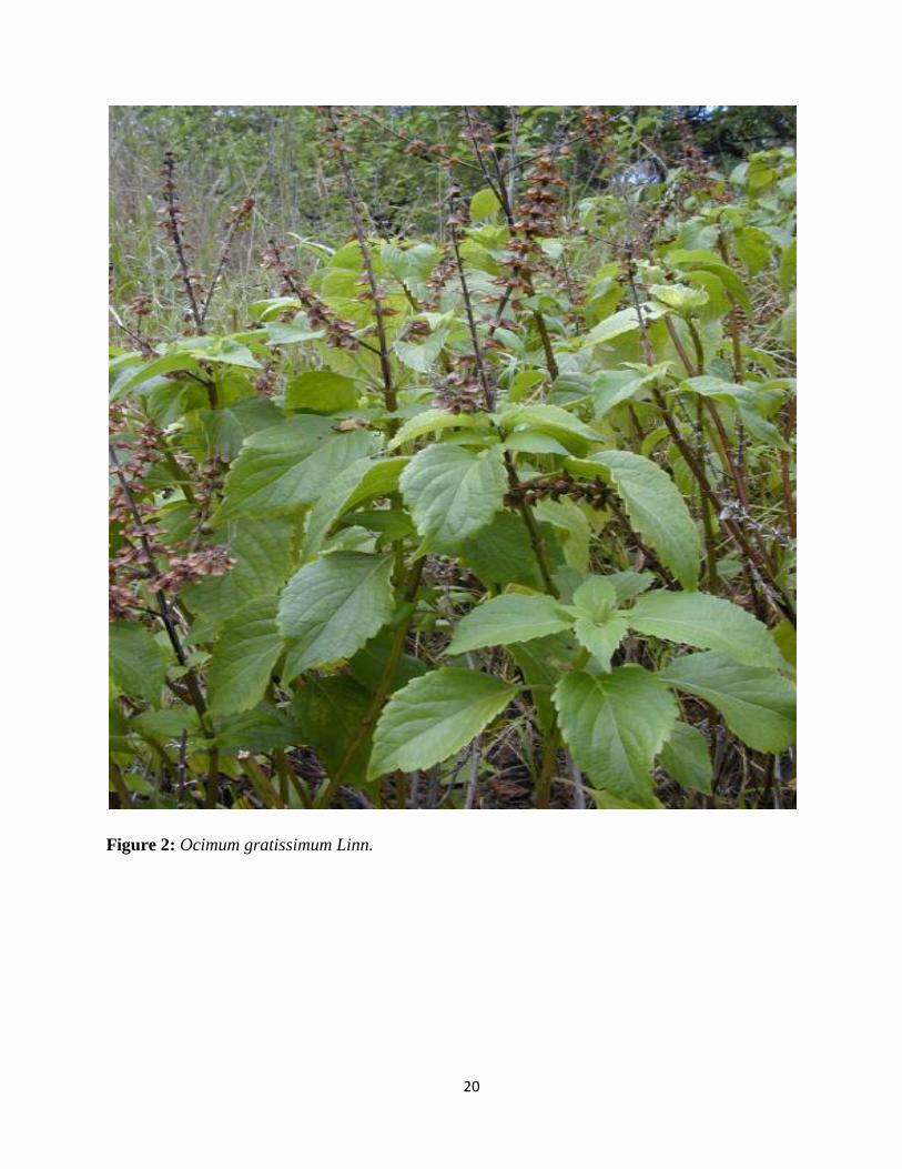
Figure 2: Ocimum gratissimum Linn.
21
2.1.3. Vernonia amygdalina Del.
Vernonia amygdalina Del. (Asteraceae) have obovate, simple and entire leaves. The flowers
occur in copious corymbose panicles white fragrant and are usually bee infested (Irvine, 1961). It is
commonly called Bitter leaf because of its bitter taste, Onugbu (Igbo), Ewuro (Ibadan,) Etidot (Cross
River) and grows predominantly in tropical Africa. The bitter taste is attributed to anti-nutritional
factors such as alkaloids, saponins, tannins and glycosides (Butler and Bailey, 1973). It is commonly
used as local iodine for wounds, in treatment of diabetes, cough, feverish condition, constipation,
hypertension, emesis, nausea, diabetes, sexually transmitted diseases, loss of appetite-induced abrosia,
dysentery and other gastrointestinal tract problems and some have scientific supports to these claims
(Gyang, et al., 2004; Nwanjo, 2005; Amira and Okubadejo, 2007; Erasto, et al., 2007; Adaramoye, et
al., 2008; Iwalokun, 2008). Its leaf extract has been demonstrated to function satisfactorily as an
alternative to hops in beer brewing (Babalola and Okoh, 1996).
Phytochemical analysis of the leaves yielded 2 known sesquesterpene lactones – Vernolide and
Vernodalol, vernodalin, hydroxyvernolide, Vernonioiside B, Myricetin saponins, steroid glycosides,
tannins, alkaloids, tannins, flavonoids, steroid glucosides (vernonioside A1-A4; for bitter tasting
constituents and vernonioside B1-B3; for nonbitter related constituents) (Babalola, et al., 2001; Erasto,
et al., 2006). The sesquesterpene lactones have an in vitro cytotoxic action against KB tumour cells
and Wilme‘s myeloma (Toubiana, 1975; Izevbigie, 2003; Izevbigie and Ernest, 2005). It is used to
stimulate the digestive system as well as reduce fever. They are used as local medicine against leeches.
The root and stem are used as chewing stick and the bitter taste has antimicrobial activity against the
oral microflora. In Nigeria, it is used in place of hops in beer making because of the bitter taste which
is due to antinutritional factors such as alkaloids, tannins, glycosides and saponins (Buttler and Bailey,
1973). In V. amygdalina, ascorbic acid decreases with storage time (Faboya, 1990). Elemental analysis
22
of the leaves revealed that they have adequate concentration of some elements e.g. Iron (1500 +
111ppm) and Calcium (10100 + 895 ppm) that are believed to be essential for normal growth (Ibrahim
et al., 2001). Oshodi, (1992) found that the dried leaves of V. amygdalina were rich in minerals,
especially in phosphorus, and that the content of ascorbic acid was temperature dependent. The
antioxidant activities of compounds (luteolin, luteolin 7-O- β -glucuronoside and luteolin 7-O- β -
glucoside flavonoid) isolated from the leaves of V. amygdalina has been reported using coupled
oxidation of β-carotene linoleic acid (Igile et al., 1994). The sesquiterpene lactone extract from the
leaves has antihepatotoxic activity in hepatotoxic damage in rats (Babalola, et al, 2001; Arhoghro, et
al., 2009).
V. amygdalina extracts may help suppress, delay, or kill cancerous cell in many ways, such as:
Induction of apoptosis as determined in cell culture and animal studies. (Sweeney, et al., 2005; Song,
et al., 2005), inhibition of the growth or growth signals of cancerous cells (Kupchan, et al., 1969;
Jisaka, et al., 1993; Izevbigie, et al., 2004; Opata and Izevbigie, 2006), suppression of metastasis of
cancerous cells in the body by the inhibition of NFҡB is an anti-apoptotic transcription factors as
demonstrated in animal studies (Song, et al., 2005), antioxidants - V. amygdalina may provide anti-
oxidant benefits (Erasto, et al., 2007), enhanced chemotherapy sensitivity - V. amygdalina extracts
may render cancerous cells to be more sensitive to chemotherapy (Sweeney, et al., 2005 ),
enhancement of the immune system - Many studies have shown that V. amygdalina extracts may
strengthen the immune system through many cytokines (including NFҡB, pro inflammatory molecule)
regulation (Sweeney, et al., 2005). http//:en.wikipedia.org/wiki/vernonia_amygdalina_–_cite note-
sweeny-1
Squeeze-washing and rinsing seem to be the best treatment to preserve both vitamins A and C.
Drying at 45 °C is best recommended while freezing for less than ten days is most appropriate for the
23
conservation of vitamin A and C (Ejoh, 2005). Several stigmastane-type saponins such as
vernoniosides A1, B1, A2, A3, B2, D3, A4 and C have been identified in the leaves (Ohigashi, et al.,
1991; Jisaka, et al., 1992). The antioxidant activities of luteolin, luteolin 7-0, β-glucuronoside and
luteolin 7-O-β-glucoside flavonoid compounds isolated from the leaves of V. amygdalina have been
reported using coupled oxidation of β-carotene linoleic acid (Igile, et al., 1994). Subsequently Jisaka,
et al. (1993) and Khalafalla, et al., (2009) demonstrated that vernodaline and vernolide elicited
antitumoral activities in leukemia cells P-388 and C-1210 with IC50 values of 0.11 and 0.17 µg/ml for
vernodaline and 0.13 and 0.11 µg/ml for vernolide respectively. V. amygdalina extracts have also been
suggested to have cell growth inhibitory effects in prostate cancer cell line (PC-3) and no effect on
normal human peripheral blood mononuclear cells (PBMC) (Izevbigie, 2003). It significantly
attenuated the hepatic triglyceride and LDL cholesterol levels of streptozotocin diabetic rats and has
hypoglycemic effect (Nwanjo, 2005; Adaramoye, et al., 2008)
The antiplasmodial (Abosi and Raseroka, 2003; Tona, et al., 2004) antihelmintic (Abosi and
Raseoka, 2005) properties of some sesquiterpene and steroidal constituents of V. amygdalina in vitro
were studied. It dose dependently restored the efficacy of chloroquine against chloroquine resistance
P. berghei malaria in mice (Iwalokun, 2008). V. amygdalina administration decreased blood glucose
by 50 % compared to untreated diabetic animals in studies conducted using streptozotocin-induced
diabetic laboratory animals (Uhegbu and Ogbechi, 2004; Nwanjo, 2005). The Fungitoxic and
phytotoxic effect of V. amygdalina on Cowpea and Cowpea seedling pathogens was reported by Alabi,
et al., (2005).
24
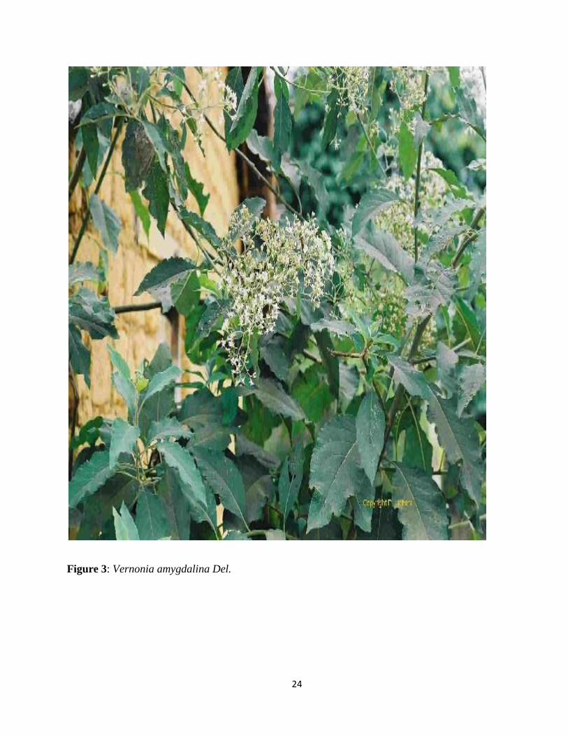
Figure 3: Vernonia amygdalina Del.

25
2.2. The Mammalian Liver
The liver is the largest glandular organ of the body and weighs approximately 1200-1500 g or,
on average, one fiftieth of the total adult body weight. It measures about 8 inches (20 cm) horizontally
(across) and 6.5 inches (17 cm) vertically (down) and is 4.5 inches (12 cm) thick (Cotran, et al., 2005).
It is a soft, pinkish-brown or reddish brown, triangular organ located on the right side of the abdomen
beneath the diaphragm and protected by the lower rib cage. The liver is covered entirely by visceral
peritoneum, a thin, double-layered membrane that reduces friction against other organs. This strategic
localization between nutrient-laden capillary beds and the general circulation is associated with
hepatic regulation of metabolite levels in the blood through storage and mobilization mechanisms
controlled by liver enzymes. It is divided into four lobes of unequal size and shape and contains as
many as 100,000 lobules that are served by two distinct blood supplies. Blood is carried to the liver via
two large vessels: the hepatic artery carries oxygen-rich blood from the aorta, and the portal vein
carries blood containing digested food from the small intestine and the descending colon (Shneider
and Sherman 2008). These blood vessels subdivide in the liver repeatedly, terminating in minute
capillaries. Each capillary leads to a lobule which is made up of hepatic cells, the basic metabolic cells
of the liver. On the surface of the lobules there are ducts, veins and arteries that carry fluids to and
from them.
The liver has important and complex functions and plays a major role in metabolism (Das,
2011). Some of which are manufacture and secretion of bile (an alkaline compound which aids in
digestion), which is stored in the gall bladder and released in the small intestine, conversion of glucose
to its stored form, glycogen, which is reconverted into glucose as the body requires it for energy (Gao,
et al., 2008), it maintains the proper level of glucose in the blood (glucose buffer function), converts
excess carbohydrates and protein into fat, it further breaks down digested proteins in the form of
26
amino acids (Deamination), synthesis, storage, and processing of fats, including fatty acids (used for
energy) and cholesterol, decomposition of red blood cells (Fanne, et al., 2010), formation and
secretion of bile that contains bile acids to aid in the intestinal absorption (taking in) of fats and the fat-
soluble vitamins A, D, E, and K, synthesis of plasma protein, it metabolizes and stores carbohydrates
(Gluconeogenesis - synthesis of glucose from certain amino acids, lactate or glycerol (Das, 2011;
Fanne, et al., 2010), Glycogenolysis - breakdown of glycogen into glucose and Glycogenesis -
formation of glycogen from glucose), which are used as the source for the sugar (glucose) in blood
that red blood cells and the brain use, synthesizes certain amino acids (the so-called nonessential
amino acids) from other amino acids (transamination) (Zakim and Boyer, 2003; Das, 2011), it filters
harmful substances from the blood (Phagocytic cells in the liver, called Kupffer cells, remove large
amounts of debris and bacteria) (Seki, et al., 2000) and detoxifies by metabolizing and/or secreting,
drugs, alcohol, and environmental toxins (Zakim and Boyer, 2003). The liver eliminates, by
metabolizing and/or secreting, the potentially harmful biochemical products produced by the body,
such as bilirubin from the breakdown of old red blood cells and ammonia from the breakdown of
proteins (Seki, et al., 2000) , manufactures about 95% of the plasma proteins (antithrombin, protein C
and S) and blood clotting agents (prothrombin, fibrinogen and other coagulation factors e.g. V, VII,
IX, X and XI) (Palmer, 2004), converts ammonia to urea, it also plays an important role in
thermoregulation; changes in metabolism of its cells varies the heat produced by the body and
synthesizes angiotensinogen, a hormone that is responsible for raising the blood pressure when
activated by renin, an enzyme that is released when the kidney senses low blood pressure and also
disposes of ammonia, an extremely toxic byproduct of protein metabolism using the amino acids
arginine and orthinine to control the ammonia levels (Zakim and Boyer, 2003; Das, 2011).
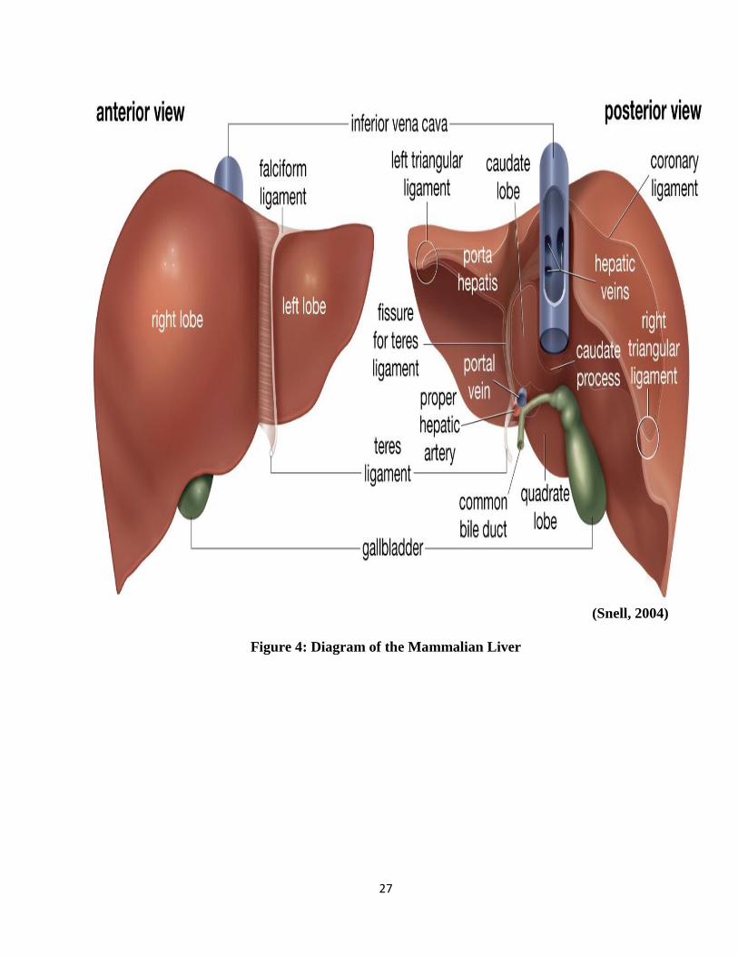
27
(Snell, 2004)
Figure 4: Diagram of the Mammalian Liver
28
In addition, the liver‘s ability to regenerate lost tissues helps maintain these functions, even in
the face of moderate damage (Murray et al., 2003; Suzuki, et al. 2008). The liver is among the few
internal human organs capable of natural regeneration of lost tissue (Out, et al., 2007). As little as 25
% of remaining liver can regenerate into a whole liver again. This is attributed to the hepatocytes
acting as unipotential stem cells (Sherlock and Dooley, 2002; Suzuki, et al., 2008). The liver is
necessary for survival and there is currently no way to compensate for the absence of liver function as
no artificial organ or device capable of emulating all the functions of the liver. Medical terms related
to the liver often start in hepato- or hepatic from the Greek word for liver.
2.2.1. Diseases of the Liver
Because of its strategic location and multidimensional functions, the liver is also prone to many
diseases (Sherlock and Dooley, 2002; Runyon, 2010). Liver disease (also called hepatic disease) is a
broad term describing any single number of diseases affecting the liver. Some of the known liver
diseases are hepatitis A, B, C, E, cancer of the liver, fatty liver, Gilbert‘s syndrome i.e. a genetic
disorder of bilirubin metabolism, Budd-Chiari syndrome i.e. an obstruction of the hepatic vein,
tumours, vascular obstruction, Wilson's disease i.e. a hereditary disease which causes the body to
retain copper an autoimmune inflammatory disease of the bile duct, formation of fibrous tissue in the
liver instead of replacing dead liver cells leading to cirrhosis, Alagille syndrome, biliary atresia,
Hemochromatosis, alcohol damage, drug damage and Glycogen storage disease type II among others
Sherlock and Dooley, 2002; Schiff et al., 2003; Schiff et al., 2007).
Jaundice is a common symptom of most liver diseases and this is caused by increase in the
bilirubin level in the system since the liver cannot remove it from the blood so it can be excreted
29
through bile (Zakim and Boyer, 2003). This bilirubin results from the breakup of the hemoglobin of
dead red blood cells. Other symptoms might include dark urine (when urine mixes with bilirubin),
fatigue, abdominal pain, loss of appetite, abdominal swelling i.e. due to ascites and / or failure of the
liver to produce albumin, itching of the skin (starts when bilirubin is deposited on the skin), nausea
and vomiting, vomiting blood, bloody/ black stools, light-colored stools (this occurs when stercobilin,
a brown pigment, is absent from the stool), and loss of libido. Some diets are believed to protect the
liver such as vitamin E, thiamine because it lowers the iron load on the liver which is present in foods
like pork, oatmeal, corn, nuts and cauliflower, higher coffee intake because it reduces the risk of
cirrhosis and supplements of omega-3 fatty acids (Yurdakok, and Kanra, 1986; Hikino and Kiso, 1988;
Wallace and Weeks, 2001).
2.2.2. Hepatitis
Hepatitis is an inflammation of the liver that can be caused by inherited diseases, viruses,
drugs, chemicals, alcohol, environmental toxins or the patient‘s own immune system causing injury or
destruction. In severe cases inflammation may interfere with the normal functions of the liver and
allow potentially toxic substances to accumulate. Hepatitis varies in severity from a self-limited
condition with total recovery to a life-threatening or life-long disease (Worman, 2001).
About two million patients infected with Hepatitis B die yearly and this makes it the ninth
leading cause of death worldwide. The risk of cirrhosis and liver cancer increases with coinfection
with hepatitis C or D. Diet influences the risk of developing liver cancer. Chronic carriers should be
strongly encouraged to avoid consuming alcohol as it increases their risk for cirrhosis and
hepatocellular carcinoma.
30
2.2.3. Causes of Hepatitis
Hepatitis is commonly caused by a virus such as hepatitis A or infectious jaundice caused by
hepatitis A virus, a picornavirus transmitted by the fecal-oral route, hepatitis B caused by hepatitis B
virus, a hepadnavirus transmissible through contact with blood or bodily fluids, tattoos, sexually or via
mother to child by breast feeding , Hepatitis C caused by hepatitis C virus an RNA virus that is a
member of the Flaviviridae family which is transmitted through the placenta and contact with blood.
Others are hepatitis D and E virus. Cytomegalovirus, Epstein-Barr virus and yellow fever may also
cause hepatitis.
Hepatitis can also be caused by an overactive immune system leading to chronic liver disease
of unknown etiology characterized by circulating autoantibodies, hyperglobulinaemia and interface
hepatitis and accounts for about 20 % of all chronic hepatitis cases (Parker and Picut, 2005), The
Epstein – Barr virus which causes mononucleosis, measles virus, drug or toxin reaction or a hepatitis
virus, can trigger off this reaction (Pieters, et al., 2002).
One of the most common causes of chronic hepatitis is fatty liver and this is mainly due to
excessive alcohol consumption. It is usually reversible and resolved by abstaining from alcohol. The
more severe form of hepatitis which is another cause of fatty liver is nonalcoholic steatohepatitis
(NASH), the most common chronic hepatitis not caused by viruses (Perrez-Carreras, et al., 2003).
While symptoms are usually fairly mild, it may cause cirrhosis. It is seen most commonly in people
with hypertension, type 2 diabetes, high triglyceride levels, metabolic syndrome and overweight
individuals (Videla, et al., 2004).
31
Other causes of hepatitis are drugs, alcoholism, chemicals, and environmental toxins. Several
inherited diseases can exhibit symptoms of acute or chronic hepatitis. Among these are deficiency of
alpha-1-antitrypsin, hemochromatosis and Wilson‘s disease.
2.2.3.1. Viral Hepatitis
The most common cause of hepatitis is an infection with one of several viruses called Hepatitis
Virus. The viruses primarily associated with hepatitis are named in the order of their discovery by the
letters A through G. Viral hepatitis is a mostly enterically transmitted liver disease caused by viral
infection. The major transmission path of the disease is through ingestion; viral hepatitis is also
transmitted through blood transfusion of virus-contaminated blood or blood products such as blood
plasma.
Hepatitis A or Infectious Hepatitis is excreted through feces and is contacted from infected
food and water. Eating shellfish taken from sewage-contaminated water is a common means of
contracting hepatitis A. Hepatitis A is contagious. It is the least serious of the common hepatitis
viruses. Symptoms are mild such as Diarrhea, nausea, fatigue and jaundice although most patients
recover fully within six months. Radioimmunoassay is generally used to identify IgM antibodies, first
produced to fight hepatitis A.
Hepatitis C formerly called non-A non-B hepatitis is spread mainly by exposure to
contaminated blood, through sexual activity that results in tissue tears or from mother to baby during
childbirth. It is the most common cause of chronic hepatitis and is contagious. Most patients with
hepatitis C do not experience symptoms but if they later appear, it develops about a month or two after
infection. Patients with chronic hepatitis C may be susceptible to non-liver disorders e.g. : Hodgkin's
32
lymphomas, Cryoglobulinemia (a disorder in which protein clumps form in the blood), Certain
autoimmune disorders, particularly hypothyroidism and rheumatoid arthritis, Porphyria cutanea tarda
(a disorder which causes skin color and texture changes and sensitivity to light.) etc. specific tests
include enzyme-linked immunosorbent assay (ELISA), They use a polymerase chain reaction (PCR) to
detect the Ribonucleic acid (RNA) of the virus and blood tests showing elevated liver enzymes,
particularly alanine aminotransferase (ALT).
Hepatitis D only causes an infection when hepatitis B is present and is spread by contact with
infected blood while hepatitis E is spread through contaminated food and water. It is not serious except
in pregnant women, when it can be life threatening. Hepatitis G is always chronic with probably the
same modes of transmission as hepatitis C. To date much research indicates it does not cause disease
or even increase the severity of any accompanying virus, including HIV or other hepatitis viruses.
Hepatitis B is a serious disease that causes scarring of the liver, liver failure, liver cancer and
even death. The hepatitis B virus is the second most prevalent cause of cancer in humans after
Tobacco smoke. Symptoms include jaundice, loss of libido, fatigue, itching, nausea, vomiting, dark
urine, light coloured stool, abdominal pain and loss of appetite (Ryan and Ray 2004). Hepatitis B is
spread through infected blood, other body fluids such as semen and vaginal secretions, use of
contaminated sharp objects such as needles, razor and from mother to child during child birth.
Hepatitis B is recognized as endemic in China and various other parts of Asia (Alberts, et al 2002).
Viral hepatitis is widespread around the world. China is estimated to have approximately thirty
million viral hepatitis patients including an estimated number of nine million new patients each year
and about one hundred million hepatitis B virus (HBV) carriers. Ten percent of the pregnant women in
33
China are estimated to be HBV carriers. About one hundred thousand people in China die each year of
liver cancer originated from liver diseases.
2.2.3.2. Autoimmune Chronic Hepatitis
Autoimmune hepatitis is a chronic liver disease of unknown etiology characterized by
circulating autoantibodies, hyperglobulinaemia and interface hepatitis and accounts for about 20 % of
all chronic hepatitis cases (Parker and Picut, 2005). Their development is as a result of genetic
defective immune system which attacks the liver after being triggered by an environmental agent,
probably a virus. Autoimmune chronic hepatitis typically occurs in women between the ages of 20 and
40 who have other autoimmune diseases, including glomerulonephritis, rheumatoid arthritis,
inflammatory bowel disease systemic lupus erythematosus, and hemolytic anemia (Pieters, et al.,
2002). Some research shows that during the postmenopausal period, there may be a rise in incidence
of autoimmune hepatitis among women (O´Connor, et al., 2009; van der Sluijs, et al., 2009). In
general, no major risk factors have been discovered for this condition. The Epstein - Barr virus which
causes mononucleosis, measles virus, drug or toxin reaction or a hepatitis virus, can trigger off this
reaction. It is treated with prednisone and azathioprine.
2.2.3.3. Nonalcoholic Fatty Liver Disease (NAFLD)
One of the most common causes of chronic hepatitis is fatty liver which causes liver
enlargement, tenderness, and abnormal liver function, and this is mainly due to excessive alcohol
consumption. It is usually a reversible, resolved by abstaining from alcohol. The more severe form of
hepatitis which is another cause of fatty liver is nonalcoholic steatohepatitis (NASH), the most
34
common chronic hepatitis not caused by viruses (Perrez-Carreras, et al., 2003). While symptoms are
usually fairly mild, it may cause cirrhosis. It is seen most commonly in people with hypertension, type
2 diabetes, high triglyceride levels, metabolic syndrome and overweight individuals.
NAFLD has features similar to alcohol-induced hepatitis, but it occurs in individuals who do
not drink significant quantity of alcohol. NAFLD is usually very slowly progressive and benign. In
certain patients, however, it can lead to cirrhosis, liver failure, or liver cancer. It can be controlled by
reducing Weight and managing any accompanying medical condition. Drugs, used to lower
triglycerides or those that increase insulin levels may help protect against liver damage. Vitamin E
may help reduce liver injury (Videla, et al 2004).
2.2.3.4. Inherited forms of Hepatitis
Several inherited diseases can exhibit symptoms of acute or chronic hepatitis. Among these are
Deficiency of alpha-1-antitrypsin which is both acute and chronic in children and can lead to cancer
and cirrhosis. Hemochromatosis is associated with accumulation of too much iron in the body and it
affects mainly the liver and it can be chronic. An accumulation of excess copper in the liver, brain, and
in other tissues commonly known as Wilson‘s disease and can be worse even fatal if not treated.
2.3. Hepatotoxicity
Approved drugs are sometimes withdrawn from the market after a post-marketing discovery of
toxicity that was not detected in the extensive preclinical and clinical testing (WHO, 2010). During the
last decade liver toxicity has been one of the most frequent reasons for pharmacovigilance safety
reports and the withdrawal from the market of an approved medicinal product (EMEA, 2008) e.g. of
35
such potentially hepatotoxic drugs are Didanosine (WHO, 2010), Olanzapine (FDA, 2009),
Bufexamac (EMA, 2010), Rosiglitazone (SFDAA, 2010), Ketoconazole tablets (MHRA, 2008a),
Moxifloxacin (MHRA, 2008b; EMEA, 2008). The occurrence of drug-induced liver toxicity is the
single most common reason for the regulatory actions concerning drugs (Boelsterli and Lim, 2007;
Kodell and Chen, 2010). The liver is the primary site for drug metabolism and hepatotoxicity is one of
the most frequently reported human adverse drug reactions and cause reactions that are similar to those
of acute viral hepatitis (Mumoli, et al., 2006). Symptoms can be observed two weeks to six months
after commencing drug treatment although most disappear when the drug is withdrawn; but, in rare
circumstances, they may progress to serious liver disease. Specifically, the existence of relatively
small, unidentified, hypersensitive subpopulations could explain the occurrence of perculiar liver
injury only after approved drugs have been prescribed for large numbers of patients (Shenton, et al.,
2004; Kaplowitz, 2005).
In some countries, hepatoprotective drugs are frequently prescribed as part of treatment for
tuberculosis e.g. China. (Nolan, et al., 1999; Ho, 2006). To treat tuberculosis (TB), traditional Chinese
medicine practitioners typically advocate a combination of biomedical treatment to eliminate bacteria,
and traditional medicine to strengthen qi (Ho, 2006) and herbs to protect the liver (Zhang, et al.,
1995).
Toxic liver injury produced by drugs and chemicals may virtually mimic any form of naturally-
occuring liver disease (Luyendyk, et al., 2003; Kaplowitz, 2005). Drug-induced hepatotoxicity is a
major issue for drug development, and toxicogenomics has the potential to predict toxicity during
early
toxicity screening (Maddox, et al., 2006; WHO, 2010). Hepatoprotective effect have been studies
against chemicals and drugs induced hepatotoxicity in laboratory animals like thioacetamide, alcohol,
rifampicin, CCl4, galactosamine, paracetamol, isoniazid, Aflatoxin B etc ( Dhuley, et al., 2002;
36
Srivastava and Shivanandappa . 2006; Lal et al., 2007; Naaz, et al., 2007; Tandon, et al., 2008;
Shaarawy, et al., 2009; Khatri, et al., 2009; Upur, et al., 2009; Somasundaram, et al., 2010; Roy
and Das , 2010).
Hepatotoxic signals including reversibility may be identified and evaluated from standard non-
clinical studies. Important and easily accessible signals may be obtained from clinical chemistry,
histopathology and ultrastructural pathology among others (Dambach, et al., 2005).Clinically, the
most relevant reactions include liver necrosis, hepatitis, cholestasis, vascular changes and steatosis
(Zimmerman, 1978). There are 3 main types of liver injuries biochemically. These are Cholestatic,
Hepatocellular and mixed hepatotoxicity (Green and Flamm, 2002; Lee and Senior, 2005; Temple,
2006)
Cholestatic hepatotoxicity is characterized by jaundice and development of pruritus. It is often
accompanied by marked elevation of serum alkaline phosphatase and total bilirubin levels.
Amoxicillin/clavulanate, Anabolic steroids, Oral contraceptives, Erythromycins and Estrogens are
classified to be a potential Cholestatic hepatotoxin. It rarely leads to chronic liver disease and
vanishing bile duct syndrome i.e. progressive destruction of intrahepatic bile ducts (Evans and
McLeod, 2003).
Hepatocellular hepatotoxicity manifests as malaise and right upper quadrant abdominal pain,
associated with marked elevation in aminotransferase levels especially ALT. In severe cases, this may
be accompanied by hyperbilirubinemia (hepatocellular jaundice). According to Hy's law,
hyperbilirubinemia is associated with mortality rates as high as 50% if hepatocellular liver injury is
accompanied by jaundice, impaired hepatic synthesis, and encephalopathy, chance of spontaneous
recovery is low, and liver transplantation should be considered (Lewis, 2006; Senior, 2006). This type
37
of injury can result from drugs such as Acetaminophen, Tetracyclines, Highly Active Antiretroviral
Therapy (HAART) drugs, Isoniazid, Ketoconazole, Lisinopril, NSAIDs and Rifampin so they are
classified to be a potential Hepatocellular hepatotoxic drugs (Borne, 1995; Goldkind and Laine, 2006).
In mixed hepatotoxicity, neither aminotransferase nor alkaline phosphatase elevations are
clearly predominant and symptoms may also be mixed. Drugs such as Azathioprine, Clindamycin,
Nitrofurantoin, Phenobarbital, Sulfonamides and Trimethoprim/sulfamethoxazole can cause this type
of injury. Liver histopathology should serve as the most important tool for identifying and
characterizing liver injury whether or not clinical chemistry changes are also identified. The presence
of significant apoptosis/necrosis should be addressed (the pattern of cellular damage, the presence of
cellular infiltrates, and the presence of necrotic and/or apoptotic cells) (Jaeschke and Lemasters, 2003;
Foster, 2005). Ultrastructural pathology can provide evidence for enzyme induction, mitochondrial
changes, drug accumulation, and early indications of cholestasis, necrosis, steatosis etc (Maddox, et
al., 2006).
2.4. Pharmacological Models for Inducing Hepatitis
Many drugs such as Acetaminophen (Chitturi and George, 2002), statins (Jacobson, 2006) can cause
asymptomatic elevation of hepatic enzymes even when taken at therapeutic doses (Setty, et al., 2007).
However, clinically significant liver injury (eg, with jaundice, abdominal pain, or pruritus) or
impaired liver functions that can result in deficient protein synthesis with prolonged Prothrombin
Time (PT) or hypoalbuminemia can occur (Boone, et al., 2005). The pathophysiology of drug
induced liver injury varies depending on the drug or hepatotoxin (Kaplowitz, 2005). Drug-induced
38
injury mechanisms include covalent binding of the drug to cellular proteins resulting in immune
injury, inhibition of cell metabolic pathways, blockage of cellular transport pumps, induction of
apoptosis, and interference with mitochondrial function (Lee, 2003; Maddox, et al., 2006).
2.4.1. Alcohol
Alcohol, the only legal and socially acceptable recreational drug is a toxin that is especially
harmful to the liver. Alcoholic liver diseases (ALD), particularly cirrhosis, is one of the leading causes
of alcohol related death. Liver cirrhosis is a major cause of death in the United States (Yoon, et al.,
2002; Minino, et al. 2002). In 2000, it was the 12th leading cause of death (Mann, et al., 2003). In an
assessment by the WHO in 2005, 4 % of the burden of disease and 3.2 % of all deaths globally were
attributable to alcohol. ALD is the foremost health risk in developing countries and ranks third in
developed countries. (WHO, 2005)
Alcohol abuse and alcoholism represents one of the major health, social and economic issues
facing the world and the liver is among the organs most susceptible to the toxic effects of ethanol
(Lieber, 2008). Alcohol (ethanol) is readily absorbed from the stomach, but most is absorbed from the
small intestine and cannot be stored. Chronic alcohol intake is known to produce hypercholesterolemia
(Polychronopoulos, et al., 2005), hyperlipidemia (Kim, et al., 2004) and hypertriglyceridemia
(Bessembinders, et al., 2011). A small amount is degraded in transit through the gastric mucosa. The
liver is the major site of ethanol metabolism and thus sustains the most injury from chronic alcohol
consumption (Srivastava and Shivanandappa, 2006; Sathaye et al., 2010).
It is metabolized primarily by alcohol dehydrogenase (ADH) but also by, by catalase in a
H2O2-dependent reaction, the microsomal enzyme oxidation system (MEOS), cytochrome P-450 2E1
39
(CYP2E1) particularly the ethanol-inducible CYP2E1, which is largely responsible for the increased
rate of alcohol metabolism associated with chronic exposure and, to a lesser extent, cytosolic aldehyde
dehydrogenases (ALDH2 and ALDH1, respectively). (Srivastava and Shivanandappa, 2006; You et
al., 2008; Sathaye et al., 2010). ADH is a cytoplasmic enzyme that oxidizes alcohol into acetaldehyde
(Ishak, et al., 1991). Acetaldehyde is a highly reactive intermediate which is further metabolized in
mitochondria to acetate by acetaldehyde dehydrogenase (ALDH), Ramchandani, et al., 2001). Chronic
alcoholism induces the MEOS (mainly in endoplasmic reticulum), increasing its activity. The main
enzyme involved is CYP2E1. When induced, the MEOS pathway can account for 20 % of alcohol
metabolism. This pathway generates harmful reactive O2 species, increasing oxidative stress and
formation of oxygen-free radicals. CYP 2E1-mediated ethanol metabolism not only leads to the
formation of acetaldehyde, but also to the formation of oxygen and hydroxyethyl radicals that in turn
promote the formation of other highly reactive intermediates (Tuma and Casey, 2003). Acetaldehyde
binds covalently to proteins forming adducts that are antigenic. Humans and animals exposed to long
term alcohol abuse develop persistent circulating antibodies that recognize acetaldehyde protein
adducts (Wehr et al., 1993; Brooks, 1997). Acetaldehyde modified self proteins may serve as
neoantigens, initiating harmful humoral and/or cellular immune responses, which lead to tissue injury
(Kannarkat et al., 2006; Lieber et al., 2008).
These oxidative reactions generate hydrogen, which converts nicotinamide-adenine
dinucleotide (NAD) to its reduced form (NADH), increasing the redox potential (NADH/NAD) in the
liver (Bondy, 1992). The increased redox potential inhibits fatty acid oxidation and gluconeogenesis,
promoting fat accumulation in the liver which may predispose to subsequent oxidative damage
(Baraona, et al., 1983; Xu, et al., 1998).
40
Alcohol consumption causes oxidative damage which is increased by liver hypermetabolism,
reduction in protective antioxidants (eg, glutathione, vitamins A and E), binding of alcohol oxidation
products, such as acetaldehyde, to liver cell proteins, forming neoantigens and resulting in
inflammation, inflammatory cytokines secreted by WBCs, free radical–induced lipid peroxidative
damage and accumulation of neutrophils and other WBCs, which are attracted by lipid peroxidative
damage (Fernandez–Checa, et al 1997; McCord, 1998 ). Removal of toxic reactive oxygen species is
achieved by three major antioxidant enzymes. These are catalase, superoxide dismutase and
glutathione peroxidase (Polavarapu, et al., 1998). The generation of high concentrations of free
radicals during the metabolism of alcohol may exceed the capacity of the antioxidant defence
mechanisms and contribute to the development of alcohol induced liver injury (Polavarapu et al, 1998;
Fattaccioli et al, 1999).
There are three kinds of liver disease related to alcohol consumption. Fatty liver (Steatosis),
alcoholic hepatitis (steatohepatitis) and cirrhosis (Sørensen, 1990) are often considered separate,
progressive manifestations of alcoholic liver disease. Steatosis is the initial and most common
consequence of alcohol–induced liver disorder and is potentially reversible with abstinence. If excess
alcohol intake persists, it may progress to cirrhosis (Teli, et al, 1996). It is marked by a build-up of fat
cells in the liver since a large proportion of the cytoplasm of affected hepatocytes is occupied by a
single large triglyceride occlusion (Nanji, et al., 1996). This causes the liver to enlarge. Usually there
are no symptoms though discomfort in the upper abdomen may be experienced.
Steatohepatitis is a combination of fatty liver, diffuse liver inflammation, and liver necrosis
(often focal)—all in various degrees of severity. Severely damaged hepatocytes become necrotic.
41
Sinusoids and terminal hepatic venules are narrowed. Symptoms may include loss of appetite, nausea,
vomiting, abdominal pain and tenderness, fever and jaundice. In its mild form, alcoholic hepatitis can
last for years and will cause progressive liver damage while the disease may occur suddenly, after
binge drinking, and it can quickly lead to life-threatening complications in its severe form. Cirrhosis
may also be present.
Cirrhosis refers to the replacement of normal liver tissue with scar tissue. Alcoholic cirrhosis is
advanced liver disease characterized by extensive fibrosis that disrupts the normal liver architecture
(Iredale, 2007; Weiler-Normann, et al., 2007). It is the most severe form of alcoholic liver injury and
is usually of the micronodular type. The feeble compensatory attempt at hepatic regeneration produces
relatively small nodules (micronodular cirrhosis). As a result, the liver usually shrinks. In time, even
with abstinence, fibrosis forms broad bands, separating liver tissue into large nodules. Many heavy
drinkers will progress from fatty liver to alcoholic hepatitis and finally to alcoholic cirrhosis, though
the progression may vary from patient to patient. At least 80% of heavy drinkers develop steatosis,
10%–35% develop alcoholic hepatitis, and approximately 10% will develop cirrhosis (Grant, et al,
1988). The risk of developing cirrhosis is particularly high for people who drink heavily and have
another chronic liver disease such as viral hepatitis C.
Alcoholic liver disease does not affect everyone who drinks alcohol (Frezza, et al., 1990).
Though it is dependent on the amount of alcohol consumed over time, some people seem to be more
susceptible to the adverse effects of alcoholic beverages (Jones–Webb, 1998). Liver damage can be
very severe after years of drinking, although genetic factors, high-fat diets and sex may play a role in
increasing or decreasing a person's risk for alcoholic hepatitis (Corrao, et al., 1994; Nanji, et al.,
2001). Poor dietary habits may also contribute to the progression of alcohol liver disease (Lieber,
42
2003). In fact, malnutrition defined by the lack of calories and protein consumed is noted in almost all
people with advanced alcohol liver disease (Stump 2002).
Diagnosis typically relies on laboratory tests of three liver enzymes: gamma–
glutamyltransferase (GGT), aspartate aminotransferase (AST), and alanine aminotransferase (ALT)
(Marsano, et al., 2003). Liver disease is the most likely diagnosis if the AST level is more than twice
that of ALT (Marsano, et al., 2003), a ratio some studies have found in more than 80 percent of
alcoholic liver disease patients.
2.4.2. Acetaminophen (APAP)
Acetaminophen (APAP), also known as paracetamol is a commonly used analgesic and
antipyretic drug (Girish, et al., 2009). It is quite safe and well tolerated in therapeutic doses, but an
acute APAP overdose can cause potentially fatal hepatic and renal necrosis or acute liver failure
characterized by centrilobular necrosis in both man and experimental animals (Rajesh, et al., 2009;
Sreedevi, et al., 2009; Roy and Das, 2010). Acetaminophen overdose contributes significantly to cases
of drug induced hepatitis (WHO, 2004) and causes hepato-renal oxidative stress (Ghosh and Sil,
2007).
Reactive oxygen (ROS) and nitrogen (RNS) species play an important part in the development
of hepatotoxicity caused by APAP (Michael et al., 1999; Knight et al., 2001; Knight and Jaeschke,
2002). Formation of an electrophilic metabolite by the cytochrome P450 system, presumably
N-acetyl-
p-benzoquinone imine (NAPQI) in the liver especially CYP2E1, is a prerequisite for the injury
(Nelson and Bruschi, 2003; Hinson, et al, 2004). NAPQI is effectively detoxified by glutathione but at
large doses of acetaminophen, it conjugates with GSH leading to its depletion in the liver causing
43
oxidative stress (Mitchell et al., 1973; Van de Straat, et al., 1987). This is because at very high doses,
the drug can no longer be metabolized by the liver, and the excess is oxidized into NAPQI leading to a
rapid depletion of the internal antioxidants glutathione and S-adenosyl-L-methionine in the liver
(Gardner, et al., 2002). NAPQI also binds to vital cellular and mitochondrial proteins and activate cells
of the immune system. When the cellular glutathione stores are depleted, cellular necrosis (Cohen, et
al., 1997), release of pro-inflammatory cytokines (Pumford, et al, 1997; Cheung, et al, 2002; Manov,
et al, 2008 ) and cytochrome c from the mitochondria (Adams, et al., 2001; Knight and Jaeschke,
2002), impaired mitochondrial respiration (Meyers, et al., 1988; Ramsay, et al., 1989), depletion of
hepatocellular ATP levels (Jaeschke, 1990; Tirmenstein and Nelson, 1990), and opening of the
mitochondrial membrane permeability transition pore (Kon et al., 2003; Hinson et al., 2004) are
observed so liver cell death begins to occur. Necrosis is recognized as the mode of cell death and
apoptosis has been ruled out (Lawson, et al., 1999; Gujral, et al., 2002).
Within 24 hours of a toxic dose of acetaminophen, nausea, vomiting, and abdominal tenderness
may be present. Elevation of liver enzymes can occur from an acute dose as soon as 36 hours after
ingestion (Ankeer, 2001). Within days, liver damage can result, followed by kidney damage. If liver
failure occurs, mortality rates are relatively high (Oz, et al., 2004). Kidney function tests and liver
enzyme measurements help assess adverse effects from acetaminophen (Wu, 1994; Brestel, 1994).
Combinations of drugs such as acetaminophen and alcohol have the potential to cause life-threatening
acute liver failure.
44
2.4.3. Carbon Tetrachloride
Carbon tetrachloride (CCl4) also called tetrachloroethane, or benziform, is a clear, colorless,
nonflammable, volatile liquid that has a high vapor density and characteristic sweetish, aromatic odour
(CPHF, 1988). Carbon tetrachloride was originally synthesised by the French chemist Henri Victor
Regnault in 1839 by the reaction of chloroform with chlorine, (Regnault, 1839) but now it is mainly
produced from methane. It is used as a grain fumigant, dry-cleaning and fabric spotting agent, used to
reduce fire hazard in combinations with either carbon disulfide or ethylene dichloride (fire-
extinguisher fluid solvent), for the recovery of tin in tin-plating waste, Industrial solvent for nylon,
cable and semiconductor manufacture (Channabasavaraj, et al., 2008; Akindele et al., 2010). It is used
as solvent for oils, fats, lacquers, varnishes, rubber waxes, resins and as a starting material in
manufacture of organic compounds (IARC, 1999).
Exposure to carbon tetrachloride results mostly from breathing contaminated air or drinking
contaminated water near manufacturing plants or waste sites, or coming in skin contact with water
while showering or cooking with contaminated water, swimming or bathing in contaminated water,
contact with or ingesting contaminated soil at waste site. Exposure to very high amounts of carbon
tetrachloride can damage the liver, kidneys, and nervous system and can also cause cancer
(Channabasavaraj, et al., 2008; Pradeep et al., 2009; Hong, et al., 2010; Akindele et al., 2010). Carbon
tetrachloride moves readily through soil and adsorbs only slightly to sediment. Releases or spills of
carbon tetrachloride on soil should result in rapid evaporation and leaching to groundwater causing
contamination, as it does not adsorb to soil well (U.S. EPA, 1998). The major sources of carbon
tetrachloride in drinking water are discharge from chemical plants and other industrial activities. Most
carbon tetrachloride in food was residual contamination from fumigation as a pesticide (U.S. EPA,
1984).
45
CCl4-induced reactive free radicals initiate cell damage through two different mechanisms of
covalent binding to the membrane proteins and cause lipid peroxidation (Kanter, et al., 2003; Kanter,
et al., 2005). CCl4 is rapidly metabolized by mixed function cytochrome P450 oxygenases of the
endoplasmic reticulum (ER) resulting in the generation of CCl3- (Plaa, 2000; Weber, et al., 2003). This
is followed by chloromethylation, saturation, peroxidation and progressive destruction of the
unsaturated fatty acid of the endoplasmic reticulum membrane phospholipids (Reynolds and Moslen,
1979). Adduct formation between CCl3- and DNA is believed to initiate hepatic cancer (Weber, et al.,
2003). CCl3- can also react with oxygen to form CCl3OO
- (Plaa, 2000; Weber, et al., 2003). CCl3
-
initiates a chain reaction leading to lipid peroxidation, changes in membrane permeability, and loss of
calcium homeostasis. In addition, tissue lipid levels increase.
Nearly half of the absorbed carbon tetrachloride is excreted unchanged, but the remainder is
metabolized by reductive dehalogenation to the trichloromethyl free radical via the cytochrome P-450
enzyme system (McCay, Lai et al., 1984). This radical may undergo both oxidative and reductive
biotransformation and the isoenzymes implicated in this process are CYP2EI and CYP2B1/2B2
(Zhang, et al., 2003; Rajesh, 2004), though the major human enzyme responsible for carbon
tetrachloride bioactivation at lower, ―environmentally relevant‖ levels is P450-2E1 (Zangar et al.,
2000). Known metabolites include phosgene, carbon monoxide, carbon dioxide, chloroform, and
hexachloroethane (Fleming and Hodgson, 1992; Pohanish, 2002). This radical quickly adds molecular
oxygen to form trichloromethylperoxy radical (Letteron, Labbe et al., 1990). Removal of hydrogen
atoms from unsaturated fatty acids by such radical creates carbon-centered lipid radicals (McCay, Lai
et al., 1984). These latter radicals quickly add molecular oxygen to form lipid peroxyl radicals,
thereby, initiating the process of lipid peroxidation (Letteron, Labbe et al., 1990). Unless scavenged by
radical scavengers, these lipid peroxyl radicals in turn abstract hydrogen atoms from other lipids
46
molecules, thereby propagating the process of lipid peroxidation (Recknagel, 1983). In addition to the
binding of carbon tetrachloride metabolites to protein and lipid macromolecules, there is also evidence
of binding to DNA (DHS, 1987).
Carbon tetrachloride is a selective hepatotoxic chemical agent (Hong, et al., 2010;
Somasundaram, et al., 2010) and one of the most widely used toxicant for experimental induction of
liver fibrosis in laboratory animals (Weiler-Normann, et al., 2007; Akindele et al., 2010). Carbon
tetrachloride is hepatotoxic in humans and the effects appear rapidly. The kidney is also a major target
of carbon tetrachloride toxicity. Characteristic renal injuries from carbon tetrachloride exposure are
nephritis, nephrosis, and renal failure. The initial cellular injury subsequently takes the form of
inflammation (Plaa, 2000; Weber, et al., 2003), and the chronic effects of CCl4 exposure include fatty
degeneration, fibrosis, hepatocellular death, and carcinogenicity (Weber, et al., 2003).
In liver fibrosis induced in rats by carbon tetrachloride (CCl4) and in human patients with an
acute liver disease, Mirza, et al., (1997) and Johnson, et al., (1997) found a dramatic rise in tissue
transglutaminases activity. After oral administration, CCl4 concentrates in the liver. CCl4 decreases
secretion of triglycerides as part of very low density lipoproteins and increases hepatic triglyceride by
195% within 3 h in rats (Boll, et al., 2001). A distinctive feature of CCl4 toxicity is the rapidity of
triglyceride accumulation in the liver due to a failure in their secretory mechanisms (Plaa, 2000;
Weber, et al., 2003). This failure has been largely attributed to unspecified functional impairment in
the Golgi apparatus (Weber, et al., 2003).
Acid and alkaline Phosphatase was a clear indication of cellular leakage and loss of functional
integrity of cell membrane (Joel and Lee, 2001) because they are cytoplasmic in location and released
into circulation after cellular damages (Rajesh, 2004; Omar et al., 2006). In humans, alterations in
47
lipid metabolism in the liver may be observed 30 minutes following administration. Histological
changes may be observed within one hour. Centrilobular necrosis and hepatic steatosis (fatty
degeneration) are characteristic toxic lesions. In mice, CCl4 decreased plasma triglyceride-rich
lipoproteins, increased cellular lipids, and reduced microsomal triglyceride transfer protein (MTP)
without diminishing mRNA levels (Pan, et al., 2007). The capacity of liver microsomes to incorporate
amino acids is depressed, causing a generalized inhibition of protein synthesis. These changes result in
rapid loss of the ability of liver to synthesize albumin (Rothschild, 1972) and other proteins causing an
overall significant decrease in the protein and serum albumin content. According to Jadon, et al.
(2007), uncoupling of oxidative phosphorylation by CCl4 causes a fall in the activity of ATPase while
reduction in SDH activity may be due to the structural and functional disorganization of the
mitochondrial assembly. Thus, when mitochondria are damaged, energy generation in them is
inevitably inhibited which contributes to the overall loss in the energy production (Gao, et al., 2004;
Chattopadhyay, et al., 2006).
Excessive exposure to carbon tetrachloride may affect the brain, digestive system, eye, nervous
system kidney, liver and skin, and may cause cancer (Takahashi, et al. 2002). Greater toxicity of
carbon tetrachloride has been reported in heavy drinkers, possibly due to induction of additional
CYP2E1 enzyme by chronic ethanol consumption (Manno and Rezzadore, 1994). Compounds that
induce cytochrome or delay tissue repair enhance CCl4 toxicity (Plaa, 2000; Weber, et al., 2003).
2.4.4. D- Galactosamine (D-GaIN)
D-Galactosamine (D-GaIN) is a hexosamine derived from galactose. It is a constituent of some
glycoprotein hormones such as follicle-stimulating hormone (FSH) and luteinizing hormone (LH).
Galactosamine (GalN) is an established experimental hepatotoxin (Upur, et al., 2009; Jaishree and
48
Badami, 2010). Liver injury induced by D-galactosamine is of clinical importance because the
multifocal necrosis produced, and the metabolic and morphological aberrations in the liver of
experimental animals closely resembles the lesions caused by acute human viral hepatitis (Kucera, et
al., 2006) and also that induced by the H-1 strain virus, one of two serologic types
of rat virus, in its
symptoms (Keppler et al., 1968).
D-GalN hepatitis is induced by a multiple step mechanism (Black et al., 1983). The cholestasis
caused by galactosamine may be from its damaging effects on bile ducts or ductless or canalicular
membrane of hepatocytes (Saraswat, et al., 1996). It mainly causes liver injury by inhibiting the
synthesis of RNA and protein via the generation of free radicals and depletion of cellular UTP
nucleotides (a substrate for RNA polymerase) (Wills and Asha, 2006; Han, et al., 2006) followed by
the formation of UDP hexosamines (Kmiec, et al., 2000). According to Mangeney-Andreani et al.
(1982), GaIN inhibits the energy metabolism of hepatocytes, it also injures the enzymes involved in
the transport of substrates to the mitochondria and modifies the phospholipid composition of
membranes (Sire, et al. 1983). These changes affect cell membranes and organelles, the synthesis of
proteins and nucleic acids (Keppler and Decker 1969) and the location of proteoglycans are changed in
the rat liver (Sasaki et al. 1996). This leads to the inhibition of transcription and consequently the
translation processes (Keppler, et al., 1974) which finally leads to the necrosis of liver cells (Decker
and Keppler, 1974; Kmiec, et al., 2000).
D-GaIN has been proposed to be hepatotoxic due to its ability to destroy liver cells possibly by
a free radical mechanism, so hepatic apoptosis and necrosis induced by GaIN intoxication are involved
in a free radical dependent fashion in hepatocytes (Quintero, et al., 2002; Ohta, et al., 2007). Oxidative
degeneration of polyunsaturated fatty acids, has been linked with altered membrane structure and
49
enzyme inactivation (Comporti, 1985). The peroxidation of endogeneous lipids is a major factor in the
cytotoxic action of GaIN (Hu and Chen, 1992: Sakaguchi and Yokota, 1995). Several reports have
suggested that oxidative stress induced by GaIN suppressed hepatic GSH levels in animal treatment
(Itokazu, et al., 1999; Daizo, et al., 2005). GaIN-induced hypoglycaemia-related glucose auto-
oxidation (Rozance and Jr, 2010) may also be responsible for the increased generation of free radicals
in GaIN hepatitis.
GaIN intoxication also induces renal dysfunction in connection with hepatic disorders (Javle, et
al., 1998). Renal failure appeared with acute liver damage where the histological status of the kidney
was normal but the renal blood flow, sodium excretion, glomerular filtration rate and creatinine
clearance was decreased while there was an increase in plasma endothelin-1 (ET-1) concentration and
also up-regulation of ET-1 receptor in renal cortex (Anand, et al., 2006). Moore, et al., (1992) reported
that levels of plasma ET-1 concentration are elevated in patients with severe liver disease and hepato-
renal syndrome.
Daizo, et al., (2005) observed that liver weight decreases 24 hr after GaIN injection as a result
of atrophy followed by necrosis. Liver damage induced by GaIN commonly reflects turbulence of liver
cell metabolism which leads to characteristic changes in the serum marker enzyme activities (Mitra, et
al 1998). Administration of GaIN caused an increase in the level of serum transaminases, hepatic
necrosis and coma in rats. (Lim, et al., 2000; Muntane, et al., 2000). GaIN induced liver injury can be
suppressed by natural compounds and dietary supplementation with plant extracts and their
constituents, such as catechin, glycoside, oligosaccharide and soluble dietary fibers (Lim, et al., 2000).
50
2.5. The Reference Drugs
Some of the herbal hepatoprotective agents used to manage and treat hepatic diseases globally
include Liv. 52®
(Livercare), Himoliv®
, silymarin, Livex®, HD-03
®, Hepatomed
® and Hepatoguard
®.
2.5.1. Liv 52®
Liv 52 was introduced in 1955 as a specially formulated proprietary ayurvedic herbal formula
available as tablets and syrup for healthy liver support by the Himalaya Drug Company, Bangalore,
India. It is a non-toxic multiherbal hepatotonic composed of Capparis spinosa, Cichorium intybus,
Solanum nigrum, Cassia occidentalis, Terminalia arjuna, Achillea millefolium, Mandur basma,
Phyllanthus amarus and Tamarix gallica and is reported to be clinically active in hepatotoxicity and a
wide range of hepatic disorders (Girish, et al., 2009), and has been used traditionally in India in the
treatment of various liver disorders (De Silva et al., 2003).
This hepatotonic has also been reported to help regulate levels of enzymes, optimizes
assimilation (Bhattacharya, et al., 2008), increases serum albumin and helps maintain healthy levels of
serum cholesterol, lipoproteins, phospholipids and triglycerides (Baijal, et al., 2004), restores the
functional efficiency of the liver by protecting the hepatic parenchyma and promoting hepatocellular
regeneration (Kolhapure and Mitra, 2004), normalizes biochemical markers indicating protective
effect on liver cells, increases synthesis of proteins revealed by increased levels of serum proteins and
normalizes gamma-globulin and gamma-glutamic transaminase (Marginean, et al., 2002). The
antiperoxidative activity of Liv.52 prevents the loss of functional integrity of the cell membrane,
maintains cytochrome P-450, hastens the recovery period and ensures early restoration of hepatic

51
functions in infective hepatitis (Kohapure and Mitra, 2004). Liv.52 facilitates rapid elimination of
acetaldehyde, the toxic intermediate metabolite of alcohol metabolism, and ensures protection from
alcohol-induced hepatic damage (Singh and Dhawan, 2000) and also diminishes the lipotropic activity
in chronic alcoholism thereby prevents fatty infiltration of the liver (De Silva, et al., 2003).
2.5.2. Silymarin
Silybum marianum, commonly known as milk thistle also called wild artichoke, holy thistle
and Mary thistle is an annual or biannual plant of the Asteraceae family and is found all over the
world. The active constituents of the plant are obtained from the dried seeds and consist of four
flavonolignans which are collectively known as silymarin (Pradhan and Girish, 2006). Silymarin is a
complex mixture of polyphenolic molecules and consists of four flavonolignan isomers namely-
silybin, isosilybin, silydianin and silychristin (Laekeman, et al., 2003).
Silymarin, the flavonoid extracted from milk thistle a member of the Daisy family, has been
studied for treating all types of liver disease. Milk Thistle has been reported to protect liver cells from
a number of toxins such as Hepatitis Virus (Mayer, et al., 2005). Silymarin has been shown to have
positive effects in treating liver diseases of various kinds, including cirrhosis, chronic hepatitis, fatty
infiltration of the liver (chemical and alcohol induced fatty liver) and inflammation of the bile duct In
human studies (Saller, et al., 2001; Mayer, et al 2005; Pradhan and Girish, 2006). It has been shown to
reverse liver cell damage and decrease elevated liver enzymes and it can also improve the symptoms
of abdominal discomfort, fatigue and decreased appetite (Magliulo, et al 1978; Crocenzi and Roma,
2006).
52
Studies on the hepatoprotection provided by silymarin indicate that it acts as an antioxidant -
exhibits activity against lipid peroxidation as a result of free radical scavenging and the ability to
increase and regulate the intracellular content of glutathione (Kosina, et al., 2002; Hassan and EL-
Gendy, 2003), as cell membrane stabilizers and permeability regulators - its ability to regulate
membrane permeability and to increase membrane stability in the presence of xenobiotic damage
(Pradhan and Girish, 2006), as promoters of ribosomal RNA synthesis - its capacity to regulate nuclear
expression by means of a steroid-like effect thereby stimulating liver regeneration (Mayer, et al., 2005;
Saller, et al., 2007), as inhibitors of the transformation of stellate hepatocytes into myofibroblasts
which are responsible for the deposition of collagen fibres leading to cirrhosis (Gazak et al., 2007). It
demonstrates anti-inflammatory activity (Saller, et al., 2001) and protects the liver against drug and
toxin related liver damage (Tasduq, et al., 2005). The antioxidant property and cell-regenerating
functions as a result of increased protein synthesis are considered as most important (Kosina, et al.,
2002).
It has also been proposed to block fibrosis (Schuppan, et al 1998). Silymarin's effect in
preventing liver destruction and enhancing liver function relates largely to its ability to inhibit the
factors that are responsible for hepatic damage, i.e., free radicals and leukotrienes, coupled with an
ability to stimulate liver protein synthesis (Wagner, 1985; Saller, et al., 2001; Kosina, et al., 2002). In
animals, experimental liver damage can be produced by diverse toxic chemicals as amanita toxin,
carbon tetrachloride, galactosamine and praseodymium nitrate (Wang, et al., 2002). Silymarin has
been shown to protect against liver damage by all of these agents (Gebhardt, 2002; Johnson, et al.,
2002). The liver can be damaged by the action of leukotrienes which are produced by the transfer of
oxygen to a polyunsaturated fatty acid, a reaction catalysed by the enzyme lipoxygenase. Crocenzi and
Roma (2006) described the impact of silymarin on normal bile secretion and its anticholestatic
53
properties in experimental models of cholestasis, with particular emphasis on the cellular/molecular
mechanisms involved, including modulation of bile salt synthesis, biotransformation/depuration of
cholestatic compounds, changes in transporter expression/activity, and evocation of signaling
pathways.
54
CHAPTER THREE
MATERIALS AND METHODS
3.1. Collection and Identification of Plant Materials
Fresh leaves of Gongronema latifolia Benth, Ocimum gratissimum Linn. and Vernonia
amygdalina Del. were purchased from Oyingbo market in Lagos metropolis during the rainy season
(May 2007). They were subsequently identified and authenticated by Dr A.B. Kadiri of Botany
Department, University of Lagos, Nigeria where voucher specimen (PCGH 444, PCGH 443 and
PCGH 432 respectively) were deposited. The identities of the leaves were also authenticated at the
Forest Research Institute of Nigeria (FRIN) Ibadan, Nigeria by Mr. A.O. Samuel and also by
comparing with herbarium collections. The voucher numbers of Gongronema latifolia Benth, Ocimum
gratissimum Linn. and Vernonia amygdalina Del. are FHI 106508, FHI 106507 and FHI 106509
respectively. The leaves were air dried at room temperature and finely ground using Corona®
hand
grinder.
3.2. Extraction and Fractionation of Plant Materials
3.2.1. Preparation of the 50 % Ethanolic Extract of Gongronema latifolia Benth, Ocimum
gratissimum Linn. and Vernonia amygdalina Del. (GOV).
For the animal studies, 3 kg of each of the powdered leaves of G. latifolia, O. gratissimum and
V. amygdalina (1:1:1) was mixed and soaked in thirty litres (30 L) of 50 % ethanol (v/v) for 24 hrs. It
was filtered using three layers of cheese cloth. The solvent was removed by rotary evaporation under
reduced pressure at temperatures below 45 °C while the water was removed by freeze-drying. This
extract is known as the triherbal formulation (GOV).

55
The percentage extract yield was estimated as:
Dry weight X 100 (Parekh and Chanda, 2007).
Dry material weight
3.2.2. Preparation of the Ethanolic Extract of Gongronema latifolia Benth, Ocimum gratissimum
Linn. and Vernonia amygdalina Del..
One hundred grams of each of the powdered leaves of G. latifolia, O. gratissimum and V.
amygdalina (1:1:1) was mixed and soaked in thirty litres (3 L) of absolute ethanol (v/v) for 24 hrs. It
was filtered using three layers of cheese cloth. The solvent was removed by rotary evaporation under
reduced pressure at temperatures below 45 °C while the water was removed by freeze-drying. This
extract is known as the ethanolic extract. The percentage extract yield was estimated.
3.2.3. Preparation of the Aqueous Extract of Gongronema latifolia Benth, Ocimum gratissimum
Linn. and Vernonia amygdalina Del..
One hundred grams of each of the powdered leaves of G. latifolia, O. gratissimum and V.
amygdalina (1:1:1) was mixed and soaked in thirty litres (3 L) of water (v/v) for 24 hrs. It was filtered
using three layers of cheese cloth. The solvent was removed by rotary evaporation under reduced
pressure at temperatures below 45 °C while the water was removed by freeze-drying. This extract is
known as the aqueous extract. The percentage extract yield was estimated.
56
3.2.4. Fractionation of 50 % Ethanolic Extract of Gongronema latifolia Benth, Ocimum
gratissimum Linn. and Vernonia amygdalina Del. (GOV)
Thirty grams of GOV was dissolved in 300 ml of distilled water in a separatory funnel, shaken
with n-hexane (150 ml) for 2 mins and allowed to settle at room temperature until a separated and
clear hexane and aqueous layers were obtained. The hexane fraction was then collected and the
aqueous layer was extracted 2 more times with hexane. The three hexane fractions were pooled and
evaporated to dryness using a rotary evaporator. The combined hexane extract was evaporated under
reduced pressure to yield the hexane fraction. The aqueous layer was then further fractionated by
successive solvent extraction with chloroform (3 x 150 ml), ethyl acetate (3 x 150 ml) and n-butanol (3
x 150 ml) using the same procedures as was performed for the hexane extraction, yielding the
chloroform, ethyl acetate, butanol and water fractions respectively .
3.3. Preliminary Phytochemical Assays
Preliminary phytochemical analysis was performed as follows.
3.3.1. Test for Tannins
The method of Trease and Evans (1989) was used to test for the presence of tannins. 5 g of
extract was stirred with 10 ml distilled water, filter and add ferric chloride to the filtrate. Presence of
blue-black, green, or blue green precipitate indicates that the extract contains Tannin.
To confirm the presence of tannins, each portion of alcohol and water extracts (5 g) were
stirred with 10 ml of alcohol and distilled water respectively, filter and add ferric chloride reagent to
the filtrate and 1ml portion of the extract was treated with bromine water. Presence of blue-black,
green, or blue green precipitate indicates that the extract contains tannin.
57
3.3.2. Test for Alkaloids
This assay was carried out using the method of Farnsworth and Euler (1962). 0.5 g of extract
was stirred with 5 ml of 1 % aqueous hydrochloric acid on a steam bath. Add a few drops of
Dragendorffs reagent to 1 ml of the extract-aqueous hydrochloric acid solution. To another 1 ml of the
solution, add a few drops of Mayer‘s reagent. Turbidity or precipitation indicates presence of alkaloids
in the extract(s).
To confirm and eliminate false results, the method of Trease and Evans (1989) was used. 1 g of
extract was treated with 40 % calcium hydroxide solution till it is clearly alkaline on litmus paper and
extracted twice with 10 ml portions of chloroform. The extract was combined and concentrated in
vacuo to about 5 ml. The chloroform extract was spotted on thin layer plates. Four (4) different solvent
systems that have wide varying polarity were used to develop the plant extract. The developed
chromatograms were sprayed with freshly prepared Dragendorff‘s spray reagent. An orange or a
darker coloured spot against a pale yellow background confirmed the presence of alkaloids. This is a
modified form of the thin layer chromatography.
3.3.3. Test for Phlobatannins
The plant extract was boiled with 1 % of aqueous hydrochloric acid. A red precipitate indicates
the presence of phlobatannins.
58
3.3.4. Test for Saponins
The ability of saponins to produce frosting in aqueous solution and to haemolyse red blood
cells was used as screening test for these compounds. The methods of Wall et al., (1954) and
Southeeswaran and Kenchington (1989) were used.
Frosting Test: 0.5 g of extract was shaken with water in a test tube. If frosting persists on
warming, it shows evidence of presence of saponins.
Haemolysis Test: The extract (0.2 g) was dissolved in 10 ml of warm water and filtered. 2 ml
of 0.8 % sodium chloride (NaCl) solution was added and it was put into two test tubes. To one of
these, 2 ml distilled water was added and to the other 2 ml of the extract was added. The concentration
of sodium chloride in each test tube was isotonic with blood serum. 5 drops of blood was added to
each test tube and the tubes were gently inverted to mix the content. Haemolysis of the red blood cells
shows that the extract contains saponins.
To eliminate all false results, this blood haemolysis test was also performed on all samples that
frosted in water. 0.5 g of positive extract(s) was boiled with 50 ml phosphate buffer of pH 7.4, cool
and filter. Pass 5 ml of extract through asbestos disc that had been previously soaked with 2 – 3 drops
of 1 % cholesterol in ether and dried for 3 hrs. The disc was washed with 0.5 ml distilled water, dried
and boiled in 20 ml oxylol for 2 hrs so as to destroy any complex formed between the cholesterol and
any saponin in the extract. Ether was used to wash the disc and it was dried and placed on a 7 %
nutrient blood agar. If after 6 hrs, there is complete haemolysis of the red blood cells around the disc
then it further proves that the extract contains saponins.
59
3.3.5. Test for Anthraquinones
Tests for the presence of free hydroxyanthraquinones and combined anthraquinones were
carried out using the method of Trease and Evans (1989).
Free hydroxyanthraquinones: The Borntrager‘s test is used. The extract (1 g) was shaken
with 2 ml benzene, filtered, and 1 ml of 10 % ammonia solution was added to the filtrate and shaken.
A pink, red or violet colour in the lower phase indicates the presence of free hydroxyanthraquinones.
Anthraquinone derivatives: 1 g of extract was boiled with 2 ml aqueous sulphuric acid and
filtered while hot. The filtrate was shaken with 1 ml benzene. The benzene layer separated. 10 %
ammonia solution was added to half its own volume. A violet, pink or red colour on the lower phase
indicates the presence of anthraquinone derivatives in the extract.
Free anthraquinone: The extract (0.1 g) was dissolved with 10 ml of hot water for the water
extract and 10 ml of alcohol for the alcohol extracts. All were put in a water bath to steam for 5 mins,
the solution was filtered while hot and.
Bound (Glycosides): Another set of mixtures were prepared with 0.1 g sample both of water
and alcohol extract with 10 ml each of ferric chloride solution and 5 ml hydrochloric acid. They were
hydrolysed by heating on a water bath for 10 mins filtered hot and the filtrates were extracted with
chloroform. The chloroform layer was removed and this layer was washed with 5 ml water and was
shaken with 5 ml dilute ammonia solution.
60
3.3.6. Tests for Cardiac Glycosides
The tests for the presence of cardiac glycosides were carried out according to the methods of
Trease and Evans (1989) and Harbone (1996)
Salkowski test: 0.5 g of extract was dissolved in 2 ml chloroform and sulphuric acid was
carefully added so as to form a lower layer. Formation of a reddish brown colour at the interface
indicates the presence of the aglycone portion of the cardiac glycoside i.e. steroidal ring.
Kedde test: 1ml of an 8 % solution of the extract was mixed in methanol with a 2 % solution
of 3, 5-dinitrobenzoic acid in methanol and 1ml of a 5.7 % aqueous sodium hydroxide. The
appearance of an immediate violet colour indicates the presence of cardenolides. The colour gradually
fades through reddish brown to brownish yellow with the precipitation of a whitish crystalline solid.
This indicates the presence of a lactone ring in the cardenolide.
Legal test: The extract was dissolved in pyridine, a few drops of 2 % sodium nitroprusside was
added and also a few drops of 20 % sodium hydroxide. Appearance of a deep red colour that fades to
brownish yellow indicates the presence of cardenolides.
Liebermann-Burchard’s test: 0.5 g of extract was dissolved in 2 ml acetic anhydride, cool on
ice and sulphuric acid was carefully added. A colour change from violet to blue to green indicates the
presence of a steroidal nucleus.
Keller-Kelian’s Test: The extract (0.5 g) was dissolved in 2 ml of glacial acetic acid
containing one drop of ferric chloride solution. This was then underplayed with 1 ml of concentrated
sulphuric acid.
61
3.3.7. Tests for Cyanogenetic Glycosides
This was performed according to the method of Sofowora (1994). The extract (0.2 g) was put
into 2 test tubes A and B. 3 ml of distilled water was added into each test tube. Sodium picrate papers
were placed at the tips of the test tubes and plugged with cotton wool in such a way that the picrate
papers did not touch the solution inside the test tubes. Test tube A was left at room temperature and
test tube B was put in hot water bath for 30 mins.
3.3.8. Identification of Flavonosides
To test for presence of flavonosides, the methods of Sofowora (1994) and Harbone, (1996)
were used.
Lead Acetate Test: To 0.2 ml of the extract, 0.2 ml of 10 % lead acetate was added and the
mixture was shaken to avoid emulsion.
Ferric Chloride Test: 0.2 ml of the 10 % ferric chloride (FeCl3) was added to the extract. The
mixture was shaken together to observe the colour.
Sodium Hydroxide Test: 0.2 ml of dilute sodium hydroxide was added to 0.2 ml of the extract
and shaken gently.
3.3.9. Tests for Reducing Compounds
The extracts (0.5 g) were distilled with 1 ml of water and 1 ml of Fehling‘s solution (A and B)
was added and then put in the hot water bath to warm. The reducing sugar reduces copper (II) ions in
these test solutions to copper (I), which then forms a brick red copper (I) oxide precipitate. Red
copper (I) oxide then precipitates out of the reaction mixture, which indicates a positive result i.e.
that redox has taken place (Trease and Evans, 1989).
62
3.4. Thin Layer Chromatography (TLC)
For TLC analyses, PET foils and Glass TLC plates with dimensions 10 x 10 and 10 x 20 coated
with Silica gel 60 F254 (Merck) were used. Preliminary experiments were performed in order to
choose the best system so as to accomplish good separation of the extracts. The TLC plates were
prewashed with methanol before use. The standard reference compounds and GOV were dissolved in
methanol (1 g in 100 mL i.e. 1 %). Pencil lines were drawn 1 cm from the bottom of the plate and 1
cm from the top. The plates were spotted with GOV and the standard reference compounds using 1 µl
microcaps and labeled using a pencil. The plates were examined under the UV light to be sure that
enough of each extract and standard were applied, if not more was added. Glass twin trough
development chambers with lid for TLC (10 x 10 and 20 x 10 by CAMAG) were used. The plates
were then developed using different solvent systems such as butanol: formic acid: water: methanol (8:
2: 3: 5), heptane: methanol (1:1), toluene: ethyl acetate (1:1) and hexane: ethyl acetate (1:1). The
plates were removed the solvent has risen to the 1cm mark on the top. The plates were left in the hood
so that the solvent will evaporate from the plates. The plates were scanned with CAMAG Scanner III®
at 270 nm using the deuterium source and examined under UV light and the Retention (Rf) values
were measured. The plates were visualized by spraying with ninhydrin and natural product reagent
(prepared by mixing 5 % aqueous aluminum chloride (AlCl3) solution and 0.05 % diphenylboric acid
– β ethylaminoester in 10 % methanol as described by Heinrich et al. (2002)). Some plates were
heated or placed in an enclosed container with a watch glass along with a few iodine crystals. Pictures
were taken using a Kodak EasyShare C433 digital camera.
63
The TLC was also used to confirm and eliminate false results especially the test for alkaloids.
To confirm the presence of alkaloids, 0.1 g of extract was treated with 40 % Calcium hydroxide
solution till it was clearly alkaline on litmus paper and extracted twice with 10 ml portions of
chloroform. The extract was combined and concentrated in vacuo to about 5 ml. The chloroform
extract was spotted on TLC plates (10 x 10 or 10 x 20 coated with Silica gel 60 F254 (Merck) ).
Different solvent systems that have wide varying polarity were used to develop spotted TLC plates.
The developed chromatograms were sprayed with freshly prepared Dragendorffs spray reagent. An
orange or a darker colored spot against a pale yellow background confirmed the presence of alkaloids.
This is a modified form of the thin layer chromatography (Farnsworth and Euler 1962).
The Retention (Rf) Values was calculated as follows
Rf value = Distance from Baseline travelled by Solute
Distance from Baseline travelled by Solvent (Solvent Front)
3.5. Flash Column Chromatography
The weight of the dry, solvent-free extract to be separated was determined. The ratio of the
silica gel used to the extract is 30:1. An appropriate column was chosen and this is determined by the
quantity of silica gel to be used and the ability to have 2-3 empty column volumes. The total volume
of the column was recorded. The stopcock end of the column was plugged with a small piece of cotton
or glass wool so that the silica will not drain out and the column was mount in the hood in a perfectly
vertical position. The stopcock was closed and 50 ml of the eluent was added. Slurry of the silica gel
was made using the eluent at a ratio of 1:5. The slurry was vigorously stirred so as to eliminate all the
air from the silica. The slurry was slowly poured into the column. A pipette containing the eluent was

64
used to remove any silica on the side of the column. The stopcock was opened and the column was
packed using compressed air while the sides of the column was been tapped with a rubber stopper so
as aid in packing the silica gel particles. The final height of the packed silica gel will be about half the
original height. A protective layer was made using glass wool and then cotton wool so as not to disturb
the packed column. Compressed air was used to reduce the volume of the eluent to where the
protective plug was. The stopcock was then closed.
The extract was dissolved in a round bottomed flask using chloroform. Silica gel of known
weight was added to the dissolved extract and evaporated to dryness using a rotary vapour at 120 rpm
till is was completely dry. A powder funnel was used to gently add the extract \ silica gel mixture to
the packed column. Sterile cotton wool was used to plug the column so as to protect the extract silica
gel mixture layer from being disturbed whenever a solvent system is added. Collection bottles were
properly labeled for easy identification of the fractions. A collecting bottle was put under the column
outlet. A known volume of the first solvent system was used to rinse the flask and the content was
gently poured into the column from the side of the flask using a pipette. The column was gently filled
with the solvent system and the source of compressed air was connected. The stopcock was opened
and the column was run. The air pressure was adjusted so as have a flow rate of 100 ml\min. the
collecting bottles were changed as the required volume was collected. Once the solvent system got to
20 – 30 mls from the extract silica gel mixture layer, the compressed air was turned off and more
solvent was added. When it was time to use another solvent system, the previous solvent system was
allowed to get to the extract silica gel mixture layer before a new solvent system was introduced. After
collection, the column was washed using methanol and compressed air.
65
The different fractions were evaporated to dryness using a rotary evaporator at 120 rpm at 40
°C and the separation was monitored by TLC and HPLC so that similar fractions can be pulled
together.
Fractions that were not pure were further purified using smaller columns. The polarity of the
solvent systems was increased gradually while the air pressure was decreased.
3.6. Reverse Phase Chromatography
The aqueous extract of all the plant leaves were subjected to reverse phase chromatography. A
Septech – Semia Preparative HPLC system with variable wavelength monitor at 210 nm, a flow rate of
105 ml\min, and a Lichrosphere column (12 µRP C18) with an empty column volume of 680 ml and a
GP 901 controller was used. The column was washed using 2000 ml of 50 % MeOH/H2O and then
equilibrated using MeOH: H2O (10:90). 1 g of the extract was dissolved in 10 ml of water and loaded
onto the column using a Q- Pump (FMI Lab Pump) at 140 ml\min. Two empty column volumes of
each solvent system were used in eluting and the fractions were collected and evaporated to dryness.
3.7. High Performance Liquid Chromatography (HPLC)
Dionex Summit Series (using 170 S multiple UV detector with an ASI- 100 automated sample
injector) and Luna 5 µ C18 (2) 100 A (150 x 4.6 mm) column was used for analysis.
3.8. High Performance Thin Layer Chromatography (HPTLC) Analysis
A HPTLC system comprising a CAMAG automatic TLC sampler 4 software, Optiquest
monitor Q7 and CAMAG scanner III was used for this study. Ethyl acetate: formic acid: acetic acid:
water (10:1.1:1.1: 2.6 v/v/v/v) was employed as mobile phase while anise aldehyde/ H2SO4 and natural
66
products reagents were used as reagent sprays. They were prepared and used fresh. Chromatography
was performed on 10 x 10 and 20 x 10 cm Bioluminex HP-TLC silica gel 60 F254 plates. The HPTLC
plates were marked using a light pencil at 70 mm showing the developing distance from the lower
edge, it was prewashed with methanol in a prewashing chamber by developing methanol off the top of
the HPTLC plate. It was dried at 100 ± 15 oC for 20 minutes.
The extracts used for HPTLC were prepared by weighing approximately 0.5 g of dried
powdered plant leaves into a 25 ml scintillation vial and 5 ml of methanol was added to it. Each vial
was sonicated for 10 min in a hot water temperature bath at 40 °C using a Telsonic Ultrasonic Bath
(TPC25). It was allowed to cool, filtered using a 0.45 µm PTFE syringe filter into a 5 ml volumetric
flask and made up to 5 ml using methanol. Using a transfer pipette (SAMCO), it was transferred into a
clean scintillation vial and mixed very well. An aliquot (1.2 – 1.8 ml) was transferred into an amber
HPLC vial with Teflon cap for analysis.
Using a CAMAG automatic TLC sampler 4® methanolic solutions of samples and standard
compounds of known concentrations were spotted on the plates at a temperature of 21°C and relative
humidity of 28 %. After spotting, the plate was placed in an oven at 100 oC for 2 minutes. The HPTLC
plate was developed and dried in an oven at 40 oC for 20 minutes. The Optiquest monitor Q7, Alpha
Innotech Optiplex GX 260 by Dell and Alpha Innotech Camera chamber was used to analyze and take
pictures of the plates at different wavelengths (365 nm and 245 nm) using the Alpha Innotech
Fluorchem® 8900 for UV detection.
The plate was sprayed with natural products reagent and the resulting bands were detected
using CAMAG® movie files at 365 nm and the 365 picture was taken. It was then sprayed with anise
aldehyde reagent (0.5 ml anisealdehyde - Fluka, 10 ml 99 % acetic acid, 85 ml methanol and 10ml
67
sulphuric acid mixed in chronological order) and placed in an oven at 115 -135 oC for 5 seconds. The
bands were detected using CAMAG®
movie files at 365 nm and derivatized 365 pictures were taken. It
was put back into the oven at 125 oC for 1 minute and the white light picture was taken. Care was
taken so that the plates will not stay more than 1 min because the whole plate will turn red. The
identification of standard compounds was confirmed by matching the UV spectra of samples and
standards within the same Rf window.
3.9. The Saturation-Transfer Difference – Nuclear Magnetic Resonance Spectroscopy (STD-
NMR) Based Screening.
Proton NMRs: 0.0035 g samples of GOV and its fractions were dissolved in a 50 mM
D2O/phosphate buffer (pH 7.4). This assumed that GOV had an average molecular weight of 700
g/mol which approximates, a 10 mM aggregate concentration. A 600 MHz Varian NMR Spectrometer,
equipped with a cryoprobe, was used to obtain these spectra at 298 K. Proton NMR (also Hydrogen-1
NMR, or 1H NMR)
spectra were acquired with 1024 scans.
STD Based Screening: Ten millimoles (10 Mm) of the triherbal formulation was prepared in
0.50 ml of 50 Mm D2O/phosphate buffer with a PH of 7.4, with 10 M Dihydrodipicolinate reductase
(DHPR) protein. To increase solubility of GOV that did not dissolve easily, 5 % of d6-dimethyl
sulfoxide (DMSO) was added to all samples. A 600 MHz varian NMR Spectrometer, equipped with a
cryoprobe, was used to obtain these spectra at 298 K, and the protein was irradiated at -0.5 ppm for the
difference in spectrum. Each STD experiment consisted of 512 scans for a total run time of 45
minutes.
68
3.10. Animal Care
Studies were carried out using Swiss mice (20-30 g) and Wistar albino rats (150-200 g) of both
sexes, obtained from the Laboratory Animal Centre of the College of Medicine, University of Lagos
Nigeria. The animals were grouped and maintained under standard laboratory conditions with dark and
light cycle (12/12 hr.). The animals were fed with standard rat chow supplied by Ladokun feeds,
Ibadan, and left for 10 days to acclimatize before commencement of experiments.
3.11. Toxicity studies:
3.11.1. Acute Toxicity Test:
This was performed according to the Organization of Economic Co-operation and
Development (OECD) guidelines for testing Chemical, TG425 (OECD, 2001). 120 mice were
randomly divided into 12 groups of 10 animals each. The animals were fasted overnight but allowed
access to water only. Groups 1-5 were administered orally with GOV at different doses (1, 2, 4, 8 and
16 g kg-1
respectively) by gastric intubation, while groups 7-11 were administered with different doses
of GOV (0.5, 1, 1.5, 2 and 2.5 g kg-1
respectively) intraperitoneally. Group 6 and 12 which are the
control groups received the dosing vehicle i.e. distilled water (10 ml kg-1
). Signs of toxicity and
mortality were observed after the administration of the extract at the first, second, fourth, sixth and
twenty fourth hrs. Mortality in each group within 24 hrs was recorded. Principle of the limit test (The
limit test is primarily used in situations where the experimenter has information indicating that the test
material is likely to be nontoxic, i.e., having toxicity only above regulatory limit doses) (OECD,
2000a; 2000b) was used to evaluate safety of GOV.
69
3.11.2. Subchronic Toxicity Test:
Based on the outcome of the acute toxicity studies, three doses of the GOV (2, 4 and 8 g kg-1
)
were chosen for further studies. Twenty eight (28) rats were randomly divided into 4 groups of 7
animals each. They were allowed standard rat chow and clean tap water ad libitum. Groups 2-4 were
administered orally with GOV at different doses (2, 4 and 8 g kg-1
) by gastric intubation for 14 days,
while animals in group 1 received the dosing vehicle i.e. 10 ml kg-1
distilled water (p.o.) and served as
the control group. After the experimental period the animals were anaesthetized mildly with ether and
blood was collected from the retro-orbital plexus. They were sacrificed and more blood samples were
collected by cardiac puncture. The blood samples were used for haematologic, biochemical,
antioxidant and apoptotic studies. The liver and kidneys were also dissected out for assay of
antioxidants and histopathologic studies. These organs were fixed in 10 % formal saline prior to the
histologic studies.
3.12. Preliminary Pharmacological Assays
3.12.1. Analgesic Activity:
3.12.1.1. Hot Plate Test:
To evaluate the analgesic activity of the triherbal preparation the hot plate method described by
Woolfe et al., (1944) was used. Twenty five mice were used for this experiment and the animals were
divided into 5 groups comprising 5 animals per group. Groups 1, 2 and 3 received GOV (2, 4 and 8 g
kg-1
p.o.), group 4 morphine (2 mg kg-1
sc.) and group 5 normal saline (10 ml kg-1
p.o). These were
70
administered to the animals 30 minutes before the tests. The animals were dropped on a hot plate
maintained at a temperature of 56 ± 1 °C. The interval between the time the animal reached the hot
plate and the moment it licked its forepaws or jumped up or out of the hot plate was recorded as the
reaction time. The percentage increase in reaction time at each time interval was calculated (Vogel,
2002)
3.12.1.2. Mouse Writhing Assay:
This experiment is based on the method described by Koster et al (1959). Swiss albino mice of
either sex were selected and divided into five groups of five animals each. Groups 1, 2, and 3 were
administered with GOV (2, 4 and 8 g kg-1
p.o), groups 4 received acetylsalicylic acid (100 mg kg-1
sc.)
while groups 5 received normal saline (10 mg kg-1
p.o.) thirty minutes before intraperitoneal injection
of 0.6 % v/v acetic acid solution. Thereafter, the mice were gently dropped inside a transparent glass
cage and the number of writhing was counted for 15 minutes. A significant reduction in the number of
writhes by acetylsalicylic acid and GOV treated animals as compared to normal saline treated animals
was considered as a positive analgesic response. The percentage inhibition of writhes was calculated
as the reduction in the number of writhes compared to the control group. Percentage inhibition of
writhes was obtained using the formula as given by Vongtau et al (2000)
Inhibition (%) = Mean number of writhing (control) – Mean number of writhing (test) X 100
Mean number of writhing (control)
3.12.1.3. Formalin Test:
The method adopted is similar to that described for the mouse writhing assay. The test animals
were administered with GOV (2, 4 and 8 g kg-1
p.o.), acetylsalicylic acid (100 mg kg-1
sc.) and normal
saline (10 ml kg-1
p.o.) thirty minutes before the injection of formalin. Twenty microliters of 1 %
71
formalin was injected subcutaneously into the right hind paw of the mice. The time spent in licking
and or biting of the injected paw was recorded as an indicator of pain response. The responses were
measured for 5 minutes after the injection of formalin (Turner 1965).
3.12.2. Anti-inflammatory Activity
3.12.2.1. Xylene Induced Ear Oedema:
The animals were divided into 5 groups of 5 animals each. Groups 1, 2 and 3 were
administered with GOV (2, 4 and 8 g kg-1
p.o.), group 4 received Dexamethasone (4 mg kg-1
p.o.) and
group 5 normal saline (10 ml kg-1
) 30 minutes before inflammation was induced. Xylene was used to
induce inflammation in mice by topical application of the xylene (2 drops) on the inner surface of the
right ear and was left to act for 15 minutes. The animals were anaesthetized using ether and the left
and right ears cut off. The difference between the 2 ears was recorded as the oedema induced by the
xylene (Junping, et al., 2005)
3.12.2.2. Carrageenan Induced Rat Paw Oedema:
The method described by Winter et al (1962) was used. Wistar rats were divided into 5 groups
with each group containing 5 test animals. Groups 1, 2 and 3 were administered with GOV (2, 4 and 8
g kg-1
, p.o.), group 4 received indomethacin (10 mg kg-1
, p.o.) while group 5 which served as control
received normal saline (10 ml kg-1
, p.o.). Carrageenan was used to induce oedema 1 hour after the
administration of the different doses of GOV, indomethacin and normal saline by injecting 0.1 ml of
freshly prepared 1 % carrageenan diluted in normal saline (1 % w\v in 0.9 % normal saline) into the
sub plantar region of the right hind paw. The paw size was measured before and immediately after the
72
administration of the carrageenan using a cotton thread at 1, 2, 3, 4, 5 and 6 hrs. intervals. Increases in
the linear diameter of the right hind paws were taken as an indication of paw oedema. Any significant
reduction in the volume of the injected hind paw of the test group(s) compared to that of the control
group was considered as an anti-inflammatory response (Satyanarayana et al., 2004). The Mean
increase in paw swelling was measured and the percentage inhibition calculated. The anti-
inflammatory effect of the extract was calculated by the following equation:
Anti-inflammatory activity (%) = (1-D/C) X 100
Where D represents the percentage difference in paw volume after the extract was administered
to the rats and C represented the percentage difference of volume in the control groups.
The percentage inhibition of the inflammation was calculated using the Newbould formula
(Moody et al., 2006).
3.13. Pharmacological Models of Inducing Hepatotoxicity
Hepatoprotective effect of many herbal products have been studies against chemical and drug
induced hepatotoxicity in laboratory animals using different hepatotoxicants e.g. alcohol, carbon
tetrachloride (CCl4), D-galactosamine (D-GaIN), paracetamol (APAP), etc (Khatri, et al., 2009; Upur,
et al., 2009; Somasundaram, et al., 2010; Roy and Das , 2010).
3.13.1. Drug Treatment Protocol for Alcohol Induced Hepatotoxicity
The rats were randomized and divided into seven groups of seven rats each.
73
Group 1: Served as normal control and the rats received 10 ml distilled water kg-1
body weight
orally (p.o.) for fourteen days.
Group 2: This is the toxin control group where the rats received distilled water (10 ml kg-1
body weight p.o.) for fourteen days and 50 % ethanol (8.5 g kg-1
body weight p.o.) on the thirteenth
day and four hrs before sacrificing on the fourteenth day.
Group 3: Rats in this group were treated with 2 g kg-1
body weight (p.o.) of GOV for fourteen
days. The extract was administered 1hr before 50 % ethanol (8.5 g kg-1
body weight p.o.) was
administered on the thirteenth day and four hrs before sacrificing on the fourteenth day.
Group 4: These rats recieved 4 g kg-1
body weight (p.o.) of GOV for fourteen days. The
extract was administered 1hr before 50 % ethanol (8.5 g kg-1
body weight p.o.) was administered on
the thirteenth day and four hrs before sacrificing on the fourteenth day.
Group 5: This group was treated with 8 g kg-1
body weight (p.o) of GOV for fourteen days.
The extract was administered 1hr before 50 % ethanol (8.5 g kg-1
body weight p.o.) was administered
on the thirteenth day and four hrs before sacrificing on the fourteenth day.
Group 6: Received LIV 52 syrup (0.3 g kg-1
body weight p.o.) for fourteen days. The drug was
administered 1hr before 50 % ethanol (8.5 g kg-1
body weight p.o.) was administered on the thirteenth
day and four hrs before sacrificing on the fourteenth day.
Group 7: Received Silymarin (0.3 g kg-1
body weight in distilled water p.o.) for fourteen days.
The drug was administered 1hr before 50 % ethanol (8.5 g kg-1
body weight p.o.) was administered on
the thirteenth day and four hrs before sacrificing on the fourteenth day.
74
3.13.2. The Effect of GOV on D-galactosamine Induced Hepatotoxicity
Forty nine rats were again randomized and divided into seven groups of seven rats each.
Group 1: These rats served as normal control group and received distilled water (10 ml kg-1
body weight p.o.) for fourteen days.
Group 2: This group served as toxin control group and received distilled water (10 ml kg-1
body weight p.o.) for fourteen days. D-GaIN (Sigma) in distilled water was administered to the rats in
a single dose of 0.5 g kg-1
body weight intraperitoneally ( i.p.) twenty four hrs before sacrificing on the
fourteenth day
Group 3: The rats in this group were treated with 2 g kg-1
body weight (p.o) of GOV for
fourteen days. The rats were administered the extract 1 hr before treatment with D-GaIN (Sigma) in
distilled water in a single dose of 0.5 g kg-1
body weight (i.p.) twenty four hrs before sacrificing on the
fourteenth day.
Group 4: These rats were treated with 4 g kg-1
body weight (p.o.) of GOV for fourteen days.
The rats were administered the extract 1 hr before treatment with D-GaIN (Sigma) in distilled water in
a single dose of 0.5 g kg-1
body weight ( i.p.) twenty four hrs before sacrificing on the fourteenth day.
Group 5: Were treated with 8 g kg-1
body weight (p.o.) of GOV for fourteen days. The rats
were administered the extract 1 hr before treatment with D-GaIN (Sigma) in distilled water in a single
dose of 0.5 g kg-1
body weight ( i.p.) twenty four hrs before sacrificing on the fourteenth day.
75
Group 6: This group of rats were administered LIV 52 syrup (0.3 g kg-1
body weight p.o.) for
fourteen days. The rats were administered LIV 52 syrup 1 hr before treatment with D-GaIN (Sigma) in
distilled water in a single dose of 0.5 g kg-1
body weight (i.p.) twenty four hrs before sacrificing on the
fourteenth day.
Group 7: This group received silymarin (0.3 g kg-1
body weight in distilled water p.o.) for
fourteen days. The rats were administered silymarin 1 hr before treatment with D-GaIN (Sigma) in
distilled water in a single dose of 0.5 g kg-1
body weight (i.p.) twenty four hrs before sacrificing on the
fourteenth day.
3.13.3. The Effect of GOV on Acetaminophen Induced Hepatotoxicity
Animals were randomized and divided into seven groups of seven animals each.
Group 1: served as normal control received distilled water (10 ml kg-1
body weight p.o.) for
fourteen days.
Group 2: served as toxin control received distilled water (10 ml kg-1
body weight p.o.) for
fourteen days and received APAP 3 g kg-1
body weight in distilled water forty eight hours before
sacrificing on the fourteenth day
Group 3: were treated with 2 g kg-1
body weight of triherbal extract (GOV) for fourteen days.
On the twelfth day, the extract was administered 1hr before treatment (p.o.) with APAP 3 g kg-1
body
weight in distilled water.
76
Group 4: were treated with 4 g/kg body weight of triherbal extract (GOV) for fourteen days.
On the twelfth day, the extract was administered 1hr before treatment (p.o.) with APAP 3 g kg-1
body
weight in distilled water.
Group 5: were treated with 8 g kg-1
body weight of triherbal extract (GOV) for fourteen days.
On the twelfth day, the extract was administered 1hr before treatment (p.o.) with APAP 3 g kg-1
body
weight in distilled water.
Group 6: received LIV 52 syrup (300 mg kg-1
body weight) for fourteen days. On the twelfth
day, the drug was administered 1hr before treatment (p.o.) with APAP 3 g kg-1
body weight in distilled
water.
Group 7: received Silymarin (300 mg kg-1
body weight in distilled water p.o.) for fourteen
days. On the twelfth day, the drug was administered 1hr before treatment (p.o.) with APAP 3 g kg-1
body weight in distilled water.
3.13.4. Drug Treatment Protocol for Carbon Tetrachloride Induced Hepatotoxicity
Animals were randomized and divided into seven groups of seven animals each.
Group 1: served as normal control received distilled water (10 ml kg-1
body weight p.o.) for
fourteen days and received a single dose liquid paraffin (1 ml kg-1
b. wt) 6 hr after the last dose
administration. Animals were sacrificed 24 hrs after the last dose and blood was collected.
Group 2: served as toxin control received distilled water (10 ml kg-1
body weight p.o.) for
fourteen days and received single intraperitoneal injection of CCl4 in a dose of 1 ml kg-1
of a 50 %
77
(v/v) solution in liquid paraffin after 6 hr of last dose administration. Animals were sacrificed 24 hrs
after the last dose and blood was collected.
Group 3: were treated with 2 g kg-1
body weight of triherbal extract (GOV) for fourteen days.
On the fourteenth day, the animals received a single intraperitoneal injection of CCl4 in a dose of 1 ml
kg-1
of a 50 % (v/v) solution in liquid paraffin after 6 hr of last dose administration. Animals were
sacrificed 24 hrs after the last dose and blood was collected.
Group 4: were treated with 4 g/kg body weight of triherbal extract (GOV) for fourteen days.
On the fourteenth day, the animals received a single intraperitoneal injection of CCl4 in a dose of 1 ml
kg-1
of a 50 % (v/v) solution in liquid paraffin after 6 hr of last dose administration. Animals were
sacrificed 24 hrs after the last dose and blood was collected.
Group 5: were treated with 8 g kg-1
body weight of triherbal extract (GOV) for fourteen days.
On the fourteenth day, the animals received a single intraperitoneal injection of CCl4 in a dose of 1 ml
kg-1
of a 50 % (v/v) solution in liquid paraffin after 6 hr of last dose administration. Animals were
sacrificed 24 hrs after the last dose and blood was collected.
Group 6: received LIV 52 syrup (300 mg kg-1
body weight) for fourteen days. On the
fourteenth day, the animals received a single intraperitoneal injection of CCl4 in a dose of 1 ml kg-1
of
a 50 % (v/v) solution in liquid paraffin after 6 hr of last dose administration. Animals were sacrificed
24 hrs after the last dose and blood was collected
Group 7: received Silymarin (300 mg kg-1
body weight in distilled water p.o.) for fourteen
days. On the fourteenth day, the animals received a single intraperitoneal injection of CCl4 in a dose of
78
1 ml kg-1
of a 50 % (v/v) solution in liquid paraffin after 6 hr of last dose administration. Animals were
sacrificed 24 hrs after the last dose and blood was collected.
After the experimental period the animals were anaesthetized mildly with ether and blood
was collected from the retro-orbital plexus. They were sacrificed and more blood samples were
collected by cardiac puncture. The blood samples were used for haematologic, biochemical,
antioxidant and caspases studies. The liver and kidneys were also dissected out for assay of oxidative
stress indicators or enzymes and histopathology.
One gram (1 g) of the liver and kidneys were weighed, washed with ice-cold normal saline and
homogenates (10 % w/v) were prepared in 0.4 M PBS using a Polytron® Ergonomic homogenizer.
The homogenates were centrifuged at 5000 × g for 10 minutes at 4 o C, after removal of the cell debris,
supernatant was used for the estimation of lipid peroxidative indices and enzymic and non-enzymic
antioxidants.
3.14. Screening for Hepatoprotective Activity of GOV
3.14.1. Haematologic indices
Blood samples were collected by cardiac puncture under mild diethyl ether anesthesia, using
21 gauge (21G) needles mounted on a 5 ml syringe into Ethylene Diamine Tetra-acetic Acid (EDTA) -
coated sample bottles for analysis of haematologic indices.
3.14.1.1. Packed Cell Volume (PCV)
Packed cell volume (PCV) is the fraction of whole blood volume that consists of red blood
cells. The packed cell volume was determined using the method of Baker (1976). A low haematocrit

79
may indicate anaemia, blood loss, bone marrow failure, leukaemia, multiple myeloma, nutritional
deficiency, over-hydration or rheumatoid arthritis, while a high haematocrit may indicate dehydration.
Procedure:
A well labelled plain capillary about three quarters filled with well mixed EDTA
anticoagulated blood sample is sealed using a sealant at the unfilled end. It was spurn in a
microhaematocrit centrifuge for 3 minutes (RCF 15,000 x g) and the PCV was read immediately by
using a transparent ruler. The length of the total column of blood and the length of the red cell column
i.e. from the base to directly below the buffy coat layer were measured. The PCV was calculated as
follows
PCV= length of red cell column (mm).
length of total column (mm)
3.14.1.2. Haemoglobin, (HB)
The principle of the method Cyanmethemoglobin method (Bain and Bates, 2001) is that when
blood is mixed with a solution containing potassium ferricyanide and potassium cyanide, the
potassium ferricyanide oxidizes iron in the haemoglobin to form methemoglobin. The potassium
cyanide then combines with methemoglobin to form cyanmethemoglobin, which is a stable colour
pigment read photometrically at a wave length of 540 nm. The colour relates to the concentration of
haemoglobin in the blood. The reading obtained corresponds to 15 g dl-1
, haemoglobin
Procedure:
Preparation of reagent blank: 5 ml of Drabkin‘s neutral diluting fluid is pipetted into a test
tube.
80
Preparation of test sample: 4 ml of Drabkin‘s neutral diluting fluid is pipetted into a test tube
containing 20 µl of EDTA anti-coagulated blood sample. The tube is allowed to stand for 4 – 5
minutes.
The absorbance of the sample is read in the spectrophotometer at 540 nm while the blank
reagent is used to zero the spectrophotometer.
3.14.1.3. Red Cell Count (RBC)
This measurement was made with a microscope, mechanical hand tally counter, counting
chamber coverslips, pipettes, and an improved Neubauer ruled counting chamber (hemacytometer).
Gower‘s solution is an isotonic solution (so that erythrocytes are not hemolyzed or crenated) that
contains a fixative which acts as a preservative and prevents agglutination of cells if counting cannot
be done within an hour.
Procedure:
Preparation of blood sample: EDTA anticoagulated blood (10 μl) was mixed with 1990 μl of
Gower‘s solution i.e. the resulting dilution is 1:200 because the normal dilution factor for a red cell
count is 200 (McPherson and Pincus, 2007).
The counting chamber and cover slip were cleaned with water and dried. The cover glass was
slide into position over the grid area and pressed down on each side until rainbow colours (Newton
rings) are seen. It was then mount on the mechanical stage of the microscope. The 20 ml pipette tip
was placed on the haemocytometer at the edge of the coverslip. The plunger of the adjustable pipette
was slowly pressed so that the sample flowed between the haemocytometer's raised shiny surface and
the coverslip i.e. the haemocytometer is charged. It was stopped when the sample touched the three
81
sides of the chamber (about 10 ml). Care was taken not to let it overflow into the moat which
surrounds the chambers. The counting chamber was undisturbed for 2-3 minutes, on the stage so that
the cells can settle down on the ruled area. The chamber was examined at 100x magnification for
evenness of the red blood cell distribution. It was turned to the 40x objective, focused and the cells in
the designated squares were counted. A chart of the squares was drawn and the number of cells in each
square was entered while counting.
Calculation:
RBC‘s Counted x Dilution x Depth x Area = RBC‘s cmm-1
Dilution = 200 Depth = l0 Area counted = 5
Or
Number of RBC's counted in 5 squares x RBC Factor = number RBC
mm3 of blood
e.g. 500 x 10,000 = 5,000,000 RBC/mm3 of blood
3.14.1.4. Mean Cell Volume or Mean Corpuscular Volume (MCV)
Mean cell volume is an estimate of the volume of red blood cells (size). It is useful for
determining the type of anaemia a person might have. A low MCV are found in microcytic anaemias
and may indicate iron deficiency, chronic disease, pregnancy, a haemoglobin disorder such as
thalassaemia, anaemia due to blood cell destruction or bone marrow disorders while a high MCV are
found in macrocytic anaemias and may indicate anaemia due to nutritional deficiencies, bone marrow
abnormalities, liver disease, alcoholism, chronic lung disease, or therapy with certain medications. It is
expressed in femtoliters (fl i.e. 10-15
litres) and is calculated
MCV fl = PCV (L/L)
RBC X 1012
/ L
82
3.14.1.5. Mean Cell Haemoglobin or Mean Corpuscular Haemoglobin (MCH)
The MCH is the haemoglobin content of the average red cell in pictograms (pg). It tells the
haematologist how many oxygen carrying proteins are in each red blood cell. It is a further guide to
the investigation of anaemia. The MCH may be low in types of anaemia where the red blood cells are
abnormally small and also when RBC‘s are microcytic and normochromic, or high in other types of
anaemia where the red blood cells are enlarged (for example, as a result of folic acid or vitamin B12
deficiency) and in macrocytic normochromic anaemias.
The index is calculated as follows:
MCH (picograms) = Hemoglobin (in g /L)
RBC count x 1012
/L
3.14.1.6. Mean Cell Haemoglobin Concentration or Mean Corpuscular Haemoglobin
Concentration (MCHC)
The MCHC is the average haemoglobin concentration in a given volume of packed red cells
and is expressed as g/L in 1 liter of packed red cells. The MCHC is low in iron deficiency, blood loss,
pregnancy and anaemias caused by chronic disease while an increased MCHC can occur in marked
spherocytosis.
The index is calculated as follows:
MCHC (%) = Haemoglobin (in gm/dl) x 100
Hematocrit (in %)
Or
MCHC (g/L) = Hb (g/L)
PCV (L/L)

83
3.14.1.7. White Cell (Leucocyte) Count
A Haemocytometer (imporved Neubauer ruled chamber), a hand tally counter, diluting fluid
(Turk‘s solution), compound microscope and watch glass were used for this assay.
Preparation of test sample: 20 μl of well mixed EDTA anticoagulated blood was pipette into a
test tube containing 0.38 ml of Turk‘s solution and mixed.
The counting chamber and cover slip were cleaned with water and dried. The cover glass was
slide into position over the grid area and pressed down on each side until rainbow colours (Newton
rings) are seen i.e. the haemocytometer is charged. It was then mount on the mechanical stage of the
microscope. A small drop of the diluted blood was used to fill one of the grids of the chamber by just
touching the edge of the cover slip with the tip of the pipette (containing the diluted blood sample) on
the chamber. Care was taken not to overfill the area. The counting chamber was undisturbed for 2-3
minutes, on the stage so that the cells can settle down on the ruled area.
White cells were counted in the four large outside squares. These squares each have an area of
1 mm2 so that the total area counted is 4 mm
2. The cells were then counted using 100x magnification.
A chart of the squares was drawn and the number of cells in each square was entered while counting.
Calculation:
The total number of cells counted were divided by 2
The number obtained was divided by 10.
The number multiplied by 109 is the white cell count.
E.g. if the number of cell counted in 4 squares = 42
42 ÷ 2 = 21.
84
21 ÷ 10 = 2.1 therefore, the WBC count = 2.1 cells X 109 –
L
3.14.1.8. Leucocyte (White Cell) Differential Count
White blood cells are classified into granulocytes, lymphocytes, and monocytes. Granulocytes
owe their name to the presence of distinct cytoplasmic granulation and three varieties are recognized.
These are neutrophils (or polymorphonuclear granulocytes), eosinophils, and basophils.
To perform a differential count, the following materials were used: plain glass microscope
slides, Wright stain solution, distilled water, microscope with light source, immersion oil and blood
cell counter.
Generally, white cells are identified by their affinity to the dye they prefer such as, cells that
prefer the acid dye (eosin) are called eosinophils. Other cells that prefer the basic dye are called
basophils. The white blood cell count was determined using the method of Houwen (2001).
Procedure:
Making the blood smear: A capillary tube was used to collect EDTA anticoagulated blood
sample and a drop of blood was deposited from the capillary tube onto a clean, grease-free slide. Using
a second microscope slide as a spreader held at 30-40 degrees, the edge of the blood drop was touched
its end and pushed toward the opposite end of the slide (the blood will be drawn into the acute
intersection of the slides and will be "pulled" across the slide). The smear was allowed to air dry for
about 5 minutes and it was properly labelled.
85
Staining the cells: the smears were placed in Wright's stain for 15 to 30 seconds, transferred to
phosphate buffer (pH 6.2) for 5 to 15 seconds and then briefly rinsed with water. The slide were
placed in a vertical support and allowed to air dry.
Results:
Red blood cells - red to pink.
Neutrophils - dark purple nuclei, pale pink cytoplasm, reddish-lilac small granules.
Eosinophils - blue nuclei, pale pink cytoplasm, red to orange-red large granules.
Basophils - purple to dark blue nucleus, dark purple, almost black large granules.
Lymphocytes - dark purple to deep bluish purple nuclei, sky blue cytoplasm.
Platelets - violet to purple granules
Counting the cells: a drop of immersion oil was placed on the blood film and it was covered
with a clean coverslip. The stained blood smear was mount on the microscope stage and examined
using the 10 x objective. A region in the "feather" end of the smear was located where the RBCs are
neither too diffuse nor overlapping then the 40 x objective was used to examine the film. The first 100
WBC encountered were counted and were classified by cell type and maturation. The tally was kept on
a multichannel counter specifically designed for WBC differential counting.
Reporting the count: the total number of each type of white cell is recorded.
For example, if 20 lymphocytes were counted among the 100 cells, the differential count for
lymphocytes is 20 %. This process was continued until the count totals 100 %. This differential count
is referred to as a relative count.
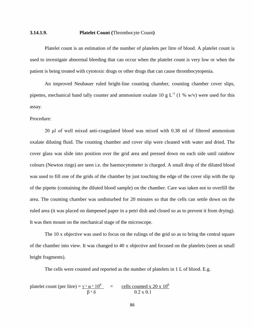
86
3.14.1.9. Platelet Count (Thrombocyte Count)
Platelet count is an estimation of the number of platelets per litre of blood. A platelet count is
used to investigate abnormal bleeding that can occur when the platelet count is very low or when the
patient is being treated with cytotoxic drugs or other drugs that can cause thrombocytopenia.
An improved Neubauer ruled bright-line counting chamber, counting chamber cover slips,
pipettes, mechanical hand tally counter and ammonium oxalate 10 g L-1
(1 % w/v) were used for this
assay.
Procedure:
20 μl of well mixed anti-coagulated blood was mixed with 0.38 ml of filtered ammonium
oxalate diluting fluid. The counting chamber and cover slip were cleaned with water and dried. The
cover glass was slide into position over the grid area and pressed down on each side until rainbow
colours (Newton rings) are seen i.e. the haemocytometer is charged. A small drop of the diluted blood
was used to fill one of the grids of the chamber by just touching the edge of the cover slip with the tip
of the pipette (containing the diluted blood sample) on the chamber. Care was taken not to overfill the
area. The counting chamber was undisturbed for 20 minutes so that the cells can settle down on the
ruled area (it was placed on dampened paper in a petri dish and closed so as to prevent it from drying).
It was then mount on the mechanical stage of the microscope.
The 10 x objective was used to focus on the rulings of the grid so as to bring the central square
of the chamber into view. It was changed to 40 x objective and focused on the platelets (seen as small
bright fragments).
The cells were counted and reported as the number of platelets in 1 L of blood. E.g.
platelet count (per litre) = γ ˣ α ˣ 106 = cells counted x 20 x 10
6
β ˣ δ 0.2 x 0.1
87
Where
γ = cells counted α = 1 in 20 dilution of blood
β = 0.2 mm2 area counted δ = 0.1 mm dept of chamber
3.14.2. Biochemical Assays
Serum alkaline phosphatase (ALP), alanine aminotransferase (ALT), aspartate
aminotransferase (AST), L-γ-glutamyltransferase (GGT) and lactate dehydrogenase (LDH) activities
were determined on serum using the standard procedures.
3.14.2.1. Alkaline Phosphatase (ALP)
This is an optimized standard method according to the Deutsche Gesellschaft fur Klinische
Chemie. Randox kit was used for this assay. Alkaline phosphatase (ALP EC 3.1.3.1:
orthophosphoricmonoester phosphohydrolase) enzymes in serum sample, converted a clear p-
nitrophenyl phosphate reagent to a yellow p-nitrophenol in a timed reaction. Colour change, measured
by light absorption, was considered a reliable indication of enzyme activity.
Procedures:
A vial of the substrate R1b (containing 10 mmol/L of p-nitrophenyl phosphate) was
reconstituted with the 10 ml of the buffer R1a (containing 1 mol/L diethanolamine buffer at pH 9.8
and 0.5 mmol/L magnesium chloride). This is the working reagent.
88
Twenty microliters (20 µl) of the serum sample was mixed with 1 ml of working reagent and
the initial absorbance was read at 405 nm. The timer was started simultaneously and the absorbance
was read after 1, 2 and 3 min.
Calculation: U/L = 2760 x Δ A 405 nm/ min
3.14.2.2. Alanine Aminotransferase (ALT)
Alanine Aminotransferase activity was measured by monitoring the concentration of pyruvate
hydrazone formed with 2,4-dinitrophenylhydrazine (Bergmeyer and Horder, 1980). Randox kit was
used for this assay. Two solutions labelled R1 (containing 100 mmol/L Phosphate buffer (pH 7.4), 200
mmol/L L-alanine and 2.0 mmol/L α-oxoglutarate) and R2 (containing 2.0 mmol/L 2,4-
dinitrophenylhydrazine) was used.
Procedure:
Preparation of reagent blank: 0.5 ml of solution R1 was mixed with 0.1 ml of distilled water
and incubated at 37 oC for 30 minutes. 0.5 ml of solution R2 was added to it, mixed and allowed to
stand for 20 minutes at 25 oC before adding 5 ml of 0.4 mol/L sodium hydroxide. After 5 minutes, the
absorbance of the test sample was measured against the reagent blank by using the reagent blank to set
the instrument to zero at 546 nm.
Preparation of test sample: 0.5 ml of solution R1 was mixed with 0.1 ml of serum sample and
incubated at 37 oC for 30 minutes. 0.5 ml of solution R2 was added to it, mixed and allowed to stand
for 20 minutes at 25 oC before adding 5 ml of 0.4 mol/L sodium hydroxide. After 5 minutes, the
89
absorbance of the test sample was measured against the reagent blank by using the reagent blank to set
the instrument to zero at 546 nm.
3.14.2.3. Aspartate Aminotransferase (AST)
Serum AST activity was measured by monitoring the concentration of oxaloacetate hydrazone
formed with 2,4-dinitrophenylhydrazine (Reitman and Frankel, 1957). Randox kit was used for this
assay. Two solutions labelled R1 (containing 100 mmol/L Phosphate buffer (pH 7.4), 100 mmol/L L-
aspartate and 2.0 mmol/L α-oxoglutarate) and R2 (containing 2.0 mmol/L 2,4-dinitrophenylhydrazine)
was used.
Procedure:
Preparation of reagent blank: 0.5 ml of solution R1 was mixed with 0.1 ml of distilled water
and incubated at 37 oC for 30 minutes. 0.5 ml of solution R2 was added to it, mixed and allowed to
stand for 20 minutes at 25 oC before adding 5 ml of 0.4mol/L sodium hydroxide. After 5 minutes, the
absorbance of the test sample was measured against the reagent blank by using the reagent blank to set
the instrument to zero at 546 nm.
Preparation of test sample: 0.5 ml of solution R1 was mixed with 0.1 ml of serum sample and
incubated at 37 oC for 30 minutes. 0.5 ml of solution R2 was added to it, mixed and allowed to stand
for 20 minutes at 25 oC before adding 5 ml of 0.4 mol/L sodium hydroxide. After 5 minutes, the
absorbance of the test sample was measured against the reagent blank by using the reagent blank to set
the instrument to zero at 546 nm.
90
3.14.2.4. Lactate Dehydrogenase (LDH)
This is an optimized standard method according to the Deutsche Gesellschaft fur Klinische
Chemie (Rec. GSCC 1972). Randox kit was used for this assay. A vial of NADH (0.18 mmol/L) (R1b)
was reconstituted with 3 ml of buffer / subtrate containing 50 mmol/L phosphate buffer at pH 7.5 and
0.6 mmol/L of pyruvate (R1a). This is the working reagent.
Procedure:
Twenty microliters (20 µl) of serum sample was pipette into cuvette containing 1 ml of the
working reagent. It was mixed and the initial absorbance was read at 340 nm after 30 sec and the the
timer was started simultaneously. The absorbance was read again after 1, 2 and 3 mins.
Calculation:
U/L = 8095 x ΔA 340 nm / min
3.14.2.5. L-γ-glutamyltransferase (GGT)
Serum L-γ-glutamyltransferase activity was determined using the method of Szasz (1969). The
substrate L-γ-glutamyl-3-carboxy-4-nitroanilide, in the presence of glycylglycine is converted by γ-GT
in the sample to 5-amino-2-nitrobenzoate which can be measured at 405 nm. Randox kit was used for
this assay. 3 ml of a solution containing Tris buffer (100 mmol/L at pH 8.25) and Glycylglycine (100
mmol/L) was used to reconstitute 2.9 mmol/L of L- γ -glutamyl-3-carboxy-4-nitroanilide substrate.
This is the working reagent.
91
Procedure:
One milliliter (1 ml) of the working reagent was mixed with 100 μl of serum sample. The
initial absorbance was read at 405 nm and a timer started simultaneously. The absorbance is read again
after 1, 2 and 3 minutes.
3.14.3. Antioxidant assays
Catalase (CAT), glutathione peroxidase (GPx), reduced glutathione (GSH), glutathione-s-
transferase (GST), thiobarbituric acid reactive substances (TBARS), superoxide dismutase (SOD) and
total protein (TP) activities were determined in liver and kidney homogenates and serum samples.
3.14.3.1. Estimation of Lipid Peroxidation Product (TBARS)
Lipid peroxidation in terms of thiobarbituric acid reactive substances (TBARS) formation was
measured using the method of Niehaus and Samuelsson (1968) with a little modification.
Malondialdehyde (MDA) is a secondary product of lipid peroxidation and is used as an indicator of
tissue damage. The MDA reacts with TBA (Thiobarbituric Acid) reagent to from a pink coloured
product which has absorption maxima at 535 nm.
Procedure:
100 μl of test sample was treated with 2ml of TCA–TBA–HCl (TBA 0.37%, 0.25N HCl and 15
% TCA (Trichloroacetic acid)) reagent (1: 1: 1) and incubated in boiling water bath for 15 minutes.
92
After cooling, the precipitate was removed by centrifugation. The absorbance of the sample was
measured at 535 nm using a blank containing all the reagents except the sample. The blank was used
to zero the spectrophotometer. TBARS concentration of the plasma, liver and kidney samples were
calculated using the extinction coefficient of MDA which is 1.56 × 10⁻⁵ mmol−1
cm−1
since 99 % of
TBARS exists as MDA using this formula.
OD x V x 106 = nmol/ml
Ʃ x v
Ʃ = molar extinction coefficient (1.56 × 10⁻⁵ mmol−1
cm−1
)
V = final volume of the reaction
v = volume of the sample
106 = optimal dilution factor
3.14.3.2. Estimation of Reduced Glutathione (GSH)
Glutathione, an antioxidant, helps protect cells from reactive oxygen species such as free
radicals and peroxides (Pompella, et al., 2003). It is the most abundant intracellular thiol compound
present in virtually all mammalian cells. GSH reacts with DTNB (5,5'-dithiobis-(2-nitrobenzoic acid)
and gets reduced to a yellow coloured complex which has an absorption maximum at 412 nm.
Reduced glutathione in plasma, kidney and liver were measured by the method of Ellman (1959).
Procedure:
Preparation of Ellmans reagents: 19.8 mg of DTNB was mixed with 100 ml of 0.1 % sodium
nitrate
93
0.5 ml of the test sample were deproteinated with 0.5 ml of 10 % TCA by centrifugation to
release GSH into the supernatant. 0.5 ml of the supernatant was derivatized with 0.25 ml of Ellmans
reagent and mixed with 0.2 M phosphate buffer (1.5 ml). Color was developed by adding the Ellmans
reagent and it was read at 412 nm.
Calculation:
OD x V x 103 = nmol/ml
Ʃ x v
Ʃ = molar extinction coefficient (13600) V = final volume of the reaction
v = volume of the sample 103 = optimal dilution factor
3.14.3.3. Glutathione Peroxidase (GPx)
Glutathione peroxidase is the general name of an enzyme family with peroxidase activity,
whose main role is to protect the organism from oxidative damage. The biochemical function of GPx
is to reduce lipid hydro peroxides to their corresponding alcohols and to reduce free hydrogen
peroxide to water. Glutathione peroxidase (GPx) activity in the test samples were determined by the
method of Ellman, (1959) based on the ability of GPx to catalyze the reduction of hydroperoxides,
including hydrogen peorxide, by reduced glutathione and functions to protect the cell from oxidative
damage.
Procedure:
A mixture containing 0.2 ml of 0.4 M phosphate buffer (pH 7), 0.1 ml of 10 mM sodium azide,
0.2 ml of the test sample, 0.2 ml of glutathione solution and 0.1 ml of 0.2 mM hydrogen peroxide was
94
incubated for 10 minutes at 37 oC. 0.4 ml of 10 % TCA was added and it was centrifuged for 10
minutes at 3,500 rpm. The supernatant was used to assay for glutathione content using 0.25 ml of
Ellmans reagent and 1.5 ml of 0.2 M phosphate buffer. It was read at 412 nm.
Calculation:
OD x V x 103 = nmol/ml
Ʃ x v
Ʃ = molar extinction coefficient (13600) V = final volume of the reaction
v = volume of the sample 103 = optimal dilution factor
3.14.3.4. Glutathione S-Transferases (GST)
Glutathione-S-transferases catalyzes the conjugation reaction with glutathione in the first step
of mercapturic acid synthesis. The activity of the enzyme was measured according to the method of
Habig and Jakoby (1974) with a little modification.
Procedure:
A mixture containing 0.5 ml of phosphate buffer (0.2 M), 50 μl of 20 mM cDNB (1-chloro-
2,4-dinitrobenzene) and distilled water was incubated at 37 oC for 5 minutes. 50 μl of the sample was
added to 50 μl of GSH reagent and it was read at 340 nm at the 1st -3
rd minutes. A blank was run in the
absence of the sample. One unit of GST activity is defined as 1 μmol product formation per minute.
95
Calculation:
∆OD/min x V x 103 = μmol/ml
Ʃ x v
Ʃ = molar extinction coefficient (9.6 mmol−1
cm−1
) v = volume of the sample
V = final volume of the reaction 103 = optimal dilution factor
3.14.3.5. Catalase (CAT)
Hydrogen peroxide (H2O2) is formed in cells by controlled pathways and elicits a broad
spectrum of cellular response ranging from mitogenic growth stimulation (Petros and Lambeth, 2001)
to apoptosis (Zhuang, et al., 2007) to necrosis (McKeague, et al., 2003) at different concentration
levels. Removal of H2O2 from cells is therefore necessary for protection against oxidative damage.
The most common definition of one catalase unit is the amount of enzyme decomposing 1.0 μmole of
hydrogen peroxide per minute at pH 7.0 and 25 ºC, with initial H2O2 concentration of 10.3 mM.
catalase level was measured according to the method of Sinha (1972).
Procedure:
The reaction mixture (1.5ml) contained 1.0 ml of 0.01M pH 7.0 phosphate buffer, 0.1 ml of
test sample and 0.4 ml of 2M H2O2 . The reaction was stopped by the addition of 2.0 ml of
dichromate-acetic acid reagent (5% potassium dichromate and glacial acetic acid were mixed in 1:3
ratio). A decrease in absorbance was observed after the addition of H2O2 to the reaction mixture
containing the test sample, which is used as the source of catalase.
96
Catalase was assayed colourimetrically at 620 nm and expressed as mmoles of H2O2
consumed/min/mg protein as described by Sinha (1972).
Calculation:
∆OD/ min x V x 103 ÷ total protein content = µmol/min/mg protein
Ʃ x v
Ʃ = molar extinction coefficient (40 mmol−1
cm−1
) v = volume of the sample
V = final volume of the reaction 103 = optimal dilution factor
3.14.3.6. Determination of Superoxide Dismutase (SOD) Activity.
SOD is a metalloenzyme with its active center occupied by copper or zinc, sometimes
manganese (or) iron. SOD plays an important role in protection of all aerobic life systems, against
oxygen toxicity. The activity of SOD was measured following the method originally developed by
Nishikimi et al., (1972) and then modified by Kakkar et al., (1984). One unit of SOD activity is
defined, as the enzyme concentration required inhibiting chromogen production by 50% in one minute
under the assay condition.
Procedure:
A mixture containing 30 μl of epinephrine (3 x 10⁴ M), 3 ml of 50 mM NaCO3 (Sodium
Carbonate) buffer (pH 10.2) and 20 μl of the test sample was read at 485 nm for the first 3 minutes i.e.
at 1 minute interval.
97
Calculation:
∆OD/min x V x 103 = nmol/ml
Ʃ x v
Ʃ = molar extinction coefficient (4.5 x 10³ M
-1 cm
-1) v = volume of the sample
V = final volume of the reaction 103 = optimal dilution factor
3.14.4. Chemical Analytes Assay
Albumin (ALB), cholesterol (CHO), creatinine (CREA), total protein, triglycerides (TG) and
blood urea nitrogen (BUN) concentrations were determined on serum using these standard procedures.
3.14.4.1. Total Protein (TP)
This method, commonly referred to as the Biuret assay (Tietz, 1995), is based on the
interaction of cupric (Cu2+
) ion with protein in an alkaline solution and the resultant development of
absorbance at 545 nm.
Procedure:
Preparation of reagent blank: To 0.02 ml of distilled water, 1 ml of the Biuret reagent was
added.
Preparation of sample: To 0.02 ml of test sample, 1 ml of the Biuret reagent was added.
Preparation of standard: To 0.02 ml of standard protein (bovine serum albumin), 1 ml of the
Biuret reagent was added.
98
They were mixed and incubated for 30 min at +20 to +25°C. Within 90 minutes after the
addition of the Biuret Reagent, the absorbance of the standard solution, reagent blank and the test
samples were determine at the wavelength of maximum absorbance at 545 nm. The absorbance of the
test sample and standard were measured against the reagent blank by using the blank to set the
instrument to zero.
3.14.4.2. Albumin (ALB)
The measurement of serum albumin is based on its quantitative binding to the indicator
3,3',5,5'-tetrabromo-m cresol sulphonephthalein (bromocresol green, BCG). The BCG albumin
procedure is based on the dye-binding properties of serum albumin with bromcresol green. The
absorbance of BCG at 630 nm increases with binding to albumin and is proportional to the albumin
concentration present (Spencer and Price, 1977).
Procedure:
Preparation of Reagent Blank: 10 μl of deionized water was mixed with 1.0 ml BCG albumin
reagent
Preparation of standard: 10 μl of albumin standard was mixed with 1.0 ml BCG albumin
reagent
Preparation of test samples: 10 μl of test sample (serum) was mixed with 1.0 ml BCG albumin
reagent.
They were incubated for 5 minutes at +20 to +25°C and the absorbance of the test sample and
standard were measured against the reagent blank at 630 nm. The absorbance of the test sample and
standard were measured against the reagent blank by using the reagent blank to set the instrument to
zero.
99
3.14.4.3. Creatinine (CREA)
Creatinine was measured according to the method of Bartels, et al., (1972). Creatinine in
alkaline solution reacts with picric acid to form a coloured complex. The amount of the complex
formed is directly proportional to the creatinine concentration.
Reagents: equal quantity of Picric acid (35 mmol/L) and Sodium hydroxide (0.32 mol/L) were
mixed and used for this assay as the working reagent.
Procedure:
Preparation of reagent blank: 1 ml of the working reagent was mixed with 100 μl of double
distilled water.
Preparation of sample: 1 ml of the working reagent was mixed with 100 μl of serum sample.
After 30 seconds, the absorbance (A1) of the reagent blank was measured against the sample.
Exactly 2 minutes later, the absorbance (A2) of the reagent blank was measured against the sample at
492 nm.
3.14.4.4. Triglycerides (TG)
Triglyceride assay is based on enzymatic reactions (GPO method) including lipoprotein lipase
(LPL), glycerol kinase (GK) and glycerol phosphate dehydrogenase (GPO). Triglycerides are
hydrolyzed by LPL to liberate glycerol and free fatty acids. Glycerol is converted to glycerol-3-
phosphate (G3P) and ADP (Adenosine diphosphate) by GK and ATP. G3P is then converted by GPO
to dihydroxyacetone phosphate (DAP) and hydrogen peroxide (H2O2). H2O2 is quantified by Trinder
reaction using 4-aminoantipyrine, 4-chlorophenol and hydrogen peroxidase (Trinder, 1969).
Triglycerides are determined after enzymatic hydrolysis with lipases. The indicator a quinoneimine, is
100
formed from hydrogen-peroxide, 4-aminophenazone and 4-chlorophenol under the catalytic influence
of peroxidase. The reaction is monitored at 500 nm.
Randox kit was used for this assay. Three reagents were used. A buffer (reagent 1) containing
Pipes buffer (40 mmol/L at pH 7.4), 4-chlorophenol (5.4 mmol/L), magnesium ions (5 mmol/L), ATP
(1 mmol/L), peroxidase ( EC 1.11.1.7; horseradish at 25°C. 0.5 U/ml. ), glycerol kinase (EC 2.7.1.30;
microbial at 25°C. 0.4 U/ml.), glycerol-3-phosphate oxidase (EC 1.1.3.21; microbial at 25°C. 1.5
U/ml.) and sodium azide (0.05 %), an enzyme reagent (reagent 2) containing 4-aminoantipyrine (0.4
mmol/L), lipases (EC 3.1.1.3; microbial, 25°C. 150 U/ml) and sodium azide (0.05 %) and a standard
(reagent 3) were used. A mixture of the enzyme reagent (250 μl) and the buffer (15 ml) was used as
the working reagent.
Procedure:
Preparation of reagent blank: 1 ml of the working reagent was mixed with 10 μl of working
reagent.
Preparation of sample: 1 ml of the working reagent was mixed with 10 μl of serum sample.
Preparation of standard: 1 ml of the working reagent was mixed with 10 μl of standard.
It was incubated for 10 minutes at 25 °C. The absorbance of the sample and standard were
measured against the reagent blank within 60 minutes.
3.14.4.5. Blood Urea Nitrogen (BUN)
Blood urea nitrogen was determined spectrophotometrically from serum samples using the
method of Kassirer (1971). Urea is hydrolyzed in the presence of water and urease to produce

101
ammonia and carbon dioxide. The ammonia produced in the first reaction combines with α-
oxoglutarate and NADH in the presence of glutamate dehydrogenase to yield glutamate and NAD+.
In the presence of glutamate dehydrogenase (GLDH) and reduced nicotinamide-adenine
dinucleotide (NADH), ammonia (as ammonium ions; NH4+) reacts with 2-oxoglutarate to form L-
glutamic acid and NAD+.
The amount of NAD+ formed is stoichiometric with the amount of ammonia or with twice the
amount of urea. It is NADH consumption which is measured by the decrease in absorbance at 340 nm.
Randox kit was used for this assay. 50 ml of tris-buffer (150 mmol/L at pH 7.6) was
reconstituted with 50 ml of an enzyme reagent containing urease (15 U/ml), GLDH (1 U/ml), NADH
(0.28 mmol/L), adenosine-5-diphosphate (2.45 mmol/L) and α-oxoglutarate (11.7 mmol/L). This is the
working reagent.
Procedure:
Preparation of reagent blank: 1 ml of the working reagent was mixed with 10 μl of distilled
water.
Preparation of sample: 1 ml of the working reagent was mixed with 10 μl of serum sample
Preparation of standard: 1 ml of the working reagent was mixed with 10 μl of standard.
The initial absorbance of the serum sample and standard were measured against the reagent
blank after 30 sec and a timer was started simultaneously. It was read again after 1 minute.
102
3.14.5. Caspase Activity Assay
3.14.5.1. Isolation of Leukocytes from Whole Blood
For each 2 ml of EDTA-treated blood (from the rats in the pharmacologically induced
hepatotoxicity groups) , the blood sample was spun at 5,000 rmp and the white blood cells i.e. the
buffy coat was transferred to a test tube containing 10 ml of 0.17 M ammonium chloride solution. It
was incubated for 5 minutes at 4 oC and then centrifuged for 5 minutes at 2000 rpm and the procedure
was repeated 4 times. The supernatant was aspirated using a Pasture pipette and the pellets were
suspended in 2 ml of 0.5 % saponin. Phosphate buffered saline was laid over the leukocytes, and tubes
were then centrifuged at 1500 rpm for 5 minutes. The supernatant was aspirated.
The concentration of the leukocyte suspension was determined using a Thoma counting
chamber. Polymorphonuclear leukocytes and mononuclear leukocytes were discerned using Giemsa
staining.
Leukocyte viability was checked using the trypan blue exclusion test since viable cells with
intact membrane integrity do not take in (exclude) certain chemical dyes. 200 μl of leucocyte
suspension being tested for viability was centrifuged for 5 min at 1500 rpm and the supernatant was
discarded. The cell pellet was resuspended in 200 μl PBS (Phosphate buffered saline) and mixed with
200 μl of 0.4 % trypan blue (GIBCO/BRL) in an Eppendorf tube. The mixture was allowed to incubate
for 3 min at room temperature (Cells should be counted within 3 to 5 min of mixing with trypan blue,
as longer incubation periods will lead to cell death and reduced viability counts). A drop of the trypan
blue/cell mixture was applied to a haemacytometer and viewed on a binocular microscope. Nuclei of
non-viable cells stained blue. The white blood cells and pellets were resuspended in 10 ml cold 1x
phosphate buffered saline (pH = 7.2), aliquot into Eppendorf tubes and stored at -20 oC.
103
3.14.5.2. Caspase-2 Assay Procedure:
The increase in caspase-2 activity was determined using the method of Hoglund, et al., (2009).
The white blood cells and pellets isolated from the rats in the pharmacologically induced
hepatotoxicity groups were used for this assay. White blood cells and pellet (2-5 x 10⁶ cells) were
counted and resuspended in 50 μl of chilled cell lysis buffer. It was incubated on ice for 10 min and
centrifuged for 1 min in a microcentrifuge at 10,000 rpm. The protein concentration of the supernatant
was assayed (Biuret method) and 100-200 μg cytosolic extract was diluted in 50 μl cell lysis buffer
(BioVision). 50 μl of 2x reaction buffer (Gentaur) (to which dithiothreitol (DTT) (Sigma) had been
added giving a final concentration of 10 mM i.e. 10 μl of 1.0 M DTT stock was added to 1 ml of 2X
reaction buffer) was added to each sample. 5 μl of 4 mM substrate comprising the amino acids L-
valyl- L-aspartyl- L-valyl- L-alanyl- L-aspartic acid- p-nitroanilide (VDVAD-pNA or Val-Asp-Val-
Ala-Asp- pNA substrate) (BioVision) was added giving a final concentration of 200 μM. It was
incubated at 37 oC for 1-2 hr. The absorbance was read using a BIO-RAD microplate reader at 405nm.
The increase in caspase-2 activity was determined by comparing the results of treated samples (groups
2-7) with the level of the untreated control (group 1).
3.14.5.3. Caspase-3 Assay Protocol:
The method of Chen, et al., (2008) was used to determine the increase in caspase-3 activity.
The white blood cells and pellets isolated from the rats in the pharmacologically induced
hepatotoxicity groups were used for this assay. White blood cells and pellet 1-5 x 10⁶ cells was
counted and resuspended in 50 μl of chilled cell lysis buffer. It was incubated on ice for 10 min and
centrifuged at 10,000 rpm for 1 min. The supernatant (cytosolic extract) was put on ice and the protein
concentration was assayed according to Biurets method. 50-200 μg cytosolic extract was diluted in 50
μl cell lysis buffer and 50 μl of 2x reaction buffer to which DTT had been added giving a final
104
concentration of 10 mM final concentration i.e. 10 μl of 1.0 M DTT stock was added to 1 ml of 2X
reaction buffer was added. 5 μl of the 4 mM DEVD (Ac-Asp-Glu-Val-Asp) p-nitroanilide substrate
(Acetyl- L-aspartyl- L-glutamyl- L-valyl- L-aspartic acid- p-nitroanilide substrate) (BioVision) was
added giving a final concentration of 200 μM and it was incubated at 37 oC for 1-2 hr. The absorbance
was read using a BIO-RAD microplate reader at 405 nm. The fold-increase in caspase-3 activity was
determined by comparing the results of hepatotoxicity induced rats (groups 2-7) with the level of the
uninduced control rats (group 1).
3.14.5.4. Caspase -9 Assay Protocol:
Fold-increase in caspase-9 activity was determined using the method of Chen, et al., (2008).
White blood cells and pellets 2-5 x 10⁶ cells were counted and resuspended in 50 μl of chilled cell
lysis buffer. It was incubated on ice for 10 min and centrifuged at 10,000 rpm for 1 min. The
supernatant (cytosolic extract) was put on ice and the protein concentration was assayed using Biurets
method. 100-200 μg of the cytosolic extract was diluted in 50 μl cell lysis buffer and 50 μl of 2x
reaction buffer (comprising of 10 μl of 1.0 M DTT stock diluted in 1 ml of 2x reaction buffer). 5 μl of
the 4 mM LEHD (Ac-Leu-Glu-His-Asp) p-nitroanilide substrate (Acetyl- L-leucyl- L-glutamyl- L-
histidyl- L-aspartic acid- p-nitroanilide substrate) (BioVision) was added giving a final concentration
of 200 μM and it was incubated at 37oC for 1-2 hr. The absorbance was read using a BIO-RAD
microplate reader at 405-nm.
Fold-increase in caspase-9 activity was determined by comparing the results of hepatotoxicity
induced rats (groups 2-7) with the level of the uninduced control rats (group 1).
3.14.6. Histopathology.
The method of Mallory (1961) was used with a little modification. A small chunk of liver and
kidney were taken from the sacrificed experimental rats used for hepatotoxicity studies and were
105
preserved in 10 % formal saline for histologic studies. The tissues were processed and sectioned in
paraffin. The paraffin sections of buffered formalin- fixed tissue samples (3 µm thick) were dewaxed
and rinsed in alcohol and also water. They were stained with Harris' haematoxylin (Sigma) for 10
minutes, washed in running tap water for 1 minute, differentiated in acid alcohol for 10 seconds and
washed again in running tap water for 5 minutes. The tissues were stained with eosin for 4 minutes and
washed in running tap water for 10 seconds. It was dehydrated and mounted for photomicroscopic
observations of the histologic architecture. The general structure of the liver and kidneys of the normal
control group (group 1) was compared with those of the treated groups (groups 2-7).
3.15. in vitro Antioxidant Assays
3.15.1. Assay of Lipid Peroxidation using Brain Homogenates
The thiobarbituric acid (TBA) assay was used to assess lipid peroxidation using the method of
Chowdhury and Soulsby (2002). For the in vitro studies, the brains of normal rats were dissected and
homogenized with a Polytron® ergonomic homogenizer (speed setting 7-8) in ice-cold Tris-HCl
buffer (20 mM, pH 7 4) to produce a 1/10 homogenate. The homogenate was centrifuged at 14,000
rpm for 15 min. One ml aliquots of the supernatant were incubated with the different fractions of
GOV, the aqueous and ethanolic extracts of GOV in the presence of 10 µM FeS04 (Ferrous Sulphate)
and 0.1 mM ascorbic acid at 37 °C for 1 hr. The reaction was stopped by addition of 1 ml
trichloroacetic acid (TCA, 28 %, w/v) and 1.5 ml thiobarbituric acid (TBA, l %, w/v) in succession,
and the solution was then heated at 100°C for 15 mins. After centrifugation to remove precipitated
protein, the colour of the malondialdehyde (MDA)-TBA complex was detected at OD 532 nm using a
Milton Roy spectronic 3000 spectrophotometer. Butylated hydroxyanisole (BHA) was used as a
positive control. The inhibition ratio (%) was calculated using the following formula.
106
Inhibition ratio (%) = (A – Al) x 100 % where
A
A = the absorbance of the control, and Al = the absorbance of the test sample
3.15.2. Assay of Inhibition of Erythrocyte Haemolysis
The method of Ramchoun, et al., (2009) was used to assay the inhibition of erythrocyte
haemolysis. Blood was obtained from mice by extirpating their eyeballs and collected in tubes
containing 0.15 M NaCl. Erythrocytes were separated from the plasma and the buffy coat, and washed
three times with 10 volumes of 0.15 M NaCl. During the last wash, the erythrocytes were centrifuged
at 2,500 rpm for 10 min to obtain a constantly packed cell preparation. Erythrocyte haemolysis was
mediated by peroxyl radicals in the assay system. A 10% suspension of erythrocytes in pH 7.4
phosphate buffered saline (PBS) was added to the same volume of 200 mM 2,2'-azo-bis (2-
amidinopropane) dihydrochloride (AAPH) (Sigma) solution (in PBS) containing different
concentrations of the samples to be tested. The reaction mixture was shaken gently while being
incubated at 37 oC for I hr. The reaction mixture was removed, diluted with 8 volumes of PBS, and
centrifuged at 2,500 rpm for 10 min.
The absorbance A of the supernatant was read at 540 nm. Similarly, the reaction mixture was
treated with 8 volumes of distilled water to achieve complete haemolysis, and the absorbance B of the
supernatant obtained after centrifugation was measured at 540 nm. L-ascorbic acid (Sigma) was used
as a positive control.
The percentage of haemolysis was calculated by using the equation
(1-A/B) × 100 %.
107
3.16. Antiproliferative Activity toward Human Hepatocellular Liver Carcinoma Cell Line
(HepG2 cells) and Nasopharyngeal Cancer Cells (CNE2 and SUME –α- Nasopharyngeal
Cells)
The human nasopharyngeal carcinoma cell lines CNE-2 (Human Nasopharyngeal Carcinoma
Cell Lines) and SUME –α- nasopharyngeal cells were bought from Sun Yat-sen University of Medical
Sciences, Guangzhou, China. Hep G2 (Human hepatocellular liver carcinoma cell line) cell lines were
purchased from The American Type Culture Collection (ATCC). All others reagents used were from
Sigma (USA) or as mentioned.
Assay of antiproliferative activity on tumor cell lines: Growth was assessed by using the
methylthiazolyldiphenyl-tetrazolium (MTT) bromide cellular proliferation assay. (Sieuwerts, et al.,
1995; Zhang, et al., 2004). Briefly, HepG 2, SUME –α- and CNE-2 cells were maintained in RPMI
(Roswell Park Memorial Institute) 1640 medium containing 10 % fetal bovine serum, 1 % penicillin
and streptomycin at 37 oC, an atmosphere of 95 % air and 5 % carbon dioxide. After the cells had
grown to 90 % confluence, the cells were trypsinized (with 0.25% (w/v) Trypsin- 0.53 mM EDTA
solution to remove all traces of serum that contains trypsin inhibitor) and seeded to a 96-well plate
(each well containing approximately 5000 cells per 100 μl) respectively. After incubation for 8 hr, the
cells were treated with a serial dilution of the purified trypsin inhibitor at concentrations from 1.6 to
100 μM for 24 and 48 hrs. Two hours (2 hrs.) before the end of each incubation interval, 50 μl of a
solution containing 5 mg ml-1
of MTT was spiked into each well. After careful removal of the
medium, 150 μl dimethyl sulfoxide (Sigma) was added to each well and then agitated on an orbital
shaker for 15 minutes. The absorbance at 595 nm was measured using a BIO-RAD microplate reader.
108
3.17. Statistical analysis
The results were expressed as Mean + SEM for seven rats. Statistical analysis of the data was
performed using ANOVA statistical SPSS package (15.0) version. The significance of differences
among all groups was determined by the Tukey HSD test. P – values less than 0.05 (p ≤ 0.05) were
considered to be statistically significant.
109
CHAPTER FOUR
RESULTS
4.1. Extraction of Plant Materials
The percentage yields of the aqueous and ethanolic extracts of Gongronema latifolia Benth,
Ocimum gratissimum Linn. and Vernonia amygdalina Del. (1: 1: 1) were 12.6 % and 14.1 %
respectively. The yield of the triherbal formulation i.e. 50 % ethanol extract (GOV) obtained was 1.42
kg (15.69 %).
4.2. Fractionation of Plant Materials
The 50 % ethanol extract (GOV) was sequentially solvent partitioned into soluble extracts of
GOV giving a % yield of Hexane (5.92 %), Chloroform (10.38 %), Ethyl acetate (20.89 %), Butanol
(24.68 %) and Water (36.63 %).
4.3. Phytochemical analysis
The results of the phytochemical screening of 50 % ethanolic extract of the triherbal
formulation (GOV) is shown in able 1. The phytochemicals alkaloids, flavonoids, tannins, free and
bound anthraquinones, saponins and reducing sugars were present in the 50 % ethanolic extract while
anthocyanides were absent.
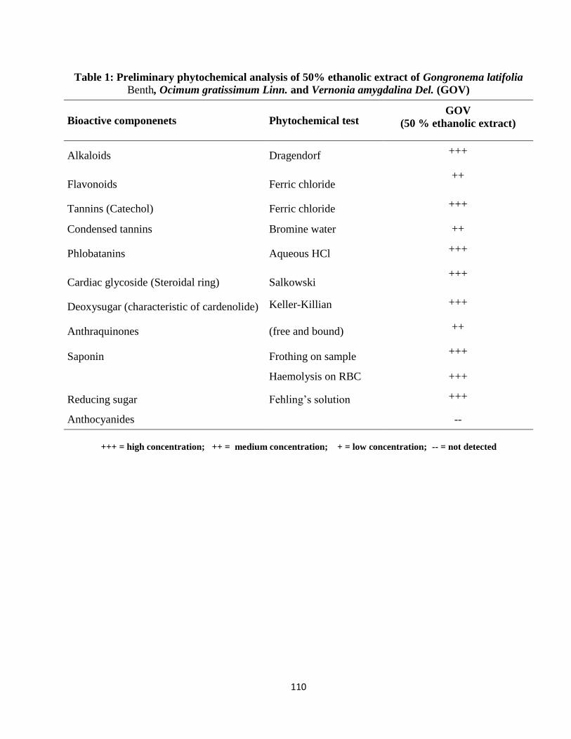
110
Table 1: Preliminary phytochemical analysis of 50% ethanolic extract of Gongronema latifolia
Benth, Ocimum gratissimum Linn. and Vernonia amygdalina Del. (GOV)
Bioactive componenets Phytochemical test GOV
(50 % ethanolic extract)
Alkaloids Dragendorf +++
Flavonoids Ferric chloride ++
Tannins (Catechol) Ferric chloride +++
Condensed tannins Bromine water ++
Phlobatanins Aqueous HCl +++
Cardiac glycoside (Steroidal ring) Salkowski +++
Deoxysugar (characteristic of cardenolide) Keller-Killian +++
Anthraquinones (free and bound) ++
Saponin Frothing on sample +++
Haemolysis on RBC +++
Reducing sugar Fehling‘s solution +++
Anthocyanides
--
+++ = high concentration; ++ = medium concentration; + = low concentration; -- = not detected
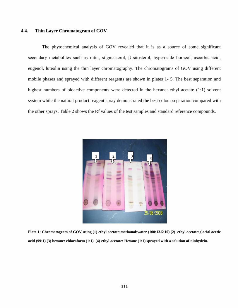
111
4.4. Thin Layer Chromatogram of GOV
The phytochemical analysis of GOV revealed that it is as a source of some significant
secondary metabolites such as rutin, stigmasterol, β sitosterol, hyperoside borneol, ascorbic acid,
eugenol, luteolin using the thin layer chromatography. The chromatograms of GOV using different
mobile phases and sprayed with different reagents are shown in plates 1- 5. The best separation and
highest numbers of bioactive components were detected in the hexane: ethyl acetate (1:1) solvent
system while the natural product reagent spray demonstrated the best colour separation compared with
the other sprays. Table 2 shows the Rf values of the test samples and standard reference compounds.
Plate 1: Chromatogram of GOV using (1) ethyl acetate:methanol:water (100:13.5:10) (2) ethyl acetate:glacial acetic
acid (99:1) (3) hexane: chloroform (1:1) (4) ethyl acetate: Hexane (1:1) sprayed with a solution of ninhydrin.
2 3 4
1
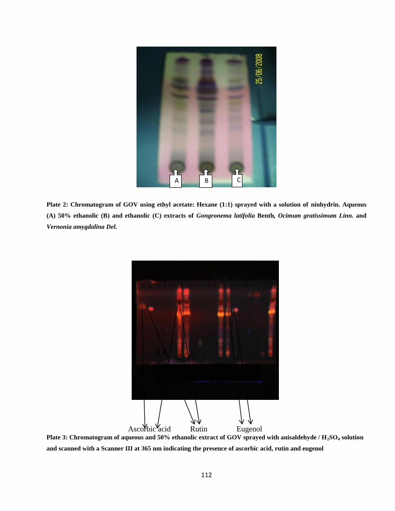
112
Plate 2: Chromatogram of GOV using ethyl acetate: Hexane (1:1) sprayed with a solution of ninhydrin. Aqueous
(A) 50% ethanolic (B) and ethanolic (C) extracts of Gongronema latifolia Benth, Ocimum gratissimum Linn. and
Vernonia amygdalina Del.
Ascorbic acid Rutin Eugenol Plate 3: Chromatogram of aqueous and 50% ethanolic extract of GOV sprayed with anisaldehyde / H2SO4 solution
and scanned with a Scanner III at 365 nm indicating the presence of ascorbic acid, rutin and eugenol
A B C
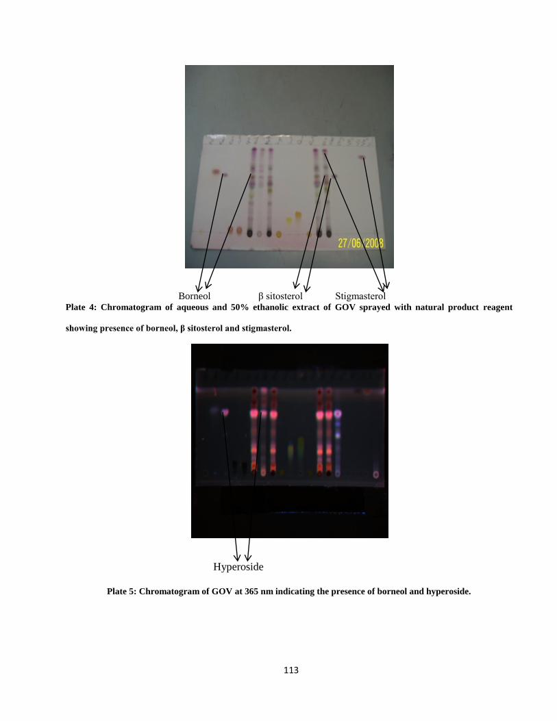
113
Borneol β sitosterol Stigmasterol Plate 4: Chromatogram of aqueous and 50% ethanolic extract of GOV sprayed with natural product reagent
showing presence of borneol, β sitosterol and stigmasterol.
Hyperoside
Plate 5: Chromatogram of GOV at 365 nm indicating the presence of borneol and hyperoside.
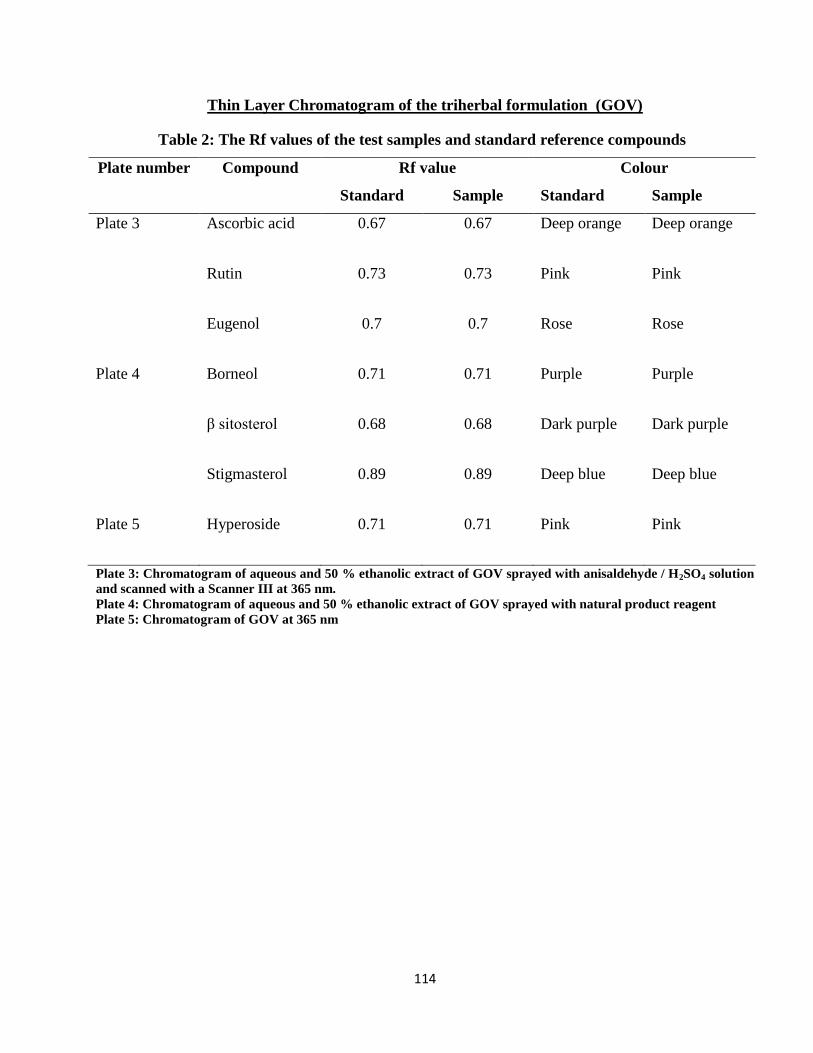
114
Thin Layer Chromatogram of the triherbal formulation (GOV)
Table 2: The Rf values of the test samples and standard reference compounds
Plate number Compound Rf value Colour
Standard Sample Standard Sample
Plate 3 Ascorbic acid 0.67 0.67 Deep orange Deep orange
Rutin 0.73 0.73 Pink Pink
Eugenol 0.7 0.7 Rose Rose
Plate 4 Borneol 0.71 0.71 Purple Purple
β sitosterol 0.68 0.68 Dark purple Dark purple
Stigmasterol 0.89 0.89 Deep blue Deep blue
Plate 5 Hyperoside 0.71 0.71 Pink Pink
Plate 3: Chromatogram of aqueous and 50 % ethanolic extract of GOV sprayed with anisaldehyde / H2SO4 solution
and scanned with a Scanner III at 365 nm.
Plate 4: Chromatogram of aqueous and 50 % ethanolic extract of GOV sprayed with natural product reagent
Plate 5: Chromatogram of GOV at 365 nm
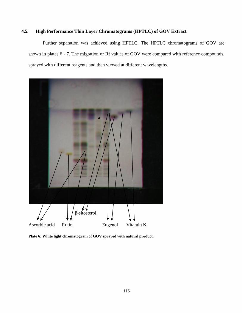
115
4.5. High Performance Thin Layer Chromatograms (HPTLC) of GOV Extract
Further separation was achieved using HPTLC. The HPTLC chromatograms of GOV are
shown in plates 6 - 7. The migration or Rf values of GOV were compared with reference compounds,
sprayed with different reagents and then viewed at different wavelengths.
β-sitosterol
Ascorbic acid Rutin Eugenol Vitamin K
Plate 6: White light chromatogram of GOV sprayed with natural product.
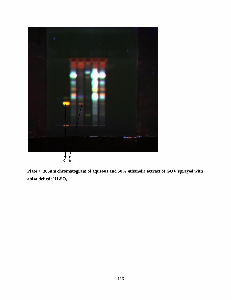
116
Rutin
Plate 7: 365nm chromatogram of aqueous and 50% ethanolic extract of GOV sprayed with
anisaldehyde/ H2SO4
117
4.6. NMR Spectra for the Binding Study of GOV and its Fractions with
Dihydrodipicolinate Reductase (DHPR)
The signals in these areas could indicate compounds that are binding to DHPR. For example,
peaks at 0.6, 1.2, and 1.3 ppm in the butanol fraction (Figure 2(A)) seemed to indicate a compound
that bound to DHPR. A follow up experiment with a proton NMR of the same sample (Figure 2 (B))
shows that the STD signals maybe due to the excitation of the protein at -0.5 ppm. This phenomenon
is due to the irradiation of the protein being near proton NMR signals of the ligands due to methyl
groups. Previous STD studies led to familiarity in irradiating DHPR at -0.5 ppm to excite the protein.
Another possible signal in the STD at 5.6 ppm was found upon close inspection of the spectrum,
Figure 5(A).
Key:
A + on a spectrum indicates that area is of interest and further experimentation is necessary to
investigate the nature of the signal.
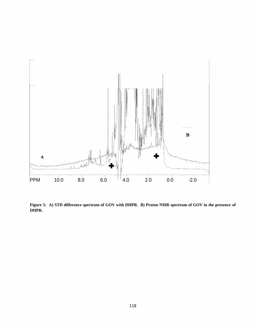
118
B
Figure 5: A) STD difference spectrum of GOV with DHPR. B) Proton NMR spectrum of GOV in the presence of
DHPR.
SpinWorks 2.5: Std proton
PPM 11.5 10.5 9.5 8.5 7.5 6.5 5.5 4.5 3.5 2.5 1.5 0.5 -0.5 -1.5 -2.5
file: C:\Documents and Settings\Terrence Neumann\Desktop\NMR\G1\6g_dhpr_1d.fid\fid block# 1 expt: "PRESAT"
transmitter freq.: 599.507170 MHz
time domain size: 19230 points
width: 9615.38 Hz = 16.038815 ppm = 0.500020 Hz/pt
number of scans: 16
freq. of 0 ppm: 599.504383 MHz
processed size: 65536 complex points
LB: 0.000 GB: 0.0000
SpinWorks 2.5: new experiment
PPM 10.0 8.0 6.0 4.0 2.0 0.0 -2.0 file: C:\Documents and Settings\Terrence Neumann\Desktop\NMR\G1\5g_dhpr_STD.fid\fid block# 1 expt: "cyclenoe"
transmitter freq.: 599.507167 MHz
time domain size: 9616 points
width: 9615.38 Hz = 16.038815 ppm = 0.999936 Hz/pt
number of scans: 512
freq. of 0 ppm: 599.504383 MHz
processed size: 16384 complex points
LB: 0.000 GB: 0.0000
A ✚
✚
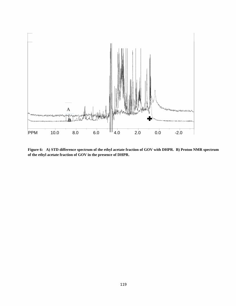
119
A
B
Figure 6: A) STD difference spectrum of the ethyl acetate fraction of GOV with DHPR. B) Proton NMR spectrum
of the ethyl acetate fraction of GOV in the presence of DHPR.
SpinWorks 2.5: Std proton
PPM 11.5 10.5 9.5 8.5 7.5 6.5 5.5 4.5 3.5 2.5 1.5 0.5 -0.5 -1.5 -2.5
file: C:\Documents and Settings\Terrence Neumann\Desktop\NMR\V\3V_presat.fid\fid block# 1 expt: "PRESAT"
transmitter freq.: 599.507170 MHz
time domain size: 19230 points
width: 9615.38 Hz = 16.038815 ppm = 0.500020 Hz/pt
number of scans: 16
freq. of 0 ppm: 599.504383 MHz
processed size: 65536 complex points
LB: 0.200 GB: 0.0000
SpinWorks 2.5: new experiment
PPM 10.0 8.0 6.0 4.0 2.0 0.0 -2.0 file: C:\Documents and Settings\Terrence Neumann\Desktop\NMR\V\3V_dhpr_std.fid\fid block# 1 expt: "cyclenoe"
transmitter freq.: 599.507167 MHz
time domain size: 9616 points
width: 9615.38 Hz = 16.038815 ppm = 0.999936 Hz/pt
number of scans: 512
freq. of 0 ppm: 599.504383 MHz
processed size: 16384 complex points
LB: 0.000 GB: 0.0000
✚
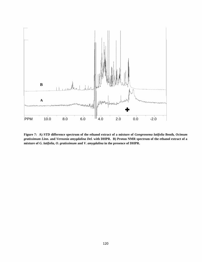
120
Figure 7: A) STD difference spectrum of the ethanol extract of a mixture of Gongronema latifolia Benth, Ocimum
gratissimum Linn. and Vernonia amygdalina Del. with DHPR. B) Proton NMR spectrum of the ethanol extract of a
mixture of G. latifolia, O. gratissimum and V. amygdalina in the presence of DHPR.
SpinWorks 2.5: new experiment
PPM 10.0 8.0 6.0 4.0 2.0 0.0 -2.0 file: C:\Documents and Settings\Terrence Neumann\Desktop\NMR\V\6v_dhpr_std.fid\fid block# 1 expt: "cyclenoe"
transmitter freq.: 599.507167 MHz
time domain size: 9616 points
width: 9615.38 Hz = 16.038815 ppm = 0.999936 Hz/pt
number of scans: 512
freq. of 0 ppm: 599.504383 MHz
processed size: 16384 complex points
LB: 0.000 GB: 0.0000
SpinWorks 2.5: Std proton
PPM 11.5 10.5 9.5 8.5 7.5 6.5 5.5 4.5 3.5 2.5 1.5 0.5 -0.5 -1.5 -2.5
file: C:\Documents and Settings\Terrence Neumann\Desktop\NMR\V\6v_presat.fid\fid block# 1 expt: "PRESAT"
transmitter freq.: 599.507170 MHz
time domain size: 19230 points
width: 9615.38 Hz = 16.038815 ppm = 0.500020 Hz/pt
number of scans: 16
freq. of 0 ppm: 599.504383 MHz
processed size: 65536 complex points
LB: 0.200 GB: 0.0000
A
B
✚
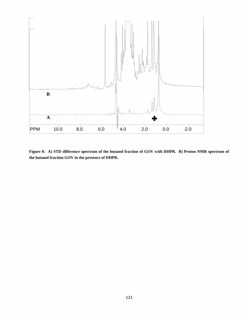
121
Figure 8: A) STD difference spectrum of the butanol fraction of GOV with DHPR. B) Proton NMR spectrum of
the butanol fraction GOV in the presence of DHPR.
SpinWorks 2.5: new experiment
PPM 10.0 8.0 6.0 4.0 2.0 0.0 -2.0 file: C:\Documents and Settings\Terrence Neumann\Desktop\NMR\1Mx_dhpr_std.fid\fid block# 1 expt: "cyclenoe"
transmitter freq.: 599.507167 MHz
time domain size: 9616 points
width: 9615.38 Hz = 16.038815 ppm = 0.999936 Hz/pt
number of scans: 512
freq. of 0 ppm: 599.504383 MHz
processed size: 16384 complex points
LB: 0.000 GB: 0.0000
SpinWorks 2.5: Std proton
PPM 10.0 8.0 6.0 4.0 2.0 0.0 -2.0 file: C:\Documents and Settings\Terrence Neumann\Desktop\NMR\1Mx_presat.fid\fid block# 1 expt: "PRESAT"
transmitter freq.: 599.507170 MHz
time domain size: 19230 points
width: 9615.38 Hz = 16.038815 ppm = 0.500020 Hz/pt
number of scans: 16
freq. of 0 ppm: 599.504383 MHz
processed size: 65536 complex points
LB: 0.200 GB: 0.0000
A
B
✚

122
Figure 9: A) STD difference spectrum of the chloroform fraction of GOV with DHPR. B) Proton NMR spectrum
of the chloroform fraction of GOV in the presence of DHPR.
SpinWorks 2.5: new experiment
PPM 10.0 8.0 6.0 4.0 2.0 0.0 -2.0 file: C:\Documents and Settings\Terrence Neumann\Desktop\NMR\Mx1\2Mx_dhpr_std.fid\fid block# 1 expt: "cyclenoe"
transmitter freq.: 599.507167 MHz
time domain size: 9616 points
width: 9615.38 Hz = 16.038815 ppm = 0.999936 Hz/pt
number of scans: 512
freq. of 0 ppm: 599.504383 MHz
processed size: 16384 complex points
LB: 0.000 GB: 0.0000
SpinWorks 2.5: Std proton
PPM 11.5 10.5 9.5 8.5 7.5 6.5 5.5 4.5 3.5 2.5 1.5 0.5 -0.5 -1.5 -2.5
file: C:\Documents and Settings\Terrence Neumann\Desktop\NMR\Mx1\2Mx_presat.fid\fid block# 1 expt: "PRESAT"
transmitter freq.: 599.507170 MHz
time domain size: 19230 points
width: 9615.38 Hz = 16.038815 ppm = 0.500020 Hz/pt
number of scans: 16
freq. of 0 ppm: 599.504383 MHz
processed size: 65536 complex points
LB: 0.200 GB: 0.0000
A
B
✚
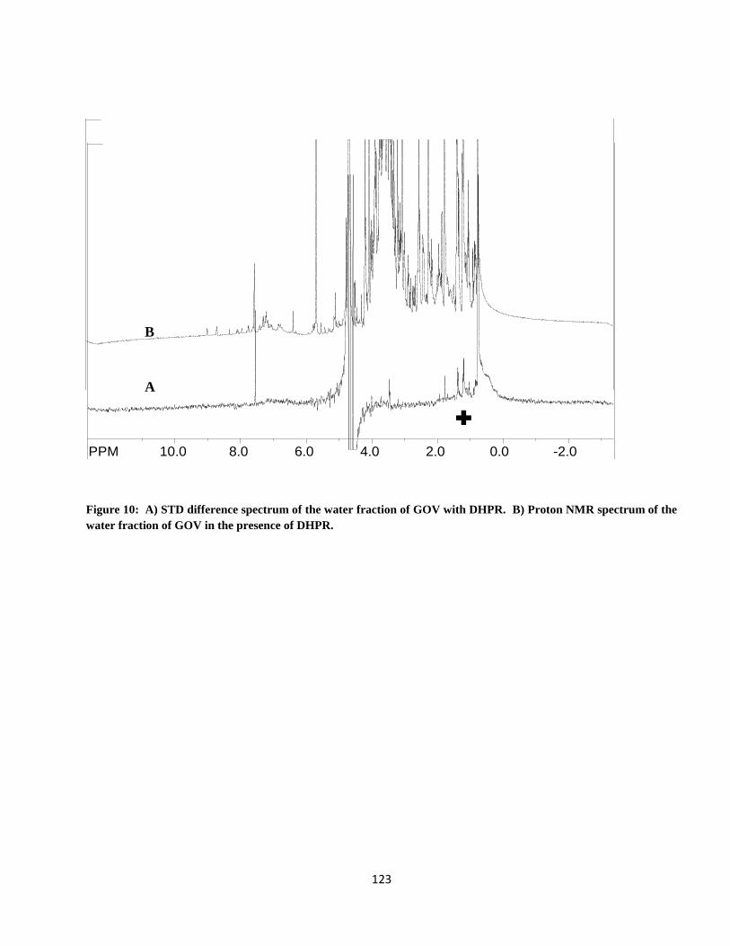
123
Figure 10: A) STD difference spectrum of the water fraction of GOV with DHPR. B) Proton NMR spectrum of the
water fraction of GOV in the presence of DHPR.
SpinWorks 2.5: new experiment
PPM 10.0 8.0 6.0 4.0 2.0 0.0 -2.0 file: C:\Documents and Settings\Terrence Neumann\Desktop\NMR\Mx1\5Mx_dhpr_std.fid\fid block# 1 expt: "cyclenoe"
transmitter freq.: 599.507167 MHz
time domain size: 9616 points
width: 9615.38 Hz = 16.038815 ppm = 0.999936 Hz/pt
number of scans: 512
freq. of 0 ppm: 599.504383 MHz
processed size: 16384 complex points
LB: 0.200 GB: 0.0000
SpinWorks 2.5: Std proton
PPM 10.0 8.0 6.0 4.0 2.0 0.0 -2.0 file: C:\Documents and Settings\Terrence Neumann\Desktop\NMR\Mx1\5Mx_presat.fid\fid block# 1 expt: "PRESAT"
transmitter freq.: 599.507170 MHz
time domain size: 19230 points
width: 9615.38 Hz = 16.038815 ppm = 0.500020 Hz/pt
number of scans: 16
freq. of 0 ppm: 599.504383 MHz
processed size: 65536 complex points
LB: 0.200 GB: 0.0000
A
B
✚
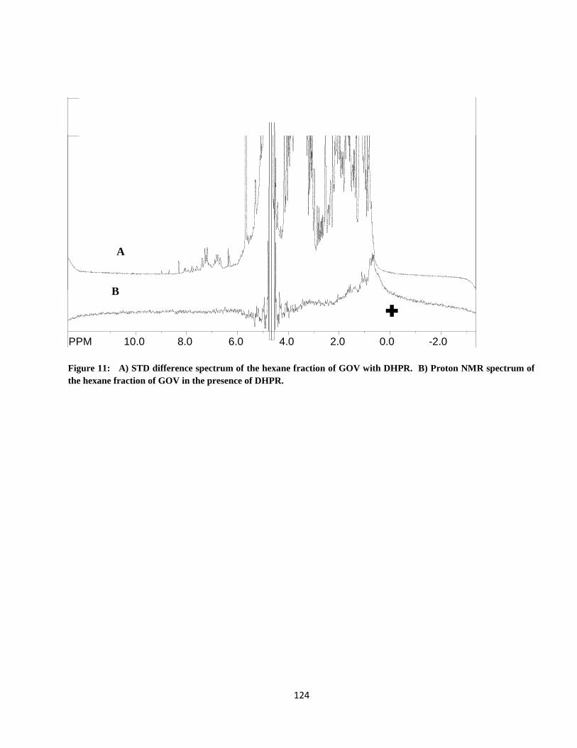
124
✚
Figure 11: A) STD difference spectrum of the hexane fraction of GOV with DHPR. B) Proton NMR spectrum of
the hexane fraction of GOV in the presence of DHPR.
SpinWorks 2.5: Std proton
PPM 11.5 10.5 9.5 8.5 7.5 6.5 5.5 4.5 3.5 2.5 1.5 0.5 -0.5 -1.5 -2.5
file: C:\Documents and Settings\Terrence Neumann\Desktop\NMR\G1\4g_dhpr_1d.fid\fid block# 1 expt: "PRESAT"
transmitter freq.: 599.507170 MHz
time domain size: 19230 points
width: 9615.38 Hz = 16.038815 ppm = 0.500020 Hz/pt
number of scans: 16
freq. of 0 ppm: 599.504383 MHz
processed size: 65536 complex points
LB: 0.000 GB: 0.0000
SpinWorks 2.5: new experiment
PPM 10.0 8.0 6.0 4.0 2.0 0.0 -2.0 file: C:\Documents and Settings\Terrence Neumann\Desktop\NMR\G1\4g_dhpr_STD.fid\fid block# 1 expt: "cyclenoe"
transmitter freq.: 599.507167 MHz
time domain size: 9616 points
width: 9615.38 Hz = 16.038815 ppm = 0.999936 Hz/pt
number of scans: 512
freq. of 0 ppm: 599.504383 MHz
processed size: 16384 complex points
LB: 0.000 GB: 0.0000
B
A
✚
125
4.7. Toxicity Tests
4.7.1. Acute Toxicity
The acute toxicity studies showed that GOV at 16 g kg-1 (p.o.) and 2.5 g kg
-1 (i.p.) did not
produce any mortality nor was there any significant change in the general behaviour of the mice.
4.7.2. Sub-chronic toxicity
There were no visible signs of deleterious effects on the rats both in behaviour and
morphology. No deaths were recorded.
4.7.2.1. Sub-chronic Effect on Haematologic Indices.
The effects of pretreatment of normal albino rats with GOV on the haematologic indices are
shown in tables 3a and 3b. At 2 g kg-1
, the haemoglobin (Hb), platelets, Mean corpuscular
haemoglobin concentration (MCHC), Mean corpuscular haemoglobin (MCH) and granulocytes were
increased while the Mean corpuscular volume (MCV) had the lowest value compared to the other
groups while at 8 g kg-1
, the packed cell volume (PCV), red blood cell (RBC), white blood cell (WBC)
and lymphocytes were the highest.
There was no significant (p ≤ 0.05) difference between the GOV treated groups and the control
group. GOV dose dependently increased the PCV, Hb, RBC, WBC, platelet, MCHC, MCH,
granulocytes and lymphocytes compared to the control group. There was no significant (p ≤ 0.05)
difference between the groups.
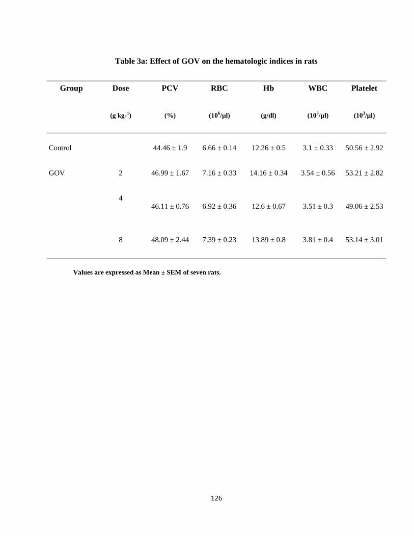
126
Table 3a: Effect of GOV on the hematologic indices in rats
Group Dose
(g kg-1)
PCV
(%)
RBC
(106/µl)
Hb
(g/dl)
WBC
(103/µl)
Platelet
(103/µl)
Control 44.46 ± 1.9 6.66 ± 0.14 12.26 ± 0.5 3.1 ± 0.33 50.56 ± 2.92
GOV 2 46.99 ± 1.67 7.16 ± 0.33 14.16 ± 0.34 3.54 ± 0.56 53.21 ± 2.82
4 46.11 ± 0.76 6.92 ± 0.36 12.6 ± 0.67 3.51 ± 0.3 49.06 ± 2.53
8 48.09 ± 2.44 7.39 ± 0.23 13.89 ± 0.8 3.81 ± 0.4 53.14 ± 3.01
Values are expressed as Mean ± SEM of seven rats.
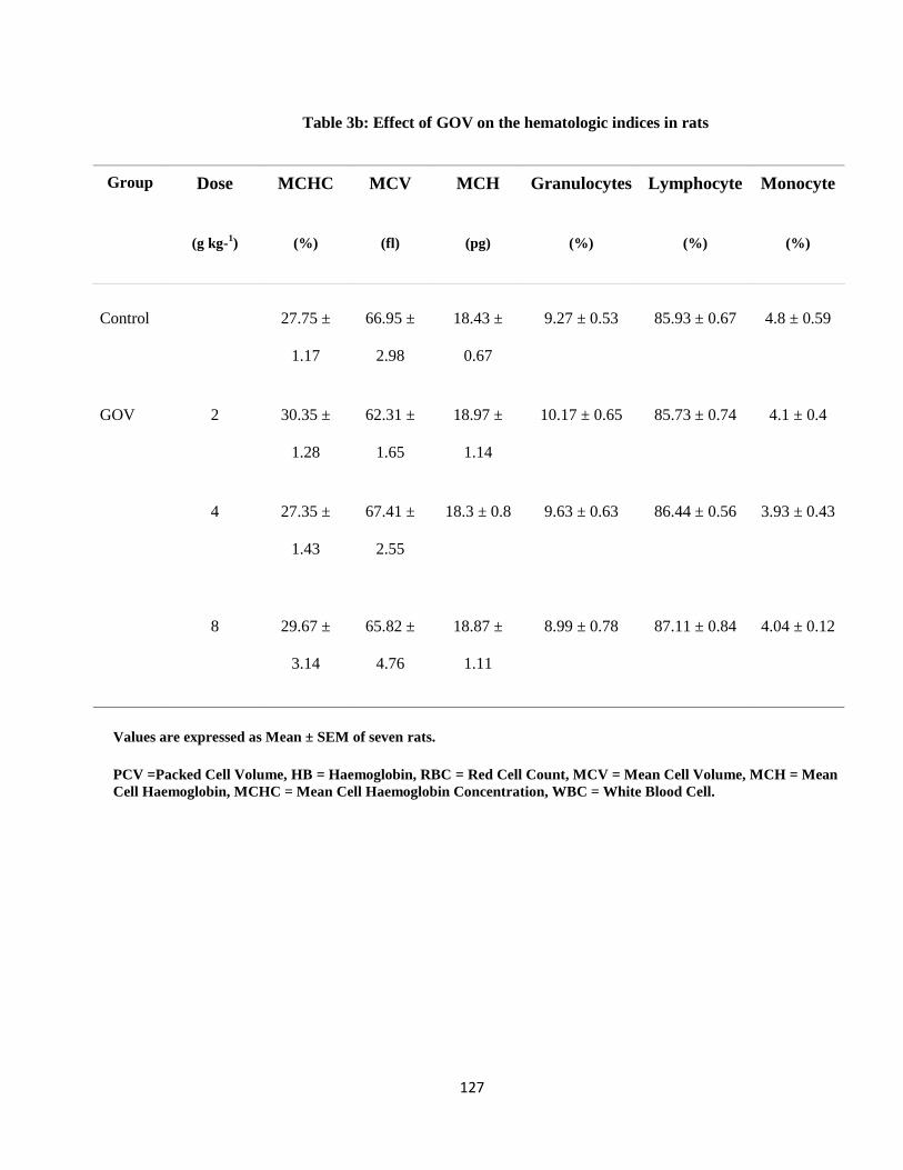
127
Table 3b: Effect of GOV on the hematologic indices in rats
Group Dose
(g kg-1)
MCHC
(%)
MCV
(fl)
MCH
(pg)
Granulocytes
(%)
Lymphocyte
(%)
Monocyte
(%)
Control 27.75 ±
1.17
66.95 ±
2.98
18.43 ±
0.67
9.27 ± 0.53 85.93 ± 0.67 4.8 ± 0.59
GOV 2 30.35 ±
1.28
62.31 ±
1.65
18.97 ±
1.14
10.17 ± 0.65 85.73 ± 0.74 4.1 ± 0.4
4 27.35 ±
1.43
67.41 ±
2.55
18.3 ± 0.8 9.63 ± 0.63 86.44 ± 0.56 3.93 ± 0.43
8 29.67 ±
3.14
65.82 ±
4.76
18.87 ±
1.11
8.99 ± 0.78 87.11 ± 0.84 4.04 ± 0.12
Values are expressed as Mean ± SEM of seven rats.
PCV =Packed Cell Volume, HB = Haemoglobin, RBC = Red Cell Count, MCV = Mean Cell Volume, MCH = Mean
Cell Haemoglobin, MCHC = Mean Cell Haemoglobin Concentration, WBC = White Blood Cell.
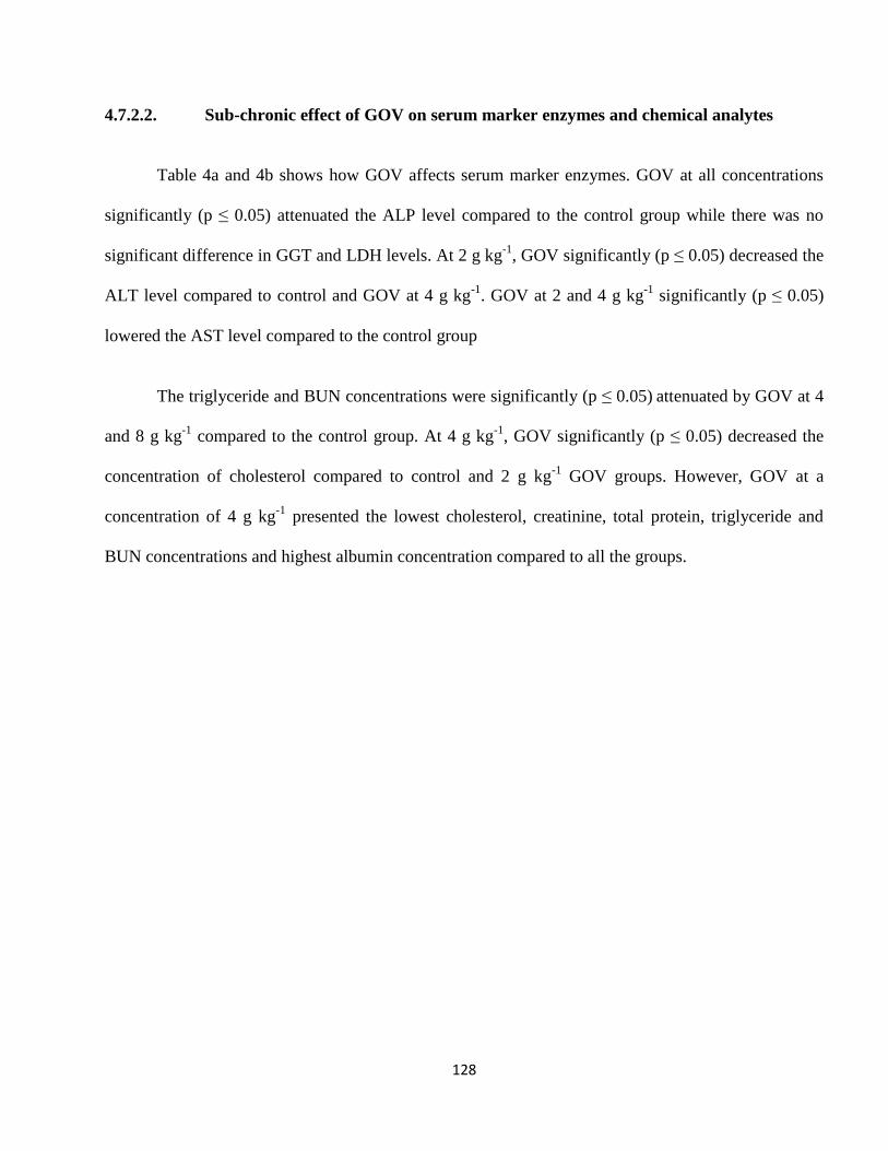
128
4.7.2.2. Sub-chronic effect of GOV on serum marker enzymes and chemical analytes
Table 4a and 4b shows how GOV affects serum marker enzymes. GOV at all concentrations
significantly (p ≤ 0.05) attenuated the ALP level compared to the control group while there was no
significant difference in GGT and LDH levels. At 2 g kg-1
, GOV significantly (p ≤ 0.05) decreased the
ALT level compared to control and GOV at 4 g kg-1
. GOV at 2 and 4 g kg-1
significantly (p ≤ 0.05)
lowered the AST level compared to the control group
The triglyceride and BUN concentrations were significantly (p ≤ 0.05) attenuated by GOV at 4
and 8 g kg-1
compared to the control group. At 4 g kg-1
, GOV significantly (p ≤ 0.05) decreased the
concentration of cholesterol compared to control and 2 g kg-1
GOV groups. However, GOV at a
concentration of 4 g kg-1
presented the lowest cholesterol, creatinine, total protein, triglyceride and
BUN concentrations and highest albumin concentration compared to all the groups.
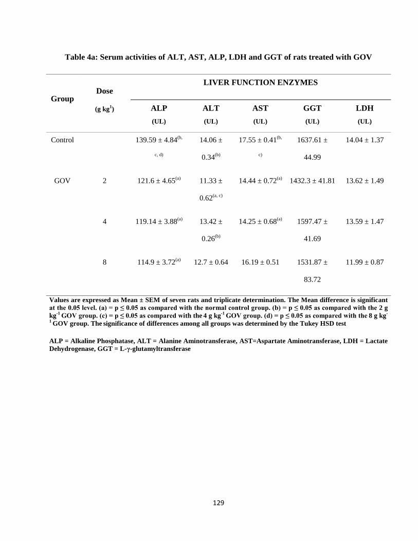
129
Table 4a: Serum activities of ALT, AST, ALP, LDH and GGT of rats treated with GOV
Group Dose
(g kg1)
LIVER FUNCTION ENZYMES
ALP
(UL)
ALT
(UL)
AST
(UL)
GGT
(UL)
LDH
(UL)
Control 139.59 ± 4.84(b,
c, d)
14.06 ±
0.34(b)
17.55 ± 0.41(b,
c)
1637.61 ±
44.99
14.04 ± 1.37
GOV 2 121.6 ± 4.65(a) 11.33 ±
0.62(a, c)
14.44 ± 0.72(a) 1432.3 ± 41.81 13.62 ± 1.49
4 119.14 ± 3.88(a) 13.42 ±
0.26(b)
14.25 ± 0.68(a) 1597.47 ±
41.69
13.59 ± 1.47
8 114.9 ± 3.72(a) 12.7 ± 0.64 16.19 ± 0.51 1531.87 ±
83.72
11.99 ± 0.87
Values are expressed as Mean ± SEM of seven rats and triplicate determination. The Mean difference is significant
at the 0.05 level. (a) = p ≤ 0.05 as compared with the normal control group. (b) = p ≤ 0.05 as compared with the 2 g
kg-1
GOV group. (c) = p ≤ 0.05 as compared with the 4 g kg
-1 GOV group. (d) = p ≤ 0.05 as compared with the
8 g kg
-
1 GOV group. The
significance of differences among all groups was determined by the Tukey HSD test
ALP = Alkaline Phosphatase, ALT = Alanine Aminotransferase, AST=Aspartate Aminotransferase, LDH = Lactate
Dehydrogenase, GGT = L-γ-glutamyltransferase
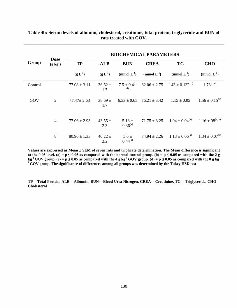
130
Table 4b: Serum levels of albumin, cholesterol, creatinine, total protein, triglyceride and BUN of
rats treated with GOV.
Group Dose (g kg
1)
BIOCHEMICAL PARAMETERS
TP
(g L-1
)
ALB
(g L-1
)
BUN
(mmol L-1
)
CREA
(mmol L-1
)
TG
(mmol L-1
)
CHO
(mmol L-1
)
Control 77.08 ± 3.11 36.62 ±
1.7 7.5 ± 0.4
(c,
d) 82.06 ± 2.75 1.43 ± 0.13
(c, d) 1.73(c, d)
GOV 2 77.47± 2.63 38.69 ±
1.7 6.53 ± 0.65 76.21 ± 3.42 1.15 ± 0.05 1.56 ± 0.15
(c)
4 77.06 ± 2.93 43.55 ±
2.3 5.18 ±
0.36(a)
71.75 ± 3.25 1.04 ± 0.04(a) 1.16 ±.08
(a, b)
8 80.96 ± 1.33 40.22 ±
2.2 5.6 ±
0.44(a)
74.94 ± 2.26 1.13 ± 0.06(a) 1.34 ± 0.07
(a)
Values are expressed as Mean ± SEM of seven rats and triplicate determination. The Mean difference is significant
at the 0.05 level. (a) = p ≤ 0.05 as compared with the normal control group. (b) = p ≤ 0.05 as compared with the 2 g
kg-1
GOV group. (c) = p ≤ 0.05 as compared with the 4 g kg
-1 GOV group. (d) = p ≤ 0.05 as compared with the
8 g kg
-
1 GOV group. The
significance of differences among all groups was determined by the Tukey HSD test
TP = Total Protein, ALB = Albumin, BUN = Blood Urea Nitrogen, CREA = Creatinine, TG = Triglyceride, CHO =
Cholesterol
131
4.7.2.3. Sub-chronic Effect on Serum Antioxidant Enzymes
The effect of GOV on serum antioxidant enzymes in rats are shown in table 5 below. GOV
increased the CAT and decreased the MDA level compared to control. At the 3 doses, GOV exhibited
similar SOD levels. At 2 g kg-1
, the total protein concentration was higher than control while the GST
levels were almost equal. The GSH levels of GOV at 4 and 8 g kg-1
are high compared to control.
There was a significant difference in GSH level of GOV at 2 g kg-1
compared to control and GOV at 4
and 8 g kg-1
.
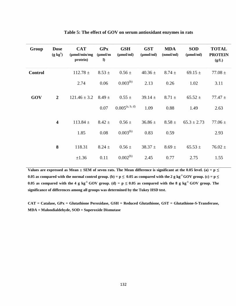
132
Table 5: The effect of GOV on serum antioxidant enzymes in rats
Group Dose
(g kg1)
CAT
(µmol/min/mg
protein)
GPx
(µmol/m
l)
GSH
(µmol/ml)
GST
(µmol/ml)
MDA
(nmol/ml)
SOD
(µmol/ml)
TOTAL
PROTEIN
(g/L)
Control 112.78 ±
2.74
8.53 ±
0.06
0.56 ±
0.003(b)
40.36 ±
2.13
8.74 ±
0.26
69.15 ±
1.02
77.08 ±
3.11
GOV 2 121.46 ± 3.2 8.49 ±
0.07
0.55 ±
0.005(a, b, d)
39.14 ±
1.09
8.71 ±
0.88
65.52 ±
1.49
77.47 ±
2.63
4 113.84 ±
1.85
8.42 ±
0.08
0.56 ±
0.003(b)
36.86 ±
0.83
8.58 ±
0.59
65.3 ± 2.73 77.06 ±
2.93
8 118.31
±1.36
8.24 ±
0.11
0.56 ±
0.002(b)
38.37 ±
2.45
8.69 ±
0.77
65.53 ±
2.75
76.02 ±
1.55
Values are expressed as Mean ± SEM of seven rats. The Mean difference is significant at the 0.05 level. (a) = p ≤
0.05 as compared with the normal control group. (b) = p ≤ 0.05 as compared with the 2 g kg
-1 GOV group. (c) = p ≤
0.05 as compared with the 4 g kg
-1 GOV group. (d) = p ≤ 0.05 as compared with the
8 g kg
-1 GOV group. The
significance of differences among all groups was determined by the Tukey HSD test.
CAT = Catalase, GPx = Glutathione Peroxidase, GSH = Reduced Glutathione, GST = Glutathione-S-Transferase,
MDA = Malondialdehyde, SOD = Superoxide Dismutase
133
4.7.2.4. The Histopathology of Normal Liver of Rats Treated with GOV
Plates 1- 2 shows that the liver architecture of rats treated with GOV at different doses and
control were characterized by polyhedral shaped hepatocytes and granulated cytoplasm. This indicates
that GOV is not toxic.
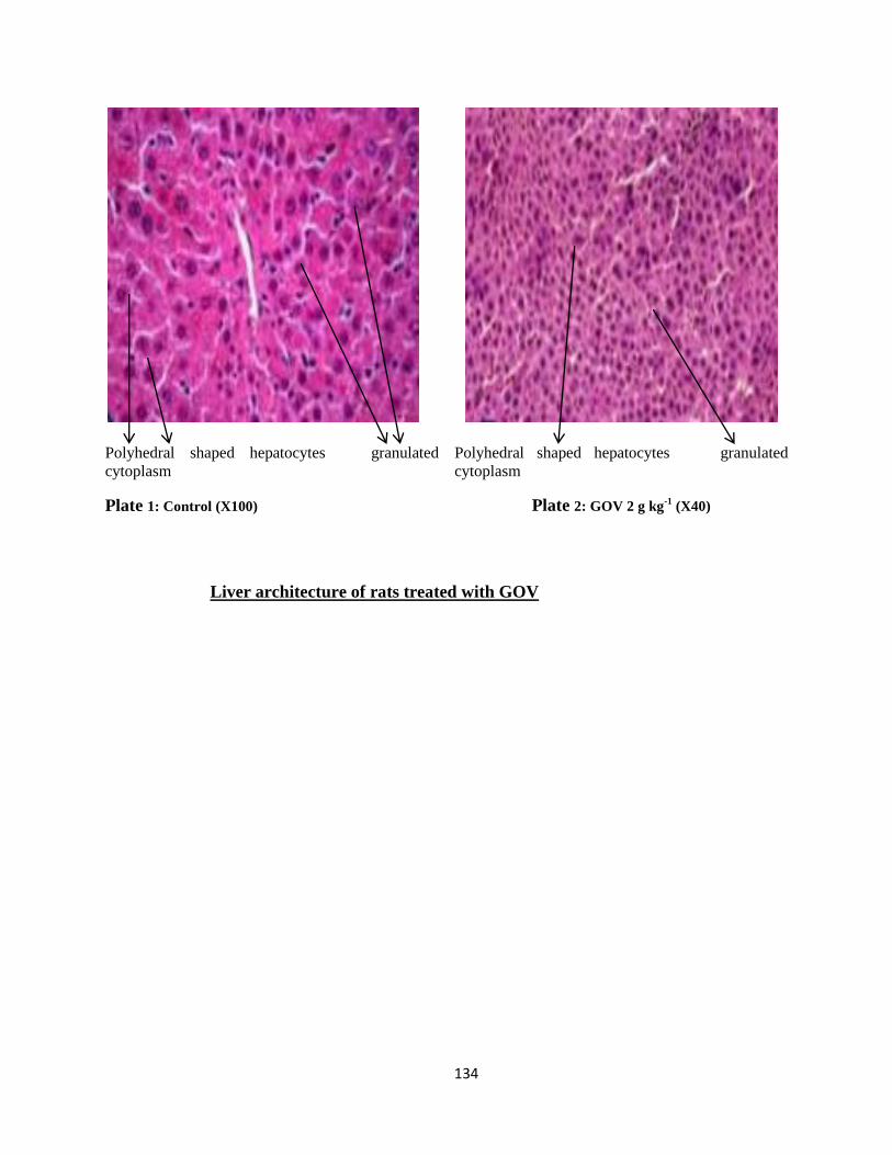
134
Polyhedral shaped hepatocytes granulated
cytoplasm
Plate 1: Control (X100)
Polyhedral shaped hepatocytes granulated
cytoplasm
Plate 2: GOV 2 g kg-1
(X40)
Liver architecture of rats treated with GOV
135
4.8. Preliminary Pharmacological Assays
4.8.1. Antinociceptive Activities of GOV
The experimental results of antinociceptive activities of the GOV are depicted in Tables 6 - 8.
The number of abdominal constrictions resulting from injection of mice with 100 mg kg -1
of 0.6 %
acetic acid intraperitoneally presented 28.4 ± 3.4 writhes which is 68.9 % in 15 minutes while
treatment with GOV at doses of 2 – 8 g kg -1
significantly (P ≤ 0.05) and dose dependently reduced the
number of writhes from 34.6 ± 3.2 to 30.4 ± 2.0 (Table 6) in 15 minutes. In control mice however, the
number of abdominal constrictions was 91 ± 12.00.
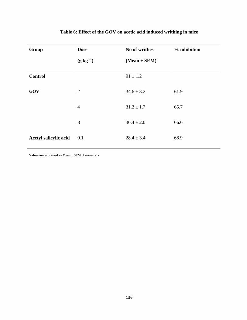
136
Table 6: Effect of the GOV on acetic acid induced writhing in mice
Group Dose
(g kg -1
)
No of writhes
(Mean ± SEM)
% inhibition
Control 91 ± 1.2
GOV 2 34.6 ± 3.2 61.9
4 31.2 ± 1.7 65.7
8 30.4 ± 2.0 66.6
Acetyl salicylic acid 0.1 28.4 ± 3.4 68.9
Values are expressed as Mean ± SEM of seven rats.
137
Table 7 indicates that in the formalin induced pain assay, the highest dose of the GOV
exhibited a significant inhibition of 46 % (42 ± 5.4) while acetylsalicylic acid produced a significant
pain inhibition of 49 % (39.6 ± 2.9).
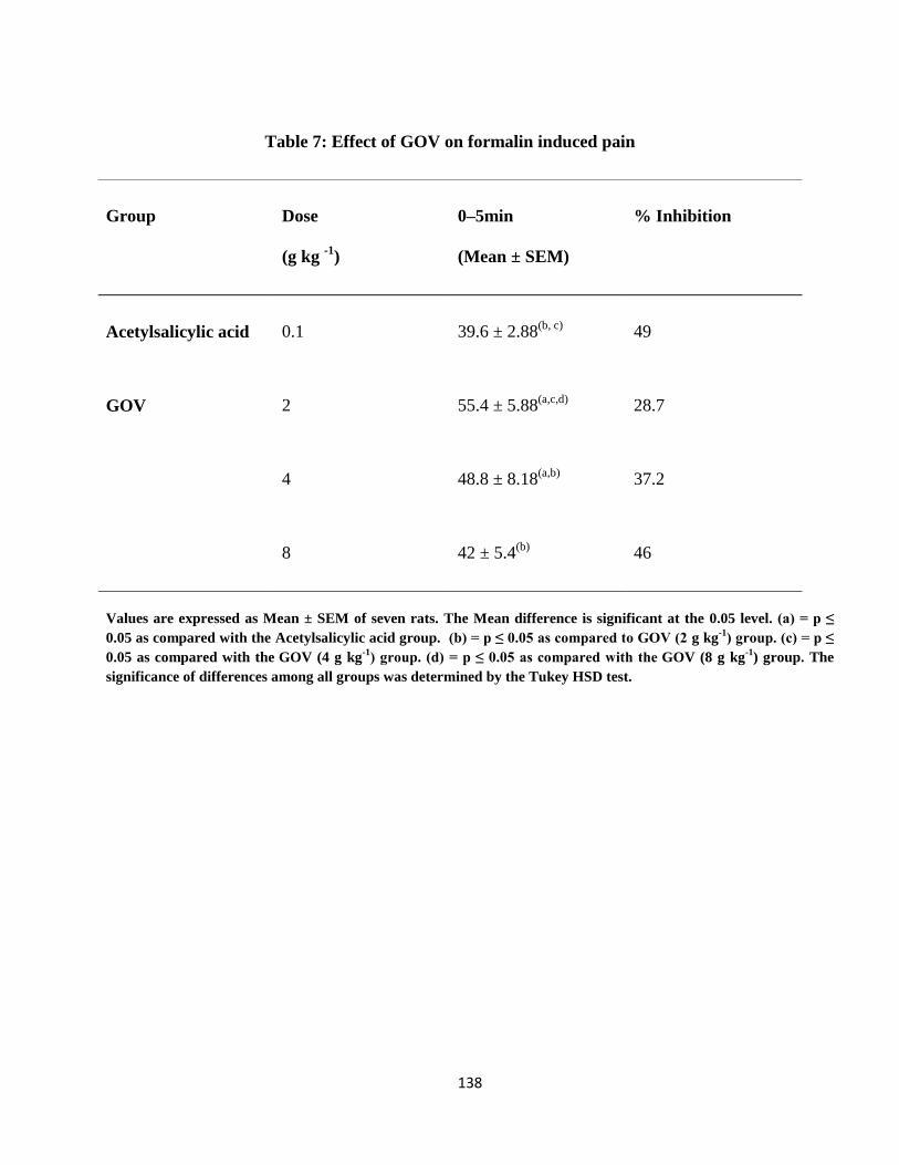
138
Table 7: Effect of GOV on formalin induced pain
Group Dose
(g kg -1
)
0–5min
(Mean ± SEM)
% Inhibition
Acetylsalicylic acid 0.1 39.6 ± 2.88(b, c)
49
GOV 2 55.4 ± 5.88(a,c,d)
28.7
4 48.8 ± 8.18(a,b)
37.2
8 42 ± 5.4(b)
46
Values are expressed as Mean ± SEM of seven rats. The Mean difference is significant at the 0.05 level. (a) = p ≤
0.05 as compared with the Acetylsalicylic acid group. (b) = p ≤ 0.05 as compared to GOV (2 g kg-1
) group. (c) = p ≤
0.05 as compared with the GOV (4 g kg
-1) group. (d) = p ≤ 0.05 as compared with the
GOV (8 g kg
-1) group. The
significance of differences among all groups was determined by the Tukey HSD test.

139
Morphine, a potent opioid agonist inhibited paw licking (180.8 %) inhibition, compared to
GOV at 8 g kg-1
(138.5 %) (Table 8) in the hot plate assay.
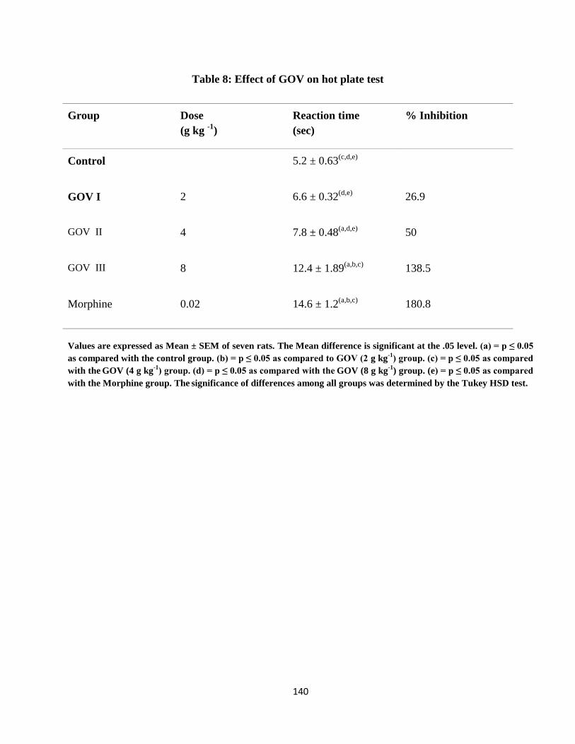
140
Table 8: Effect of GOV on hot plate test
Group Dose
(g kg -1
)
Reaction time
(sec)
% Inhibition
Control 5.2 ± 0.63(c,d,e)
GOV I 2 6.6 ± 0.32(d,e)
26.9
GOV II 4 7.8 ± 0.48(a,d,e)
50
GOV III 8 12.4 ± 1.89(a,b,c)
138.5
Morphine 0.02 14.6 ± 1.2(a,b,c)
180.8
Values are expressed as Mean ± SEM of seven rats. The Mean difference is significant at the .05 level. (a) = p ≤ 0.05
as compared with the control group. (b) = p ≤ 0.05 as compared to GOV (2 g kg-1
) group. (c) = p ≤ 0.05 as compared
with the GOV (4 g kg
-1) group. (d) = p ≤ 0.05 as compared with the
GOV (8 g kg
-1) group. (e) = p ≤ 0.05 as compared
with the Morphine group. The significance of differences among all groups was determined by the Tukey HSD test.
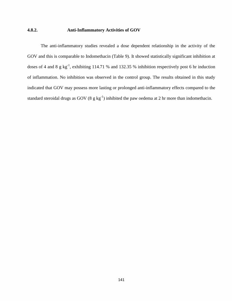
141
4.8.2. Anti-Inflammatory Activities of GOV
The anti-inflammatory studies revealed a dose dependent relationship in the activity of the
GOV and this is comparable to Indomethacin (Table 9). It showed statistically significant inhibition at
doses of 4 and 8 g kg-1
, exhibiting 114.71 % and 132.35 % inhibition respectively post 6 hr induction
of inflammation. No inhibition was observed in the control group. The results obtained in this study
indicated that GOV may possess more lasting or prolonged anti-inflammatory effects compared to the
standard steroidal drugs as GOV (8 g kg-1
) inhibited the paw oedema at 2 hr more than indomethacin.
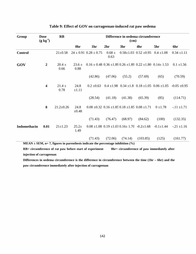
142
Table 9: Effect of GOV on carrageenan-induced rat paw oedema
Group Dose (g kg
-1)
RB
0hr
Difference in oedema circumference
(cm)
1hr 2hr 3hr 4hr 5hr 6hr
Control 21±0.58 24 ± 0.91 0.28 ± 0.75 0.68 ±
0.63 0.58±1.03 0.52 ±0.95 0.4 ±1.08 0.34 ±1.11
GOV 2 20.4 ±
0.66 23.6 ±
0.88 0.16 ± 0.48 0.36 ±1.89 0.26 ±1.89 0.22 ±1.80 0.14± 1.53 0.1 ±1.56
(42.86) (47.06) (55.2) (57.69) (65) (70.59)
4 21.4 ±
0.78 24.8
±1.11 0.2 ±0.63 0.4 ±1.98 0.34 ±1.8 0.18 ±1.05 0.06 ±1.05 -0.05 ±0.95
(28.54) (41.18) (41.38) (65.39) (85) (114.71)
8 21.2±0.26 24.8
±0.48 0.08 ±0.32 0.16 ±1.85 0.18 ±1.85 0.08 ±1.71 0 ±1.78 -.11 ±1.71
(71.43) (76.47) (68.97) (84.62) (100) (132.35)
Indomethacin 0.01 21±1.23 25.2±
1.49 0.08 ±1.08 0.19 ±1.03 0.16± 1.70 -0.2±1.68 -0.1±1.44 -.21 ±1.16
(71.43) (72.06) (74.14) (103.85) (125) (161.77)
MEAN ± SEM, n= 7, figures in parenthesis indicate the percentage inhibition (%)
RB= circumference of rat paw before start of experiment 0hr= circumference of paw immediately after
injection of carrageenan
Differences in oedema circumference is the difference in circumference between the time (1hr – 6hr) and the
paw circumference immediately after injection of carrageenan
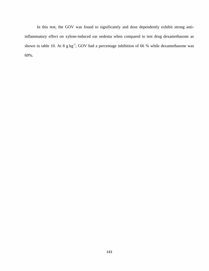
143
In this test, the GOV was found to significantly and dose dependently exhibit strong anti-
inflammatory effect on xylene-induced ear oedema when compared to test drug dexamethasone as
shown in table 10. At 8 g kg
-1, GOV had a percentage inhibition of 66 % while dexamethasone was
69%.
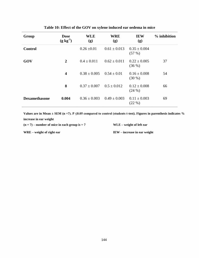
144
Table 10: Effect of the GOV on xylene induced ear oedema in mice
Group Dose
(g kg-1
)
WLE
(g)
WRE
(g)
IEW
(g)
% inhibition
Control 0.26 ±0.01 0.61 ± 0.013 0.35 ± 0.004
(57 %)
GOV 2 0.4 ± 0.011 0.62 ± 0.011 0.22 ± 0.005
(36 %)
37
4 0.38 ± 0.005 0.54 ± 0.01 0.16 ± 0.008
(30 %)
54
8 0.37 ± 0.007 0.5 ± 0.012 0.12 ± 0.008
(24 %)
66
Dexamethasone 0.004 0.36 ± 0.003 0.49 ± 0.003 0.11 ± 0.003
(22 %)
69
Values are in Mean ± SEM (n =7). P ≤0.05 compared to control (students t-test). Figures in parenthesis indicates %
increase in ear weight
(n = 7) – number of mice in each group is = 7 WLE – weight of left ear
WRE – weight of right ear IEW – increase in ear weight
145
4.9. Alcohol Induced Hepatotoxicity
4.9.1. Effect of GOV on the Haematologic Indices on Alcohol Induced Hepatotoxicity
on Rats.
The administration of GOV dose dependently caused a significant (p≤ 0.05) increase in PCV,
RBC, Hb, WBC, platelet, MCHC, granulocytes and lymphocytes and decrease in MCV and
monocytes compared to the ethanol induced toxin control group (tables 11a and 11b) resulting in GOV
associated protection of the haematopoietic system. There was an increase in levels of Hb, platelet
count and MCHC and granulocytes on administration of GOV at 2 g kg-1
compared to Liv 52 group,
all the other groups and control and silymarin groups respectively. However a decrease in MCV was
observed compared to control and silymarin group. There was an increase in PCV at the
administration of GOV at 4 g kg-1
compared to Liv 52 and silymarin groups and it showed an increase
in RBC when compared to silymarin treated rats but was almost equal to Liv 52 treated rats. The
numbers of monocytes observed in the 4 g kg-1
group were lowered compared to control and Liv 52
groups and it exhibited the lowest and highest value of MCH and lymphocytes respectively compared
to all the groups.
On administration of GOV (8 g kg-1
), the PCV and WBC levels were elevated compared to Liv
52 and silymarin also, its RBC was high compared to silymarin and almost equal to Liv 52 treated rats.
The Hb and platelet counts of GOV (8 g kg-1
) were elevated compared to Liv 52 though it attenuated
the MCH level compared to control and silymarin. It boosted and abated the lymphocyte and
granulocytes levels respectively compared to control and Liv 52 experimental groups. There was a
significant increase in the PCV and WBC values at administration of GOV at a dose of 8 g kg-1
,
compared to 2 g kg-1
.
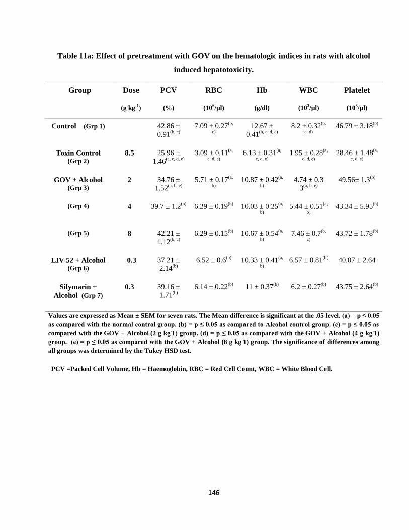
146
Table 11a: Effect of pretreatment with GOV on the hematologic indices in rats with alcohol
induced hepatotoxicity.
Group Dose
(g kg-1
)
PCV
(%)
RBC
(106/µl)
Hb
(g/dl)
WBC
(103/µl)
Platelet
(103/µl)
Control (Grp 1) 42.86 ±
0.91(b, c)
7.09 ± 0.27(b,
c)
12.67 ±
0.41(b, c, d, e)
8.2 ± 0.32(b,
c, d)
46.79 ± 3.18(b)
Toxin Control (Grp 2)
8.5 25.96 ±
1.46(a, c, d, e)
3.09 ± 0.11(a,
c, d, e)
6.13 ± 0.31(a,
c, d, e)
1.95 ± 0.28(a,
c, d, e)
28.46 ± 1.48(a,
c, d, e)
GOV + Alcohol (Grp 3)
2 34.76 ±
1.52(a, b, e)
5.71 ± 0.17(a,
b)
10.87 ± 0.42(a,
b)
4.74 ± 0.3
3(a, b, e)
49.56± 1.3(b)
(Grp 4) 4 39.7 ± 1.2(b)
6.29 ± 0.19(b)
10.03 ± 0.25(a,
b)
5.44 ± 0.51(a,
b)
43.34 ± 5.95(b)
(Grp 5) 8 42.21 ±
1.12(b, c)
6.29 ± 0.15(b)
10.67 ± 0.54(a,
b)
7.46 ± 0.7(b,
c)
43.72 ± 1.78(b)
LIV 52 + Alcohol (Grp 6)
0.3 37.21 ±
2.14(b)
6.52 ± 0.6(b)
10.33 ± 0.41(a,
b)
6.57 ± 0.81(b)
40.07 ± 2.64
Silymarin +
Alcohol (Grp 7)
0.3 39.16 ±
1.71(b)
6.14 ± 0.22(b)
11 ± 0.37(b)
6.2 ± 0.27(b)
43.75 ± 2.64(b)
Values are expressed as Mean ± SEM for seven rats. The Mean difference is significant at the .05 level. (a) = p ≤ 0.05
as compared with the normal control group. (b) = p ≤ 0.05 as compared to Alcohol control group. (c) = p ≤ 0.05 as
compared with the GOV + Alcohol (2 g kg
-1) group. (d) = p ≤ 0.05 as compared with the
GOV + Alcohol (4 g kg
-1)
group. (e) = p ≤ 0.05 as compared with the GOV + Alcohol (8 g kg
-1) group. The
significance of differences among
all groups was determined by the Tukey HSD test.
PCV =Packed Cell Volume, Hb = Haemoglobin, RBC = Red Cell Count, WBC = White Blood Cell.
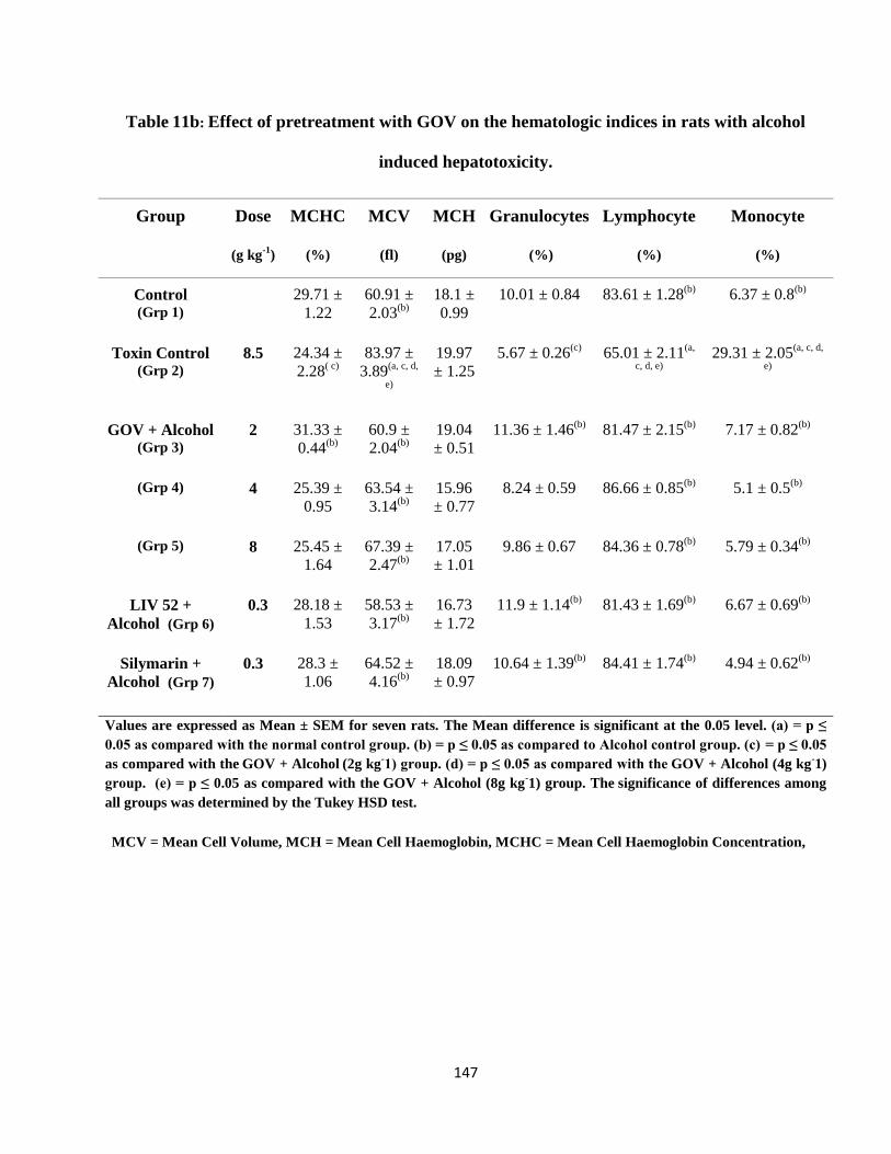
147
Table 11b: Effect of pretreatment with GOV on the hematologic indices in rats with alcohol
induced hepatotoxicity.
Group Dose
(g kg-1
)
MCHC
(%)
MCV
(fl)
MCH
(pg)
Granulocytes
(%)
Lymphocyte
(%)
Monocyte
(%)
Control (Grp 1)
29.71 ±
1.22
60.91 ±
2.03(b)
18.1 ±
0.99
10.01 ± 0.84 83.61 ± 1.28(b)
6.37 ± 0.8(b)
Toxin Control (Grp 2)
8.5 24.34 ±
2.28( c)
83.97 ±
3.89(a, c, d,
e)
19.97
± 1.25
5.67 ± 0.26(c)
65.01 ± 2.11(a,
c, d, e)
29.31 ± 2.05(a, c, d,
e)
GOV + Alcohol (Grp 3)
2 31.33 ±
0.44(b)
60.9 ±
2.04(b)
19.04
± 0.51
11.36 ± 1.46(b)
81.47 ± 2.15(b)
7.17 ± 0.82(b)
(Grp 4) 4 25.39 ±
0.95
63.54 ±
3.14(b)
15.96
± 0.77
8.24 ± 0.59 86.66 ± 0.85(b)
5.1 ± 0.5(b)
(Grp 5) 8 25.45 ±
1.64
67.39 ±
2.47(b)
17.05
± 1.01
9.86 ± 0.67 84.36 ± 0.78(b)
5.79 ± 0.34(b)
LIV 52 +
Alcohol (Grp 6)
0.3 28.18 ±
1.53
58.53 ±
3.17(b)
16.73
± 1.72
11.9 ± 1.14(b)
81.43 ± 1.69(b)
6.67 ± 0.69(b)
Silymarin +
Alcohol (Grp 7)
0.3 28.3 ±
1.06
64.52 ±
4.16(b)
18.09
± 0.97
10.64 ± 1.39(b)
84.41 ± 1.74(b)
4.94 ± 0.62(b)
Values are expressed as Mean ± SEM for seven rats. The Mean difference is significant at the 0.05 level. (a) = p ≤
0.05 as compared with the normal control group. (b) = p ≤ 0.05 as compared to Alcohol control group. (c) = p ≤ 0.05
as compared with the GOV + Alcohol (2g kg
-1) group. (d) = p ≤ 0.05 as compared with the
GOV + Alcohol (4g kg
-1)
group. (e) = p ≤ 0.05 as compared with the GOV + Alcohol (8g kg
-1) group. The
significance of differences among
all groups was determined by the Tukey HSD test.
MCV = Mean Cell Volume, MCH = Mean Cell Haemoglobin, MCHC = Mean Cell Haemoglobin Concentration,
148
4.9.2. Effect of GOV on Serum Hepatic Enzymes and Chemical Analytes on Alcohol intoxicated
rats
The effects of the triherbal formulation on the serum hepatic enzymes and chemical analytes
on alcohol induced toxicity in all the groups are given in tables 12a and 12b. There was an increase in
the levels of serum ALP, AST, GGT, ALT, and LDH in the toxin control group compared to the
normal control group. Pretreatment of rats with GOV, dose dependently decreased the levels of serum
hepatic enzymes activities significantly (p≤ 0.05) in experimental rats as compared to the toxin control
group. In the 8 g kg-1
group, ALP activity was lower than that of silymarin and was significantly (p≤
0.05) increased when compared to Liv 52. In 8 g kg-1
group, LDH and ALP activities were decreased
when compared to the rats that received silymarin, while its ALT activities were lower than that of Liv
52 group. In the 2 g kg-1
group, GOV lowered the AST activity compared to Liv 52 and Silymarin.
There was a 76 %, 67.39 %, 66.44 %, 52.5 % and 63.67 % increase in ALP, ALT, AST, GGT and
LDH activities respectively in group 2 as compared to group 1 with a 36.8 % decrease in total protein
level.
At 4 g kg-1
, GOV attenuated the concentrations of cholesterol and creatinine more than Liv 52
and silymarin, lowered the triglyceride concentration more than that of Liv 52 and had almost the
same BUN concentration as Liv 52. The protein concentration of GOV at 8 g kg-1
was almost equal to
that of Liv 52 and silymarin. Silymarin significantly (p≤ 0.05) increased the albumin concentration
compared to GOV at 2 and 4 g kg-1
. The levels of serum albumin and total protein concentrations in
alcohol induced toxin control group were significantly (p≤ 0.05) decreased while increase in
cholesterol, creatinine, triglyceride and BUN concentrations were observed compared to the three
doses of GOV.
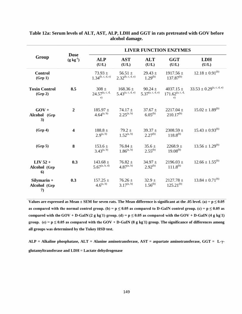
149
Table 12a: Serum levels of ALT, AST, ALP, LDH and GGT in rats pretreated with GOV before
alcohol damage.
Group Dose
(g kg-1
)
LIVER FUNCTION ENZYMES
ALP
(U/L)
AST
(U/L)
ALT
(U/L)
GGT
(U/L)
LDH
(U/L)
Control (Grp 1)
73.93 ±
1.34(b, c, d, e)
56.51 ±
2.32(b, c, d, e)
29.43 ±
1.29(b)
1917.56 ±
137.87(b)
12.18 ± 0.91(b)
Toxin Control (Grp 2)
8.5 308 ±
24.57(a, c, d,
e)
168.36 ±
5.43(a, c, d, e)
90.24 ±
5.37(a, c, d, e)
4037.15 ±
171.62(a, c, d,
e)
33.53 ± 0.29(a, c, d, e)
GOV +
Alcohol (Grp
3)
2 185.97 ±
4.64(a, b)
74.17 ±
2.25(a, b)
37.67 ±
6.05(b)
2217.04 ±
210.17(b)
15.02 ± 1.89(b)
(Grp 4) 4 188.8 ±
2.9(a, b)
79.2 ±
1.52(a, b)
39.37 ±
2.27(b)
2308.59 ±
118.8(b)
15.43 ± 0.93(b)
(Grp 5) 8 153.6 ±
3.43(a, b)
76.84 ±
1.86(a, b)
35.6 ±
2.55(b)
2268.9 ±
19.08(b)
13.56 ± 1.29(b)
LIV 52 +
Alcohol (Grp
6)
0.3 143.68 ±
5.67(a, b, d)
76.82 ±
4.87(a, b)
34.97 ±
2.92(b)
2196.03 ±
111.8(b)
12.66 ± 1.55(b)
Silymarin +
Alcohol (Grp
7)
0.3 157.25 ±
4.6(a, b)
76.26 ±
3.17(a, b)
32.9 ±
1.56(b)
2127.78 ±
125.21(b)
13.84 ± 0.71(b)
Values are expressed as Mean ± SEM for seven rats. The Mean difference is significant at the .05 level. (a) = p ≤ 0.05
as compared with the normal control group. (b) = p ≤ 0.05 as compared to D-GaIN control group. (c) = p ≤ 0.05 as
compared with the GOV + D-GaIN (2 g kg
-1) group. (d) = p ≤ 0.05 as compared with the
GOV + D-GaIN (4 g kg
-1)
group. (e) = p ≤ 0.05 as compared with the GOV + D-GaIN (8 g kg
-1) group. The
significance of differences among
all groups was determined by the Tukey HSD test.
ALP = Alkaline phosphatase, ALT = Alanine aminotransferase, AST = aspartate aminotransferase, GGT = L-γ-
glutamyltransferase and LDH = Lactate dehydrogenase
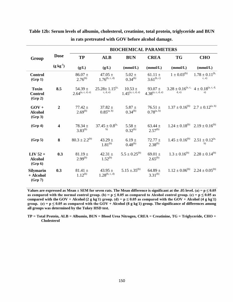
150
Table 12b: Serum levels of albumin, cholesterol, creatinine, total protein, triglyceride and BUN
in rats pretreated with GOV before alcohol damage.
Group Dose
(g kg-1
)
BIOCHEMICAL PARAMETERS
TP
(g/L)
ALB
(g/L)
BUN
(mmol/L)
CREA
(mmol/L)
TG
(mmol/L)
CHO
(mmol/L)
Control (Grp 1)
86.07 ±
2.76(b)
47.05 ±
1.76(b, c, d)
5.02 ±
0.34(b)
61.11 ±
3.61(b, c)
1 ± 0.03(b)
1.78 ± 0.11(b,
c, e)
Toxin
Control (Grp 2)
8.5 54.39 ±
2.64(a, c, d, e)
25.28± 1.15(a,
c, d, e)
10.53 ±
1.45(a, c, d, e)
93.07 ±
4.38(a, c, d, e)
3.28 ± 0.16(a, c,
d, e)
4 ± 0.18(a, c, d,
e)
GOV +
Alcohol (Grp 3)
2 77.42 ±
2.69(b)
37.82 ±
0.85(a, b)
5.87 ±
0.34(b)
76.51 ±
0.78(a, b)
1.37 ± 0.16(b)
2.7 ± 0.12(a, b)
(Grp 4) 4 78.34 ±
3.83(b)
37.45 ± 0.8(a,
b)
5.58 ±
0.32(b)
63.44 ±
2.57(b)
1.24 ± 0.18(b)
2.19 ± 0.16(b)
(Grp 5) 8 80.3 ± 2.2(b)
43.29 ±
1.81(b)
6.19 ±
0.48(b)
72.77 ±
2.38(b)
1.45 ± 0.16(b)
2.51 ± 0.12(a,
b)
LIV 52 +
Alcohol (Grp 6)
0.3 81.19 ±
2.99(b)
42.31 ±
1.52(b)
5.5 ± 0.25(b)
69.01 ±
2.65(b)
1.3 ± 0.16(b)
2.28 ± 0.14(b)
Silymarin
+ Alcohol (Grp 7)
0.3 81.41 ±
1.12(b)
43.95 ±
1.28(b, c, d)
5.15 ±.35(b)
64.89 ±
3.31(b)
1.12 ± 0.06(b)
2.24 ± 0.05(b)
Values are expressed as Mean ± SEM for seven rats. The Mean difference is significant at the .05 level. (a) = p ≤ 0.05
as compared with the normal control group. (b) = p ≤ 0.05 as compared to Alcohol control group. (c) = p ≤ 0.05 as
compared with the GOV + Alcohol (2 g kg
-1) group. (d) = p ≤ 0.05 as compared with the
GOV + Alcohol (4 g kg
-1)
group. (e) = p ≤ 0.05 as compared with the GOV + Alcohol (8 g kg
-1) group. The
significance of differences among
all groups was determined by the Tukey HSD test.
TP = Total Protein, ALB = Albumin, BUN = Blood Urea Nitrogen, CREA = Creatinine, TG = Triglyceride, CHO =
Cholesterol
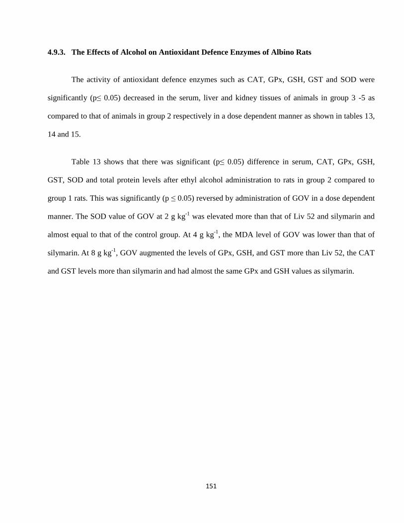
151
4.9.3. The Effects of Alcohol on Antioxidant Defence Enzymes of Albino Rats
The activity of antioxidant defence enzymes such as CAT, GPx, GSH, GST and SOD were
significantly (p≤ 0.05) decreased in the serum, liver and kidney tissues of animals in group 3 -5 as
compared to that of animals in group 2 respectively in a dose dependent manner as shown in tables 13,
14 and 15.
Table 13 shows that there was significant (p≤ 0.05) difference in serum, CAT, GPx, GSH,
GST, SOD and total protein levels after ethyl alcohol administration to rats in group 2 compared to
group 1 rats. This was significantly (p ≤ 0.05) reversed by administration of GOV in a dose dependent
manner. The SOD value of GOV at 2 g kg-1
was elevated more than that of Liv 52 and silymarin and
almost equal to that of the control group. At 4 g kg-1
, the MDA level of GOV was lower than that of
silymarin. At 8 g kg
-1, GOV augmented the levels of GPx, GSH, and GST more than Liv 52, the CAT
and GST levels more than silymarin and had almost the same GPx and GSH values as silymarin.
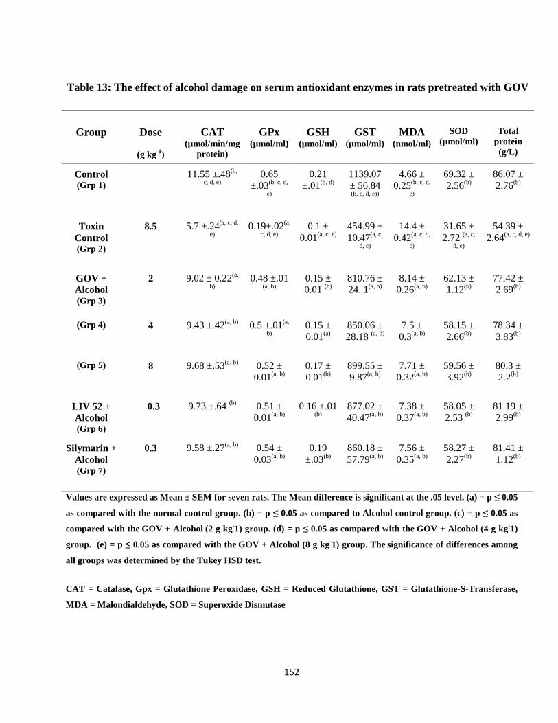
152
Table 13: The effect of alcohol damage on serum antioxidant enzymes in rats pretreated with GOV
Group Dose
(g kg-1
)
CAT
(µmol/min/mg
protein)
GPx
(µmol/ml)
GSH
(µmol/ml)
GST
(µmol/ml)
MDA
(nmol/ml)
SOD
(µmol/ml)
Total
protein
(g/L)
Control (Grp 1)
11.55 ±.48(b,
c, d, e)
0.65
±.03(b, c, d,
e)
0.21
±.01(b, d)
1139.07
± 56.84 (b, c, d, e))
4.66 ±
0.25(b, c, d,
e)
69.32 ±
2.56(b)
86.07 ±
2.76(b)
Toxin
Control (Grp 2)
8.5 5.7 ±.24(a, c, d,
e)
0.19±.02(a,
c, d, e)
0.1 ±
0.01(a, c, e)
454.99 ±
10.47(a, c,
d, e)
14.4 ±
0.42(a, c, d,
e)
31.65 ±
2.72 (a, c,
d, e)
54.39 ±
2.64(a, c, d, e)
GOV +
Alcohol (Grp 3)
2 9.02 ± 0.22(a,
b)
0.48 ±.01 (a, b)
0.15 ±
0.01 (b)
810.76 ±
24. 1(a, b)
8.14 ±
0.26(a, b)
62.13 ±
1.12(b)
77.42 ±
2.69(b)
(Grp 4) 4 9.43 ±.42(a, b)
0.5 ±.01(a,
b)
0.15 ±
0.01(a)
850.06 ±
28.18 (a, b)
7.5 ±
0.3(a, b)
58.15 ±
2.66(b)
78.34 ±
3.83(b)
(Grp 5) 8 9.68 ±.53(a, b)
0.52 ±
0.01(a, b)
0.17 ±
0.01(b)
899.55 ±
9.87(a, b)
7.71 ±
0.32(a, b)
59.56 ±
3.92(b)
80.3 ±
2.2(b)
LIV 52 +
Alcohol (Grp 6)
0.3 9.73 ±.64 (b)
0.51 ±
0.01(a, b)
0.16 ±.01 (b)
877.02 ±
40.47(a, b)
7.38 ±
0.37(a, b)
58.05 ±
2.53 (b)
81.19 ±
2.99(b)
Silymarin +
Alcohol (Grp 7)
0.3 9.58 ±.27(a, b)
0.54 ±
0.03(a, b)
0.19
±.03(b)
860.18 ±
57.79(a, b)
7.56 ±
0.35(a, b)
58.27 ±
2.27(b)
81.41 ±
1.12(b)
Values are expressed as Mean ± SEM for seven rats. The Mean difference is significant at the .05 level. (a) = p ≤ 0.05
as compared with the normal control group. (b) = p ≤ 0.05 as compared to Alcohol control group. (c) = p ≤ 0.05 as
compared with the GOV + Alcohol (2 g kg
-1) group. (d) = p ≤ 0.05 as compared with the
GOV + Alcohol (4 g kg
-1)
group. (e) = p ≤ 0.05 as compared with the GOV + Alcohol (8 g kg
-1) group. The
significance of differences among
all groups was determined by the Tukey HSD test.
CAT = Catalase, Gpx = Glutathione Peroxidase, GSH = Reduced Glutathione, GST = Glutathione-S-Transferase,
MDA = Malondialdehyde, SOD = Superoxide Dismutase
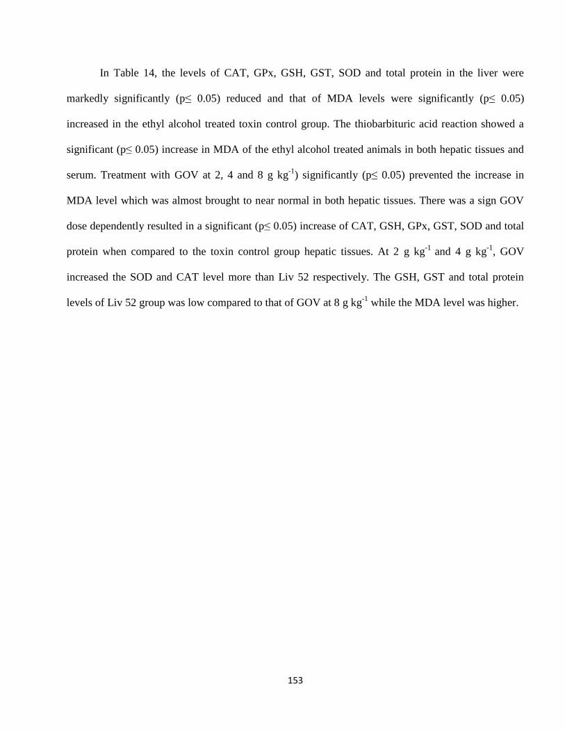
153
In Table 14, the levels of CAT, GPx, GSH, GST, SOD and total protein in the liver were
markedly significantly (p≤ 0.05) reduced and that of MDA levels were significantly (p≤ 0.05)
increased in the ethyl alcohol treated toxin control group. The thiobarbituric acid reaction showed a
significant (p≤ 0.05) increase in MDA of the ethyl alcohol treated animals in both hepatic tissues and
serum. Treatment with GOV at 2, 4 and 8 g kg-1
) significantly (p≤ 0.05) prevented the increase in
MDA level which was almost brought to near normal in both hepatic tissues. There was a sign GOV
dose dependently resulted in a significant (p≤ 0.05) increase of CAT, GSH, GPx, GST, SOD and total
protein when compared to the toxin control group hepatic tissues. At 2 g kg-1
and 4 g kg-1
, GOV
increased the SOD and CAT level more than Liv 52 respectively. The GSH, GST and total protein
levels of Liv 52 group was low compared to that of GOV at 8 g kg-1
while the MDA level was higher.
154
Table 14: The effect of alcohol damage on liver antioxidant enzymes in rats pretreated with
GOV.
Group Dose
(g kg-1
)
CAT
(µmol/min/mg
protein)
GPx
(µmol/ml)
GSH
(µmol/ml)
GST
(µmol/ml)
MDA
(nmol/ml)
SOD
(µmol/ml)
Total
protein
(g/L)
Control (Grp 1)
206.68 ±
5.56(b, c)
8.82 ±
0.24(b, c, d,
e)
26.64 ±
1.13(b, c)
424.58 ±
17.46(b, c, d,
e)
10.77 ±
0.56(b)
115.81 ±
10.46(b, c, d,
e)
84.2 ±
2.24(b, c, d)
Toxin Control (Grp 2)
8.5 55.93 ± 2.83(a,
c, d, e)
3.94 ±
0.18(a, d, e)
10.76 ±
0.8(a, c, d, e)
90.2 ±
5.96(a, c, d,
e)
35.65 ±
2.76(a, c, d,
e)
42.7 ±
2.49(a, c, d,
e)
53.97 ±
1.94(a, d, e)
GOV +
Alcohol (Grp 3)
2 157.16 ±
11.48(a, b)
4.43 ±
0.1(a, e)
18.75 ±
1.34(a, b)
265.38 ±
12.24(a, b)
14.46 ±
0.46(b)
85.49 ±
4.18(a, b)
63.13 ±
1.72(a, e)
(Grp 4) 4 191.27 ±
4.49(b)
5.16 ±
0.43 (a, b, e)
21.07 ±
1.36(b)
214.23 ±
4.24(a, b, e)
15.02 ±
0.89(b)
71.45 ±
4.2(a, b)
67.05 ±
2.23(a, b, e)
(Grp 5) 8 176.63 ±
10.2(b)
6.66 ±
0.28(a, b, c,
d)
24.22 ±
2.57(b)
292.14 ±
34.04(a, b, d)
12.66 ±
1.29(b)
82.52 ±
6.49(a, b)
77.87 ±
3.32(b, c, d)
LIV 52 +
Alcohol (Grp 6)
0.3 185.08 ±
5.38(b)
6.72 ±
0.13(a, b, c,
d)
22.19 ±
1.44(b)
275.33 ±
4.54(a, b)
12.95 ±
0.61(b)
85.37 ±
3.91(a, b)
76.9 ±
1.85(b, c, d)
Silymarin +
Alcohol (Grp 7)
0.3 192.71 ±
12.32(b)
7.03 ± 0.4 (a, b, c, d)
21.01 ±
1.29(b)
293.93 ±
9.31(a, b, d)
12.31 ±
0.44(b)
93.14 ±
4.15(b)
78.27 ±
1.67(b, c, d)
Values are expressed as Mean ± SEM for seven rats. The Mean difference is significant at the .05 level. (a) = p ≤ 0.05
as compared with the normal control group. (b) = p ≤ 0.05 as compared to Alcohol control group. (c) = p ≤ 0.05 as
compared with the GOV + Alcohol (2g kg
-1) group. (d) = p ≤ 0.05 as compared with the
GOV + Alcohol (4g kg
-1)
group. (e) = p ≤ 0.05 as compared with the GOV + Alcohol (8g kg
-1) group. The
significance of differences among
all groups was determined by the Tukey HSD test.
CAT = Catalase, GPx = Glutathione Peroxidase, GSH = Reduced Glutathione, GST = Glutathione-S-Transferase,
MDA = Malondialdehyde, SOD = Superoxide Dismutase
155
Table 15 shows the effect of alcohol induced toxicity on rat kidney. At 2 g kg-1
, GOV exhibited
higher concentration of total protein compared to Liv 52 while the GST was almost equal to that of
Liv 52 and silymarin. The CAT, GPx and SOD levels of GOV at 4 g kg-1
were increased while the
MDA was lowered compared to Liv 52 and silymarin. When compared to Liv 52, the GSH and GPx
activities of GOV at 4 and 8 g kg-1
were increased respectively. The CAT activity of GOV at 4 g kg-1
was significantly (p ≤ 0.05) higher than that of 2 and 8 g kg-1
. GOV significantly reversed the effects
of alcohol on the rats in a dose dependent manner.
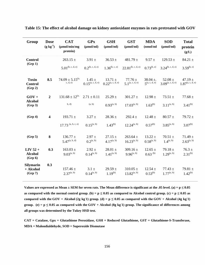
156
Table 15: The effect of alcohol damage on kidney antioxidant enzymes in rats pretreated with GOV
Group Dose
(g kg-1
)
CAT
(µmol/min/mg
protein)
GPx
(µmol/ml)
GSH
(µmol/ml)
GST
(µmol/ml)
MDA
(nmol/ml
SOD
(µmol/ml)
Total
protein
(g/L)
Control (Grp 1)
263.15 ±
5.01(b, c, d, e)
3.91 ±
0.2(b, c, d, e)
36.53 ±
1.36(b, c, e)
481.79 ±
22.81(b, c, d, e)
9.57 ±
0.73(b, e)
129.53 ±
3.24(b, c, d, e)
84.21 ±
3.59(b, e)
Toxin
Control (Grp 2)
8.5 74.09 ± 5.15(a,
c, d, e)
1.45 ±
0.15(a, c, d, e)
13.71 ±
0.22(a, c, d, e)
77.76 ±
5.1(a, c, d, e)
38.04 ±.
5(a, c, d, e)
52.08 ±
3.09(a, c, d, e)
47.19 ±
1.87(a, c, d, e)
GOV +
Alcohol (Grp 3)
2 131.68 ± 12(a,
b, d)
2.71 ± 0.11
(a, b)
25.29 ±
0.93(a, b)
301.27 ±
17.03(a, b)
12.98 ±
1.63(b)
73.51 ±
3.11(a, b)
77.68 ±
3.41(b)
(Grp 4) 4 193.71 ±
17.73 (a, b, c, e)
3.27 ±
0.15(a, b)
28.36 ±
1.43(b)
292.4 ±
12.24(a, b)
12.48 ±
0.57(b)
80.57 ±
3.85(a, b)
79.72 ±
3.07(b)
(Grp 5) 8 136.77 ±
5.47(a, b, d)
2.97 ±
0.2(a, b)
27.15 ±
4.17(a, b)
263.64 ±
16.23(a, b)
13.22 ±
0.58(a, b)
70.51 ±
1.4(a, b)
71.49 ±
2.63(a, b)
LIV 52 +
Alcohol (Grp 6)
0.3 163.03 ±
9.03(a, b)
2.92 ±
0.14(a, b)
28.01 ±
1.41(a, b)
309.16 ±
9.96(a, b)
12.65 ±
0.63 (b)
79.18 ±
1.29(a, b)
76.3 ±
2.31(b)
Silymarin
+ Alcohol (Grp 7)
0.3 157.46 ±
2.37(a, b)
3.1 ±
0.14(a, b)
29.59 ±
1.19(b)
310.05 ±
13.82(a, b)
12.54 ±
0.53(b)
77.43 ±
1.77(a, b)
79.81 ±
1.42(b)
Values are expressed as Mean ± SEM for seven rats. The Mean difference is significant at the .05 level. (a) = p ≤ 0.05
as compared with the normal control group. (b) = p ≤ 0.05 as compared to Alcohol control group. (c) = p ≤ 0.05 as
compared with the GOV + Alcohol (2g kg
-1) group. (d) = p ≤ 0.05 as compared with the
GOV + Alcohol (4g kg
-1)
group. (e) = p ≤ 0.05 as compared with the GOV + Alcohol (8g kg
-1) group. The
significance of differences among
all groups was determined by the Tukey HSD test.
CAT = Catalase, Gpx = Glutathione Peroxidase, GSH = Reduced Glutathione, GST = Glutathione-S-Transferase,
MDA = Malondialdehyde, SOD = Superoxide Dismutase
157
4.9.4. Effect of Alcohol on Caspase Activities
Table 16 shows the fold increase in caspase 2, 3 and 9 activities in the white blood cell of rats
pretreated with GOV before ethanol damage. At 2 g kg-1
, GOV significantly (p≤ 0.05) increased the
caspase 9 activities compared to control, GOV (4 and 8 g kg-1
), Liv 52 and silymarin groups though
there was a significant (p≤ 0.05) decrease compared to the toxin control group.

158
Table 16: Caspase - 2, 3 and 9 activities in the white blood cell of rats treated with ethanol.
Group Dose
(g kg-1)
Caspase 2 (Units/mg of total
protein).
Caspase 3 (Units/mg of total
protein)
Caspase 9 (Units/mg of total
protein)
Control
(Grp 1)
0.5 ± 0.06(b, c, d, e) 0.95 ±.03(b, c, d, e) 1.20 ± .09(b, c, d, e)
Toxin Control (Grp 2)
8.5 2.96 ± 0.31(a, c, d, e) 5.85 ± 0.12(a, c, d, e) 7.3 ± 0.45(a, c, d, e)
(4.92) (5.16) (5.08)
GOV + Alcohol (Grp 3)
2 0.91 ± 0.05(a, b) 1.42 ± 0.03(a, b) 2.05 ± 0.02(a, b, d, e)
(0.81) (0.5) (0.71)
(Grp 4) 4 0.98 ± 0.12(a, b) 1.39 ± 0.03(a, b) 1.69 ± 0.03(a, b, c)
(0.95) (0.46) (0.41)
(Grp 5) 8 0.88 ± 0.04(a, b) 1.34 ± 0.04(a, b) 1.56 ± 0.1(a, b, c)
(0.76) (0.41) (0.3)
LIV 52 + Alcohol (Grp 6)
0.3 0.942 ± 0.05(a, b) 1.38 ± 0.04(a, b) 1.57 ± 0.07(a, b, c)
(0.84) (0.45) (0.31)
Silymarin +
Alcohol (Grp 7)
0.3 0.82± 0.09(b) 1.37 ± 0.03(a, b) 1.48 ± 0.02(b, c)
(0.65) (0.45) (0.23)
Values are expressed as Mean ± SEM for seven rats. The Mean difference is significant at the .05 level. (a) = p ≤ 0.05
as compared with the normal control group. (b) = p ≤ 0.05 as compared to Alcohol control group. (c) = p ≤ 0.05 as
compared with the GOV + Alcohol (2g kg
-1) group. (d) = p ≤ 0.05 as compared with the
GOV + Alcohol (4g kg
-1)
group. (e) = p ≤ 0.05 as compared with the GOV + Alcohol (8g kg
-1) group. The
significance of differences among
all groups was determined by the Tukey HSD test. The figures in parenthesis = fold-increase in caspase activity
compared to the uninduced control.
159
4.9.5. Histopathology of Liver of Rats Intoxicated with Alcohol
The photomicrograph of the liver of animals in the control group showed normal histology.
Pretreatment with GOV at 8 g kg-1
before administration of alcohol showed almost normal liver
architecture while at 4 g kg-1
, it showed almost normal liver with unremarkable central vein and
hepatic lobule, with a focus of inflammatory infiltrate but no necrosis. The toxin control group showed
loss of architecture, fibrosis, micro- and macrovesicular steatosis and fatty infiltration with extensive
necrosis.
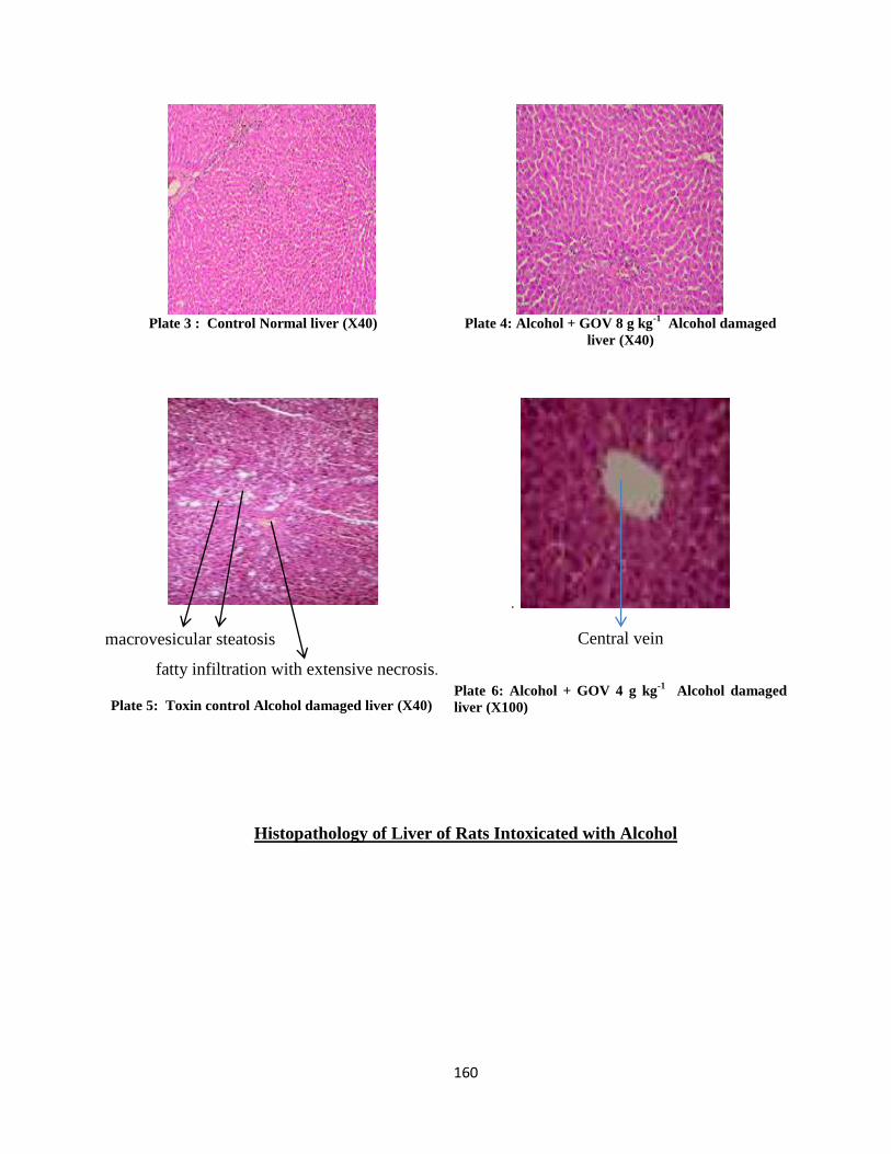
160
Plate 3 : Control Normal liver (X40)
Plate 4: Alcohol + GOV 8 g kg
-1 Alcohol damaged
liver (X40)
macrovesicular steatosis
fatty infiltration with extensive necrosis.
Plate 5: Toxin control Alcohol damaged liver (X40)
.
Central vein
Plate 6: Alcohol + GOV 4 g kg-1
Alcohol damaged
liver (X100)
Histopathology of Liver of Rats Intoxicated with Alcohol
161
4.10. D- galactosamine (D-GaIN) Induced Hepatotoxicity
Administration D-GalN on normal rats induced liver injury, as indicated by an increase of
serum liver marker enzymes, decrease of albumin and total serum, liver and kidney proteins, decreased
serum, liver and kidney antioxidant activities, increased serum and tissue lipid peroxidation, increase
in caspase activities and distorted liver and kidney histopathology
4.10.1. Effect of GOV on the Haematologic Indices on D-GaIN Induced Hepatotoxicity on
Rats.
Tables 17a and 17b shows the effect of GOV on the hematologic indices of rats with D-GaIN
induced hepatotoxicity. There was a significantly (p≤ 0.05) increase in PCV, RBC, Hb, WBC, platelet
count, MCHC, granulocytes and lymphocytes while the MCV and monocytes significantly (p≤ 0.05)
depreciated in a dose dependent fashion compared to the toxin control group.
At 2 g kg-1
, GOV did not demonstrate any significant (p≤ 0.05) difference in PCV, MCHC,
MCH and granulocyte compared to the toxin control group. The administration of GOV at 4 g kg-1
and
8 g kg-1
increased the MCHC and MCH values and RBC and WBC values compared to all the groups
respectively. Rats that received GOV at a dose of 4 g kg-1
exhibited increments in PCV, RBC, Hb,
WBC and lymphocyte but the platelet numbers, MCV, granulocyte and monocytes depreciated
compared to Liv 52 group. Compared to rats in the silymarin group, the rats in GOV (8 g kg-1
) group
manifested an increment in PCV, RBC, WBC, MCV and monocytes while the Hb, platelet count,
MCHC, MCH, granulocytes and lymphocytes were decreased. Groups 5 and 7 demonstrated
significant (p≤ 0.05) increase in RBC compared to group 3.
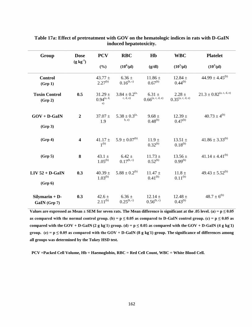
162
Table 17a: Effect of pretreatment with GOV on the hematologic indices in rats with D-GaIN
induced hepatotoxicity.
Group Dose
(g kg-1
)
PCV
(%)
RBC
(106/µl)
Hb
(g/dl)
WBC
(103/µl)
Platelet
(103/µl)
Control
(Grp 1)
43.77 ±
2.27(b)
6.36 ±
0.16(b, c)
11.86 ±
0.67(b)
12.84 ±
0.44(b)
44.99 ± 4.45(b)
Toxin Control
(Grp 2)
0.5 31.29 ±
0.94(a, d,
e)
3.84 ± 0.2(a,
c, d, e)
6.31 ±
0.66(a, c, d, e)
2.28 ±
0.35(a, c, d, e)
21.3 ± 0.82(a, c, d, e)
GOV + D-GaIN
(Grp 3)
2 37.07 ±
1.9
5.38 ± 0.3(a,
b, e)
9.68 ±
0.48(b)
12.39 ±
0.47(b)
40.73 ± 4(b)
(Grp 4) 4 41.17 ±
1(b)
5.9 ± 0.07(b)
11.9 ±
0.32(b)
13.51 ±
0.18(b)
41.86 ± 3.33(b)
(Grp 5) 8 43.1 ±
1.05(b)
6.42 ±
0.17(b, c)
11.73 ±
0.52(b)
13.56 ±
0.99(b)
41.14 ± 4.41(b)
LIV 52 + D-GaIN
(Grp 6)
0.3 40.39 ±
1.03(b)
5.88 ± 0.2(b)
11.47 ±
0.41(b)
11.8 ±
0.11(b)
49.43 ± 5.52(b)
Silymarin + D-
GaIN (Grp 7)
0.3 42.6 ±
2.11(b)
6.36 ±
0.25(b, c)
12.14 ±
0.56(b, c)
12.48 ±
0.43(b)
48.7 ± 6(b)
Values are expressed as Mean ± SEM for seven rats. The Mean difference is significant at the .05 level. (a) = p ≤ 0.05
as compared with the normal control group. (b) = p ≤ 0.05 as compared to D-GaIN control group. (c) = p ≤ 0.05 as
compared with the GOV + D-GaIN (2 g kg
-1) group. (d) = p ≤ 0.05 as compared with the
GOV + D-GaIN (4 g kg
-1)
group. (e) = p ≤ 0.05 as compared with the GOV + D-GaIN (8 g kg
-1) group. The
significance of differences among
all groups was determined by the Tukey HSD test.
PCV =Packed Cell Volume, Hb = Haemoglobin, RBC = Red Cell Count, WBC = White Blood Cell.
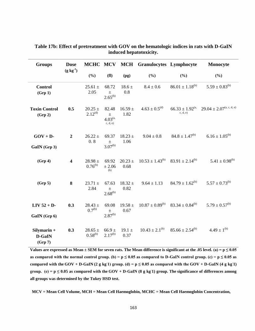
163
Table 17b: Effect of pretreatment with GOV on the hematologic indices in rats with D-GaIN
induced hepatotoxicity.
Groups Dose
(g kg-1
)
MCHC
(%)
MCV
(fl)
MCH
(pg)
Granulocytes
(%)
Lymphocyte
(%)
Monocyte
(%)
Control
(Grp 1)
25.61 ±
2.05
68.72
±
2.65(b)
18.6 ±
0.8
8.4 ± 0.6 86.01 ± 1.18(b)
5.59 ± 0.83(b)
Toxin Control
(Grp 2)
0.5 20.25 ±
2.12(d)
82.48
±
4.03(a,
c, d, e)
16.59 ±
1.82
4.63 ± 0.5(d)
66.33 ± 1.92(a,
c, d, e)
29.04 ± 2.07(a, c, d, e)
GOV + D-
GaIN (Grp 3)
2 26.22 ±
0. 8
69.37
±
3.07(b)
18.23 ±
1.06
9.04 ± 0.8 84.8 ± 1.47(b)
6.16 ± 1.05(b)
(Grp 4) 4 28.98 ±
0.76(b)
69.92
± 2.06 (b)
20.23 ±
0.68
10.53 ± 1.43(b)
83.91 ± 2.14(b)
5.41 ± 0.98(b)
(Grp 5) 8 23.71 ±
2.84
67.63
±
2.68(b)
18.32 ±
0.82
9.64 ± 1.13 84.79 ± 1.62(b)
5.57 ± 0.73(b)
LIV 52 + D-
GaIN (Grp 6)
0.3 28.43 ±
0.7(b)
69.08
±
2.87(b)
19.58 ±
0.67
10.87 ± 0.89(b)
83.34 ± 0.84(b)
5.79 ± 0.57(b)
Silymarin +
D-GaIN
(Grp 7)
0.3 28.65 ±
0.58(b)
66.9 ±
2.17(b)
19.1 ±
0.37
10.43 ± 2.1(b)
85.66 ± 2.54(b)
4.49 ± 1(b)
Values are expressed as Mean ± SEM for seven rats. The Mean difference is significant at the .05 level. (a) = p ≤ 0.05
as compared with the normal control group. (b) = p ≤ 0.05 as compared to D-GaIN control group. (c) = p ≤ 0.05 as
compared with the GOV + D-GaIN (2 g kg
-1) group. (d) = p ≤ 0.05 as compared with the
GOV + D-GaIN (4 g kg
-1)
group. (e) = p ≤ 0.05 as compared with the GOV + D-GaIN (8 g kg
-1) group. The
significance of differences among
all groups was determined by the Tukey HSD test.
MCV = Mean Cell Volume, MCH = Mean Cell Haemoglobin, MCHC = Mean Cell Haemoglobin Concentration,
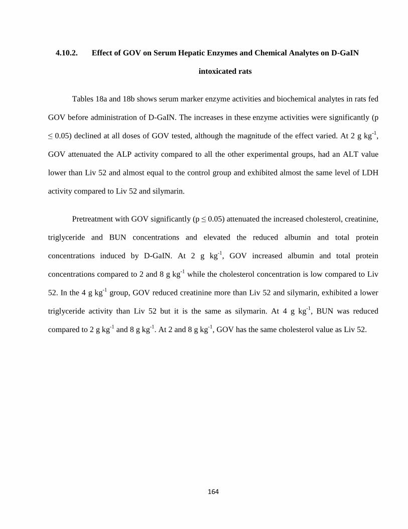
164
4.10.2. Effect of GOV on Serum Hepatic Enzymes and Chemical Analytes on D-GaIN
intoxicated rats
Tables 18a and 18b shows serum marker enzyme activities and biochemical analytes in rats fed
GOV before administration of D-GaIN. The increases in these enzyme activities were significantly (p
≤ 0.05) declined at all doses of GOV tested, although the magnitude of the effect varied. At 2 g kg-1
,
GOV attenuated the ALP activity compared to all the other experimental groups, had an ALT value
lower than Liv 52 and almost equal to the control group and exhibited almost the same level of LDH
activity compared to Liv 52 and silymarin.
Pretreatment with GOV significantly (p ≤ 0.05) attenuated the increased cholesterol, creatinine,
triglyceride and BUN concentrations and elevated the reduced albumin and total protein
concentrations induced by D-GaIN. At 2 g kg-1
, GOV increased albumin and total protein
concentrations compared to 2 and 8 g kg-1
while the cholesterol concentration is low compared to Liv
52. In the 4 g kg-1
group, GOV reduced creatinine more than Liv 52 and silymarin, exhibited a lower
triglyceride activity than Liv 52 but it is the same as silymarin. At 4 g kg-1
, BUN was reduced
compared to 2 g kg-1
and 8 g kg-1
. At 2 and 8 g kg-1
, GOV has the same cholesterol value as Liv 52.
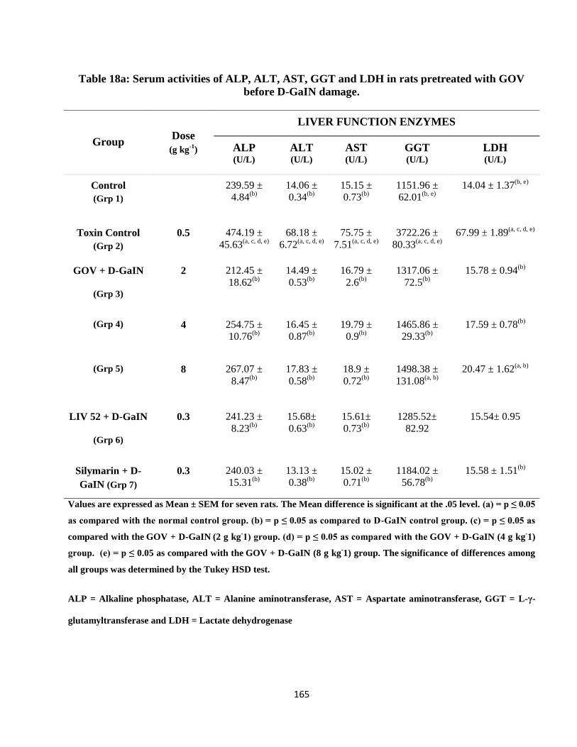
165
Table 18a: Serum activities of ALP, ALT, AST, GGT and LDH in rats pretreated with GOV
before D-GaIN damage.
Group Dose
(g kg-1
)
LIVER FUNCTION ENZYMES
ALP
(U/L)
ALT
(U/L)
AST
(U/L)
GGT
(U/L)
LDH
(U/L)
Control
(Grp 1)
239.59 ±
4.84(b)
14.06 ±
0.34(b)
15.15 ±
0.73(b)
1151.96 ±
62.01(b, e)
14.04 ± 1.37(b, e)
Toxin Control
(Grp 2)
0.5 474.19 ±
45.63(a, c, d, e)
68.18 ±
6.72(a, c, d, e)
75.75 ±
7.51(a, c, d, e)
3722.26 ±
80.33(a, c, d, e)
67.99 ± 1.89(a, c, d, e)
GOV + D-GaIN
(Grp 3)
2 212.45 ±
18.62(b)
14.49 ±
0.53(b)
16.79 ±
2.6(b)
1317.06 ±
72.5(b)
15.78 ± 0.94(b)
(Grp 4) 4 254.75 ±
10.76(b)
16.45 ±
0.87(b)
19.79 ±
0.9(b)
1465.86 ±
29.33(b)
17.59 ± 0.78(b)
(Grp 5) 8 267.07 ±
8.47(b)
17.83 ±
0.58(b)
18.9 ±
0.72(b)
1498.38 ±
131.08(a, b)
20.47 ± 1.62(a, b)
LIV 52 + D-GaIN
(Grp 6)
0.3 241.23 ±
8.23(b)
15.68±
0.63(b)
15.61±
0.73(b)
1285.52±
82.92
15.54± 0.95
Silymarin + D-
GaIN (Grp 7)
0.3 240.03 ±
15.31(b)
13.13 ±
0.38(b)
15.02 ±
0.71(b)
1184.02 ±
56.78(b)
15.58 ± 1.51(b)
Values are expressed as Mean ± SEM for seven rats. The Mean difference is significant at the .05 level. (a) = p ≤ 0.05
as compared with the normal control group. (b) = p ≤ 0.05 as compared to D-GaIN control group. (c) = p ≤ 0.05 as
compared with the GOV + D-GaIN (2 g kg
-1) group. (d) = p ≤ 0.05 as compared with the
GOV + D-GaIN (4 g kg
-1)
group. (e) = p ≤ 0.05 as compared with the GOV + D-GaIN (8 g kg
-1) group. The
significance of differences among
all groups was determined by the Tukey HSD test.
ALP = Alkaline phosphatase, ALT = Alanine aminotransferase, AST = Aspartate aminotransferase, GGT = L-γ-
glutamyltransferase and LDH = Lactate dehydrogenase
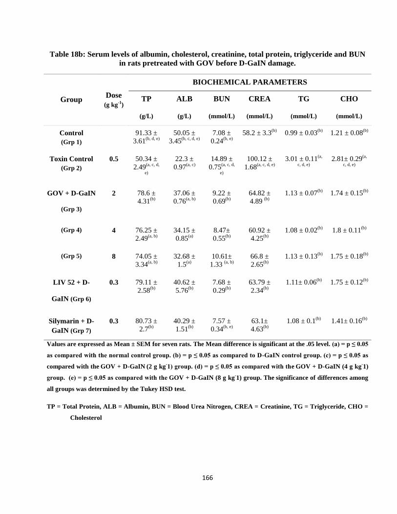
166
Table 18b: Serum levels of albumin, cholesterol, creatinine, total protein, triglyceride and BUN
in rats pretreated with GOV before D-GaIN damage.
Group Dose
(g kg-1
)
BIOCHEMICAL PARAMETERS
TP
(g/L)
ALB
(g/L)
BUN
(mmol/L)
CREA
(mmol/L)
TG
(mmol/L)
CHO
(mmol/L)
Control
(Grp 1)
91.33 ±
3.61(b, d, e)
50.05 ±
3.45(b, c, d, e)
7.08 ±
0.24(b, e)
58.2 ± 3.3(b)
0.99 ± 0.03(b)
1.21 ± 0.08(b)
Toxin Control
(Grp 2)
0.5 50.34 ±
2.49(a, c, d,
e)
22.3 ±
0.97(a, c)
14.89 ±
0.75(a, c, d,
e)
100.12 ±
1.68(a, c, d, e)
3.01 ± 0.11(a,
c, d, e)
2.81± 0.29(a,
c, d, e)
GOV + D-GaIN
(Grp 3)
2 78.6 ±
4.31(b)
37.06 ±
0.76(a, b)
9.22 ±
0.69(b)
64.82 ±
4.89 (b)
1.13 ± 0.07(b)
1.74 ± 0.15(b)
(Grp 4) 4 76.25 ±
2.49(a, b)
34.15 ±
0.85(a)
8.47±
0.55(b)
60.92 ±
4.25(b)
1.08 ± 0.02(b)
1.8 ± 0.11(b)
(Grp 5) 8 74.05 ±
3.34(a, b)
32.68 ±
1.5(a)
10.61±
1.33 (a, b)
66.8 ±
2.65(b)
1.13 ± 0.13(b)
1.75 ± 0.18(b)
LIV 52 + D-
GaIN (Grp 6)
0.3 79.11 ±
2.58(b)
40.62 ±
5.76(b)
7.68 ±
0.29(b)
63.79 ±
2.34(b)
1.11± 0.06(b)
1.75 ± 0.12(b)
Silymarin + D-
GaIN (Grp 7)
0.3 80.73 ±
2.7(b)
40.29 ±
1.51(b)
7.57 ±
0.34(b, e)
63.1±
4.63(b)
1.08 ± 0.1(b)
1.41± 0.16(b)
Values are expressed as Mean ± SEM for seven rats. The Mean difference is significant at the .05 level. (a) = p ≤ 0.05
as compared with the normal control group. (b) = p ≤ 0.05 as compared to D-GaIN control group. (c) = p ≤ 0.05 as
compared with the GOV + D-GaIN (2 g kg
-1) group. (d) = p ≤ 0.05 as compared with the
GOV + D-GaIN (4 g kg
-1)
group. (e) = p ≤ 0.05 as compared with the GOV + D-GaIN (8 g kg
-1) group. The
significance of differences among
all groups was determined by the Tukey HSD test.
TP = Total Protein, ALB = Albumin, BUN = Blood Urea Nitrogen, CREA = Creatinine, TG = Triglyceride, CHO =
Cholesterol
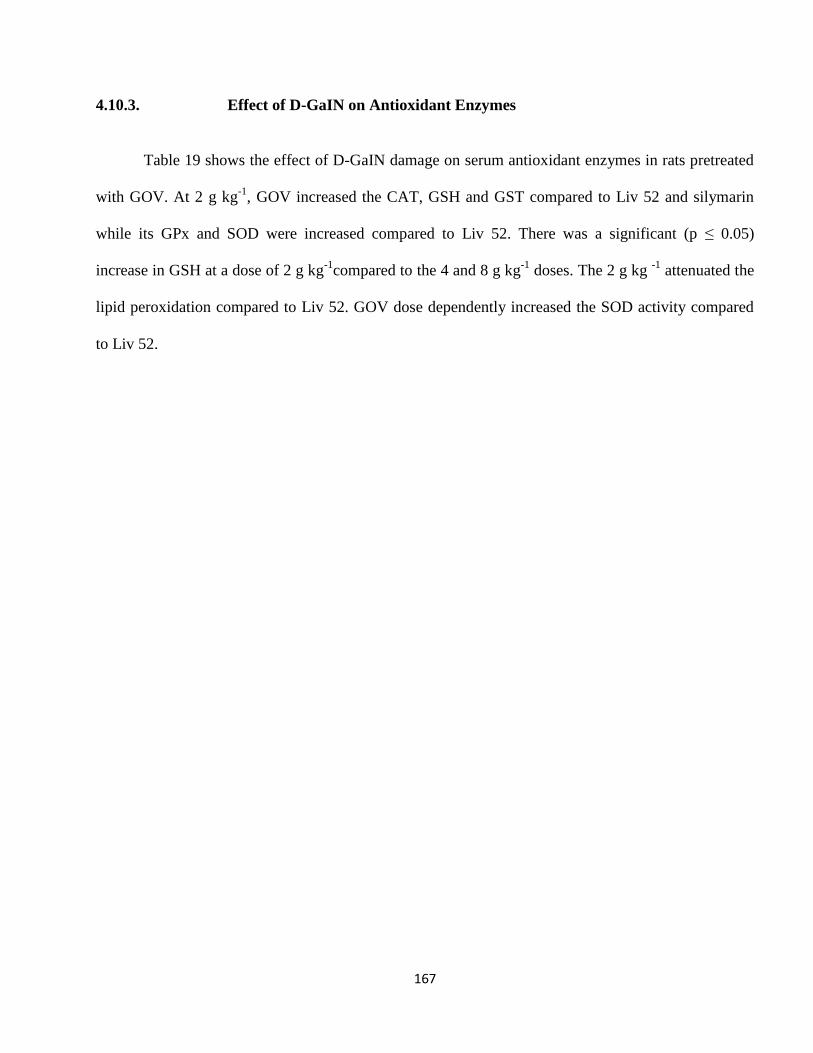
167
4.10.3. Effect of D-GaIN on Antioxidant Enzymes
Table 19 shows the effect of D-GaIN damage on serum antioxidant enzymes in rats pretreated
with GOV. At 2 g kg-1
, GOV increased the CAT, GSH and GST compared to Liv 52 and silymarin
while its GPx and SOD were increased compared to Liv 52. There was a significant (p ≤ 0.05)
increase in GSH at a dose of 2 g kg-1
compared to the 4 and 8 g kg-1
doses. The 2 g kg -1
attenuated the
lipid peroxidation compared to Liv 52. GOV dose dependently increased the SOD activity compared
to Liv 52.
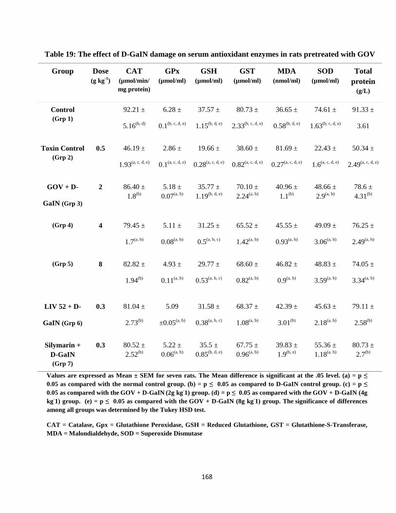
168
Table 19: The effect of D-GaIN damage on serum antioxidant enzymes in rats pretreated with GOV
Group Dose
(g kg-1
)
CAT
(µmol/min/
mg protein)
GPx
(µmol/ml)
GSH
(µmol/ml)
GST
(µmol/ml)
MDA
(nmol/ml)
SOD
(µmol/ml)
Total
protein
(g/L)
Control
(Grp 1)
92.21 ±
5.16(b, d)
6.28 ±
0.1(b, c, d, e)
37.57 ±
1.15(b, d, e)
80.73 ±
2.33(b, c, d, e)
36.65 ±
0.58(b, d, e)
74.61 ±
1.63(b, c, d, e)
91.33 ±
3.61
Toxin Control
(Grp 2)
0.5 46.19 ±
1.93(a, c, d, e)
2.86 ±
0.1(a, c, d, e)
19.66 ±
0.28(a, c, d, e)
38.60 ±
0.82(a, c, d, e)
81.69 ±
0.27(a, c, d, e)
22.43 ±
1.6(a, c, d, e)
50.34 ±
2.49(a, c, d, e)
GOV + D-
GaIN (Grp 3)
2 86.40 ±
1.8(b)
5.18 ±
0.07(a, b)
35.77 ±
1.19(b, d, e)
70.10 ±
2.24(a, b)
40.96 ±
1.1(b)
48.66 ±
2.9(a, b)
78.6 ±
4.31(b)
(Grp 4) 4 79.45 ±
1.7(a, b)
5.11 ±
0.08(a, b)
31.25 ±
0.5(a, b, c)
65.52 ±
1.42(a, b)
45.55 ±
0.93(a, b)
49.09 ±
3.06(a, b)
76.25 ±
2.49(a, b)
(Grp 5) 8 82.82 ±
1.94(b)
4.93 ±
0.11(a, b)
29.77 ±
0.53(a, b, c)
68.60 ±
0.82(a, b)
46.82 ±
0.9(a, b)
48.83 ±
3.59(a, b)
74.05 ±
3.34(a, b)
LIV 52 + D-
GaIN (Grp 6)
0.3 81.04 ±
2.73(b)
5.09
±0.05(a, b)
31.58 ±
0.38(a, b, c)
68.37 ±
1.08(a, b)
42.39 ±
3.01(b)
45.63 ±
2.18(a, b)
79.11 ±
2.58(b)
Silymarin +
D-GaIN
(Grp 7)
0.3 80.52 ±
2.52(b)
5.22 ±
0.06(a, b)
35.5 ±
0.85(b, d, e)
67.75 ±
0.96(a, b)
39.83 ±
1.9(b, e)
55.36 ±
1.18(a, b)
80.73 ±
2.7(b)
Values are expressed as Mean ± SEM for seven rats. The Mean difference is significant at the .05 level. (a) = p ≤
0.05 as compared with the normal control group. (b) = p ≤ 0.05 as compared to D-GaIN control group. (c) = p ≤
0.05 as compared with the GOV + D-GaIN (2g kg
-1) group. (d) = p ≤ 0.05 as compared with the
GOV + D-GaIN (4g
kg-1) group. (e) = p ≤ 0.05 as compared with the
GOV + D-GaIN (8g kg
-1) group. The
significance of differences
among all groups was determined by the Tukey HSD test.
CAT = Catalase, Gpx = Glutathione Peroxidase, GSH = Reduced Glutathione, GST = Glutathione-S-Transferase,
MDA = Malondialdehyde, SOD = Superoxide Dismutase
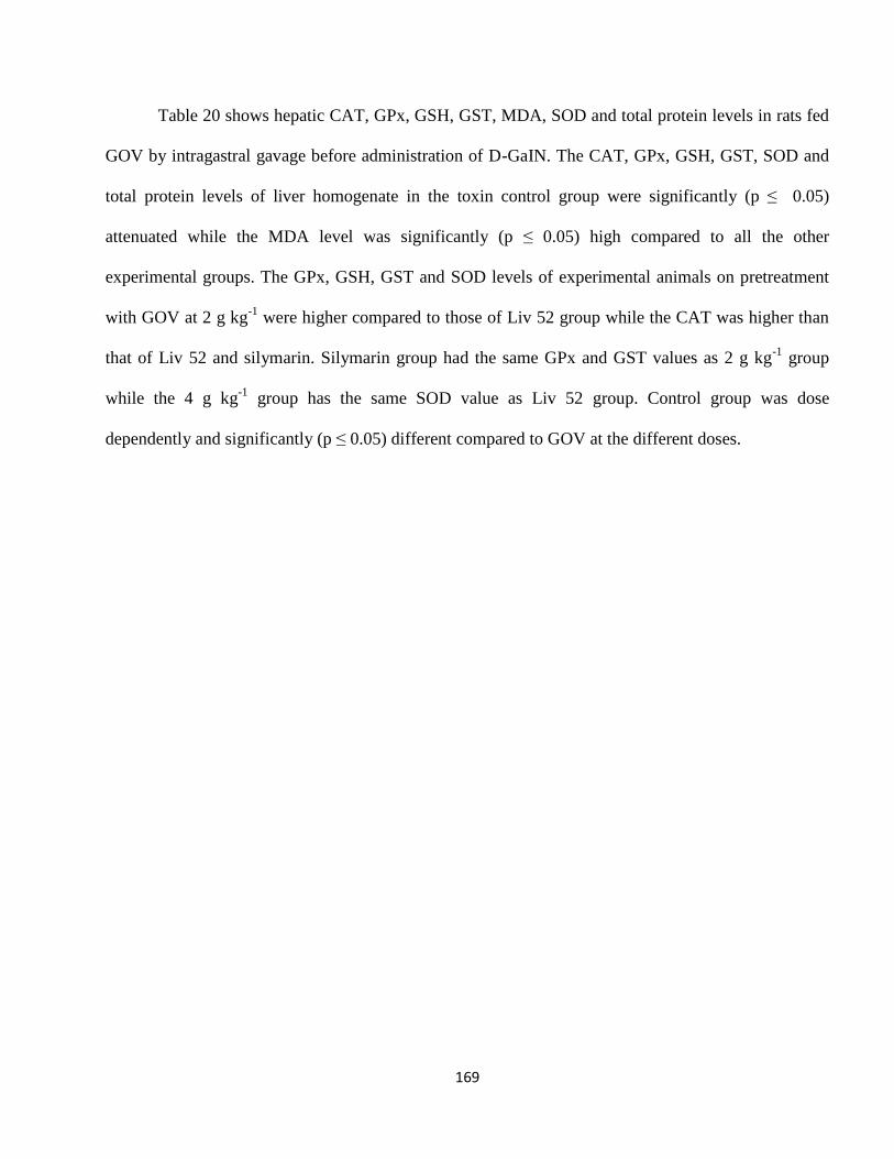
169
Table 20 shows hepatic CAT, GPx, GSH, GST, MDA, SOD and total protein levels in rats fed
GOV by intragastral gavage before administration of D-GaIN. The CAT, GPx, GSH, GST, SOD and
total protein levels of liver homogenate in the toxin control group were significantly (p ≤ 0.05)
attenuated while the MDA level was significantly (p ≤ 0.05) high compared to all the other
experimental groups. The GPx, GSH, GST and SOD levels of experimental animals on pretreatment
with GOV at 2 g kg-1
were higher compared to those of Liv 52 group while the CAT was higher than
that of Liv 52 and silymarin. Silymarin group had the same GPx and GST values as 2 g kg-1
group
while the 4 g kg-1
group has the same SOD value as Liv 52 group. Control group was dose
dependently and significantly (p ≤ 0.05) different compared to GOV at the different doses.
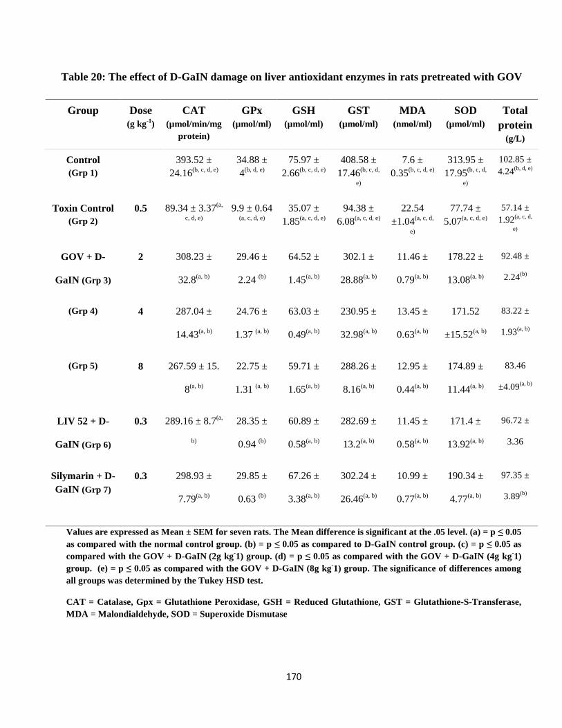
170
Table 20: The effect of D-GaIN damage on liver antioxidant enzymes in rats pretreated with GOV
Group Dose
(g kg-1
)
CAT
(µmol/min/mg
protein)
GPx
(µmol/ml)
GSH
(µmol/ml)
GST
(µmol/ml)
MDA
(nmol/ml)
SOD
(µmol/ml)
Total
protein
(g/L)
Control
(Grp 1)
393.52 ±
24.16(b, c, d, e)
34.88 ±
4(b, d, e)
75.97 ±
2.66(b, c, d, e)
408.58 ±
17.46(b, c, d,
e)
7.6 ±
0.35(b, c, d, e)
313.95 ±
17.95(b, c, d,
e)
102.85 ±
4.24(b, d, e)
Toxin Control
(Grp 2)
0.5 89.34 ± 3.37(a,
c, d, e)
9.9 ± 0.64
(a, c, d, e)
35.07 ±
1.85(a, c, d, e)
94.38 ±
6.08(a, c, d, e)
22.54
±1.04(a, c, d,
e)
77.74 ±
5.07(a, c, d, e)
57.14 ±
1.92(a, c, d,
e)
GOV + D-
GaIN (Grp 3)
2 308.23 ±
32.8(a, b)
29.46 ±
2.24 (b)
64.52 ±
1.45(a, b)
302.1 ±
28.88(a, b)
11.46 ±
0.79(a, b)
178.22 ±
13.08(a, b)
92.48 ±
2.24(b)
(Grp 4) 4 287.04 ±
14.43(a, b)
24.76 ±
1.37 (a, b)
63.03 ±
0.49(a, b)
230.95 ±
32.98(a, b)
13.45 ±
0.63(a, b)
171.52
±15.52(a, b)
83.22 ±
1.93(a, b)
(Grp 5) 8 267.59 ± 15.
8(a, b)
22.75 ±
1.31 (a, b)
59.71 ±
1.65(a, b)
288.26 ±
8.16(a, b)
12.95 ±
0.44(a, b)
174.89 ±
11.44(a, b)
83.46
±4.09(a, b)
LIV 52 + D-
GaIN (Grp 6)
0.3 289.16 ± 8.7(a,
b)
28.35 ±
0.94 (b)
60.89 ±
0.58(a, b)
282.69 ±
13.2(a, b)
11.45 ±
0.58(a, b)
171.4 ±
13.92(a, b)
96.72 ±
3.36
Silymarin + D-
GaIN (Grp 7)
0.3 298.93 ±
7.79(a, b)
29.85 ±
0.63 (b)
67.26 ±
3.38(a, b)
302.24 ±
26.46(a, b)
10.99 ±
0.77(a, b)
190.34 ±
4.77(a, b)
97.35 ±
3.89(b)
Values are expressed as Mean ± SEM for seven rats. The Mean difference is significant at the .05 level. (a) = p ≤ 0.05
as compared with the normal control group. (b) = p ≤ 0.05 as compared to D-GaIN control group. (c) = p ≤ 0.05 as
compared with the GOV + D-GaIN (2g kg
-1) group. (d) = p ≤ 0.05 as compared with the
GOV + D-GaIN (4g kg
-1)
group. (e) = p ≤ 0.05 as compared with the GOV + D-GaIN (8g kg
-1) group. The
significance of differences among
all groups was determined by the Tukey HSD test.
CAT = Catalase, Gpx = Glutathione Peroxidase, GSH = Reduced Glutathione, GST = Glutathione-S-Transferase,
MDA = Malondialdehyde, SOD = Superoxide Dismutase
171
Table 21 shows the activities and levels of CAT, GPx, GSH, GST, MDA and total protein in
kidney tissue homogenates obtained from all the experimental mice. MDA level (34.64 ± 4.79 nmol/g
tissue) in kidney tissue homogenates of D-GaIN treated rats was found to be significantly (p≤ 0.05)
higher than that level compared to normal control rats (7.43 ± 0.2 nmol/g tissue). Pre-treatment with
GOV followed by toxin administration decreased the level significantly (p≤ 0.05). GOV significantly
(p≤ 0.05) attenuated TBARS formation and elevated the activities of CAT, GPx, GSH, GST, SOD
and total protein compared to D-GaIN intoxicated control group. GOV at 4 g kg-1
had higher CAT
activity than Liv 52, increased GSH and GST compared to 2 and 8 g kg-1
, attenuated TBARS
formation in D-GaIN induced kidney damaged rats compared to toxin control and GOV at 2 and 8 g
kg-1
. The total protein concentration in GOV at 2 g kg-1
was almost equal to that of Liv 52. The GPx
activity in the 8 g kg-1
group is significantly (p≤ 0.05) low compared to 4 g kg-1
, Liv 52 and silymarin
groups while the total protein concentration is significantly (p≤ 0.05) low compared to silymarin.
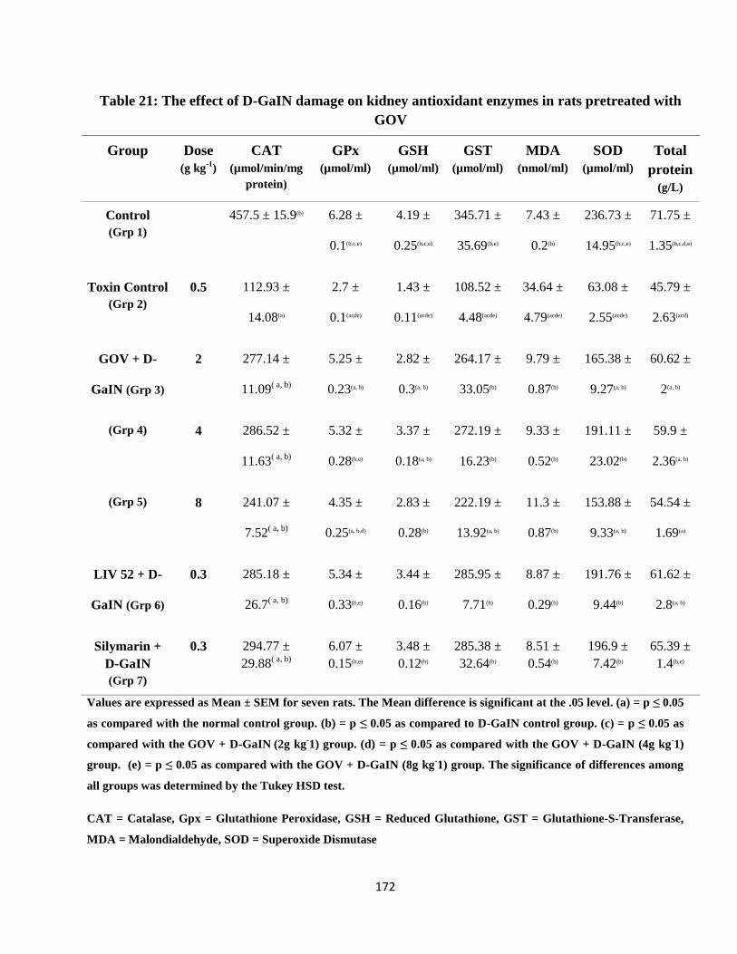
172
Table 21: The effect of D-GaIN damage on kidney antioxidant enzymes in rats pretreated with
GOV
Group Dose
(g kg-1
)
CAT
(µmol/min/mg
protein)
GPx
(µmol/ml)
GSH
(µmol/ml)
GST
(µmol/ml)
MDA
(nmol/ml)
SOD
(µmol/ml)
Total
protein
(g/L)
Control
(Grp 1)
457.5 ± 15.9(b) 6.28 ±
0.1(b,c,e)
4.19 ±
0.25(b,c,e)
345.71 ±
35.69(b,e)
7.43 ±
0.2(b)
236.73 ±
14.95(b,c,e)
71.75 ±
1.35(b,c,d,e)
Toxin Control
(Grp 2)
0.5 112.93 ±
14.08(a)
2.7 ±
0.1(acde)
1.43 ±
0.11(acde)
108.52 ±
4.48(acde)
34.64 ±
4.79(acde)
63.08 ±
2.55(acde)
45.79 ±
2.63(acd)
GOV + D-
GaIN (Grp 3)
2 277.14 ±
11.09( a, b)
5.25 ±
0.23(a, b)
2.82 ±
0.3(a, b)
264.17 ±
33.05(b)
9.79 ±
0.87(b)
165.38 ±
9.27(a, b)
60.62 ±
2(a, b)
(Grp 4) 4 286.52 ±
11.63( a, b)
5.32 ±
0.28(b,e)
3.37 ±
0.18(a, b)
272.19 ±
16.23(b)
9.33 ±
0.52(b)
191.11 ±
23.02(b)
59.9 ±
2.36(a, b)
(Grp 5) 8 241.07 ±
7.52( a, b)
4.35 ±
0.25(a, b,d)
2.83 ±
0.28(b)
222.19 ±
13.92(a, b)
11.3 ±
0.87(b)
153.88 ±
9.33(a, b)
54.54 ±
1.69(a)
LIV 52 + D-
GaIN (Grp 6)
0.3 285.18 ±
26.7( a, b)
5.34 ±
0.33(b,e)
3.44 ±
0.16(b)
285.95 ±
7.71(b)
8.87 ±
0.29(b)
191.76 ±
9.44(b)
61.62 ±
2.8(a, b)
Silymarin +
D-GaIN
(Grp 7)
0.3 294.77 ±
29.88( a, b)
6.07 ±
0.15(b,e)
3.48 ±
0.12(b)
285.38 ±
32.64(b)
8.51 ±
0.54(b)
196.9 ±
7.42(b)
65.39 ±
1.4(b,e)
Values are expressed as Mean ± SEM for seven rats. The Mean difference is significant at the .05 level. (a) = p ≤ 0.05
as compared with the normal control group. (b) = p ≤ 0.05 as compared to D-GaIN control group. (c) = p ≤ 0.05 as
compared with the GOV + D-GaIN (2g kg
-1) group. (d) = p ≤ 0.05 as compared with the
GOV + D-GaIN (4g kg
-1)
group. (e) = p ≤ 0.05 as compared with the GOV + D-GaIN (8g kg
-1) group. The
significance of differences among
all groups was determined by the Tukey HSD test.
CAT = Catalase, Gpx = Glutathione Peroxidase, GSH = Reduced Glutathione, GST = Glutathione-S-Transferase,
MDA = Malondialdehyde, SOD = Superoxide Dismutase
173
4.10.4. Effect of D-GaIN on Caspase Activities
Table 22 shows the fold increase in caspase 2, 3 and 9 activities in the white blood cell of rats
pretreated with GOV before D-GaIN damage. At 2 and 4 g kg-1
, GOV lowered the fold increase in the
caspase 3 activities compared to toxin control, GOV (8 g kg-1
), Liv 52 and silymarin groups. GOV
significantly (p≤ 0.05) decrease the fold increase in caspase 2, 3 and 9 activities compared to the toxin
control group.
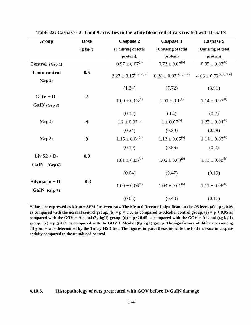
174
Table 22: Caspase - 2, 3 and 9 activities in the white blood cell of rats treated with D-GaIN
Group Dose
(g kg-1)
Caspase 2
(Units/mg of total
protein).
Caspase 3
(Units/mg of total
protein)
Caspase 9
(Units/mg of total
protein)
Control (Grp 1) 0.97 ± 0.07(b)
0.72 ± 0.07(b)
0.95 ± 0.02(b)
Toxin control
(Grp 2)
0.5 2.27 ± 0.15
(a, c, d, e) 6.28 ± 0.33
(a, c, d, e) 4.66 ± 0.72
(a, c, d, e)
(1.34) (7.72) (3.91)
GOV + D-
GaIN (Grp 3)
2 1.09 ± 0.03
(b) 1.01 ± 0.1
(b) 1.14 ± 0.07
(b)
(0.12) (0.4) (0.2)
(Grp 4) 4 1.2 ± 0.07(b)
1 ± 0.07(b)
1.22 ± 0.04(b)
(0.24) (0.39) (0.28)
(Grp 5) 8 1.15 ± 0.04(b)
1.12 ± 0.05(b)
1.14 ± 0.02(b)
(0.19) (0.56) (0.2)
Liv 52 + D-
GaIN (Grp 6)
0.3 1.01 ± 0.05
(b) 1.06 ± 0.09
(b) 1.13 ± 0.08
(b)
(0.04) (0.47) (0.19)
Silymarin + D-
GaIN (Grp 7)
0.3 1.00 ± 0.06
(b) 1.03 ± 0.01
(b) 1.11 ± 0.06
(b)
(0.03) (0.43) (0.17)
Values are expressed as Mean ± SEM for seven rats. The Mean difference is significant at the .05 level. (a) = p ≤ 0.05
as compared with the normal control group. (b) = p ≤ 0.05 as compared to Alcohol control group. (c) = p ≤ 0.05 as
compared with the GOV + Alcohol (2g kg
-1) group. (d) = p ≤ 0.05 as compared with the
GOV + Alcohol (4g kg
-1)
group. (e) = p ≤ 0.05 as compared with the GOV + Alcohol (8g kg
-1) group. The
significance of differences among
all groups was determined by the Tukey HSD test. The figures in parenthesis indicate the fold-increase in caspase
activity compared to the uninduced control.
4.10.5. Histopathology of rats pretreated with GOV before D-GaIN damage
175
The plates of the histopathologic studies on rat liver damaged with D-GaIN are shown in plates
7-10. The control group showed normal hepatocytes arranged in roughly rod or pillar-shaped pattern
while the Toxin control group showed extensive hepatocellular necrosis, leukocyte infiltration,
steatosis, centrizonal necrosis, hepatocellular swelling and vacuolation. Pretreatment with GOV 2 g
kg-1
before intoxication using D-GaIN showed no remarkable changes while at 4 g kg-1
it showed mild
steatosis. Pretreatment with Liv 52 and silymarin before damage with D-GaIN showed no remarkable
changes.
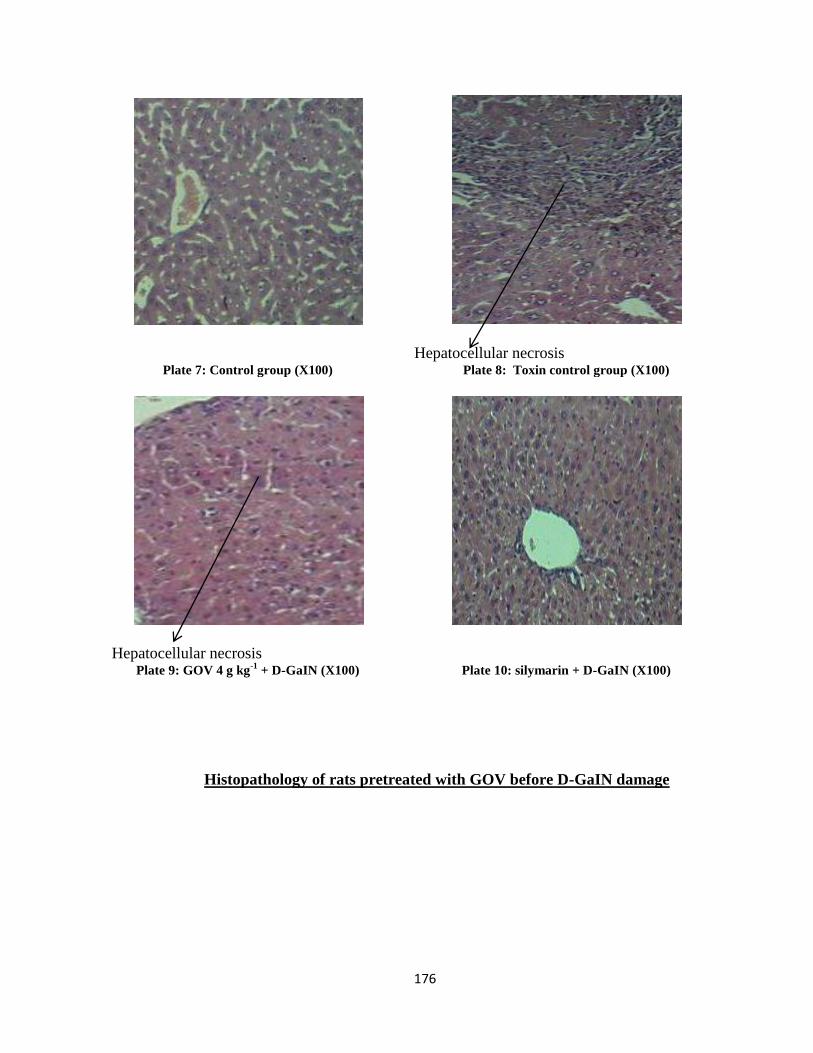
176
Plate 7: Control group (X100)
Hepatocellular necrosis Plate 8: Toxin control group (X100)
Hepatocellular necrosis Plate 9: GOV 4 g kg
-1 + D-GaIN (X100)
Plate 10: silymarin + D-GaIN (X100)
Histopathology of rats pretreated with GOV before D-GaIN damage
177
4.11. Acetaminophen Induced Hepatotoxicity
4.11.1. Effect of GOV on Hematological Parameter Acetaminophen-Induced Hepatotoxic Rats
The triherbal formulation (GOV) dose dependently caused a significant (P≤0.05) increase in
the levels of Hb, PCV, RBC, WBC, Platelet count, MCHC, granulocytes and lymphocytes and also
significantly (P≤0.05) reduced the levels of MCV and monocytes when compared to the APAP
induced toxin control group (Table 23a and 23b). At 2 g kg-1
, GOV increased the Hb, MCHC and
neutrophil level while the PCV was almost equal compared to Liv 52 and Silymarin groups. At 4 g kg-
1, GOV increased the platelet count compared to Liv 52 group while it was restored to normal with
reference to the control group. When compared to the Liv 52 and silymarin groups, GOV at 8 g kg-1
increased the Hb and MCHC levels while the RBC, platelet counts and granulocytes levels were
increased compared to Liv 52. Administration of GOV at 8 g kg-1
reduced the MCV level compared to
all the groups.
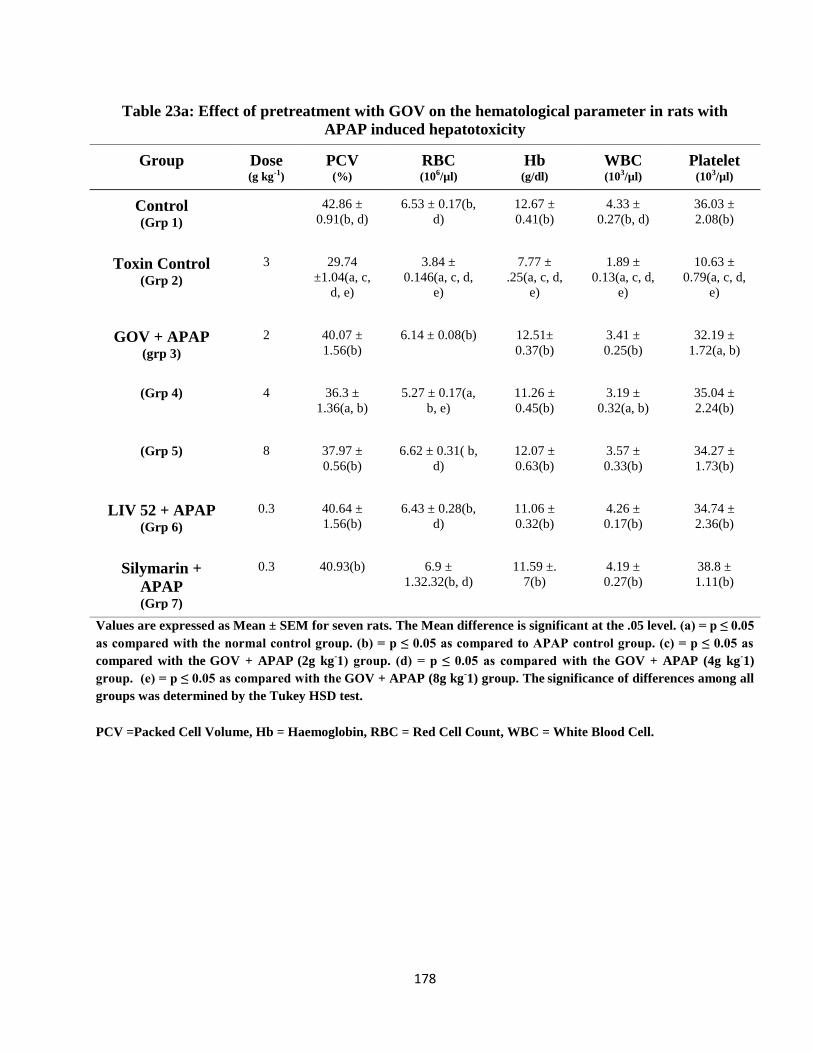
178
Table 23a: Effect of pretreatment with GOV on the hematological parameter in rats with
APAP induced hepatotoxicity
Group Dose
(g kg-1)
PCV
(%) RBC
(106/µl) Hb
(g/dl) WBC
(103/µl) Platelet
(103/µl)
Control (Grp 1)
42.86 ±
0.91(b, d)
6.53 ± 0.17(b,
d)
12.67 ±
0.41(b)
4.33 ±
0.27(b, d)
36.03 ±
2.08(b)
Toxin Control (Grp 2)
3 29.74
±1.04(a, c,
d, e)
3.84 ±
0.146(a, c, d,
e)
7.77 ±
.25(a, c, d,
e)
1.89 ±
0.13(a, c, d,
e)
10.63 ±
0.79(a, c, d,
e)
GOV + APAP
(grp 3)
2 40.07 ±
1.56(b)
6.14 ± 0.08(b)
12.51±
0.37(b)
3.41 ±
0.25(b)
32.19 ±
1.72(a, b)
(Grp 4) 4 36.3 ±
1.36(a, b)
5.27 ± 0.17(a,
b, e)
11.26 ±
0.45(b)
3.19 ±
0.32(a, b)
35.04 ±
2.24(b)
(Grp 5) 8 37.97 ±
0.56(b)
6.62 ± 0.31( b,
d)
12.07 ±
0.63(b)
3.57 ±
0.33(b)
34.27 ±
1.73(b)
LIV 52 + APAP
(Grp 6)
0.3 40.64 ±
1.56(b)
6.43 ± 0.28(b,
d)
11.06 ±
0.32(b)
4.26 ±
0.17(b)
34.74 ±
2.36(b)
Silymarin +
APAP
(Grp 7)
0.3 40.93(b)
6.9 ±
1.32.32(b, d)
11.59 ±.
7(b)
4.19 ±
0.27(b)
38.8 ±
1.11(b)
Values are expressed as Mean ± SEM for seven rats. The Mean difference is significant at the .05 level. (a) = p ≤ 0.05
as compared with the normal control group. (b) = p ≤ 0.05 as compared to APAP control group. (c) = p ≤ 0.05 as
compared with the GOV + APAP (2g kg
-1) group. (d) = p ≤ 0.05 as compared with the
GOV + APAP (4g kg
-1)
group. (e) = p ≤ 0.05 as compared with the GOV + APAP (8g kg
-1) group. The
significance of differences among all
groups was determined by the Tukey HSD test.
PCV =Packed Cell Volume, Hb = Haemoglobin, RBC = Red Cell Count, WBC = White Blood Cell.
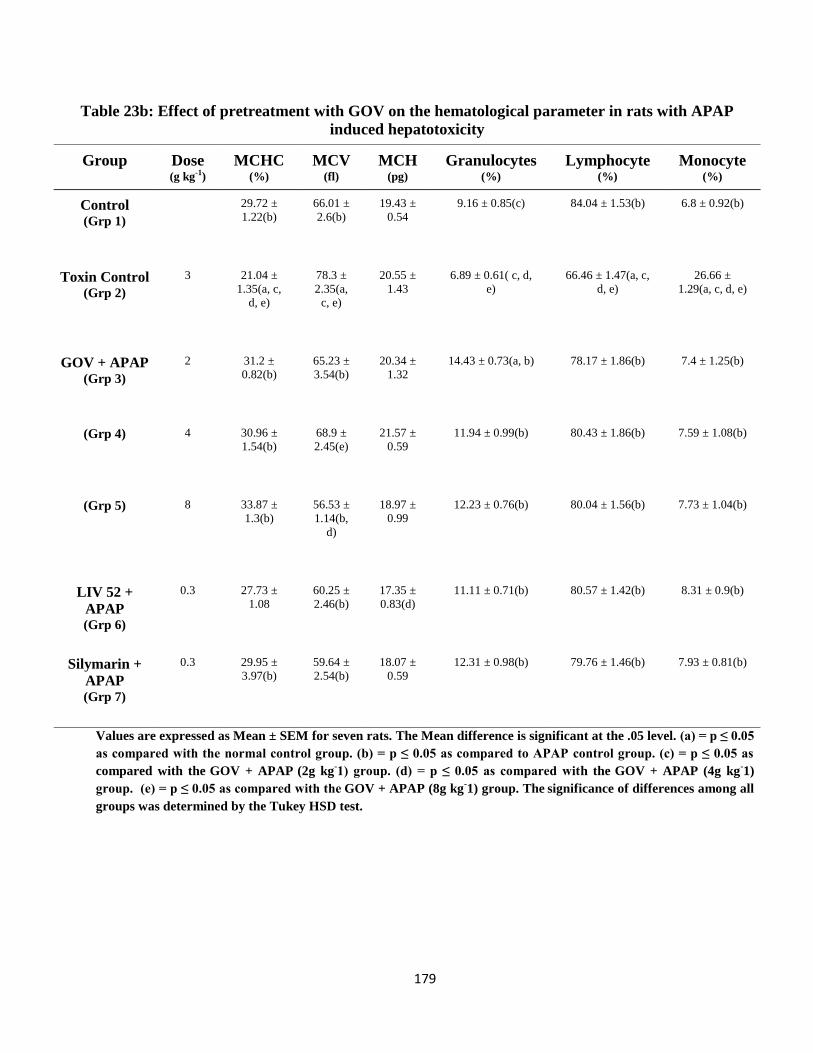
179
Table 23b: Effect of pretreatment with GOV on the hematological parameter in rats with APAP
induced hepatotoxicity
Group Dose
(g kg-1)
MCHC
(%) MCV
(fl) MCH
(pg) Granulocytes
(%) Lymphocyte
(%) Monocyte
(%)
Control (Grp 1)
29.72 ±
1.22(b)
66.01 ±
2.6(b)
19.43 ±
0.54
9.16 ± 0.85(c) 84.04 ± 1.53(b)
6.8 ± 0.92(b)
Toxin Control (Grp 2)
3 21.04 ±
1.35(a, c,
d, e)
78.3 ±
2.35(a,
c, e)
20.55 ±
1.43
6.89 ± 0.61( c, d,
e)
66.46 ± 1.47(a, c,
d, e)
26.66 ±
1.29(a, c, d, e)
GOV + APAP (Grp 3)
2 31.2 ±
0.82(b)
65.23 ±
3.54(b)
20.34 ±
1.32
14.43 ± 0.73(a, b)
78.17 ± 1.86(b)
7.4 ± 1.25(b)
(Grp 4) 4 30.96 ±
1.54(b)
68.9 ±
2.45(e)
21.57 ±
0.59
11.94 ± 0.99(b)
80.43 ± 1.86(b)
7.59 ± 1.08(b)
(Grp 5) 8 33.87 ±
1.3(b)
56.53 ±
1.14(b,
d)
18.97 ±
0.99
12.23 ± 0.76(b)
80.04 ± 1.56(b)
7.73 ± 1.04(b)
LIV 52 +
APAP (Grp 6)
0.3 27.73 ±
1.08
60.25 ±
2.46(b)
17.35 ±
0.83(d)
11.11 ± 0.71(b)
80.57 ± 1.42(b)
8.31 ± 0.9(b)
Silymarin +
APAP (Grp 7)
0.3 29.95 ±
3.97(b)
59.64 ±
2.54(b)
18.07 ±
0.59
12.31 ± 0.98(b)
79.76 ± 1.46(b)
7.93 ± 0.81(b)
Values are expressed as Mean ± SEM for seven rats. The Mean difference is significant at the .05 level. (a) = p ≤ 0.05
as compared with the normal control group. (b) = p ≤ 0.05 as compared to APAP control group. (c) = p ≤ 0.05 as
compared with the GOV + APAP (2g kg
-1) group. (d) = p ≤ 0.05 as compared with the
GOV + APAP (4g kg
-1)
group. (e) = p ≤ 0.05 as compared with the GOV + APAP (8g kg
-1) group. The
significance of differences among all
groups was determined by the Tukey HSD test.
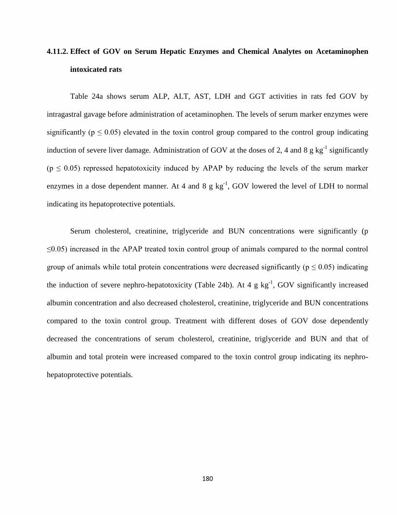
180
4.11.2. Effect of GOV on Serum Hepatic Enzymes and Chemical Analytes on Acetaminophen
intoxicated rats
Table 24a shows serum ALP, ALT, AST, LDH and GGT activities in rats fed GOV by
intragastral gavage before administration of acetaminophen. The levels of serum marker enzymes were
significantly (p ≤ 0.05) elevated in the toxin control group compared to the control group indicating
induction of severe liver damage. Administration of GOV at the doses of 2, 4 and 8 g kg-1
significantly
(p ≤ 0.05) repressed hepatotoxicity induced by APAP by reducing the levels of the serum marker
enzymes in a dose dependent manner. At 4 and 8 g kg-1
, GOV lowered the level of LDH to normal
indicating its hepatoprotective potentials.
Serum cholesterol, creatinine, triglyceride and BUN concentrations were significantly (p
≤0.05) increased in the APAP treated toxin control group of animals compared to the normal control
group of animals while total protein concentrations were decreased significantly (p ≤ 0.05) indicating
the induction of severe nephro-hepatotoxicity (Table 24b). At 4 g kg-1
, GOV significantly increased
albumin concentration and also decreased cholesterol, creatinine, triglyceride and BUN concentrations
compared to the toxin control group. Treatment with different doses of GOV dose dependently
decreased the concentrations of serum cholesterol, creatinine, triglyceride and BUN and that of
albumin and total protein were increased compared to the toxin control group indicating its nephro-
hepatoprotective potentials.
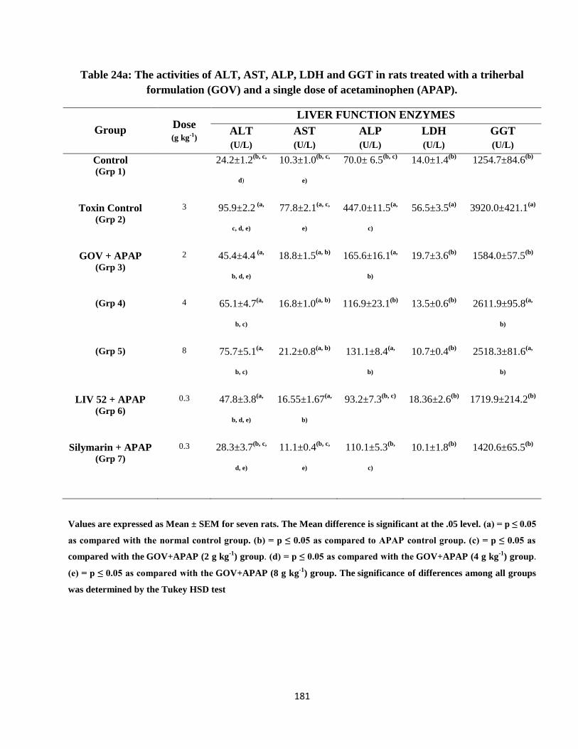
181
Table 24a: The activities of ALT, AST, ALP, LDH and GGT in rats treated with a triherbal
formulation (GOV) and a single dose of acetaminophen (APAP).
Group Dose
(g kg-1)
LIVER FUNCTION ENZYMES
ALT
(U/L)
AST
(U/L)
ALP
(U/L)
LDH
(U/L)
GGT
(U/L)
Control (Grp 1)
24.2±1.2(b, c,
d)
10.3±1.0(b, c,
e)
70.0± 6.5(b, c)
14.0±1.4(b)
1254.7±84.6(b)
Toxin Control (Grp 2)
3 95.9±2.2 (a,
c, d, e)
77.8±2.1(a, c,
e)
447.0±11.5(a,
c)
56.5±3.5(a)
3920.0±421.1(a)
GOV + APAP (Grp 3)
2 45.4±4.4 (a,
b, d, e)
18.8±1.5(a, b)
165.6±16.1(a,
b)
19.7±3.6(b)
1584.0±57.5(b)
(Grp 4) 4 65.1±4.7(a,
b, c)
16.8±1.0(a, b)
116.9±23.1(b)
13.5±0.6(b)
2611.9±95.8(a,
b)
(Grp 5) 8 75.7±5.1(a,
b, c)
21.2±0.8(a, b)
131.1±8.4(a,
b)
10.7±0.4(b)
2518.3±81.6(a,
b)
LIV 52 + APAP (Grp 6)
0.3 47.8±3.8(a,
b, d, e)
16.55±1.67(a,
b)
93.2±7.3(b, c)
18.36±2.6(b)
1719.9±214.2(b)
Silymarin + APAP (Grp 7)
0.3 28.3±3.7(b, c,
d, e)
11.1±0.4(b, c,
e)
110.1±5.3(b,
c)
10.1±1.8(b)
1420.6±65.5(b)
Values are expressed as Mean ± SEM for seven rats. The Mean difference is significant at the .05 level. (a) = p ≤ 0.05
as compared with the normal control group. (b) = p ≤ 0.05 as compared to APAP control group. (c) = p ≤ 0.05 as
compared with the GOV+APAP (2 g kg
-1) group. (d) = p ≤ 0.05 as compared with the
GOV+APAP (4 g kg
-1) group.
(e) = p ≤ 0.05 as compared with the GOV+APAP (8 g kg
-1) group. The
significance of differences among all groups
was determined by the Tukey HSD test
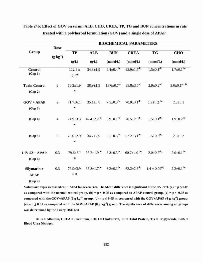
182
Table 24b: Effect of GOV on serum ALB, CHO, CREA, TP, TG and BUN concentrations in rats
treated with a polyherbal formulation (GOV) and a single dose of APAP.
Group
Dose
(g kg-1
)
BIOCHEMICAL PARAMETERS
TP
(g/L)
ALB
(g/L)
BUN
(mmol/L)
CREA
(mmol/L)
TG
(mmol/L)
CHO
(mmol/L)
Control (Grp 1)
112.8 ±
12.5(b)
34.3±1.9 6.4±0.4(b)
63.9±1.5(b)
1.5±0.1(b)
1.7±0.1(b)
Toxin Control
(Grp 2)
3 56.2±1.9(
a)
28.9±1.9 13.6±0.7(a)
89.8±3.5(a)
2.9±0.2(a)
3.0±0.1(a, d)
GOV + APAP
(Grp 3)
2 71.7±6.1(
a)
35.1±0.8 7.5±0.3(b)
70.0±3.1(b)
1.9±0.2 (b)
2.3±0.1
(Grp 4) 4 74.9±3.3(
a)
42.4±2.2(b)
5.9±0.1(b)
70.5±2.9(b)
1.5±0.1(b)
1.9±0.2(b)
(Grp 5) 8 73.0±2.9(
a)
34.7±2.9 6.1±0.5(b)
67.2±3.1(b)
1.5±0.3(b)
2.3±0.2
LIV 52 + APAP
(Grp 6)
0.3 79.6±5(a,
b)
38.2±1.9(b)
6.3±0.3(b)
60.7±4.0 (b)
2.0±0.2(b)
2.0±0.1(b)
Silymarin +
APAP
(Grp 7)
0.3 79.9±3.9(
a, b)
38.8±1.7(b)
6.2±0.1(b)
62.2±2.6(b)
1.4 ± 0.09(b)
2.2±0.1(b)
Values are expressed as Mean ± SEM for seven rats. The Mean difference is significant at the .05 level. (a) = p ≤ 0.05
as compared with the normal control group. (b) = p ≤ 0.05 as compared to APAP control group. (c) = p ≤ 0.05 as
compared with the GOV+APAP (2 g kg
-1) group. (d) = p ≤ 0.05 as compared with the
GOV+APAP (4 g kg
-1) group.
(e) = p ≤ 0.05 as compared with the GOV+APAP (8 g kg
-1) group. The
significance of differences among all groups
was determined by the Tukey HSD test
ALB = Albumin, CREA = Creatinine, CHO = Cholesterol, TP = Total Protein, TG = Triglyceride, BUN =
Blood Urea Nitrogen
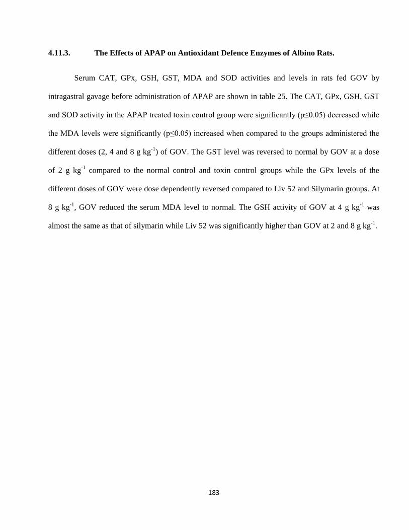
183
4.11.3. The Effects of APAP on Antioxidant Defence Enzymes of Albino Rats.
Serum CAT, GPx, GSH, GST, MDA and SOD activities and levels in rats fed GOV by
intragastral gavage before administration of APAP are shown in table 25. The CAT, GPx, GSH, GST
and SOD activity in the APAP treated toxin control group were significantly (p≤0.05) decreased while
the MDA levels were significantly (p≤0.05) increased when compared to the groups administered the
different doses (2, 4 and 8 g kg-1
) of GOV. The GST level was reversed to normal by GOV at a dose
of 2 g kg-1
compared to the normal control and toxin control groups while the GPx levels of the
different doses of GOV were dose dependently reversed compared to Liv 52 and Silymarin groups. At
8 g kg-1
, GOV reduced the serum MDA level to normal. The GSH activity of GOV at 4 g kg-1
was
almost the same as that of silymarin while Liv 52 was significantly higher than GOV at 2 and 8 g kg-1
.
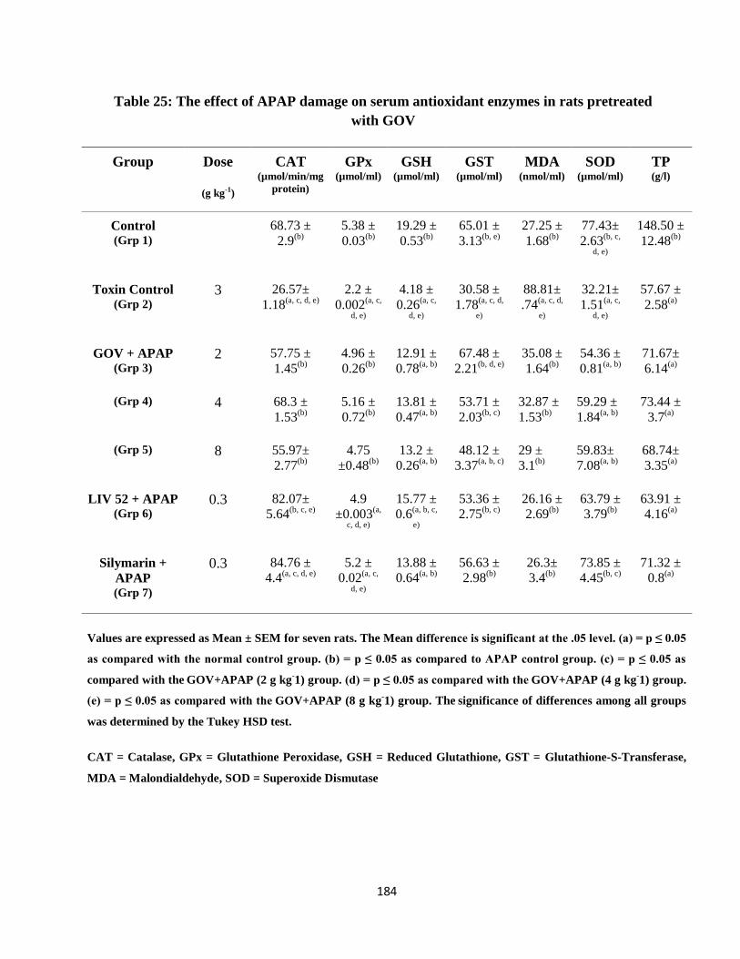
184
Table 25: The effect of APAP damage on serum antioxidant enzymes in rats pretreated
with GOV
Group Dose
(g kg-1)
CAT
(µmol/min/mg
protein)
GPx
(µmol/ml) GSH
(µmol/ml) GST
(µmol/ml) MDA
(nmol/ml) SOD
(µmol/ml) TP
(g/l)
Control (Grp 1)
68.73 ±
2.9(b)
5.38 ±
0.03(b)
19.29 ±
0.53(b)
65.01 ±
3.13(b, e)
27.25 ±
1.68(b)
77.43±
2.63(b, c,
d, e)
148.50 ±
12.48(b)
Toxin Control (Grp 2)
3 26.57±
1.18(a, c, d, e)
2.2 ±
0.002(a, c,
d, e)
4.18 ±
0.26(a, c,
d, e)
30.58 ±
1.78(a, c, d,
e)
88.81±
.74(a, c, d,
e)
32.21±
1.51(a, c,
d, e)
57.67 ±
2.58(a)
GOV + APAP (Grp 3)
2 57.75 ±
1.45(b)
4.96 ±
0.26(b)
12.91 ±
0.78(a, b)
67.48 ±
2.21(b, d, e)
35.08 ±
1.64(b)
54.36 ±
0.81(a, b)
71.67±
6.14(a)
(Grp 4) 4 68.3 ±
1.53(b)
5.16 ±
0.72(b)
13.81 ±
0.47(a, b)
53.71 ±
2.03(b, c)
32.87 ±
1.53(b)
59.29 ±
1.84(a, b)
73.44 ±
3.7(a)
(Grp 5) 8 55.97±
2.77(b)
4.75
±0.48(b)
13.2 ±
0.26(a, b)
48.12 ±
3.37(a, b, c)
29 ±
3.1(b)
59.83±
7.08(a, b)
68.74±
3.35(a)
LIV 52 + APAP (Grp 6)
0.3 82.07±
5.64(b, c, e)
4.9
±0.003(a,
c, d, e)
15.77 ±
0.6(a, b, c,
e)
53.36 ±
2.75(b, c)
26.16 ±
2.69(b)
63.79 ±
3.79(b)
63.91 ±
4.16(a)
Silymarin +
APAP (Grp 7)
0.3 84.76 ±
4.4(a, c, d, e)
5.2 ±
0.02(a, c,
d, e)
13.88 ±
0.64(a, b)
56.63 ±
2.98(b)
26.3±
3.4(b)
73.85 ±
4.45(b, c)
71.32 ±
0.8(a)
Values are expressed as Mean ± SEM for seven rats. The Mean difference is significant at the .05 level. (a) = p ≤ 0.05
as compared with the normal control group. (b) = p ≤ 0.05 as compared to APAP control group. (c) = p ≤ 0.05 as
compared with the GOV+APAP (2 g kg
-1) group. (d) = p ≤ 0.05 as compared with the
GOV+APAP (4 g kg
-1) group.
(e) = p ≤ 0.05 as compared with the GOV+APAP (8 g kg
-1) group. The
significance of differences among all groups
was determined by the Tukey HSD test.
CAT = Catalase, GPx = Glutathione Peroxidase, GSH = Reduced Glutathione, GST = Glutathione-S-Transferase,
MDA = Malondialdehyde, SOD = Superoxide Dismutase

185
The effect of GOV on CAT, GPx, GSH, GST, SOD and total protein activity were significantly
(p ≤ 0.05) and dose dependently increased compared to the toxin control group, while the MDA
activities were significantly (p ≤ 0.05) reduced as shown in table 26. At 8 g kg-1
, GOV significantly (p
≤ 0.05) increased the activity of GSH compared to all the groups while at 4 g kg-1
, GOV increased the
levels of GSH, GST and total protein compared to Liv 52 and Silymarin groups. The GPx levels of the
different doses of GOV were restored to normal. The triherbal formulation dose dependently reversed
the nephro-toxic activity of APAP on rats compared to the control and toxin control groups.
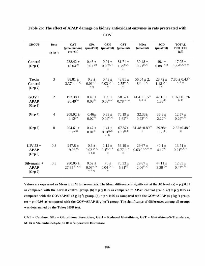
186
Table 26: The effect of APAP damage on kidney antioxidant enzymes in rats pretreated with
GOV
GROUP Dose
(g kg-1)
CAT
(µmol/min/mg
protein)
GPx
(µmol/ml) GSH
(µmol/ml) GST
(µmol/ml) MDA
(nmol/ml) SOD
(µmol/ml) TOTAL
PROTEIN
(g/l)
Control (Grp 1)
238.42 ±
18.04(b)
0.46 ±
0.01 (b)
0.91 ±
0.08(b, c,
e)
81.71 ±
1.79(b, c,
e)
30.48 ±
0.71(b, c)
49.1±
0.88 (b, d)
17.95 ±
0.32(b, c, d, e)
Toxin
Control (Grp 2)
3 88.81 ±
3.37(a ,c, d, e)
0.3 ±
0.01(a, c,
d, e)
0.43 ±
0.03 (a, d,
e)
43.81 ±
2.55(a, d,
e)
56.64 ± 2.
8(a, c, d, e)
28.72 ±
1.18 (a, c,
e)
7.86 ± 0.43(a,
c, d, e)
GOV +
APAP (Grp 3)
2 193.38 ±
20.49(b)
0.49 ±
0.03(b)
0.59 ±
0.03(a, e)
58.57±
0.78 (a, b)
41.4 ± 1.5(a,
b, d, e)
42.16 ±
1.88(b)
11.69 ±0 .76 (a, b)
(Grp 4) 4 208.92 ±
4.12(b)
0.46±
0.02(b)
0.83 ±
0.04(b, e)
70.19 ±
1.62(b)
32.33±
0.92(b, c)
36.8 ±
2.22(a)
12.57 ±
0.29(a, b)
(Grp 5) 8 204.61 ±
3.17(b)
0.47 ±
0.01(b)
1.41 ±
0.01(a, b,
c, d)
67.87±
1.31(a, b)
31.48±0.89(b,
c)
39.98±
1.59(b)
12.32±0.48(a,
b)
LIV 52 +
APAP (Grp 6)
0.3 247.8 ±
19.03 (b)
0.6 ±
0.02 (a, b,
c, d, e)
1.12 ±
0.1(b, c, d,
e)
56.19 ±
0.77 (a, b,
d)
29.67 ±
0.63(a, b, c, d, e)
40.1 ±
4.12(b)
13.71 ±
0.21(a, b, c)
Silymarin +
APAP (Grp 7)
0.3 280.05 ±
27.85 (b, c, e)
0.62 ±
0.03(a, b,
c, d, e)
.76 ±
0.04 (a, b,
e)
70.33 ±
5.91(b)
29.87 ±
2.06(b, c)
44.11 ±
3.39 (b)
12.85 ±
0.47(a, b)
Values are expressed as Mean ± SEM for seven rats. The Mean difference is significant at the .05 level. (a) = p ≤ 0.05
as compared with the normal control group. (b) = p ≤ 0.05 as compared to APAP control group. (c) = p ≤ 0.05 as
compared with the GOV+APAP (2 g kg
-1) group. (d) = p ≤ 0.05 as compared with the
GOV+APAP (4 g kg
-1) group.
(e) = p ≤ 0.05 as compared with the GOV+APAP (8 g kg
-1) group. The
significance of differences among all groups
was determined by the Tukey HSD test.
CAT = Catalase, GPx = Glutathione Peroxidase, GSH = Reduced Glutathione, GST = Glutathione-S-Transferase,
MDA = Malondialdehyde, SOD = Superoxide Dismutase
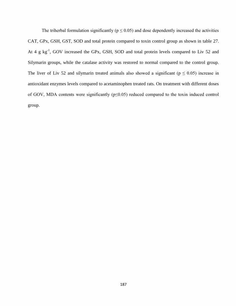
187
The triherbal formulation significantly (p ≤ 0.05) and dose dependently increased the activities
CAT, GPx, GSH, GST, SOD and total protein compared to toxin control group as shown in table 27.
At 4 g kg-1
, GOV increased the GPx, GSH, SOD and total protein levels compared to Liv 52 and
Silymarin groups, while the catalase activity was restored to normal compared to the control group.
The liver of Liv 52 and silymarin treated animals also showed a significant (p ≤ 0.05) increase in
antioxidant enzymes levels compared to acetaminophen treated rats. On treatment with different doses
of GOV, MDA contents were significantly (p≤0.05) reduced compared to the toxin induced control
group.
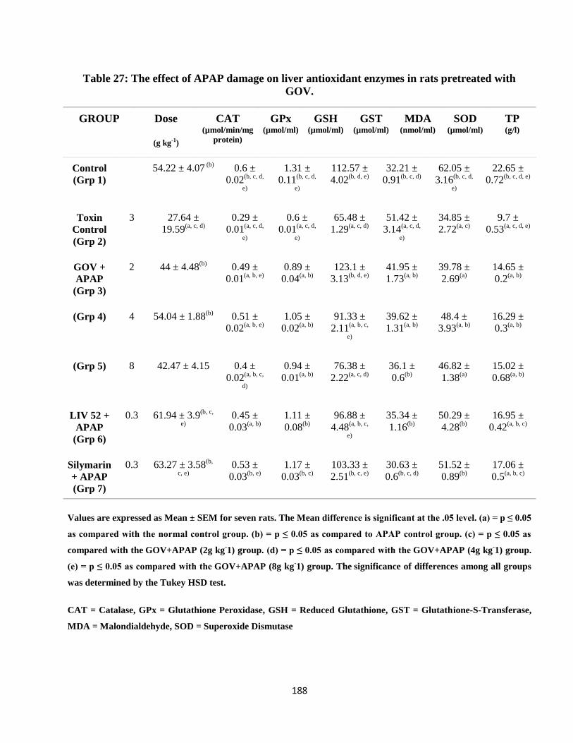
188
Table 27: The effect of APAP damage on liver antioxidant enzymes in rats pretreated with
GOV.
GROUP Dose
(g kg-1)
CAT
(µmol/min/mg
protein)
GPx
(µmol/ml)
GSH
(µmol/ml)
GST
(µmol/ml)
MDA
(nmol/ml)
SOD
(µmol/ml)
TP
(g/l)
Control
(Grp 1)
54.22 ± 4.07 (b)
0.6 ±
0.02(b, c, d,
e)
1.31 ±
0.11(b, c, d,
e)
112.57 ±
4.02(b, d, e)
32.21 ±
0.91(b, c, d)
62.05 ±
3.16(b, c, d,
e)
22.65 ±
0.72(b, c, d, e)
Toxin
Control
(Grp 2)
3 27.64 ±
19.59(a, c, d)
0.29 ±
0.01(a, c, d,
e)
0.6 ±
0.01(a, c, d,
e)
65.48 ±
1.29(a, c, d)
51.42 ±
3.14(a, c, d,
e)
34.85 ±
2.72(a, c)
9.7 ±
0.53(a, c, d, e)
GOV +
APAP
(Grp 3)
2 44 ± 4.48(b)
0.49 ±
0.01(a, b, e)
0.89 ±
0.04(a, b)
123.1 ±
3.13(b, d, e)
41.95 ±
1.73(a, b)
39.78 ±
2.69(a)
14.65 ±
0.2(a, b)
(Grp 4) 4 54.04 ± 1.88(b)
0.51 ±
0.02(a, b, e)
1.05 ±
0.02(a, b)
91.33 ±
2.11(a, b, c,
e)
39.62 ±
1.31(a, b)
48.4 ±
3.93(a, b)
16.29 ±
0.3(a, b)
(Grp 5) 8 42.47 ± 4.15 0.4 ±
0.02(a, b, c,
d)
0.94 ±
0.01(a, b)
76.38 ±
2.22(a, c, d)
36.1 ±
0.6(b)
46.82 ±
1.38(a)
15.02 ±
0.68(a, b)
LIV 52 +
APAP
(Grp 6)
0.3 61.94 ± 3.9(b, c,
e)
0.45 ±
0.03(a, b)
1.11 ±
0.08(b)
96.88 ±
4.48(a, b, c,
e)
35.34 ±
1.16(b)
50.29 ±
4.28(b)
16.95 ±
0.42(a, b, c)
Silymarin
+ APAP
(Grp 7)
0.3 63.27 ± 3.58(b,
c, e)
0.53 ±
0.03(b, e)
1.17 ±
0.03(b, c)
103.33 ±
2.51(b, c, e)
30.63 ±
0.6(b, c, d)
51.52 ±
0.89(b)
17.06 ±
0.5(a, b, c)
Values are expressed as Mean ± SEM for seven rats. The Mean difference is significant at the .05 level. (a) = p ≤ 0.05
as compared with the normal control group. (b) = p ≤ 0.05 as compared to APAP control group. (c) = p ≤ 0.05 as
compared with the GOV+APAP (2g kg
-1) group. (d) = p ≤ 0.05 as compared with the
GOV+APAP (4g kg
-1) group.
(e) = p ≤ 0.05 as compared with the GOV+APAP (8g kg
-1) group. The
significance of differences among all groups
was determined by the Tukey HSD test.
CAT = Catalase, GPx = Glutathione Peroxidase, GSH = Reduced Glutathione, GST = Glutathione-S-Transferase,
MDA = Malondialdehyde, SOD = Superoxide Dismutase
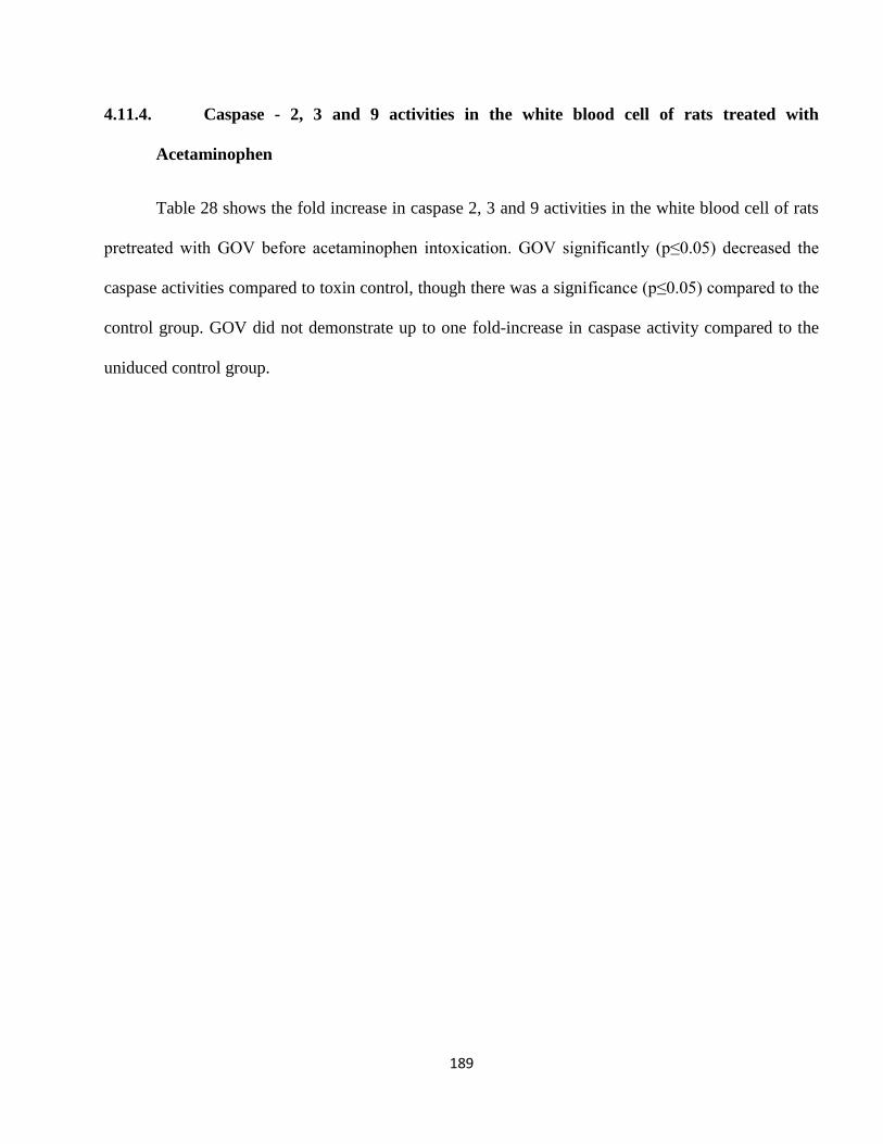
189
4.11.4. Caspase - 2, 3 and 9 activities in the white blood cell of rats treated with
Acetaminophen
Table 28 shows the fold increase in caspase 2, 3 and 9 activities in the white blood cell of rats
pretreated with GOV before acetaminophen intoxication. GOV significantly (p≤0.05) decreased the
caspase activities compared to toxin control, though there was a significance (p≤0.05) compared to the
control group. GOV did not demonstrate up to one fold-increase in caspase activity compared to the
uniduced control group.
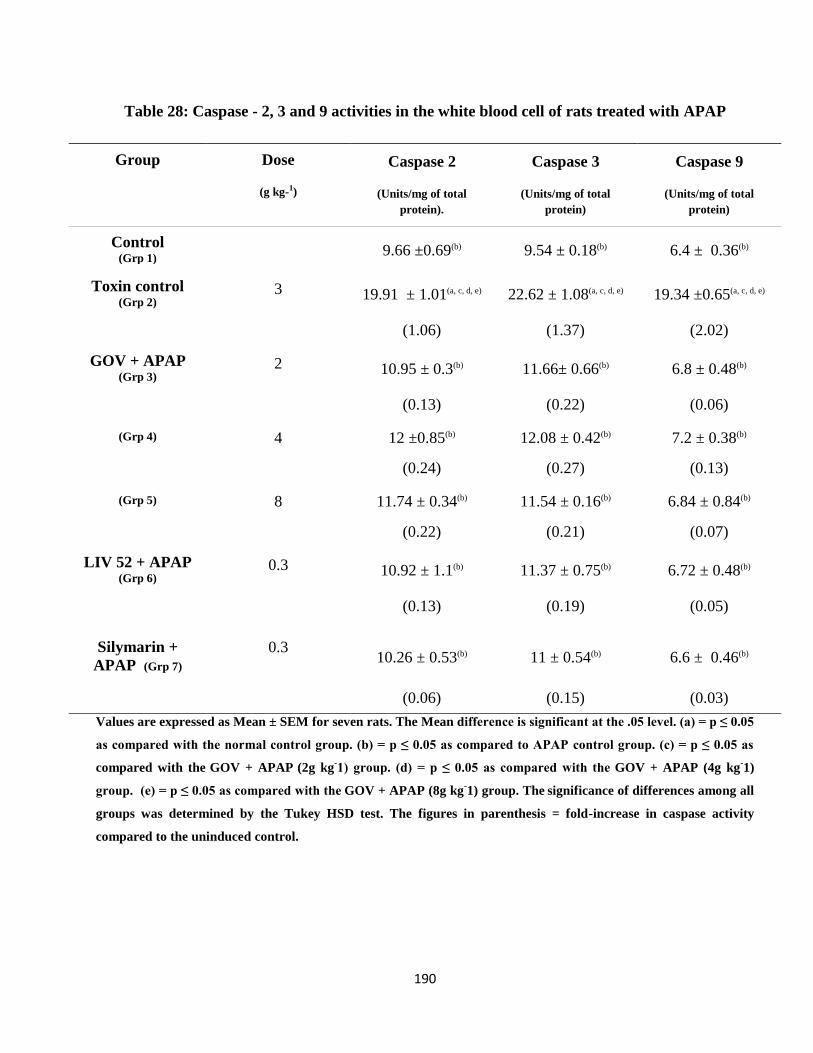
190
Table 28: Caspase - 2, 3 and 9 activities in the white blood cell of rats treated with APAP
Group Dose
(g kg-1)
Caspase 2
(Units/mg of total
protein).
Caspase 3
(Units/mg of total
protein)
Caspase 9
(Units/mg of total
protein)
Control (Grp 1)
9.66 ±0.69(b) 9.54 ± 0.18(b) 6.4 ± 0.36(b)
Toxin control (Grp 2)
3 19.91 ± 1.01(a, c, d, e) 22.62 ± 1.08(a, c, d, e) 19.34 ±0.65(a, c, d, e)
(1.06) (1.37) (2.02)
GOV + APAP (Grp 3)
2 10.95 ± 0.3(b) 11.66± 0.66(b) 6.8 ± 0.48(b)
(0.13) (0.22) (0.06)
(Grp 4) 4 12 ±0.85(b) 12.08 ± 0.42(b) 7.2 ± 0.38(b)
(0.24) (0.27) (0.13)
(Grp 5) 8 11.74 ± 0.34(b) 11.54 ± 0.16(b) 6.84 ± 0.84(b)
(0.22) (0.21) (0.07)
LIV 52 + APAP (Grp 6)
0.3 10.92 ± 1.1(b) 11.37 ± 0.75(b) 6.72 ± 0.48(b)
(0.13) (0.19) (0.05)
Silymarin +
APAP (Grp 7)
0.3 10.26 ± 0.53(b) 11 ± 0.54(b) 6.6 ± 0.46(b)
(0.06) (0.15) (0.03)
Values are expressed as Mean ± SEM for seven rats. The Mean difference is significant at the .05 level. (a) = p ≤ 0.05
as compared with the normal control group. (b) = p ≤ 0.05 as compared to APAP control group. (c) = p ≤ 0.05 as
compared with the GOV + APAP (2g kg
-1) group. (d) = p ≤ 0.05 as compared with the
GOV + APAP (4g kg
-1)
group. (e) = p ≤ 0.05 as compared with the GOV + APAP (8g kg
-1) group. The
significance of differences among all
groups was determined by the Tukey HSD test. The figures in parenthesis = fold-increase in caspase activity
compared to the uninduced control.
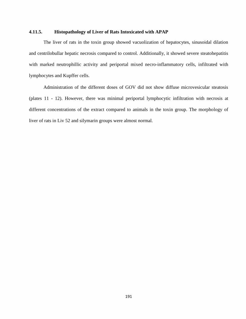
191
4.11.5. Histopathology of Liver of Rats Intoxicated with APAP
The liver of rats in the toxin group showed vacuolization of hepatocytes, sinusoidal dilation
and centrilobullar hepatic necrosis compared to control. Additionally, it showed severe steatohepatitis
with marked neutrophillic activity and periportal mixed necro-inflammatory cells, infiltrated with
lymphocytes and Kupffer cells.
Administration of the different doses of GOV did not show diffuse microvesicular steatosis
(plates 11 - 12). However, there was minimal periportal lymphocytic infiltration with necrosis at
different concentrations of the extract compared to animals in the toxin group. The morphology of
liver of rats in Liv 52 and silymarin groups were almost normal.
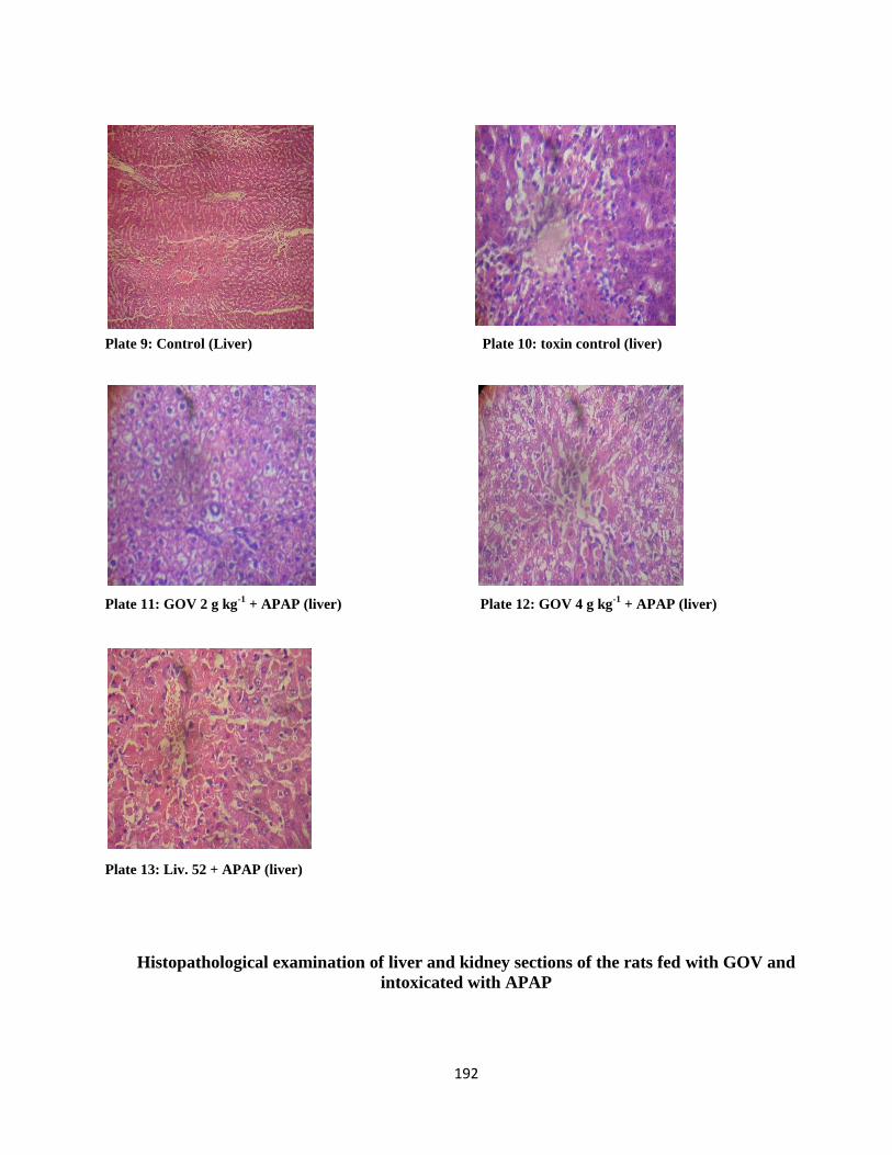
192
Plate 9: Control (Liver) Plate 10: toxin control (liver)
Plate 11: GOV 2 g kg
-1 + APAP (liver) Plate 12: GOV 4 g kg
-1 + APAP (liver)
Plate 13: Liv. 52 + APAP (liver)
Histopathological examination of liver and kidney sections of the rats fed with GOV and
intoxicated with APAP
193
4.12. Carbon Tetrachloride Induced Hepatotoxicity (CCl4)
4.12.1. Effect of pretreatment with GOV on the hematological parameter in rats with
CCl4 induced hepatotoxicity
The administration of GOV dose dependently caused a increase in PCV, RBC, Hb, WBC,
platelet, MCHC, granulocytes and lymphocytes and decrease in MCV and monocytes compared to the
CCl4 induced toxin control group as shown in tables 29a and 29b. There was an increase in levels of
Hb, PCV, platelet count, MCV, MCH and MCHC and monocytes on administration of GOV at 2 g kg-
1 compared to Liv 52 group. At 4 g kg
-1, GOV lowered the WBC count while at 8 g kg
-1, GOV
increased the WBC count comparable to Liv 52 and silymarin. GOV increased platelet count and
MCHC compared to Liv 52. At 8 g kg-1
, GOV increased the number of granulocytes and monocytes
compared to control, Liv 52 and silymarin groups. The PCV level of the control group was
significantly (p≤0.05) increased comparable to the toxin control and GOV at 4 and 8 g kg-1
. At 8 g kg-
1, GOV significantly (p≤0.05) increased WBC count comparable to the control group. There was no
significant (p≤0.05) difference in MCV, MCHC and MCH in this experiment.
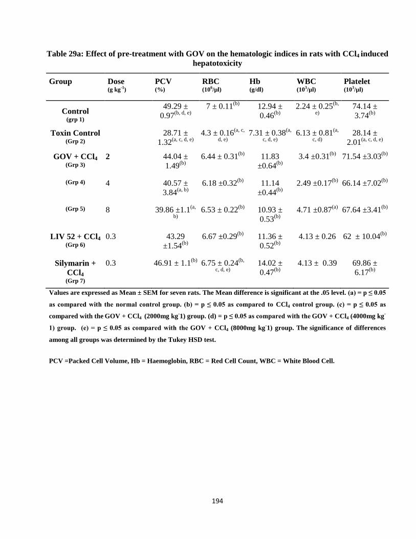
194
Table 29a: Effect of pre-treatment with GOV on the hematologic indices in rats with CCl4 induced
hepatotoxicity
Group Dose
(g kg-1)
PCV
(%)
RBC
(106/µl)
Hb
(g/dl)
WBC
(103/µl)
Platelet
(103/µl)
Control (grp 1)
49.29 ±
0.97(b, d, e)
7 ± 0.11(b)
12.94 ±
0.46(b)
2.24 ± 0.25(b,
e)
74.14 ±
3.74(b)
Toxin Control (Grp 2)
28.71 ±
1.32(a, c, d, e)
4.3 ± 0.16(a, c,
d, e)
7.31 ± 0.38(a,
c, d, e)
6.13 ± 0.81(a,
c, d)
28.14 ±
2.01(a, c, d, e)
GOV + CCl4
(Grp 3)
2 44.04 ±
1.49(b)
6.44 ± 0.31(b)
11.83
±0.64(b)
3.4 ±0.31(b)
71.54 ±3.03(b)
(Grp 4) 4 40.57 ±
3.84(a, b)
6.18 ±0.32(b)
11.14
±0.44(b)
2.49 ±0.17(b)
66.14 ±7.02(b)
(Grp 5) 8 39.86 ±1.1(a,
b)
6.53 ± 0.22(b)
10.93 ±
0.53(b)
4.71 ±0.87(a)
67.64 ±3.41(b)
LIV 52 + CCl4
(Grp 6)
0.3 43.29
±1.54(b)
6.67 ±0.29(b)
11.36 ±
0.52(b)
4.13 ± 0.26 62 ± 10.04(b)
Silymarin +
CCl4
(Grp 7)
0.3 46.91 ± 1.1(b)
6.75 ± 0.24(b,
c, d, e)
14.02 ±
0.47(b)
4.13 ± 0.39 69.86 ±
6.17(b)
Values are expressed as Mean ± SEM for seven rats. The Mean difference is significant at the .05 level. (a) = p ≤ 0.05
as compared with the normal control group. (b) = p ≤ 0.05 as compared to CCl4 control group. (c) = p ≤ 0.05 as
compared with the GOV + CCl4 (2000mg kg
-1) group. (d) = p ≤ 0.05 as compared with the
GOV + CCl4 (4000mg kg
-
1) group. (e) = p ≤ 0.05 as compared with the GOV + CCl4 (8000mg kg
-1) group. The
significance of differences
among all groups was determined by the Tukey HSD test.
PCV =Packed Cell Volume, Hb = Haemoglobin, RBC = Red Cell Count, WBC = White Blood Cell.
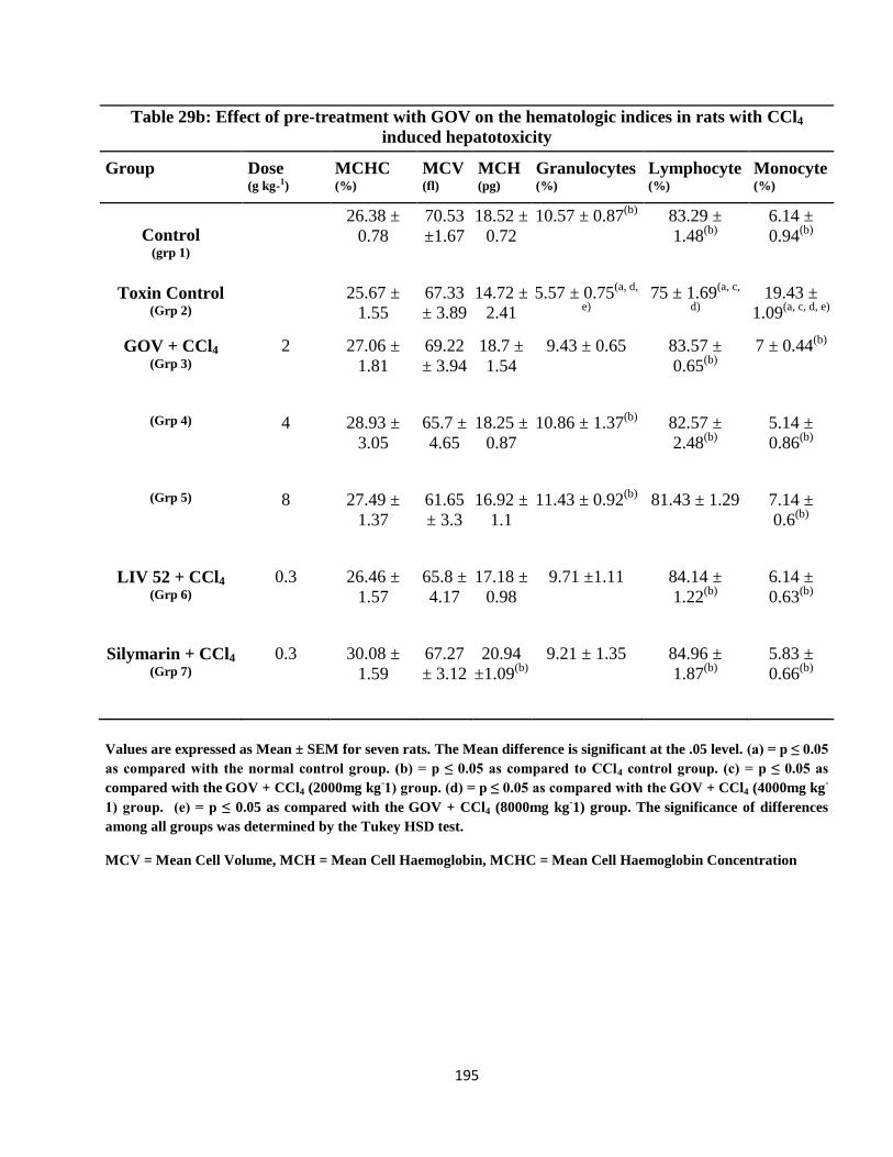
195
Table 29b: Effect of pre-treatment with GOV on the hematologic indices in rats with CCl4
induced hepatotoxicity
Group Dose
(g kg-1)
MCHC
(%)
MCV
(fl)
MCH
(pg)
Granulocytes
(%)
Lymphocyte (%)
Monocyte
(%)
Control (grp 1)
26.38 ±
0.78
70.53
±1.67
18.52 ±
0.72
10.57 ± 0.87(b)
83.29 ±
1.48(b)
6.14 ±
0.94(b)
Toxin Control (Grp 2)
25.67 ±
1.55
67.33
± 3.89
14.72 ±
2.41
5.57 ± 0.75(a, d,
e)
75 ± 1.69(a, c,
d)
19.43 ±
1.09(a, c, d, e)
GOV + CCl4
(Grp 3)
2 27.06 ±
1.81
69.22
± 3.94
18.7 ±
1.54
9.43 ± 0.65 83.57 ±
0.65(b)
7 ± 0.44(b)
(Grp 4) 4 28.93 ±
3.05
65.7 ±
4.65
18.25 ±
0.87
10.86 ± 1.37(b)
82.57 ±
2.48(b)
5.14 ±
0.86(b)
(Grp 5) 8 27.49 ±
1.37
61.65
± 3.3
16.92 ±
1.1
11.43 ± 0.92(b)
81.43 ± 1.29 7.14 ±
0.6(b)
LIV 52 + CCl4
(Grp 6)
0.3 26.46 ±
1.57
65.8 ±
4.17
17.18 ±
0.98
9.71 ±1.11 84.14 ±
1.22(b)
6.14 ±
0.63(b)
Silymarin + CCl4
(Grp 7) 0.3 30.08 ±
1.59
67.27
± 3.12
20.94
±1.09(b)
9.21 ± 1.35 84.96 ±
1.87(b)
5.83 ±
0.66(b)
Values are expressed as Mean ± SEM for seven rats. The Mean difference is significant at the .05 level. (a) = p ≤ 0.05
as compared with the normal control group. (b) = p ≤ 0.05 as compared to CCl4 control group. (c) = p ≤ 0.05 as
compared with the GOV + CCl4 (2000mg kg
-1) group. (d) = p ≤ 0.05 as compared with the
GOV + CCl4 (4000mg kg
-
1) group. (e) = p ≤ 0.05 as compared with the GOV + CCl4 (8000mg kg
-1) group. The
significance of differences
among all groups was determined by the Tukey HSD test.
MCV = Mean Cell Volume, MCH = Mean Cell Haemoglobin, MCHC = Mean Cell Haemoglobin Concentration
196
4.12.2. Effect of GOV on Serum Hepatic Enzymes and Chemical Analytes on CCl4
intoxicated rats
The effect of GOV on serum hepatic enzymes and chemical analytes on CCl4 intoxicated rats
are shown in Tables 30a and 30b. Pretreatment with GOV at a dose of 8 g kg-1
decreased the activity
of ALP compared to Liv 52 group. At a dose of 4 g kg-1
, GOV significantly (p≤ 0.05) decreased serum
ALT activity compared to the 8 g kg-1
group and also its activity compared to Liv 52 and silymarin
groups. There was a significant increase in AST activity of GOV at a dose of 8 g kg-1
compared to
silymarin and control groups. GOV at a dose of 2 g kg-1
decreased the level of total bilirubin compared
to all the other experimental groups and it was significant (p≤ 0.05) compared to the 8 g kg-1
and toxin
control groups. At 2 and 8 g kg-1
, GOV exhibited a decrease in GGT activities compared to Liv 52
group. The LDH activity of the Liv 52 group was increased compared to GOV at 4 and 8 g kg-1
.
Administration of GOV at a dose of 4 g kg-1
exhibited the lowest concentration of cholesterol
and BUN compared to all the other groups. It showed a decreased concentration in triglyceride level
and an increased concentration in total protein compared to Liv 52 group. It also demonstrated almost
the same level of albumin concentration as the Liv 52 group. The level of creatinine concentration
exhibited by GOV at a dose of 8 g kg-1
is comparable to those of Liv 52 and silymarin groups while its
triglyceride concentration is decreased compared to Liv 52 group.
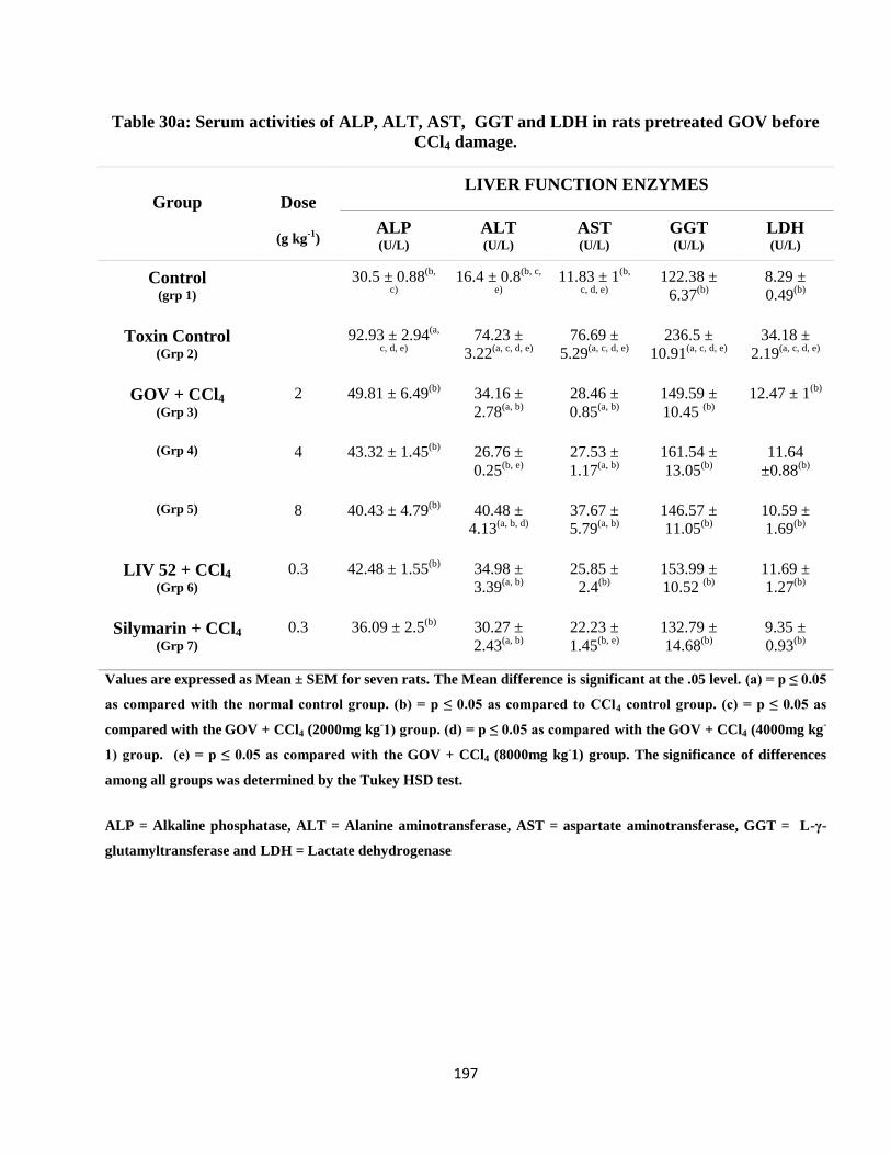
197
Table 30a: Serum activities of ALP, ALT, AST, GGT and LDH in rats pretreated GOV before
CCl4 damage.
Group Dose
(g kg-1
)
LIVER FUNCTION ENZYMES
ALP (U/L)
ALT (U/L)
AST (U/L)
GGT (U/L)
LDH (U/L)
Control (grp 1)
30.5 ± 0.88(b,
c)
16.4 ± 0.8(b, c,
e)
11.83 ± 1(b,
c, d, e)
122.38 ±
6.37(b)
8.29 ±
0.49(b)
Toxin Control (Grp 2)
92.93 ± 2.94(a,
c, d, e)
74.23 ±
3.22(a, c, d, e)
76.69 ±
5.29(a, c, d, e)
236.5 ±
10.91(a, c, d, e)
34.18 ±
2.19(a, c, d, e)
GOV + CCl4
(Grp 3)
2 49.81 ± 6.49(b)
34.16 ±
2.78(a, b)
28.46 ±
0.85(a, b)
149.59 ±
10.45 (b)
12.47 ± 1(b)
(Grp 4) 4 43.32 ± 1.45(b)
26.76 ±
0.25(b, e)
27.53 ±
1.17(a, b)
161.54 ±
13.05(b)
11.64
±0.88(b)
(Grp 5) 8 40.43 ± 4.79(b)
40.48 ±
4.13(a, b, d)
37.67 ±
5.79(a, b)
146.57 ±
11.05(b)
10.59 ±
1.69(b)
LIV 52 + CCl4
(Grp 6)
0.3 42.48 ± 1.55(b)
34.98 ±
3.39(a, b)
25.85 ±
2.4(b)
153.99 ±
10.52 (b)
11.69 ±
1.27(b)
Silymarin + CCl4
(Grp 7) 0.3 36.09 ± 2.5
(b) 30.27 ±
2.43(a, b)
22.23 ±
1.45(b, e)
132.79 ±
14.68(b)
9.35 ±
0.93(b)
Values are expressed as Mean ± SEM for seven rats. The Mean difference is significant at the .05 level. (a) = p ≤ 0.05
as compared with the normal control group. (b) = p ≤ 0.05 as compared to CCl4 control group. (c) = p ≤ 0.05 as
compared with the GOV + CCl4 (2000mg kg
-1) group. (d) = p ≤ 0.05 as compared with the
GOV + CCl4 (4000mg kg
-
1) group. (e) = p ≤ 0.05 as compared with the GOV + CCl4 (8000mg kg
-1) group. The
significance of differences
among all groups was determined by the Tukey HSD test.
ALP = Alkaline phosphatase, ALT = Alanine aminotransferase, AST = aspartate aminotransferase, GGT = L-γ-
glutamyltransferase and LDH = Lactate dehydrogenase
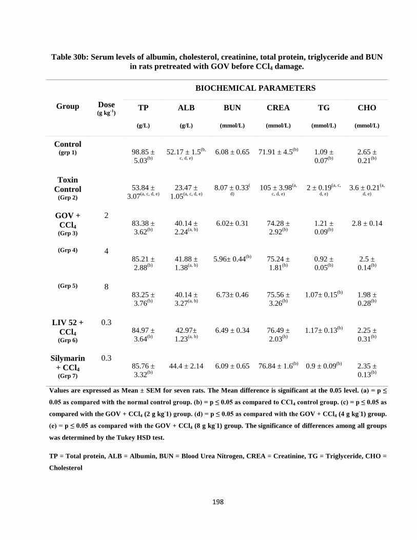
198
Table 30b: Serum levels of albumin, cholesterol, creatinine, total protein, triglyceride and BUN
in rats pretreated with GOV before CCl4 damage.
Group Dose
(g kg-1)
BIOCHEMICAL PARAMETERS
TP
(g/L)
ALB
(g/L)
BUN
(mmol/L)
CREA
(mmol/L)
TG
(mmol/L)
CHO
(mmol/L)
Control (grp 1)
98.85 ±
5.03(b)
52.17 ± 1.5(b,
c, d, e)
6.08 ± 0.65 71.91 ± 4.5(b)
1.09 ±
0.07(b)
2.65 ±
0.21(b)
Toxin
Control (Grp 2)
53.84 ±
3.07(a, c, d, e)
23.47 ±
1.05(a, c, d, e)
8.07 ± 0.33(
d)
105 ± 3.98(a,
c, d, e)
2 ± 0.19(a, c,
d, e)
3.6 ± 0.21(a,
d, e)
GOV +
CCl4
(Grp 3)
2 83.38 ±
3.62(b)
40.14 ±
2.24(a, b)
6.02± 0.31 74.28 ±
2.92(b)
1.21 ±
0.09(b)
2.8 ± 0.14
(Grp 4) 4 85.21 ±
2.88(b)
41.88 ±
1.38(a, b)
5.96± 0.44(b)
75.24 ±
1.81(b)
0.92 ±
0.05(b)
2.5 ±
0.14(b)
(Grp 5) 8 83.25 ±
3.76(b)
40.14 ±
3.27(a, b)
6.73± 0.46 75.56 ±
3.26(b)
1.07± 0.15(b)
1.98 ±
0.28(b)
LIV 52 +
CCl4
(Grp 6)
0.3 84.97 ±
3.64(b)
42.97±
1.23(a, b)
6.49 ± 0.34 76.49 ±
2.03(b)
1.17± 0.13(b)
2.25 ±
0.31(b)
Silymarin
+ CCl4
(Grp 7)
0.3 85.76 ±
3.32(b)
44.4 ± 2.14 6.09 ± 0.65 76.84 ± 1.6(b)
0.9 ± 0.09(b)
2.35 ±
0.13(b)
Values are expressed as Mean ± SEM for seven rats. The Mean difference is significant at the 0.05 level. (a) = p ≤
0.05 as compared with the normal control group. (b) = p ≤ 0.05 as compared to CCl4 control group. (c) = p ≤ 0.05 as
compared with the GOV + CCl4 (2 g kg
-1) group. (d) = p ≤ 0.05 as compared with the
GOV + CCl4 (4 g kg
-1) group.
(e) = p ≤ 0.05 as compared with the GOV + CCl4 (8 g kg
-1) group. The
significance of differences among all groups
was determined by the Tukey HSD test.
TP = Total protein, ALB = Albumin, BUN = Blood Urea Nitrogen, CREA = Creatinine, TG = Triglyceride, CHO =
Cholesterol
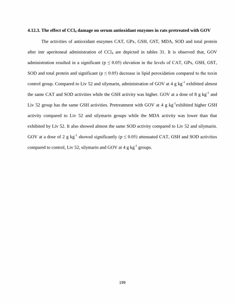
199
4.12.3. The effect of CCl4 damage on serum antioxidant enzymes in rats pretreated with GOV
The activities of antioxidant enzymes CAT, GPx, GSH, GST, MDA, SOD and total protein
after intr aperitoneal administration of CCl4 are depicted in tables 31. It is observed that, GOV
administration resulted in a significant (p ≤ 0.05) elevation in the levels of CAT, GPx, GSH, GST,
SOD and total protein and significant (p ≤ 0.05) decrease in lipid peroxidation compared to the toxin
control group. Compared to Liv 52 and silymarin, administration of GOV at 4 g kg-1
exhibited almost
the same CAT and SOD activities while the GSH activity was higher. GOV at a dose of 8 g kg-1
and
Liv 52 group has the same GSH activities. Pretreatment with GOV at 4 g kg-1
exhibited higher GSH
activity compared to Liv 52 and silymarin groups while the MDA activity was lower than that
exhibited by Liv 52. It also showed almost the same SOD activity compared to Liv 52 and silymarin.
GOV at a dose of 2 g kg-1
showed significantly (p ≤ 0.05) attenuated CAT, GSH and SOD activities
compared to control, Liv 52, silymarin and GOV at 4 g kg-1
groups.
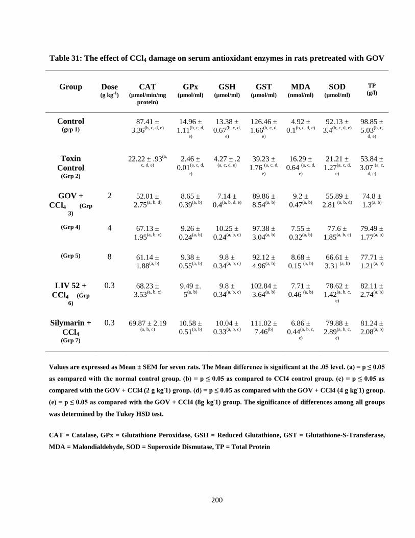
200
Table 31: The effect of CCl4 damage on serum antioxidant enzymes in rats pretreated with GOV
Group Dose (g kg-1)
CAT
(µmol/min/mg
protein)
GPx
(µmol/ml)
GSH
(µmol/ml)
GST
(µmol/ml)
MDA
(nmol/ml)
SOD
(µmol/ml)
TP
(g/l)
Control (grp 1)
87.41 ±
3.36(b, c, d, e)
14.96 ±
1.11(b, c, d,
e)
13.38 ±
0.67(b, c, d,
e)
126.46 ±
1.66(b, c, d,
e)
4.92 ±
0.1(b, c, d, e)
92.13 ±
3.4(b, c, d, e)
98.85 ±
5.03(b, c,
d, e)
Toxin
Control (Grp 2)
22.22 ± .93(a,
c, d, e)
2.46 ±
0.01(a, c, d,
e)
4.27 ± .2 (a, c, d, e)
39.23 ±
1.76 (a, c, d,
e)
16.29 ±
0.64 (a, c, d,
e)
21.21 ±
1.27(a, c, d,
e)
53.84 ±
3.07 (a, c,
d, e)
GOV +
CCl4 (Grp
3)
2 52.01 ±
2.75(a, b, d)
8.65 ±
0.39(a, b)
7.14 ±
0.4(a, b, d, e)
89.86 ±
8.54(a, b)
9.2 ±
0.47(a, b)
55.89 ±
2.81 (a, b, d)
74.8 ±
1.3(a, b)
(Grp 4) 4 67.13 ±
1.95(a, b, c)
9.26 ±
0.24(a, b)
10.25 ±
0.24(a, b, c)
97.38 ±
3.04(a, b)
7.55 ±
0.32(a, b)
77.6 ±
1.85(a, b, c)
79.49 ±
1.77(a, b)
(Grp 5) 8 61.14 ±
1.88(a, b)
9.38 ±
0.55(a, b)
9.8 ±
0.34(a, b, c)
92.12 ±
4.96(a, b)
8.68 ±
0.15 (a, b)
66.61 ±
3.31 (a, b)
77.71 ±
1.21(a, b)
LIV 52 +
CCl4 (Grp
6)
0.3 68.23 ±
3.53(a, b, c)
9.49 ±.
5(a, b)
9.8 ±
0.34(a, b, c)
102.84 ±
3.64(a, b)
7.71 ±
0.46 (a, b)
78.62 ±
1.42(a, b, c,
e)
82.11 ±
2.74(a, b)
Silymarin +
CCl4
(Grp 7)
0.3 69.87 ± 2.19 (a, b, c)
10.58 ±
0.51(a, b)
10.04 ±
0.33(a, b, c)
111.02 ±
7.46(b)
6.86 ±
0.44(a, b, c,
e)
79.88 ±
2.89(a, b, c,
e)
81.24 ±
2.08(a, b)
Values are expressed as Mean ± SEM for seven rats. The Mean difference is significant at the .05 level. (a) = p ≤ 0.05
as compared with the normal control group. (b) = p ≤ 0.05 as compared to CCl4 control group. (c) = p ≤ 0.05 as
compared with the GOV + CCl4 (2 g kg
-1) group. (d) = p ≤ 0.05 as compared with the
GOV + CCl4 (4 g kg
-1) group.
(e) = p ≤ 0.05 as compared with the GOV + CCl4 (8g kg
-1) group. The
significance of differences among all groups
was determined by the Tukey HSD test.
CAT = Catalase, GPx = Glutathione Peroxidase, GSH = Reduced Glutathione, GST = Glutathione-S-Transferase,
MDA = Malondialdehyde, SOD = Superoxide Dismutase, TP = Total Protein

201
Results presented in Table 32 indicates that the elevated level of MDA due to CCl4 intoxication
were significantly (p≤0.05) attenuated in rats, after treatment with GOV. GOV exhibited lower MDA
activity compared the standard reference drug groups. The GOV pretreated rats in the 4 g kg-1
group,
showed increased GPx activity compared to the standard reference drugs. It also showed significantly
(p≤0.05) decreased SOD activity compared to the control group. At 2 g kg-1
and 8 g kg-1
, GOV
showed significant (p≤0.05) decrease in the concentration of total protein compared to the standard
reference drugs. The catalase activity of GOV was significantly (p≤0.05) decreased compared to
control and standard reference drug groups.
202
Table 32: The effect of CCl4 damage on liver antioxidant enzymes in rats pretreated with GOV
Group Dose (g kg-1)
CAT
(µmol/min/mg
protein)
GPx
(µmol/ml) GSH
(µmol/ml) GST
(µmol/ml) MDA
(nmol/ml) SOD
(µmol/ml) TP (g/l)
Control (grp 1)
210.73 ±
10.96(b, c, d, e)
55.12 ±
4.49(b)
137.53 ±
14. 2(b, d, e)
296.91 ±
31.32(b)
13.64 ±
1.27(b)
106.65 ±
4.04(b, c, d,
e)
114.79
± 5.06(b,
c, d, e)
Toxin Control (Grp 2)
69.8 ± 5.89 (a,
c, d, e)
28.57 ±
0.99(a, c, d,
e)
58.94 ±
4.77(a, c, e)
67.86 ±
5.12(a, c, d,
e)
36.35 ±
3.37 (a, c, d,
e)
52.76 ±
4.45(a, c, d,
e)
56.07 ±
2.73(a, c,
d, e)
GOV + CCl4
(Grp 3)
2 148.03 ±
1.6(a, b)
43.37 ±
1.31(b)
91.06 ±
2.28(a, b)
222.45 ±
15.86 (b)
15.59 ±
0.89(b)
85.68 ±
2.1(a, b)
80.15 ±
2.23(a, b)
(Grp 4) 4 151.98 ±
6.86(a, b)
46.81 ±
4.01 (b)
103.52 ±
12.21(b)
250.43 ±
16.89(b)
15.6 ±
1.71(b)
80.7 ±
2.65(a, b)
84.25 ±
3.15(a, b)
(Grp 5) 8 151.76 ±
2.07(a, b)
44.78 ±
2.31 (b)
100.57 ±
4.35(a, b)
262.41 ±
8.09(b)
15.14 ±
1.2(b)
84.63 ±
4.78(a, b)
77.93 ±
1.03(a, b)
LIV 52 +
CCl4 (Grp 6)
0.3 187.98 ±
7.57(b, c, d, e)
45.33 ±
2.65(b)
115.6 ±
5.01(b)
296.33 ±
10.67 (b)
16.39 ±
0.8(b)
93.89 ±
3.73(b)
98.31 ±
3.94(a, b,
c, e)
Silymarin +
CCl4
(Grp 7)
0.3 190.45 ±
5.81(b, c, d, e)
45.71 ±
2.21(b)
117.54 ±
4.52(b)
287.64 ±
35.15 (b)
16.56 ±
0.6 (b)
98.28 ±
3.19(b, c)
95.03 ±
1.96 (a, b,
c, e)
Values are expressed as Mean ± SEM for seven rats. The Mean difference is significant at the .05 level. (a) = p ≤ 0.05
as compared with the normal control group. (b) = p ≤ 0.05 as compared to CCl4 control group. (c) = p ≤ 0.05 as
compared with the GOV + CCl4 (2 g kg
-1) group. (d) = p ≤ 0.05 as compared with the
GOV + CCl4 (4 g kg
-1) group.
(e) = p ≤ 0.05 as compared with the GOV + CCl4 (8g kg
-1) group. The
significance of differences among all groups
was determined by the Tukey HSD test.
CAT = Catalase, GPx = Glutathione Peroxidase, GSH = Reduced Glutathione, GST = Glutathione-S-Transferase,
MDA = Malondialdehyde, SOD = Superoxide Dismutase, TP = Total Protein
203
In Table 33, pretreatment of rats with different doses of GOV 1 hr before CCl4 injection,
showed that there was a significant (p≤0.05) increase in CAT, GPx, GSH, GST, SOD and total
protein and also decreased MDA activity compared to the CCl4 intoxicated toxin group. GOV at a
dose of 8 g kg-1
showed increased CAT, GPx and SOD activities compared to 2 g kg-1
and 4 g kg-1
. At
a dose of 2 g kg-1
, GOV demonstrated a significant (p≤0.05) decrease in CAT activity compared to
toxin control and Liv 52 groups. There was a significant (p≤0.05) increase in GPx activity in the
control group compared to all other groups. At 4 g kg-1
, GOV exhibited increased GSH activity and
decreased MDA activity compared to Liv 52 group. It was also significantly (p≤0.05) elevated
compared to the 2 g kg-1
group. At a dose of 8 g kg-1
, GOV showed almost the same level of SOD
activity and total protein concentration as the Liv 52 and silymarin groups and the Liv 52 group
respectively.
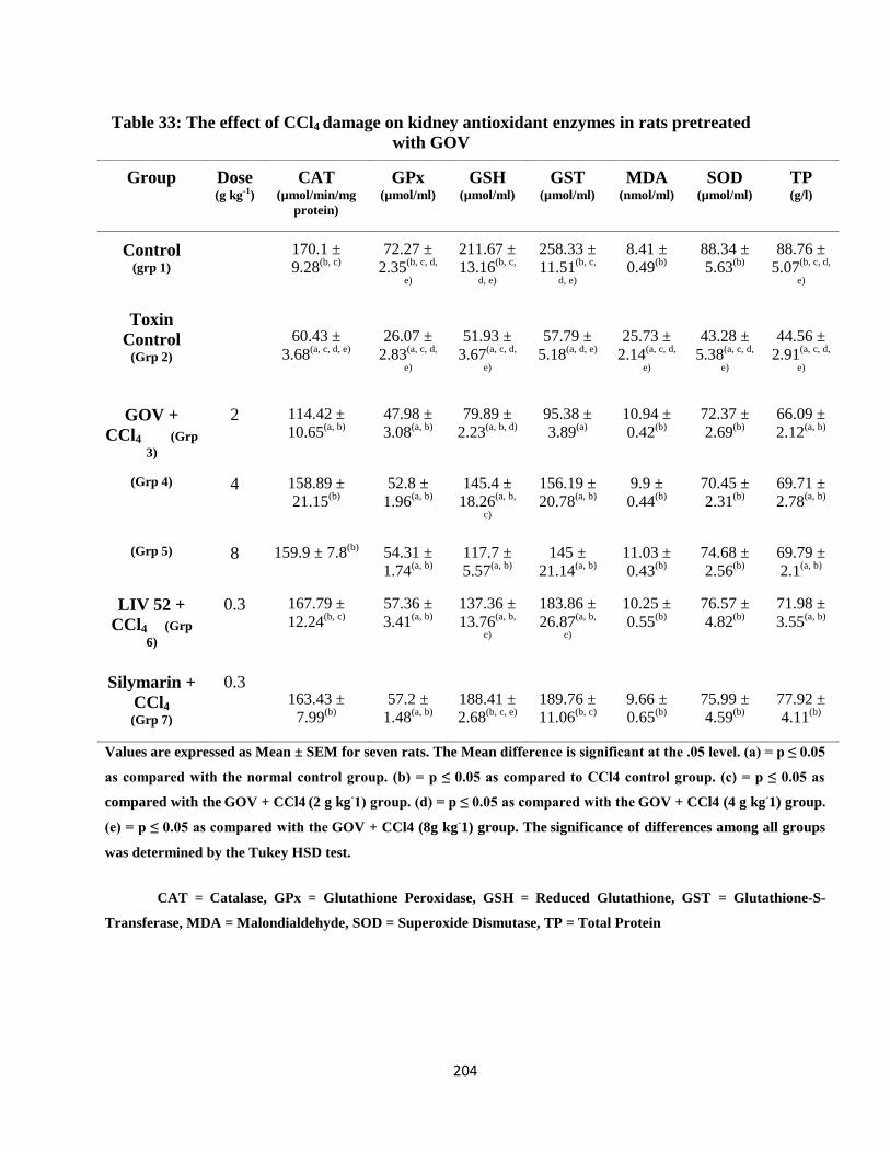
204
Table 33: The effect of CCl4 damage on kidney antioxidant enzymes in rats pretreated
with GOV
Group Dose (g kg-1)
CAT
(µmol/min/mg
protein)
GPx
(µmol/ml)
GSH
(µmol/ml)
GST
(µmol/ml)
MDA
(nmol/ml)
SOD
(µmol/ml)
TP (g/l)
Control (grp 1)
170.1 ±
9.28(b, c)
72.27 ±
2.35(b, c, d,
e)
211.67 ±
13.16(b, c,
d, e)
258.33 ±
11.51(b, c,
d, e)
8.41 ±
0.49(b)
88.34 ±
5.63(b)
88.76 ±
5.07(b, c, d,
e)
Toxin
Control (Grp 2)
60.43 ±
3.68(a, c, d, e)
26.07 ±
2.83(a, c, d,
e)
51.93 ±
3.67(a, c, d,
e)
57.79 ±
5.18(a, d, e)
25.73 ±
2.14(a, c, d,
e)
43.28 ±
5.38(a, c, d,
e)
44.56 ±
2.91(a, c, d,
e)
GOV +
CCl4 (Grp
3)
2 114.42 ±
10.65(a, b)
47.98 ±
3.08(a, b)
79.89 ±
2.23(a, b, d)
95.38 ±
3.89(a)
10.94 ±
0.42(b)
72.37 ±
2.69(b)
66.09 ±
2.12(a, b)
(Grp 4) 4 158.89 ±
21.15(b)
52.8 ±
1.96(a, b)
145.4 ±
18.26(a, b,
c)
156.19 ±
20.78(a, b)
9.9 ±
0.44(b)
70.45 ±
2.31(b)
69.71 ±
2.78(a, b)
(Grp 5) 8 159.9 ± 7.8(b)
54.31 ±
1.74(a, b)
117.7 ±
5.57(a, b)
145 ±
21.14(a, b)
11.03 ±
0.43(b)
74.68 ±
2.56(b)
69.79 ±
2.1(a, b)
LIV 52 +
CCl4 (Grp
6)
0.3 167.79 ±
12.24(b, c)
57.36 ±
3.41(a, b)
137.36 ±
13.76(a, b,
c)
183.86 ±
26.87(a, b,
c)
10.25 ±
0.55(b)
76.57 ±
4.82(b)
71.98 ±
3.55(a, b)
Silymarin +
CCl4
(Grp 7)
0.3 163.43 ±
7.99(b)
57.2 ±
1.48(a, b)
188.41 ±
2.68(b, c, e)
189.76 ±
11.06(b, c)
9.66 ±
0.65(b)
75.99 ±
4.59(b)
77.92 ±
4.11(b)
Values are expressed as Mean ± SEM for seven rats. The Mean difference is significant at the .05 level. (a) = p ≤ 0.05
as compared with the normal control group. (b) = p ≤ 0.05 as compared to CCl4 control group. (c) = p ≤ 0.05 as
compared with the GOV + CCl4 (2 g kg
-1) group. (d) = p ≤ 0.05 as compared with the
GOV + CCl4 (4 g kg
-1) group.
(e) = p ≤ 0.05 as compared with the GOV + CCl4 (8g kg
-1) group. The
significance of differences among all groups
was determined by the Tukey HSD test.
CAT = Catalase, GPx = Glutathione Peroxidase, GSH = Reduced Glutathione, GST = Glutathione-S-
Transferase, MDA = Malondialdehyde, SOD = Superoxide Dismutase, TP = Total Protein
205
4.12.4. Caspase - 2, 3 and 9 activities in the white blood cell of rats treated with CCl4
Table 34 shows the results of the colorimetric assay of caspase - 2, 3 and 9 activities in the
white blood cell of rats pretreated with CCl4. GOV significantly attenuated the fold increase resulting
from CCl4 induced hepatotoxicity compared to the toxin control. At 4 g kg-1, GOV demonstrated
lower caspase 2 and 9 activities compared to Liv 52.
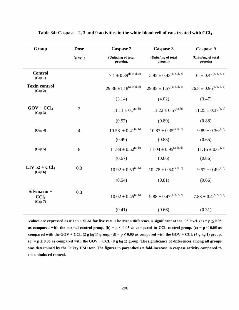
206
Table 34: Caspase - 2, 3 and 9 activities in the white blood cell of rats treated with CCl4
Group Dose
(g kg-1)
Caspase 2
(Units/mg of total
protein).
Caspase 3
(Units/mg of total
protein)
Caspase 9
(Units/mg of total
protein)
Control (Grp 1)
7.1 ± 0.39(b, c, d, e)
5.95 ± 0.43(a, c, d, e)
6 ± 0.44(a, c, d, e)
Toxin control (Grp 2)
29.36 ±1.18(a, c, d, e)
29.85 ± 1.57(a, c, d, e)
26.8 ± 0.96(a, c, d, e)
(3.14) (4.02) (3.47)
GOV + CCl4
(Grp 3) 2 11.11 ± 0.7
(a, b) 11.22 ± 0.57
(a, b) 11.25 ± 0.37
(a, b)
(0.57) (0.89) (0.88)
(Grp 4) 4 10.58 ± 0.41(a, b)
10.87 ± 0.35(a, b, e)
9.89 ± 0.36(a, b)
(0.49) (0.83) (0.65)
(Grp 5) 8 11.88 ± 0.62(a, b)
11.04 ± 0.95(a, b, d)
11.16 ± 0.6(a, b)
(0.67) (0.86) (0.86)
LIV 52 + CCl4
(Grp 6) 0.3 10.92 ± 0.53
(a, b) 10. 78 ± 0.54
(a, b, e) 9.97 ± 0.49
(a, b)
(0.54) (0.81) (0.66)
Silymarin +
CCl4
(Grp 7)
0.3 10.02 ± 0.45
(a, b) 9.88 ± 0.47
(a, b, c, e) 7.88 ± 0.4
(b, c, d, e)
(0.41) (0.66) (0.31)
Values are expressed as Mean ± SEM for five rats. The Mean difference is significant at the .05 level. (a) = p ≤ 0.05
as compared with the normal control group. (b) = p ≤ 0.05 as compared to CCl4 control group. (c) = p ≤ 0.05 as
compared with the GOV + CCl4 (2 g kg
-1) group. (d) = p ≤ 0.05 as compared with the
GOV + CCl4 (4 g kg
-1) group.
(e) = p ≤ 0.05 as compared with the GOV + CCl4 (8 g kg
-1) group. The
significance of differences among all groups
was determined by the Tukey HSD test. The figures in parenthesis = fold-increase in caspase activity compared to
the uninduced control.

207
4.12.5. Histopathology of rats pretreated with GOV before CCl4 damage
The photomicrographs of liver sections of the experimental rat from the different groups
stained with haemotoxylin and eosin (x100) are shown below. The Control shows the normal
architecture while the carbon tetrachloride treated control rats showed steatosis and necrosis. At 2 g
kg-1
GOV showed moderate restoration of liver architecture while at 4 g kg-1
it demonstrated almost
normal of liver architecture. The Liv 52 and Silymarin groups exhibited significant restoration of liver
architecture comparable with control rat group.
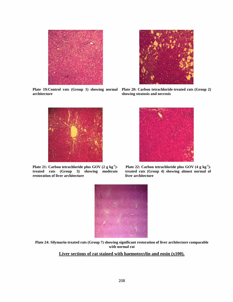
208
Plate 19:Control rats (Group 1) showing normal
architecture
Plate 20: Carbon tetrachloride-treated rats (Group 2)
showing steatosis and necrosis
Plate 21: Carbon tetrachloride plus GOV (2 g kg-1
)-
treated rats (Group 3) showing moderate
restoration of liver architecture
Plate 22: Carbon tetrachloride plus GOV (4 g kg-1
)-
treated rats (Group 4) showing almost normal of
liver architecture
Plate 24: Silymarin-treated rats (Group 7) showing significant restoration of liver architecture comparable
with normal rat
Liver sections of rat stained with haemotoxylin and eosin (x100).
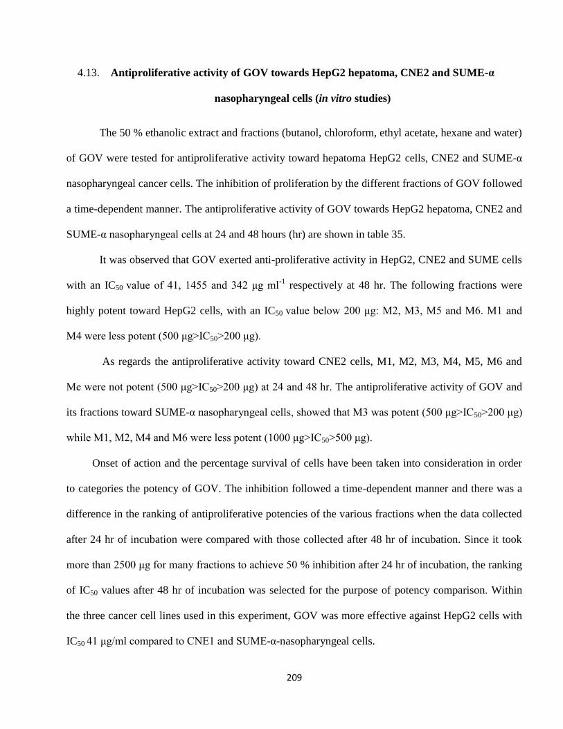
209
4.13. Antiproliferative activity of GOV towards HepG2 hepatoma, CNE2 and SUME-α
nasopharyngeal cells (in vitro studies)
The 50 % ethanolic extract and fractions (butanol, chloroform, ethyl acetate, hexane and water)
of GOV were tested for antiproliferative activity toward hepatoma HepG2 cells, CNE2 and SUME-α
nasopharyngeal cancer cells. The inhibition of proliferation by the different fractions of GOV followed
a time-dependent manner. The antiproliferative activity of GOV towards HepG2 hepatoma, CNE2 and
SUME-α nasopharyngeal cells at 24 and 48 hours (hr) are shown in table 35.
It was observed that GOV exerted anti-proliferative activity in HepG2, CNE2 and SUME cells
with an IC50 value of 41, 1455 and 342 μg ml-1
respectively at 48 hr. The following fractions were
highly potent toward HepG2 cells, with an IC50 value below 200 μg: M2, M3, M5 and M6. M1 and
M4 were less potent (500 μg>IC50>200 μg).
As regards the antiproliferative activity toward CNE2 cells, M1, M2, M3, M4, M5, M6 and
Me were not potent (500 μg>IC50>200 μg) at 24 and 48 hr. The antiproliferative activity of GOV and
its fractions toward SUME-α nasopharyngeal cells, showed that M3 was potent (500 μg>IC50>200 μg)
while M1, M2, M4 and M6 were less potent (1000 μg>IC50>500 μg).
Onset of action and the percentage survival of cells have been taken into consideration in order
to categories the potency of GOV. The inhibition followed a time-dependent manner and there was a
difference in the ranking of antiproliferative potencies of the various fractions when the data collected
after 24 hr of incubation were compared with those collected after 48 hr of incubation. Since it took
more than 2500 μg for many fractions to achieve 50 % inhibition after 24 hr of incubation, the ranking
of IC50 values after 48 hr of incubation was selected for the purpose of potency comparison. Within
the three cancer cell lines used in this experiment, GOV was more effective against HepG2 cells with
IC50 41 μg/ml compared to CNE1 and SUME-α-nasopharyngeal cells.
210
Table 35: Antiproliferative activity of GOV towards HepG2 hepatoma cells, CNE2 and
SUME-α nasopharyngeal cells
Antiproliferative activity
toward HepG2 hepatoma cells Antiproliferative activity toward
CNE2 nasopharyngeal cells Antiproliferative activity toward
SUME-α-nasopharyngeal cells
IC50 at 24hr (μg)
IC50 at 48hr (μg)
IC50 at 24hr (μg)
IC50 at 48hr (μg)
IC50 at 24hr (μg)
IC50 at 48hr (μg)
Me >2500 1975 >2500 >2500 >2500 1000
M1 >2500 251 >2500 1455 >2500 611
M2 >2500 73 >2500 1831 >2500 928
M3 2000 130 >2500 2206 2000 342
M4 >2500 448 >2500 >2500 >2500 799
M5 >2500 60 >2500 >2500 >2500 1021
M6 >2500 41 >2500 >2500 >2500 533
M1 – butanol fraction of GOV M2 – chloroform fraction of GOV
M3 – ethyl acetate fraction of GOV M4 – hexane fraction of GOV
M5 – water fraction of GOV M6 – 50% ethanolic extract of GOV
Me – ethanolic extract of a mixture of G. latifolia, O. gratissimum and V. amygdalina.
Antiproliferative activity of GOV toward HepG2
hepatoma cells:
Antiproliferative activity of GOV toward CNE2
nasopharyngeal cells:
Ranking of potencies at 24h: M3> the rest. Ranking of potencies at 24h: all are equal
Ranking of potencies at 48h: M6>M5>M2>M3>M1>M4
>Me
Ranking of potencies at 48h: M1>M2>M3
Antiproliferative activity of GOV toward SUME-α nasopharyngeal cell:
Ranking of potencies at 24h: M3> the rest Ranking of potencies at 48h: M3>M6>M1>M4
>Me>M5
.
211
4.14. in vitro antioxidant assays
4.14.1. Inhibition of erythrocyte haemolysis using Swiss mice
Table 36 shows anti-oxidant activity of GOV and its fraction using red blood cell. In the
screening of various fractions of GOV and ethanolic extracts of a mixture of G. latifolia, O.
gratissimum and V. amygdalina for antioxidant activity using the assay of inhibition of haemolysis of
mice erythrocytes, it was found that M1, M2, M3, M5, M6 and Me exhibited antioxidant activity. A
40% inhibition of erythrocyte haemolysis or more was taken as the cut-off point for possession of
antioxidant activity.
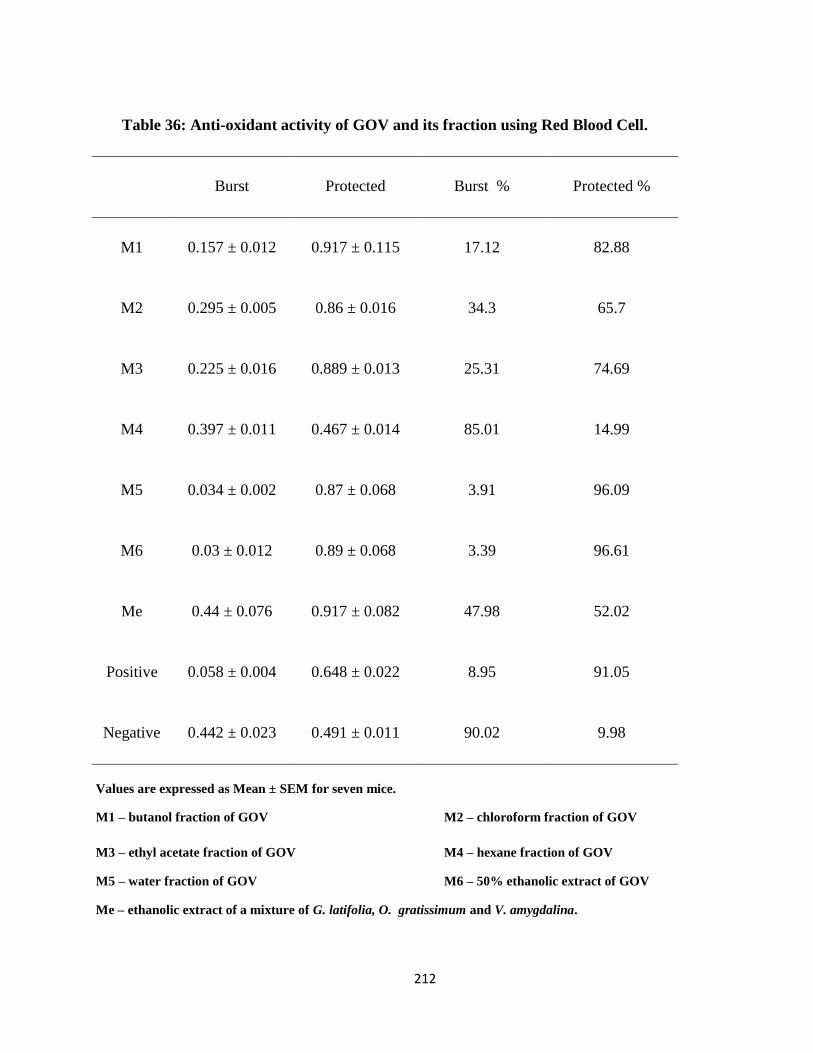
212
Table 36: Anti-oxidant activity of GOV and its fraction using Red Blood Cell.
Burst Protected Burst % Protected %
M1 0.157 ± 0.012 0.917 ± 0.115 17.12 82.88
M2 0.295 ± 0.005 0.86 ± 0.016 34.3 65.7
M3 0.225 ± 0.016 0.889 ± 0.013 25.31 74.69
M4 0.397 ± 0.011 0.467 ± 0.014 85.01 14.99
M5 0.034 ± 0.002 0.87 ± 0.068 3.91 96.09
M6 0.03 ± 0.012 0.89 ± 0.068 3.39 96.61
Me 0.44 ± 0.076 0.917 ± 0.082 47.98 52.02
Positive 0.058 ± 0.004 0.648 ± 0.022 8.95 91.05
Negative 0.442 ± 0.023 0.491 ± 0.011 90.02 9.98
Values are expressed as Mean ± SEM for seven mice.
M1 – butanol fraction of GOV M2 – chloroform fraction of GOV
M3 – ethyl acetate fraction of GOV M4 – hexane fraction of GOV
M5 – water fraction of GOV M6 – 50% ethanolic extract of GOV
Me – ethanolic extract of a mixture of G. latifolia, O. gratissimum and V. amygdalina.
213
4.14.2. Lipid Peroxidative Activity of GOV using Brain Homogenates of Wistar Albino Rats
Lipid peroxidative activity of GOV using brain homogenates of Wistar albino rats are shown in
Table 37. When the extracts and fractions of GOV were tested for antioxidant activity using rat brain
homogenate, it was found that only fractions all the extracts and fractions manifested sufficiently high
antioxidant activity to allow estimation of the IC50 value (i.e. the concentration that brings about 50 %
inhibition). All other fractions had very low inhibition activity on oxidation in rat brain homogenate.
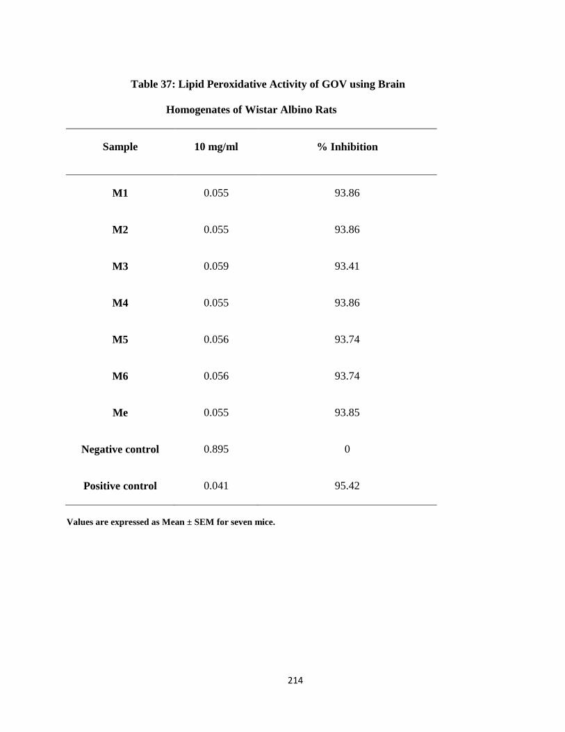
214
Table 37: Lipid Peroxidative Activity of GOV using Brain
Homogenates of Wistar Albino Rats
Sample 10 mg/ml % Inhibition
M1 0.055 93.86
M2 0.055 93.86
M3 0.059 93.41
M4 0.055 93.86
M5 0.056 93.74
M6 0.056 93.74
Me 0.055 93.85
Negative control 0.895 0
Positive control 0.041 95.42
Values are expressed as Mean ± SEM for seven mice.
215
CHAPTER FIVE
DISCUSSION
Preliminary phytochemical screening of GOV revealed the presence of different
phytoconstituents like flavonoids, triterpenoids, saponins, terpenoids and phenolics and alkaloids and
these compounds are known to possess antioxidant and hepatoprotective activity.
Thin layer chromatographic (TLC) analysis of GOV, the ethanolic and water extracts of G.
latifolia, O. gratissimum and V. amygdalina showed that water is not the best solvent for extracting
polyphenolic compounds. This is in concordance with the findings reported by Marwah et al. (2007)
that aqueous alcohols are the best solvents for extracting polyphenolic compounds from plant
materials. Stigmasterol which is one of the phytoconstituents suspected has been reported to possess
anti-osteoarthritic (Gabay, et al., 2010), antioxidant, hypoglycemic and thyroid inhibiting properties
(Panda et al., 2009) while β-sitosterol another compound suspected, exhibits anti-inflammatory
activity in human aortic endothelial cells (Loizou et al., 2010) and is used in Europe for the treatment
of breast cancer (Awad, et al 2000). Rutin is an antioxidant as reported by Jaganath et al., (2009),
shows antinociceptive (Lapa et al 2009) and anti-inflammatory activity in some animal and in vitro
models (Chan, et al., 2007). Hyperoside could have a protective antioxidant effect on cultured PC12
cells (a cell line derived from a pheochromocytoma of the rat adrenal medulla) (Zhiyong et al., 2005)
and also in lung fibroblast cells (Piao, et al., 2008). Eugenol shows protective activity against oxidized
LDL-induced cytotoxicity and adhesion molecule expression in endothelial cells (Ou, et al 2006) and
exhibited antioxidant, anti-inflammatory and DNA-protective properties in thioacetamide-induced
liver injury in rats (Yogalakshmi, et al., 2010). Ascorbic acid selectively kills cancer cells and acts as a
pro-drug to deliver hydrogen peroxide to tissues (Chen, et al., 2005) while linoleic acid demonstrates
antiapoptotic activities (Miner, et al., 2001). Borneol exhibits antihypertensive and antioxidant
activities (Kumar, et al., 2010).
216
The antioxidant, hepatoprotective, antiproliferative, anti-inflammatory, antinociceptive and
antiapoptotic activities of GOV could be adduced to the presence of some of these compounds
identified from our phytoscreening.
Saturation-transfer difference NMR spectroscopy (STD NMR) studies of the various fractions
showed many areas of interest. Initial proton NMR experiments showed the presence of various
aromatic compounds in each fraction, due to proton NMR signals between 6-8 ppm. Representative
proton NMR spectra of the butanol fraction of GOV are shown in Figures 4. Proton NMR spectra of
the other fractions also showed many aromatic protons in the 6-8 ppm range. Other areas of the spectra
showing signals, the carbohydrate region, 3-4.5 ppm and the methyl region, 0.5-1 ppm, showed
numerous peaks. Previous STD studies have led to familiarity in irradiating DHPR at -0.5 ppm to
excite the protein (Ge and Sem, 2007). Because of the anisotropic effect in proteins, some signals can
show up in the negative scale of the spectrum. Often these are methylene signals and can be
characteristic of lipids. Knowing that these plants produce medicinally relevant chemicals and that
aromatic type substructures must be present, it was reasonable to test these fractions for binding
against a drug target of interest- initially, we are screening against DHPR. These regions of interest
require further experimentation to examine the nature of the origin of the signal (ie. if it indicates
binding or if it comes from irradiation of the protein).
The models (hot-plate, tail-flick, mouse writhing test, formalin, capsaicin, carrageenan,
turpentine, UV-irradiation or Freund's complete adjuvant) of inflammatory or neuropathic pain could
be used to identify effective and potent drugs that may be successful in the clinic while leaving the
animals in good health and without any visible changes e.g. excessive weight loss, distress or any
behavioural changes.
217
The analgesic activity assessed by hot plate test, formalin induced pain test and acetic acid-
induced writhing test to ascertain if GOV possesses peripheral and/or central action. Aspirin interferes
with synthesis of prostaglandins and thromboxanes by irreversibly inhibiting cyclo-oxygenase 1
(COX-1) and modifies the enzymatic activity of COX-2 (Wu, 2003). Aspirin-modified COX-2
produces lipoxins, most of which are anti-inflammatory. Morphine acts directly on the central nervous
system (CNS) to relieve pain. Commonly used non-steroidal anti-inflammatory drugs (NSAID) such
as aspirin and indomethacin are widely used to reduce swelling associated with pain and inflammation
through inhibition of prostaglandin synthesis by direct effect on cyclo-oxygenase (COX) in the
arachidonic acid metabolism (Nwafor and Okwuasaba, 2003).
Acute pain is modeled by the hot-plate test while persistent pain by the formalin tests. The hot
plate method is very effective for evaluating drugs possessing analgesic property, which act centrally
(Al-Naggar et al., 2003). Prolongation of reaction time in hot plate test inferred possible central
analgesic effects of GOV. The acetic acid induced mouse writhing test is used for rapid evaluation of
peripheral type of analgesic action in drugs (Viana et al., 2000). The abdominal constriction is related
to the sensitization of nociceptive receptors to prostaglandins. Therefore, GOV may have exhibited
analgesic effect in mice by inhibiting the acetic acid induced writhes, which is a model of visceral
pain, as much as that of the standard drug used-acetylsalicyclic acid. According to Rang et al., (1999),
acetic acid has been reported to cause hyperalgesia by liberating endogenous substances such as
prostaglandins, leukotrieines, 5-HT, histamine, kinins, H+ and K
+, etc. which have been implicated in
the mediation of pain perception (Oyemitran, et al., 2008). Consequently it is suggested that GOV may
be exhibiting its analgesic effects in a similar manner to opioids or opiates. Therefore, it is likely that
one of the bioactive components of GOV may be useful as an analgesic.
218
It had been reported that nociceptive activity of the first phase of the formalin test is mediated
by the effect on peripheral nociceptors activating primary afferent fibers to transmit the pain signals
while the second phase occurs through the activation of ventral horn neurons at the spinal cord levels
(Ridtitid et al., 2008). Opioid analgesics have been reported to possess antinociceptive effects in both
phases having more effect at the 2nd
phase (Le Bars et al., 2001). GOV has analgesic effects at both
phases of the formalin induced paw-licking episodes in mice (Iroanya et al., 2010), pointing to the
central mediation of the anti-inflammatory activity of GOV due to its effects on the 2nd
phase of the
reaction. The results further demonstrated that GOV has comparable effects with the standard drugs
used. GOV dose dependently showed significant antinociceptive activity, indicating that it possessed a
significant anti-oedematogenic effect that might be interfering with the prostaglandin pathways.
Inflammation is a pathophysiologic response of living tissue to injuries that leads to the local
accumulation of plasmatic fluid and blood cells or the reaction of the body to tissue injury or infection.
Carrageenan induced rat paw oedema is a multimediated phenomenon that liberates diversity of
mediators and is taken as a prototype of exudative phase of inflammation. It is believed to be biphasic;
the first phase (1 h) involves the release of serotonin and histamine while the second phase (over 1 h)
is caused by the release of bradykinin, protease, prostaglandin, and lysosome (Perianayagam et al.,
2004). Xylene-induced mouse ear oedema reflects the oedematization during the early stages of acute
inflammation, which was probably related with the release and inhibition of the inflammation factors.
Xylene and carrageenan-induced inflammation model are significant predictive tests for anti-
inflammatory agents acting by the mediators of acute inflammation (Perianayagam et al., 2004). The
inhibitory effect of GOV on carrageenan-induced inflammation and xylene induced ear oedema is
likely to be due to inhibition of the enzyme cyclooxygenase, leading to the inhibition of prostaglandin
synthesis.
219
The antinociceptive and anti-inflammatory effects of GOV may be attributed to the presence of
phytochemicals such as flavonoids, tannins, alkaloids and saponins. These bioactive constituents also
present in GOV have been reported to possess antioxidant, analgesic and/or anti-inflammatory
properties on different in vivo and in vitro models.
It has been shown that ethanol intake may lead to oxidative damage in several tissues such as
liver, erythrocyte or brain (Hernandez-Munoz, et al., 2000). When the amount of alcohol is high in the
body, imbalances are created which can lead to hypoglycemia, hyperuricemia, fatty liver, and or
hyperlipemia. Blood is a liquid tissue and suspended in the watery plasma are seven types of cells and
cell fragments viz red blood cells (RBCs) or erythrocytes, platelets or thrombocytes , five kinds of
white blood cells (WBCs) or leukocytes (three kinds of granulocytes - neutrophils, eosinophils,
basophils and two kinds of leukocytes without granules in their cytoplasm-lymphocytes and
monocytes. The vital function that blood cells perform (such as transport and defence) increases its
susceptibility to intoxication by xenobiotics and makes the hematopoeitic system a unique target
organ. It ranks with liver and kidney as one of the most important considerations in the risk assessment
of potential environmental toxicants or xenobiotics (Adeneye, et al., 2008). Hematological parameters
namely PCV, WBC and differentials were monitored in this study because of their diagnostic
significance and role in providing information concerning hematological changes caused by drug and
toxin-induced toxicity (Patrick-Iwuanyanwu et al., 2007).
The significant increase in serum Alkaline Phosphatase (ALP), Alanine Aminotransferase
(ALT), Aspartate Aminotransferase (AST), L-γ-Glutamyltransferase (GGT) and Lactate
dehydrogenase (LDH) activity levels shows that there is leakage of cellular enzymes into the serum
indicating hepatic injury or damage. The observed decrease in the ALP, ALT, AST, GGT and LDH
levels and increased total protein level may be attributed to the reduced leakage of these marker
220
enzymes in serum. It is likely that GOV could be acting by modulating the repair of hepatic injury and
or restore cellular permeability thereby lowering the toxicity of ethyl alcohol.
It is possible that GOV dose dependently prevented glutathione depletion by up-regulating the
biosynthesis of glutathione in the liver. Many phytochemical constituents such as steroidal saponins
could play vital roles as anti-inflammatory agents, in the induction of protein synthesis, and in tissue
regeneration and repair (Matsuura, 2001) while flavonoids are the most common and widely
distributed group of plant phenolic compounds and are usually very effective antioxidants (Urquiaga
and Leighton, 2000). Silymarin has been reported to help stabilize mast cells and inhibit neutrophil
migration, Kupffer cells, and the formation of inflammatory prostaglandins and leukotrienes (Luper,
1998). Therefore GOV is likely to produce the same effects since its activities are similar and in some
cases better than silymarin. Pretreatment with GOV, Liv 52 and silymarin also showed increased
synthesis of protein which may suggest the regeneration of liver.
Inhibition of the generation of free radicals or antioxidation activity is important in the
protection against alcohol induced liver damage because it increases free radical induced lipid
peroxidative damage, accumulation of granulocytes and other WBCs, which are attracted by lipid
peroxidative damage and neoantigens and Inflammatory cytokines secreted by WBCs. The ability of
GOV to significantly (P≤ 0.05) increase the CAT, GPx, GSH, GST, SOD and total protein activity and
significantly (P≤ 0.05) deplete the levels of TBARS in both serum and liver tissue after intoxication
with ethyl alcohol may be ascribed to decreased lipid peroxidation and improvement of the serum and
tissue antioxidant defence enzymes activity levels. GOV may have attenuated the generation of free
radicals and also increased the ability to scavenge free radicals thereby stabilizing the structure of the
cell membrane by significantly (P≤ 0.05) reducing the level of the protective enzymes CAT, GPx,
GSH, GST and SOD, previously elevated by ethyl alcohol damage in the serum and liver homogenate.
221
It is also possible that the mechanism of hepatoprotective action of GOV might be due to its anti-
oxidant properties. The significant increase in the level of total protein of rats in GOV, Liv 52 and
silymarin groups suggests that there may have been a repair of damaged hepatocytes and restoration of
normal functions of liver after alcohol induced hepatotoxicity.
The hepatoprotective formulation may have a role in the process of regeneration, thus, our data
indicates that treatment with GOV offers protection against free radical-mediated oxidative stress in
serum, liver and kidney of animals subjected to ethanol-induced injury. The antioxidant activity may
be by inhibiting the formation of the free radicals or scavenging of the formed radical assisted by the
presence of the phenolic compounds.
D-GaIN produces diffuse type of liver injury simulating viral hepatitis (Srinath, et al., 2010).
Its toxicity increases cell membrane permeability leading to enzyme leakage which eventually causes
cell death. Pretreatment with GOV reduced the increased enzyme activities of liver marker enzymes
and malondialdehyde induced by D-GaIN damage. D-GaIN causes cholestasis and this can be
attributed to its damaging effects on bile ducts. The bile duct obstruction caused pronounced elevation
in ALP activity as ALP is mainly produced in the bile duct and its release is enhanced by cholestasis.
The significant reduction in ALP on pretreatment with GOV can be attributed to its attenuation of the
damaging effect on the bile ducts thereby preventing cholestasis. Elevated levels of serum enzymes are
indicative of cellular leakage and loss of functional integrity of cell membrane in liver (Rajkapoor, et
al 2008). Serum ALP, bilirubin and total protein levels on other hand are related to the function of
hepatic cell.
It has been shown that the lowering of serum albumin level is attributed to the reduction of
albumin mRNA expression. The reduction in serum albumin concentration in D-GaIN treated rats
after GOV treatment could be attributed to its ability to increase the blood flow and irrigation to liver
222
hence contribute to liver vitality. Serum cholesterol and triglyceride are elevated in cholestasis because
metabolic degradation and excretion are impaired. Pretreatment with GOV significantly decreased
serum cholesterol and triglyceride concentrations indicating its ability to enhance metabolic
degradation and excretion. As D-GalN induced renal failure seems to occur at the end stage of liver
cirrhosis, the protective role of GOV against D-GalN induced renal damages is likely to be an indirect
effect probably coming into play via the protection against hepatic disorders. Decline in protein
synthesis may have arisen due to disruption in synthesis of essential uridylate nucleotides which
causes organelle injury and finally cell death, since depletion of essential uridylate nucleotides impede
the normal synthesis of RNA. GOV attenuated the decrease in the total serum, kidney and liver
proteins demonstrating that it participates in improving the conditions of liver and kidney. It shows
that GOV may support optimum metabolic conditions for the high rate of energy dependent recovery
processes required for repairing the tissues damaged by D-GaIN intoxication.
The restoration by GOV of the decreased serum, hepatic and renal levels of glutathione as well
as decreased activities of glutathione S-transferase and glutathione peroxidase by GaIN towards
normalization is suggestive of its hepatoprotective activity. This hepatoprotectivity may consist of
maintaining adequate levels of hepatic glutathione for xenobiotics removal and increased blood flow
to liver thus increasing its antioxidant capacity.
The protective role of GOV against D-GaIN induced renal damages is likely to be an indirect
effect, since GOV possesses hepatoprotective activity, it may first ameliorate liver damage and
subsequently the renal disorders are reduced.
Caspases are known to mediate the apoptotic pathway (Kikuchi, et al., 2010). The results
obtained from this experiment indicated that D-GaIN induced cell death occurs through activation of
caspases-2, 3 and 9. Zhivotovsky and Orrenius (2005), reported that genotoxic stress causes activation
223
of caspase-2 upstream of mitochondria and that this caspase is the apical caspase which is required for
apoptosis. In rats, it has been shown that D-GaIN causes apoptosis in the liver by activating caspase-3,
which is released to the plasma by secondary necrosis, as indicated by the concomitant
AST increase
(Sun et al., 2003). The reduction in caspase 3 activity by GOV supports the attenuation of AST level
earlier observed. Chan et al., (2006) and Kang and Reynolds, (2009) reported that cytotoxic stress
either from DNA damage or mitochondrial impairment leads to apoptosis via the intrinsic pathway.
The intrinsic pathway involves the release of proapoptotic proteins including cytochrome c from the
inner membrane of mitochondria to the cytosol leading to activation of caspase-9 (Riedl and Salvesen,
2007). It is likely that GOV may have decreased the extent of cytotoxic stress induced by these toxins
by lowering the extent of release of proapoptotic proteins including cytochrome c and subsequent
decrease in caspase activity. Thus it is suggested that GOV may inhibit apoptosis by down-regulating
caspase-2, 3 and 9 activities.
In this toxicity study, treatment of rats with a toxic dose of acetaminophen caused significant
(p<0.05) alterations in most of the measured hematological parameters, except for the MCH which
were not significantly (p<0.05) different (table 23a and 23b) compared to the normal control group.
Oral treatment with graded doses of GOV reversed the significant (p<0.05) decrease in the Hb, PCV,
RBC, WBC, platelet count, MCHC, granulocytes and lymphocytes value recorded for APAP
hematotoxicity and also caused a significant (p<0.05) dose related decrease in the MCV, MCH and
monocytes value compared to the toxin control group. This result shows that APAP induced
hematotoxicity can cause leukopenia, granulocytosis and neutropenia, thrombocytopenia and
pancytopenia in rats. The recorded hematotoxicity could be secondary to the deleterious effect of
APAP on organs of hematopoeisis in the body which include liver and kidneys. Results of this study
showed that the extract could contain active biological principle(s) annulling the hematotoxic effect of
224
APAP, with ensuing improvement of hematopoiesis. The biological principle(s) could also be
arbitrating hematopoietin-like effect or augmenting the release of hematopoietin from hematopoetic
organs such as the kidneys or liver.
The laboratory feature of hepatotoxicity induced by APAP resembles other kinds of acute
inflammatory liver disease with prominent increase of AST, ALT, and ALP levels (Davidson and
Eastham, 1966). The effects of GOV on serum marker enzymes are presented in table 24a. In the
present study, the serum level of hepatic enzymes AST, ALT, ALP, GGT and LDH of the APAP
induced toxin control group were significantly (p< 0.05) increased compared to the control group. This
reflected the hepatocellular damage in the APAP-induced hepatotoxicity animal model and is
indicative of cellular leakage and loss of functional integrity of cell membrane in liver (Drotman and
Lawhan, 1978). Increase in serum level of ALP is due to increased synthesis, in presence of increasing
biliary pressure (Muriel and Garcipiana, 1992). Administration of GOV at the doses of 2, 4 and 8 g kg-
1 significantly (p<0.05) prevented acetaminophen-induced hepatotoxicity in rats. The hepatoprotective
effect of the different doses of GOV was shown by the decreases in the activities of ALT, ALP, AST,
LDH and GGT. This is an indication of stabilization of plasma membrane as well as repair of hepatic
tissue damage caused by acetaminophen. The reversal of increased serum enzymes in acetaminophen-
induced liver damage by GOV may be due to the prevention of the leakage of intracellular enzymes by
its membrane stabilizing activity. Acting as a stabilizing agent, GOV increased the stability of
membrane and simultaneously prevented the intracellular leakage of enzymes. This is in agreement
with the commonly accepted view that serum levels of transaminases return to normal with the healing
of hepatic parenchyma and the regeneration of hepatocytes (Thabrew and Joice, 1987). The effects of
the different doses of GOV were comparable with that of the standard drugs Liv 52 and silymarin.

225
In Table 24b, APAP induced toxicity showed a significant (P<0.05) increase in the serum
cholesterol, triglyceride, urea and creatinine concentrations in the APAP induced toxin control group
when compared to the normal control group. Elevation of urea and creatinine levels in the serum was
taken as the index of nephrotoxicity (Ali, et al., 2001). The reduction in albumin and protein
concentrations and increase in triglyceride concentration is attributed to the initial damage produced
and localized in the endoplasmic reticulum which results in the loss of P450 leading to its functional
failure with a decrease in protein synthesis and accumulation of triglycerides leading to fatty liver.
Oral administration of GOV dose dependently and significantly (P<0.05) attenuated serum cholesterol,
triglyceride, urea and creatinine concentrations and increased the albumin and total protein
concentrations when compared to the toxin control group. At 4 g kg-1
, GOV increased the albumin
concentration, lowered cholesterol and urea concentrations more than the standard drugs. The rise in
protein and albumin concentrations and decrease in triglyceride concentration suggests the
stabilization of endoplasmic reticulum leading to protein synthesis and the anti-hyperlipidemic effect
of GOV. Silymarin, Liv 52 and the different doses of GOV decreased acetaminophen induced elevated
enzyme levels in tested groups thereby, indicating the protection of structural integrity of serum,
kidney and liver cell membrane or regeneration of damaged liver cells.
SOD, CAT and GPx enzymes are important scavengers of superoxide ion and hydrogen
peroxide. The non-enzymic antioxidant, glutathione is one of the most abundant tripeptides present in
the liver and its functions are mainly concerned with the removal of free radical species such as
hydrogen peroxide, superoxide radicals and alkoxy radicals, maintenance of membrane protein thiols
and as a substrate for glutathione peroxidase and GST (Prakash et al., 2001). These enzymes prevent
generation of hydroxyl radical and protect the cellular constituents from oxidative damage (Scott, et
226
al., 1991). Recent studies have demonstrated that oxidative stress is a major mechanism in the
development of APAP-induced hepatotoxicity (Rajkapoor, et al., 2008: Hanczko, et al., 2009).
In the present study, the elevations in the levels of end products of lipid peroxidation in the
serum, kidney and liver of rats treated with paracetamol were observed (tables 25 -27). The increase in
malondialdehyde (MDA) levels in serum, kidney and liver of the APAP induced toxin control groups
suggests enhanced lipid peroxidation leading to tissue damage and failure of antioxidant defense
mechanisms to prevent formation of excessive free radicals. The result in table 25 - 27 indicates the
anti-lipid peroxidation and/or adaptive nature of the systems as brought about by different doses of the
polyherbal formulation against the damaging effects of free radical produced by APAP.
It was observed that the different doses of GOV significantly (p< 0.05) and dose dependently
increased the serum, kidney and hepatic CAT, GPx, GSH, GST, SOD and total protein activity in
APAP induced damage in rats compared to the toxin control groups. Administration of GOV
significantly (p<0.05) increased the level of GPx and GST in a dose dependent manner. It also caused
a significant (p< 0.05) increase in SOD, CAT and GSH activity and thus reduces reactive free radical
induced oxidative damage, protected the tissues from highly reactive hydroxyl radicals and attenuated
lipid peroxidation in the liver, kidney and blood APAP treated rat. This shows that GOV can dose
dependently reduce reactive free radicals that might lessen oxidative damage to the tissues and
improve the activities of the hepatic antioxidant enzyme.
The biochemical results obtained agrees with the histological studies of the liver and kidney
sections. The hepatoprotective property of this polyherbal mixture can be attributed to the presence of
these active principles which alone or in combination may be responsible for the hepatoprotection
227
demonstrated in this study. This indicates that GOV has hepato- and nephro-protective effects against
APAP induced toxicity.
The experimentally induced cirrhotic response by carbon tetrachloride (CCl4) in rats and mice
has been shown to be similar to human cirrhosis of the liver (Weiler-Normann et al., 2007). CCl4 can
induce liver damage through the formation of reactive free radicals that can bind covalently to cellular
macromolecules forming nucleic acid, protein and lipid adducts, through the induction of
hypomethylated ribosomal RNA, resulting in inhibition of protein synthesis and it can also affect
hepatocellular calcium homeostasis (Weber, et al., 2003: Manibusan et al., 2007). CCl4 treatment can
result in centrilobular steatosis, inflammation, apoptosis and necrosis (Shi et al., 1998: Weber, et al.,
2003: Manibusan et al., 2007). The liver will progress to fibrosis and cirrhosis if the damage exceeds
the ability of the liver to repair (Manibusan et al., 2007).
Mansour (2000) reported that a single dose of CCl4 induced hepatotoxicity manifested
biochemically by significant elevation of activities of liver functions, such as ALT and AST. The
results of the present study shows that GOV at different doses (2, 4 and 8 g kg−1
) significantly
(P<0.05) protected the rats against CCl4-induced hepatotoxicity as demonstrated by its inhibition of
the elevation of serum AST ALT, ALP, GGT, and LDH activities (Tables 30a and 30b). CCl4-induced
acute liver damage may be initiated by the trichloromethyl free radical, which is formed by
cytochrome P450 and could induce peroxidation of the unsaturated fatty acids of cell membrane. This
leads to membrane injury and leakage of enzymes e.g. AST and ALT (Lee et al., 2002).
Aminotransferase levels are sensitive indicators of liver-cell injury and are helpful in recognizing
hepatocellular diseases while gamma glutamyl transferase a sensitive indicator of cholestasis is useful
to help determine if elevations of alkaline phosphatase are due to bone or liver disorder.
228
The main mechanism by which CCl4 is known to mediate its toxic effects is through oxidative
stress and oxidative damage due to an increased production of Reactive Oxygen Species (ROS)
(Arteel, 2003). A variety of enzymatic and non-enzymatic mechanisms have evolved to protect cells
against ROS, including the superoxide dismutases, which remove O2; catalase and the glutathione
(GSH) peroxidase system which remove H2O2; glutathione transferases which can remove reactive
intermediates and lipid aldehydes (Yu, 1994: Halliwell, 1999). GOV significantly (P<0.05) increased
the activities of catalase, GSH, GPx, GST and superoxide dismutase but decreased the lipid
peroxidation level compared to the toxin control rats (Tables 31 – 33). Malondialdehyde (MDA) is a
cytotoxic reactive aldehyde (Manibusan, et al 2007) formed as a byproduct of lipid peroxidation. The
ability of GOV to attenuate the elevated levels of MDA indicates it hepatoprotective potentials.
Depletion of GSH has been shown to enhance CYP2E1 resulting in CYP2E1-derived ROS leading to
toxicity (Zhuge and Cederbaum, 2007). Since CYP2E1 is a key contributor to injury produced by
CCl4, one possible mechanism involved in the prevention of this toxicity by GOV could have been an
inhibition of CYP2E1 catalytic activity. The mechanism of protection can be attributed to decreased
production of ROS and lipid peroxidation when the CYP2E1 mediated oxidative stress was produced
in hepatocytes with pro-oxidant as CCl4.
Rat hepatocytes undergo apoptosis after injection of CCl4 (Shi, et al., 1998). It increased
accumulation of apoptotic cells and is thought to play an important role in the progression of liver
injury. A great deal of evidence indicates that cell exposure to ROS, in addition to producing direct
chemical injury to cell components, also induces apoptosis, which leads to cell disruption and tissue
necrosis (Ozben, 2007). Caspase 9 initiates apoptosis following mitochondrial dysfunction while
caspase 2 has been implicated in cell death by endoplasmic reticulum stress (the unfolded protein
response) and following DNA damage (Kumar, 2009). GOV significantly (P<0.05) inhibited the fold
229
increase attributed to CCl4 induced hepatotoxicity compared to the toxin control group as shown in
table 34, thereby inhibiting death receptor-mediated apoptosis (Witek, et al., 2009). GOV can be
suggested to attenuate hepatic injury and fibrosis in preclinical models of cholestasis and nonalcoholic
liver disease because of its potential as a caspase inhibitor.
GOV administration led to amelioration of the effects of CCl4 on liver function tests including
serum levels of albumin, lipids, liver enzymes, antioxidant enzymes and histology of the liver and
kidneys. These benefits were seen with the three doses used. These reports, shows that GOV can
protect against CCl4-induced toxicity and oxidative stress.
There is an increasing realization that chemotherapeutic agents act primarily by inducing
cancer cell death through the mechanism of apoptosis, though there are many cancers that are
intrinsically resistant to apoptosis (Muscella, et al., 2008), thereby making it vital to develop novel
drugs for combination chemotherapy.
The ranking of antiproliferative activity of the various fractions toward hepatoma cells is
grossly similar to that toward nasopharyngeal cancer cells although not completely identical. It is
known that ribosome inactivating proteins have different antiproliferative activities toward different
tumor cell lines (Chu and Ng, 2006). The ability of GOV to inhibit cancer cells proliferation further
purports it as a potential anticancer agent in liver and nasopharyngeal carcinomas, thus making it a
promising agent for chemotherapy, which merits further study. This finding may be a prelude to the
development of new drugs as an alternative in the treatment liver diseases.
230
CONCLUSION
This study complements the on-going activities of evaluation of different uses of medicinal
plants and the development of new improved traditional medicine in Nigeria. Phytochemical,
pharmacological and toxicologic studies were carried on this triherbal formulation in order to ascertain
its effectiveness as a hepatoprotective agent. The traditional knowledge of plants with medicinal
properties supported by experimental studies could serve as an innovative and powerful discovery
engine for newer, safer and affordable medicines. The development of these traditional systems of
medicines with the perspectives of safety, efficacy and quality will help not only to preserve this
traditional heritage but also to rationalize the use of natural products in the health care. Thus, we can
easily identify rare and extinct plants for conservation and preserve the traditional heritage of the
traditional practitioners.
The ability of a hepatoprotective agent to reduce the injurious effects, or to preserve the normal
hepatic physiologic mechanisms which have been disturbed by a hepatotoxin, is an index of its
protective effects. Although serum enzyme levels are not a direct measure of hepatic injury, they show
the status of the liver. The lowering of enzyme levels is an indication of the hepatoprotective action of
this triherbal formulation. The in vitro and histopathologic studies are also direct evidence of the
efficacy of this formulation as a hepatoprotectant. The results of the analgesic and antiinflammatory
properties of GOV further justify the use of the plant in ethnomedicine for alleviating pain and treating
inflammatory disorders. Thus, the presence of rutin, borneol, stigmasterol, beta sitosterol, eugenol,
hyperoside, ascorbic acid and other antioxidants in GOV may be the contributing factor towards its
hepatoprotective activity and justifies the folkloric use of the plant in treatment of liver diseases. The
231
hepatoprotective properties of GOV may be attributed to the individual or combined action of these
bioactive constituents.
The data presented here indicate that GOV exhibits antioxidant, liver and kidney protective
properties both in ethyl alcohol and D-galactosamine treated rats and antiproliferative properties in
Hep G2 cells. These findings show the prophylactic efficacy of GOV in maintaining the integrity and
functional status of hepatocytes in rats and its antiproliferative property on Hep G2 cells. In addition,
the in vivo studies carried out using GOV also proved to be highly efficient in terms of dosage,
tolerability, and restoring the liver.
It can be said that this triherbal formulation (GOV) has demonstrated liver protective effect
against ethyl alcohol and D- galactosamine-induced hepatotoxicity. It exhibited antioxidant activities
in a dose dependent manner and demonstrated significant protection to the liver thus justifying its use
as a hepatoprotective agent. The present findings provide scientific evidence to the ethnomedicinal use
of this trihebal formulation by the tribal group of Eastern Nigeria in treating liver diseases.
232
Contributions to Knowledge
1. The hepatoprotective potentials of GOV were established using different models of inducing
hepatotoxicity in Wistar albino rats.
2. This study has demonstrated that GOV modulates liver cancer proliferation using HepG2
hepatoma cells.
3. This study has demonstrated the reduced release of proapoptotic proteins and subsequent decrease
in caspase activity on administration of GOV to Wistar albino rats.
4. GOV exhibited antinociceptive and anti-oedematogenic activity comparable to some standard
reference drugs, such as Liv 50, Silymarin, Acetaminophen.
5. This study has shown that GOV offered protection to the liver in cases of alcohol or toxin induced
damage as reflected from histopathologic studies and also against free radical mediated oxidative
stress.

233
REFERENCES
Adaramoye, O. A., Akintayo, O., Achem, J. and Fafunso, M. A. (2008). Lipid-lowering effects of
methanolic extract of Vernonia amygdalina leaves in rats fed on high cholesterol diet. Journal
of Vascular Health and Risk Management. 4(1): 235–241.
Adesunkanmi, A.R.K., Akinkuolie, A.A. and Badru, O.S. (2002). A five year analysis of death in
accident and emergency room of a semi-urban hospital. West African Journal of Medicine.
21(2):99–104.
Aguiyi, J.C., Obi, C.I., Gang, S.S. and Igweh, A.C. (2000). Hypoglycaemic activity of Ocimum
gratissimum in rats. Fitoterapia. 71(4):444-446.
Ainslie J.R. (1937). A List of plant used in Native Medicine in Nigeria. Imperial forestry Institute,
Oxford, paper 7 (Mimeographed), pg 30.
Akah, P.A. and Odo C. I. (2010). Hepatoprotective effect of the solvent fractions of the stem of
Hoslundia opposita Vahl (Lamiaceae) against carbon tetrachloride- and paracetamol-induced
liver damage in rats. International Journal of Green Pharmacy. 4:54-8.
Akindele, A.J., Ezenwanebe, K.O., Anunobi, C.C. and Adeyemi, O.O. (2010). Hepatoprotective and in
vivo antioxidant effects of Byrsocarpus coccineus Schum. and Thonn. (Connaraceae). Journal
of Ethnopharmacology. 129(1):46-52.
Akuodor, G.C., Idris-Usman, M., Anyalewechi, N., Odo, E., Ugwu, C. T., Akpan, J.L., Gwotmut,
M.D. and Osunkwo, U.A. (2010). in vivo antimalarial activity of ethanolic leaf extract of
Verbena hastata against Plasmodium berghei berghei in mice. Journal of Herbal Medicine and
Toxicology. 4 (2): 17-23
234
Alabi, D.A., Oyero, L.A. and Jimoh, A.N.A (2005). Fungitoxic and phytotoxic effect of Vernonia
amygdalina Del., Bryophyllum pinnantus Kurz, Ocimum gratissimum (Closium) L and
Eucalypta globules (Caliptos) Labill water extracts on cowpea and cowpea seedling pathogens
in Ago-Iwoye, South Western Nigeria. World Journal of Agricultural Sciences. 1: 70-75.
Albano, E. (2006). ―Alcohol, oxidative stress and free radical damage‖. Proceeding of the Nutrition
Society. 65(3) 278-290.
Al-Naggar, T.B., Gomez-Serranillos, M.F., Carretero, M. E. and Villar, A.M. (2003).
Neuropharmacological activity of Nigella sativa L. extracts. International Journal of
Neuropharmacology. 88:63–68.
Amira, O.C. and Okubadejo, N.U. (2007). Frequency of complementary and alternative medicine
utilization in hypertensive patients attending an urban tertiary care centre in Nigeria.
BioMedCentral Complementary and Alternative Medicine. 7: 30-40.
Anand, R., Harry, D., Holt, S., Milner, P., Dashwood, M., Goodier, D., Jarmulowicz, M. and Moore,
K. (2006). Endothelin is an important determinant of renal function in a rat model of acute
liver and renal failure. International Journal in Gastroenterology. 50:111–117.
Antai, A.B., Ofem, O.E., Ikpi, D.E., Ukafia, S. and Agiang, E.A. (2009). Phytochemistry and some
haematological changes following oral administration of ethanolic root extract of Gongronema
latifolia Benth in rats. Nigerian Journal of Physiological Sciences. 24(1):79-83.
Arhoghro, E.M., Ekpo, K.E., Anosike, E.O. and Ibeh, G.O. (2009). Effect of aqueous extract of bitter
leaf (Vernonia amygdalina Del.) on carbon tetrachloride induced liver damage in albino wistar
rats. European Journal of Scientific Research. 26: 122-130.
235
Aruna, K. and Sivaramakrishnan, V.M. (1990). Plant products as protective agent against cancer.
Indian Journal of Experimental Biology. 28: 1008-1011.
Awad, A. B., Chan, K.C., Downie, A.C. and Fink, C.S. (2000). Peanuts as a source of β-sitosterol, a
sterol with anticancer properties. Nutrition and Cancer. 36:238-241.
Babalola, O.O., Anetor, J.I. and Adeniyi, F.A. (2001). Amelioration of carbon tetrachloride induced
hepatotoxicity of terpenoid extract from leaves of Vernonia amygdalina. African Journal of
Medical Sciences. 30(2): 91-93.
Baijal, R., Patel, N. and Kolhapure, S. A. (2004). Evaluation of efficacy and safety of Liv 52 DS
tablets in acute viral hepatitis: A prospective, double-blind, randomized, placebo-controlled,
phase III clinical trial. Medicine Update; 12(5): 41-53.
Bain, B. and Bates, I. (2001). Basic haematological techniques. Dacie and Lewis practical
haematology. London: Church, Livingstone. pp. 20–22.
Bartels, H., Bohmer, M. and Heierli, C. (1972). Serum creatinine determination without protein
precipitation. The International Journal of Clinical Chemistry. 37:193–197.
Bhattacharya, S. S., Patki, P. S. and Mitra, S.K. (2008). Clinical evaluation and safety of Liv.52 drops
in the management of loss of appetite in children: A subset analysis. The Antiseptic. 105(3):
139-141.
Black, D.D., Tso, P., Weidman, S. and Sabesin, S.M. (1983), Intestinal lipoproteins in the rat with D-
(+)-galactosamine hepatitis. Journal of Lipid Research. 24:977-992.
Boone, L., Meyer, D., Cusick, P., Ennulat, D., Provencher, B. A., Everds, N., Meador, V., Elliott, G.,
Honor, D. and Bounous, D. (2005). Recommendations from the regulatory affairs committee of
the American society for veterinary clinical pathology: identification and interpretation of
236
indicators of hepatic injury in preclinical studies. International Journal of Veterinary Clinical
Pathology. 34:182-188.
Buttler, G. W. and Bailey, R. W. (1973). Chemistry and Biochemistry of Herbage, Vol.1 Academic
Press, London and New York.
Chan, K.M., Rajab, N.F., Ishak, M.H., Ali, A.M., Yusoff, K., Din, L.B. and Inayat-Hussain, S.H.
(2006). Goniothalamin induces apoptosis in vascular smooth muscle cells. Chemico-Biological
Interactions. 159:129–140.
Chattopadhyay, P., Agrawal, S.S. and Garg, A. (2006). Liver regenerative effect of Phyllanthus
amarus Linn. against alcohol induced liver cell injury in partially hepatectomised albino rats.
International Journal of Pharmacology. 2: 426-430.
Chen, T.H., Pan, S.L., Guh, J.H., Liao, C.H., Huang, D.Y., Chen, C.C. and Teng, C.M. (2008).
Moscatilin induces apoptosis in human colorectal cancer cells: A crucial role of c-Jun NH2-
terminal protein kinase activation caused by tubulin depolymerization and DNA damage.
Clinical Cancer Research. 14: 4250-4258.
Cheung, C.Y., Poon, L.L., Lau, A.S., Luk, W., Lau, Y.L., Shortridge, K.F., Gordon, S., Guan, Y. and
Peiris, J.S. (2002). "Induction of proinflammatory cytokines in human macrophages by
influenza A (H5N1) viruses: a mechanism for the unusual severity of human disease?". Lancet.
360 (9348):1831–1837.
Chitturi, S. and George, J. (2002). Hepatotoxicity of commonly used drugs: nonsteroidal anti-
inflammatory drugs, antihypertensives, antidiabetic agents, anticonvulsants, lipid-lowering
agents, psychotropic drugs. Seminars in Liver Disease. 22(2):169-183.
237
Chowdhury, P. and Soulsby, M. (2002). Lipid Peroxidation in Rat Brain is Increased by Simulated
Weightlessness and Decreased by a Soy-Protein Diet. Annals of Clinical and Laboratory
Science. 32(2): 188-192.
Chu, K.T. and Ng, T.B. (2006). Smilaxin, a novel protein with immunostimulatory, antiproliferative,
and HIV-1-reverse transcriptase inhibitory activities from fresh Smilax glabra rhizomes.
Biochemical and Biophysical Research Communications. 340(1): 118-124
Comporti, M. (1985). Lipid peroxidation and cellular damage in toxic liver injury. Laboratory
Investigation. 53(6):599–623.
Cotran, R. S., Kumar, V., Fausto, N., Nelso F., Robbins, S. L. and Abbas, A. K. (2005). Robbins and
Cotran pathologic basis of disease. St. Louis, MO: Elsevier Saunders. pp. 878.
CPHF (California Public Health Foundation) (1988). Health Risk Assessment of Carbon Tetrachloride
(CTC) in California Drinking Water. State of California Contract No. 84-84571 to the
California Public Health Foundation.
Cravotto, G., Boffa, L., Genzini, L. and Garella, D. (2010). "Phytotherapeutics: an evaluation of the
potential of 1000 plants". Journal of Clinical Pharmacy and Therapeutics. 35 (1): 11–48.
Crocenzi, F.A. and Roma, M.G. (2006). Silymarin as a new hepatoprotective agent in experimental
cholestasis: New possibilities for an ancient medication. Current Medicinal
Chemistry.13:1055–1074.
Daizo, A., Egashira, Y. and Sanada, H. (2005). Suppressive effect of corn bran hemicelluloses on liver
induced by D-galactosamine in rats. Nutrition. 21:1044-1051.
Dandagi, P. Patil, M., M. B., Mastiholimath, V. S., Gadad, A. P. and Dhumansure, R. H. (2008)
Development and Evaluation of Hepatoprotective Polyherbal Formulation Containing Some
Indigenous Medicinal Plants. Indian Journal of Pharmaceutical Sciences. 70(2): 265–268
238
Das, U.N. (2011). Influence of polyunsaturated fatty acids and their metabolites on stem cell biology.
Nutrition. 27(1):21-5.
De Silva, H.A., Saparamadu, P.A., Thabrew, M.I., Pathmeswaran, A., Fonseka, M.M. and de Silva,
H.J. (2003). Liv-52 in alcoholic liver disease: a prospective, controlled trial. The Journal of
Ethnopharmacology. 84: 47–50.
Dhiman, R.K. and Chawla, Y.K. (2005). Herbal medicines for liver diseases. Digestive Diseases and
Sciences. 50: 1807-1812.
DHS (Department of Health Services) (1987). Part B. Health effects of carbon tetrachloride. Report to
the Air Resources Board on Carbon Tetrachloride Submitted to the Scientific Review Panel for
Review. California Department of Health Services. Currently the Air Toxicology and
Epidemiology Section, Office of Environmental Health Hazard Assessment, California
Environmental Protection Agency.
Ejoh, R.A., Tanya, A.N., Djuikwo, V.N. and Mbofung, C.M. (2005). Effect of processing and
preservation on the iron and vitamin A (total carotenoid) levels of some species of Vernonia.
Sciences des Aliments. 25: 185-192.
Eleyinmi, A. F. (2007). Chemical composition and antibacterial activity of Gongronema latifolia
Benth. Journals of Zhejiang University-Science B. 8(5): 352–358.
Ellman, G.L. (1959). Tissue sulphydryl groups. Archives of Biochemistry and Biophysics. 82: 70-77.
Elujoba, A.A. (2000). Studies on the antidiarrhoea activity of Ocimum gratissimum and quavu.
University of Ile Ife press. Pp 112-113.
EMEA (European Medicines Agency) (2008). Assessment report for Tyverb. Evaluation of medicines
for human Use. Doc.Ref.: EMEA/302222/2008. http://www.emea.europa.eu
EMEA (European Medicines Agency) (2010). European Medicines Agency press release dated 22
April 2010. EMA/246395/2010 at http:// www.ema.europa.eu
239
EPA (United States Environmental Protection Agency) (1998). Technical Fact Sheet on Carbon
Tetrachloride. National Primary Drinking Water Regulations. www://epa.gov/OGWDW/dwh/t-
voc/carbonte.html.
Erasto, P., Grierson, D. S. and Afolayan, A. J. (2006). Bioactive sesquiterpene lactones from the
leaves of Vernonia amygdalina. The Journal of Ethnopharmacology. 106 (1); 117-20.
Erasto, P., Grierson, D.S. and Afolayan, A.J. (2007). Evaluation of antioxidant activity and the fatty
acid profile of the leaves of Vernonia amygdalina growing in South Africa. Food Chemistry.
104: 636-642.
Essien, J. P., Ebong, G. A. and Akpan, E. J. (2007). Antioxidant and antitussive Properties
of Gongronema latifolium leaves used locally for the treatment of Fowl Cough in Nigeria.
Journal of Applied Science Environment and Management. 11(4): 47 – 50.
Etim, N. A., Okon, S. and Akpabi, I. A. (2011). Determinants of Deprivation among Part–time
Cassava Farming Households in the Humid Tropic. International Journal of Agricultural
Management and Development. 1(1): 45-51.
Faboya, O. (1990). The effect of process handling condition on the ascorbic acid content of green leafy
vegetables. Food Chemistry. 38: 297-303.
Fanne, R. A., Nassar, T., Mazuz, A., Waked, O., Heyman, S. N., Hijazi, N., Goelman, G. and Higazi,
A. (2011). Neuroprotection by glucagon: role of gluconeogenesis. Journal of Neurosurgery.
114 (1): 85-91
Foster, G. R. (2005). Apoptotic cell death: the capase-clevage ―gold rush‖. Lancet. 365: 1293-1294.
Gabay, O., Sanchez, C., Salvat, C., Chevy, F., Breton, M., Nourissat, G., Wolf, C., Jacques, C. and
Berenbaum, F. (2010). Stigmasterol: a phytosterol with potential anti-osteoarthritic properties.
Osteoarthritis Cartilage. 18(1):106-16
240
Gao J, Garulacan L A, Storm S M, Hefta S A, Opiteck G J, Lin J-H, Moulin F, Dambach D M. 2004.
Identification of in vitro biomarkers of idiosyncratic liver toxicity. Toxicology in vitro. 18: 533-
541.
Gao, B., Jeong, W. and Tian, Z. (2008). Liver: an organ with predominant innate immunity (Review).
Hepatology; 47:729-736
Gao, H.K., Yin, Z., Zhou, N., Feng, X.Y., Gao, F. and Wang, H.C. (2008). Glycogen synthase kinase 3
inhibition protects the heart from acute ischemia-reperfusion injury 161 via inhibition of
inflammation and apoptosis. Journal of Cardiovascular Pharmacology. 52(3):286-92.
Gardner, C. R., Laskin, J. D., Dambach, D. M., Sacco, M., Durham, S, K., Bruno, M. K., Cohen, S. D.,
Gordon, M. K., Gerecke, D. R., Zhou, P. and Laskin, D. L. (2002). Reduced hepatotoxicity of
acetaminophen in mice lacking inducible nitric oxide synthase: Potential role of tumor necrosis
factor alpha and Interleukin-10. Toxicology and Applied Pharmacology. 184: 27-36.
Gazak, R., Walterova, D. and Kren, V. (2007). Silybin and silymarin—new and emerging applications
in medicine. Current Medicinal Chemistry. 14: 315-338.
Ghosh, A. and Sil, P. C. (2007). Anti-oxidative effect of a protein from Cajanus indicus L against
acetaminophen-induced hepato-nephro toxicity. Journal of Biochemistry and Molecular
Biology. 40 (6): 1039-1049.
Gill, L.S. (1992). Ethnomedical Uses of Plants in Nigeria. Uniben Press, Benin City, Nigeria. p. 226.
Girish, C., Koner, B.C., Jayanthi, S., Rao, K.R., Rajesh, B. and Pradhan, S.C. (2009).
Hepatoprotective activity of six polyherbal formulations in CCl4-induced liver toxicity in
mice. Indian Journal of Experimental Biology. 47(4):257-63.
Grabmann, J. (2005). Terpenoids as plant antioxidants. Vitamins and Hormones.72:505-35
241
Hagib, W.H., Pabst, M.J. and Jakoby, W.B. (1974). Glutathione-S-Transferase, the first enzymatic
step in mercapturic acid formation. Journal of Biological Chemistry. 249:7130-7139.
Halim, N.K. and Ajayi, O.I. (2000). Factors and sero prevalence of hepatitis C antibody in blood
donors in Nigeria. African Medical Journal. 77:410–412
Haller, C.A., Benowitz, N.L. and Jacob, P. (2005). Hemodynamic effects of ephedra-free weight-loss
supplements in humans. The American Journal of Medicine. 118:998-1003.
Han, K.H., Hashimoto, N., Shimada, K., Sekikawa, M., Noda, T., Yamauchi, H., Hashimoto, M.,
Chiji, H., Topping, D. and Fukushima, M. (2006). Hepatoprotective effect of purple potato
extract against D galactosamine induced liver injury in rats. Bioscience, Biotechnology, and
Biochemistry. 70:1432–1437.
Harborne, J.B. (1996). Phytochemical methods. London: Chapman and Hall Ltd; pp 52-105.
Hassan, H.A. and El- Gendy, A.M. (2003). Evaluation of silymarine and / or ginger effect on induced
hepatotoxicity by carbon tetrachloride in male albino rats. The Egyptian Journal of Medicine.
12: 101-112.
Heinrich, G., Pfeifhofer, H. W., Stabentheiner, E. and Sawidis, T. (2002). Glandular hairs of
Sigesbeckia jorullensis Kunth (Asteraceae): morphology, histochemistry and composition of
essential oil. Annals of Botany. 89:459-469.
Hernandez-Munoz, R., Montiel-Ruiz, C. and Vazquez-Martinez, O. (2000). Gastric mucosal cell
proliferation in ethanol-induced chronic mucosal injury is related to oxidative stress and lipid
peroxidation in rats. Laboratory Investigation. 80, 1161–1169.
242
Hinson, J.A., Reid, A.B., McCullough, S.S. and James, L.P. (2004). Acetaminophen - induced
hepatotoxicity: role of metabolic activation, reactive oxygen/nitrogen species, and
mitochondrial permeability transition. Drug Metabolism Review. 36:805-822.
Ho, M.J. (2006). Perspectives on tuberculosis among traditional Chinese medical practitioners in New
York City's Chinatown. Culture, Medicine and Psychiatry. 30:105-22
Hoglund, A., Nilsson, L. M., Forshell, L. P., Maclean, K. H. and Nilsson, J.A. (2009). Myc sensitizes
p53-deficient cancer cells to the DNA-damaging effects of the DNA methyltransferase
inhibitor decitabine. Blood. 113 (18): 4281-4288.
Holets, F.B., Ueda-Nakamura, T., Filho, B.P.D., Cortez, D.A.G., Morgado-Diaz, J.A. and Nakamura,
C.V. (2003). Effect of essential oil of Ocimum gratissimum on the
trypanosomatid Herpetomonas samuelpessoai. Acta Protonzoologica. 42: 269-276.
Hong, Z., Chen, W., Zhao, J., Wu, Z., Zhou, J. H., Li, T. and Hu, J. (2010). Hepatoprotective effects
of Rubus aleaefolius Poir. and identification of its active constituents. Journal of
Ethnopharmacology. 129(2): 267-272
Houwen, B. (2001). The differential cell count. Laboratory Hematology. 7:89-100.
Hu, H.L. and Chen, R.D. (1992). Changes in free radicals trace elements and neurophysiological
function in rats with liver damage induced by D galactosamine. Biological Trace
Element Research. 34:19–25.
Hwang, S. J. and Chen, L. K. (2006). Chronic hepatitis C and diabetes mellitus. Journal of the Chinese
Medical Association. 69:143–145.
IARC (International Agency for Research on Cancer) (1999). IARC monographs on the evaluation of
carcinogenic risks to humans. 71: 401–432
243
Ibrahim, N.D.G., Abdurahhman, E.M. and Ibrahim, G. (2001). Elemental analysis of the leaves of
Vernonia amygdalina and its biological evaluation in rats. Nigerian Journal of Natural
Products and Medicine. 2: 13-16.
Igile, G. O., Oleszels, W., Juzysta, M., Burda, S., Fafunso, M. and Fesanmade, A. A. (1994)
Flavonoids from Vernonia amygdalina and their antioxidant activities. The Journal of
Agricultural and Food Chemistry. 42: 2445 – 2448.
Igile, G. O., Olesezk, W., Burda, S. and Jurzysta, M. (1995). Nutritional assessment of Vernonia
amygdalina leaves in growing mice. The Journal of Agricultural and Food Chemistry. 93:
2162 – 2166.
Iroanya, O., Okpuzor, J. and Mbagwu, H. (2010). Anti-nociceptive and anti-phlogistic actions of a
polyherbal formulation. International Journal of Pharmacology. 6: 31-36.
Itokazu, Y., Segawa, Y., Inoue, N., and Omita, T. (1999). D-galactosamine induced mouse hepatic
apoptosis: possible involvement with tumor necrosis factor, but not with caspase 3 activitty.
Biological and Pharmaceutical Bulletin. 22: 1127-1130
Iwalokun, B.A. (2008). Enhanced antimalarial effects of chloroquine by aqueous Vernonia
amygdalina leaf extract in mice infected with chloroquine resistant and sensitive Plasmodium
berghei strains. The African Health Sciences. 8(1): 25–35.
Iweala, E. and Obidoa, O. (2009). Effect of a long term consumption of a diet supplemented with
leaves of Gongronema latifolia Benth on some biochemical and histological parameters in
male Albino Rats. Journal of Biological Science. 9 (8):859-865.
Izevbigie, E.B. (2003). Discovery of Water-Soluble Anticancer Agents From a Vegetable Found in
Nigeria. Experimental Biology and Medicine. 228(3): 293-298,
244
Izevbigie, E.B. (2003). Discovery of water-soluble anticancer agents (Edotides) from a vegetable
fround in Benin City, Nigeria. Experimental Biology and Medicine. 228:293-298
Izevbigie, E.B. and Ernest, O. (2005). Phytochemotherapy for cancer. United States Patent. 6(8):49-
60.
Jacobson, T.A. (2006). Statin safety: lessons from new drug applications for marketed statins. The
American Journal of Cardiology. 97:44–51
Jaeschke, H. and Lemasters, J. J. (2003). Apoptosis versus oncotic necrosis in hepatic
ischaemia/reperfusion injury. Gastroenterology. 125: 1246-1257.
Jaganath, I.B., Mullen, W., Lean, M.E., Edwards, C.A. and Crozier, A. (2009). In vitro catabolism of
rutin by human faecal bacteria and the antioxidant capacity of its catabolites. Free Radical
Biology and Medicine. 47 (8): 1180-1189
Jaishree, V. and Badami, S. (2010). Antioxidant and hepatoprotective effect of swertiamarin from
Enicostemma axillare against d-galactosamine induced acute liver damage in rats. Journal of
Ethnopharmacology. 130(1):103-6.
Javle, P., Yates, J. and Kynaston, H.G. (1998). Hepatoplanchnic hemodynamics and renal blood flow
and function in rats with liver failure. Gut. 43:272–279.
Jisaka, M., Ohigashi, H., Takagaki, T., Nozaki, H., Tada, T., Hirota, M., Irie, R., Huffman, M.A.,
Nishida, T., Kaji, M. and Koshimizu, K. (1992). Bitter steroid glucosides, Vernoniosides A1,
A2 and A3 and realted B1 from a possible medicinal plant, Vernonia amygdalina, used by wild
chimpanzees. Tetrahedron. 48: 625-632.
245
Jisaka, M., Ohigashi, H., Takegawa, K., Huffman, M.A. and Koshimizu, K. (1993). Antitumoral and
antimicrobial activities of bitter sesquiterpene lactones of Vernonia amygdalina, a possible
medicinal plant used by wild chimpanzees. Bioscience, Biotechnology, and Biochemistry. 57:
833–834.
Johnson, T.S., El-Koraie, A.F., Skill, N.J., Baddour, N.M., El Nahas, A.M., Njloma, M., Adam, A.G.
and Griffin, M. (2003). Tissue transglutaminase and the progression of human renal scarring.
Journal of the American Society of Nephrology. 14(8):2052-2062.
Johnson, T.S., Griffin, M., Thomas, G.L., Skill, J., Cox, A., Yang, B., Nicholas, B., Birckbichler, P.J.,
Muchaneta-Kubara, C., Meguid El Nahas, A. (1997). The role of transglutaminase in the rat
subtotal nephrectomy model of renal fibrosis. The Journal of Clinical Investigation. 99:2950-
2960.
Junping, K., Yun, N., Wang, N., Liang, L. and Zhi-Hong, H. (2005). Analgesic and anti-inflammatory
activities of total extract and individual fractions of Chinese medicinal plants Polyrhachis
lamellidens. Biological Pharmaceutical Bulletin. 28: 176-180.
Kakkar, P., Das, B. and Viswanathan, P.N. (1984). A modified spectrophotometric assay of
superoxide dismutase (SOD). Indian Journal of Biochemistry and Biophysics. 21: 130-132.
Kaner, E., Newbury-Birch, D., Avery, L., Jackson, K., Brown, N. and Mason, H. (2007). A rapid
review of liver disease epidemiology, treatment and service provision in England. Final Report.
Institute of Health and Society, Newcastle University. Pp 2-4
Kang, M.H. and Reynolds, C.P. (2009). Bcl-2 inhibitors: targeting mitochondrial apoptotic pathways
in cancer therapy. Clinical Cancer Research. 15:1126–1132.
246
Kanter, M., Coskun, O. and Budancamanak, M. (2005). Hepatoprotective effects of Nigella sativa L
and Urtica dioica L on lipid peroxidation, antioxidant enzyme systems and liver enzymes in
carbon tetrachloride-treated rats. World Journal of Gastroenterology. 11(42): 6684– 6688.
Kaplowitz, N. (2005). Idiosyncratic drug hepatotoxicity. Nature Reviews Drug Discovery. 4: 489–499
Kassirer, J.P. (1971). Clinical evaluation of kidney function-glomerular function. . The New England
Journal of Medicine. 285:385–389.
Keita, S.M., Vincent, C., Schmit, J. P., Arnason, J.T. and Belanger, A. (2001). Efficacy of essential oil
of Ocimum basilicum L. and O. gratissimum L. applied as an insecticidal fumigant and powder
to control Callosobruchus maculatus (Fab.) [Coleoptera: Bruchidae]. Journal of Stored
Products Research. 37(4): 339-349.
Keppler, D. and Decker, K. (1969). Studies on the mechanism of galactosamine hepatitis:
accumulation of galactosamine-1- phosphate and its inhibition of UDP-glucose
pyrophosphorylase. European Journal of Biochemistry. 10: 219-225.
Keppler, D., Lesch, R and Reutter, W. (1968). Experimental hepatitis induced by D-galactosamine.
Experimental and Molecular Pathology. 9:279–290.
Keppler, D., Pausch, J. and Decker, K. (1974). Selective uridine triphosphate deficiency induced by D-
galactosamine in liver and reversed by pyrimidine nucleotide precursors. Effect on ribonucleic
acid synthesis. Journal of Biological Chemistry. 249:211–216.
Khalafalla, M. M., Abdellatef, E., Daffalla, H. M.,, Nassrallah, A. A., Aboul-Enein, K. M., Lightfoot,
D. A., Cocchetto, A. and El-Shemy, H. A. (2009). Antileukemia activity from root cultures of
Vernonia amygdalina. Journal of Medicinal Plants Research. 3(8): 556-562.
247
Khatri, A., Garg, A. and Agrawal, S.S. (2009). Evaluation of hepatoprotective activity of aerial parts
of Tephrosia purpurea L. and stem bark of Tecomella undulata. Journal of
Ethnopharmacology. 25(122):1-5.
Kikuchi, T., Nihei, M., Nagai, H., Fukushi, H., Tabata, K., Suzuki, T. and Akihisa, T. (2010). Albanol
A from the root bark of Morus alba L. induces apoptotic cell death in HL60 human leukemia
cell line. Chemical and Pharmaceutical Bulletin. 58 (4): 568—571.
Kmiec, Z., Smolenski, T.R., Zych, M. and Mysliwski, A. (2000). The effects of galactosamine on UTP
levels in the liver of young, adult and old rats. Acta Biochimica Polonica. 47:349–353.
Knight, T. R., and Jaeschke, H. (2002). Acetaminophen-induced inhibition of Fas receptor-mediated
liver cell apoptosis: Mitochondrial dysfunction versus glutathione depletion. Toxicology and
Applied Pharmacology. 181:133–141
Kodell, R. L and Chen, J. J. (2010). Is Premarket identification of hepatotoxic drugs and sensitive
patients possible based on high-dimensional 'Omic Data?. Personalized Medicine. 7 (2):171-
178.
Kolhapure, S. A. and Mitra, S. K. (2004). Meta-analysis of 50 phase III clinical trials in evaluation of
efficacy and safety of Liv.52 infective hepatitis. Medicine Update. 12(2): 51-61.
Kosina, P., Kren, V., Gebhardt, R., Grambal, F., Ulrichova, J. and Walterova, D. (2002). Antioxidant
properties of silybin glycosides. Phytotherapy Research. 16 : S33-S39.
Koster, R., Anderson, M. and Debeer, E. (1959). Acetic acid for analgesic screening. Federation
Proceedings.18:412.
Kshirsagar, A., Vetal, Y., Ashok, P., Bhosle, P. and Ingawale, D. (2009). Drug induced hepatotoxicity:
A comprehensive review. The Internet Journal of Pharmacology. 7 (1)23-29.
248
Kumar, M. S., Kumar, S. and Raja, B. (2010). Antihypertensive and antioxidant potential of borneol-a
natural terpene in L-NAME – induced hypertensive rats. International Journal of
Pharmaceutical and Biological Archives. 1(3): 271 – 279.
Kume, H., Okazaki, K. And Sasaki, H. (2006). Hepatoprotective effects of whey protein on D-
galactosamine –induced hepatitis and liver fibrosis in rats. Bioscience Biotechnology and
Biochemistry. 70(5)1281-1285.
Laekeman, G., De Coster, S. and De Meyer, K. (2003). St. Mary‘s Thistle: an overview. Journal de
Pharmacie de Belgique. 58(1):28-31.
Lapa, F. R., Gadotti, V.M., Missau, F.C., Pizzolatti, M.G., Marques, M.C., Dafre, A.L., Farina, M.,
Rodrigues, A.L. and Santos, A.R. (2009). Antinociceptive properties of the hydroalcoholic
extract and the flavonoid rutin obtained from Polygala paniculata L. in mice. Basic and
Clinical Pharmacology and Toxicology. 104(4):306-15.
Le-Bars, D., Gozariu, M. and Cadden, S.W. (2001). Animal models of nociception. Pharmacological
Reviews. 53:597–652.
Lee, W.M. (2007). Acetaminophen toxicity: changing perceptions on a social/medical issue.
Hepatology. 46:966–970.
Lemos, J.A., Passos, X.S., Fernandes, O.F., Paul, J.R., Ferri, J.P., Ferri, P.H., Souza, L.K., Lemos,
A.A. and Silva, K.R.M. (2005). Antifungal activity of Ocimum gratissimum towards
Cryptococcus neoformans. Journal of Ethnopharmacology. 20:4-13.
Letteron, P., Labbe, G., Degott, C., Berson, A., Fromenty, B., Delafarge, M., Larrey, D., and Pessayre,
D. (1990). Mechanism for protective effects of silymarin against carbon tetrachloride induced
lipid peroxidation and hepatotoxicity in mice: Evidence that silymarin acts both an inhibitor of
249
metabolic activation and as a chain-breaking antioxidant. Biochemical Pharmacology 39:
2027-2034.
Lewis, M., Barbara, S., Bain, J. and Bates, I. (2006). Dacie and Lewis practical haematology. 10th
Edn, Churchill Livingstone Elsevier Ltd, London. ISBN-10:0443066604.
Lieber, C.S. (2003). Relationships between nutrition, alcohol use, and liver disease. Alcohol Research
and Health. 27(3):220–231.
Lieber, C.S., Leo, M.A., Wang, X. and Decarli, L.M. (2008). Effect of chronic alcohol consumption
on hepatic SIRT1 and PGC-1alpha in rats. Biochemical and Biophysical Research
Communications. 370: 44-48
Lim, H.K., Kim, H.S., Choi, H.S., Oh, S., Jang, C.G., Choi, J., Kim, S. H., and Chang, M. J. (2000).
Effects of acetylbergenin against D- galactosamine induced hepatotoxicity in rats.
Pharmacological Research. 42: 471-474
Liu, Q., Garner, P., Wang, Y., Huang, B. and Smith, H. (2008). Drugs and herbs given to prevent
hepatotoxicity of tuberculosis therapy: systematic review of ingredients and evaluation studies.
BioMed Central Public Health. 8:365-373.
Loizou, S., Lekakis, I., Chrousos, G. P. and Moutsatsou, P. (2010). Beta-sitosterol exhibits anti-
inflammatory activity in human aortic endothelial cells. Molecular Nutrition and Food
Research. 54 (4) 551-558.
Luper, S. (1998). A review of plants used in the treatment of liver disease; part 1. Alternative Medicine
Review. 6:410-421.
Maddox, J. F., Luyendyk, J. P., Cosma, G. N., Breau, A. P., Bible Jr, R. H., Harrigan, G. G.,
Goodacre, R., Ganey, P. E., Cantor, G. H., Cockerell, G. L. and Roth, R. A. (2006).
250
Metabonomic evaluation of idiosyncracy-like liver injury in rats co-treated with ranitidine and
lipopolysaccharide. Toxicology and Applied Pharmacology. 212: 35-44.
Madeira, S.V.F., Rabelo, M., Soares, P.M.G., Souza, E.P., Meireles, A.V.P., Montenegro, C., Lima,
R.F., Assreuy, A.M.S. and Criddle, D.N. (2005). Temporal variation of chemical composition
and relaxant action of the essential oil of Ocimum gratissimum L. (Labiatae) on guinea-pig
ileum. Phytomedicine. 12 (6-7): 506-509.
Magliulo, E., Gagliardi, B. and Fiori, G.P. (1978). Results of a Double Blind Study on the effect of
Silymarin in the Treatment of Acute Viral Hepatitis carried out at two Medical Centres. Med
Klin; 73:1060–1065.
Mallory, F.B. (1961). Pathological Technique, Hafner Publishing., New York, c., pp. 152
Manach, C., Scalbert, A., Morand, C., Remesy, C. and Jimenez, H. (2004). Polyphenols: food sources
and bioavailability. The American Journal of Clinical Nutrition. 79: 727- 747.
Mangeney-Andreani, M., Sire, O., Montagne-Clavel, J., Nordmann, R. and Nodmanna, J. (1982).
Inhibitory effect of D galactosamine administration on fatty acid oxidation in rat hepatocytes.
Federation of European Biochemical Societies Letters. 145:267–270.
Manno, M. and Rezzadore, M. (1994). Critical role of ethanol abuse in carbon tetrachloride poisoning.
Lancet. 343(891): 232.
Mansour, H. H., Hafez, H. F. and Fahmy, N. M. (2006). Silymarin modulates Cisplatin-induced
oxidative stress and hepatotoxicity in rats. International Journal of Biochemistry and
Molecular Biology. 39: 656-661.
251
Marginean, O., Micle, I., Lesovici, M., Ioana, B. and Kulkarni, K. S. (2002). Use of hepatoprotective
agents in hepatomegaly syndrome in children: An experience with Liv.52 syrup. Medicine
Update. 8 (10) 57-60.
Marsano, L.S., Mendez, C., Hill, D., Barve, S. and McClain, C.J. (2003). Diagnosis and treatment of
alcoholic liver disease and its complications. Alcohol Research and Health. 27(3):247–256.
Marwah, R.G., Fatope, M.O., Mahrooqi, R.A., Varma, G.B., Abadi, H.A. and Al-Burtamani, S.K.S.
(2007). Antioxidant capacity of some edible and wound healing plants in Oman. Food
Chemistry. 101(2):465–470.
Matsuura, H. (2001). Saponins in garlic as modifiers of the risk of cardiovascular disease. The Journal
of Nutrition. 131:1000S-1005S
Mayer, K.E., Myers, R.P. and Lee, S.S. (2005). Silymarin treatment of viral hepatitis: A systematic
review. Journal of Viral Hepatitis. 12: 559-567.
McPherson, R.A. and Pincus, M.R. (2007). Henry's Clinical Diagnosis and Management by
Laboratory Methods. 21st ed. Philadelphia, Pa: WB Saunders: pp 461-2.
MHRA (Medicines and Healthcare products Regulatory Agency) (2008a). Communication from
Janssen- Cilag Ltd January 2008 \\mhralonfs01\home$\ tilstonec\Website\Feb 2008\Nizoral
DDL.doc on the Medicines and Healthcare products Regulatory Agency (MHRA). website at
http:// www.mhra.gov.uk/Safetyinformation
MHRA (Medicines and Healthcare products Regulatory Agency) (2008b).Communication from Bayer
Schering Pharma, Bayer plc February 2008, at http://www.mhra.gov.uk
Miner, J.L., Cederberg, C.A., Nielsen, M.K., Chen, X. and Baile, C.A. (2001). Conjugated linoleic
acid (CLA), body fat and apoptosis. Obesity Research. 9:129-34.
252
Minino, A., Arias, E., Kochanek, K.D., Murphy, S. L. and Smith, B. L., (2002). Deaths: Final data for
2000. National Vital Statistics Reports. 50 (15): 1–107.
Mitra, S.K., Venkataranganna, M.V., Sundaram, R. and Gopumadhavan, S. (1998). Protective effect of
HD-03, a herbal formulation, against various hepatotoxic agents in rats. Journal of
Ethnopharmacology. 63(3):181-186.
Mohamed-Saleem, T.S., Chetty, C. M., Ramkanth, S., Rajan, V.S.T., Kumar, K. M. and Gauthaman
K. (2010). Hepatoprotective herbs – A Review. International Journal of Research in
Pharmaceutical Sciences. 1(1) 1-5.
Montilla, P., Barcos, M., Munoz, M. C., Bujalance, I., MunozCastaneda, J. R. and Tunez, I. (2005)
Red wine prevents brain oxidative stress and nephropathy in streptozotocin-induced diabetic
rats. Journal of Biochemistry and Molecular Biology. 38: 539-544.
Moore, K., Wendon, J., Frazer, M., Karani, J., Willams, R. and Badr, K. (1992). Plasma endothelin
immunoreactivity in liver disease and the hepatorenal syndrome. The New England Journal
of Medicine. 327:1774–1778.
Morebise, O., Fafunso, M.A., Makinde, J.M., Olajide, O.A. and Awe, E.O. (2002). Anti-inflammatory
properties of the leaves of Gongronema latifolium; Phytotherapy Research.16: (s1):75-77.
Moulds, R.F. and Malani, J. (2003). Kava: herbal panacea or liver poison? The Medical Journal of
Australia.178:451-3
Mumoli, N., Cei, M. and Cosimi, A. (2006). "Drug-related hepatotoxicity". The New England Journal
of Medicine. 354 (20): 2191–3
253
Muntane, J., Rodriguez, F. J., Segado, O., Quintero, A., Lozano, J. M., Siendones, E., Pedraza, C. A.,
Delgado, M., and Mino, G. (2000). TNF-α dependent production of inducible nitric oxide is
involved in PGE1 protection against acute liver injury. Gut. 47: 553-562.
Muscella, A., Calabriso, N., Fanizzi, F. P., De Pascali, S. A., Urso, L., Ciccarese, A., Migoni, D. and
Marsigliante, S. (2008). (Pt(O,O′-acac)(γ-acac)(DMS)), a new Pt compound exerting fast
cytotoxicity in MCF-7 breast cancer cells via the mitochondrial apoptotic pathway. British
Journal of Pharmacology. 153: 34–49.
Nakamura, C.V., Ishida, K., Faccin, L.C., Filho, B.P., Cortez, D.A., Rozental, S., de Souza, W.
and Ueda-Nakamura, T. (2004). In vitro activity of essential oil from Ocimum gratissimum L.
against four Candida species. Research in Microbiology. 155: 579-586.
Nanji, A.A., Jokelainen, K., Fotouhinia, M., Rahemtulla, A., Thomas, P., Tipoe, G. L., Su, G. L. and
Dannenberg, A. J. (2001). Increased severity of alcoholic liver injury in female rats: Role of
oxidative stress, endotoxin, and chemokines. American Journal of Physiology: Gastrointestinal
and Liver Physiology. 281(6):G1348–G1356.
Ncube, N.S., Afolayan, A.J. and Okoh, A. (2008). Assessment techniques of antimicrobial properties
of natural compounds of plant origin: current methods and future trends. African Journal of
Biotechnology. 7(12):1797-1806
Negi, J.S., Singh, P., Pant, G.J., Rawat, M.S.M. and Bisht, V.K. (2010). Chemical constituents
of Asparagus. Pharmacological Reviews. 4: 215-220.
Nelson, S. D., and Bruschi, S. A. (2003). Mechanisms of acetaminophen-induced liver disease. In
Drug-induced Liver Disease. (N. Kaplowitz, and L. D. DeLeve, Eds.), Marcel Dekker, Inc.,
New York. pp. 287–325.
254
Niehaus, W.G. and Samuelsson, B. (1968). Formation of malondialdehyde from phospholipid
arachidonate during microsomal lipid peroxidation. European Journal of Biochemistry. 6:
126-130.
Nwafor, P.A. and Okwuasaba, F.K. (2003). Antinociceptive and anti-inflammmatory effects of
methanolic extract of Asparagus pubescens root in rodents. The Journal of
Ethnopharmacology. 84:125–129.
Nwanjo, H. U. and Alumanah, E. O. (2005). Effect of aqueous extract of Gongronema latifolia Benth
leaf on some indices of liver function in rats. Global Journal of Medical Science. 4 (1): 29-32.
OECD (2000a) Guidance Document on Acute Oral Toxicity . Environmental Health and Safety
Monograph Series on Testing and Assessment No 24.
OECD (2000b) Guidance Document on the Recognition, Assessment and Use of Clinical Signs as
Humane Endpoints for Experimental Animals Used in Safety Evaluation. Environmental
Health and Safety Monograph Series on Testing and Assessment No 19.
OECD. (2001). The Organisation for Economic Co-operation and Development (OECD) 425
Guidelines for testing of chemicals acute oral toxicity up and down procedure. 26:1-26.
Ohigashi, H., Jisaka, M., Takagaki, T., Nozaki, H., Tada, T., Huffman, M.A., Nishida, T., Kaji, M. and
Koshimizu, K. (1991). Bitter principle and a related steroid glucoside from Vernonia
amygdalina a possible medicinal plant for wild chimpanzees. Agricultural and Biological
Chemistry. 55: 1201-1203
Ohta, Y., Matsura, T., Kitagawa, A., Tokunaga, K., Yamada, K. (2007). Xanthine oxidase-derived
reactive oxygen species contribute to the development of D-galactosamine-induced liver injury
in rats. Free Radical Research. 41:135-44.
255
Okwu, D.O (2003). Potentials of Ocimum gratissimum, Penrgularia extensa and Tetrapleura
tetraptera as spice and flavouring agents. Nigerian Agricultural Journal. 34: 143-148.
Omale, J. and Okafor P. N. (2009). Cytotoxicity and antioxidant screening of some selected Nigerian
medicinal plants. Asian Journal of Pharmaceutical and Clinical Research. 2 (4) 48-53.
Onajobi, F.D. (1986). Smooth muscle contracting lipid soluble principles in chromatographic
fractions of Ocimum gratissimum. Journal of Ethnopharmacology. 18: 3-11.
Opata, M.M. and Izevbigie, E.B. (2006). Aqueous V. amygdalina extracts alter MCF-7 cell membrane
permeability and efflux. International Journal of Environmental Research and Public Health.
3(2): 174-179.
Orafidiya, L.O.,Agbani, E.O.,Iwalewa, E.O.,Adelusola, K.A.,Oyedapo, O.O. (2004). Studies on the
acute and sub-chronic toxicity of the essential oil of Ocimum gratissimum L. leaf. Phytomedicine.
11 (1): 71-76.
O'Shea, R.S., Dasarathy, S. and McCullough, A.J. (2010). Alcoholic liver disease. Hepatology.
51(1):307-28.
Oshodi, A.A. (1992). Comparison of proteins, minerals and vitamin C content of some dried leafy
vegetables. Pakistan Journal of Scientific and Industrial Research. 35: 267-269.
Ou, H.C., Chou, F.P., Lin, T.M., Yang, C.H. and Sheu, W.H. (2006). Protective effects of eugenol
against oxidized LDL-induced cytotoxicity and adhesion molecule expression in endothelial
cells. Food and Chemical Toxicology. 44(9):1485-95.
Oyemitan, I. A., Iwalewa, E. O., Akanmu, M. A. and Olugbade, T. A. (2008). Antinociceptive and
antiinflammatory effects of essential oil of Dennettia tripetala g. Baker (Annonaceae) in
256
rodents. African Journal of Traditional, Complimentary and Alternative Medicines. 5 (4)355 –
362.
Oz, H.S., McClain, C.J., Nagasawa, H.T., Ray, M.B., de Villiers, W.J. and Chen, T.S. (2004). Diverse
antioxidants protect against acetaminophen hepatotoxicity. Journal of Biochemical and
Molecular Toxicology. 18(6):361–8.
Padhy, B.M., Srivastava, A. and Kumar, V.L. (2007). Calotropis procera latex affords protection
against carbon tetrachloride induced hepatotoxicity in rats. Journal of Ethnopharmacology.
113: 498-502.
Panda, S., Jafri, M., Kar, A. and, Meheta, B.K. (2009). Thyroid inhibitory, antiperoxidative and
hypoglycemic effects of stigmasterol isolated from Butea monosperma. Fitoterapia. 80(2):123-
6.
Perianayagam, J.B., Sharma, S.K., Joseph, A. and Christina, A.J.M. (2004). Evaluation of antipyretic
and analgesic activity of Emblica officinalis Gaertn. Journal of Ethnopharmacology. 95:83-87.
Perrisoud, D. (1986). The development of cianidanol and 3-palmitoyl-9(+)-Catechin as drugs for the
treatment of liver diseases. In: Plant Flavanoids in Biology and medicine: Biochemical,
Pharmacological and structure- Activity Relationships, Cody, V., E. Middleton and J.B.
Harborne (Eds.). Alan R. Liss, Inc. New York, pp: 559-569.
Pessoa, L.M., Morais, S.M., Bevilaqua, C.M.L. and Luciano, J.H.S. (2002). Antihelmintic activity of
essential oil of Ocimum gratissimum Linn and eugenol against Haemoachus contortus.
Veterinary Parasitology. 109: 59-63
Pinzani, M. and Rombouts, K. (2004). Liver fibrosis: from the bench to clinical targets. Digestive
and Liver Disease. 36:231-242.
257
Plaa, G. L. (2000). Chlorinated methanes and liver injury: Highlights of the past 50 years. The Annual
Review of Pharmacology and Toxicology. 40: 43–65
Polavarapu, R., Spitz, D.R., Sim, J.E., Follansbee, M.H., Oberley, L.W., Rahemtulla, A., Nanji, A.A.
(1998). Increased lipid peroxidation and impaired antioxidant enzyme function is associated
with pathological liver injury in experimental alcoholic liver disease in rats fed diets high in
corn oil and fish oil. Hepatology. 27:1317–1323.
Pradhan, S.C. and Girish, C. (2006). Hepatoprotective herbal drug, silymarin from experimental
pharmacology to clinical medicine. The Indian Journal of Medical Research. 124: 491-504.
Quintero, A., Pedraja, C.A., Siendones, E., Kamal, EISaid, A.M., Colell, A., Garcia-Ruiz, C.,
Montero, J.L., De la Mata, M., Fernandez-Checa, J.C., Mino, G. and Muntane, J. (2002). PGE1
protection against apoptosis induced by D-galactosamine is not related to the modulation of
intracellular free radical production in primary culture of rat hepatocytes. Free Radical
Research. 36:345–355.
Rabelo, M., Souza, E.P., Soares, P.M.G., Miranda, A.V., Matos, F.J.A. and Criddle, D.N. (2003).
Antinociceptive properties of the essential oil of Ocimum gratissimum L. (Labiatae) in ice.
Brazilian Journal Medical Biolology Reservee. 36: 521- 524.
Rajkapoor, B., Venugopal, Y., Anbu, J., Harikrishnan, N., Gobinath, M. and Ravichandran, V. (2008).
Protective effect of Phyllanthus polyphyllus on acetaminophen induced hepatotoxicity in rats.
Journal of Pharmaceutical Sciences. 21 (1)57-62.
Ramchandani, V.A., Bosron, W.F. and Li, T.K. (2001). Research advances in ethanol metabolism.
Pathologie Biologie. 49:676–682.
258
Ramchoun, M., Harnafi, H., Alem, C., Benlyas, M., Elrhaffari, L. and Amrani, S. (2009).Study on
antioxidant and hypolipidemic effects of polyphenol-rich extracts from Thymus vulgaris and
Lavendula multifida. Pharmacognosy Research. 1:106-112.
Rehm, J., Mathers, C., Popova, S., Thavorncharoensap, M., Teerawattananon, Y. and Patra, J. (2009).
Global burden of disease and injury and economic cost attributable to alcohol use and alcohol-
use disorders. The Lancet. 373 (9682): 2223 – 2233.
Ridtitid, W., Sae-Wong, C., Reanmongkol, W. and Wongnawa, M. (2008). Antinociceptive activity of
the methanolic extract of Kaempferia galanga Linn. in experimental animals. The Journal of
Ethnopharmacology. 118: 225-30.
Riedl, S..J. and Salvesen, G.S. (2007). The apoptosome: signalling platform of cell death. Nature
Reviews Molecular Cell Biology. 8:405–413.
Rodríguez, E. and West, J. E. (1995). International research on biomedicines from the tropical rain
forest. Interciencia. 20(3): 140-143.
Roeschlau, P., Bernt, E. and Gruber, W. (1974). Enzymatic determination of total cholesterol in serum.
Journal of Clinical Chemistry and Biochemistry. 12:403.
Roy, C.K. and Das, A.K. (2010). Comparative evaluation of different extracts of leaves of Psidium
guajava Linn. for hepatoprotective activity. Pakistan Journal of Pharmaceutical Sciences.
23(1):15-20.
Rozance, P.J. and Hay, W.W. Jr (2010). Describing hypoglycemia - Definition or operational
threshold?. Early Human Development. 86 (5): 275-280.
Russmann, S., Kullak-Ublick, G. A. and Grattagliano, I. (2009). Current concepts of mechanisms in
drug-induced hepatotoxicity. Current Medicinal Chemistry. 16(23): 3041–3053.
259
Sakaguchi, S. and Yokota, K. (1995). A role of Ca2+ on endotoxin-sensitivity by galactosamine
challenge: Lipid peroxide formation and hepatotoxicity in zymosan-primed mice.
Pharmacology and Toxicology. 77: 81-86.
Saller, R., Meier, R. and Brignoli, R. (2001). The use of silymarin in the treatment of liver
diseases. Drugs. 61(41): 2035-2063.
Saller, R., Meizer, J., Reichling, J., Brignoli, R. and Meier, R. (2007). An updated systematic review
of the pharmacology of silymarin. Forsch Komplementarmed. 14(2): 70-80.
Saraswat, B., Visen, P.K.S., Dayal, R., Agarwal, P.P. and Patnaik, G.K. (1996). Protective action of
ursolic acid against chemical induced hepatotoxicity in rats, Indian Journal of Pharmacology.
28:232-239
Sasaki, S., Koide, N., Shinji, T. and Tsuji, T. (1996). Immunohistochemical study of proteoglycans in
D-galactosamine-induced acute liver injury in rats. Journal of Gastroenterology. 31: 46-54.
Sathaye, S., Bagul, Y., Gupta, S., Kaur, H. and Redkar, R. (2011). Hepatoprotective effects of aqueous
leaf extract and crude isolates of Murraya koenigii against in vitro ethanol-induced
hepatotoxicity model. Experimental and Toxicologic Pathology. 63(6): 587-591
Schiff, E. R., Sorrell, M. F., Maddrey, W. C. eds. (2007). The liver in systemic diseases. In Schiff's
diseases of the liver, 10th ed. Philadelphia: Lippincott, Williams and Wilkins. 2:329-344
Schuppan, D., Strösser, W., Burkard, G. and Walosek, G. (1998). Legalon® lessens Fibrosing
Activity in Patients with Chronic Liver Diseases. Zeits Allgemeinmed. 74:577–584.
Seki, S., Habu, Y., Kawamura, T., Takeda, K., Dobashi, H., Ohkawa, T. and Hiraide, H. (2000). The
liver as a crucial organ in the first line of host defense: the role of Kuppfer cells, natural killer
260
(NK) cells and NK1.1 Ag+ T cells in T helper 1 immune responses. Immunological Reviews.
174: 35-46.
SFDAA (Saudi Food and Drug Authority Advisory) (2010). 17 March 2010 http://dpic.sfda. gov.sa
Shakya, R., Rao, B.S. and Shrestha, B. (2006). Evaluation of risk factors for antituberculosis drugs-
induced hepatotoxicity in Nepalese population. Kathmandu University Journal of Science,
Engineering and Technology. 2(1): 1-8.
Sherlock, S. and Dooley, J. (2002). Diseases of the Liver and Biliary System. 11th
ed. Oxford, UK;
Malden, MA: Blackwell Science. Pp. 706.
Shneider, B. L. and Sherman, P. M. (2008). Pediatric Gastrointestinal Disease. Connecticut: PMPH-
USA. pp. 751. ISBN 1-55009-364-9.
Sieuwerts, A.M., Klijn, J.G., Peters, H.A. and Foekens, J.A. (1995). The MTT tetrazolium salt assay
scrutinized: how to use this assay reliably to measure metabolic activity of cell cultures in vitro
for the assessment of growth characteristics, IC50-values and cell survival. European Journal
of Clinical Chemistry and Clinical Biochemistry. 33(11):813–823.
Silva, M.R.M., Oliveira, J.G. Jr, Fernades, O.F.L., Passos, X.S., Costa, C.R., Souza, L.K.H., Lemos,
J.A. and Paula, J.R. (2005). Antifungal activities of Ocimum gratisimum towards
dermatophytes. The Journal of Ethnopharmacology. 48(1):72-86.
Singh, B. and Dhawan, D. (2000). Role of Liv.52 - A herbal formulation on 14
C-ethanol metabolism
and 14
C-acetaldehyde accumulation in rat liver. Indian Journal of Nuclear Medicine. 1(15):27-
29.
Sinha, K.A. (1972). Colorimetric assay of catalase. Analytical Biochemistry. 47: 389-394.
261
Sinha, M., Manna, P. And Sil, P.C. (2007). Amelioration of galactosamine-induced nephrotoxicity by
a protein isolated from the leaves of the herb, Cajanus indicus L. BioMed Central
Complementary and Alternative Medicine. 7: 7-11
Sire, O., Mangenev, M., Montagne, J., Nordmann, R. and Nordmann, J. (1983). Carnitine
palmitoyltransferase I. Inhibition by D-galactosamine and role of phospholipids. European
Journal of Biochemistry. 136:371–375.
Snell, R.S. (2004). Clinical anatomy. Lippicott Williams & Wilkins. Pg 305.
Sofowara, E.A. (1993). Medicinal plants and Traditional medicine in Africa. Spectrum Books Ltd,
Ibadan, Nigeria. pp. 145 -152.
Sofowora, E.A. (1993). Medicinal plants and traditional medicine in Africa. Spectrum Books Ltd,
Ibaban. pp. 55-71.
Sofowora, E.A. (1994). Medical Plant and Traditional Medicine in Africa. University of Ife Press,
Nigeria: pp. 1 – 23.
Sofowora, E.A. (2000). Medicinal Plants and Traditional Medicine in Africa. John Wiley and Sons
Canada, Ltd., Ibadan, Nigeria, ISBN-13: 9780471902447.
Somasundaram, A., Karthikeyan, R., Velmurugan, V., Dhandapani, B. and Raja, M. (2010).
Evaluation of hepatoprotective activity of Kyllinga nemoralis (Hutch and Dalz) rhizomes.
Journal of Ethnopharmacology. 127(2):555-7.
Song, Y.J., Lee, D.Y., Kim, S.N., Lee, K.R., Lee, H.W., Han, J.W., Kang, D.W., Lee, H.Y. and Kim,
Y.K. (2005). "Apoptotic potential of seequiterpene lactone ergolide through the inhibition of
NF-κB signaling pathway". Journal of Pharmacology. 57 (12): 1591-1597.
262
Southeeswaran, S. and Kenchington, W. (1989). Hemolysis test for saponins: A caution. Journal of
Chemical Education Online. 66 (12):1058-1560.
Srinath, A., Jyothi, V. and Asha- jyothi, V. (2010). Hepatoprotective activity – a review. International
Journal of Pharmacy and Technology. 2 (3): 354-366.
Sun, F., Hamagawa, E., Tsutsui, C., Sakaguchi, N., Kakuta, Y., Tokumaru, S., Kojo, S. (2003).
Evaluation of oxidative stress during apoptosis and necrosis caused by D-Galactosamine in rat
liver. Biochemical Pharmacology. 65: 101–107.
Suzuki, K., Tanaka, M., Watanabe, N., Saito, S., Nonaka, H. and Miyajima, A. (2008). "p75
Neurotrophin receptor is a marker for precursors of stellate cells and portal fibroblasts in
mouse fetal liver". Gastroenterology. 135 (1): 270–281.
Sweeney, C.J., Mehrotra, S., Sadaria, M.R., Kumar, S., Shortle, N.H., Roman, Y., Sheridan, C.,
Campbell, R.A., Murray, D.J., Badve, S. and Nakshatri, H. (2005). The sesquiterpene lactone
parthenolide in combination with docetaxel reduces metastasis and improves survival in a
xenograft model of breast cancer. Molecular Cancer Therapeutics. 4(6):1004-1011.
Szaszi, G. (1969). A kinetic photometric method for serum gammaglutamyl transpeptidase. Clinical
Chemistry. 15:124-126.
Takahashi, S., Takahashi, T., Mizobuchi, S., Matsumi, M., Morita, K., Miyazaki, M., Namba,
M., Akagi, R. and Hirakawa, M. (2002). Increased cytotoxicity of carbon tetrachloride in a
human hepatoma cell line overexpressing cytochrome P450 2E1. Journal of International
Medical Research. 30(4): 400-405.
Tarpey, M.M. and Fridovich I. (2001) Methods of detection of vascular reactive species. Nitric oxide,
superoxde hydrogen peroxide and peroxynitrite. Circulation Research. 89:224–236.
263
Tasduq, S.A., Peerzada, K., Koul, S., Bhat, R. and Johri, R.K. (2005). Biochemical manifestations of
anti-tuberculosis drugs induced hepatotoxicity and the effect of silymarin. Hepatology
Research. 31: 132-135.
Tietz, N.W. (1986a). Fundamentals of Clinical Chemistry, 2nd ed, W.B. Saunders Co., Philadelphia.
pp. 589.
Tietz, N.W. (1986b). Textbook of Clinical Chemistry, W.B. Saunders Co., Philadelphia, pp. 26.
Tietz, N.W. (1990): Clinical Guide to laboratory Tests. 2nd ed. Philadelphia: WB Saunders; pp 26-29.
Tona, L., Cimanga, R.K., Mesia, K., Musuamba, C.T., Bruyne, T.D., Apers, S., Hernans, N., Miert,
S.V., Pieters, L., Totte, J. and Vlietinck, A.J. (2004). In vitro antiplasmodial activity of extracts
and fractions from seven medicinal plants used in the Democratic Republic of Congo. Journal
of Ethnopharmacology. 93: 27-32
Trease, G.E. and Evans, W.C. (1989). A Text-book of Pharmacognosy. 13th
edn Bailliere Tinall Ltd,
London. pp 53.
Trevisan, M.T.S., Silva, M.G.V., Pfundstein, B., Spiegelhalder, B. and Owen, R.W.
(2006). Characterization of the volatile pattern and antioxidant capacity of essential oils from
different species of the genus Ocimum. The Journal of Agricultural and Food Chemistry. 54:
4378-4382.
Tuma, D.J. and Casey, C.A. (2003). Dangerous byproducts of alcohol breakdown - focus on adducts.
Alcohol Research and Health. 27: 285-290
Turner, R. (1965). Screening method in pharmacology. Anti-inflammatory agent. Academic Press
New York , London. 13:158.
Turrens, J. F. (2003). Mitochondrial formation of reactive oxygen species. The Journal of Physiology.
552: 335-344.
264
Uhuegbu, F.O. and Ogbuehi, K.J. (2004). Effect of aqueous extract (crude) of leaves of Vernonia
amygdalina (Del.) on blood glucose, serum albumin and cholesterol levels in diabetic albino
rats. Global Journal of Pure and Applied Sciences. 10: 189-194.
Upur, H., Amat, N., Blazeković, B. and Talip, A. (2009). Protective effect of Cichorium glandulosum
root extract on carbon tetrachloride-induced and galactosamine-induced hepatotoxicity in mice.
Food and Chemical Toxicology. 47(8):2022-2030.
Urquiaga, I. and Leighton, F. (2000). Plant polyphenol antioxidants and oxidative stress. Biological
Research. 33 (2):55-64.
Viana, G.S.B., Vale, T.G., Pinho, R.S.N. and Matos, F.J.A. (2000). Anti-nociceptive effect of the
essential oil from Cymbopogon citratus in mice. The Journal of Ethnopharmacology. 70:323–
327.
Vierra, R.F and Simon, J.E. (2000). Chemical characterization of Ocimum gratissimum found in the
market and used in Traditional medicine in Brazil. Journal of Economic Botany. 20:5-6.
Vijayan, P., Prashanth, H.C., Vijayaraj, P., Dhanaraj, S.A., Badami, S. and Suresh, B. (2003).
Hepatoprotective Effect of the Total Alkaloid Fraction of Solanum pseudocapsicum Leaves.
Pharmaceutical Biology. 41(6): 443-448.
Wall, M. E., Eddy, C. R., McClenna, M. L. and Klump, M. E. (1952). ‗Detection and estimation of
steroidal sapogenins in plant tissue‘. Analytical Chemistry. 24: 1337.
Wang, T., Shankar, K., Ronis, M. J., and Mehendale, H. M. (2000). Potentiation of thioacetamide liver
injury in diabetic rats is due to induced CYP2E1. Journal of Pharmacology and Experimental
Therapeutics. 294: 473–479.
265
Weber, L. W., Boll, M., and Stampfl, A. (2003). Hepatotoxicity and mechanism of action of
haloalkanes: carbon tetrachloride as a toxicological model. Critical Reviews in Toxicology. 33:
105–136.
Weiler-Normann, C., Herkel, J. and Lohse, A.W. (2007). Mouse models of liver fibrosis. Zeitschrift
fur Gastroenterologie. 45(1):43-50.
WHO (World Health Organization) (2001a). Traditional medicine strategy 2002-2005, World Health
Organization, WHO/EDM/TRM/2002.1, Geneva, pp.7
WHO (World Health Organization) (2001b). Legal Status of Traditional Medicine and
Complementary/Alternative Medicine: A Worldwide Review.
whqlibdoc.who.int/hq/2001/WHO_EDM_TRM_2001.2.pdf
WHO (World Health Organization) (2004). Hepatitis B vaccines. The Weekly Epidemiological
Record. 79:255-63.
WHO (World Health Organization) (2005). Public health problems caused by harmful use of alcohol:
report by the Secretariat. Fifty-eighth World Health assembly, Provisional agenda item 13.
Geneva, Switzerland: WHO. 14(1):4-19.
WHO (World Health Organization) (2008). Safety and Efficacy. Drug Information. 22 (3):4-6.
WHO (World Health Organization) (2010). Regulatory action and news. Drug Information. 24(1):13-
18.
Wills, P. J. and Asha, V.V. (2006). Protective effect of lygodium flexusum (L) Sw. (Lygodiaceae)
extract against D-Galactosamine induced liver injury in rats. Journal of Ethnopharmacology.
108: 116-123
266
Winter, C.A., Risley, E. and Nuss, G. (1962). Carrageenan-induced edema in hind aw of the rat as an
assay for anti-inflammatory drugs. Proceedings of the Society for Experimental Biology and
Medicine. 111:544-547.
Woolfe, G. and MacDonald, A.D. (1944). The evaluation of the analgesic action of pethidin
hydrochloride (DEMEROL). Journal of Pharmacology and Experimental Therapeutics. 80:
300-307.
Wu, Y., Tian-Shan, W., Fang-Zhou, Y. and Bao-Chang. C. (2003). Analgesic and anti-inflammatory
properties of brucine and brucine-N extracted from seeds of Strychnos nux-vomica. The
Journal of Ethnopharmacology. 88:205–214.
Yogalakshmi, B., Viswanathan, P. and Anuradha, C.V. (2010). Investigation of antioxidant, anti-
inflammatory and DNA-protective properties of eugenol in thioacetamide-induced liver injury
in rats. Toxicology. 268(3):204-12.
Zakim, D. and Boyer, T.D. (2003). Hepatology - A textbook of liver disease, Volume 1. USA:
Saunders; 445-470.
Zeashan, H., Amresh, G., Singh, S. and Rao, C.V. (2009). Hepatoprotective and antioxidant activity of
Amaranthus spinosus against CCl4 induced toxicity. Journal of Ethnopharmacology.
125(2):364-6.
Zhang, S., Lin, Z. N., Yang, C. F., Shi, X., Ong, C. N., and Shen, H. M. (2004). Suppressed NF-kappa
B and sustained JNK activation contribute to the sensitization effect of parthenolide to TNF-
alpha-induced apoptosis in human cancer cells. Carcinogenesis. 25: 2191–2199.
Zhivotovsky, B. and Orrenius, S. (2005). Caspase-2 function in response to DNA damage.
Biochemical and Biophysical Research Communications. 331: 859-857.
267
Zhiyong, L., Xinyi, T., Chongwei, Z., Yanhua, L. and Dongzhi, W. (2005). Protective effects of
hyperoside (quercetin-3-o-galactoside) to PC12 cells against cytotoxicity induced by hydrogen
peroxide and tert-butyl hydroperoxide. Biomedicine and Pharmacotherapy. 59(9):481-90.