A Gip1p-Glc7p phosphatase complex regulates septin organization and spore wall formation
12
The Rockefeller University Press, 0021-9525/2001/11/797/12 $5.00 The Journal of Cell Biology, Volume 155, Number 5, November 26, 2001 797–808 http://www.jcb.org/cgi/doi/10.1083/jcb.200107008 JCB Article 797 A Gip1p–Glc7p phosphatase complex regulates septin organization and spore wall formation Hiroyuki Tachikawa, 1 Andrew Bloecher, 2 Kelly Tatchell, 2 and Aaron M. Neiman 1 1 Department of Biochemistry and Cell Biology, and Institute for Cell and Developmental Biology, State University of New York at Stony Brook, Stony Brook, NY 11794 2 Department of Biochemistry and Molecular Biology, Louisiana State University Medical Center, Shreveport, LA 71130 porulation of Saccharomyces cerevisiae is a develop- mental process in which a single cell is converted into four haploid spores. GIP1, encoding a developmentally regulated protein phosphatase 1 interacting protein, is required for spore formation. Here we show that GIP1 and the protein phosphatase 1 encoded by GLC7 play essential roles in spore development. The gip1 mutant undergoes meiosis and prospore membrane formation normally, but is specifically defective in spore wall synthesis. We demonstrate that in wild-type cells, distinct layers of the spore wall are deposited in a specific temporal order, and that gip1 cells S display a discrete arrest at the onset of spore wall deposition. Localization studies revealed that Gip1p and Glc7p colocalize with the septins in structures underlying the growing prospore membranes. Interestingly, in the gip1 mutant, not only is Glc7p localization altered, but septins are also delocalized. Similar phenotypes were observed in a glc7–136 mutant, which expresses a Glc7p defective in interacting with Gip1p. These results indicate that a Gip1p–Glc7p phosphatase complex is required for proper septin organization and initiation of spore wall formation during sporulation. Introduction Sporulation of Saccharomyces cerevisiae is a developmental process in which a single diploid cell is converted into an ascus containing four haploid spores. Diploid cells subjected to nitrogen starvation in the presence of a nonfermentable carbon source undergo two meiotic nuclear divisions, meio- sis I and II, and packaging of the four resulting haploid nu- clei into spores. Spore formation occurs in a series of well- defined steps (Lynn and Magee, 1970; Moens, 1971; Moens and Rapport, 1971; Neiman, 1998; Kupiec et al., 1997). Late in meiosis I, the spindle pole body (SPB)* undergoes a structural change and its outer plaque becomes enlarged. As cells enter meiosis II, secretory vesicles coalesce on the cyto- plasmic surface of each outer plaque to form a novel intra- cellular membrane, termed the prospore membrane. The prospore membranes extend out as double membranes from the SPBs and engulf the adjacent nuclear lobes. After the completion of meiosis II, nuclear division gives rise to four haploid nuclei. At the same time, the ends of each double membrane meet and fuse with themselves, encapsulating the haploid nuclei to form prospores. Membrane closure triggers the final step of spore morphogenesis, spore wall formation. Spore wall materials are deposited in the luminal space between the two membranes derived from the prospore membrane. The spore wall consists of four layers. The two inner layers are composed of glucan and mannan, components of the vegetative cell wall (Klis, 1994). The third layer is composed largely of chitosan, a glucosamine polymer, produced by the combined action of the chitin synthetase, Chs3p, and chitin deacetylases (Briza et al., 1988; Pammer et al., 1992; Christodoulidou et al., 1996; Mishra et al., 1997). The out- ermost layer consists largely of cross-linked dityrosine mole- cules (Briza et al., 1986). Dityrosine precursors are synthe- sized in the spore cytosol by the enzymes encoded by DIT1 and DIT2 (Briza et al., 1990, 1994). These genes are in- duced specifically around the time of prospore membrane closure. The chitosan and dityrosine layers are specific to spores, and provide much of the spore’s resistance to envi- ronmental stress. Several mutants and genes related to spore wall formation have been isolated and characterized. Among these are genes coding for protein kinases, SPS1, SMK1, CAK1, and MPS1, and a gene encoding a nuclear protein, The online version of this article contains supplemental material. Address correspondence to Aaron Neiman, 332 Life Sciences, Dept. of Biochemistry and Cell Biology, SUNY Stony Brook, Stony Brook, NY 11794-5215. Tel.: (631) 632-1543. Fax: (631) 632-8575. E-mail: [email protected] *Abbreviations used in this paper: GFP, green fluorescent protein; HA, hemagglutinin; PP1, protein phosphatase type 1; SPB, spindle pole body. A. Bloecher’s present address is Division of Basic Sciences, Fred Hutchinson Cancer Research Center, 1100 Fairview Ave. North, Seattle, WA 98109. Key words: sporulation; GIP1; GLC7; septin; spore wall on March 14, 2014 jcb.rupress.org Downloaded from Published November 26, 2001 http://jcb.rupress.org/content/suppl/2001/11/14/jcb.200107008.DC1.html Supplemental Material can be found at:
-
Upload
independent -
Category
Documents
-
view
2 -
download
0
Transcript of A Gip1p-Glc7p phosphatase complex regulates septin organization and spore wall formation
The Rockefeller University Press, 0021-9525/2001/11/797/12 $5.00The Journal of Cell Biology, Volume 155, Number 5, November 26, 2001 797–808http://www.jcb.org/cgi/doi/10.1083/jcb.200107008
JCB
Article
797
A Gip1p–Glc7p phosphatase complex regulatesseptin organization and spore wall formation
Hiroyuki Tachikawa,
1
Andrew Bloecher,
2
Kelly Tatchell,
2
and Aaron M. Neiman
1
1
Department of Biochemistry and Cell Biology, and Institute for Cell and Developmental Biology, State University of New York at Stony Brook, Stony Brook, NY 11794
2
Department of Biochemistry and Molecular Biology, Louisiana State University Medical Center, Shreveport, LA 71130
porulation of
Saccharomyces cerevisiae
is a develop-mental process in which a single cell is converted intofour haploid spores.
GIP1
, encoding a developmentallyregulated protein phosphatase 1 interacting protein, isrequired for spore formation. Here we show that
GIP1
andthe protein phosphatase 1 encoded by
GLC7
play essential
roles in spore development. The
gip1
�
mutant undergoesmeiosis and prospore membrane formation normally, but isspecifically defective in spore wall synthesis. We demonstratethat in wild-type cells, distinct layers of the spore wall aredeposited in a specific temporal order, and that
gip1
�
cells
S
display a discrete arrest at the onset of spore wall deposition.
Localization studies revealed that Gip1p and Glc7pcolocalize with the septins in structures underlying thegrowing prospore membranes. Interestingly, in the
gip1
�
mutant, not only is Glc7p localization altered, but septinsare also delocalized. Similar phenotypes were observed ina
glc7–136
mutant, which expresses a Glc7p defective ininteracting with Gip1p. These results indicate that aGip1p–Glc7p phosphatase complex is required for properseptin organization and initiation of spore wall formationduring sporulation.
Introduction
Sporulation of
Saccharomyces cerevisiae
is a developmentalprocess in which a single diploid cell is converted into anascus containing four haploid spores. Diploid cells subjectedto nitrogen starvation in the presence of a nonfermentablecarbon source undergo two meiotic nuclear divisions, meio-sis I and II, and packaging of the four resulting haploid nu-clei into spores. Spore formation occurs in a series of well-defined steps (Lynn and Magee, 1970; Moens, 1971; Moensand Rapport, 1971; Neiman, 1998; Kupiec et al., 1997).Late in meiosis I, the spindle pole body (SPB)* undergoes astructural change and its outer plaque becomes enlarged. Ascells enter meiosis II, secretory vesicles coalesce on the cyto-plasmic surface of each outer plaque to form a novel intra-cellular membrane, termed the prospore membrane. Theprospore membranes extend out as double membranes fromthe SPBs and engulf the adjacent nuclear lobes. After the
completion of meiosis II, nuclear division gives rise to fourhaploid nuclei. At the same time, the ends of each doublemembrane meet and fuse with themselves, encapsulating thehaploid nuclei to form prospores. Membrane closure triggersthe final step of spore morphogenesis, spore wall formation.Spore wall materials are deposited in the luminal spacebetween the two membranes derived from the prosporemembrane.
The spore wall consists of four layers. The two inner layersare composed of glucan and mannan, components of thevegetative cell wall (Klis, 1994). The third layer is composedlargely of chitosan, a glucosamine polymer, produced by thecombined action of the chitin synthetase, Chs3p, and chitindeacetylases (Briza et al., 1988; Pammer et al., 1992;Christodoulidou et al., 1996; Mishra et al., 1997). The out-ermost layer consists largely of cross-linked dityrosine mole-cules (Briza et al., 1986). Dityrosine precursors are synthe-sized in the spore cytosol by the enzymes encoded by
DIT1
and
DIT2
(Briza et al., 1990, 1994). These genes are in-duced specifically around the time of prospore membraneclosure. The chitosan and dityrosine layers are specific tospores, and provide much of the spore’s resistance to envi-ronmental stress. Several mutants and genes related to sporewall formation have been isolated and characterized. Amongthese are genes coding for protein kinases,
SPS1
,
SMK1
,
CAK1
, and
MPS1
, and a gene encoding a nuclear protein,
The online version of this article contains supplemental material.Address correspondence to Aaron Neiman, 332 Life Sciences, Dept. ofBiochemistry and Cell Biology, SUNY Stony Brook, Stony Brook, NY11794-5215. Tel.: (631) 632-1543. Fax: (631) 632-8575. E-mail:[email protected]*Abbreviations used in this paper: GFP, green fluorescent protein; HA,hemagglutinin; PP1, protein phosphatase type 1; SPB, spindle pole body.
A. Bloecher’s present address is Division of Basic Sciences, Fred HutchinsonCancer Research Center, 1100 Fairview Ave. North, Seattle, WA 98109. Key words: sporulation; GIP1; GLC7; septin; spore wall
on March 14, 2014
jcb.rupress.orgD
ownloaded from
Published November 26, 2001
http://jcb.rupress.org/content/suppl/2001/11/14/jcb.200107008.DC1.html Supplemental Material can be found at:

798 The Journal of Cell Biology
|
Volume 155, Number 5, 2001
SWM1
, raising the possibility that signal transduction path-ways might regulate the spore wall formation (Friesen et al.,1994;
Krisak et al., 1994; Wagner et al., 1997; Ufano et al.,1999; Straight et al., 2000).
Septins are a conserved family of proteins characterized bya central core domain and P-loop nucleotide-binding motif(Longtine et al., 1996; Kartmann and Roth, 2001). Most ofthem also contain a coiled-coil domain that could be in-volved in their assembly into filaments. In vegetatively grow-ing yeast cells, septins localize to the bud neck and functionas a scaffold interacting with a variety of proteins (DeMariniet al., 1997; Longtine et al., 2000). There are seven septingenes in
S. cerevisiae
, four of which are transcriptionally up-regulated during sporulation:
SPR3
and
SPR28
are sporula-tion specific, and
CDC3
and
CDC10
are induced more thantenfold (Kaback and Feldberg, 1985; Chu et al., 1998; DeVirgilio et al., 1996; Fares et al., 1996; Primig et al., 2000).During spore development, septin localization is differentthan that seen in vegetative cells (Fares et al., 1996). No sep-tin localization to the cell periphery is seen. Rather, the sep-tins appear in early meiosis II as four ring-like structuresaround each SPB. As the prospore membrane extends, sep-tins disappear from the SPB region and display an extendedband-like pattern underlying the prospore membrane. Afterclosure of the prospore membrane, the septins become dif-fusely localized in the spore periphery (Fares et al., 1996).Surprisingly, disruption studies have revealed only modestsporulation defects in septin mutants;
spr3
�
spr28
�
ho-mozygous diploids and
cdc10
�
diploids sporulate well (DeVirgilio et al., 1996; Fares et al., 1996). Although no strongsporulation phenotype has been observed in septin mutants,their expression profile and specific localization suggest thatthey have some function in sporulation.
Protein phosphatase type 1 (PP1) is a highly conservedphosphoserine-/phosphothreonine-specific protein phos-phatase that plays important roles in a variety of cellular pro-
cesses including cell cycle progression, glycogen metabolism,glucose repression, and sporulation (Mumby and Walter,1993; Depaoli-Roach et al., 1994; Shenolikar, 1994). In
S.cerevisiae
, PP1 is encoded by the essential gene
GLC7
(Fenget al., 1991). Consistent with the multiple functions ofGlc7p, the localization of this protein is dynamic through-out the cell cycle. In addition to a predominant nuclear lo-calization, it is also observed at the SPB, bud neck, and acto-myosin ring at distinct times in the cell cycle (Bloecher andTatchell, 2000). The localization and substrate specificity ofGlc7p are thought to be regulated by interaction with differ-ent targeting or regulatory subunits. For instance, bud necklocalization of Glc7p requires the septin-binding proteinBni4p (unpublished data), and Gac1p is required to localizeGlc7p to glycogen particles (Francois et al., 1992; Stuart etal., 1994). As in vegetative cells, there are multiple points atwhich
GLC7
is required during sporulation, including pre-meiotic DNA synthesis and passage through meiosis I (Ra-maswamy et al., 1998; Bailis and Roeder, 2000).
GIP1
encodes a potential developmentally regulated target-ing subunit of Glc7p.
GIP1
was isolated as a
GLC7
interact-ing gene in a two-hybrid screen (Tu et al., 1996). This inter-action was confirmed by coimmunoprecipitation (Tu et al.,1996).
GIP1
is a sporulation-specific gene and diploid strainshomozygous for a deletion of this gene are blocked in sporula-tion and fail to induce
SPS100
, one of the very late sporula-tion genes (Tu et al., 1996). Additionally, several
glc7
mutantalleles defective in sporulation have been isolated, and in mostinstances sporulation efficiency correlates with the strength ofthe two-hybrid interaction between the
glc7
mutant proteinand Gip1p (Baker et al., 1997; Ramaswamy et al., 1998).These previous reports raise the possibility that
GIP1
depen-dent regulation of Glc7p might be important for sporulation.
In this study, we examine the roles of
GIP1
and
GLC7
insporulation. Cytological analyses of mutant strains revealedthat
GIP1
and
GLC7
are required for spore wall formation.
Table I.
Strains used in this study
Strain Genotype Source
NH144
MATa/MAT
�
ura3/ura3 leu2
�
::hisG/leu2-k his4-x/HIS4 arg4-NspI/ARG4 lys2/lys2 ho
�
::LYS2/ho
�
::LYS2
Neiman, 1998AN103
MATa/MAT
�
his3
�
SK/his3
�
SK ura3/ura3 leu2
�
::hisG/leu2
�
::hisG trp1::hisG/trp1::hisG arg6/ARG6 lys2/lys2 ho
�
::LYS2/ho
�
::LYS2 rme1::LEU2/rme1::LEU2 gip1::HIS3/gip1::HIS3
This study
SB247
MATa/MAT
�
ura3-52/ura3-52 trp1-1/trp1-1 leu2/leu2 glc7::LEU2/glc7::LEU2 [pNC160-glc7-136 (TRP1)]
Baker et al., 1997
AN117-4B
MAT
�
his3 ura3 trp1::hisG leu2 arg4-NspI lys2 ho
�
::LYS2 rme1::LEU2
Neiman et al., 2000AN117-16D
MATa his3 ura3 trp1::hisG leu2 lys2 ho
�
::LYS2
Neiman et al., 2000AN120
MATa/MAT
�
his3/his3 ura3/ura3 trp1::hisG/trp1::hisG leu2/leu2 arg4-NspI/ARG4 lys2/lys2 ho
�
::LYS2/ho
�
::LYS2 rme1::LEU2/RME1
Neiman et al., 2000
NY1
MAT
�
his3 ura3 trp1::hisG leu2 arg4-NspI lys2 ho
�
::LYS2 rme1::LEU2 gip1::HIS3
This studyNY2
MATa his3 ura3 trp1::hisG leu2 lys2 ho
�
::LYS2 gip1::HIS3
This studyNY501
MATa/MAT
�
his3/his3 ura3/ura3 trp1::hisG/trp1::hisG leu2/leu2 arg4-NspI/ARG4 lys2/lys2 ho
�
::LYS2/ho
�
::LYS2 rme1::LEU2/RME1 gip1::HIS3/gip1::HIS3
NY1
�
NY2
NY13
MAT
�
his3 ura3 trp1::hisG leu2 arg4-NspI lys2 ho
�
::LYS2 rme1::LEU2 gip1::his5
+
-spo20pr-HA-GIP1
This studyNY14
MATa his3 ura3 trp1::hisG leu2 lys2 ho
�
::LYS2 gip1::his5
+
-spo20pr-HA-GIP1
This studyNY509
MATa/MAT
�
his3/his3 ura3/ura3 trp1::hisG/trp1::hisG leu2/leu2 arg4-NspI/ARG4 lys2/lys2 ho
�
::LYS2/ho
�
::LYS2 rme1::LEU2/RME1 gip1::his5
+
-spo20pr-HA-GIP1 / gip1::his5
+
-spo20pr-HA-GIP1
NY13
�
NY14
NY523
MATa/MAT
�
his3/his3 ura3/ura3 trp1::hisG/trp1::hisG leu2/leu2 arg4-NspI/ARG4 lys2/lys2 ho
�
::LYS2/ho
�
::LYS2 rme1::LEU2/RME1 glc7::his5
+
/GLC7
This study
NY525
MATa/MAT
�
his3/his3 ura3:glc7-136:URA3/ura3 trp1::hisG/trp1::hisG leu2/leu2 arg4-NspI/ARG4 lys2/lys2 ho
�
::LYS2/ho
�
::LYS2 rme1::LEU2/RME1 glc7::his5
+
/GLC7
This study
NY527
MATa/MAT
�
his3/his3 ura3:glc7-136:URA3/ura3:glc7-136:URA3 trp1::hisG/trp1::hisG leu2/leu2 arg4-NspI/ARG4 lys2/lys2 ho
�
::LYS2/ho
�
::LYS2 rme1::LEU2/RME1 glc7::his5
+
/glc7::his5
+
This study
on March 14, 2014
jcb.rupress.orgD
ownloaded from
Published November 26, 2001
Gip1p–Glc7p phosphatase complex |
Tachikawa et al. 799
Both proteins are found to colocalize with septins and are re-quired for septin assembly during sporulation. These resultssuggest that
GIP1
and
GLC7
, and perhaps the septins, par-ticipate in a signal transduction pathway necessary to moni-tor prospore membrane growth and initiate the synthesis ofthe spore wall.
Results
Spore wall formation is blocked in
gip1
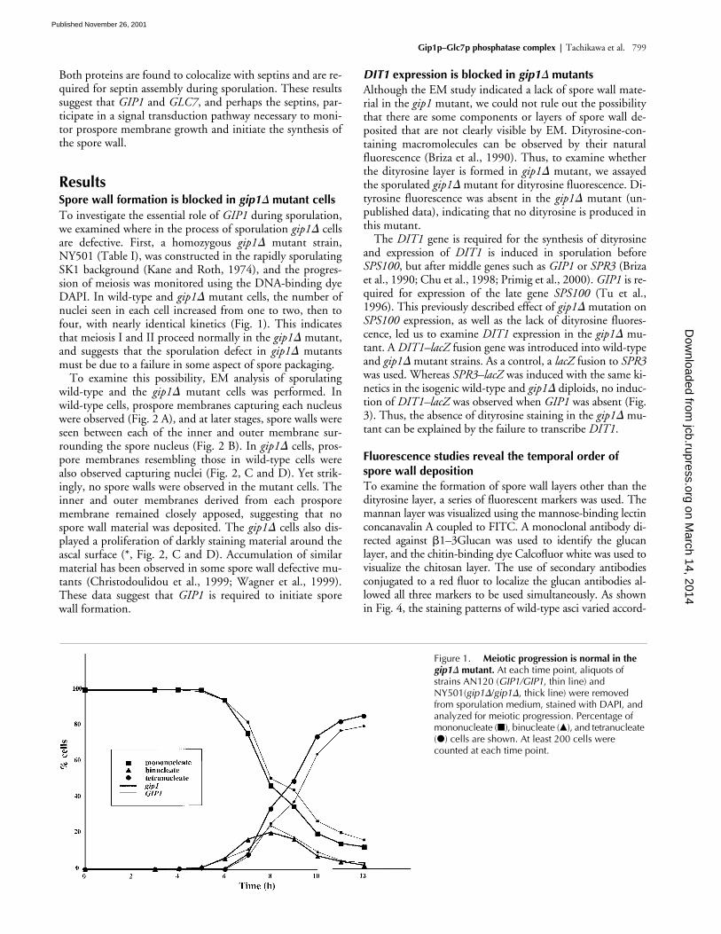
� mutant cellsTo investigate the essential role of GIP1 during sporulation,we examined where in the process of sporulation gip1� cellsare defective. First, a homozygous gip1� mutant strain,NY501 (Table I), was constructed in the rapidly sporulatingSK1 background (Kane and Roth, 1974), and the progres-sion of meiosis was monitored using the DNA-binding dyeDAPI. In wild-type and gip1� mutant cells, the number ofnuclei seen in each cell increased from one to two, then tofour, with nearly identical kinetics (Fig. 1). This indicatesthat meiosis I and II proceed normally in the gip1� mutant,and suggests that the sporulation defect in gip1� mutantsmust be due to a failure in some aspect of spore packaging.
To examine this possibility, EM analysis of sporulatingwild-type and the gip1� mutant cells was performed. Inwild-type cells, prospore membranes capturing each nucleuswere observed (Fig. 2 A), and at later stages, spore walls wereseen between each of the inner and outer membrane sur-rounding the spore nucleus (Fig. 2 B). In gip1� cells, pros-pore membranes resembling those in wild-type cells werealso observed capturing nuclei (Fig. 2, C and D). Yet strik-ingly, no spore walls were observed in the mutant cells. Theinner and outer membranes derived from each prosporemembrane remained closely apposed, suggesting that nospore wall material was deposited. The gip1� cells also dis-played a proliferation of darkly staining material around theascal surface (*, Fig. 2, C and D). Accumulation of similarmaterial has been observed in some spore wall defective mu-tants (Christodoulidou et al., 1999; Wagner et al., 1999).These data suggest that GIP1 is required to initiate sporewall formation.
DIT1 expression is blocked in gip1� mutantsAlthough the EM study indicated a lack of spore wall mate-rial in the gip1 mutant, we could not rule out the possibilitythat there are some components or layers of spore wall de-posited that are not clearly visible by EM. Dityrosine-con-taining macromolecules can be observed by their naturalfluorescence (Briza et al., 1990). Thus, to examine whetherthe dityrosine layer is formed in gip1� mutant, we assayedthe sporulated gip1� mutant for dityrosine fluorescence. Di-tyrosine fluorescence was absent in the gip1� mutant (un-published data), indicating that no dityrosine is produced inthis mutant.
The DIT1 gene is required for the synthesis of dityrosineand expression of DIT1 is induced in sporulation beforeSPS100, but after middle genes such as GIP1 or SPR3 (Brizaet al., 1990; Chu et al., 1998; Primig et al., 2000). GIP1 is re-quired for expression of the late gene SPS100 (Tu et al.,1996). This previously described effect of gip1� mutation onSPS100 expression, as well as the lack of dityrosine fluores-cence, led us to examine DIT1 expression in the gip1� mu-tant. A DIT1–lacZ fusion gene was introduced into wild-typeand gip1� mutant strains. As a control, a lacZ fusion to SPR3was used. Whereas SPR3–lacZ was induced with the same ki-netics in the isogenic wild-type and gip1� diploids, no induc-tion of DIT1–lacZ was observed when GIP1 was absent (Fig.3). Thus, the absence of dityrosine staining in the gip1� mu-tant can be explained by the failure to transcribe DIT1.
Fluorescence studies reveal the temporal order of spore wall depositionTo examine the formation of spore wall layers other than thedityrosine layer, a series of fluorescent markers was used. Themannan layer was visualized using the mannose-binding lectinconcanavalin A coupled to FITC. A monoclonal antibody di-rected against �1–3Glucan was used to identify the glucanlayer, and the chitin-binding dye Calcofluor white was used tovisualize the chitosan layer. The use of secondary antibodiesconjugated to a red fluor to localize the glucan antibodies al-lowed all three markers to be used simultaneously. As shownin Fig. 4, the staining patterns of wild-type asci varied accord-
Figure 1. Meiotic progression is normal in the gip1� mutant. At each time point, aliquots of strains AN120 (GIP1/GIP1, thin line) and NY501(gip1�/gip1�, thick line) were removed from sporulation medium, stained with DAPI, and analyzed for meiotic progression. Percentage of mononucleate (�), binucleate (�), and tetranucleate (�) cells are shown. At least 200 cells were counted at each time point.
on March 14, 2014
jcb.rupress.orgD
ownloaded from
Published November 26, 2001

800 The Journal of Cell Biology | Volume 155, Number 5, 2001
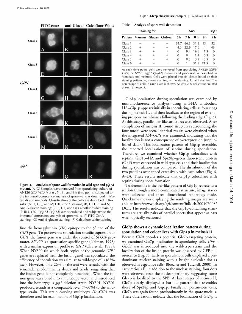
ing to the stage of spore development. After counting and clas-sifying these asci, we ordered the patterns as follows (Table II):when cells are in the stage of the prospore membrane forma-tion and extension, those membranes are stained only withFITC-ConA (Class 2). Approximately 30% of cells showed
this pattern at the seven hour time point. Subsequently, a pop-ulation of cells appears that is positive for both FITC-ConAand anti-�1–3Glucan (Class 3). At the 7- and 8-h time points,�20% of the cells showed staining with anti-�1–3Glucan inthe spore precursors. After a transient period of triply positivestaining (Class 4), the ascus loses anti-�1–3Glucan staining(Class 5). At this point, the FITC-conA staining patternchanges to stain only the spore periphery. Finally, Calcofluorwhite no longer stains the spore wall (Class 6). Therefore,these markers allow all the major spore wall polysaccharides tobe visualized during the course of spore wall formation andsuggest that these components are deposited in a specific se-quence: mannan before glucan before chitosan.
The gip1� mutant fails to form the inner layers of spore wallTo examine the spore wall defect of gip1�, we applied thismethod to gip1� mutant cells. As in wild-type cells, the de-veloping prospore membranes stained with FITC-ConA(Fig. 4, P–R). However, the gip1� mutant cells never pro-gressed to show anti-�1–3Glucan or Calcofluor white stain-ing. Taken together with the lack of the dityrosine layer,these results indicate that the prospore wall in gip1� mutantscontains only those mannoproteins present during prosporemembrane formation. No additional spore wall material isdeposited at the completion of prospore membrane synthesis.
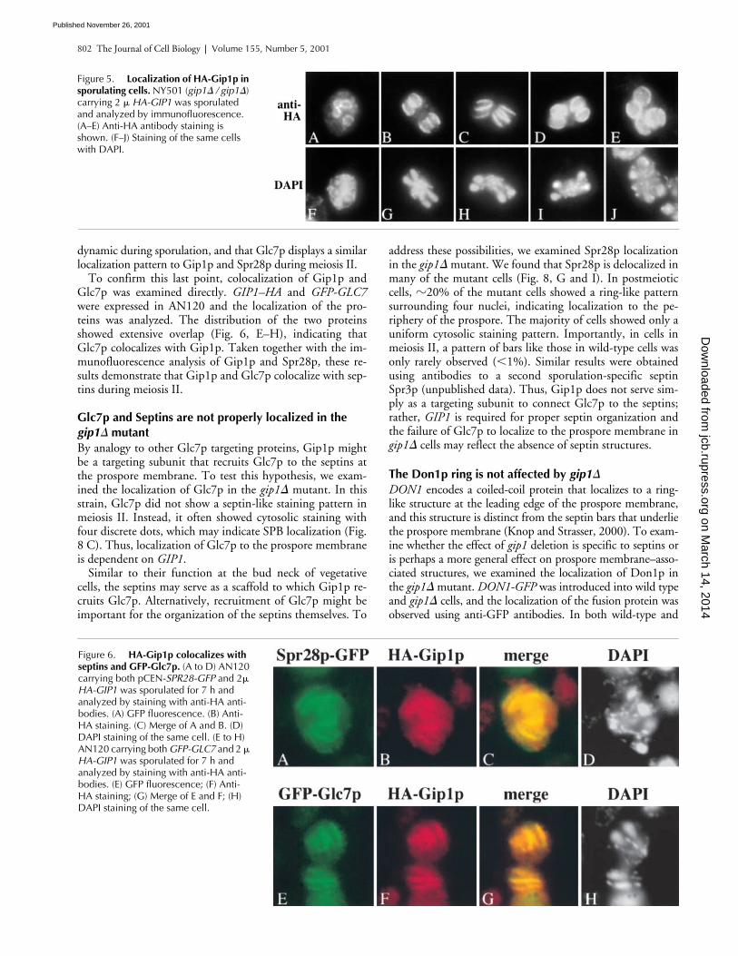
Gip1p colocalizes with Spr28p during spore formationTo examine where in the cell Gip1p functions, the Gip1 pro-tein was localized by immunofluorescence analysis. COOH-terminal–tagged versions of Gip1p were nonfunctional (un-published data). Therefore, targeted integration was used to
Figure 2. The gip1� mutant fails to form spore walls. EM analysis of GIP1 (NH144, A and B) and homozygous gip1� mutant (AN103, C and D) cells at a late stage of sporulation. Prospore membranes (PrM); spore walls (SW); and nuclei (N) are indicated. *, Darkly staining material formed in the ascal cytoplasm of gip1 mutants. Bar, 500 nm.
Figure 3. DIT1–lacZ is not induced in gip1� cells. Sporulating cultures of AN120 (GIP1/GIP1, �) and NY501 (gip1�/gip1�, �) carrying either (A) SPR3–lacZ or (B) DIT1–lacZ were analyzed at various time points for �-galactosidase activity. �-galactosidase activity is given in Miller Units.
on March 14, 2014
jcb.rupress.orgD
ownloaded from
Published November 26, 2001
Gip1p–Glc7p phosphatase complex | Tachikawa et al. 801
fuse the hemagglutinin (HA) epitope to the 5� end of theGIP1 gene. To preserve the sporulation-specific expression ofGIP1, the fusion gene was under the control of SPO20 pro-moter. SPO20 is a sporulation specific gene (Neiman, 1998)with a similar expression profile to GIP1 (Chu et al., 1998).When NY509 (in which both copies of the genomic GIP1genes are replaced with the fusion gene) was sporulated, theefficiency of sporulation was similar to wild-type cells (82%asci). However, only 26% of the asci were tetrads, with theremainder predominantly dyads and triads, suggesting thatthe fusion gene is not completely functional. When the fu-sion gene was cloned into a multicopy vector and introducedinto the homozygous gip1 deletion strain, NY501, NY501produced tetrads at a comparable level (�60%) to the wild-type strain. This strain carrying multicopy HA-GIP1 wastherefore used for examination of Gip1p localization.
Gip1p localization during sporulation was examined byimmunofluorescence analysis using anti-HA antibodies.HA–Gip1p appears initially in sporulating cells as four ringsduring meiosis II, and then localizes to the region of extend-ing prospore membranes following the leading edge (Fig. 5).At this stage, parallel bar-like structures were observed. Aftercompletion of meiosis II, round structures surrounding thefour nuclei were seen. Identical results were obtained whenthe integrated HA–GIP1 was examined, indicating that thelocalization is not a consequence of overexpression (unpub-lished data). This localization pattern of Gip1p resemblesthe reported localization of septins during sporulation.Therefore, we examined whether Gip1p colocalizes withseptins. Gip1p–HA and Spr28p–green fluorescent protein(GFP) were expressed in wild type cells and their localizationduring sporulation was compared. The distribution of thetwo proteins overlapped extensively with each other (Fig. 6,A–D). These results indicate that Gip1p colocalizes withseptins during spore formation.
To determine if the bar-like pattern of Gip1p represents asection through a more complicated structure, image stackswere collected and three dimensional renderings made.Quicktime movies displaying the resulting images are avail-able at http://www.jcb.org/cgi/content/full/jcb.200107008/DC1. The results indicate that the Gip1p-containing struc-tures are actually pairs of parallel sheets that appear as barswhen optically sectioned.
Glc7p shows a dynamic localization pattern during sporulation and colocalizes with Gip1p in meiosis IIBecause GIP1 encodes a potential Glc7p targeting protein,we examined Glc7p localization in sporulating cells. GFP–GLC7 was introduced into the wild-type strain and thelocalization of the fusion protein was observed by GFP flu-orescence (Fig. 7). Early in sporulation, cells displayed a pre-dominant nuclear staining with a bright nucleolar dot asobserved in vegetative cells (Bloecher and Tatchell, 2000). Inearly meiosis II, in addition to the nuclear staining, four dotswere observed near the nuclear periphery suggesting someGlc7p is localized to the SPB. At later stages of meiosis II,Glc7p clearly displayed a bar-like pattern that resemblesthose of Spr28p and Gip1p. Finally, in postmeiotic cells,Glc7p was again found predominantly in the spore nucleus.These observations indicate that the localization of Glc7p is
Figure 4. Analysis of spore wall formation in wild type and gip1� mutant. (A–O) Samples were removed from sporulating culture of AN120 (GIP1/GIP1) at 6-, 7-, 8-, and 9-h time points, subjected to the immunofluorescence analysis of spore walls as described in Ma-terials and methods. Classification of the cells are described in Re-sults. (A, D, G, J, and M) FITC-ConA staining; (B, E, H, K, and N) Anti-�-glucan staining; (C, F, I, L, and O) Calcofluor white staining. (P–R) NY501 (gip1� / gip1�) was sporulated and subjected to the immunofluorescence analysis of spore walls. (P) FITC-ConA staining; (Q) Anti–�-glucan staining; (R) Calcofluor white staining.
Table II. Analysis of spore wall deposition
Staining for GIP1 gip1
Pattern Mannan Glucan Chitosan 6 h 7 h 8 h 9 h 9 h
Class 1 � � � 95.7 66.3 31.8 13 52Class 2 � � 4.3 22.8 17.8 4 48Class 3 F 0 9.4 16.8 7.5 0Class 4 0 0 1.4 0.5 0Class 5 � 0 0.5 0.9 3.5 0Class 6 � F 0 1 31.3 71.5 0
At each time point, cells were removed from sporulating AN120 (GIP1/GIP1) or NY501 (gip1�/gip1�) cultures and processed as described inMaterials and methods. Cells were placed into six classes based on theirstaining pattern. ; strong staining, �, no staining; F, faint staining. Thepercentage of cells in each class is shown. At least 200 cells were countedat each time point.
on March 14, 2014
jcb.rupress.orgD
ownloaded from
Published November 26, 2001
802 The Journal of Cell Biology | Volume 155, Number 5, 2001
dynamic during sporulation, and that Glc7p displays a similarlocalization pattern to Gip1p and Spr28p during meiosis II.
To confirm this last point, colocalization of Gip1p andGlc7p was examined directly. GIP1–HA and GFP-GLC7were expressed in AN120 and the localization of the pro-teins was analyzed. The distribution of the two proteinsshowed extensive overlap (Fig. 6, E–H), indicating thatGlc7p colocalizes with Gip1p. Taken together with the im-munofluorescence analysis of Gip1p and Spr28p, these re-sults demonstrate that Gip1p and Glc7p colocalize with sep-tins during meiosis II.
Glc7p and Septins are not properly localized in the gip1� mutantBy analogy to other Glc7p targeting proteins, Gip1p mightbe a targeting subunit that recruits Glc7p to the septins atthe prospore membrane. To test this hypothesis, we exam-ined the localization of Glc7p in the gip1� mutant. In thisstrain, Glc7p did not show a septin-like staining pattern inmeiosis II. Instead, it often showed cytosolic staining withfour discrete dots, which may indicate SPB localization (Fig.8 C). Thus, localization of Glc7p to the prospore membraneis dependent on GIP1.
Similar to their function at the bud neck of vegetativecells, the septins may serve as a scaffold to which Gip1p re-cruits Glc7p. Alternatively, recruitment of Glc7p might beimportant for the organization of the septins themselves. To
address these possibilities, we examined Spr28p localizationin the gip1� mutant. We found that Spr28p is delocalized inmany of the mutant cells (Fig. 8, G and I). In postmeioticcells, �20% of the mutant cells showed a ring-like patternsurrounding four nuclei, indicating localization to the pe-riphery of the prospore. The majority of cells showed only auniform cytosolic staining pattern. Importantly, in cells inmeiosis II, a pattern of bars like those in wild-type cells wasonly rarely observed (1%). Similar results were obtainedusing antibodies to a second sporulation-specific septinSpr3p (unpublished data). Thus, Gip1p does not serve sim-ply as a targeting subunit to connect Glc7p to the septins;rather, GIP1 is required for proper septin organization andthe failure of Glc7p to localize to the prospore membrane ingip1� cells may reflect the absence of septin structures.
The Don1p ring is not affected by gip1�
DON1 encodes a coiled-coil protein that localizes to a ring-like structure at the leading edge of the prospore membrane,and this structure is distinct from the septin bars that underliethe prospore membrane (Knop and Strasser, 2000). To exam-ine whether the effect of gip1 deletion is specific to septins oris perhaps a more general effect on prospore membrane–asso-ciated structures, we examined the localization of Don1p inthe gip1� mutant. DON1-GFP was introduced into wild typeand gip1� cells, and the localization of the fusion protein wasobserved using anti-GFP antibodies. In both wild-type and
Figure 5. Localization of HA-Gip1p in sporulating cells. NY501 (gip1� / gip1�) carrying 2 � HA-GIP1 was sporulated and analyzed by immunofluorescence. (A–E) Anti-HA antibody staining is shown. (F–J) Staining of the same cells with DAPI.
Figure 6. HA-Gip1p colocalizes with septins and GFP-Glc7p. (A to D) AN120 carrying both pCEN-SPR28-GFP and 2� HA-GIP1 was sporulated for 7 h and analyzed by staining with anti-HA anti-bodies. (A) GFP fluorescence. (B) Anti-HA staining. (C) Merge of A and B. (D) DAPI staining of the same cell. (E to H) AN120 carrying both GFP-GLC7 and 2 � HA-GIP1 was sporulated for 7 h and analyzed by staining with anti-HA anti-bodies. (E) GFP fluorescence; (F) Anti-HA staining; (G) Merge of E and F; (H) DAPI staining of the same cell.
on March 14, 2014
jcb.rupress.orgD
ownloaded from
Published November 26, 2001

Gip1p–Glc7p phosphatase complex | Tachikawa et al. 803
gip1� cells, ring-like structures were observed at the lip of theprospore membranes (Fig. 9). Thus, assembly of the Don1pring is not affected by gip1 deletion, suggesting that the effectsof gip1� are specific to assembly of the septin complex.
The glc7–136 mutant shows similar defect to the gip1� mutantTo determine whether the phenotypes of the gip1� mutantare caused by a failure to properly localize Glc7p or a secondfunction of the Gip1 protein, we examined the sporulationphenotype of a strain carrying the glc7–136 allele. The glc7–136 mutant causes a sporulation defect (Baker et al., 1997),and the protein is defective in interaction with Gip1p (un-published data). Thus, if the sporulation defect of the gip1�mutant is due to altered localization of Glc7p, the glc7–136mutant should display similar phenotypes. EM analysis ofSB247, a glc7–136 mutant in the JC482 background (Bakeret al., 1997), was performed. The cytological phenotype ofglc7–136 was more heterogeneous than gip1�, probably dueto the leaky sporulation defect of the mutant (2% asci).Nonetheless, a significant fraction of the cells (50%) dis-played prospores with no apparent deposition of spore wallmaterial (Fig. 10 A), similar to the gip1� mutant (Fig. 2).Thus, glc7–136 mutant cells display a block to spore walldeposition similar to that seen in the gip1 deletion strain.
To examine the localization of Spr28p and Gip1p in theglc7–136 strain, the glc7–136 mutation was introduced into thesame SK1 strains used for the gip1 analysis. As in the JC482background, the glc7–136 mutant is leaky in SK1, giving 5%asci. SPR28-GFP and HA–GIP1 were introduced into NY527,and the proteins were localized by immunofluorescence. Mostof the cells expressing SPR28-GFP displayed uniform cytosolicstaining. In �30% of the cells, staining of the spore peripherywas observed, as is the case with septin localization in the gip1�mutant (Fig. 10, B and D). HA–Gip1p displayed similar local-ization defects to Spr28p-GFP in the glc7–136 strain (Fig. 10, Fand H). Thus, as with the spore wall, the glc7–136 mutantshows similar septin organization defects to the gip1� mutant.Further, Gip1p and Glc7p are interdependent for localization;interaction of the two proteins is required for either to localizeto the prospore membrane. Taken together, these data indicatethat Gip1p and Glc7p function together to promote both orga-nization of the septins and spore wall formation.
DiscussionGIP1 and GLC7 are required to signal the onset of spore wall formationStrains deleted for GIP1 display two clear phenotypes, a blockto spore wall formation and delocalization of the septins from
Figure 7. Localization of GFP-Glc7p during sporulation. AN120 carrying GFP-GLC7 was sporulated and GFP fluorescence was observed. (A–F) GFP fluorescence; (G–L) DAPI staining of corresponding cells in A–F.
Figure 8. Localization pattern of GFP-Glc7p and Spr28p-GFP are altered in the gip1 mutant. AN120 (GIP1/GIP1) and NY501 (gip1� / gip1�) carrying either GFP-GLC7 or pCEN-SPR28-GFP were sporulated and analyzed by fluorescence microscopy. (A) GFP-Glc7p localization in AN120. (B) DAPI staining of cell in A. (C) GFP-Glc7p localization in NY501. (D) DAPI staining of cells in C. (E) Spr28p-GFP localization in AN120. (F) DAPI staining of cells in E. (G and I) Spr28p-GFP localization in NY501 (H and J) DAPI staining of cells in G and I, respectively.
on March 14, 2014
jcb.rupress.orgD
ownloaded from
Published November 26, 2001

804 The Journal of Cell Biology | Volume 155, Number 5, 2001
the forming prospore membrane. The spore wall phenotypeof GIP1 mutants is a discrete arrest at the initiation of sporewall formation. In wild-type cells, deposition of spore wallmaterial is never seen until after the closure of the prosporemembrane (Lynn and Magee, 1970). Consistent with this cy-tological observation, expression of the DIT1 gene appears tooccur after prospore closure (Briza et al., 1990). These obser-vations suggest that the cell must have some mechanism totrigger spore wall formation in response to prospore mem-brane closure. Given their colocalization with septins, theGip1p–Glc7p holoenzyme would be ideally located to partici-pate in this process. Though we favor the idea that this phos-phatase complex acts as a monitor to signal the closure of the
prospore membrane, we cannot at present rule out the possi-bility that the Gip1p–Glc7p phosphatase is required formembrane closure itself. A defect in either membrane closureor the ability to sense that the membrane has closed would re-sult in a failure to initiate spore wall formation.
CHS3 encodes the chitin synthase responsible for the syn-thesis of the chitosan layer. In contrast to DIT1, the CHS3gene is not transcriptionally induced during sporulation(Pammer et al., 1992). Therefore, the failure of gip1� mu-tants to produce chitosan suggests that GIP1 may be re-quired for posttranslational regulation of this enzyme. Invegetative cells, Chs3p activity is controlled in part by regu-lated recycling through an endosomal compartment (Zimanet al., 1998). It will be of interest to examine the localizationof Chs3p in sporulating wild-type and gip1� cells.
Spore wall formation is an ordered processPrevious studies have shown that Calcofluor white tran-siently stains the chitosan layer of the developing spore(Briza et al., 1990). The loss of Calcoflour white staining inmature asci is due to maturation of the overlying dityrosinelayer blocking access of the dye to its ligand (Briza et al.,1988, 1990). We interpret the transient staining of sporeswith the anti–�-glucan antibody in the same way. That is,spores are positive for staining until the overlying chitosanlayer forms and obscures access of the antibody to the glucanlayer. Surprisingly, staining with FITC-ConA is not lost asspores mature, even though the inner mannan layer shouldalso be obscured as the outer layers form. However, at thesame time that �-glucan staining is lost, the appearance ofthe FITC-ConA staining changes markedly. This changeprobably reflects a loss of access to the mannan layer of theinner spore wall, but residual staining of mannoproteinspresent in the outer prospore membrane or ascal cytoplasmcondensed around the spores. Thus, our immunofluores-cence assay of spore wall formation suggests a definitive or-der of spore wall assembly. Mannoproteins are deposited asthe prospore membrane forms. After prospore membraneclosure, the glucan layer forms, followed by the chitosanlayer and finally the dityrosine layer.
Figure 9. Don1p ring is formed normally in the gip1� mutant. AN120 (GIP1/GIP1) and NY501 (gip1� / gip1�) carrying DON1-GFP were sporulated and analyzed by immunofluorescence using anti-GFP antibodies. (A) Don1p-GFP localization in AN120. (B) DAPI staining of cell in A. (C) Don1p-GFP localization in NY501. (D) DAPI staining of cell in C.
Figure 10. glc7–136 mutants show a similar phenotype to the gip1� mutant. (A) SB247 (glc7–136/glc7–136) at a late stage of sporulation was analyzed by transmission EM. Prospore membranes (PrM) and nucleus (N) are indicated. (B–I) NY527 (glc7–136/glc7–136) harboring either pCEN-SPR28-GFP or 2 � HA-GIP1 was sporulated and analyzed by fluorescence microscopy. (B and D) anti-GFP staining; (C and E) DAPI staining of cells in B and D, respectively; (F and H) Anti-HA staining; (G and I) DAPI staining of cells in F and H, respectively. Bar, 500 nm.
on March 14, 2014
jcb.rupress.orgD
ownloaded from
Published November 26, 2001

Gip1p–Glc7p phosphatase complex | Tachikawa et al. 805
Surprisingly, the layers are deposited from innermost tooutermost, with respect to the spore cytoplasm. This sug-gests that precursors to the chitosan and dityrosine layermust be able to pass through the glucan and mannan layersbefore assembling. Alternatively, some of the spore wall lay-ers may be constructed from precursors introduced from theascal cytoplasm rather than the spore cytosol. This possibil-ity has been suggested previously based on specific localiza-tion of some sporulation-induced transcripts to the ascal cy-toplasm (Kurtz and Lindquist, 1986), and is also consistentwith the accumulation of darkly staining vesicles in the re-gion of the ascal cytoplasm surrounding the prospores that isseen in gip1� (Fig. 2) and other spore wall–defective mu-tants (Christodoulidou et al., 1999; Wagner et al., 1999).
Relationship of GIP1 to other spore wall synthesis genes.Our data indicate that not only is spore wall deposition coor-dinated with respect to prospore membrane closure, but alsoa temporal order of deposition, glucan before chitosan beforedityrosine, must be maintained. Previously reported sporewall–defective mutants fall broadly into one of two catego-ries: those such as dit1 or chs3 that remove specific spore walllayers (Briza et al., 1990; Pammer et al., 1992), or those suchas smk1 and swm1 in which individual spores within an ascusdisplay heterogeneous wall defects (Krisak et al., 1994; Ufanoet al., 1999). The heterogeneous spore wall defects seen inthese latter mutants could result from an inability to coordi-nate the timing of deposition within individual spores.
SMK1 is particularly interesting in this regard. Though dele-tion of SMK1 causes a heterogeneous spore wall phenotype,point mutations produce more homogeneous arrests (Wagneret al., 1999). In the most severe allele, smk1–4, this arrest ap-pears similar to gip1�. Further, it has been reported that forma-tion of the glucan, chitosan, and dityrosine layers requires in-creasing levels of Smk1p activity, respectively (Wagner et al.,1999). As this matches the temporal order of deposition ofthese layers, SMK1 activity might provide a mechanism to con-trol the timing of synthesis of the different layers. Gip1p–Glc7pphosphatase could control the initiation of spore wall synthesis,at least in part, by regulating the activation of Smk1p.
Gip1p–Glc7p phosphatase regulates septin organizationThe Gip1p–Glc7p complex colocalizes with the septins duringprospore membrane growth and the first evident phenotype ofthe gip1� mutation is not the spore wall defect, but a failure toorganize the septins onto the prospore membrane. However,at times after prospore closure, in a fraction of the gip1� mu-tant cells the septins can be found in the spore periphery as inwild-type cells. These results indicate a specific requirementfor the Gip1p–Glc7p phosphatase to organize the septins intostructures associated with the growing prospore membrane.Whether this effect is mediated by direct dephosphorylation ofthe septin proteins or some other modifier of septin organiza-tion is not yet known. In vegetative cells, mutation of severaldifferent protein kinases has effects on organization of the sep-tin ring (Bouquin et al., 2000; Longtine et al., 2000). For in-stance, mutation of GIN4 leads to the arrangement of the sep-tins as a series of parallel bars (Longtine et al., 1998a). It has
not yet been reported whether any of the septin proteins aredirect substrates of any of these kinases. Nonetheless, taken to-gether with our results, it seems likely that phosphorylationand dephosphorylation regulate septin architecture.
The fact that septins are lost from the prospore membranein the gip1� mutant but that the prospore membrane none-theless forms, indicates that the septins are not required formembrane assembly or growth. One possibility is that theseptins function to keep Gip1p–Glc7p associated with theprospore membrane. In this instance, mutation of the septingenes might cause a similar spore wall phenotype to thegip1� mutation. However, no significant sporulation phe-notype has been reported in septin mutants (De Virgilio etal., 1996; Fares et al., 1996). Therefore, it remains to be de-termined whether the spore wall and septin phenotypes ofthe gip1� mutant are linked, or whether they represent twoindependent aspects of Gip1p–Glc7p function.
In higher cells, as in yeast, septins are found at sites of cy-tokinesis (Neufeld and Rubin, 1994; Kinoshita et al., 1997).However, in contrast to mitotic yeast cells, the septins ofhigher cells exhibit dynamic localization and are often foundin association with regions of active membrane growth(Fares et al., 1995; Kinoshita et al., 1997; Hsu et al., 1998).Thus, the behavior of septins during sporulation is similar tothat in higher cells. Our results indicate that the Gip1p–Glc7p complex is required to organize the septins onto theprospore membrane. Given the high degree of conservationof PP1, it will be of interest to learn if septin organization isalso regulated by this enzyme in higher cells.
Bailis and Roeder (2000) have reported that GLC7-dependent dephosphorylation of the synaptonemal complexcomponent Red1p is required for progression through themeiotic pachytene checkpoint. In contrast to our findings,this study reported that gip1� mutants arrest in pachytene.Further, GIP1 was required for the localization of Glc7p tomeiotic chromosomes and overexpression of GLC7 sup-pressed the gip1� sporulation defect. From these results itwas suggested that the function of Gip1p is to target Glc7pto meiotic chromosomes. We found no evidence of a mei-otic arrest in gip1� in two different strain backgrounds, SK1(Fig. 1) and JC482. Additionally, overexpression of GLC7does not relieve the spore formation defect of gip1� mutantsin our strains (unpublished data), as was observed in the BRstrain background (Bailis and Roeder, 2000). The reason forthe differing phenotypes of gip1� in different strain back-grounds remains unclear; however, our results clearly dem-onstrate roles for GIP1 and GLC7 in postmeiotic events.
In sum, our data suggest a model in which Gip1p is in-duced during sporulation, binds to Glc7p, and the Gip1p–Glc7p complex then organizes the septins onto the prosporemembrane. The Gip1p–Glc7p complex remains associatedwith the septins during meiosis II, and this places the phos-phatase in a position to act as a monitor of prospore mem-brane growth. Upon closure of the prospore membrane, wepropose that Gip1p and Glc7p initiate a signaling cascadethat triggers events leading to the synthesis of the spore wall.This includes the induction of DIT1 as well as the activationof chitin and �-glucan synthases. Further work will be nec-essary to elucidate the nature of this intracellular signalingpathway.
on March 14, 2014
jcb.rupress.orgD
ownloaded from
Published November 26, 2001
806 The Journal of Cell Biology | Volume 155, Number 5, 2001
Materials and methodsYeast strains and mediaS. cerevisiae strains used in this work are listed in Table I. AN103 was createdby selection of 5-fluoroorotic acid–resistant derivatives of MCY3259 (Tu et al.,1996). To construct NY501, AN117–4B and AN117–16D (Neiman et al.,2000) were transformed with PvuII-digested pJT26-HIS3 (Tu et al., 1996) toproduce NY1 and NY2, respectively. For this and all other integrations, properarrangement of the genomic locus was confirmed by PCR. These strains aremated to create diploid NY501. To create a strain carrying NH2 terminallyHA-tagged GIP1 under control of the SPO20 promoter, pFA6a-His3MX6-prSPO20-HA was subjected to PCR with HT1 (5�-CTATTTTCTTCCT-CCTTGCCTATTCGTTTGGTGGCTTTCTTTATAATTCGGAATTCGAGCTC-GTTTAAAC-3�) and HT2 (5�-CTTTTCAAAGACTCAAATGGTCTAGCCTTTG-GCTGCAAAATAGTTTCCATTTTGTATAGTTCATCCATGC-3�), and the prod-uct was used to transform AN117–4B and AN117–16D to obtain NY13 andNY14, respectively. These strains were mated to create diploid NY509. Tocreate the DON1–GFP fusion gene in yeast, pFA6a-GFP (S65T)-His3MX6(Longtine et al., 1998b) was amplified with HT15 (5�-AAGATGAAAAAAA-GCAGGTTCATCCATCTAGCAAGAATTAAGTTTTACGCGGATCCCCGGGT-TAATTAA-3�) and HT16 (5�-AAAGGCAAATAAGCGCAGAAGAAGCAA-TGGTGGTGTCTGTTCATTGCGAGGAATTCGAGCTCGTTTAAAC-3�), andthe product was used to transform AN117–4B to create NY17. To constructNY527, the his5 gene of Schizosaccharomyces pombe was amplified frompFA6a-His3MX6 (Longtine et al., 1998b) with HT28 (5�-AACTGGGG-AGAGGAAAGCAGATTACCACAATATACATTCAAATTAAAGAACGGAT-CCCCGGGTTAATTAA-3�) and HT29 (5�-TAATAAGTATTTTCCTTTTTAAA-CTTTGATTTAGGACGTGAATCTATTTAGAATTCGAGCTCGTTTAAAC-3�),and the product was used to transform AN120 to create the glc7 heterozygousdeletion strain, NY523. NY523 was then transformed with pSB14 to createNY525, which has glc7–136 integrated at the ura3 locus. NY525 was sporu-lated and dissected. Progeny that had both the glc7 deletion and the glc7–136integration were identified and mated to produce NY527. Unless otherwisenoted, standard media and growth conditions were used (Rose et al., 1990).
PlasmidsTo create pFA6a-His3MX6-prSPO20-HA, the promoter sequence ofthe SPO20 gene was amplified directly from yeast genome using ANO195(5�-CCTTGAGATCTAAGTCTAGGCGCTTTCAAC-3�) and ANO201(5�-CCTTGTTAATTAAAGACATTATATATCTAAAAATGGC-3�). The PCRproduct was then digested with PacI and BglII and used to replace GAL1promoter region of pFA6a-His3MX6-PGAL1-3HA (Longtine et al., 1998b).To clone the prSPO20-HA-GIP1 fusion gene, genomic DNA from NY13was subjected to PCR with ANO195 and HT6 (5�-CAGTTGCCTACCAAT-GTTTC-3�). The product was cloned into SmaI sites of pRS424 andpRS426 (Christianson et al., 1992) to create pSB5 and pSB6, respectively.pNC160-GFP-GLC7 (Bloecher and Tatchell, 2000) was used to expressGFP-tagged version of Glc7p. pSB7 was created by excising the SPR28–GFP fusion gene from YEplac181-SPR28-GFP (De Virgilio et al., 1996)with EcoRI and BamHI and cloning it into pRS314 (Sikorski and Hieter,1989). To clone DON1–GFP into plasmid, genomic DNA prepared fromNY17 was subjected to PCR using HT10 (5�-AAACAGATCTATATTAC-CCTG-3�) and HT19 (5�-GAAGAATTCGATATAGCTCTGAACAATTC-3�).The product was digested with EcoRI and BglII and ligated into similarlydigested pRS314 to create pSB8. To integrate glc7–136 at the ura3 locus,the glc7–136 gene was cut out from pSB56 (Baker et al., 1997) withHindIII and SacI, and cloned into pRS306, creating pSB14. pSB14 waslinearized with PstI before transformation to target integration to the ura3locus. The plasmids pHindIII-DIT1-lacZ, a gift of J. Segall (University ofToronto, Toronto, Canada), and pGK16 (Holaway et al., 1987) carryingthe DIT1–lacZ and SPR3–lacZ reporters, respectively, were used to moni-tor gene induction.
MicroscopyFor analysis of meiotic progression, cells were sporulated as described(Neiman, 1998). At intervals, aliquots were removed and fixed with 3.7%formaldehyde at 4�C overnight. Cells were collected and mounted in me-dium containing DAPI (Molecular Probes). EM was performed as described(Neiman, 1998). Samples were analyzed using a JEOL 1200EX transmis-sion electron microscope at the Stony Brook University Microscopy Imag-ing Center (Stony Brook, NY).
For immunofluorescence analysis, cells were sporulated and fixedwith 3.7% formaldehyde for 2 h, collected, resuspended in SHA buffer (1 MSorbitol, 0.1 M Hepes, pH 7.5, 5 mM NaN3) and stored at 4�C. Immu-nofluorescence analysis of proteins was performed as described previ-ously (Gao and Dean, 2000). Monoclonal anti-HA antibodies (12CA5)
were provided by N. Dean (SUNY Stony Brook, Stony Brook, NY), andaffinity-purified anti-GFP antibodies, a gift of N. Davis (Louisiana StateUniversity Medical Center, Shreveport, LA), were used at a 1:10 and1:200 dilution, respectively. Anti-Spr3p antibodies (Fares et al., 1996)were used at a 1:10 dilution. Alexa Fluor 488 goat anti–mouse IgG con-jugate, Alexa Fluor 488 goat anti–rabbit IgG conjugate, Alexa Fluor 546goat anti–rabbit IgG conjugate, and Texas red goat anti–rabbit IgG conju-gate (Molecular Probes) were used as secondary antibodies at a 1:400 di-lution.
Immunofluorescence analysis of spore walls was performed as follows.Fixed sporulating cells were suspended in Zymolyase solution (27 �g/mlZymolyase and 0.2% �-mercaptoethanol in SHA buffer) and incubated at30� for 1 h. Cells were then treated 5 min with SHA buffer containing0.1% Triton X-100, and 10 min with SHA buffer containing 0.2 mg/mlCalcofluor white (Sigma-Aldrich), washed twice with SHA buffer, and ad-hered to wells of a polylysine-coated slide. Cells on the slide were treatedwith methanol for 6 min, followed by acetone for 30 s, and then dried. Af-ter blocking with WT buffer (50 mM Hepes, pH 7.5, 150 mM NaCl, 1%Milk, 5 mg/ml bovine serum albumin), cells were incubated with a mono-clonal anti–�-1,3 glucan antibody (Biosupplies) (Humbel et al., 2001) at a1:300 dilution in WT buffer overnight. Wells were washed with PBS-BSA(7 mM Na2HPO4, 3 mM NaH2PO4, 130 mM NaCl, 5 mg/ml bovine serumalbumin) and incubated with PBS-BSA containing 1:400 diluted AlexaFluor 546 goat anti–mouse IgG and 0.1 mg/ml FITC-ConA (Sigma-Aldrich)for 2 h. Finally, wells were washed with PBS-BSA and covered withmounting media. For detection of native GFP fluorescence, cells werefixed with 3.7% formaldehyde for 10 min, washed once with PBS, andmounted with mounting media containing DAPI or processed for immu-nofluorescence.
Immunofluorescence images were acquired using a Zeiss Axioskop withattached SPOT camera (Diagnostic Instruments) and Adobe Photoshop5.0, or on a Zeiss Axioplan 2 microscope with a Zeiss Axiocam and ZeissAxiovision 3.0.6 software. Figures were prepared using Adobe Photoshop6.0 and Canvas 5.0 (Deneba Software).
�-Galactosidase and dityrosine assaysNatural fluorescence of the dityrosine molecules was observed as de-scribed previously (Briza et al., 1990). DIT1–lacZ and SPR3–lacZ wereseparately introduced into both AN120 and NY501. The resulting cellswere sporulated and aliquots were collected at each time point and sub-jected to �-galactosidase assay as described (Stern et al., 1984).
Online supplemental materialTo create three-dimensional projections of Gip1 immunofluorescence, cellscarrying HA–GIP1 were prepared after 7 h in sporulation medium, andstacks of images were collected with a Bio-Rad Radiance 2000 confocal mi-croscroscope (25mW Arlaser, Nikon TE300, 100 X Plan Apo objective 1.4NA). Images were collected at 0.2-�m intervals in the Z-axis at 1.5% laserpower, pinhole aperture at 2.6, Kalman filtration of four images. Three-dimensional projections were created using Bio-Rad Lasersharp2000 soft-ware. Projected images were tilted at angles of �45�–45� at 1�-intervals.
To create three-dimensional projects of HA-Gip1p and DAPI fluores-cence, stacks of images were collected through an Olympus UPlanFl 100 �1.3 NA objective with a Roper Coolmax HQ CCD Camera using Scana-lytics IPlab Spectrum software. Images were collected at 0.2-�m intervalsin the Z-axis, and three-dimensional projections were created using Iplabsoftware. Projected images were tilted at angles of �45�–45� at 5�-inter-vals. Both projections were saved as Apple QuickTime movies, availableat http://www.jcb.org/cgi/content/full/jcb.200107008/DC1
The authors are grateful to J. Pringle (University of North Carolina, ChapelHill, NC), N. Dean (SUNY Stony Brook, Stony Brook, NY), and N. David(Louisiana State University Medical Center, Shreveport, LA) for antibodies,and to H. Friesen and J. Segall (University of Toronto, Toronto, Canada)and N. Hollingsworth (SUNY Stony Brook) for reagents. A.M. Neiman isgrateful to R. Sternglanz (SUNY Stony Brook) for material support duringthe early phases of this work. The authors are also grateful to N. Hollings-worth and J. Engebrecht for comments on the manuscript, and to membersof the Neiman Lab for discussions during the course of this work.
This work was supported by National Institutes of Health grantsGM62184 to A.M. Neiman and GM47789 to K. Tatchell.
Submitted: 2 July 2001Revised: 24 September 2001Accepted: 15 October 2001
on March 14, 2014
jcb.rupress.orgD
ownloaded from
Published November 26, 2001
Gip1p–Glc7p phosphatase complex | Tachikawa et al. 807
ReferencesBailis, J.M., and G.S. Roeder. 2000. Pachytene exit controlled by reversal of Mek1-
dependent phosphorylation. Cell. 101:211–221.Baker, S.H., D.L. Frederick, A. Bloecher, and K. Tatchell. 1997. Alanine-scanning
mutagenesis of protein phosphatase type 1 in the yeast Saccharomyces cerevi-siae. Genetics. 145:615–626.
Bloecher, A., and K. Tatchell. 2000. Dynamic localization of protein phosphatasetype 1 in the mitotic cell cycle of Saccharomyces cerevisiae. J. Cell Biol. 149:125–140.
Bouquin, N., Y. Barral, R. Courbeyrette, M. Blondel, M. Snyder, and C. Mann.2000. Regulation of cytokinesis by the Elm1 protein kinase in Saccharomycescerevisiae. J. Cell Sci. 113:1435–1445.
Briza, P., M. Breitenbach, A. Ellinger, and J. Segall. 1990. Isolation of two devel-opmentally regulated genes involved in spore wall maturation in Saccharomy-ces cerevisiae. Genes Dev. 4:1775–1789.
Briza, P., M. Eckerstorfer, and M. Breitenbach. 1994. The sporulation-specific en-zymes encoded by the DIT1 and DIT2 genes catalyze a two-step reactionleading to a soluble LL-dityrosine-containing precursor of the yeast sporewall. Proc. Natl. Acad. Sci. USA. 91:4524–4528.
Briza, P., A. Ellinger, G. Winkler, and M. Breitenbach. 1988. Chemical composi-tion of the yeast ascospore wall. The second outer layer consists of chitosan.J. Biol. Chem. 263:11569–11574.
Briza, P., G. Winkler, H. Kalchhauser, and M. Breitenbach. 1986. Dityrosine is aprominent component of the yeast ascospore wall. A proof of its structure. J.Biol. Chem. 261:4288–4294.
Christianson, T.W., R.S. Sikorski, M. Dante, J.H. Shero, and P. Hieter. 1992.Multifunctional yeast high-copy-number shuttle vectors. Gene. 110:119–122.
Christodoulidou, A., V. Bouriotis, and G. Thireos. 1996. Two sporulation-specificchitin deacetylase-encoding genes are required for the ascospore wall rigidityof Saccharomyces cerevisiae. J. Biol. Chem. 271:31420–31425.
Christodoulidou, A., P. Briza, A. Ellinger, and V. Bouriotis. 1999. Yeast ascosporewall assembly requires two chitin deacetylase isozymes. FEBS Lett. 460:275–279.
Chu, S., J. DeRisi, M. Eisen, J. Mulholland, D. Botstein, P.O. Brown, and I. Her-skowitz. 1998. The transcriptional program of sporulation in budding yeast.Science. 282:699–705.
De Virgilio, C., D.J. DeMarini, and J.R. Pringle. 1996. SPR28, a sixth member ofthe septin gene family in Saccharomyces cerevisiae that is expressed specificallyin sporulating cells. Microbiology. 142:2897–2905.
DeMarini, D.J., A.E. Adams, H. Fares, C. De Virgilio, G. Valle, J.S. Chuang, andJ.R. Pringle. 1997. A septin-based hierarchy of proteins required for local-ized deposition of chitin in the Saccharomyces cerevisiae cell wall. J. Cell Biol.139:75–93.
Depaoli-Roach, A.A., I.K. Park, V. Cerovsky, C. Csortos, S.D. Durbin, M.J.Kuntz, A. Sitikov, P.M. Tang, A. Verin, and S. Zolnierowicz. 1994. Serine/threonine protein phosphatases in the control of cell function. Adv. EnzymeRegul. 34:199–224.
Fares, H., L. Goetsch, and J.R. Pringle. 1996. Identification of a developmentallyregulated septin and involvement of the septins in spore formation in Sac-charomyces cerevisiae. J. Cell Biol. 132:399–411.
Fares, H., M. Peifer, and J.R. Pringle. 1995. Localization and possible functions ofDrosophila septins. Mol. Biol. Cell. 6:1843–1859.
Feng, Z.H., S.E. Wilson, Z.Y. Peng, K.K. Schlender, E.M. Reimann, and R.J.Trumbly. 1991. The yeast GLC7 gene required for glycogen accumulationencodes a type 1 protein phosphatase. J. Biol. Chem. 266:23796–23801.
Francois, J.M., S. Thompson-Jaeger, J. Skroch, U. Zellenka, W. Spevak, and K.Tatchell. 1992. GAC1 may encode a regulatory subunit for protein phos-phatase type 1 in Saccharomyces cerevisiae. EMBO J. 11:87–96.
Friesen, H., R. Lunz, S. Doyle, and J. Segall. 1994. Mutation of the SPS1-encodedprotein kinase of Saccharomyces cerevisiae leads to defects in transcription andmorphology during spore formation. Genes Dev. 15:2162–2175.
Gao, X.D., and N. Dean. 2000. Distinct protein domains of the yeast Golgi GDP-mannose transporter mediate oligomer assembly and export from the endo-plasmic reticulum. J. Biol. Chem. 275:17718–17727.
Holaway, B.L., G. Kao, M.C. Finn, and M.J. Clancy. 1987. Transcriptional regu-lation of sporulation genes in yeast. Mol. Gen. Genet. 210:449–459.
Hsu, S.C., C.D. Hazuka, R. Roth, D.L. Foletti, J. Heuser, and R.H. Scheller.1998. Subunit composition, protein interactions and structures of the mam-malian brain sec6/8 complex and septin filaments. Neuron. 20:1111–1122.
Humbel, B.M., M. Konomi, T. Takagi, N. Kamasawa, S.A. Ishijima, and M.Osumi. 2001. In situ localization of beta-glucans in the cell wall of Schizosac-
charomyces pombe. Yeast. 18:433–444.Kaback, D.B., and L.R. Feldberg. 1985. Saccharomyces cerevisiae exhibits a sporula-
tion-specific temporal pattern of transcript accumulation. Mol. Cell. Biol.5:751–761.
Kane, S., and R. Roth. 1974. Carbohydrate metabolism during ascospore develop-ment in yeast. J. Bacteriol. 118:8–14.
Kartmann, B., and D. Roth. 2001. Novel roles for mammalian septins: from vesicletrafficking to oncogenesis. J. Cell Sci. 114:839–844.
Kinoshita, M., S. Kumar, A. Mizoguchi, C. Ide, A. Kinoshita, T. Haraguchi, Y.Hiraoka, and M. Noda. 1997. Nedd5, a mammalian septin, is a novel cyto-skeletal component interacting with actin-based structures. Genes Dev. 11:1535–1547.
Klis, F.M. 1994. Review: cell wall assembly in yeast. Yeast. 10:851–869.Knop, M., and K. Strasser. 2000. Role of the spindle pole body of yeast in mediat-
ing assembly of the prospore membrane during meiosis. EMBO J. 19:3657–3667.
Krisak, L., R. Strich, R.S. Winters, J.P. Hall, M.J. Mallory, D. Kreitzer, R.S. Tuan,and E. Winter. 1994. SMK1, a developmentally regulated MAP kinase, isrequired for spore wall assembly in Saccharomyces cerevisiae. Genes Dev.8:2151–2161.
Kupiec, M., B. Byers, R.E. Esposito, and A.P. Mitchell. 1997. Meiosis and sporula-tion in Saccharomyces cerevisiae. In The Molecular and Cellular Biology ofthe Yeast Saccharomyces. Vol. 3. J.R. Pringle, J.R. Broach, and E.W. Jones,editors. Cold Spring Harbor Laboratory Press. Cold Spring Harbor, NY889–1036.
Kurtz, S., and S. Lindquist. 1986. Subcellular differentiation in sporulating yeastcells. Cell. 45:771–779.
Longtine, M.S., D.J. DeMarini, M.L. Valencik, O.S. Al-Awar, H. Fares, C. DeVirgilio, and J.R. Pringle. 1996. The septins: roles in cytokinesis and otherprocesses. Curr. Opin. Cell Biol. 8:106–119.
Longtine, M.S., H. Fares, and J.R. Pringle. 1998a. Role of the yeast Gin4p proteinkinase in septin assembly and the relationship between septin assembly andseptin function. J. Cell Biol. 143:719–736.
Longtine, M.S., A. McKenzie III, D.J. Demarini, N.G. Shah, A. Wach, A. Brachat,P. Philippsen, and J.R. Pringle. 1998b. Additional modules for versatile andeconomical PCR-based gene deletion and modification in Saccharomyces cere-visiae. Yeast. 14:953–961.
Longtine, M.S., C.L. Theesfeld, J.N. McMillan, E. Weaver, J.R. Pringle, and D.J.Lew. 2000. Septin-dependent assembly of a cell cycle-regulatory module inSaccharomyces cerevisiae. Mol. Cell. Biol. 20:4049–4061.
Lynn, R.R., and P.T. Magee. 1970. Development of the spore wall during as-cospore formation in Saccharomyces cerevisiae. J. Cell Biol. 44:688–692.
Mishra, C., C.E. Semino, K.J. McCreath, H. de la Vega, B.J. Jones, C.A. Specht,and P.W. Robbins. 1997. Cloning and expression of two chitin deacetylasegenes of Saccharomyces cerevisiae. Yeast. 13:327–336.
Moens, P.B. 1971. Fine structure of ascospore development in the yeast Saccharo-myces cerevisiae. Can. J. Microbiol. 17:507–510.
Moens, P.B., and E. Rapport. 1971. Spindles, spindle plaques, and meiosis in theyeast Saccharomyces cerevisiae (Hansen). J. Cell Biol. 50:344–361.
Mumby, M.C., and G. Walter. 1993. Protein serine/threonine phosphatases: struc-ture, regulation, and functions in cell growth. Physiol. Rev. 73:673–699.
Neiman, A.M. 1998. Prospore membrane formation defines a developmentallyregulated branch of the secretory pathway in yeast. J. Cell Biol. 140:29–37.
Neiman, A.M., L. Katz, and P.J. Brennwald. 2000. Identification of domains re-quired for developmentally regulated SNARE function in Saccharomyces cere-visiae. Genetics. 155:1643–1655.
Neufeld, T.P., and G.M. Rubin. 1994. The Drosophila peanut gene is required forcytokinesis and encodes a protein similar to yeast putative bud neck filamentproteins. Cell. 77:371–379.
Pammer, M., P. Briza, A. Ellinger, T. Schuster, R. Stucka, H. Feldmann, and M.Breitenbach. 1992. DIT101 (CSD2, CAL1), a cell cycle-regulated yeast generequired for synthesis of chitin in cell walls and chitosan in spore walls. Yeast.8:1089–1099.
Primig, M., R.M. Williams, E.A. Winzeler, G.G. Tevzadze, A.R. Conway, S.Y.Hwang, R.W. Davis, and R.E. Esposito. 2000. The core meiotic transcrip-tome in budding yeasts. Nat. Genet. 26:415–423.
Ramaswamy, N.T., L. Li, M. Khalil, and J.F. Cannon. 1998. Regulation of yeastglycogen metabolism and sporulation by Glc7p protein phosphatase. Genet-ics. 149:57–72.
Rose, M.D., F. Winston, and P. Hieter. 1990. Methods in yeast genetics. ColdSpring Harbor Laboratory Press. Cold Spring Harbor, NY. 198 pp.
Shenolikar, S. 1994. Protein serine/threonine phosphatases—new avenues for cell
on March 14, 2014
jcb.rupress.orgD
ownloaded from
Published November 26, 2001
808 The Journal of Cell Biology | Volume 155, Number 5, 2001
regulation. Annu. Rev. Cell Biol. 10:55–86.Sikorski, R.S., and P. Hieter. 1989. A system of shuttle vectors and yeast host
strains designed for efficient manipulation of DNA in Saccharomyces cerevi-siae. Genetics. 122:19–27.
Stern, M., R. Jensen, and I. Herskowitz. 1984. Five SWI genes are required for ex-pression of the HO gene in yeast. J. Mol. Biol. 178:853–868.
Straight, P.D., T.H. Giddings, Jr., and M. Winey. 2000. Mps1p regulates meioticspindle pole body duplication in addition to having novel roles duringsporulation. Mol. Biol. Cell. 11:3525–3537.
Stuart, J.S., D.L. Frederick, C.M. Varner, and K. Tatchell. 1994. The mutant type1 protein phosphatase encoded by glc7-1 from Saccharomyces cerevisiae failsto interact productively with the GAC1-encoded regulatory subunit. Mol.Cell. Biol. 14:896–905.
Tu, J., W. Song, and M. Carlson. 1996. Protein phosphatase type 1 interacts withproteins required for meiosis and other cellular processes in Saccharomyces
cerevisiae. Mol. Cell. Biol. 16:4199–4206.Ufano, S., P. San-Segundo, F. del Rey, and C.R. Vazquez de Aldana. 1999.
SWM1, a developmentally regulated gene, is required for spore wall assem-bly in Saccharomyces cerevisiae. Mol. Cell. Biol. 19:2118–2129.
Wagner, M., P. Briza, M. Pierce, and E. Winter. 1999. Distinct steps in yeast sporemorphogenesis require distinct SMK1 MAP kinase thresholds. Genetics. 151:1327–1340.
Wagner, M., M. Pierce, and E. Winter. 1997. The CDK-activating kinase CAK1can dosage suppress sporulation defects of smk1 MAP kinase mutants and isrequired for spore wall morphogenesis in Saccharomyces cerevisiae. EMBO J.16:1305–1317.
Ziman, M., J.S. Chuang, M. Tsung, S. Hamamoto, and R. Schekman. 1998.Chs6p-dependent anterograde transport of Chs3p from the chitosome to theplasma membrane in Saccharomyces cerevisiae. Mol. Biol. Cell. 9:1565–1576.
on March 14, 2014
jcb.rupress.orgD
ownloaded from
Published November 26, 2001