A Functional Variant in MicroRNA146a Promoter Modulates Its Expression and Confers Disease Risk for...
11
A Functional Variant in MicroRNA-146a Promoter Modulates Its Expression and Confers Disease Risk for Systemic Lupus Erythematosus Xiaobing Luo 1,2. , Wanling Yang 3. , Dong-Qing Ye 4 , Huijuan Cui 1,2 , Yan Zhang 3 , Nattiya Hirankarn 5 , Xiaoxia Qian 1 , Yuanjia Tang 1 , Yu Lung Lau 3 , Niek de Vries 6 , Paul Peter Tak 6 , Betty P. Tsao 7 , Nan Shen 1 * 1 Joint Molecular Rheumatology Laboratory of the Institute of Health Sciences and Shanghai Renji Hospital, Shanghai Institutes for Biological Sciences, Chinese Academy of Sciences, Shanghai Jiaotong University School of Medicine, Shanghai, China, 2 Key Laboratory of Stem Cell Biology, Shanghai Institutes for Biological Sciences, Chinese Academy of Sciences, Shanghai, China, 3 Department of Paediatrics and Adolescent Medicine, LKS Faculty of Medicine, University of Hong Kong, Hong Kong, China, 4 Department of Epidemiology and Biostatistics, Anhui Medical University School of Public Health, Hefei, Anhui, China, 5 Lupus Research Unit, Department of Microbiology, Faculty of Medicine, Chulalongkorn University, Bangkok, Thailand, 6 Academic Medical Center, University of Amsterdam, Amsterdam, The Netherlands, 7 Division of Rheumatology, Department of Medicine, David Geffen School of Medicine, University of California Los Angeles, Los Angeles, California, United States of America Abstract Systemic lupus erythematosus (SLE) is a complex autoimmune disease with a strong genetic predisposition, characterized by an upregulated type I interferon pathway. MicroRNAs are important regulators of immune homeostasis, and aberrant microRNA expression has been demonstrated in patients with autoimmune diseases. We recently identified miR-146a as a negative regulator of the interferon pathway and linked the abnormal activation of this pathway to the underexpression of miR-146a in SLE patients. To explore why the expression of miR-146a is reduced in SLE patients, we conducted short parallel sequencing of potentially regulatory regions of miR-146a and identified a novel genetic variant (rs57095329) in the promoter region exhibiting evidence for association with SLE that was replicated independently in 7,182 Asians (P meta = 2.74 6 10 28 , odds ratio = 1.29 [1.18–1.40]). The risk-associated G allele was linked to reduced expression of miR-146a in the peripheral blood leukocytes of the controls. Combined functional assays showed that the risk-associated G allele reduced the protein-binding affinity and activity of the promoter compared with those of the promoter containing the protective A allele. Transcription factor Ets-1, encoded by the lupus-susceptibility gene ETS1, identified in recent genome- wide association studies, binds near this variant. The manipulation of Ets-1 levels strongly affected miR-146a promoter activity in vitro; and the knockdown of Ets-1, mimicking its reduced expression in SLE, directly impaired the induction of miR-146a. We also observed additive effects of the risk alleles of miR-146a and ETS1. Our data identified and confirmed an association between a functional promoter variant of miR-146a and SLE. This risk allele had decreased binding to transcription factor Ets-1, contributing to reduced levels of miR-146a in SLE patients. Citation: Luo X, Yang W, Ye D-Q, Cui H, Zhang Y, et al. (2011) A Functional Variant in MicroRNA-146a Promoter Modulates Its Expression and Confers Disease Risk for Systemic Lupus Erythematosus. PLoS Genet 7(6): e1002128. doi:10.1371/journal.pgen.1002128 Editor: Edward K. Wakeland, University of Texas Southwestern Medical Center, United States of America Received October 21, 2010; Accepted April 30, 2011; Published June 30, 2011 Copyright: ß 2011 Luo et al. This is an open-access article distributed under the terms of the Creative Commons Attribution License, which permits unrestricted use, distribution, and reproduction in any medium, provided the original author and source are credited. Funding: This work was partially supported by the National Natural Science Foundation of China 30971632, 81025016; the National High Technology Research and Development Program of China (863 Program) 2007AA02Z123; the Program of the Shanghai Commission of Science and Technology 08JC1414700, 10JC1409300; the National Basic Research Program of China (973 Program) 2007CB947900 (to NS); the Program of the Shanghai Commission of Science and Technology 10ZR1434900 (to HC); the Research Grant Council of Hong Kong GRF HKU781709M (to WY); the National Natural Science Foundation of China 30830089 (to D-QY); and the US National Institutes of Health grant RO1AR043814 (to BPT). YLL thanks the generous donations from Shun Tak District Min Yuen Tong of Hong Kong. NH thanks the Higher Education Research Promotion and National Research University Project of Thailand, Office of the Higher Education Commission (HR1163A). The funders had no role in study design, data collection and analysis, decision to publish, or preparation of the manuscript. Competing Interests: The authors have declared that no competing interests exist. * E-mail: [email protected] . These authors contributed equally to this work. Introduction Systemic lupus erythematosus (SLE) is a chronic autoimmune disease with a complex etiology and diverse clinical manifestations [1]. The role of genetic factors in the SLE risk has long been established, and demonstrated in familial aggregations, twin studies, and sibling recurrence rates [2]. Recently, high-throughput technologies have facilitated genome-wide association studies (GWASs) across different populations. This approach, accompanied by large-scale replications, has not only confirmed the association of many established susceptibility genes, but has also presented convincing evidence of novel genetic loci involved in SLE [3–8]. As members of the Asian Lupus Genetics Consortium, we have also performed a GWAS in Asian populations and have identified variants in ETS1 and WDFY4 that are associated with SLE [9]. A combination of GWAS data from different ethnic groups will clearly provide new insights into the genetics of SLE and further our understanding of the pathogenesis of lupus [10,11]. To use genomic tools to study the mechanisms of SLE, we and others have independently identified a gene expression signature for lupus patients using microarray profiling [12–14], which highlights the pathogenic role of the abnormal activation of the PLoS Genetics | www.plosgenetics.org 1 June 2011 | Volume 7 | Issue 6 | e1002128
Transcript of A Functional Variant in MicroRNA146a Promoter Modulates Its Expression and Confers Disease Risk for...

A Functional Variant in MicroRNA-146a PromoterModulates Its Expression and Confers Disease Risk forSystemic Lupus ErythematosusXiaobing Luo1,2., Wanling Yang3., Dong-Qing Ye4, Huijuan Cui1,2, Yan Zhang3, Nattiya Hirankarn5,
Xiaoxia Qian1, Yuanjia Tang1, Yu Lung Lau3, Niek de Vries6, Paul Peter Tak6, Betty P. Tsao7, Nan Shen1*
1 Joint Molecular Rheumatology Laboratory of the Institute of Health Sciences and Shanghai Renji Hospital, Shanghai Institutes for Biological Sciences, Chinese Academy
of Sciences, Shanghai Jiaotong University School of Medicine, Shanghai, China, 2 Key Laboratory of Stem Cell Biology, Shanghai Institutes for Biological Sciences, Chinese
Academy of Sciences, Shanghai, China, 3 Department of Paediatrics and Adolescent Medicine, LKS Faculty of Medicine, University of Hong Kong, Hong Kong, China,
4 Department of Epidemiology and Biostatistics, Anhui Medical University School of Public Health, Hefei, Anhui, China, 5 Lupus Research Unit, Department of
Microbiology, Faculty of Medicine, Chulalongkorn University, Bangkok, Thailand, 6 Academic Medical Center, University of Amsterdam, Amsterdam, The Netherlands,
7 Division of Rheumatology, Department of Medicine, David Geffen School of Medicine, University of California Los Angeles, Los Angeles, California, United States of
America
Abstract
Systemic lupus erythematosus (SLE) is a complex autoimmune disease with a strong genetic predisposition, characterizedby an upregulated type I interferon pathway. MicroRNAs are important regulators of immune homeostasis, and aberrantmicroRNA expression has been demonstrated in patients with autoimmune diseases. We recently identified miR-146a as anegative regulator of the interferon pathway and linked the abnormal activation of this pathway to the underexpression ofmiR-146a in SLE patients. To explore why the expression of miR-146a is reduced in SLE patients, we conducted short parallelsequencing of potentially regulatory regions of miR-146a and identified a novel genetic variant (rs57095329) in thepromoter region exhibiting evidence for association with SLE that was replicated independently in 7,182 Asians(Pmeta = 2.7461028, odds ratio = 1.29 [1.18–1.40]). The risk-associated G allele was linked to reduced expression of miR-146ain the peripheral blood leukocytes of the controls. Combined functional assays showed that the risk-associated G allelereduced the protein-binding affinity and activity of the promoter compared with those of the promoter containing theprotective A allele. Transcription factor Ets-1, encoded by the lupus-susceptibility gene ETS1, identified in recent genome-wide association studies, binds near this variant. The manipulation of Ets-1 levels strongly affected miR-146a promoteractivity in vitro; and the knockdown of Ets-1, mimicking its reduced expression in SLE, directly impaired the induction ofmiR-146a. We also observed additive effects of the risk alleles of miR-146a and ETS1. Our data identified and confirmed anassociation between a functional promoter variant of miR-146a and SLE. This risk allele had decreased binding totranscription factor Ets-1, contributing to reduced levels of miR-146a in SLE patients.
Citation: Luo X, Yang W, Ye D-Q, Cui H, Zhang Y, et al. (2011) A Functional Variant in MicroRNA-146a Promoter Modulates Its Expression and Confers Disease Riskfor Systemic Lupus Erythematosus. PLoS Genet 7(6): e1002128. doi:10.1371/journal.pgen.1002128
Editor: Edward K. Wakeland, University of Texas Southwestern Medical Center, United States of America
Received October 21, 2010; Accepted April 30, 2011; Published June 30, 2011
Copyright: � 2011 Luo et al. This is an open-access article distributed under the terms of the Creative Commons Attribution License, which permits unrestricteduse, distribution, and reproduction in any medium, provided the original author and source are credited.
Funding: This work was partially supported by the National Natural Science Foundation of China 30971632, 81025016; the National High Technology Researchand Development Program of China (863 Program) 2007AA02Z123; the Program of the Shanghai Commission of Science and Technology 08JC1414700,10JC1409300; the National Basic Research Program of China (973 Program) 2007CB947900 (to NS); the Program of the Shanghai Commission of Science andTechnology 10ZR1434900 (to HC); the Research Grant Council of Hong Kong GRF HKU781709M (to WY); the National Natural Science Foundation of China30830089 (to D-QY); and the US National Institutes of Health grant RO1AR043814 (to BPT). YLL thanks the generous donations from Shun Tak District Min YuenTong of Hong Kong. NH thanks the Higher Education Research Promotion and National Research University Project of Thailand, Office of the Higher EducationCommission (HR1163A). The funders had no role in study design, data collection and analysis, decision to publish, or preparation of the manuscript.
Competing Interests: The authors have declared that no competing interests exist.
* E-mail: [email protected]
. These authors contributed equally to this work.
Introduction
Systemic lupus erythematosus (SLE) is a chronic autoimmune
disease with a complex etiology and diverse clinical manifestations
[1]. The role of genetic factors in the SLE risk has long been
established, and demonstrated in familial aggregations, twin studies,
and sibling recurrence rates [2]. Recently, high-throughput
technologies have facilitated genome-wide association studies
(GWASs) across different populations. This approach, accompanied
by large-scale replications, has not only confirmed the association of
many established susceptibility genes, but has also presented
convincing evidence of novel genetic loci involved in SLE [3–8].
As members of the Asian Lupus Genetics Consortium, we have also
performed a GWAS in Asian populations and have identified
variants in ETS1 and WDFY4 that are associated with SLE [9]. A
combination of GWAS data from different ethnic groups will clearly
provide new insights into the genetics of SLE and further our
understanding of the pathogenesis of lupus [10,11].
To use genomic tools to study the mechanisms of SLE, we and
others have independently identified a gene expression signature
for lupus patients using microarray profiling [12–14], which
highlights the pathogenic role of the abnormal activation of the
PLoS Genetics | www.plosgenetics.org 1 June 2011 | Volume 7 | Issue 6 | e1002128

type I interferon (IFN) pathway in human lupus [15–17].
Intriguingly, recent investigations suggest a genetic contribution
to the variability observed among individuals in the production
and signaling of IFN [17], and advances in the genetics of SLE
highlight the strong association between the risk of developing
lupus and gene variants connected to the production and effects of
type I IFN [11,18].
We recently used a microRNA (miRNA) profiling assay to
examine the involvement of miRNAs in SLE, because miRNAs
are novel gene expression regulators [19] and important players in
shaping the immune response [20–22]. This profiling identified a
reduction in miR-146a expression in lupus patients, and we showed
that the underexpression of miR-146a contributes to lupus
pathogenesis by deregulating the activation of the IFN pathway
[23]. However, why miR-146a levels are reduced in patients with
SLE remains unresolved. miR-146a is encoded at 5q33.3.
Interestingly, recent data from GWASs in both European and
Asian populations have indicated that this region is a novel
susceptibility locus for SLE [3,7,9], suggesting a plausible role for a
genetic variant around miR-146a in modulating its expression and
thus the disease risk.
Several studies have demonstrated unambiguously that genetic
variants in miRNA precursors (pre-miRNA) can affect miRNA
expression levels by interfering with the miRNA maturation
process and are thus associated with disease susceptibility [24–26].
We postulate that genetic variants in both the miRNA promoter
and the precursor region may alter mature miRNA production.
Given the critical regulatory role of miR-146a in the type I IFN
pathway and the abovementioned genetic association between this
pathway and SLE susceptibility, polymorphisms in the miR-146a
gene could also potentially confer a disease risk. To assess whether
genetic variants modulate miR-146a expression and thus contrib-
ute to the risk of developing SLE, we sequenced the promoter and
key regulatory regions of the miR-146a precursor to identify
potential functional variants that might be associated with SLE
susceptibility. Our subsequent replication and functional studies
provide evidence that single-nucleotide polymorphism (SNP)
rs57095329 in the miR-146a promoter, which affects its mature
level, can confer SLE susceptibility.
Results
Discovery of SLE-associated miR-146a promoter SNPsmiR-146a is located at 5q33.3. The transcription start site (TSS)
of its primary transcript (pri-miR-146a) has been identified [27]. To
characterize the essential regulatory region for subsequent genetic
analysis, we first cloned miR-146a upstream fragments with
variable 59 ends into the pGL3-basic reporter plasmid to analyze
its promoter activity. We found that the inclusion of a fragment
from nucleotide (nt) 21,091 to nt 2611, which contains a known
NF-kB-binding site characterized in THP-1 cells [27], was
consistently robust to promote luciferase activity in HeLa cells
(Figure S1). The inclusion of the more distal region (nt 21,998 to
nt 21,091) enhanced neither the basal nor phorbol myristate
acetate and ionomycin (hereafter referred to as ‘‘PMA+Iono’’) -
induced activity of the promoter (Figure S1B). Therefore, to look
for new genetic variants and to characterize their potential
association with SLE, we designed four pairs of primers with
which to sequence the upstream region that spans the 1,105-bp
promoter (nt 21,091 to nt +14) and the consecutive first exon of
pri-miR-146a (Figure S1A), in 360 individuals (180 SLE patients
and 180 controls), which served as the discovery panel. We also
sequenced the 452-bp region centered on miR-146a precursor or
exon 2 of pri-miR-146a, because it potentially affects mature miR-
146a production [26], in the same discovery panel. A total of 12
variants were identified, with nine already reported in the dbSNP
database Build 130 (Table S1). Five variants had a minor allele
frequency (MAF) of .1% (rs17057381, rs73318382, rs57095329,
rs6864584, and rs2910164; Figure S1A). Therefore, we extended
our sequencing analysis to examine these five SNPs in up to 816
patients and 1,080 controls, who were all Chinese Han individuals
living in Shanghai. In this expanded study panel, only rs73318382
and rs57095329 showed an association with SLE (Table S2).
These two SNPs are separated by 304 bp and are in strong linkage
disequilibrium (LD; r2 = 0.81; Figure S2). When a Bonferroni
correction was applied, the association of rs57095329 with SLE
remained highly significant (P = 461024). Given that rs57095329
is identified through our candidate region sequencing approach
and not included in the HapMap database, it is not surprising that
this SNP has not been included in commercial SNP arrays.
Because published GWASs in SLE of both Asian and European
populations detected association signals at rs2431697 and
rs2431099 (Figure S3), 15 kb and 8 kb upstream from miR-146a
TSS, respectively, we extended our genotyping of rs2431697 and
rs2431099 using 1,896 Shanghai samples. Both SNPs showed
significant association with the disease (Table S2), while
rs57095329 produced the best association signal among the three
SNPs in the same dataset (Table S2). Therefore, we focused on
rs57095329 in the subsequent experiments.
Replicated association of rs57095329 with SLE inindependent cohorts
We replicated the association between rs57095329 and SLE
using a TaqMan genotyping assay in another two panels from
Hong Kong, China, and Bangkok, Thailand. We also added 1,536
patients from the central China area to our mainland China
cohort, and the newly added patients showed an allele frequency
for rs57095329 very similar to that in the discovery panel (MAFs
of 20.53% and 20.77%, respectively). This replication provided
consistent evidence for the association, revealed by an allelic
association analysis (Table 1). When all the samples were included
Author Summary
Genome-wide association studies have identified quite anumber of susceptibility loci associated with complexdiseases such as systemic lupus erythematosus (SLE).However, for most of them, the intrinsic link betweengenetic variation and disease mechanism is not fullyunderstood. SLE is characterized by a significantly upre-gulated type I interferon (IFN) pathway, and we havepreviously reported that underexpression of a microRNA,miR-146a, contributes to alterations in the type I IFNpathway in lupus patients. Here we identified a novelgenetic variant in the promoter region of miR-146a that isdirectly related to reduced expression of miR-146a and isassociated with SLE susceptibility. The risk allele of thisvariant confers weaker binding affinity for Ets-1, which is atranscription factor encoded by a lupus susceptibility genefound in recent GWAS. These findings suggest thatreduced expression of Ets-1 and its reduced bindingaffinity to the miR-146a promoter both may contribute tolow levels of this microRNA in SLE patients, which maycontribute to the upregulated type I IFN pathway in thesepatients. To our knowledge, this is also the first piece ofevidence showing association between a genetic variant ina promoter region of a miRNA gene and a human disease.
Functional Variant in MiR-146a Associated with SLE
PLoS Genetics | www.plosgenetics.org 2 June 2011 | Volume 7 | Issue 6 | e1002128
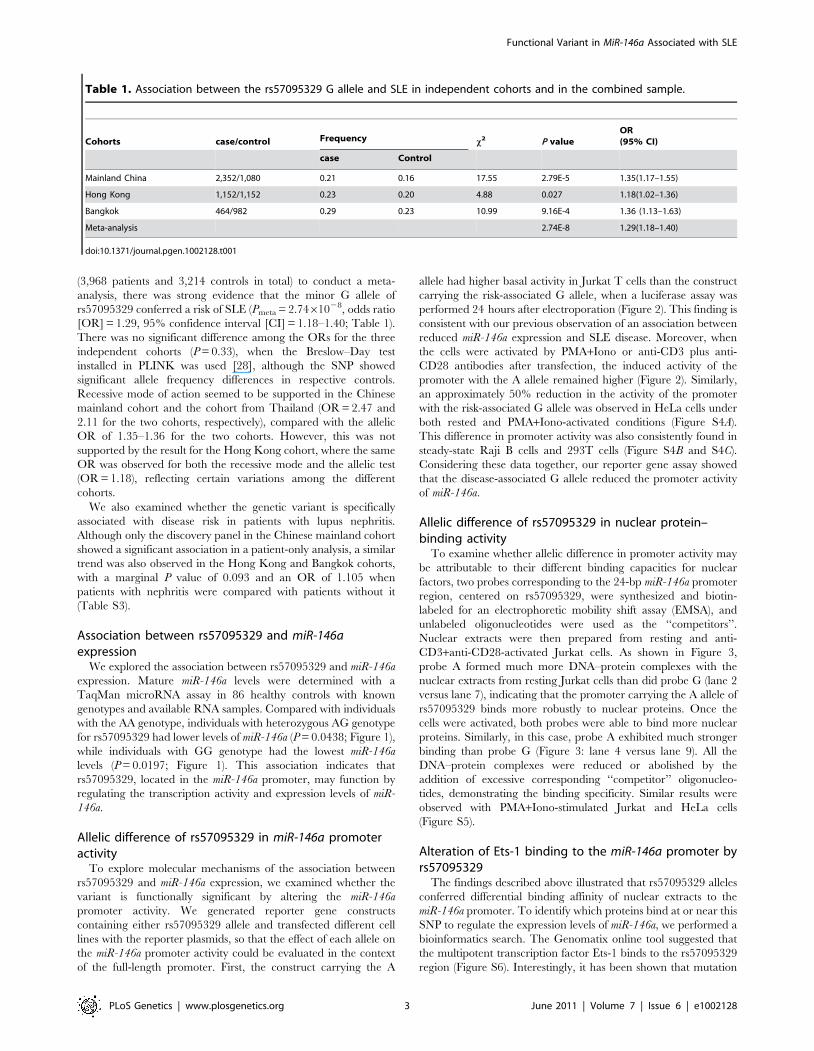
(3,968 patients and 3,214 controls in total) to conduct a meta-
analysis, there was strong evidence that the minor G allele of
rs57095329 conferred a risk of SLE (Pmeta = 2.7461028, odds ratio
[OR] = 1.29, 95% confidence interval [CI] = 1.18–1.40; Table 1).
There was no significant difference among the ORs for the three
independent cohorts (P = 0.33), when the Breslow–Day test
installed in PLINK was used [28], although the SNP showed
significant allele frequency differences in respective controls.
Recessive mode of action seemed to be supported in the Chinese
mainland cohort and the cohort from Thailand (OR = 2.47 and
2.11 for the two cohorts, respectively), compared with the allelic
OR of 1.35–1.36 for the two cohorts. However, this was not
supported by the result for the Hong Kong cohort, where the same
OR was observed for both the recessive mode and the allelic test
(OR = 1.18), reflecting certain variations among the different
cohorts.
We also examined whether the genetic variant is specifically
associated with disease risk in patients with lupus nephritis.
Although only the discovery panel in the Chinese mainland cohort
showed a significant association in a patient-only analysis, a similar
trend was also observed in the Hong Kong and Bangkok cohorts,
with a marginal P value of 0.093 and an OR of 1.105 when
patients with nephritis were compared with patients without it
(Table S3).
Association between rs57095329 and miR-146aexpression
We explored the association between rs57095329 and miR-146a
expression. Mature miR-146a levels were determined with a
TaqMan microRNA assay in 86 healthy controls with known
genotypes and available RNA samples. Compared with individuals
with the AA genotype, individuals with heterozygous AG genotype
for rs57095329 had lower levels of miR-146a (P = 0.0438; Figure 1),
while individuals with GG genotype had the lowest miR-146a
levels (P = 0.0197; Figure 1). This association indicates that
rs57095329, located in the miR-146a promoter, may function by
regulating the transcription activity and expression levels of miR-
146a.
Allelic difference of rs57095329 in miR-146a promoteractivity
To explore molecular mechanisms of the association between
rs57095329 and miR-146a expression, we examined whether the
variant is functionally significant by altering the miR-146a
promoter activity. We generated reporter gene constructs
containing either rs57095329 allele and transfected different cell
lines with the reporter plasmids, so that the effect of each allele on
the miR-146a promoter activity could be evaluated in the context
of the full-length promoter. First, the construct carrying the A
allele had higher basal activity in Jurkat T cells than the construct
carrying the risk-associated G allele, when a luciferase assay was
performed 24 hours after electroporation (Figure 2). This finding is
consistent with our previous observation of an association between
reduced miR-146a expression and SLE disease. Moreover, when
the cells were activated by PMA+Iono or anti-CD3 plus anti-
CD28 antibodies after transfection, the induced activity of the
promoter with the A allele remained higher (Figure 2). Similarly,
an approximately 50% reduction in the activity of the promoter
with the risk-associated G allele was observed in HeLa cells under
both rested and PMA+Iono-activated conditions (Figure S4A).
This difference in promoter activity was also consistently found in
steady-state Raji B cells and 293T cells (Figure S4B and S4C).
Considering these data together, our reporter gene assay showed
that the disease-associated G allele reduced the promoter activity
of miR-146a.
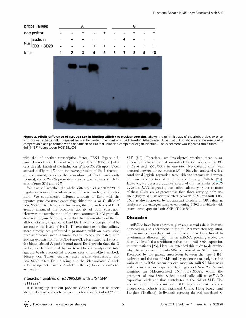
Allelic difference of rs57095329 in nuclear protein–binding activity
To examine whether allelic difference in promoter activity may
be attributable to their different binding capacities for nuclear
factors, two probes corresponding to the 24-bp miR-146a promoter
region, centered on rs57095329, were synthesized and biotin-
labeled for an electrophoretic mobility shift assay (EMSA), and
unlabeled oligonucleotides were used as the ‘‘competitors’’.
Nuclear extracts were then prepared from resting and anti-
CD3+anti-CD28-activated Jurkat cells. As shown in Figure 3,
probe A formed much more DNA–protein complexes with the
nuclear extracts from resting Jurkat cells than did probe G (lane 2
versus lane 7), indicating that the promoter carrying the A allele of
rs57095329 binds more robustly to nuclear proteins. Once the
cells were activated, both probes were able to bind more nuclear
proteins. Similarly, in this case, probe A exhibited much stronger
binding than probe G (Figure 3: lane 4 versus lane 9). All the
DNA–protein complexes were reduced or abolished by the
addition of excessive corresponding ‘‘competitor’’ oligonucleo-
tides, demonstrating the binding specificity. Similar results were
observed with PMA+Iono-stimulated Jurkat and HeLa cells
(Figure S5).
Alteration of Ets-1 binding to the miR-146a promoter byrs57095329
The findings described above illustrated that rs57095329 alleles
conferred differential binding affinity of nuclear extracts to the
miR-146a promoter. To identify which proteins bind at or near this
SNP to regulate the expression levels of miR-146a, we performed a
bioinformatics search. The Genomatix online tool suggested that
the multipotent transcription factor Ets-1 binds to the rs57095329
region (Figure S6). Interestingly, it has been shown that mutation
Table 1. Association between the rs57095329 G allele and SLE in independent cohorts and in the combined sample.
Cohorts case/control Frequency x2 P valueOR(95% CI)
case Control
Mainland China 2,352/1,080 0.21 0.16 17.55 2.79E-5 1.35(1.17–1.55)
Hong Kong 1,152/1,152 0.23 0.20 4.88 0.027 1.18(1.02–1.36)
Bangkok 464/982 0.29 0.23 10.99 9.16E-4 1.36 (1.13–1.63)
Meta-analysis 2.74E-8 1.29(1.18–1.40)
doi:10.1371/journal.pgen.1002128.t001
Functional Variant in MiR-146a Associated with SLE
PLoS Genetics | www.plosgenetics.org 3 June 2011 | Volume 7 | Issue 6 | e1002128

of this predicted Ets-1-binding site resulted in a great reduction in
the activity of an miR-146a promoter–reporter gene [29]. Here, we
performed the following assays and further confirmed the pivotal
role of Ets-1 in regulating miR-146a expression: the transient
expression of Ets-1 greatly enhanced the reporter gene activity
from the full-length miR-146a promoter in Jurkat cells, compared
Figure 1. Comparison of miR-146a expression levels between groups of healthy individuals with different rs57095329 genotypes.The horizontal line indicates the mean expression level within each group. * indicates P,0.05.doi:10.1371/journal.pgen.1002128.g001
Figure 2. Allelic difference of rs57095329 in miR-146a promoter activity. Shown is a schematic representation of reporter gene constructsdriven by the full-length miR-146a promoter containing one or other of the rs57095329 alleles (upper) and the relative luciferase activity of the twoconstructs in Jurkat cells in both the steady (medium) and activated states (lower). For activation, the cells were stimulated with PMA+Iono or withanti-CD3+anti-CD28 antibodies for 6 hours (see Materials and Methods). The data shown are means 6 SEM and are representative of threeindependent experiments performed in triplicate. ** indicates P,0.01.doi:10.1371/journal.pgen.1002128.g002
Functional Variant in MiR-146a Associated with SLE
PLoS Genetics | www.plosgenetics.org 4 June 2011 | Volume 7 | Issue 6 | e1002128
with that of another transcription factor, PBX1 (Figure 4A);
knockdown of Ets-1 by small interfering RNA (siRNA) in Jurkat
cells directly impaired the induction of pri-miR-146a upon T-cell
activation (Figure 4B); and the overexpression of Ets-1 dramati-
cally enhanced, whereas the knockdown of Ets-1 consistently
reduced, the miR-146a promoter–reporter gene activity in HeLa
cells (Figure S7A and S7B).
We assessed whether the allelic difference of rs57095329 in
regulatory activity is attributable to different binding affinity for
Ets-1. We cotransfected different amounts of Ets-1 with the
reporter gene construct containing either the A or G allele of
rs57095329 into HeLa cells. Increasing the protein levels of Ets-1
greatly enhanced the promoter activity of both constructs.
However, the activity ratios of the two constructs (G/A) gradually
decreased (Figure S8), suggesting that the inferior ability of the G-
allele-containing sequence to bind Ets-1 could be compensated by
increasing the levels of Ets-1. To examine the binding affinity
more directly, we performed a promoter pulldown assay using
streptavidin-conjugated agarose beads. When incubated with
nuclear extracts from anti-CD3+anti-CD28-activated Jurkat cells,
the biotin-labeled A probe bound more Ets-1 protein than the G
probe, as demonstrated by western blotting analysis of total
agarose beads precipitated proteins with an anti-Ets-1 antibody
(Figure 4C). Taken together, these results demonstrate that
rs57095329 alters Ets-1 binding, and the risk-associated G allele
is less competent than the A allele in the regulation of miR-146a
expression.
Interaction analysis of rs57095329 with ETS1 SNPrs1128334
It is intriguing that our previous GWAS and that of others
identified an association between a functional variant of ETS1 and
SLE [8,9]. Therefore, we investigated whether there is an
interaction between the risk variants of the two genes, rs1128334
in ETS1 and rs57095329 in miR-146a. No epistatic effect was
detected between the two variants (P = 0.46), when analyzed with a
conditional logistic regression test, with the interaction between
the two variants treated as a covariate using PLINK [28].
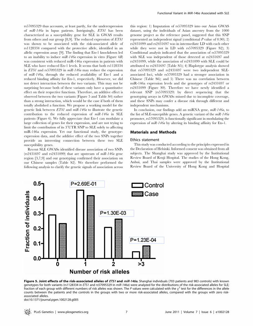
However, we observed additive effects of the risk alleles of miR-
146a and ETS1, suggesting that individuals carrying two or more
of these alleles are at greater risk than those carrying only one
allele (Figure 5). This additive effect between ETS1 and miR-146a
SNPs is also supported by a consistent increase in OR values in
analysis of the enlarged samples containing 4,302 individuals with
known genotypes for both SNPs (Table S4).
Discussion
miRNAs have been shown to play an essential role in immune
homeostasis, and aberrations in the miRNA-mediated regulation
of immune-cell development and function has been linked to
autoimmune diseases [30]. In an miRNA profiling study, we
recently identified a significant reduction in miR-146a expression
in lupus patients [23]. Here, we extended this study to determine
why the expression of miR-146a is reduced in SLE patients.
Prompted by the genetic association between the type I IFN
pathway and the risk of SLE and by evidence that polymorphic
variants in miRNA precursors can modulate miRNA biogenesis
and disease risk, we sequenced key regions of pri-miR-146a and
identified an SLE-associated SNP, rs57095329, within the
promoter of miR-146a, which functionally affects miR-146a
expression levels and thus contributes to the risk of SLE. The
association of this variant with SLE was consistent in three
independent cohorts from mainland China, Hong Kong, and
Bangkok (Thailand). Individuals carrying the risk-associated G
Figure 3. Allelic difference of rs57095329 in binding affinity to nuclear proteins. Shown is a gel-shift assay of the allelic probes (A or G)with nuclear extracts (N.E.) prepared from either rested (medium) or anti-CD3+anti-CD28-activated Jurkat cells. Also shown are the results of acompetition assay performed with the addition of 100-fold unlabeled competitor oligonucleotides. The experiment was repeated three times.doi:10.1371/journal.pgen.1002128.g003
Functional Variant in MiR-146a Associated with SLE
PLoS Genetics | www.plosgenetics.org 5 June 2011 | Volume 7 | Issue 6 | e1002128

allele tended to express lower levels of miR-146a. To the best of our
knowledge, this is the first report of an association between a
functional genetic variant in an miRNA promoter and a human
disease. It will be interesting to investigate the association between
rs57095329 and SLE in other ethnic groups. Another functional
variant located in the miR-146a precursor, rs2910164, has been
associated with cancer development [26,31,32], but showed no
significant association with SLE in our initial sequencing
experiments.
Among the multiple immunological aberrations present in lupus
patients, the type I IFN system is thought to play a crucial role in
its pathogenesis [15–17]. Intriguingly, a number of genes involved
in IFN signaling have already been associated with various
autoimmune diseases, including SLE [33]. Functional variants in
genes encoding key components of the IFN pathway, such as
TYK2, IRF5, and STAT4, have been identified and characterized,
and their association with SLE has been extensively replicated
[34–39]. Our recent work characterized the role of miR-146a as a
negative regulator of the type I IFN pathway by targeting key
signaling proteins [23]. Here, the delineation of an SLE-
susceptible variant of the miR-146a promoter further supports
the notion that polymorphic variants linked to IFN pathway
molecules contribute to the pathogenesis of lupus. miR-146a is
embedded in a non-coding RNA with a previously unknown
function, so our findings highlight the importance of exploring
genetic variants in such regions, which have been more or less
ignored in previous genetic studies.
Our findings underline the regulatory role of Ets-1 in miR-146a
expression, and attribute the allelic difference of rs57095329 to
different affinity for Ets-1. Rs57095329 is not located at the core
sequence of the Ets-1-binding site (Figure S6), so it only causes an
affinity difference, whereas Ets-1 recognition is still well preserved.
Nevertheless, the risk-associated G allele of rs57095329 does affect
the strongly conserved A residue near the Ets-1 core motif (Figure
S6), highlighting the relevance of this SNP. Besides, this is a germ-
line regulatory polymorphism and thus potentially functions in
each cell type, as reflected in our consistent observation of the
reduced activity of a reporter gene carrying the risk-associated G
allele in various cell lines (Figure 2 and Figure S4). The attenuation
of the promoter activity by the risk-associated G allele of
Figure 4. Ets-1 regulates miR-146a expression and accounts for the different regulatory activities of the rs57095329 alleles. (A)Comparison of the miR-146a promoter–reporter gene activities in Jurkat cells after cotransfection with an equal amount of irrelevant carrier vector orETS1- or PBX1-expressing vector. The data shown are means 6 SEM and are representative of three independent experiments. * indicates P,0.05. (B)Real-time PCR analysis of the fold induction of pri-miR-146a in Jurkat cells after the transfection of ETS1 siRNA (#1 and #2) or a negative control (NC),followed by cell activation with anti-CD3+anti-CD28 antibodies for 9 hours. The data shown are means 6 SEM and are representative of threeindependent experiments. * indicates P,0.05. (C) Streptavidin–agarose pulldown assay of transcription factors bound to allelic probes of rs57095329.Biotinylated A or G probes were incubated with nuclear extracts from anti-CD3+anti-CD28-antibody-activated Jurkat cells in the presence ofstreptavidin–agarose beads. The precipitated proteins were analyzed by western blotting with an anti-Ets-1 antibody (upper). The assay was repeatedthree times. For experiment #1, the nuclear extracts (N.E.) that were used as input for the pulldown assay were also directly blotted as the control.Also shown is a comparison of the relative densitometry values for the blotted bands corresponding to the allelic probes (lower). * indicates P,0.05.(D) Western blot analysis of Ets-1 levels in Jurkat cells after transfection of the same amounts of the indicated expression vectors or siRNA as used in Aor B, respectively. The cells were collected 24 hours or 72 hours after transfection for overexpression or for siRNA-mediated knockdown assays,respectively. GAPDH was used as the loading control.doi:10.1371/journal.pgen.1002128.g004
Functional Variant in MiR-146a Associated with SLE
PLoS Genetics | www.plosgenetics.org 6 June 2011 | Volume 7 | Issue 6 | e1002128
rs57095329 thus accounts, at least partly, for the underexpression
of miR-146a in lupus patients. Intriguingly, ETS1 has been
characterized as a susceptibility gene for SLE in GWAS results
from others and our group [8,9]. The reduced expression of ETS1
was shown to be associated with the risk-associated allele of
rs1128334 compared with the protective allele, identified in an
allelic expression assay [9]. The finding that Ets-1 knockdown led
to an inability to induce miR-146a expression in vitro (Figure 4B)
was consistent with reduced miR-146a expression in patients with
SLE who have reduced Ets-1 levels. It seems that both rs1128334
in ETS1 and rs57095329 in miR-146a may reduce the expression
of miR-146a, through the reduced availability of Ets-1 and a
reduced binding affinity for Ets-1, respectively. However, we did
not detect interaction between the two variants. This may not be
surprising because both of these variants only have a quantitative
effect on their respective functions. Therefore, an additive effect is
observed between the two variants (Figure 5 and Table S4) rather
than a strong interaction, which would be the case if both of them
totally abolished a function. We propose a working model for the
genetic link between ETS1 and miR-146a to illustrate the genetic
contribution to the reduced expression of miR-146a in SLE
patients (Figure 6). We fully appreciate that Ets-1 can modulate a
large collection of genes for their expression, and are not trying to
limit the contribution of its 39UTR SNP to SLE solely to affecting
miR-146a expression. Yet our functional study, the genotype-
expression data, and the additive effect of the two SNPs together
provide an interesting connection between these two SLE
susceptibility genes.
Recent SLE GWASs identified disease association of two SNPs
(rs2431697 and rs2431099) that are upstream of miR-146a gene
region [3,7,9] and our genotyping confirmed their association on
our Chinese samples (Table S2). We therefore performed the
following analysis to clarify the genetic signals of association across
this region: 1) Imputation of rs57095329 into our Asian GWAS
dataset, using the individuals of Asian ancestry from the 1000
genome project as the reference panel, suggested that this SNP
represented an independent signal (conditional P value of 0.90); 2)
rs2431099 and rs2431697 was in intermediate LD with each other
while they were not in LD with rs57095329 (Figure S2); 3)
Conditional analysis indicated that the association of rs57095329
with SLE was independent of those detected at rs2431697 and
rs2431099, while the association of rs2431099 with SLE could be
attributed to rs2431697 (Table S5); 4) Haplotype analysis showed
that rs57095329 and rs2431697 were two independent SLE-
associated loci, while rs57095329 had a stronger association in
Chinese (Table S6); and 5) There was no correlation between
miR-146a expression levels and the genotypes of rs2431697 or
rs2431099 (Figure S9). Therefore we have newly identified a
relevant SNP (rs57095329) by direct sequencing that the
genotyping arrays in GWASs missed due to incomplete coverage,
and these SNPs may confer a disease risk through different and
independent mechanisms.
In conclusion, our findings add an miRNA gene, miR-146a, to
the list of SLE-susceptible genes. A genetic variant of the miR-146a
promoter, rs57095329, is functionally significant in modulating the
expression of miR-146a by altering its binding affinity for Ets-1.
Materials and Methods
Ethics statementThis study was conducted according to the principles expressed in
the Declaration of Helsinki. Informed consent was obtained from all
subjects. The Shanghai study was approved by the Institutional
Review Board of Renji Hospital. The studies of the Hong Kong,
Anhui, and Thai samples were approved by the Institutional
Review Board of the University of Hong Kong and Hospital
Figure 5. Joint effects of the risk-associated alleles of ETS1 and miR-146a. Shanghai individuals (703 patients and 983 controls) with knowngenotypes for both variants (rs1128334 in ETS1 and rs57095329 in miR-146a) were analyzed for the distributions of the risk-associated alleles for SLE;fraction of each group with different numbers of risk alleles was shown. The P values were calculated with the x2 test for the differences in the allelecounts between the patients and the controls in the groups with two or more risk-associated alleles, compared with the groups with zero risk-associated alleles.doi:10.1371/journal.pgen.1002128.g005
Functional Variant in MiR-146a Associated with SLE
PLoS Genetics | www.plosgenetics.org 7 June 2011 | Volume 7 | Issue 6 | e1002128

Authority, Hong Kong West Cluster, New Territory West Cluster,
and Hong Kong East Cluster; the Research Ethics Committee of
Anhui Medical University; and the Ethics Committee of the Faculty
of Medicine, Chulalongkorn University, respectively.
SubjectsWe recruited 816 SLE patients and 1,080 sex- and age-matched
controls, all of whom were from the Chinese Han population in
Shanghai, China. Other Chinese mainland samples consisted of
1,536 SLE patients living in central China, collected by
collaborators in Anhui province. For the independent replications,
samples collected by collaborators in Hong Kong (case vs control:
1,152 vs 1,152) and Bangkok, Thailand (464 vs 982, respectively)
were included. All SLE patients fulfilled the American College of
Rheumatology (ACR) classification criteria for SLE, and 1,254
patients met the ACR criteria for lupus nephritis.
DNA sequencing and genotypingConsecutive overlapping amplicons corresponding to the miR-
146a promoter region were amplified from genomic DNA
Figure 6. Diagram of the genetic link between ETS1 and miR-146a and genetic contribution to miR-146a expression. Expression ofcellular miR-146a depends on the binding of Ets-1 to the miR-146a promoter, so both the availability of and its binding affinity for Ets-1 could affectmiR-146a expression. We previously found that the A allele of rs1128334 located in the ETS1 39 untranslated region is associated with SLE by reducingEts-1 expression (upper panel). In this study, we show that the binding affinity for Ets-1 is affected by the risk-associated G allele of rs57095329, whichcauses an incompetent compositional change (lower panel), and therefore represents another genetic factor that contributes to the reducedexpression of miR-146a in SLE patients.doi:10.1371/journal.pgen.1002128.g006
Functional Variant in MiR-146a Associated with SLE
PLoS Genetics | www.plosgenetics.org 8 June 2011 | Volume 7 | Issue 6 | e1002128
extracted from peripheral blood leukocytes. The products were
purified and directly sequenced on a 3730 automated sequencer
(Applied Biosystems). The 452-bp DNA region around the miR-
146a precursor was also amplified and sequenced. The primer
pairs used are shown in Table S7.
In the replication stage, SNP rs57095329 was genotyped with
the specified TaqMan SNP genotyping probes (Applied Biosys-
tems). The assay was run on a 7900HT sequence detection system
(Applied Biosystems) and the data were analyzed with the affiliated
SDS software, version 2.3. The genotypes of rs57095329 were
found to be in Hardy–Weinberg equilibrium (P.0.01) in the
controls of all three cohorts. The average call rate for all samples
was 92%.
Real-time PCRTotal RNA was extracted from peripheral blood leukocytes or
cultured cells using TRIzol (Invitrogen), followed by reverse
transcription using a reverse transcriptase kit obtained from
Takara. To determine the quantity of pri-miR-146a, the cDNA was
amplified by real-time PCR with SYBR Green RT–PCR kit
(Takara), and the expression of RPL13A was used as the internal
control. The primers used are shown in Table S7. To determine
the quantity of mature miR-146a, the specific TaqMan MicroRNA
Assay kit (Applied Biosystems) was used, and the expression levels
were normalized to snRNA U6. The assays were performed on a
7900HT real-time instrument (Applied Biosystems). Relative
expression levels were calculated using the 22DDCt method.
ConstructsTo create the miR-146a promoter–luciferase reporter con-
structs, three fragments of variable lengths, corresponding to the
upstream region of the TSS of pri-miR-146a, were amplified and
cloned into the pGL3-basic luciferase vector (Promega). To
compare the activities of miR-146a promoters containing the
different rs57095329 alleles, the full-length 1105-bp fragment was
amplified from individual homozygous templates. The ETS1
overexpression vector was a kind gift from Dr Gang Pei, and the
PBX1 overexpression plasmid was created by replacing the
inserted ETS1 sequence with the PBX1 coding sequence. The
primers used are shown in Table S7. All constructs were verified
by sequencing.
Cell culture, transfection, and stimulationJurkat and Raji cells were grown in RPMI 1640 medium
supplemented with 10% fetal bovine serum. These two cell lines
were electroporated with 2 mg of the indicated luciferase reporter
vector and 0.2 mg of a modified pRL-TK vector, using a
nucleofector device (Amaxa). Alternatively, the reporter gene
vectors were electroporated in combination with 1.5 mg of an
ETS1- or PBX1-expressing vector. For the knockdown of ETS1,
3 mg of ETS1 siRNA or negative control oligonucleotides (all from
GenePharma, Shanghai) were transfected. HeLa and 293T cells
were grown in Dulbecco’s modified Eagle’s medium supplemented
with 10% fetal bovine serum. These two cell lines were transfected
using Lipofectamine 2000 (Invitrogen), with the ETS1- or PBX1-
expressing vector or ETS1 siRNA alone, or in combination with
50 ng of the indicated luciferase reporter vector and 5 ng of a
modified pRL-TK plasmid. Where indicated, an irrelevant
‘‘carrier’’ vector was added to ensure that equal total amounts of
plasmid DNA were transfected among the groups. For cell
activation, Jurkat and HeLa cells were stimulated with PMA
(100 ng/mL; Sigma) and ionomycin (1 mM; Sigma) for the
indicated times. Alternatively, Jurkat cells were activated with
plate-bound anti-CD3 antibody (coating solution: 5 mg/mL;
eBioscience) and soluble anti-CD28 antibody (2 mg/mL;
eBioscience).
Reporter gene assayCells were cultured for 24 hours or 48 hours after transfec-
tion with the reporter gene vectors together with the ETS1
expression vector or siRNA, respectively. The cells were then
maintained resting or activated for 6 hours and lysed. Their
luciferase activity was measured on a luminometer (LB960;
Berthold) using the Dual-Luciferase Reporter Assay System
(Promega). The ratio of firefly luciferase to Renilla luciferase was
calculated for each well.
EMSAJurkat and HeLa cells (16107) were activated or left to rest for
2 hours, and then their nuclear proteins were extracted with a
Nuclear Extract Kit (Active Motif), according to the manufactur-
er’s protocol. The protein concentrations were determined with
the DC Protein Assay Kit (Bio-Rad). Double-stranded allelic
probes were synthesized and labeled with biotin by Takara (the
sequence is shown in Figure S6). EMSA was carried out with a gel-
shift kit purchased from Active Motif. The competition assay was
performed by adding cognate unlabeled oligonucleotides. After
incubation, the protein–DNA complexes were separated on a
nondenaturing 6% polyacrylamide gel and then transferred to a
nitrocellulose membrane (Millipore). The signals were detected
using a luminoimage analyzer.
Streptavidin–agarose pulldown and western blottingThe pulldown assay was performed following a protocol
described elsewhere [40], with slight modification. Biotin-labeled
allelic probes were incubated with equal amounts of nuclear
extract from activated Jurkat cells for 2 hours at room tempera-
ture, in the presence of streptavidin–agarose beads (GE Health-
care) and protein inhibitors. The precipitated protein–DNA
complex was dissociated from the agarose beads by suspending
the pellet in Laemmli sample buffer (Bio-Rad) and heating it. The
supernatants were then subjected to SDS–PAGE. The proteins
were transferred onto a PVDF membrane (Bio-Rad), blotted with
an anti-Ets-1 antibody, and detected with ECL solution (Pierce).
To evaluate the Ets-1 protein levels after the transfection of the
overexpression vectors or siRNAs, the Jurkat and HeLa cells were
lysed in RIPA buffer (Thermo Scientific), and the supernatants
were similarly used for immunoblotting. Anti-ETS1, anti-
GAPDH, and horseradish-peroxidase-conjugated secondary anti-
bodies were all obtained from Santa Cruz Biotechnology.
Data analysisFor single SNP analysis, PLINK was used for the basic allelic
test and other tests in the patients and the controls [28]. LD
patterns were analyzed and displayed with HaploView [41].
Review manager was used to perform meta-analysis. IMPUTE
version 2 was used to perform imputation. Other data were
analyzed with GraphPad Prism 4 software, version 4.03. The
nonparametric Mann–Whitney test was used to compare miR-
146a expression between the genotype groups, and an unpaired t
test was used to compare reporter gene activities. Two-tailed P
values,0.05 were considered to be statistically significant.
Supporting Information
Figure S1 Illustration of the miR-146a genomic region investi-
gated to identify common variants by direct sequencing. (A)
Functional Variant in MiR-146a Associated with SLE
PLoS Genetics | www.plosgenetics.org 9 June 2011 | Volume 7 | Issue 6 | e1002128

Genomic structure of the miR-146a gene. The two gray boxes
represent exons of pri-miR-146a, whereas the black box represents
pre-miR-146a. The gray lines (underneath the genomic structure)
indicate the genomic regions that were amplified for sequence
analysis. The locations of the five SNPs with minor allele
frequencies of .0.01 are shown. TSS, transcription start site. (B)
Schematic representation of reporter gene constructs driven by
various miR-146a upstream fragments (left) and their corresponding
relative luciferase activities in HeLa cells (right), under rested
(medium) and PMA+Iono-activated conditions. The data shown
are means 6 SEM and are representative of three independent
experiments performed in triplicate.
(TIF)
Figure S2 Linkage disequilibrium of six common SNPs in or
upstream of the miR-146a promoter. Data are based on 816 SLE
patients and 1,080 controls from Shanghai and were analyzed with
HaploView.
(PNG)
Figure S3 Plot of 2log10 P values for SNPs genotyped in the
GWAS spanning 5q33.3 region. Data were from three GWAS on
both Asian (A) and European population (B and C). The linkage
disequilibrium in the region derived from the Asian GWAS data is
shown with r2 values as indicated.
(PNG)
Figure S4 Reporter gene activity of constructs containing either
rs57095329 allele in different cell lines. Shown are the relative
luciferase activities of the two constructs driven by the miR-146a
promoter containing either rs57095329 allele (A or G) in rested
(medium) and activated (with the addition of PMA+Iono for 6 hours)
HeLa cells (A), steady-state Raji cells (B), and steady-state 293T cells
(C). The data shown are means 6 SEM and are representative of
three independent experiments performed in triplicate or quadrupli-
cate. * indicates P,0.05, ** P,0.01, *** P,0.0001.
(TIF)
Figure S5 Gel-shift assay of allelic probes with nuclear proteins
from different cell lines. Shown is a comparison of the binding
affinities of different rs57095329 alleles for the nuclear extracts
(N.E.) from rested (medium) or PMA+Iono-activated HeLa cells
(left), and from PMA+Iono-activated Jurkat cells (right). Also shown
are the results of a competition assay, which was performed with
the addition of 50- to 200-fold unlabeled cognate oligonucleotides.
The assays were repeated at least three times.
(TIF)
Figure S6 Predicted binding sites of Ets-1 on the miR-146a
promoter. (A) The 200-nt sequence around rs57095329 (A/G) was
used as the input for the Genomatix online tool, which predicted a
nearby Ets-1-binding site (indicated by the blue box, with the red
letters indicating the core sequence). Also shown is the probe
sequence for the EMSA, indicated by the pink line below the
sequence. (B) Conservation of rs57095329 residue and Ets-1
binding site. Shown is the UCSC Genome Bioinformatics search
result by alignment of the sequence around rs5705329 (indicated
by the red box) in 7 species.
(TIF)
Figure S7 Analysis of the regulation of miR-146a expression by
Ets-1 in HeLa cells. (A) Comparisons of the miR-146a promoter–
reporter gene activity after cotransfection of an equal amount of an
irrelevant carrier vector or an ETS1- or PBX1-expressing vector.
The data shown are means 6 SEM and are representative of three
independent experiments performed in triplicate. *** indicates
P,0.001. (B) Comparisons of the miR-146a promoter–reporter gene
activity after the cotransfection of ETS1 siRNA (siETS1 #1 and
siETS1 #2) or a negative control (NC). The data shown are means
6 SEM and are representative of three independent experiments
performed in triplicate. * indicates P,0.05. (C) Western blot analysis
of Ets-1 levels after the transfection of the indicated expression
vectors or siRNA. In the overexpression assay, the cells were
collected 24 hours after transfection; in the siRNA-mediated
knockdown assay, the cells were collected 48 hours after transfec-
tion. GAPDH was used as the loading control.
(TIF)
Figure S8 Effect of ectopic Ets-1 expression on the activity of the
allelic miR-146a promoter–reporter gene constructs. Reporter gene
constructs containing the A or G miR-146a sequence were
cotransfected into HeLa cells with different amounts of ETS1-
expressing vector (0, 50, or 200 ng). For these three groups, 200 ng,
150 ng, or 0 ng of an irrelevant carrier vector was cotransfected,
respectively, so that equal amounts of total plasmid DNA were used
in all groups. The relative luciferase activity was measured 24 hours
after transfection (upper). Cotransfection of a PBX1-expressing vector
was used as the negative control. Also shown are the average G/A
ratios of the luciferase activity of the allelic constructs (lower).
(TIF)
Figure S9 Comparison of miR-146a expression levels in healthy
individuals with different genotypes of rs2431697 or rs2431099.
The horizontal line indicates the mean expression level within
each group.
(JPG)
Table S1 A list of the variants identified by the initial sequencing
of the miR-146a region.
(DOC)
Table S2 Association between the seven common SNPs around
miR-146a and SLE.
(DOC)
Table S3 Association between the rs57095329 G allele and
lupus nephritis.
(DOC)
Table S4 Analysis of OR in case-control groups carrying
different numbers of risk alleles of either miR-146a or ETS1 SNP.
(DOC)
Table S5 Conditional analysis of three SNPs in 5q33.3 in SLE
cases and controls.
(DOC)
Table S6 Haplotypic association of three SNPs in 5q33.3 with
SLE.
(DOC)
Table S7 A list of the primers used for the various assays.
(DOC)
Acknowledgments
We thank all the subjects for their participation in this study. We also thank
Dr. Gang Pei (Tongji University) for providing the ETS1-expressing vector
and Dr. Jian Zhao and Dr. Yun Deng (University of California, Los
Angeles) for assistance with data analysis.
Author Contributions
Conceived and designed the experiments: XL NS. Performed the
experiments: XL WY HC YZ XQ. Analyzed the data: XL WY HC NS.
Contributed reagents/materials/analysis tools: NS WY D-QY NH YLL
BPT XL HC YZ YT NdV PPT. Wrote the paper: XL WY BPT NS.
Functional Variant in MiR-146a Associated with SLE
PLoS Genetics | www.plosgenetics.org 10 June 2011 | Volume 7 | Issue 6 | e1002128
References
1. D’Cruz DP, Khamashta MA, Hughes GR (2007) Systemic lupus erythematosus.Lancet 369: 587–596.
2. Rahman A, Isenberg DA (2008) Systemic lupus erythematosus. N Engl J Med358: 929–939.
3. Harley JB, Alarcon-Riquelme ME, Criswell LA, Jacob CO, Kimberly RP, et al.(2008) Genome-wide association scan in women with systemic lupus erythema-
tosus identifies susceptibility variants in ITGAM, PXK, KIAA1542 and other
loci. Nat Genet 40: 204–210.4. Hom G, Graham RR, Modrek B, Taylor KE, Ortmann W, et al. (2008)
Association of systemic lupus erythematosus with C8orf13-BLK and ITGAM-ITGAX. N Engl J Med 358: 900–909.
5. Graham RR, Cotsapas C, Davies L, Hackett R, Lessard CJ, et al. (2008) Genetic
variants near TNFAIP3 on 6q23 are associated with systemic lupuserythematosus. Nat Genet 40: 1059–1061.
6. Kozyrev SV, Abelson AK, Wojcik J, Zaghlool A, Linga Reddy MV, et al. (2008)Functional variants in the B-cell gene BANK1 are associated with systemic lupus
erythematosus. Nat Genet 40: 211–216.
7. Gateva V, Sandling JK, Hom G, Taylor KE, Chung SA, et al. (2009) A large-scale replication study identifies TNIP1, PRDM1, JAZF1, UHRF1BP1 and
IL10 as risk loci for systemic lupus erythematosus. Nat Genet 41: 1228–1233.8. Han JW, Zheng HF, Cui Y, Sun LD, Ye DQ, et al. (2009) Genome-wide
association study in a Chinese Han population identifies nine new susceptibilityloci for systemic lupus erythematosus. Nat Genet 41: 1234–1237.
9. Yang W, Shen N, Ye DQ, Liu Q, Zhang Y, et al. (2010) Genome-wide
association study in Asian populations identifies variants in ETS1 and WDFY4associated with systemic lupus erythematosus. PLoS Genet 6: e1000841.
doi:10.1371/journal.pgen.1000841.10. Crow MK (2008) Collaboration, genetic associations, and lupus erythematosus.
N Engl J Med 358: 956–961.
11. Flesher DL, Sun X, Behrens TW, Graham RR, Criswell LA (2010) Recentadvances in the genetics of systemic lupus erythematosus. Expert Rev Clin
Immunol 6: 461–479.12. Bennett L, Palucka AK, Arce E, Cantrell V, Borvak J, et al. (2003) Interferon
and granulopoiesis signatures in systemic lupus erythematosus blood. J Exp Med197: 711–723.
13. Han GM, Chen SL, Shen N, Ye S, Bao CD, et al. (2003) Analysis of gene
expression profiles in human systemic lupus erythematosus using oligonucleotidemicroarray. Genes Immun 4: 177–186.
14. Baechler EC, Batliwalla FM, Karypis G, Gaffney PM, Ortmann WA, et al.(2003) Interferon-inducible gene expression signature in peripheral blood cells of
patients with severe lupus. Proc Natl Acad Sci U S A 100: 2610–2615.
15. Pascual V, Farkas L, Banchereau J (2006) Systemic lupus erythematosus: allroads lead to type I interferons. Curr Opin Immunol 18: 676–682.
16. Ronnblom L, Eloranta ML, Alm GV (2006) The type I interferon system insystemic lupus erythematosus. Arthritis Rheum 54: 408–420.
17. Crow MK (2007) Type I interferon in systemic lupus erythematosus. Curr TopMicrobiol Immunol 316: 359–386.
18. Ronnblom L, Alm GV, Eloranta ML (2009) Type I interferon and lupus. Curr
Opin Rheumatol 21: 471–477.19. Bartel DP (2004) MicroRNAs: genomics, biogenesis, mechanism, and function.
Cell 116: 281–297.20. Baltimore D, Boldin MP, O’Connell RM, Rao DS, Taganov KD (2008)
MicroRNAs: new regulators of immune cell development and function. Nat
Immunol 9: 839–845.21. Lodish HF, Zhou B, Liu G, Chen CZ (2008) Micromanagement of the immune
system by microRNAs. Nat Rev Immunol 8: 120–130.22. Xiao C, Rajewsky K (2009) MicroRNA control in the immune system: basic
principles. Cell 136: 26–36.23. Tang Y, Luo X, Cui H, Ni X, Yuan M, et al. (2009) MicroRNA-146A
contributes to abnormal activation of the type I interferon pathway in human
lupus by targeting the key signaling proteins. Arthritis Rheum 60: 1065–1075.
24. Duan R, Pak C, Jin P (2007) Single nucleotide polymorphism associated with
mature miR-125a alters the processing of pri-miRNA. Hum Mol Genet 16:
1124–1131.
25. Mencia A, Modamio-Hoybjor S, Redshaw N, Morin M, Mayo-Merino F, et al.
(2009) Mutations in the seed region of human miR-96 are responsible for
nonsyndromic progressive hearing loss. Nat Genet 41: 609–613.
26. Jazdzewski K, Murray EL, Franssila K, Jarzab B, Schoenberg DR, et al. (2008)
Common SNP in pre-miR-146a decreases mature miR expression and
predisposes to papillary thyroid carcinoma. Proc Natl Acad Sci U S A 105:
7269–7274.
27. Taganov KD, Boldin MP, Chang KJ, Baltimore D (2006) NF-kappaB-
dependent induction of microRNA miR-146, an inhibitor targeted to signaling
proteins of innate immune responses. Proc Natl Acad Sci U S A 103:
12481–12486.
28. Purcell S, Neale B, Todd-Brown K, Thomas L, Ferreira MA, et al. (2007)
PLINK: a tool set for whole-genome association and population-based linkage
analyses. Am J Hum Genet 81: 559–575.
29. Curtale G, Citarella F, Carissimi C, Goldoni M, Carucci N, et al. (2010) An
emerging player in the adaptive immune response: microRNA-146a is a
modulator of IL-2 expression and activation-induced cell death in T
lymphocytes. Blood 115: 265–273.
30. Luo X, Tsai LM, Shen N, Yu D (2010) Evidence for microRNA-mediated
regulation in rheumatic diseases. Ann Rheum Dis 69 Suppl 1: i30–36.
31. Xu T, Zhu Y, Wei QK, Yuan Y, Zhou F, et al. (2008) A functional
polymorphism in the miR-146a gene is associated with the risk for hepatocellular
carcinoma. Carcinogenesis 29: 2126–2131.
32. Xu B, Feng NH, Li PC, Tao J, Wu D, et al. (2010) A functional polymorphism
in Pre-miR-146a gene is associated with prostate cancer risk and mature miR-
146a expression in vivo. Prostate 70: 467–472.
33. Delgado-Vega AM, Alarcon-Riquelme ME, Kozyrev SV (2010) Genetic
associations in type I interferon related pathways with autoimmunity. Arthritis
Res Ther 12 Suppl 1: S2.
34. Sigurdsson S, Nordmark G, Goring HH, Lindroos K, Wiman AC, et al. (2005)
Polymorphisms in the tyrosine kinase 2 and interferon regulatory factor 5 genes
are associated with systemic lupus erythematosus. Am J Hum Genet 76:
528–537.
35. Graham RR, Kozyrev SV, Baechler EC, Reddy MV, Plenge RM, et al. (2006) A
common haplotype of interferon regulatory factor 5 (IRF5) regulates splicing and
expression and is associated with increased risk of systemic lupus erythematosus.
Nat Genet 38: 550–555.
36. Graham RR, Kyogoku C, Sigurdsson S, Vlasova IA, Davies LR, et al. (2007)
Three functional variants of IFN regulatory factor 5 (IRF5) define risk and
protective haplotypes for human lupus. Proc Natl Acad Sci U S A 104:
6758–6763.
37. Remmers EF, Plenge RM, Lee AT, Graham RR, Hom G, et al. (2007) STAT4
and the risk of rheumatoid arthritis and systemic lupus erythematosus.
N Engl J Med 357: 977–986.
38. Kariuki SN, Kirou KA, MacDermott EJ, Barillas-Arias L, Crow MK, et al.
(2009) Cutting edge: autoimmune disease risk variant of STAT4 confers
increased sensitivity to IFN-alpha in lupus patients in vivo. J Immunol 182:
34–38.
39. Rullo OJ, Woo JM, Wu H, Hoftman AD, Maranian P, et al. (2010) Association
of IRF5 polymorphisms with activation of the interferon alpha pathway. Ann
Rheum Dis 69: 611–617.
40. Wu KK (2006) Analysis of protein-DNA binding by streptavidin-agarose
pulldown. Methods Mol Biol 338: 281–290.
41. Barrett JC, Fry B, Maller J, Daly MJ (2005) Haploview: analysis and
visualization of LD and haplotype maps. Bioinformatics 21: 263–265.
Functional Variant in MiR-146a Associated with SLE
PLoS Genetics | www.plosgenetics.org 11 June 2011 | Volume 7 | Issue 6 | e1002128