A dielectric dispersion technique for measuring the ionic permeability of internal membranes of...
11
J. Membrane Biol. 8, 97-107 (1972) by Springer-Verlag New York Inc. 1972 A Dielectric Dispersion Technique for Measuring the Ionic Permeability of Internal Membranes of Isolated Chloroplasts Wendy Gordon Biophysics Department, Tate & Lyle Ltd. Research Centre, Westerham Rd., Keston, Kent, England Received 22 July 1971 Summary. A method has been developed in which a chloroplast suspension is placed between electrodes to which a variable AC potential is applied. The dielectric constant of the suspension varies inversely as the square root of frequency within the range 0.5 to 50 MHz. Results are consistent with the view that this dielectric dispersion is due to ion movement across the chloroplast internal membranes, under the influence of the applied potential. The slope of the dispersion depends on the permeability of the membranes, and thus enables the mobility of externally added ions to be calculated. Results imply that the internal membranes of sugar cane chloroplasts are more permeable to K + than Na +, and more permeable to CI- than CH3COO-. Results also confirm the view that Triton X-100 increases the ionic permeability of membranes. Information about the structure and ionic conductance of the mem- branes of organelles may be obtained from the dielectric dispersion of a suspension of the organelles. (The dielectric dispersion is the variation in dielectric constant or conductivity of the suspension, when placed in a suitable cell, as the frequency of the AC potential applied to the electrodes is varied.) Such techniques have been developed by Schwan (1963) and Cole (1968a) and applied to red blood cells (Fricke & Morse, 1925), mitochondria (Pauli, Packer & Schwan, 1960), and other biological materials. In this paper the method has been extended to include suspensions of chloroplasts. Materials and Methods Equipment The cell shown in Fig. 1 was designed to hold a 50 ~diter sample which was confined to the center of the electrodes, thereby ensuring linear field lines through the sample. A Churchill Chiller Thermo-Circulator was used to pump fluid through the perspex 7 a J. Membrane Biol. 8
-
Upload
independent -
Category
Documents
-
view
3 -
download
0
Transcript of A dielectric dispersion technique for measuring the ionic permeability of internal membranes of...
J. Membrane Biol. 8, 97-107 (1972) �9 by Springer-Verlag New York Inc. 1972
A Dielectric Dispersion Technique for Measuring the Ionic Permeability of Internal Membranes
of Isolated Chloroplasts
Wendy G o r d o n
Biophysics Department, Tate & Lyle Ltd. Research Centre, Westerham Rd., Keston, Kent, England
Received 22 July 1971
Summary. A method has been developed in which a chloroplast suspension is placed between electrodes to which a variable AC potential is applied. The dielectric constant of the suspension varies inversely as the square root of frequency within the range 0.5 to 50 MHz. Results are consistent with the view that this dielectric dispersion is due to ion movement across the chloroplast internal membranes, under the influence of the applied potential. The slope of the dispersion depends on the permeability of the membranes, and thus enables the mobility of externally added ions to be calculated. Results imply that the internal membranes of sugar cane chloroplasts are more permeable to K + than Na +, and more permeable to CI- than CH3COO-. Results also confirm the view that Triton X-100 increases the ionic permeability of membranes.
In fo rmat ion abou t the structure and ionic conductance of the mem-
branes of organelles may be obta ined f rom the dielectric dispersion of a
suspension of the organelles. (The dielectric dispersion is the var ia t ion
in dielectric constant or conduct ivi ty of the suspension, when placed in a
suitable cell, as the f requency of the AC potent ia l applied to the electrodes
is varied.) Such techniques have been developed by Schwan (1963) and
Cole (1968a) and applied to red b lood cells (Fricke & Morse, 1925),
mi tochondr ia (Pauli, Packer & Schwan, 1960), and other biological materials.
In this paper the m e t h o d has been extended to include suspensions of
chloroplasts.
Materials and Methods
Equipment
The cell shown in Fig. 1 was designed to hold a 50 ~diter sample which was confined to the center of the electrodes, thereby ensuring linear field lines through the sample. A Churchill Chiller Thermo-Circulator was used to pump fluid through the perspex
7 a J. Membrane Biol. 8
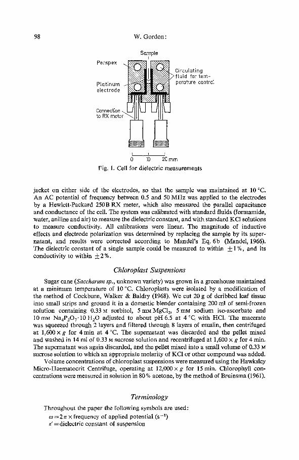
98 W. Gordon:
Sam )re
22. to RX meter ~
0
t~ Circulating ~X~,~N~]/>ftuid for tem-
perature controt
10 20 mm
Fig. 1. Cell for dielectric measurements
jacket on either side of the electrodes, so that the sample was maintained at 10 ~ An AC potential of frequency between 0.5 and 50 MHz was applied to the electrodes by a Hewlett-Packard 250B RX meter, which also measured the parallel capacitance and conductance of the cell. The system was calibrated with standard fluids (formamide, water, aniline and air) to measure the dielectric constant, and with standard KC1 solutions to measure conductivity. All calibrations were linear. The magnitude of inductive effects and electrode polarization was determined by replacing the sample by its super- natant, and results were corrected according to Mandel's Eq. 6b (Mandel, 1966). The dielectric constant of a single sample could be measured to within + 1%, and its conductivity to within 4- 2 %.
Chloroplast Suspensions
Sugar cane (Saccharum sp., unknown variety) was grown in a greenhouse maintained at a minimum temperature of 10 ~ Chloroplasts were isolated by a modification of the method of Cockburn, Walker & Baldry (1968). We cut 20 g of deribbed leaf tissue into small strips and ground it in a domestic blender containing 200 ml of semi-frozen solution containing 0.33 M sorbitol, 5 n~MgC12, 5 mM sodium iso-ascorbate and 10 mM Na4P20 7" 10 H20 adjusted to about pI--I 6.5 at 4 ~ with HC1. The macerate was squeezed through 2 layers and filtered through 8 layers of muslin, then centrifuged at 1,600 x g for 4 rain at 4 ~ The supernatant was discarded and the pellet mixed and washed in 14 ml of 0.33 M sucrose solution and recentrifuged at 1,600 x g for 4 min. The supernatant was again discarded, and the pellet mixed into a small volume of 0.33 M sucrose solution to which an appropriate molarity of KC1 or other compound was added.
Volume concentrations of chloroplast suspensions were measured using the Hawksley Micro-Haematocrit Centrifuge, operating at 12,000 x g for 15 rain. Chlorophyll con- centrations were measured in solution in 80 % acetone, by the method of Bruinsma (1961).
Terminology
Throughout the paper the following symbols are used: co = 2 n • frequency of applied potential (s-l) g = dielectric constant of suspension
Permeability of Chloroplast Membranes 99
eo = permittivity for free space (Fm -1) a = conductivity of suspension (~-1 m-l ) p =volume concentration of chloroplasts in suspension e~ = dielectric constant of supernatant a s = conductivity of supernatant (f~-lm-X)
t e T = dielectric constant of chloroplast stroma a r = conductivity of stroma (~-1 m-l )
For dectrodiffusion across a membrane: = time constant or relaxation time (s)
V= mobility of ions moving across the membrane (m s-1/V m-l) e=ionic charge (coulombs)
D = diffusion coefficient (m 2 s-i) c o =concentration of ions in region of membrane (m -3)
= membrane thickness (m) A =total area of membranes per unit volume of suspension (m -1) k = Boltzmann's constant (J ~ T= temperature (~
C M, G u = parallel capacitance, conductance per unit area of membrane (Fm -2, f~ -1 m -2) Z M = impedance per unit area of membrane (f~-I m-2)
Y, Ys, Yc = effective complex conductivities of the suspension, supernatant, chloroplasts.
Results and Discussion
D i s p e r s i o n C u r v e s
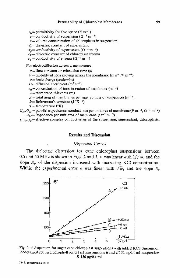
The dielectric dispersion for cane chloroplast suspensions between
0.5 and 50 M H z is shown in Figs. 2 and 3. e' was linear with 1 / ] /~ , and the
slope S~, of the dispersion increased with increasing KC1 concentrat ion.
Within the experimental error ~ was linear with ]/~-, and the slope S~
250 ~--" KCI
200 ~ + 91 mM
. . . ' " 150
/ ~..~.m.~ + 30 mM ~ , . . . . ~ __.~.~.~- + 6 mM
100 ~ - t - 0 rnM
, , , , 1 / 4 2 0
0 1 2 3 4 5 6x10 -4
Fig. 2. ~' dispersion for sugar cane chloroplast suspensions with added KCI. Suspension A contained 280 I~g chlorophyll per 0.1 ml; suspensions B and C 152 pg/0.1 ml; suspension
D 150 ~g/0.1 ml
7 b Jr. M e m b r a n e B i o l , 8

100 W. Gordon:
150
100
0.64
0.63
0.62
0.61
I Ir ,
3 XlO-' 0 10 -4 0
(7
(~-~m-~)
,toa ; ,
10x103
Fig. 3. e' and a dispersions for cane chloroplasts, demonstrating a unity loss tangent for the dispersion. Suspension contained 219 lag chlorophyll per 0.1 ml, and 96 mM KCI
s,,
2O X104
10
O' i 100 200
CHLOROPHYLL ( pg/o.1 ml )
p2 2+p)ZA
100 200 CHLOROPHYLL (pg/o.lml)
b
Fig. 4. (a) Effect of dilution on slope of 8' dispersion for cane chloroplasts. Suspension contained 50 mM added KC1. (b) p2/(2 + p)2A versus chlorophyll concentration during
dilution experiment
for the conductivity dispersion was related to S,, by
Sa = '30 Se'
that is, the dispersion had a unity loss tangent within the error. However, S, could not be measured as accurately as S~,, and therefore in further experiments the e' dispersion alone was measured.
In Fig. 4, a chloroplast suspension was diluted by adding more re- suspending medium at constant molarity of KC1. S~, decreased approximately linearly with the suspension chlorophyll concentration. The dielectric constant of the supernatant did not vary between 0.5 and 50 MHz.
Permeability of Chloroplast Membranes 101
Electrodiffusion Theory
The observed linearity of d with 1/V-~, and the unity loss tangent for the dispersion, is reminiscent of the electrodiffusion theory for ions moving across a membrane under the influence of an applied potential. The impedance of such a membrane is given (Appendix I) by
ZM- ]/2kT 1 . ( l_ j ) . (1) 2coe]/(Ve) l/-~
Assuming that such electrodiffusion occurs mainly across the chloroplast internal membranes (evidence for this is given in the next section), d and a for a chloroplast suspension would be (Appendix II)
d 2 - 2 p , . 6pe:r [ _~_]+ 3p: coeVV--e 1 2+p oAV-ffr V;' (2)
2 - 2 p 6paT coe]/V-ee a~- -2+p o ' ~ + ~ [ 1 - - - ~ ] + ~ . 3p2 A]/2kT "V~. (3)
This would therefore explain the linearity of d with 1/VN at constant Co, and the linearity of a with ]/~-.
From Eq. (2) 3p 2 coe~/Ve
S~,-'- (2-~p) z (4)
From Eq. (3) eo AV2kT
3 p Z Co e (5)
A1/zkr
and therefore S, = eo S,, as shown experimentally. On diluting a chloroplast suspension, A and p are decreased, and since S,, is linear with chlorophyll concentration (Fig. 4a) Eq. (4) indicates that p2/(2+p)2A should also be linear with chlorophyll concentration. This was demonstrated experi- mentally (Fig. 4b).
Location of Electrodiffusion
Electrodiffusion might occur at the chloroplast outer envelope, or within the internal lamellar system. However, there is good evidence that the observed dispersions were due to diffusion at the internal membranes:
(1) Cane chloroplasts as isolated here were considered entirely outer- membrane-free, since they did not have the highly refractive appearance of intact spinach or pea chloroplasts viewed in the phasecontrast microscope under light ground illumination (Walker, 1967).
102 W. Gordon:
C I
110
100
90
80 11/'oo
o 1 ; xlo-,, Fig. 5. e' dispersion for outer-membrane-free pea chloroplasts. Suspension contained
51 i.tg chlorophyll per 0.1 ml, and 77 mM added KCI
B 15 ~ A 10
, ,
0 1 2 3 x10⁻⁴
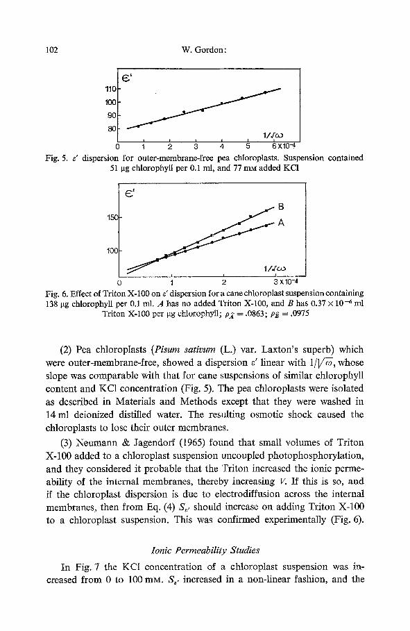
Fig. 6. Effect of Triton X-100 on e' dispersion for a cane chloroplast suspension containing 138 ~tg chlorophyll per 0.1 ml. A has no added Triton X-100, and B has 0.37 x 10 -6 ml
Triton X-100 per gg chlorophyll; p2 = .0863; p~ = .0975
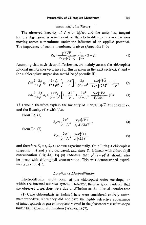
(2) Pea chloroplasts (Pisum sativum (L.) var. Laxton's superb) which were outer-membrane-free, showed a dispersion s' linear with 1/]/~, whose slope was comparable with that for cane suspensions of similar chlorophyll content and KC1 concentration (Fig. 5). The pea chloroplasts were isolated as described in Materials and Methods except that they were washed in 14 ml deionized distilled water. The resulting osmotic shock caused the chloroplasts to lose their outer membranes.
(3) Neumann & Jagendorf (t965) found that small volumes of Triton X-100 added to a chloroplast suspension uncoupled photophosphorylation, and they considered it probable that the Triton increased the ionic perme- ability of the internal membranes, thereby increasing V. If this is so, and if the chloroplast dispersion is due to electrodiffusion across the internal membranes, then from Eq. (4) S,, should increase on adding Triton X-100 to a chloroplast suspension. This was confirmed experimentally (Fig. 6).
Ionic Permeability Studies
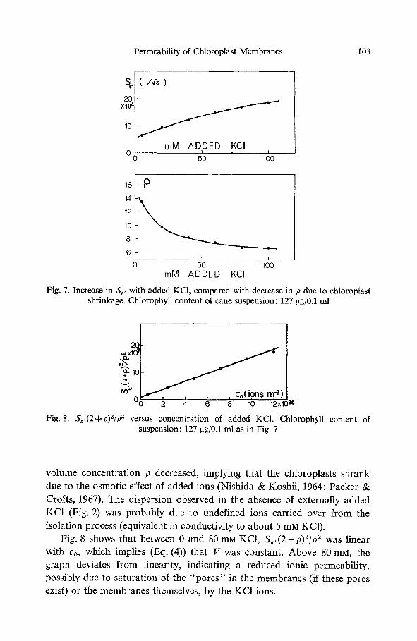
In Fig. 7 the KCI concentration of a chloroplast suspension was in- creased from 0 to 100 mM. S,, increased in a non-linear fashion, and the
Permeability of Chloroplast Membranes 103
~
20 XlO 4
10
0 0
(1/,r. )
I
J mM ADDED KCI
I I
50 100
16! 14
12
10
8
6
0
2_ I I
50 100 mM ADDED KCI
Fig. 7. Increase in S,, with added KCI, compared with decrease in p due to chloroplast shrinkage. Chlorophyll content of cane suspension: 127 gg/0.1 rnl
2G x101 ~ 3 2- t0
~ ) 00 2 4 6 8 10 12x1( 25
Fig. 8. S,,(2+p)2/p z versus concentration of added KCI. Chlorophyl l content of suspension: 127 ~g/0.1 ml as in Fig. 7
volume concentration p decreased, implying that the chloroplasts shrank due to the osmotic effect of added ions (Nishida & Koshii, 1964; Packer & Crofts, 1967). The dispersion observed in the absence of externally added KC1 (Fig. 2) was probably due to undefined ions carried over from the isolation process (equivalent in conductivity to about 5 mM KC1).
Fig. 8 shows that between 0 and 80 mM KCI, S~,(2+p)2/p 2 was linear with co, which implies (Eq. (4)) that V was constant. Above 80 mM, the graph deviates from linearity, indicating a reduced ionic permeability, possibly due to saturation of the "pores" in the membranes (if these pores exist) or the membranes themselves, by the KC1 ions.
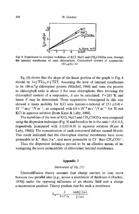
104 W. Gordon"
IN
~ 5 §
f KCl NaCI CH3COONa
, ,Co ( ions m -3) 4 ' 6 '8XI0 25
Fig. 9. Experiment to compare mobilities of KC1, NaC1 and CH3COONa ions, through the internal membranes of cane chloroplasts. Chlorophyll content of suspension:
163 ~tg/0.I mI
Eq. (4) shows that the slope of the linear portion of the graph in Fig. 8 should be 3e]/F-e/eoA ]/2--k-T. Assuming the area of internal membranes to be 160 m2/g chloroplast protein (Mitchell, 1966) and since the protein to chlorophyll ratio is about 5 for cane chloroplasts, then knowing the chlorophyll content of a suspension, A can be calculated. T=283 ~ and hence V may be determined. Three suspensions investigated in this way showed a mean mobility for KC1 ions (anions+cations) of (3.1_+0.4)x 10 -11 m s-~/V m -~, as compared with 6.9 x 10 - s m s-1/V m -1 for 50 mM KC1 in aqueous solution (from Kaye & Laby, 1966).
The mobilities of the ions of KC1, NaC1 and CH3COONa were compared using the dispersion technique (Fig. 9) and found to be in the ratio 1:0.8:0.5, respectively [compared with 1:0.83:0.58 in aqueous solution (Kaye & Laby, 1966)]. The concentration of each compound did not exceed 60 m s . This result indicated that the chloroplast internal membranes were more permeable to K § than Na § and more permeable to C1- than CH3COO-.
Thus the dispersion technique proved to be an effective means of in- vestigating the ionic permeability of chloroplast internal membranes.
Appendix I
Derivation of Eq. (1)
Electrodiffusion theory assumes that charge carriers or ions move between two parallel sites (e.g., across a membrane of thickness 6 (Racker, 1970)) under the opposing influences of an electric field and a charge concentration gradient. Theory predicts that for such a membrane
6 tanh VJ a~ zM= .
Permeability of Chloroplast Membranes 105
where "r=,52/4D, v = V / e and D = v k T (Cole, 1968b). at high frequencies, then
ZM V 2 k T 1 ( l - j ) 2coel/ 1/7
1/Zu = GM +jo) C~t and, therefore,
CM c~ 1
V2kr V
G M - c o e V -~ee V ~" ]//2kT
If tanh Vj--~ --, 1
(I-l)
Thus the constant value for CM [typically 1 gF/cm 2 for some biological membranes (Cole, 1968a)] is not encountered in this type of membrane, where CM varies with frequency, ionic concentration and permeability.
A p p e n d i x H
Derivation of Eqs. (2) and (3)
For a suspension of chloroplasts, Maxwell's equation (Cole, 1968b) shows that
1 -y/y~ 1 -y~/ys for small values of p 2+y/y~ - p 2+yJy~
where y = a + j ~ e ' e o
ys=~s+ jCoe~eo �9 !
yc=-~rc + J O9 ~c~o .
Maxwell's equation would strictly apply only for spherical chloroplasts, but it is a good approximation for non-spherical particles, provided the eccentricity is not too large (Fricke, 1924).
Solving Maxwell's equation for y,
2 y s ( 1 - p ) + y c ( l + 2 p ) Y=Ys ( 2 + p ) y s + ( l _ p ) y c
2(1-p) 9pyc - (2+p) ys-~ (2+p)Z t-terms of higher power in yc.
Assuming y~ ~ys, the terms of higher power in y~ may be ignored, and
2(i--p) . 9py~ (2+p
106 W. Gordon:
A chloroplast contains membrane lamellae lying parallel to each other, and surrounded by stroma. On the assumption that the stroma is highly conducting compared with the membranes, chloroplasts which lie with their membranes perpendicular to the applied field [effectively one-third of the total population (Pauli etal. , 1960)] will offer greater resistance to ionic movement than those which lie with their membranes parallel to the field.
Since the volume of membranes per unit volume of suspension is A 6, the density of membranes within a chloroplast is A 6/p. It can be shown that the effective conductivity of the chloroplasts lying perpendicular to the applied field approximates to G~t6/(A~/p) and the effective conductivity of the remaining two-thirds of the chloroplast population approximates to at(1 - A 6/p). Thus, the mean chloroplast conductivity
ac~�89 (GM p/A) + z3 ar (1 - A ~lp). Similarly,
t 1 2 t ~ c ~ ( CM p/~o A) + ~ ~ r (1 - A ~/p).
Expressions for CM and G~t were derived in Appendix I.
Substituting for y, Ys and yc in the equation for y, and equating real and imaginary parts of the equation
e, 2 - 2 p , 6pe'r I- -I
[--;-] oAV2kr ~ 1 A5 3p z coe]/Ve 1
2+p + (2-2-+~ + _ (II-1)
a ~ 2-2P2+p as+ (2~--~6Par [1- -~- -] + 3#2 A I / 2 k T (II-2)
I would like to thank Dr. S. E. Keefe and Dr. C. Bucke for helpful discussion, and Miss Sylvie Stachenko for technical assistance.
References
Bruinsma, J. 1961. A comment on the spectrophotometric determination of chlorophyll. Bioehem. Biophys. Acta 52:576.
Cockburn, W., Walker, D. A., Baldry, C. W. 1968. The isolation of spinach chloroplasts in pyrophosphate media. Plant PhysioL 43, no. 9:1415.
Cole, K. S. 1968a. Membranes, Ions and Impulses. University of California Press, Berkeley and Los Angeles.
Cole, K. S. 1968b. Membranes, Ions, and Impulses. University of California Press, Berkeley and Los Angeles. pp. 22, 174, 182, 186.
Fricke, H. 1924. A mathematical treatment of the electrical conductivity of colloids and cell suspensions, g. Gen. Physiol. 6:375.
Permeability of Chloroplast Membranes 107
Fricke, H., Morse, S. 1925. The electric resistance and capacity of blood for frequencies between 800 and 4�89 million cycles. J. Gen. Physiol. 9:153.
Kaye, G. W. C., Laby, T. H. 1966. Tables of Physical and Chemical Constants. Longmans, Green and Co. Ltd., London. p. 162.
Neumann, J., Jagendorf, A. T. 1965. Uncoupling photophosphorylation by detergents. Biochem. Biophys. Acta 109:382.
Nishida, K., Koshii, K. 1964. Volume changes of isolated spinach chloroplasts caused by electrolytes and sugars. Physiol. PI. 17: 846.
Packer, L., Crofts, A. R. 1967. The energised movement of ions and water by chloro- plasts, ln: Current Topics in Bioenergetics. Vol. 2. D .R. Sanadi, editor, p. 23. Academic Press Inc., New York, London.
Pauli, H., Packer, L., Schwan, H. P. 1960. Electrical properties of mitochondrial mem- branes. J. Biophys. Biochem. Cytol. 7, no. 4:589.
Racker, E. 1970. Membranes of Mitochondria and Chloroplasts. Van Nostrand Reinhold Co., New York. p. 26.
Schwan, H. P. 1963. Determination of biological impedances, ln: Physical Techniques in Biological Research. W. L. Nastuk, editor, p. 323. Academic Press Inc., New York.
Walker, D. 1967. Photosynthetic activity of isolated pea chloroplasts. In: The Bio- chemistry of Chloroplasts II. T. W. Goodwin, editor, p. 53. Academic Press Inc., New York and London.