Shepard (2001) Perceptual-cognitive universals as reflections ...
Upload
khangminh22Category
view
1download
0
MASTER IN COGNITIVE SCIENCE AND LANGUAGE
MASTER THESIS September 2021
Functional brain correlates of auditory verbal
hallucinations in schizophrenia: a design of an
fMRI study testing perceptual and cognitive
models.
by Olivia Gawel
Under the supervision of:
Dr. Paola Fuentes Claramonte and Dr. Joana Rosselló Ximenes

1
ABSTRACT
Auditory verbal hallucinations (AVHs, or ‘hearing voices’) are a cardinal symptom of
schizophrenia, and yet, their biological basis has not been fully determined. As of now,
theories that attempt to disentangle the origins of AVHs can be separated into two main
types (or models): perceptual versus cognitive. The former has considered AVHs to be
due to malfunction in perceptual processing, namely an abnormal activation in the
auditory cortex, as well as having a top-down cognitive influence. The latter considers
AVHs to be due to one of two cognitive processes: the misinterpretation of intrusive
memories (which posit that AVHs are the result of a breakdown in the processes
monitoring the source of memories) or to the malfunction of inner speech (which posits
that AVHs are due to dysfunction of speech monitoring). The current study aims to
propose an adequate experimental design of a prospective fMRI study that will test both
the perceptual and cognitive approaches allowing to fill the gaps in the general framework
for AVHs. Firstly, to test the perceptual model, an experimental design borrowed from
Fuentes-Claramonte and colleagues (2021) will be adapted, with a modification
controlling for motor activity. To test one side of the Cognitive Model, the theory of
intrusive memory, the experimental paradigm that has been created by Fuentes-
Claramonte and colleagues (2019) and validated by Martin-Subero and colleagues (2021)
will be adapted to test schizophrenic patients with AVHs, which has not been done before.
It will elicit negatively valenced autobiographical memories, which has been shown to
activate parts of the default mode network, a circuit thought to be impaired in
schizophrenia. Thirdly, to test the other side of the Cognitive Model, namely the theory
of inner speech, an experimental paradigm will be proposed called the Rhyming task, a
phonological encoding task that is known to activate brain regions involved in subvocal
rehearsal and short-term storage of information. However, because the stimuli for this
task is lacking for the Spanish population, a pilot study (an online survey) was conducted,
presenting healthy participants with pairs of objects (created partly from a personalized
corpus), and asked them to do three tasks: to decide whether the names of both objects
rhyme; provide the name of each object; and rate the object on a 1 to 5 Likert scale for
the purposes of determining emotional valence. The results of the pilot study guided the
selection of the appropriate stimuli for the prospective imaging study. The proposed fMRI
study tackles the biological basis of AVHs from different perspectives, helping to
improve patients' lives that are touched by this cardinal symptom, and thus enabling future
research to sculpt appropriate clinical intervention thanks to pinpointing the exact
biological basis of hearing voices.

2
ACKNOWLEDGEMENTS
“We all carry the seeds of greatness within us, but we need an image as a point
of focus in order that they may sprout.”
Epictetus as interpreted by Lebell (2007, p. 95).
Such “images as a point of focus” play the most important role in one’s life, as
without them one is made up of missing pieces – and I am lucky enough to have a few
that watered my seeds for them to sprout.
To Dr. Fuentes and Dr. Rosselló: you have fully exposed me to the concept of
auditory verbal hallucinations, providing me with all the necessary aptitudes to dive into
scientific investigation: from theory to practice, from statistical analyses to critical
thinking, from advice to warmth and understanding. Your support throughout my
academic endeavors (and not only) are gifts that I shall cherish. In this way, I would like
to thank you very sincerely for your mentorships.
To my family: you have given me many fruits: those of perseverance, faith,
strength, and without your most cherished one, constant support, I would not be here
today. Although it seems that words cannot express the depth of my gratitude, it is still
through language that I hope to try to meet you on the bridge to your heart: Dziękuję wam
bardzo za wszystko1.
To my beloved father - the lighthouse that always guided me to the shore: your
light shall be eternally missed. This thesis is dedicated to you.
1 Polish to English translation: Thank you so much for everything.

3
This work was supported by the project PI21/00416, funded by the Instituto de Salud
Carlos III and co-founded by the European Union (FEDER/FSE) "A way to make
Europe"/"Investing in your future" as well as by the Catalonian Government, Generalitat
de Catalunya (2017-SGR-1271 to EP-C from AGAUR), and by a Sara Borrell Research
contract (CD19/00149 to PF-C) funded by the ISCIII and co-founded by the European
Union (FEDER/FSE) "A way to make Europe"/"Investing in your future".
.

4
TABLE OF CONTENTS
LIST OF TABLES 5
LIST OF FIGURES 6
ABBREVIATIONS 7
1. INTRODUCTION 8
2. LITERATURE REVIEW 11
2.1. The Phenomenological Basis of AVHs 11
2.2. Brain Activation Correlates Underlying AVHs 14
2.2.1. The Perceptual Model 16
2.2.1.1. Aberrant Perception Theory 16
2.2.1.2. Proposal of Symptom Capture Paradigm 26
2.2.2. The Cognitive Model 28
2.2.2.1. Intrusive Memory Theory 28
2.2.2.2. Proposal of Autobiographical Recall Task 34
2.2.2.3. Inner Speech Theory 36
2.2.2.4. Proposal of Rhyming Task 43
3. METHODOLOGY 46
3.1. Data Acquisition 46
3.2. Participants 46
3.3. Stimuli 46
3.4. Procedure 47
3.5. Data Cleaning 48
3.6. Statistical Analysis 49
4. RESULTS 49
4.1. Demographic Information 49
4.2. Stimuli Selection 52
4.2.1. Rhyming Judgement Task 52
4.2.2. Object Naming Task 53
4.2.3. Rating Task 55
5. DISCUSSION 55
6. CONCLUSIONS 61
REFERENCES 62
APPENDICES 71
Appendix 1 72
Appendix 2 73
Appendix 3 81
Appendix 4 83

5
LIST OF TABLES
Table Page
1 Demographic Information about Participants 50
2 Frequency of Rhyming Pairs 52
3 Frequency of Non-rhyming Pairs 53
4 Rhyming Pairs with Highest Agreement 54
5 Non-rhyming Pairs with Highest Agreement 54
6
LIST OF FIGURES
Figure Page
1 The Primary Auditory Cortex 17
2 Fuentes-Claramonte and Colleagues (2021) Results 22
3 Proposal of Symptom Capture Paradigm 27
4 AM and DMN Circuits 33
5 Proposal of Autobiographical Recall Task 35
6 Proposal of Rhyming Task 44

7
ABBREVIATIONS
ACC Anterior Cingulate Cortex
ALE Activation Likelihood Estimation
AM Autobiographical Memory
AMI Autobiographical Memory Interview
AVH Auditory Verbal Hallucination
BOLD Blood Oxygen Level Dependent
DMN Default Mode Network
DSM-5 Diagnostic and Statistical Manual of Mental Disorders
EEG Electroencephalography
EMG Electromagnetic
fMRI Functional Magnetic Resonance Imaging
GLM General Linear Models
IQ Intelligence Quotient
MTG Middle Temporal Gyrus
MTL Medial Temporal Lobe
PAC Primary Auditory Cortex
PET Positron Emission Tomography
PFC Prefrontal Cortex
PTSD Post-Traumatic Stress Disorder
ROI Region of Interest
SD Standard Deviation
SMA Supplementary Motor Area
STG Superior Temporal Gyrus
VH Visual Hallucination

8
1. INTRODUCTION
“Always deters me from the course of action I was intending to engage in, but it never
gives me positive advice.”
Socrates on his purported auditory verbal hallucinations
as told by Plato (Long, 2009, p. 64)
Hearing voices, or experiencing auditory verbal hallucinations (AVHs), is one of
the main symptoms of schizophrenia, and occurs in about 70% of patients (David, 1999;
McCarthy-Jones, 2012). Despite its clinical significance, the biological basis underlying
AVHs is undetermined, and the findings portrayed by scientific literature remain
inconclusive till this day.
The present multidisciplinary framework (from Psychiatry to Cognitive Science)
has mainly taken two approaches to explain the origins of AVHs.
The first approach proposes that hallucinations appear as a result of abnormal
activity in the primary auditory cortex, the brain region that processes auditory perception,
leading to the perception of these voices that characterize the symptom. This theory has
received mixed experimental support, as some single studies and meta-analyses have
found activation in the auditory cortex during AVHs (Dierks et al., 1999, Lennox et al.,
1999; Kompus et al., 2011 ), while others have not (Silbersweig et al., 1995; Lennox et
al., 2000; Copolov et al., 2003; Diederen et al. 2010; Hoffman et al., 2011; Fuentes-
Claramonte et al. 2021; Jardri et al., 2011; Kühn and Gallinat, 2012); Zmigrod et al.,
2016).
The second approach proposes that hallucinations arise from two distinct
cognitive processes. The first is a theory of the misinterpretation of intrusive or traumatic
memories, which posits that AVHs are due to the malfunction in “the processes
monitoring the source of memories” (Seal et al., 2004, p. 53). The second is a theory on
the malfunction in inner speech, particularly short-term verbal memory. However, as of
now, there are very few studies that have attempted to test this model, and thus a complete
answer is still lacking.

9
For these exact reasons, the current study aims to establish the building blocks
needed to test the biological basis of AVHs in a prospective fMRI study. This shall be
done in the following way.
Firstly, I will embark upon a review of the available literature by briefly diving
into the phenomenological basis of AVHs, where I look at the structure and content of
such hallucinations. Next, I outline how such studies on AVHs are conducted, by
explaining different design approaches and focusing on one technique in particular,
known as the ‘symptom capture’ technique, which compares brain states or activation
patterns in periods of time when a schizophrenic patient hallucinates and when said
patient does not.
Once this brief background is provided, I then elaborate on the methodology and
findings of individual studies and meta-analyses that have targeted the Perceptual Model.
Throughout my review, I will show that studies which have implemented the symptom
capture technique have mixed findings with regards to the genesis of brain activation
whilst schizophrenic patients experience AVHs. Because of this factor, we propose
implementing an experimental paradigm, the Symptom Capture Paradigm, which has
been formulated by Fuentes-Claramonte and colleagues (2021). Essentially, patients are
to indicate when they experience AVHs by pressing a button with their right index finger,
and they are also to indicate when they perceive real speech by pressing a button with
their left index finger. Numerous previous studies have been posed with some limitations,
one in particular being the confounding effect of motor activity on brain activation during
AVHs. To counteract this limitation, we propose a modification that controls motor
activity by asking participants to press two buttons with both their index fingers when
they are not experiencing AVHs. In this way, the influence of motor activity during button
press will be nullified.
Following this, I move to the Cognitive Model, where I begin by discussing the
two dominant theories: the misinterpretation of intrusive or traumatic memories, and the
malfunction of inner speech. I provide a theoretical background for both by reviewing a
handful of available studies that have attempted to test this particular model. For each
cognitive theory, we propose an experimental paradigm. For the former theory, we
propose implementing the Autobiographical Recall Task, an experimental paradigm
created by Fuentes-Claramonte and colleagues (2019) and validated by Martin-Subero
and colleagues (2021). It tests whether there are any alterations linked to hallucinations
and the intrusiveness of traumatic memories. For the latter theory, I will focus on the

10
Rhyming Task, which involves internal speech, in which subjects must determine whether
two words rhyme or not. As this task involves processes of phonological coding that
involve internal speech and short-term verbal memory, it can test whether there are inner
speech alterations linked to hallucinations, namely whether patients with hallucinations
experience an altered processing of inner speech, or if there is alteration in brain activation
in the inner speech task whilst patients are experiencing AVHs. It is worth noting,
however, that because no previous study has used rhyming pairs in Spanish, this pilot
study attempts to design this stimuli.
For this reason, I focus on the design of the above-mentioned Rhyming Task in
Section 3, and explain how we created and validated the stimuli, as well as providing the
chosen stimulus pairs that will be used in the prospective fMRI study.
In the last Section, I end with a discussion and conclusions on the prospective
fMRI study as well as the pilot study by pointing out the advantages and potential
limitations of both the prospective fMRI study and pilot study, as well as the possible
direction of future research and our speculation.
With all that in consideration, only by first thoroughly building a set of
foundations, specifically the experimental design that will take AVHs apart, can the
presence of heard illusory voices be ultimately understood as being due to abnormal
auditory activity, intrusive or traumatic memories or alterations in inner speech. By
identifying the basis of AVHs, the key can be found to designing new treatments, such as
neuromodulation techniques, aimed at acting on the precise brain areas involved -
therefore having a direct impact on this symptom. Thanks to this, the development of
interventions to treat a specific symptom or set of symptoms is a further step towards
personalized medicine which, in a disease as heterogeneous as schizophrenia, is
imperative in adjusting treatments to the needs of each patient. All this is endeavored so
that the maximum clinical improvement is obtained, minimizing potential health risks
and other adverse effects.
11
2. LITERATURE REVIEW
2.1. The Phenomenological Basis of AVHs
From figures like Socrates to Sigmund Freud, auditory verbal hallucinations
(AVHs) do not spare anyone on their warpath. Following contemporary psychiatric
classifications, AVHs are considered a characteristic symptom of a range of psychotic
and mood disorders as in, for instance, schizophrenia (McCarthy-Jones, 2012). This
particularly disabling mental disorder comprises “a collection of signs and symptoms of
unknown aetiology, predominantly defined by observed signs of psychosis” (Insel, 2010,
p. 187). Although the peak onset age of schizophrenia is said to be in early adulthood (Li
et al., 2009), only recently has it been considered as a neurodevelopmental disorder (Insel,
2010). When considering AVHs in schizophrenia, the first question that arises is
regarding their phenomenological basis. In order to cover some explanatory ground, here
is a demonstration of a few examples of AVHs:
Example (1): “You're not crazy, she tells me. I love you, she tells me. You're going to
change the world, she tells me (laughs). You really don't believe me, she
tells me. It's weird but it's like that, she tells me. Jose, she tells me again. I
have always loved you. Don't be ashamed. You have them all freaking
out. Jose, again, repeats a little, you know? Really, even if you don't
believe me.” [Transcription of a patient’s AVHs by Tovar et al., (2019)]
Example (2): “You killed your father and you want to kill your sister, you want to stay
in power, you want to kill your mother, you want to hurt everyone around
you. Because you're bad, you're cruel. You are a very bad person (...) You're
disgusting. You want to take power. You want to kill everyone. You want
to destroy the whole world” [Transcription of a patient’s AVHs by Tovar et
al., (2019)]
Example (3): “He is an astronomy fanatic. Here is a taste of his own medicine. He is
getting up now. He is going to wash. It is about time” - “ [Transcription of
a patient’s AVHs by Frith (1992, p. 66)].
AVHs are also considered to be “perceptual experiences that occur in the absence
of a triggering external sensory stimulus” and are “perceived to originate from another

12
agency” (Hugdahl et al., 2008, p. 2; Waters et al., 2006, p. 66; Woodruff, 2004; Nayani
& David, 1996). Thus, Examples (1) - (3) of AVHs must not be thought of as the
individual’s imagination running wild, but as significant elements of their realities of the
world that accompany them every step of their way.
In most cases, AVHs are perceived as human voices external to the individual,
spoken most commonly in second-person, as seen in Examples (1) and (2), or in third-
person, as seen in Example (3) (Tovar et al., 2019; McCarthy-Jones, 2012). In other
instances, AVHs have been said to be “more like ideas than external sensations”,
“soundless voices, absolutely silent and could not be heard” (McCarthy-Jones, 2012, p.
108). It must be underlined, however, that schizophrenic patients do not tend to confuse
AVHs with the voices of real individuals or of their own thoughts (McCarthy-Jones,
2012; Mullen, 1997; Hoffman et al., 2008). What is more, the vocalizations of these
hallucinations may be clear, mumbling, whisper-like, or shouting, and their location
variable — coming to the patient internally (i.e. located in the brain, body, belly, feet),
externally, or both (McCarthy-Jones, 2012; Nayani & David, 1996; Moritz & Laroi, 2008;
Copolov et al., 2004). The number of voices depends on the patient, ranging from only
hearing a single voice to a multitude of them (Tovar et al., 2019; Nayani & David, 1996;
McCarthy-Jones, 2012). Furthermore, the content of such voices is diffuse and can range
from being very positive, such as in Example (1), to being very negative, such as in
Example (2) in terms of valence, and said voices can discuss a range of topics, such as in
Example (3).
Based on Examples (1) - (3), it can be said that these voices have a distinctive
linguistic profile. A recent study by Tovar and colleagues (2019) explored the formal
linguistic aspects of AVHs in 18 patients with either schizophrenia or schizo-affective
disorder. The first fundamental factor found was that ‘voice talk’ is on a personal level,
whereby “speech participants or other objects of the immediate context are the subject of
the utterance” rather than on an impersonal level, whereby voices describe “facts about
the world relatively independent of the speech content” (Tovar et al., 2019, p. 8). For
instance, in Example (2) the voices speak of the patient, their mother, and “everyone
around them”. Thus, AVHs mostly concern aspects of the patient’s life that are ‘close to
home’, as opposed to external world events. Furthermore, the authors (2019) also
discovered that their voice sample encompassed parataxis, which is “language with a
strong tendency to be reduced to the single sentence level, lacking connectivity and
embedding” as exemplified in Example (1): “[Don't be ashamed.] [You have them all

13
freaking out.] (...) [Really, even if you don't believe me]”. In addition, the content of
“voice talk” is normally noted to be free of semantic or syntactic error, which may be due
to “the low grammatical complexity of such speech” as seen in all the above-mentioned
examples (Tovar et al., 2019, p. 15). As such, the details of the phenomenology of AVHs
do not just concern the content of what patients hear, but also the linguistic profile of
hearing voices as well, which should always be considered in analysis, as there is much
more to consider than is immediately apparent.
On another note, even though it has been mentioned that the voice-hearer can
separate the voices from the real world, they can have properties which make them seem
to have their own sense of agency that is separate from the patient’s intentions (McCarthy-
Jones, 2012). There are instances in which voices are in pursuit of self-preservation and
try to persuade the patient, for example, to avoid antipsychotic medication, collaboration
in treatment and other forms of aid. At times, if the patient refuses to comply with their
commands, the voices bully them. As a result of this process, there is a variability in the
degree of command that patients have over the voices, and consequently patients are, at
times, engaged in battles of power or dominance. Such a push-and-pull relationship is
demonstrated in the following examples:
Example (4): “It makes me feel like a Spanish fly. It makes me feel bad. I could not
block them out. There was not anything I could do about it. They make
me feel bad just the same” (Modell, 1958, p. 451).
Example (5): “I did not know but I did not want to listen. It was just the sound of a
certain person’s voice that gave me pleasure. I was scared when it all first
started so I was trying to blank them out and I kept pushing them back
because I did not want to hear them. I just did not want to and I kept
blocking my ears. I was scared. I really was” (Modell, 1958, p. 452).
Example (6): “When I make mistakes she is right with me. She (voice 1) gets too
impatient with me, and I try to figure out something all by myself. I am
trying to make a clothespin apron, and she is right there with me and she
is trying to tell me what to do. The fellow (voice 2) leaves me alone. She
is a little but too overanxious. I want to be left alone. If I asked her for
help, it would be different but I do not want her bothering me” (Modell,
1958, p. 452).
Based on these examples, it can be stated without any doubt that the voices which
the patient experiences are not imagined, but instead are abnormal perceptions - a part of
their reality, which reflects the inherent subjectiveness of the situation. This in turn

14
precisely demonstrates the significant role that AVHs can play in a patient's life: they are
detrimental, as such individuals experience a loss of “basic human needs”, including but
not limited to: privacy, homeostasis, hope, motivation, social relationships, self-esteem,
autonomy, professional opportunities, respect, or freedom (McCarthy-Jones, 2012).
Moreover, individuals experiencing AVHs not only suffer from a loss of consensual
reality in which they feel as if they are no longer living in the same world as the rest of
society (Dilks et al., 2010; Mauritz & van Neijel, 2009; Jarosinski, 2008; McCarthy-
Jones, 2012), but they also lose their own sense of self.
Unfortunately, those that experience such voices are in a way prisoners of their
own minds. Despite the widespread affliction of schizophrenic individuals hearing voices,
little is understood regarding the biological origins of such hallucinations, and thus, the
‘optimal’ form of therapy for such patients has still not been fully laid out.
2.2. Brain Activation Correlates Underlying AVHs
In order to delineate the neurobiological basis of this phenomenon, the following
subsection aims to shed light on how brain activation correlates with underlying AVHs.
With the advent of bountiful technological advancements, many studies have
undertaken functional neuroimaging. This has lent a helping hand in determining the
concrete brain activation patterns during AVHs, which would not have been detectable to
such an extent by other techniques (i.e. EEG) - this is in part due to the high spatial
resolution of functional neuroimaging techniques, such as Functional Magnetic
Resonance Imaging (fMRI for short) or Positron Emission Tomography (PET for short).
The former is a neuroimaging technique that “depicts changes in deoxyhemoglobin
concentration consequent to task-induced or spontaneous modulation of neural
metabolism” (Glover, 2011, p.1). The latter is a neuroimaging technique that measures
“physiological function by looking at blood flow, metabolism, neurotransmitters and
radiolabeled drugs” (Berger, 2003, p. 1449). Together these neuroimaging techniques
provide the opportunity to conduct two types of studies that may provide a detailed
portrayal of brain activation whilst experiencing AVHs.
One set of studies attempts to measure brain activity precisely during the
occurrence of AVHs. These are the so-called symptom capture studies, also known as
state studies, and are conducted on the basis of a within-subject design in which there is
a comparison made between brain states or activation patterns in periods of time when a
schizophrenic patient hallucinates and when the same patient does not (Kühn & Gallinat,

15
2012). Such studies have been used to examine the perceptual model, whose hypothesis
is that the mechanism responsible for AVHs is abnormal auditory perception in auditory
brain regions, such as the primary auditory cortex. The second type of study, known as
trait studies, are conducted on the basis of a between-subjects design in which there is a
comparison made between brain states or activation patterns of one group of
schizophrenic patients who hallucinate with another group of schizophrenic patients that
do not have this symptom or a group of healthy controls (Kühn & Gallinat, 2012). The
comparison between groups can be done either in resting state, or when exposing the
participants to an experimental task that involves processes presumably linked to AVHs,
such as sound or speech perception (thus targeting the perceptual model) or tasks
involving language and memory (thus testing the cognitive model) (Allen et al., 2008).
As these studies attempt to measure brain activity dedicated to cognitive processes
“underlying the disposition to hallucinate”, their hypothesis is that the mechanisms
responsible for AVHs are due to the inability to detect speech monitoring (inner speech
theory), or to the activation of traumatic memories (intrusive memory theory) (Allen et
al., 2008, p. 179). Thanks to the fact that whole brain analytical processes are
implemented across the above-mentioned studies, deciphering which particular model is
the building block of AVHs can be discreetly determined.
On a methodological level, the most common approach2 used to capture AVHs in
functional neuroimaging studies is known as the button press. This method allows
subjects to press one or two buttons with one or both hands to indicate the onset, duration,
or diminishing of their hallucinations (i.e. Silbersweig et al., 1995; Dierks et al., 1999;
Lennox et al., 1999, 2000; Diederen et al., 2010; Hoffman et al., 2011; Fuentes-
Claramonte et al., 2021). This approach became utilized post-hoc Serafetinides et al.
(1986), who demonstrated that manually reporting AVHs is a much more viable method
than verbally reporting AVHs, as had been done in electrophysiological studies, the
results of which are, for the most part, inconclusive (i.e. Marjerrison et al., 1968; Stevens
et al., 1979; Stevens & Livermore, 1982). Despite the fact that the indication of AVHs in
2 It must be noted that there are other approaches to capturing AVHs which are not as common as the button
press approach. The first, known as random sampling, is a discontinuous acquisition method, “in which
many fMRI volumes are acquired at random intervals” during the experiment (Leroy et al., 2017, p. 2; i.e.
Shergill et al., 2000a). The second, known as independent component analysis, “allows the co-activated
brain regions to be separated without predefined temporal model of brain activity” (Leroy et al., 2017, p.
2; van de Ven et al., 2005; Jardri et al., 2007, 2009; Jardri et al., 2013). However, because the proposal of
this thesis is in part based on the button press approach, emphasis will only be placed in this particular
approach.

16
functional neuroimaging studies is based on self-reports, it has been rigorously shown
that patients are highly accurate at providing feedback to unexpected auditory stimuli in
an fMRI environment, demonstrating the ability to capture similar unexpected AVHs with
similar accuracy (e.g. van de Ven et al., 2005; Sommer et al., 2008; Diederen et al., 2013;
van Lutterveld et al., 2013; Foucher, 2013; Jardri et al., 013; Leroy et al., 2017).
As the Perceptual Model deserves the same level of scrutiny, it shall be examined
henceforth.
2.2.1. The Perceptual Model
The forthcoming section targets the Perceptual Model, which maintains that
AVHs are genuinely perceptual, that is, that they arise as a result of pathological activity
in auditory perception regions of the brain. This theory has come together through a
multitude of studies across time (of which only a select few shall be mentioned) whose
approach was to understand and investigate AVHs exploratorily, their primary aim being
to capture and compare brain activation during the experience of AVHs or lack thereof.
Accounting for this premise, what must be kept in mind is that rather than there first
being a model that all studies followed, what took place was common exploration that
then formed the model.
2.2.1.1. Aberrant Perception Theory
Human reality is constructed ‘brick by brick’ by perception, which is a set of
complex processes that cooperate to determine one’s experience and one’s reaction to a
given stimulus in one’s environment. The neural engine processes incoming information
from the environment, or in other words, engages in bottom-up processing. This type of
processing stimulates the brain’s receptors, thanks to which a given percept is created by
“recombining features from sensory input” (de Boer et al., 2019, p. 2772; Engel et al.,
2001). This particular framework is the starting point of all perception, as without it, the
neural engine would not have information to process to begin with. The brain is not a
‘blank’ slate, but is instead equipped with existing knowledge about previous experience
which is set into play by having certain expectations about the world, or in other words,
it engages in top-down processing. This type of processing allows for “a faster processing
of sensory information” (de Boer et al., 2019; Fenske et al., 2006; O’Callaghan et al.,
2017). Worth noting is the constant dynamic interplay between both types of processing
which sculpts an accurate perception of the world (de Boer et al., 2019; Stocker &

17
Simoncelli, 2006). Conversely, any imbalance prompts the opposite: errors in perception
in which certain percepts become activated without any external stimulation from the
environment.
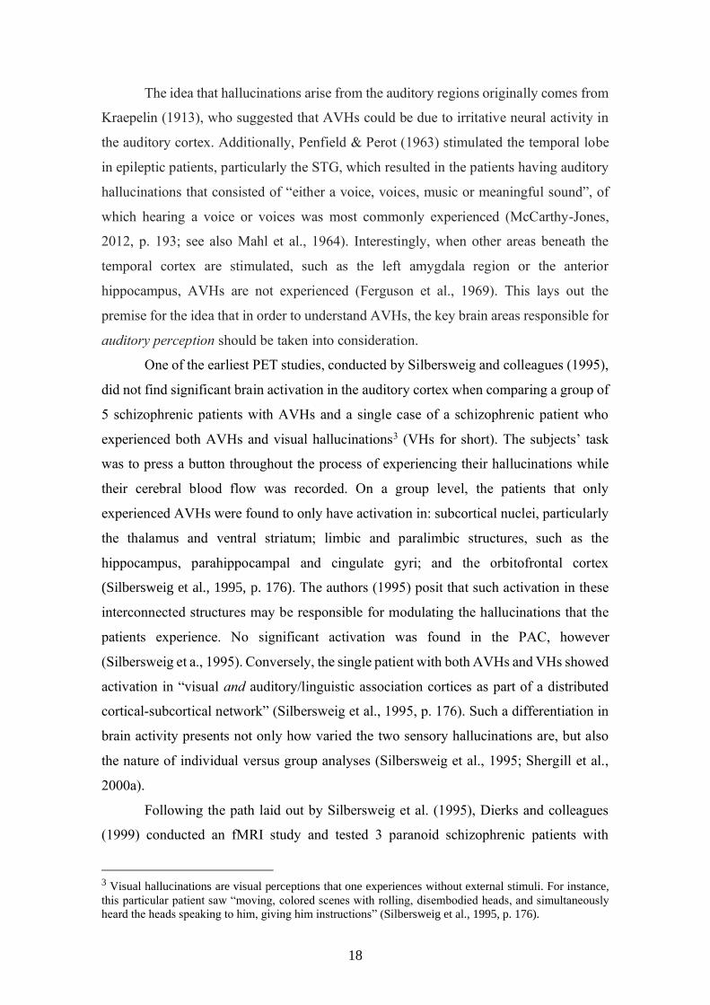
On this basis, one principal mechanism that has been thought to be behind the
underlying foundation of AVHs is the phenomenon of “aberrant perceptions generated
in the auditory regions”, particularly in the primary auditory cortex (PAC for short), as
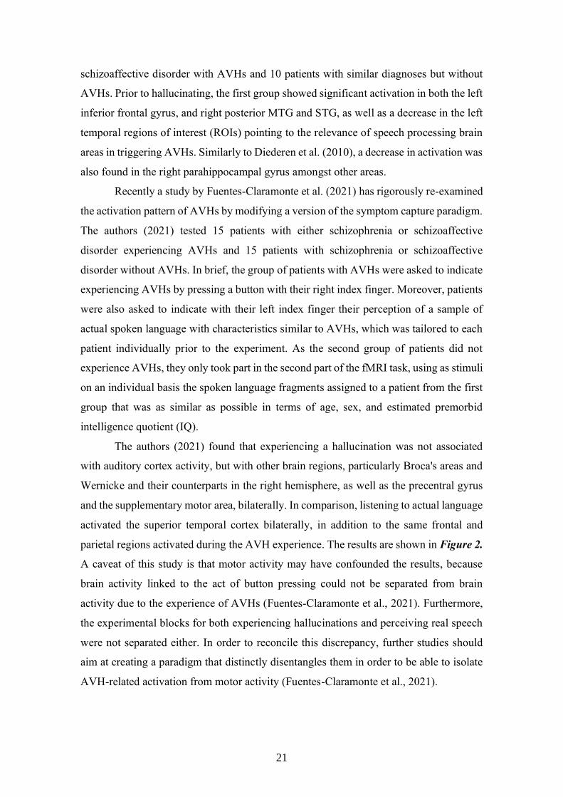
presented in Figure 1 (Jardri et al., 2011, p. 73; Kompus et al., 2013; Ćurčić-Blake et al.,
2017). The PAC “is located in the superior plane of the superior temporal gyri called
Heschl’s gyrus” (Ćurčić-Blake et al., 2017, p. 13; Zatorre et al., 2002). It is responsible
for primary sound perception and processing. It then sends further information down to
the secondary auditory cortex (consisting of the superior temporal gyrus (STG for short)
and middle temporal gyrus (MTG for short)) (Ćurčić-Blake et al., 2017). Finally, thanks
to the thalamus, the information travels up to the higher-order areas involving language,
allowing for a full processing of the original auditory percept (Belin et al., 2000; Zatorre,
et al., 2002, 2007; Javitt & Sweet, 2015; Ćurčić-Blake et al., 2017). Consequently, it is
hypothesized that “AVHs that form even stronger sensory experiences accordingly,
activate the auditory cortex (including the left STG), which houses the linguistic auditory
perception regions of the brain” (Ćurčić-Blake et al., 2017, p. 13; Allen et al., 2012;
Woodruff et al., 1995, 1997).
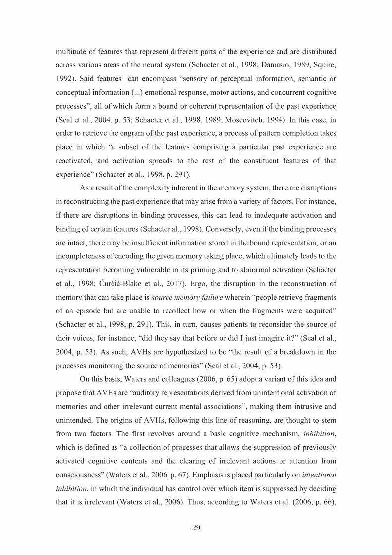
Figure 1. The Primary Auditory Cortex (PAC for short) presented on a brain template by Kompus
et al. (2013, p. 2). PAC occupies a large portion of Heschl's gyrus (or the transverse temporal
gyrus) (red), and is surrounded by the superior temporal gyrus (blue).
18
The idea that hallucinations arise from the auditory regions originally comes from
Kraepelin (1913), who suggested that AVHs could be due to irritative neural activity in
the auditory cortex. Additionally, Penfield & Perot (1963) stimulated the temporal lobe
in epileptic patients, particularly the STG, which resulted in the patients having auditory
hallucinations that consisted of “either a voice, voices, music or meaningful sound”, of
which hearing a voice or voices was most commonly experienced (McCarthy-Jones,
2012, p. 193; see also Mahl et al., 1964). Interestingly, when other areas beneath the
temporal cortex are stimulated, such as the left amygdala region or the anterior
hippocampus, AVHs are not experienced (Ferguson et al., 1969). This lays out the
premise for the idea that in order to understand AVHs, the key brain areas responsible for
auditory perception should be taken into consideration.
One of the earliest PET studies, conducted by Silbersweig and colleagues (1995),
did not find significant brain activation in the auditory cortex when comparing a group of
5 schizophrenic patients with AVHs and a single case of a schizophrenic patient who
experienced both AVHs and visual hallucinations3 (VHs for short). The subjects’ task
was to press a button throughout the process of experiencing their hallucinations while
their cerebral blood flow was recorded. On a group level, the patients that only
experienced AVHs were found to only have activation in: subcortical nuclei, particularly
the thalamus and ventral striatum; limbic and paralimbic structures, such as the
hippocampus, parahippocampal and cingulate gyri; and the orbitofrontal cortex
(Silbersweig et al., 1995, p. 176). The authors (1995) posit that such activation in these
interconnected structures may be responsible for modulating the hallucinations that the
patients experience. No significant activation was found in the PAC, however
(Silbersweig et a., 1995). Conversely, the single patient with both AVHs and VHs showed
activation in “visual and auditory/linguistic association cortices as part of a distributed
cortical-subcortical network” (Silbersweig et al., 1995, p. 176). Such a differentiation in
brain activity presents not only how varied the two sensory hallucinations are, but also
the nature of individual versus group analyses (Silbersweig et al., 1995; Shergill et al.,
2000a).
Following the path laid out by Silbersweig et al. (1995), Dierks and colleagues
(1999) conducted an fMRI study and tested 3 paranoid schizophrenic patients with
3 Visual hallucinations are visual perceptions that one experiences without external stimuli. For instance,
this particular patient saw “moving, colored scenes with rolling, disembodied heads, and simultaneously
heard the heads speaking to him, giving him instructions” (Silbersweig et al., 1995, p. 176).

19
transitory AVHs who were asked to indicate the onset of their AVHs by pressing a button
with their left hand, and continue pressing it until the end of their hallucinations in two
conditions. The first consisted of the patients being in a resting state. The second consisted
of the patients being presented with spoken text which was played backwards and
presenting them with “a modulated tone of 2000 Hz”, during which all patients were to
indicate their AVHs (Dierks et al., 1999, p. 615). When experiencing AVHs, the authors
found significant activation in Heschl’s gyrus, the posterior STG, the MTG, the
frontoparietal operculum, the hippocampus, the amygdala as well as the sensorimotor
cortex. According to the authors (1999, p. 617) such activation increased “when the
patient signalled the onset of an episode of hallucination, remained at a high level during
that episode, and returned to baseline levels immediately after the patient had signalled
the hallucination ended”. Furthermore, when comparing the experience of AVHs and
acoustic and tonal stimulation, the highest correlation of the BOLD signal was found in
Heschl’s gyrus “at the same location” in both, suggesting a mutual location for their
genesis (Dierks et al., 1999, p. 617).
Next in this sequence of studies, Lennox and colleagues (1999) tested one
paranoid schizophrenic with AVHs who was to press a button as an indication of both the
onset and end of his AVHs in an fMRI state study. In the time periods associated with
AVHs, the authors (1999, p. 644) found significant activation in the right MTG; bilateral
STG; right middle and inferior frontal gyri, right ACC; right cuneus; and the primary
motor cortex (related to the manual response). Worth mentioning is that the strongest
activation was not found in the primary auditory cortex, but rather in the MTG, part of
the secondary auditory cortex. In a follow-up fMRI study, the same research group (2000,
p. 17) asked four schizophrenic patients with AVHs to press the left button for the onset
of their hallucination, and the right button for the end of the hallucination. The authors
demonstrated that whilst experiencing hallucinations, significant activation was found “in
the left and right STG, left inferior parietal and left middle frontal gyrus” (Lennox et al.,
2000, p. 15). As this study implemented a different methodology for the button press
when compared to previous studies, the authors argue that it serves as an advantage in the
technical aspect of the analysis as there is less contamination in image acquisition.
Together, Lennox and colleagues (1999, 2000) place emphasis on the role of brain regions
responsible for auditory perception in the origins of AVHs.
A lack of activation in the primary auditory cortex during AVHs was found by
Copolov et al. (2003), who compared 7 schizophrenic patients with hallucinations and 1

20
patient with schizoaffective disorder with hallucinations, 7 schizophrenic patients without
hallucinations, and 8 healthy controls. The first group of participants was to press a button
with their right index finger during the onset and duration of their AVHs, whilst the
second and third groups of participants were presented with auditory stimuli that
“consisted of multi-speak babble, a crowd noise, in which many human voices could be
heard talking about different topics with no one dominating” and were instructed to also
press a button with their right index finger as an indication that they perceived the
stimulus (Copolov et al., 2003, p. 141). Interestingly, pure exposure to human speech in
the second and third group crucially revealed “bilateral activation of the superior temporal
gyri, involving the primary and association auditory areas (...) and bilaterally in the
superior temporal gyri”, as well as other areas (Copolov et al., 2003, p. 143). For those
patients that experienced AVHs, however, the areas of significant activation were
localized in “the right medial frontal region, most probably reflecting a combination of
anterior cingulate and cingulum activity (...) lateral surface of the right prefrontal cortex,
the left posterior STG, and the right posterior MTG (...) left hippocampal formation” and
so on, yet no such activity was demonstrated in the auditory cortex (Copolov et al., 2003,
p. 146-147).
A different fMRI study by Diederen and colleagues (2010) tested 24 psychotic
patients with AVHs, and made a comparison of brain activation between them squeezing
a balloon as an indication of onset of AVHs and 15 healthy individuals squeezing a
balloon randomly across trials. The patients presented significant activation during their
AVHs in the bilateral language areas, predominantly in “bilateral insula and inferior
frontal gyrus (including Broca’s homologue) as well as the MTG, STG, and
supramarginal gyri” (Diederen et al, 2010, p. 430-431). What is more, 6 seconds prior to
the onset of hallucinations, 15 out of the 24 patients showed significant “deactivation in
the left parahippocampal gyrus” - an area involved in memory recollection4 (Diederen et
al., 2010, p. 432). Subsequently, the authors conclude the presence of AVHs can be
mainly found in bilateral language areas.
Following Diederen et al. (2010), Hoffman and colleagues (2011) aimed at
improving the “understanding of the chain of brain events leading to AVHs”. For this
reason, the authors (2011) compared 11 patients with either schizophrenia or
4 The fact that brain regions responsible for memory recollection become activated prior to AVHs is an
interesting finding and shall be discussed further on in Section 2.2.2.1..
21
schizoaffective disorder with AVHs and 10 patients with similar diagnoses but without
AVHs. Prior to hallucinating, the first group showed significant activation in both the left
inferior frontal gyrus, and right posterior MTG and STG, as well as a decrease in the left
temporal regions of interest (ROIs) pointing to the relevance of speech processing brain
areas in triggering AVHs. Similarly to Diederen et al. (2010), a decrease in activation was
also found in the right parahippocampal gyrus amongst other areas.
Recently a study by Fuentes-Claramonte et al. (2021) has rigorously re-examined
the activation pattern of AVHs by modifying a version of the symptom capture paradigm.
The authors (2021) tested 15 patients with either schizophrenia or schizoaffective
disorder experiencing AVHs and 15 patients with schizophrenia or schizoaffective
disorder without AVHs. In brief, the group of patients with AVHs were asked to indicate
experiencing AVHs by pressing a button with their right index finger. Moreover, patients
were also asked to indicate with their left index finger their perception of a sample of
actual spoken language with characteristics similar to AVHs, which was tailored to each
patient individually prior to the experiment. As the second group of patients did not
experience AVHs, they only took part in the second part of the fMRI task, using as stimuli
on an individual basis the spoken language fragments assigned to a patient from the first
group that was as similar as possible in terms of age, sex, and estimated premorbid
intelligence quotient (IQ).
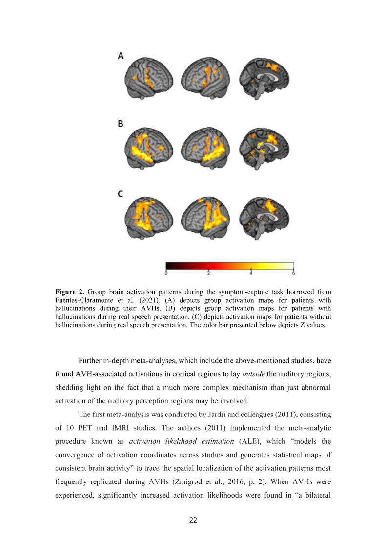
The authors (2021) found that experiencing a hallucination was not associated
with auditory cortex activity, but with other brain regions, particularly Broca's areas and
Wernicke and their counterparts in the right hemisphere, as well as the precentral gyrus
and the supplementary motor area, bilaterally. In comparison, listening to actual language
activated the superior temporal cortex bilaterally, in addition to the same frontal and
parietal regions activated during the AVH experience. The results are shown in Figure 2.
A caveat of this study is that motor activity may have confounded the results, because
brain activity linked to the act of button pressing could not be separated from brain
activity due to the experience of AVHs (Fuentes-Claramonte et al., 2021). Furthermore,
the experimental blocks for both experiencing hallucinations and perceiving real speech
were not separated either. In order to reconcile this discrepancy, further studies should
aim at creating a paradigm that distinctly disentangles them in order to be able to isolate
AVH-related activation from motor activity (Fuentes-Claramonte et al., 2021).
22
Figure 2. Group brain activation patterns during the symptom-capture task borrowed from
Fuentes-Claramonte et al. (2021). (A) depicts group activation maps for patients with
hallucinations during their AVHs. (B) depicts group activation maps for patients with
hallucinations during real speech presentation. (C) depicts activation maps for patients without
hallucinations during real speech presentation. The color bar presented below depicts Z values.
Further in-depth meta-analyses, which include the above-mentioned studies, have
found AVH-associated activations in cortical regions to lay outside the auditory regions,
shedding light on the fact that a much more complex mechanism than just abnormal
activation of the auditory perception regions may be involved.
The first meta-analysis was conducted by Jardri and colleagues (2011), consisting
of 10 PET and fMRI studies. The authors (2011) implemented the meta-analytic
procedure known as activation likelihood estimation (ALE), which “models the
convergence of activation coordinates across studies and generates statistical maps of
consistent brain activity” to trace the spatial localization of the activation patterns most
frequently replicated during AVHs (Zmigrod et al., 2016, p. 2). When AVHs were
experienced, significantly increased activation likelihoods were found in “a bilateral
23
neural network, including the Broca’s area, anterior insula, precentral gyrus, frontal
operculum, MTG, STG, inferior parietal lobule, and hippocampus and parahippocampal
region” (Jardri et al., 2011, p. 73). To conclude, despite the widespread activation in
language-related areas, no such significant activation was found in the PAC when AVHs
are experienced.
In the second meta-analysis, Kühn and Gallinat (2012) compared 12 functional
state and 8 trait studies by implementing ALE. For the state studies, convergent patterns
of activation were observed “in bilateral inferior frontal gyrus, bilateral postcentral gyrus,
left parietal operculum that is part of the left inferior parietal lobule” (Kühn & Gallinat,
2012, p. 781). Even after the authors (2012) decreased the threshold, no significant
convergence was found in the STG. For the trait studies, convergent patterns of activation
were observed in “left STG, left MTG, ACC, left premotor cortex” (Kühn & Gallinat,
2012, p. 781). With such findings, this meta-analysis does not lend credence to the theory
of AVHs being due to abnormal neuronal activity in the auditory cortex, but rather
implicates speech production areas.
The third meta-analysis, executed by Kompus and colleagues (2011), gathered
data from fMRI and PET studies and employed ALE. The aim of this meta-analysis was
to compare brain activations across studies in which patients experienced AVHs “in the
absence of a corresponding external stimulus” and when patients were exposed to
“external auditory stimulus” (Kompus et al., 2011, p. 3364). The authors (2011, p. 3362)
found that “the areas of significant convergence of increased activation when
experiencing auditory hallucinations included a cluster in the STG, corresponding to left
PAC, extending to parietal operculum”, and the left primary sensory/motor cortex, as well
as other areas (to a lesser extent) such as “left insula, left posterior hippocampus, right
MTG, right inferior parietal lobule, opercular part of the right inferior frontal gyrus and
rostral portion of right superior frontal gyrus”. Additionally, the left primary auditory
cortex was also implicated in the auditory stimulation tasks, yet its activation was
decreased in schizophrenic patients when compared to healthy controls, pointing to a
deficit in auditory processing. Based on this, the authors (2011, p. 3365) concluded that
both such a deficit in auditory processing and experiencing AVHs share common
mechanisms in speech perception areas, which may point to “competition between
internally generated and externally originating neural activity in the auditory cortex for
the attentional resources in hallucinating individuals”.

24
The fourth meta-analysis, consisting of 14 studies, done by Zmigrod and
colleagues (2016), aimed to examine both AVHs and VHs in not only schizophrenia, but
also other clinical and non-clinical populations (i.e. psychotic disorders, Parkinson’s
disease, Alice in Wonderland syndrome5, Charles Bonnet syndrome6). A distinct pattern
of activation was found for both AVHs and VHs, with some areas of overlap indicating
that hallucinations of different sensory modalities have little brain activity in common
(Zmigrod et al., 2016). Most importantly, during AVHs, large clusters of activation were
observed in the “ bilateral somatosensory cortex, bilateral insula, STG, Broca’s area and
its right hemisphere homologue, and in Wernicke’s area/secondary auditory cortex, (...)
left hippocampus/parahippocampal gyrus, the right motor cortex” and so on (Zmigrod et
al., 2016, p. 5). In comparison, significant activation for VHs was found in “extrastriate
visual areas around the ventral lingual and fusiform gyri” demonstrating a distinction
between brain regions associated with AVHs versus VHs (Zmigrod et al., 2016, p. 5).
Although activity was found in speech production areas and the secondary auditory cortex
(and others) during AVHs, this analysis may overgeneralize brain activation across
groups, as a variety of both clinical and nonclinical studies were converged, rather than
just those of schizophrenic patients. As of now, the degree to which AVHs in
schizophrenia (or psychosis) mirrors AVHs in other populations remains unknown (i.e.
Frith and Dolan, 1997; Waters et al., 2006; McCarthy-Jones, 2012; Choong, et al., 2007;
Diederen et al., 2012).
The conclusions of the data presented points to a commonality: brain activity
whilst experiencing AVHs is inconsistent in the sense that the same brain regions,
particularly those involved in auditory perception, are not activated across all studies. By
putting together the data from single case studies to meta-analyses, the most common
brain regions that have been found to be involved in AVHs are: the PAC (including
Heschl’s gyrus (Dierks et al., 1999; Lennox et al., 1999; Kompus et al., 2011), Wernicke’s
area and its homologue (Dierks et al., 1999; Lennox et al., 1999, 2000; Copolov et al.,
2003; Hoffman et al., 2011; Fuentes-Claramonte et al., 2021; Kompus et al., 2011;
Zmigrod et al., 2016), Broca’s area and its homologue (Lennox et al., 1999, 2000;
Copolov et al., 2003; Diederen et al., 2010; Hoffman et al., 2011; Jardri et al., 2011; Kühn
5 Alice in Wonderland Syndrome is characterized as “a perceptual disorder characterized by distortions of
visual perception, the body schema, and the experience of time” (Blom, 2016.). 6 Charles Bonnet Syndrome is characterized “by the presence of complex visual hallucinations in
psychologically healthy but visually impaired people” (Schwartz and Vahget, 1998).

25
and Gallinat, 2012; Fuentes-Claramonte et al., 2021), superior temporal gyri (Lennox et
al., 1999, 2000; Copolov et al., 2003; Diederen et al., 2010; Jardri et al., 2011; Zmigrod
et al., 2016), middle temporal gyri (Dierks et al., 1999; Lennox et al., 1999, 2000;
Copolov et al., 2003; Hoffman et al. 2011; Kompus, et al. 2011; Jardri et al., 2011),
hippocampus (Silbersweig et al., 1995; Dierks et al., 1999; Copolov et al., 2003; Jardri et
al., 2011; Kompus and colleagues (2011), Zmigrod et al., 2016); parahippocampus
(Silbersweig et al., 1995; Copolov et al., 2003; Diederen et al. 2010; Jardri et al., 2011;
Zmigrod et al., 2016), precentral gyrus (Dierks et al., 1999; Diederen et al., 2010;
Fuentes-Claramonte et al., 2021; Jardri et al., 2011), postcentral gyrus (Dierks et al.,
1999; Diederen et al., 2010; Jardri et al., 2011; Kühn and Gallinat, 2012).
This variation in findings across studies may be due to a variety of reasons that
mostly stem from methodology. For instance, the number of participants that have taken
part in the (earlier) studies is insufficient as the number does not go over 8 in many cases
(i.e. Silbersweig et al., 1995; Dierks et al., 1995; Lennox et al., 1999, 2000; Copolov et
al., 2003; Hoffman et al., 2007). Such a limited number of participants has a detrimental
impact on the statistical effect of the findings, and the recommendation for fMRI studies
is of at least 20 subjects per group (Thirion et al., 2007; Desmond & Glover, 2002; Jardri
& Sommer, 2013). In connection to this, the statistical threshold used in earlier studies
(such as Silbersweig et al., 1995; Dierks et al., 1999; Lennox et al., 1999, 2000) was
liberal (p = .001, uncorrected), and may have resulted in type I and type II errors
(Sommers et al., (2008). Therefore, the results of such studies should be taken with
caution.
On another note, the BOLD activity found across certain studies may have been
confounded by motor behavior, which is an essential element in the experimental
paradigm to test AVHs, as it serves as an indication of onset and end of AVHs. In some
cases, the motor activity for button press during AVHs onset/duration was not compared
with the motor activity for button press without AVHs — not allowing for motor activity
to be cancelled out (e.g. Fuentes-Claramonte et al., 2021). Because of this, the activation
behind the button press may affect the activation during AVHs. It may be worthwhile in
future experimental design to implement blocks in which patients conduct the button
press during time periods of when they do not experience AVHs.
Lastly, it would be beneficial to supplement one technique, such as fMRI, with
other techniques, such as EEG, that target other resolutions better in order to provide the
full AVH picture.

26
In order to test the perceptual model thoroughly and overcome potential
limitations as found across previously mentioned studies, the following experimental
paradigm is suggested.
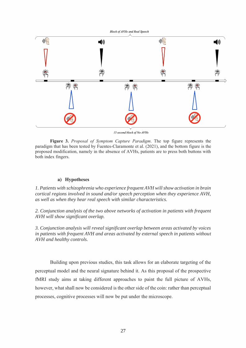
2.2.1.2. Proposal of Symptom Capture Paradigm
Three groups of participants will take part in the prospective fMRI study and all
tasks involved: 25 right-handed patients diagnosed with schizophrenia or schizoaffective
disorder according to DSM-5 with frequent AVHs (Group 1); 25 right-handed patients
diagnosed with schizophrenia or schizoaffective disorder without AVHs for at least 6
months (Group 2); and 25 right-handed healthy controls (Group 3). The task that will be
implemented is based on Fuentes-Claramonte and colleagues (2021), but with one notable
modification, and is presented in Figure 3. Group 1 will press a button with their right
index finger at the onset of the AVHs, and press a button with their left index finger at
the onset of real speech. To be more precise, randomly and within a 10-minute period, 40
instances of real speech will be presented through headphones with contents individually
tailored to resemble each patient’s AVH. Patients will be required to press a button when
they hear these examples of real speech with their left index finger. Furthermore, four 15-
second blocks will be interspersed throughout the task in which participants will also be
asked to randomly and alternatively press both buttons in time periods when they do not
hear AVHs. This enables activations associated with experiencing AVHs to be compared
with those when none are experienced, and thus overcome confounding activation for
AVHs potentially leaking from motor activity. With regards to Group 2 and Group 3, as
they do not experience AVHs, they will only be presented and respond to real speech, and
the content of the stimuli will be based on a composite version of voice content from
Group 1. Both groups will also perform the 15-second blocks to control for motor activity.
The statistical analyses will be performed at a level of p < .05, corrected at the
cluster level using Gaussian field methods, with a voxel level threshold of z > 3.1 (p <
.001, uncorrected).
27
Figure 3. Proposal of Symptom Capture Paradigm. The top figure represents the
paradigm that has been tested by Fuentes-Claramonte et al. (2021), and the bottom figure is the
proposed modification, namely in the absence of AVHs, patients are to press both buttons with
both index fingers.
a) Hypotheses
1. Patients with schizophrenia who experience frequent AVH will show activation in brain
cortical regions involved in sound and/or speech perception when they experience AVH,
as well as when they hear real speech with similar characteristics.
2. Conjunction analysis of the two above networks of activation in patients with frequent
AVH will show significant overlap.
3. Conjunction analysis will reveal significant overlap between areas activated by voices
in patients with frequent AVH and areas activated by external speech in patients without
AVH and healthy controls.
Building upon previous studies, this task allows for an elaborate targeting of the
perceptual model and the neural signature behind it. As this proposal of the prospective
fMRI study aims at taking different approaches to paint the full picture of AVHs,
however, what shall now be considered is the other side of the coin: rather than perceptual
processes, cognitive processes will now be put under the microscope.

28
2.2.2. The Cognitive Model
The following section targets two separate theories that make up the Cognitive
model, which maintain that AVHs arise from a dysfunction that causes internal, non-
perceptual processes to be incorrectly interpreted as external. The first is Intrusive
Memory Theory, which puts forth the argument that AVHs are unstable memories
activated due to “a breakdown in the processes monitoring the source of memories” (Seal
et al., 2004, p. 53). The second is Inner Speech Theory, which puts forth the argument
that AVHs are due to deficits in speech monitoring. It must be kept in mind that, although
both could be considered ‘cognitive’ models of AVHs, the theoretical groundwork behind
them has not been fully laid out as of yet, and the associated experimental work is still in
its nascent stage.
2.2.2.1. Intrusive Memory Theory
If in good condition, the mind has a peculiar gift: the ability to travel back into its
past experiences - recalling the places it has seen, the smells it has processed, and most
importantly, delving into the footprints left behind by other minds. As Elden Turving
(2005, p. 15) once said, “There can be no travel without a traveler”, and it is the travelling
self, the mind, that can wander from memory to memory recalling the pieces of the puzzle
of its existence (or episodic memory).
If, on the other hand, the mind’s condition has deteriorated, a malfunction of
storing and retrieving such memories takes place - a variant known as the theory of
misattribution of intrusive or traumatic memories. The proposition that hallucinations are
reactivated memories could be said to date back to Aristotle’s age:
“Sometimes we do not know when such stimuli occur in our soul from an earlier sensation, and
we are in doubt whether it is memory or not. But sometimes it happens that we reflect and
remember that we have heard or seen something before. Now, this occurs whenever we first think of it as itself, and then change and think of it as referring to something else (...) The opposite also
occurs, as happened to Antiphenon of Oreus, and other deranged people (egistamenois); for they
spoke of their mental pictures (fantasmata) as if they had actually taken place, and as if they
actually remembered them” (Berrios, 1996, p. 210; Bicknell, 1981).
The theory of misattribution of intrusive or traumatic memories originates in the
theory of episodic memory retrieval. Essentially, an experience embedded in one’s
memory is never a reproduction of the same replica, as it undergoes error and
reconstruction (Seal et al., 2004; Chalfonte & Johnson, 1993; Schacter, 1995; Schacter et
al., 1998). The entire episodic memory engram of the given experience consists of a
29
multitude of features that represent different parts of the experience and are distributed
across various areas of the neural system (Schacter et al., 1998; Damasio, 1989, Squire,
1992). Said features can encompass “sensory or perceptual information, semantic or
conceptual information (...) emotional response, motor actions, and concurrent cognitive
processes”, all of which form a bound or coherent representation of the past experience
(Seal et al., 2004, p. 53; Schacter et al., 1998, 1989; Moscovitch, 1994). In this case, in
order to retrieve the engram of the past experience, a process of pattern completion takes
place in which “a subset of the features comprising a particular past experience are
reactivated, and activation spreads to the rest of the constituent features of that
experience” (Schacter et al., 1998, p. 291).
As a result of the complexity inherent in the memory system, there are disruptions
in reconstructing the past experience that may arise from a variety of factors. For instance,
if there are disruptions in binding processes, this can lead to inadequate activation and
binding of certain features (Schacter al., 1998). Conversely, even if the binding processes
are intact, there may be insufficient information stored in the bound representation, or an
incompleteness of encoding the given memory taking place, which ultimately leads to the
representation becoming vulnerable in its priming and to abnormal activation (Schacter
et al., 1998; Ćurčić-Blake et al., 2017). Ergo, the disruption in the reconstruction of
memory that can take place is source memory failure wherein “people retrieve fragments
of an episode but are unable to recollect how or when the fragments were acquired”
(Schacter et al., 1998, p. 291). This, in turn, causes patients to reconsider the source of
their voices, for instance, “did they say that before or did I just imagine it?” (Seal et al.,
2004, p. 53). As such, AVHs are hypothesized to be “the result of a breakdown in the
processes monitoring the source of memories” (Seal et al., 2004, p. 53).
On this basis, Waters and colleagues (2006, p. 65) adopt a variant of this idea and
propose that AVHs are “auditory representations derived from unintentional activation of
memories and other irrelevant current mental associations”, making them intrusive and
unintended. The origins of AVHs, following this line of reasoning, are thought to stem
from two factors. The first revolves around a basic cognitive mechanism, inhibition,
which is defined as “a collection of processes that allows the suppression of previously
activated cognitive contents and the clearing of irrelevant actions or attention from
consciousness” (Waters et al., 2006, p. 67). Emphasis is placed particularly on intentional
inhibition, in which the individual has control over which item is suppressed by deciding
that it is irrelevant (Waters et al., 2006). Thus, according to Waters et al. (2006, p. 66),

30
there is “a fundamental deficit in intentional inhibition which leads to auditory mental
representation intruding into consciousness in a manner that is beyond the control of the
sufferer” (Waters et al., 2006, p. 66). The second revolves around contextual memory,
which “provides cues that allow us to differentiate one memory from another” (Waters et
al., 2006, p. 72). It follows, according to the authors (2006, p. 66), that AVHs emerge as
a result of “a deficit in binding contextual cues, resulting in an inability to form a complete
representation of the origins of mental events”. Combining the deficits in intentional
inhibition and contextual memory, Waters and colleagues (2006, p. 66) propose a model
in which AVHs in schizophrenia are the result of “mental events which are experienced
as involuntary and intrusive and are not recognized because the contextual cues that allow
them to be identified correctly are missing or incomplete”.
The potential significance of memory in AVHs has been somewhat reflected in
the brain activity preceding AVHs as well as in their duration. As already mentioned in
Section 2.2.1.1., two studies showed the involvement of the parahippocampus, the brain
area responsible for spatial memory and temporal memory, and which processes context
information of a past event, prior to patients hearing voices. Diederen and colleagues
(2010) showed that 6 seconds prior to the hallucinations, 15 out of the 24 patients showed
significant “deactivation in the left parahippocampal gyrus” (Kompus et al., 2013, p.
432). On the other hand, Hoffman et al. (2011) showed a decrease in activation in the
right parahippocampal gyrus. What is more, brain activation throughout the duration of
AVHs has been shown by single studies and meta-analyses to not only involve
parahippocampal gyri (i.e. Silbersweig et al., 1995; Diederen et al., 2010; Jardri et al.,
2011; Kompus et al., 2011; Zmigrod et al., 2016), but also the hippocampus - the brain
region fundamental for the formation of long-term episodic memory, particularly context
memory, and the construction of mental images. Perhaps most importantly, it binds item
information and context information together, creating the detailed episodic memory (i.e.
Silbersweig et al., 1995; Dierks et al., 1999; Copolov et al., 2003; Jardri et al., 2011;
Kompus et al., 2011; Zmigrod et al., 2016; Bird & Burgess, 2008; Slotnick, 2017). Such
findings of brain activation suggest a potential neural link between memory retrieval and
AVHs which encompasses the hippocampal complex - this link may be involved in the
onset and duration of voice hearing. The way this may come to be, for instance, as
proposed by Diederen (2010), is that the disinhibiting state of the parahippocampus in
particular may potentially “trigger the bilateral language-related areas originally involved
in the perception of speech”, and it may also prepare other areas, such as the inferior

31
temporal gyrus or the insula “for activation in the course of hallucination” (McCarthy-
Jones, 2012, p. 273). As it stands, it may be that the dysfunction in memory inhibition
may allow the memory fragments (such as auditory representations) to become
unintentionally and intrusively activated, making the patient relive their past experience,
not being able to differentiate the source of the given memory.
Such reliving of past experience in the form of AVHs has been proposed to
involve not just memories per se, but traumatic memories in particular (i.e. McCarthy-
Jones et al., 2014; Steel, 2015). This goes hand in hand with the link between being
diagnosed with schizophrenia and the prevalence of traumatic memories in a given patient
(Steel, 2015; Grubaugh et al., 2011). What is more, 15% of patients that are diagnosed
with schizophrenia will also have suffered from post-traumatic stress disorder (PTSD), a
mental disorder based on “an intrusive memory of a traumatic event, most likely in the
form of a visual image” (Steel, 2015, p. 1; Achim et al., 2011). Such intrusive memories
have been found to be present in the phenomenology of AVHs in patients diagnosed with
schizophrenia. For instance, Morrison and colleagues (2002, p. 3) interviewed 35 patients
with either schizophrenia, schizoaffective disorder or schizophreniform disorder, and
found that “74.3% (n=26) were able to identify an image in relation to their psychotic
symptoms, (...) patients who were able to identify idiosyncratic images experienced in
conjunction with their hallucinations or delusions, 69.2% (18 out of 26) reported that their
images were recurrent, 96.2% (n =25) were able to link the image to the experience of a
particular emotion and to a particular belief, and 70.8% (n=17) were able to associate the
image with a memory for a particular event in their past”. For instance, Morrison et al.
(2002) provides an example of a patient who was assaulted with a gun at a club, and he
experienced voices that related to that traumatic event.
Hardy et al. (2005) assessed 75 patients with either schizophrenia, schizoaffective
disorder or delusional disorder who experienced AVHs and found that 53.3% reported a
trauma, of which the most common were bullying (30%), adult sexual abuse (20%), and
child sexual abuse (17.5%). Most significantly, the authors (2005, p. 506) found that, of
these 53.5% patients, “just over half of these (30.6%) had at least one type of
phenomenological association between their traumas and hallucinations”. This is seen in
Example (7) of a patient who has gone through such trauma and experiences intrusive
AVHs:

32
Example (7) : “My mother psychologically abused me, like mental cruelty, she
constantly interfered with and controlled my life” (Experienced Trauma).
“I hear my neighbor giving me some form of therapy. She makes
comments and tells me what to do. Sometimes it is neutral but sometimes
she is more critical and I feel persecuted by her” (Experienced AVHs)
(Hardy et al., 2005, p. 505).
McCarthy-Jones et al. (2014) interviewed 199 patients with AVHs, of which
80.9% had schizophrenia, 13.6% had affective psychosis, 3% had other nonorganic
psychoses, and 2.5% had borderline personality disorder. The authors (2014, p. 231)
found that “12% of participants reported AVHs as like identical replays from memory,
31% reported AVHs as similar to memories”.
En masse, the collected data from the above-mentioned interviews point to an
important fact, namely that only some AVHs in schizophrenia can be accounted for by
trauma. This, however, does not point to a categorical description of AVHs. Furthermore,
to date there is little functional neuroimaging research that has precisely and directly
tested the mechanism of intrusive memories despite the fact that the brain regions
involved in conscious memory recall are well known (Seal et al., 2004; Slotnick, 2017).
Were the theory of intrusive memory thoroughly tested, this could lend a helping hand in
understanding the associations between AVHs composed of rather negative valence and
its potential link to psychological trauma.
One approach that can be taken is by considering autobiographical memory (AM),
which is the active retrieval of personal past experiences. It involves highly complex
“retrieval processes, semantic content, personal significance, subjective re-experience,
spatiotemporal context, emotion, social interactions and varying levels of specificity,
remoteness and rehearsal” (Jacques & de Brigard, 2015, p. 265). Interestingly, it activates
a network of regions known as the “AM retrieval network” or “core network”, which
consists of regions that consistently interact with one another, specifically “the medial
and lateral prefrontal cortices, lateral and medial temporal lobes (MTL; hippocampus,
parahippocampal gyrus), ventral parietal cortex and posterior cingulate cortex” (Jacques
& de Brigard, 2015, p. 265).
Interestingly, the complex AM circuits largely overlap with the neural network
that is activated during resting or passive states, known as the Default Mode Network
(DMN), as shown in Figure 4 (Raichle et al., 2001; Buckner et al., 2008). Midline regions
(medial prefrontal, posterior cingulate and precuneus cortices) are considered ‘core’

33
regions of the network and have been associated with self-referential processes during
AM retrieval. Additionally, some authors also define an MTL sub-network within the
DMN that comprises hippocampal, ventromedial prefrontal, retrosplenial and medial
parietal cortices, which “has been linked to constructing a scene based on memory”
(Jacques & de Brigard, 2015, p. 266). Moreover, the frontoparietal or central executive
network, which includes lateral PFC, anterior cingulate, and inferior parietal cortices, also
contributes to the retrieval of AM and is associated with “adaptive cognitive control
processes” (Jacques & de Brigard, 2015, p. 266).
.
Figure 4. Brain networks that make up AM as borrowed from Jacques & de Brigard
(2015, p. 266). The contribution of large-scale networks to AM that share parts of DMN: Frontoparietal network (top), Medial PFC network (bottom-left); Medial temporal lobe network
(bottom-right).
Of particular current interest is the dysfunction of the default network in
schizophrenia, as a number of studies have found evidence for the failure to activate or
deactivate particular regions during attentional tasks as well as abnormality found in
functional connectivity (i.e. Pomarol-Clotet et al., 2008; Salgado-Pineda et al., 2011;
Guerrero-Pedraza et al., 2012; Hu et al. 2017). The link between both the AM and DMN
networks lies not only in their spatial overlap, but in that autobiographical memory is one
of the proposed roles for the DMN, among others, that include processing “self-relevant
information, including self-reflection, making social and emotional judgements about

34
oneself and others, envisioning the future or performing theory of mind operations”
(Fuentes-Claramonte et al., 2019, p. 2).
As of now, only a handful of studies have tested AM in schizophrenia by
implementing the autobiographical recall task (which will be discussed in the next
section), that targets the AM and DMN circuits. Such studies’ interests lay in either
narrowing down the neural basis of AM (e.g. Cuervo-Lombard et al., 2012); analyzing
the activation pattern of the DMN (e.g. Fuentes-Claramonte et al., 2019); or
understanding how the DMN “behaves during a task like autobiographical memory”
(Martin-Subero et al., 2021, p. 2). However, no study as of yet has implemented the
autobiographical recall task to test the biological basis of AVHs by presenting the task to
a group of schizophrenic patients who specifically experience hearing voices.
Therefore, to test one side of the Cognitive Model, the following paradigm
targeting the question of whether intrusive or traumatic memories act as the main
generator of AVHs shall be proposed.
2.2.2.2. Proposal of Autobiographical Recall Task
The same three groups of participants as in the perceptual paradigm will be
presented with keywords that are designed to either evoke or not evoke autobiographical
memories previously described by the subjects - a task that has already been validated in
healthy individuals and schizophrenic patients (e.g. Fuentes-Claramonte et al., 2019;
Martin-Subero et al., 2021). Prior to the fMRI session, each participant will be given the
Crovitz test (Crovitz & Schiffman, 1974) and the autobiographical memory interview
(AMI for short) (Kopelman et al., 1989) in order to obtain a description of a total of 20
autobiographical memories from different times in life (childhood, adolescence,
adulthood and the last year) which will be of negative valence. Memories will be
evaluated based on their clarity, specificity and descriptive richness according to the
criteria of the AMI, and must receive the maximum score to be included. For each
memory, 2 keywords will be selected that identify it, and that will be displayed during
the task to indicate that that particular memory should be recalled. The control condition
will be to present keywords that are not associated with any specific memory.
During the fMRI scan and the task, 10 autobiographical memory blocks will be
presented, interspersed with 10 control blocks, with a duration of 20 seconds per block.
Each block will contain 2 memory-evoking or non-evoking keywords. After each block,
2 questions will be presented on the screen to the participant: the first is whether they

35
managed to recall the memories; the second regarding whether any AVHs occurred
during the block. As in the previous task, the participant must provide a manual response,
indicating "yes" (right index finger) or "no" (left index finger). What is more, the hand
assigned to each answer will be counterbalanced between participants. Lastly, there will
be a fixation point between blocks, presented for 16 seconds, that will act as the baseline.
The paradigm is presented in Figure 5.
Figure 5. Proposal of Autobiographical Recall Task based on Fuentes-Claramonte and
colleagues (2019) and validated by Martin-Subero et al. (2021).
For the statistical analysis, at the intra-subject level, the memory blocks and the
control blocks will be compared, using General Linear Models (GLMs), to delimit the
regions involved in the evocation of negative autobiographical memories. At the group
level, mean activation maps will be generated for each group that will then be compared
in order to identify differences in autobiographical memory between all three groups of
participants.
In Group 1 (the patient group with AVHs), the autobiographical memory blocks
with the presence of hallucinations will be compared with autobiographical memory
blocks without hallucinations. This shall be done in order to assess whether hallucinations
modulate the activity of the brain regions that are involved in the experience. The
statistical analysis will be performed at a level of p < 0.05, corrected at the cluster level
using Gaussian field methods, with a voxel level threshold of z > 3.1 (p < 0.001,
uncorrected).

36
a) Hypotheses
1. Patients with AVHs will show a brain activation pattern in response to cues to evoke
negative memories that will differ from that observed in patients without AVHs and
healthy controls.
2. Activation differences will be located in brain regions involved in autobiographical
memory, including DMN regions such as the medial prefrontal cortex, the posterior
cingulate and precuneus, among others.
3. Alternatively or additionally, hallucinations will transitorily alter the activity of these
regions.
Although this experimental paradigm tests the cognitive model, it only does so up
to a certain extent. Thus, the other side of the coin, which heavily relies on a different
cognitive mechanism, shall now be demonstrated.
2.2.2.3. Inner Speech Theory
Memory is the vehicle that permits one to travel back in time, yet it is the
subjective voice inside one’s head that puts forth the narration of one’s reality. The
discussion of inner dialogue or inner speech is illustrated in the Platonic dialogue, The
Theaetetus, which is a question-and-answer dialogue that arises between Socrates and
Theatetus, a young and brilliant mathematician (Chappell, 2021). In essence, Socrates
considers judgment (doxa) to be a silent statement (logos), and judging (doxazein) to be
silent speaking (legein):
“(Socrates): Very nice. But do you call thinking exactly what I call it?
(Theaetetus): What do you call it?
(Socrates): A talk which the soul carries on itself with itself about the things
it examines. This I tell you in ignorance. For this is how it seems
to me. The soul, when thinking, does nothing other than discuss,
asking itself questions and answering them, both affirming and
denying. Whenever it first determines something, either sluggishly
or sharply, then says the same thing and does not hesitate, we posit
that as its judgement. So I, at least, call judging, speaking and
judgment a state, not articulated to someone else but in silence to
oneself.” (Duncombe, 2016, p. 106).

37
Centuries later, the father of behaviorism, Watson (1913), recognized inner
speech as subvocalized language, or more precisely as “a purely mechanical process in
which speech becomes quieter and quieter until it is merely a whisper, and then silent
thought” - thus designating inner speech as thinking (Alderson-Day & Fernyhough, 2015,
p. 932). Vygotsky (1934, 1987) disagreed with Watson (1913) and posited that inner
speech is a much more complex process than simply consisting of behavioral components
of overt speech. To the contrary, Vygotsky (1934, 1987) believed that inner speech was
subvocalized language, formed in the development stage, as a developmental process,
and that what becomes internalized conversation with the self is based on the
“linguistically mediated social exchanges such as those between the child and a
caregiver” (Alderson-Day & Fernyhough, 2015, p. 932).
The early link between both inner speech and AVHs comes from a series of
studies that examined patients’ own subvocal speech via a technique, the electromyogram
(EMG), which records muscle potentials. Of particular interest are those movements of
speech organs during overt speech, as well as the silent articulation of speech. The
workings of the electrogram “represents the outcome of a succession of processes
unfolding consecutively in the central nervous system, which find a correspondence in
the alternate processes occurring in muscle elements” (Sokolov, 1975, p. 158). One of the
very first subvocalization studies conducted by Jacobson (1931, 1932) and Max (1937)
demonstrated that the participants who took part in their study had hidden muscle tensions
while being fully engaged in finding solutions to various mental problems. Such findings
on healthy individuals demonstrate that verbal thinking coincides with subvocalization
and has been confirmed by further research (i.e. Davis, 1938; Shaw, 1940; Aserinsky &
Kleitman, 1953; Woodworth & Schlosberg, 1962; Sokolov, 1975).
In this framework, AVHs were proposed to be due to the malfunction of internally
generated speech, and thus should be accompanied by subvocalization that can be
detected by EMG (Kandinskii, 1890). One of the very first to demonstrate this link
between AVHs in schizophrenia and subvocalization was done by Gould (1948, 1949,
1950). By recording muscle potentials from both the chin and lips, Gould showed that
“80% of schizophrenic patients with AVHs or with probable AVHs exhibited an increase
in muscle potentials as they were hallucinating” (Ditman & Kuperberg, 2005, p. 282).
Unfortunately, due to the fact that the author did not measure motor activity in nonvocal
areas, there was a constraint in determining whether AVHs do increase motor activity,
generally speaking (Ditman & Kuperberg, 2005). A different study by McGuigan and

38
colleagues (1966) chose “the chin, tongue and nondominant arm from one psychotic
patient during periods of on and off hallucinations”, and found that there was increased
EMG activity prior to reporting the hallucinations, and such activity was at its peak in the
midst of hallucination (Ditman & Kuperberg, 2015). This was further replicated by
Inouye and Shimizu (1970). Further studies have shown that disrupting, blocking (i.e.
Bick & Kinsbourne, 1987; Green & Kinsbourne, 1990), or suppressing subvocalization
leads to a decrease in AVHs (Slade, 1974; Margo et al., 1981; Gallagher et al., 1994).
Recently, Rapin and colleagues (2013, p. 2) recorded lip muscle activity in 11
schizophrenic patients throughout the duration of their AVHs and found “an increase in
EMG activity in the orbicularis oris inferior muscle, during covert AVHs relative to rest
state”.
It must be pointed out that the evident link in the form of a temporal relationship
between AVHs and subvocalizations or inner speech has not been found consistently
across these studies, which may come from a combination of limitations (e.g. Green &
Kinsbourne, 1990). This may be because when these studies were conducted, the
development of the precise diagnostic definitions of schizophrenia were recently
undertaken, and thus the precise establishment of patient intake may have confounded the
findings (Ditman & Kuperbeg, 2005). Secondly, the patient sample was small and thus
insufficient to provide statistical significance (Ditman & Kuperbeg, 2005). Adding on,
because of the sensitivity of EMG, the data obtained from the already small number of
patients was further reduced (Ditman & Kuperbeg, 2005). Lastly, there was potential for
high inter-subject variability that may have impacted surface electrode placement and
affected signal detection, such as skin thickness, facial hair, subcutaneous fat,
temperature, or humidity (Rapin, 2013).
The next theory was based on auditory verbal imagery, which “refers to the
process of imagining speech”, the malfunction of which could potentially be attributed to
AVHs (Shergill et al., 2001, p. 241). Based on this, a patient with AVHs is thought to
experience “vivid and lifelike mental imagery” and “this experience was theorized to be
so convincing that the hallucinating individual would be led to believe that they actually
heard a voice speaking; the vivid imagery explanation of AVHs” (Seal, 2004, p. 45). A
review conducted by Seal (2004) found that a number of early studies tried their hand at
investigating the link between AVHs and aberrant auditory imagery (i.e. Coren, 1938;
Roman & Landis, 1945). Until now, only one study has been able “to identify the
relationship between vividness of auditory mental imagery and predisposition to AVHs”
39
(e.g. Mintz & Alpert, 1972). Further complicating matters, some studies have found that
schizophrenic patients with AVHs have decreased auditory imagery compared to those
without AVHs (e.g. Seitz & Molholm, 1947; Starker & Jolin, 1982; Seal, 2004).
More recent theoretical background proposes that inner speech is reflected as
AVHs because of the malfunction in verbal self-monitoring, as proposed by Frith (1987,
1992). The basis of this theory is that there is a malfunction in the brain’s monitoring
system which does not distinguish self-generated action from externally generated
actions. This ‘self-monitoring’ system “misattributes self-generated actions to an external
agent”, and as a consequence patients with AVHs fail to monitor their own actions, in this
case speech (Frith, 1992, p. 73). This may stem from a mechanism known as ‘corollary
discharge’ or ‘re-afference copy’, first observed by Helmholtz (1866), which “depends
upon a comparison between intentions to move and actual movements” that is based on
an ‘internal forward model’ (Frith, 1992, p. 74). In addition, it is proposed that “efference
copy signals are used to make a prediction of the sensory consequences of the motor act
and this prediction is then compared with the actual consequences of such an act”, and
that “if the prediction matches the actual sensory consequences then the sensation is likely
to be self-generated” (Blakemore et al., 2000, p. 1131-1132). Thanks to the workings of
this mechanism, self-produced action can be distinguished from other factors, such as the
stimuli created by the environment. Any form of malfunction in the self-monitoring
system thus causes given thoughts or actions to be ‘detached’ or isolated from the
individual and their sense of will (Frith, 1992; Blakemore et al., 2000). In this manner,
the voices that schizophrenic patients experience seem to be coming from ‘outside’ of the
individual rather than from ‘within’ the individual. Despite having the intention to speak,
the patient is unaware of having initiated speaking, and so from their perspective it seems
that the sense of initiation comes from an external source.
Due to this phenomenon, only a handful of studies have attempted to test the
theory of auditory verbal imagery and speech monitoring as the basis for AVHs by
implementing different tasks.
One such study by McGuire and colleagues (1995) examined three groups of
participants (6 schizophrenic patients with AVHs, 6 schizophrenic patients without
AVHs, and 6 healthy controls). They were presented with three tasks. The control tasks
consisted of the participants silently reading single words which consisted of adjectives
salient to the experienced AVHs (from derogatory, complementary to neutral), such as
‘stupid’, ‘clever’, ‘awake’. In the inner speech task, patients were presented with the same
40
stimuli as in the control task, but were also asked to “mentally recite a sentence ending
with the presented words in their usual inner voice, without making articulatory
movements ”, such as ‘You are [stupid]’ (McGuire et al., 1995, p. 597). In the auditory
verbal imagery task, subjects engaged in the inner speech task, but were asked to “imagine
the sentences being spoken to them in an unknown expressionless voice that they had
heard on a tape while practising the task” (McGuire et al., 1995, p. 598). Once the
participant completed the two active experimental conditions, they were asked to rate
“their ability to perform the task as instructed on analogues scales of 0-10” (McGuire et
al., 1995, p. 598).
The authors (1995, p. 596) found that during the auditory verbal imagery task,
which involves generating and monitoring of inner speech, patients with AVHs
demonstrated “reduced activation in the left middle temporal gyrus (MTG) and the rostral
supplementary motor area (SMA) which were activated by both normal subjects and non-
hallucinators”. Such results are interesting as the left MTG is activated “by tasks which
entail speech analysis including alien speech when it may be involved in the internal
perception of speech” (McGuire et al., 1995, p. 599). Furthermore, the SMA “is involved
in the initiation of movement, including articulation, and lesions in this area can produce
the alien limb syndrome, when the limb contralateral to the lesion makes apparently
purposeful movements without the patients’ intent”. The authors (1995, p. 599) conclude
by stating that “a predisposition to AVHs might reflect aberrant connectivity between
areas concerned with the generation and monitoring of inner speech, and hence defective
communication between the mind’s voice and the mind’s ear”.
Shergill et al. (2000b) examined the internal speech model with a paradigm in
which 8 schizophrenic patients experiencing AVHs were presented with four conditions
in an fMRI study. In the inner speech condition, “subjects silently articulated a sentence
in the form “I like…” or “I like being…” ending with the presented word” (Shergill et al.,
2000, p. 1691). In the first person imagery conditions, the subjects were exposed to the
same stimuli as in the previous task, but were asked to imagine “the sentence spoken in
their own voice”. In the second-person imagery condition, participants were to imagine a
sentence “in the form ‘You Like…’ or ‘You like being…’ ending with the presented word
and spoken to them in the voice heard during screening” (Shergill et al., 2000b, p. 169).
In the third person imagery condition, participants were to do the same procedure as in
the second-person imagery condition, except they were to imagine a sentence “in the form
‘He likes…’ or ‘He likes being…’ ” (Shergill et al., 2000b, p. 1691). In a total of 6 (of 8)

41
patients with AVHs, the authors (2000b, p. 1691) found that “patients showed no
differences while generating inner speech but experienced a relatively attenuated
response in the posterior cerebellar cortex, hippocampi, and lenticular nuclei bilaterally
and the right thalamus, middle and superior temporal cortex, and left nucleus accumbens
during auditory verbal imagery”. The authors (2000b) concluded by emphasizing the
implication of functional abnormalities or defects in areas responsible for verbal self-
monitoring.
Fu and colleagues (2001) conducted a PET study in three groups of patients (10
patients with acute psychotic symptoms, 6 patients in remission, and 10 healthy controls).
They were asked to read adjectives out loud, and “heard their voice which was either:
undistorted; distorted (by a pitch change); replaced by another male (“alien”) voice;
replaced by a distorted male voice” (Fu et al., 2001, p. S1052). The participants were
asked to indicate the source of the heard voice as either ‘self’, ‘other’ or ‘unsure’ by
pressing a button. The authors (2001, p. S1052) demonstrated that when healthy controls
were exposed to their own voice in a distorted manner, “the hippocampus, cingulate and
cerebellum were particularly activated”, whereas when acutely psychotic patients were
exposed to the stimuli “they mis-identified their distorted voice as ‘other’ and failed to
engage these areas”, and thus showed impairment in their performance. Patients in
remission, however, exhibited intermediate results between the other two groups. The
authors (2001, p. S1052) concluded that the verbal self-monitoring system consists of a
network of brain regions “implicated in the generation and perception of speech”, and the
group of patients with acute psychotic symptoms demonstrates a defect in the engagement
of the brain regions found in the given network. Thus, such patients have deficits in verbal
self-monitoring which may be the underlying mechanism behind AVHs.
Similarly, Sommer and colleagues (2008) attempted to capture AVHs in an fMRI
study by asking 24 psychotic patients to squeeze a balloon when experiencing AVHs, and
to release it when the AVHs end. Patients were to indicate their AVHs not only when in
a resting state (thus testing the perceptual model in this condition), but also during a paced
letter fluency task in which the subjects had to silently generate a word that started with
a given letter of the alphabet that was displayed on a screen inside the scanner. As this
task targeted language brain areas, this allowed the authors to directly compare brain
activity during hallucinations and determine whether such areas responsible for language
processing are responsible. Brain activation for AVHs was mostly found in the right
hemisphere, specifically in “the right inferior frontal area, including the right insula, and

42
Broca’s homologue, (...) left insula, bilateral supramarginal gyri, and the right STG”
(Sommer et al., 2008, p. 4). However, no such activation was found in Broca’s area or the
left STG. Brain activation for the language task was found “in the Broca’s area and to a
lesser degree its contralateral homologue, both extending to the left insula, the STG, the
MTG (...) the ACC” (Sommers et al., 2008, p. 4). In this way, activation was generally
found more on the left side. With the lack of activation in the PAC, the authors (2008, p.
7) concluded by stating that “hallucinations mainly activate the right homologues of the
language areas, especially the insula and the right homologue of Broca’s area, while
normal language production predominantly activates frontal and temporal language areas
in the left hemisphere”.
Such results are significant, as this particular study is one of the first functional
neuroimaging studies to recruit over 20 participants, allowing for sufficient statistical
power when compared to previous studies, which recruited between 1 to 8 participants
(e.g. Sielbersweig et al., 1995). One potential confounding factor may be that the authors
substituted the traditional button press with squeezing a balloon for the purposes of
indicating the onset and end of AVHs. Such alterations in the methodology may create a
difference in the specific timing of AVHs when comparing button press versus balloon
squeezing, particularly as participants were to release pressure on the balloon to indicate
the end of their hallucination. The limiting details of the motor task may be reflected in
the peculiarities of the elicited brain activation - motor activity in particular is generally
expected “in the motor cortex and SMA of the contralateral hemisphere and in the right
cerebellum”, whilst this study only showed significant activation in the right inferior
frontal area (Sommer et al., 2008, p. 8; Cramer et al., 2002). To move forward, future
research could aim at comparing the timing of both techniques in one experimental
paradigm.
Waters and colleagues (2012) conducted a review of studies using a wide variety
of tasks, all aimed at determining whether the perceived stimuli (such as words or the
patient's own distorted voice) were self-generated or externally generated. They found
that performance on these tasks was worse in schizophrenic patients with AVHs than in
those without AVHs, although the number of studies was low. Yet, as demonstrated
above, to date, very few functional imaging studies have examined cognitive models of
AVHs.
To sum up, a number of studies have attempted to investigate inner speech via
different experimental paradigms, yet not only do results remain inconclusive, but there

43
is also a lack of such experimentation. In addition, the theory that would underlie such
experimentation is in the process of being formulated.
One way of tackling the question of whether inner speech and its malfunction
results in AVHs in schizophrenia is by taking into consideration the verbal component of
working memory, or the phonological loop (Frith, 1992, p. 72). The phonological loop is
“the central component to working memory of verbal material” (Buchsbaum, 2016, p.
866). It “is used for the temporal usage of verbal material and to hold the inner speech
needed for short-term memory tasks”, being “a slave system that is under the supervisory
control of the central executive component” (Frith, 1992, p. 72; Buchsbaum, 2016, p. 866;
Baddeley, 1986, 2000; Baddeley & Hitch, 1974). What is more, it is composed of two
elements: the phonological store, or “passive buffer wherein verbal information can be
stored for brief (approximately 2 s) periods”; and the articulatory rehearsal process,
which “serves to refresh the contents of the store, thereby allowing the system to maintain
sequences of verbal items in memory over some interval of time” (Buchsbaum, 216, p.
866). As there is a division of labor, both components consistently interact with one
another.
The idea to consider the phonological loop comes from Baddley and colleagues
(1986), who posit that “the loop and its rehearsal processes are operating at a much deeper
level...apparently relying on central speech control codes which appear to be able to
function in the absence of peripheral feedback… it is not surprising that attempts to study
inner speech through the monitoring of the peripheral speech musculature have had only
limited success”. The phonological loop is thought to be responsible for subvocal
repetition (such as “remembering a string of digits for a short-time which can be achieved
by repeatedly saying the digits subvocally”).
Based on these premises, to test the other side of the Cognitive Model a paradigm
that generates rhyming judgements which dips into phonological encoding, should
provide the answer as to whether malfunction to inner speech acts as the main generator
of AVHs shall be proposed.
2.2.2.4. Proposal of Rhyming Task Experimental Paradigm
The same three groups of participants will take part in this paradigm. All subjects
will perform a rhyming task during the fMRI scan. In the experimental condition,
participants will be presented with 2 images of 2 different objects and will be instructed
to think about the name of each of the objects and decide whether they rhyme or not. One

44
crucial element is that illustrations of all objects will be used instead of written words.
This is because Spanish is a language in which the task of rhymes can be done based on
visually comparing the sequence of letters, thus orthographically speaking, pictures will
be used to avoid this. Participants will provide their answer for every pair by pressing a
button. For “yes”, they will press a button with their right index finger, and for “no” they
will press a button with their left index finger. The assignment of the buttons will be
counterbalanced across all participants in all three groups. The rhyming task will be
presented in blocks of 15s. At the end of each block, the participant will be asked if they
experienced AVHs, to which they will answer with the same manual response. In the
control condition, 2 distorted images will be presented in each trial, and the participant
must decide if they are the same, giving the same manual response. As before, at the end
of the block they will be asked if they experienced AVHs during that block. Lastly,
between blocks, a fixating point will be presented for 15s that will act as a baseline. The
paradigm is presented in Figure 6.
Figure 6. Proposal of the Rhyming Task.
The significance underlying this task is twofold. Firstly, rhyming requires
processes of phonological coding that dip into internal speech and short-term verbal
memory. Secondly, deciding whether the distorted images are the same or not requires
only perceptual processes because, by not being able to identify the object represented,
phonological and semantic processing are avoided.
Therefore, in Group 1 (the patient group with AVHs), the rhyming judgement
blocks with the presence of hallucinations will be compared with the rhyming judgement
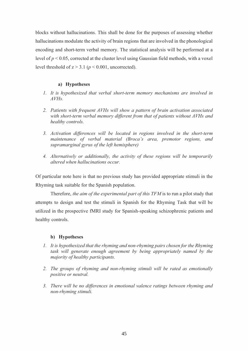
45
blocks without hallucinations. This shall be done for the purposes of assessing whether
hallucinations modulate the activity of brain regions that are involved in the phonological
encoding and short-term verbal memory. The statistical analysis will be performed at a
level of p < 0.05, corrected at the cluster level using Gaussian field methods, with a voxel
level threshold of z > 3.1 (p < 0.001, uncorrected).
a) Hypotheses
1. It is hypothesized that verbal short-term memory mechanisms are involved in
AVHs.
2. Patients with frequent AVHs will show a pattern of brain activation associated
with short-term verbal memory different from that of patients without AVHs and
healthy controls.
3. Activation differences will be located in regions involved in the short-term
maintenance of verbal material (Broca’s area, premotor regions, and
supramarginal gyrus of the left hemisphere)
4. Alternatively or additionally, the activity of these regions will be temporarily
altered when hallucinations occur.
Of particular note here is that no previous study has provided appropriate stimuli in the
Rhyming task suitable for the Spanish population.
Therefore, the aim of the experimental part of this TFM is to run a pilot study that
attempts to design and test the stimuli in Spanish for the Rhyming Task that will be
utilized in the prospective fMRI study for Spanish-speaking schizophrenic patients and
healthy controls.
b) Hypotheses
1. It is hypothesized that the rhyming and non-rhyming pairs chosen for the Rhyming
task will generate enough agreement by being appropriately named by the
majority of healthy participants.
2. The groups of rhyming and non-rhyming stimuli will be rated as emotionally
positive or neutral.
3. There will be no differences in emotional valence ratings between rhyming and
non-rhyming stimuli.

46
3. METHODOLOGY
3.1. Data acquisition
All data was acquired via Psychtoolkit (Stoet, 2010, 2017), which saved all data
inputs, and extracted them in an excel file.
3.2. Participants
Participants were recruited via different social media outlets by researchers from
their circles of relatives and acquaintances, as well as independent sources in the
community. All participants provided their consent online right before beginning the
survey. This pilot study was approved by FIDMAG Hermanas Hospitalarias Research
Foundation’s Ethical Committee.
3.3. Stimuli
The original stimuli consisted of 21 rhyming pairs, and 19 non-rhyming pairs. The
stimuli were first created by putting together Spanish nouns that are frequent in everyday
use in either rhyming or non-rhyming pairs. The criteria for two words to form a rhyming
pair consisted of them having perfect rhyme, meaning that both words were to have an
identical stressed vowel sound, any following sound was identical, and the onset of the
stressed syllables in both words would be completely varied. The criteria for two words
to form a non-rhyming pair consisted of them not meeting the aforementioned conditions.
In either case, both words ought to have had the same number of syllables. What is more,
the number of categories of objects was aimed to be even across rhyming and non-
rhyming pairs (i.e. people, animals, food etc.) as demonstrated in Appendix 1. In addition,
all pairs consisted of two different categories (i.e. animals and fruit) and could not be
semantically related (i.e. not cat & dog but dog & pencil).
Once the word pairlist was created, 2 image corpi representing objects were used
for all rhyming and non-rhyming pairs, as demonstrated in Appendix 2. The first corpus
was borrowed from Hebrat and colleagues (2019), and the second corpus was specifically
created for this study. The images taken from the first corpus, initially colored, were first
desaturated, the background was erased, and a white background was applied so that the
main object was in the foreground, and then the whole image was resized to 400 x 400
pixels. The images taken from the second corpus were first created, smoothed, desatured,
the background was erased, and a white background was applied so that the main object

47
was in the foreground, and then the whole image was resized to 400 x 400 pixels. Next,
2 sets of the same images were made. The first set was used to put together images
according to the word pair they belonged to (implemented in Task 1 and Task 2). The
second set was used for each image to be separated (implemented in Task 3).
3.4. Procedure
Participants were sent a direct link to the online survey via social media outlets.
When the link was opened, the survey began with the participants being exposed to a
detailed account of the aim of the survey, which was followed by them providing their
consent to gather data. The first 6 questions, aimed at acquiring demographic information,
in which participants were asked to provide feedback on their gender, age, birthplace,
place of residence, mother tongue, second language, and other potential foreign languages
that they spoke. Next, the participants were provided with a detailed account of the
instructions to the first task, and they were to check a box acknowledging that they
understood the instructions well, allowing them to move onto the next question.
Having done all the above, the participants were then able to begin the first part
of the survey, which consisted of two tasks. In the first task, Rhyming Judgement Task,
they were presented with 2 objects and were asked whether their names rhyme (i.e. niña
- piña). If the pair of objects rhymed, the participants had to tick yes (“Sí”), and if they
did not rhyme (i.e. zapato - mochila), they had to tick no (“No”). Next, participants were
presented with the second task, Object Naming Task, in which the same pair was
presented again and were asked to provide the specific name for each object separately.
After the participants completed the first part of the online study, they were then
presented with the second part of the survey, which consisted of the third and final task,
Rating Task, in which they were presented with each of the pictures used in the task and
were asked to rate their emotional valence on a scale from 1 to 5 (1 = very negative; 2 =
negative; 3 = neutral; 4 = positive; 5 = very positive), using the self-assessment manikin
(Bradley and Jang, 1994)
Once the participants finished the survey, they were asked whether they wanted
to leave a comment for the researchers voluntarily, after which they were redirected to
the end of the survey.

48
3.5. Data Cleaning
Once the data of the survey was collected from Psychtoolkit (Stoet, 2010, 2017),
it was diligently cleaned in excel.
Firstly, data was not considered from participants who did not provide their
consent (5 out of 84). Secondly, data was also excluded from participants who: did not
understand the instructions (1 out of 84); provided responses to more than one example
of either task due to closing the survey (27 out of 84); providing an answer inconsistent
with the requirements (e.g. 1 participant wrote “z” for the first couple of questions) (1 out
of 84). Here it must be noted that the survey was designed specifically for participants
not to skip examples, yet 1 participant missed their response to one example. This must
have been due to a glitch in the program, and so their data was not included in the analysis.
On a different note, 2 other participants whose birthplace was not in Spain (i.e. Veracruz,
Mexico; Odessa, Ukraine) were not included due to the possibility of deviation from
European Spanish (2 out of 84). In addition, 1 participant conducted the study fully in
Catalan rather than Spanish, so their results were omitted (1 out of 84). Thus, out of 84
participants, a total of 47 participants, having completed the survey and being born in
Spain, were included in the analyses. We also excluded responses for 2 rhyming pairs due
to an error in the number or structure of syllables (i.e. delfín - calcetin) and erroneous
sound pattern (avión - bastón). Thus, the final set of stimuli consisted of 19 rhyming pairs,
and 19 non-rhyming pairs.
All participant responses were then unified, particularly in the Object Naming
Task, in which participants were to provide the name for every object in every pair.
Firstly, all responses were transformed from upper-case to lower-case (e.g. Archivador to
archivador; LIBRETA to libreta). In addition, because Psychtoolkit (Stoet, 2010, 2017)
did not save the Spanish spelling appropriately, all words were accented where necessary
(e.g. JudÃa to judía; sueter to suéter). All misspellings were then corrected for, such as
missing letters (e.g. jean to jeans; goma de borar to goma de borrar; crocil to crocodilo);
additive letters (mochilla to mochila; copia to copa); spelling mistakes (aveja to abeja;
tecladi to teclado; calcetij to calcetín; blobo to globo). What must be mentioned is that
all descriptive data, particularly place of birth, place of residence, mother tongue and all
foreign languages were translated to English, whereas the participant responses for all
tasks were left in Spanish. Lastly, some participants provided more than one name for an
object, and thus, only the first noun was taken into consideration, and any informative

49
responses, such as descriptive adjectives, were dropped (i.e. vaca, buey, ternera to vaca;
rosa roja to rosa).
3.6. Statistical Analysis
All statistical analyses were conducted by utilizing SPSS for Mac (version 2018)
and Python (3.8 version). The level of statistical significance was set at 0.05 across all
statistical tests. Tests were used to conduct comparison between rhyme and non-rhyme
stimuli and potential effects on rating.
4. Results
4.1. Demographic information
Demographic information for all 47 participants can be found in Table 1.
The total number of participants in the online survey was 84. The reported results,
however, come from 47 participants, after data cleaning, who successfully completed the
survey and come from the territory of Spain. Out of 47 participants, 19 were male, 26
were female, and 2 identified as other, of which the youngest was aged 16 and the oldest
was aged 70 (data from participants under 18 was included since no personal data was
collected in this study). Their mean age was 36.12 (SD = 15.83). Such an average age of
healthy participants in this study is similar to the average age of schizophrenic patients
with AVHs who take part in functional neuroimaging studies (i.e. Shergill et al., 2001;
Copolov et al., 2003; Diederen et al., 2010; McCarthy-Jones et al., 2014; de Boer et al.,
2019).
The most common birthplaces were the Balearic Islands (10 participants), Madrid
(10 participants), and Barcelona (9 participants). The most common place of residence
were Barcelona (12 participants), Madrid (11 participants), and Balearic Islands (10
participants).
Education-wise, 35 participants were university educated, 8 received high school
education, and 4 received vocational training. With regards to occupation, 23 participants
were employed, 11 were students, 7 were students and workers, 3 were unemployed and
3 were self-employed.
The mother tongue for 30 participants was Spanish, and for 17 participants was
Catalan. Next, 46 participants spoke a second language, the most common being English
(22 participants), Spanish (14 participants), and Catalan (7 participants). Furthermore, 29

50
participants spoke a third language, the most common being English (16 participants) and
French (6 participants). Moreover, 10 participants spoke a fourth language, the most
common being French (3 participants) and English (2 participants). Lastly, the total time
that it took all 47 participants to complete the survey is approximately 26 minutes (mean
25.78, SD 10.89).
Table 1. Demographic information of all participants.
N %
Sex female 26 55.3%
male 19 40.3%
other 2 4.3%
Place of Birth baleares 10 21.3%
madrid 10 21.3%
barcelona 9 19.1%
granada 2 4.3%
zaragoza 2 4.3%
cádiz 2 4.3%
valencia 1 2.1%
zamora 1 2.1%
alicante 1 2.1%
sevilla 1 2.1%
mallorca 1 2.1%
ciudad real 1 2.1%
lleida 1 2.1%
tarragona 1 2.1%
pontevedra 1 2.1%
asturias 1 2.1%
cadiz 1 2.1%
castellón 1 2.1%
Place of Residence barcelona 12 25.5%
madrid 11 23.4%
baleares 10 21.3%
granada 3 6.3%
alicante 3 6.3%
tarragona 1 2.1%
pontevedra 1 2.1%
araba 1 2.1%
cáceres 1 2.1%
ciudad real 1 2.1%
mallorca 1 2.1%
cádiz 1 2.1%
almería 1 2.1%
Education university 35 74.5%
vocational training 8 17.0%
high school 4 8.5%

51
Occupation employed 23 48.9%
student 11 23.4%
student/employed 7 14.9%
self-employed 3 6.4%
unemployed 3 6.4%
Mother Tongue
spanish 30 63.8%
catalan 17 36.2%
Second Language 1
english 22 46.8%
spanish 14 29.8%
catalan 7 14.9%
french 2 4.3%
galician 1 2.1%
- 1 2.1%
Second Language 2
- 18 38.3%
english 16 34.0%
french 6 12.8%
sign language 2 4.3%
catalan 1 2.1%
italian 1 2.1%
japenese 1 2.1%
russian 1 2.1%
spanish 1 2.1%
Second Language 3
- 37 78.7%
french 3 6.4%
english 2 4.3%
arabic 1 2.1%
basque 1 2.1%
danish 1 2.1%
german 1 2.1%
italian 1 2.1%

52
4.2. Stimuli Selection
Out of the total rhyming and non-rhyming pairs presented to participants, the pairs
that were chosen for the prospective fMRI study were based on high agreement of object
names as appropriately named by the majority of participants and positive or neutral
emotional ratings as shown in. The total number of chosen pairs of both groups is 24 (12
rhyming pairs and 12 non-rhyming pairs), as the rhyming judgement condition in the
prospective fMRI study consists of 24 trials.
The descriptive analysis is shown below.
4.2.1. Rhyming Judgment Task
Based on the chosen pairs for both groups, the frequency of rhyming judgements
(Table 2) and non-rhyming judgments (Table 3) for every pair was calculated as
presented below.
Table 2. Frequency of rhyming judgements for every pair.
Rhyming Pairs
Pair "Yes" Frequency
"No"
Frequency
pato - gato 47 0
cama - rama 47 0
nariz - maiz 46 1
oreja - abeja 45 2
ventana -
manzana 45 2
fresa - mesa 43 4
queso - hueso 47 0
nina - pina 46 1
globo - lobo 46 1
capa - mapa 46 1
cubo - tubo 45 2
helado -
teclado 42 5

53
Table 3. Frequency of non-rhyming judgements for every pair.
4.2.2. Object Naming Task
The objective of this analysis was to determine the frequency of responses of
names for every object in every pair (rhyme and non-rhyme) and in this way chose the 12
pairs of both categories with the highest agreement.
With the help of Python (version 3.8), the first step that was taken was counting
the frequency of a given word for every object in the rhyme-pairs. For instance, for the
object folder, 27 participants said carpeta, 14 participants said libreta, 4 participants said
cuaderno, 1 participant said clasificador, 1 participant said archivador. A summary of all
responses for the rhyme objects can be found in Appendix 3, and all responses for the
non-rhyme objects can be found in Appendix 4. Once said frequency was counted, the
percentage of participants selecting the most frequent word (percentage of agreement)
was calculated. For instance, in the previously mentioned example (which consists of a
rhyming pair carpeta-galleta) for the object folder, the most frequent is carpeta according
to 27 participants (57%), and for the object biscuit, the most frequent is galleta according
to 47 participants (100%). This was done for every object of every rhyming pair as. The
percentage of agreement for each pair was calculated as the average of the individual
percentages of agreement for the 2 objects forming the pair (e.g. 78.5% in the previous
example). All pairs were then sorted in a descending fashion, and the top 12 with the
highest agreement were selected (as the rhyming judgement condition in the fMRI
Non-Rhyming Pairs
Pair "Yes" Frequency "No" Frequency
gorra - dado 1 46
vaso - cuna 1 46
rosa- regla 0 47
llave - cerdo 0 47
mochila - zapato 1 46
tenedor - lechuga 0 47
falda - coche 0 47
perro - lapiz 1 46
calabaza - elefante 0 47
esqueleto - cocodrilo 0 47
tomate - cuchara 0 47
goma - jersey 0 47

54
scanner requires 24 trials, thus 12 pairs of both groups are needed). The chosen pairs as
shown in Table 4.
Table 4. Rhyming Pairs with highest agreement. The chosen 12 are highlighted in red.
The same procedure was applied to the non-rhyming pairs and the results are
shown in Table 5.
Table 5. Non-Rhyming Pairs with highest agreement. The chosen 12 are highlighted in
red.

55
4.2.3. Rating Task
Since the aim of the task is to engage phonological encoding processes, the
selected stimuli need to be emotionally neutral to avoid the confounding effects of
emotional responses to the pictures. To ensure that this was the case, we extracted
descriptive measures from the picture ratings, in terms of affective valence, and also
compared the ratings of rhyming and non-rhyming pairs, to make sure no significant
differences were found between the two groups of stimuli. Only the ratings of objects that
are part of the 12 rhyming pairs and 12 non-rhyming pairs selected for the fMRI task were
included.
Firstly, with the use of Python (version 3.8), data from outliers, or participants
whose responses were extreme, were omitted. Thus, for this task, data from 45 out 47
participants were considered.
Secondly, an independent two sample t-test between both the rhyming and non-
rhyming groups was carried out (for which all assumptions were met). Firstly, to test for
normality, the Shapiro-Wilk Test of Normality was applied which showed that p > .05 (p
= .71). Thus, the sample is gaussian and the null hypothesis failed to be rejected, as both
groups are approximately normally distributed. Similarly, there was homogeneity of
variance of rating for rhyming and non-rhyming, as assessed by Levene’s Test for
Equality of Variances which showed p > .05 (p = .48). Lastly, the Independent two sample
t-test showed no significant difference between both groups, t(22) = -1.22, p = .23.
5. DISCUSSION
Hearing voices, or experiencing auditory verbal hallucinations (AVHs) is a
detrimental symptom of schizophrenia (among other conditions). Not having the
opportunity to experience tranquility or the ability to control what is whispered in one’s
‘ear’ affects the patient to a large extent. The complex phenomenological basis of AVHs
demonstrates that they are not part of the patient’s imagination, but are rather an
additional feature of their perception of the world.
Despite their clinical significance, the origins of AVHs are still unknown. In order
to find the key to their biological basis, an experimental design of a prospective fMRI
study has been proposed that takes into consideration different perspectives that have
targeted AVHs exploratorily of which two main approaches or models have been built.
56
The first, perceptual model, aims at understanding whether AVHs are due to the
abnormal activity of the primary auditory cortex (which is part of the superior temporal
gyrus), a brain area responsible for primary sound perception and processing. On the one
hand, some single studies and meta-analyses have found activation in the auditory cortex
during AVHs (Dierks et al., 1999, Lennox et al., 1999; Kompus et al., 2011 ), while others
have not (Silbersweig et al., 1995; Lennox et al., 2000; Copolov et al., 2003; Diederen et
al. 2010; Hoffman et al., 2011; Fuentes-Claramonte et al. 2021; Jardri et al., 2011; Kühn
and Gallinat (2012); Zmigrod et al., 2016). The limitations of these studies, which range
from not recruiting enough patients to not precisely controlling for motor activity, should
not be understated, as they may have significantly confounded the main results. This
demonstrates the conflicting nature of such studies, and does not fully provide a sufficient
answer to whether aberrant activation in the PAC leads to AVHs. For this reason, the
paradigm proposed to test this model is based on Fuentes-Claramonte and colleagues
(2021), with a modification to control for motor activity. This is done by asking patients
to press a button with both index fingers in the fMRI scanner during those time periods
in which they do not experience AVHs.
The second, the Cognitive Model, has two sides which aim at understanding
whether AVHs are due to either misattributions of intrusive or traumatic memories, or
malfunction in inner speech. It must be pointed out that both the theoretical and
experimental work behind both theories are still under construction, and many questions
remain open.
One side of the Cognitive Model lies in the theory of episodic memory and more
particularly, source memory failure, in which the retrieval of memories is fragmented. As
such, the patient is confused regarding the origins of the memory. The activation of
intrusive memories is theorized to come from the deficits in intentional inhibition and
contextual memory, which causes auditory mental representation to intrude into
consciousness - a phenomenon out of control for the patient. This goes hand in hand with
the fact that some studies have shown that the parahippocampus becomes activated a few
seconds before patients experience AVHs (e.g. Diederen et al., 2010; Hoffman et al.,
2011), and that both the parahippocampus and hippocampus become activated during the
experience of hearing voices (Silbersweig et al., 1995; Dierks et al., 199; Diederen et al.,
2010; Jardri et al., 2011; Kompus et al. 2011; Zmigrod et al. 2016). One way of
determining whether the intrusion of traumatic memories is the reason behind AVHs is
by considering autobiographical memory - the active retrieval of personal past

57
experiences involving highly complex retrieval processes. This type of episodic memory
activates a network of regions known which overlap with the neural network that is
activated during resting or passive states, known as the DMN (which has been thought be
impaired in schizophrenia, though the findings are inconclusive (e.g. Starck et al., 2005;
Zhou et al., 2018; Martin-Subero et al., 2021). Although some studies have explored AM
in schizophrenia, no previous study has placed enough emphasis on AVHs in
schizophrenia and the potential link to AM. Because of this, the paradigm proposed to
test this model, the Autobiographical Recall Task, relies on Fuentes-Claramonte and
colleagues (2019), which has been validated by Martin-Subero and colleagues (2021).
Here, patients will be presented with keywords that are designed to either evoke or inhibit
autobiographical memories which the patients will share with the researchers prior to the
study. In this way, the stimuli will be tailored individually to every patient taking part in
the study.
The other side of the Cognitive Model lies in the malfunction of inner speech - a
malfunction in the brain’s monitoring system which does not recognize self-generated
speech and attributes it to an external agent. Due to this, the voices that schizophrenic
patients experience seem to be coming from ‘outside’ from the individual, rather than
interpreting voices as coming from ‘within’ the individual. Despite having the intention
to speak, the patient is unaware of having initiated speaking, and so for them it seems that
the sense of initiation comes from an external source. The way in which this theory has
been studied has been based on implementing a variety of tasks that target auditory verbal
imagery, wherein the patient imagines speech or inner speech tasks which attempt to
understand whether there is a breakdown in self-monitoring. Because of this, the
paradigm proposed to test this model, the Rhyming Judgement Task, will present
participants with 2 images of 2 different objects and will be instructed to think about the
name of each of the objects and decide whether they rhyme or not. If the pair rhyme, they
are to press a button with their right index finger, if the pair does not rhyme, they are to
press a button with their left index finger.
Because of the fact that no study as of yet has tested the Spanish population with
such image stimuli, a pilot study was conducted to determine the precise picture stimuli
that will be used in the prospective fMRI study in the Inner Speech testing of the cognitive
model. For this reason, an online survey asking participants whether the presented pairs
rhymed, the names of both objects in each pair, as well as rating each object separately
on the Likert scale was conducted. Based on the results, we were able to select the 12

58
rhyming and non-rhyming pairs with the greatest agreement in terms of rhyming
(Yes/No) and the name assigned to the presented object. Furthermore, additional
statistical analyses showed that there was no significant difference between rhyme and
non-rhyme pairs in affective valence ratings. This will ensure that the stimuli used in the
task will elicit, on average, the same verbal responses in most of the participants, and
confounding factors like unclear object naming will not contaminate the observed brain
activations in the fMRI study.
Only by taking a variety of approaches can the appropriate brain regions and
circuits underlying AVHs be determined which may provide the appropriate clinical
intervention, such as therapeutic targets for pharmacological or brain stimulation
interventions which can lead to the improvement of the patient’s life. With this in mind,
this study has many advantages that can make this happen.
With regards to the prospective fMRI study, the major advantages are that it tests
different approaches to find the biological basis of AVHs. In this way, the two major
approaches (or three theories) that have been common across literature have been put
together into one experimental design. Firstly, the entire prospective fMRI study aims to
have 25 participants per group which will allow for statistically significant results.
Gathering this number of participants per group will be doable as the FIDMAG research
team cooperates with many different hospitals in Catalunya, and many of their previous
studies have successfully included such a number of participants (i.e. Fuentes-Claramonte
et al., 2019, 2021; Martin-Subero et al., 2021 ). In connection to this, apart from testing
schizophrenic patients with AVHs and healthy controls, the prospective fMRI study will
include schizophrenic patients without AVHs which will allow comparisons across a
broad range of subjects, two groups having similar pathologies and divergent symptoms.
With regards to the experimental design, it has been designed to last around an hour,
which allows the participants to be tested in one sitting. Furthermore, the prospective
fMRI study consists of paradigms that have already been tested for. For instance, the
Symptom Capture Paradigm, which tests the perceptual model, and the Autobiographical
Recall Task, which tests the intrusive memory theory (one side of the Cognitive Model),
have both been meticulously experimented with by the research group that developed this
TFM (i.e. Fuentes-Claramonte, 2019, 2021; Martin-Subero et al., 2021). What this TFM
does suggest, however, are certain modifications to those paradigms that enable results
overcoming previous limitations as shown in Figures 4, 5, and 6 in Section 2. In turn, the

59
experimental paradigm that targets inner speech theory (the other side of the Cognitive
Model), has not been tested before on the Spanish population.
Similarly, the pilot study adds to the advantages of the fMRI framework. The
online survey tested healthy participants who will most likely be matched to the recruited
groups of schizophrenic patients with regards to age, as many studies outline that the
mean age of schizophrenic patients recruited in studies targeting AVHs is around 35 (e.g.
Sommer et al., 2008; Kompus et al., 2013 etc.). Furthermore, as the recruitment of patients
will take place in hospitals residing in Catalunya, the majority of prospective
schizophrenic patients may have a similar place of birth, place of residence, mother
tongue and second language. In addition, the total number of participants whose data was
considered was 47, which is a considerable sample size for a pilot study. With regards to
the stimuli chosen, a sufficient number of both rhyming and non-rhyming pairs were
tested to select the most appropriate ones, as the rhyming judgement condition in the
prospective fMRI study will include 24 trials, hence the 12 pairs of each group.
Conversely, both the prospective fMRI study and pilot study may have some
possible caveats that shall be mentioned.
With regards to the prospective fMRI study, testing schizophrenic patients in a
functional neuroimaging study poses challenges because the severity of symptoms for
some patients might not allow them to follow through with the entire experiment.
Consequently, some patients might not be able to tolerate the conditions of fMRI
experiment. Furthermore, the prospective schizophrenic patients will most likely be
receiving antipsychotic drug treatment, which is a limitation in most functional imaging
studies that investigate schizophrenia. On top of that, studying hallucinations is a complex
procedure, as patients experience brief AVHs that are unpredictable, and thus are difficult
to investigate when compared to other sensory experiences. Lastly, any potential
limitations regarding each proposed paradigm will come to light only once the
prospective fMRI is conducted.
With regards to the pilot study, even though the instructions of the study stated to
use Spanish, what could have been more precisely mentioned is to not to use Catalan, as
in some examples in the Object Naming Task, participants provided the names of objects
in this language rather than Spanish (such as that 1 participant whose data was excluded
as they used Catalan for the majority of responses). This may have been due to the
frequency of usage of the given name of the object, and thus the most dominant word for
a given object became activated, and so participants provided the first name that came to

60
mind. Similarly, what could also have been mentioned more explicitly is to only use 1
word for every object, in this way, participants would focus their attention on thinking of
one name rather than four or five as took place in some cases. For instance, some
participants added descriptive adjectives to the object (e.g. rosa roja ). On a different
note, the images that were chosen may not have been as well illustrated despite the pair
being frequently used in Spanish, which caused some participants to have a difficulty
with providing the names for objects as found in the pairs that were not chosen (i.e. casa-
gasa or mano-vaca). Lastly, as the mean time of all participants is 25 minutes long, this
may have been too long for the participants who were not rewarded for their participation.
Taking these factors into consideration, participants’ attention spans might have declined,
along with their motivation, when they were conducting the pilot study - particularly the
last task they were exposed to (the Rating task) - and thus some ratings may have been
impacted by this.
Therefore, future research should precisely consider these implications when
trying their hand at exploring AVHs, and when developing appropriate stimuli and corpi.
One way to overcome these limitations would be to conduct the fMRI study itself, and
test the mentioned experimental paradigms that target different perspectives of AVHs. By
doing so, previous limitations found across many previous studies can be overcome.
Once conducted, our speculation is that brain activation during the symptom
capture paradigm will not show significant activation in the PAC, but rather point to the
significance of language areas, as they are the ones responsible for inner speech.
Furthermore, although significant activation will be found in the autobiographical recall
task during the evocation of negative memories, no significant difference will be found
across all three groups, meaning that traumatic memories are less likely to be the cause
of AVHs which will be reflected in brain activity. Therefore, the one likely premise is
that malfunctions in inner speech, or self-monitoring, is the reason why schizophrenia
patients experience voices. In this way, there will be activation differences in brain areas
that are responsible for short-term maintenance of verbal material, such as the Broca’s
area and others. Taken together, our speculation is in line with Frith’s (1992, p. 76)
proposal: “some auditory hallucinations are based on inner speech which the patient
misattributes to an external source”.

61
6. CONCLUSIONS
In summary, this study has attempted to make two significant contributions to the
fields of Cognitive Science and Psychiatry. The first attempt was to propose an
experimental design of a prospective fMRI study which targets different aspects of the
biological basis of AVHs: abnormal activation in the auditory cortex; the
misinterpretations of intrusive or traumatic memories; the malfunction in inner speech,
or speech monitoring. This was done by proposing three different experimental
paradigms, created and validated by previous research, and by implementing certain
modifications to overcome previous limitations. The second attempt was to develop novel
stimuli (12 rhyming and 12 non-rhyming pairs) that will be implemented in the rhyming
judgement condition in the fMRI study, which tackles the malfunctioning in inner speech.
The chosen stimuli (12 rhyming pairs and 12 non-rhyming pairs) was based on having
high agreement of object names as appropriately named by the majority of participants
and having positive or neutral emotional ratings.
Solely by taking such a diverse approach to understanding the biological basis of
AVHs can the most fundamental aim of research on hallucinations be addressed:
pinpointing the brain regions which are responsible for the genesis of AVHs. In doing so,
there can be a direct impact on this symptom of schizophrenia at hand - only then can
sufficient clinical intervention be devised, such as personalized medicine in the form of
pharmaceutical intervention or brain stimulation. In treating a disease as heterogeneous
as schizophrenia, such knowledge is imperative in adjusting treatments to the needs of
each patient so that the maximum clinical improvement is obtained minimizing the risks
and the adverse effects.
Those, like Socrates, whose voices deter them from their actions and intentions,
by whispering ill omens in the vacuum of tranquility, may finally achieve the silence of
their “soul”.
62
REFERENCES
Achim, A. M., Maziade, M., Raymond, É., Olivier, D., Mérette, C., & Roy, M. A. (2011). How prevalent
are anxiety disorders in schizophrenia? A meta-analysis and critical review on a significant
association. Schizophrenia bulletin, 37(4), 811-821.
Alderson-Day, B., & Fernyhough, C. (2015). Inner speech: development, cognitive functions,
phenomenology, and neurobiology. Psychological bulletin, 141(5), 931.
Allen, P., Larøi, F., McGuire, P. K., & Aleman, A. (2008). The hallucinating brain: a review of structural
and functional neuroimaging studies of hallucinations. Neuroscience & Biobehavioral Reviews, 32(1), 175-
191.
Allen, P., Modinos, G., Hubl, D., Shields, G., Cachia, A., Jardri, R., ... & Hoffman, R. (2012).
Neuroimaging auditory hallucinations in schizophrenia: from neuroanatomy to neurochemistry and
beyond. Schizophrenia bulletin, 38(4), 695-703.
Aserinsky, E., & Kleitman, N. (1953). Regularly occurring periods of eye motility, and concomitant
phenomena, during sleep. Science, 118(3062), 273-274.
Baddeley, A. D., & Hitch, G. (1974). Working memory. In Psychology of learning and motivation (Vol. 8,
pp. 47-89). Academic press.
Badeley, A., Logie, R., Bressi, S., Sala, S. D., & Spinnler, H. (1986). Dementia and working memory. The
Quarterly Journal of Experimental Psychology Section A, 38(4), 603-618.
Badeley, A., Gathercole, S., & Papagno, C. (1998). The phonological loop as a language learning
device. Psychological review, 105(1), 158.
Barta, P. E., Pearlson, G. D., Powers, R. E., Richards, S. S., & Tune, L. E. (1990). Auditory hallucinations
and smaller superior temporal gyral volume in schizophrenia. The American journal of psychiatry, 147(11),
1457–1462.
Bradley, M. M., & Lang, P. J. (1994). Measuring emotion: the Self-Assessment Manikin and the Semantic
Differential. Journal of behavior therapy and experimental psychiatry, 25(1), 49–59.
Belin, P., Zatorre, R. J., Lafaille, P., Ahad, P., & Pike, B. (2000). Voice-selective areas in human auditory
cortex. Nature, 403(6767), 309-312.
Berger, A. (2003). How does it work?: Positron emission tomography. BMJ: British Medical
Journal, 326(7404), 1449.
Berrios, G. E. (1996). The history of mental symptoms: Descriptive psychopathology since the nineteenth
century. Cambridge University Press.
Bick, P. A., & Kinsbourne, M. (1987). Auditory hallucinations and subvocal speech in schizophrenic
patients. The American Journal of Psychiatry.
Bicknell, P. (1981). DÉJÀ VU, Autoscopia, and Antipheron: Notes on, Memory and Recollection ", I, 451a
8–12 and " Meterologica", III, 4, 373b 1–10. Acta Classica, 24, 156-159.
Bird, C. M., & Burgess, N. (2008). The hippocampus and memory: insights from spatial processing. Nature
Reviews Neuroscience, 9(3), 182-194.
Blakemore, S. J., Smith, J., Steel, R., Johnstone, E. C., & Frith, C. D. (2000). The perception of self-
produced sensory stimuli in patients with auditory hallucinations and passivity experiences: evidence for a
breakdown in self-monitoring. Psychological medicine, 30(5), 1131-1139.

63
Blom, J. D. (2016). Alice in Wonderland syndrome: a systematic review. Neurology: Clinical
Practice, 6(3), 259-270.
Buckner, R. L., Andrews-Hanna, J. R., & Schacter, D. L. (2008). The brain's default network: anatomy,
function, and relevance to disease.
Buchsbaum, B. R. (2016). Working Memory and Language. In G. Hickok & S. L. Small (Eds.),
Neurobiology of Language (pp. 863-875). Elsevier.
Chalfonte, B. L., & Johnson, M. K. (1996). Feature memory and binding in young and older adults. Memory
& cognition, 24(4), 403–416. https://doi.org/10.3758/bf03200930
Chappell, S. G. (2005). Plato on Knowledge in the Theaetetus.
Choong, C., Hunter, M. D., & Woodruff, P. W. R. (2007). Auditory hallucinations in those populations that
do not suffer from schizophrenia. Current Psychiatry Reports, 9(3), 206-212.
Chin, J. T., Hayward, M., & Drinnan, A. (2009). Relating to voices: Exploring the relevance of this concept
to people who hear voices. Psychology and Psychotherapy: Theory, Research and Practice, 82(1), 1-17.
Copolov, D. L., Seal, M. L., Maruff, P., Ulusoy, R., Wong, M. T., Tochon-Danguy, H. J., & Egan, G. F.
(2003). Cortical activation associated with the experience of auditory hallucinations and perception of
human speech in schizophrenia: a PET correlation study. Psychiatry Research: Neuroimaging, 122(3), 139-
152.
Copolov, D., Trauer, T., & Mackinnon, A. (2004). On the non-significance of internal versus external
auditory hallucinations. Schizophrenia Research, 69(1), 1-6.
Coren, L. H. (1938). Imagery and its relation to schizophrenic symptoms. Journal of Mental Science, 84,
284±346.
Cramer, S. C., Weisskoff, R. M., Schaechter, J. D., Nelles, G., Foley, M., Finklestein, S. P., & Rosen, B.
R. (2002). Motor cortex activation is related to force of squeezing. Human brain mapping, 16(4), 197-205.
Crovitz, H. F., & Schiffman, H. (1974). Frequency of episodic memories as a function of their age. Bulletin
of the Psychonomic Society, 4(5), 517-518.
Cuervo-Lombard, C., Lemogne, C., Gierski, F., Bera-Potelle, C., Tran, E., Portefaix, C., ... & Limosin, F.
(2012). Neural basis of autobiographical memory retrieval in schizophrenia. The British Journal of
Psychiatry, 201(6), 473-480.
Ćurčić-Blake, B., Ford, J. M., Hubl, D., Orlov, N. D., Sommer, I. E., Waters, F., ... & Aleman, A. (2017).
Interaction of language, auditory and memory brain networks in auditory verbal hallucinations. Progress
in neurobiology, 148, 1-20.
Damasio, A. R. (1989). Time-locked multiregional retroactivation: A systems-level proposal for the neural
substrates of recall and recognition. Cognition, 33(1-2), 25–62.
David, A. S. (1999). Auditory hallucinations: phenomenology, neuropsychology and neuroimaging
update. Acta Psychiatrica Scandinavica, 99, 95-104.
Davis, R. C. (1938). The relation of muscle action potentials to difficulty and frustration. Journal of
Experimental Psychology, 23(2), 141.
de Boer, J. N., Linszen, M. M., de Vries, J., Schutte, M. J., Begemann, M. J., Heringa, S. M., ... & Sommer,
I. E. C. (2019). Auditory hallucinations, top-down processing and language perception: a general population
study. Psychological medicine, 49(16), 2772-2780.

64
Diederen, K. M., Neggers, S. F., Daalman, K., Blom, J. D., Goekoop, R., Kahn, R. S., & Sommer, I. E.
(2010). Deactivation of the parahippocampal gyrus preceding auditory hallucinations in
schizophrenia. American Journal of Psychiatry, 167(4), 427-435.
Diederen, K. M., Daalman, K., de Weijer, A. D., Neggers, S. F., van Gastel, W., Blom, J. D., ... & Sommer,
I. E. (2012). Auditory hallucinations elicit similar brain activation in psychotic and nonpsychotic
individuals. Schizophrenia bulletin, 38(5), 1074-1082.
Diederen, K. M. J., Neggers, S. F. W., De Weijer, A. D., Van Lutterveld, R., Daalman, K., Eickhoff, S. B.,
... & Sommer, I. E. C. (2013). Aberrant resting-state connectivity in non-psychotic individuals with auditory
hallucinations. Psychological medicine, 43(8), 1685-1696.
Dierks, T., Linden, D. E., Jandl, M., Formisano, E., Goebel, R., Lanfermann, H., & Singer, W. (1999).
Activation of Heschl’s gyrus during auditory hallucinations. Neuron, 22(3), 615-621.
Dilks, S., Tasker, F., & Wren, B. (2010). Managing the impact of psychosis: A grounded theory exploration
of recovery processes in psychosis. British Journal of Clinical Psychology, 49(1), 87-107.
Ditman, T., & Kuperberg, G. R. (2005). A source-monitoring account of auditory verbal hallucinations in
patients with schizophrenia. Harvard review of psychiatry, 13(5), 280-299.
Engel, A. K., Fries, P., & Singer, W. (2001). Dynamic predictions: oscillations and synchrony in top–down
processing. Nature Reviews Neuroscience, 2(10), 704-716.
Fenske, M. J., Aminoff, E., Gronau, N., & Bar, M. (2006). Top-down facilitation of visual object recognition: object-based and context-based contributions. Progress in brain research, 155, 3-21.
Ferguson, S. M., Rayport, M., KASS, W., GARDNER, R., Weiner, H., & Reiser, M. F. (1969). Similarities
in mental content of psychotic states, spontaneous seizures, dreams, and responses to electrical brain
stimulation in patients with temporal lobe epilepsy. Psychosomatic Medicine, 31(6), 479-498.
Foucher, J. R. (2013). Perspectives in brain imaging and computer-assisted technologies for the treatment
of hallucinations. In The Neuroscience of Hallucinations (pp. 529-547). Springer, New York, NY.
Frith, C. D. (1987). The positive and negative symptoms of schizophrenia reflect impairments in the
perception and initiation of action. Psychological medicine, 17(3), 631-648.
Frith, C. D. (1992). The cognitive neuropsychology of schizophrenia.
Frith, C., & Dolan, R. J. (1997). Brain mechanisms associated with top-down processes in
perception. Philosophical Transactions of the Royal Society of London. Series B: Biological
Sciences, 352(1358), 1221-1230.
Fu, C. H., Vythelingum, N., Andrew, C., Brammer, M. J., Amaro, E., Williams, S. C., & McGuire, P. K.
(2001). Alien voices... who said that? Neural correlates of impaired verbal self-monitoring in
schizophrenia. NeuroImage, 13(6), S1052-S1052.
Fuentes-Claramonte, P., Martín-Subero, M., Salgado-Pineda, P., Alonso-Lana, S., Moreno-Alcázar, A.,
Argila-Plaza, I., ... & Salvador, R. (2019). Shared and differential default-mode related patterns of activity
in an autobiographical, a self-referential and an attentional task. Plos one, 14(1), e0209376.
Fuentes-Claramonte, P., Soler-Vidal, J., Salgado-Pineda, P., Garcia-León, M. A., Ramiro, N., Santo-
Angles, A., Torres, M. L., Tristany, J., Guerrero-Plaza, A., Munuera, J., Sarró, S., Salvador, R., Hinzen,
W., McKenna,P., Pomarol-Clotet, E. (2021). Auditory hallucinations activate language and verbal short-
term memory, but not auditory, brain regions. Scientific Reports.
Gallagher, A. G., Dinan, T. G., & Baker, L. J. V. (1994). The effects of varying auditory input on
schizophrenic hallucinations: A replication. British Journal of Medical Psychology, 67(1), 67-75.

65
Garrett, M., & Silva, R. (2003). Auditory hallucinations, source monitoring, and the belief that “voices” are
real. Schizophrenia Bulletin, 29(3), 445-457.
Glover, G. H. (2011). Overview of functional magnetic resonance imaging. Neurosurgery Clinics, 22(2),
133-139.
Gould, L. N. (1949). Auditory hallucinations and subvocal speech; objective study in a case of
schizophrenia. Journal of Nervous and Mental Disease.
Gould, L. N. (1950). Verbal hallucinations as automatic speech: The reactivation of dormant speech
habit. American Journal of Psychiatry, 107(2), 110-119.
Grubaugh, A. L., Zinzow, H. M., Paul, L., Egede, L. E., & Frueh, B. C. (2011). Trauma exposure and
posttraumatic stress disorder in adults with severe mental illness: A critical review. Clinical psychology
review, 31(6), 883-899.
Green, M. F., & Kinsbourne, M. (1990). Subvocal activity and auditory hallucinations: Clues for behavioral
treatments?. Schizophrenia Bulletin, 16(4), 617-625.
Guerrero-Pedraza, A., McKenna, P. J., Gomar, J. J., Sarro, S., Salvador, R., Amann, B., ... & Pomarol-
Clotet, E. (2012). First-episode psychosis is characterized by failure of deactivation but not by hypo-or
hyperfrontality. Psychological medicine, 42(1), 73-84.
Hardy, A., Fowler, D., Freeman, D., Smith, B., Steel, C., Evans, J., ... & Dunn, G. (2005). Trauma and hallucinatory experience in psychosis. The Journal of nervous and mental disease, 193(8), 501-507.
Helmholtz, H. V. (1866). Concerning the perceptions in general. Treatise on physiological optics,.
Hoffman, R. E., Varanko, M., Gilmore, J., & Mishara, A. L. (2008). Experiential features used by patients
with schizophrenia to differentiate ‘voices’ from ordinary verbal thought. Psychological medicine, 38(8),
1167-1176.
Hoffman, R. E., Pittman, B., Constable, R. T., Bhagwagar, Z., & Hampson, M. (2011). Time course of
regional brain activity accompanying auditory verbal hallucinations in schizophrenia. The British Journal
of Psychiatry, 198(4), 277-283.
Hu, M. L., Zong, X. F., Mann, J. J., Zheng, J. J., Liao, Y. H., Li, Z. C., ... & Tang, J. S. (2017). A review
of the functional and anatomical default mode network in schizophrenia. Neuroscience bulletin, 33(1), 73-
84.
Hugdahl, K., Løberg, E. M., Specht, K., Steen, V. M., Wageningen, H. V., & Jørgensen, H. A. (2008).
Auditory hallucinations in schizophrenia: the role of cognitive, brain structural and genetic disturbances in
the left temporal lobe. Frontiers in Human Neuroscience, 2, 6.
Inouye, T., & Shimizu, A. (1970). The electromyographic study of verbal hallucination. Journal of Nervous
and Mental Disease.
Insel, T. R. (2010). Rethinking schizophrenia. Nature, 468(7321), 187-193.
Jacobson, E. (1930). Electrical measurements of neuromuscular states during mental activities: I.
Imagination of movement involving skeletal muscle. American Journal of Physiology-Legacy
Content, 91(2), 567-608.
Jacobson, E. (1932). Electrophysiology of mental activities. The American Journal of Psychology, 44(4),
677-694.
Jardri, R., Pins, D., Delmaire, C., Goeb, J. L., & Thomas, P. (2007). Activation of bilateral auditory cortex
during verbal hallucinations in a child with schizophrenia. Molecular psychiatry, 12(4), 319.

66
Jardri, R., Pins, D., Bubrovszky, M., Lucas, B., Lethuc, V., Delmaire, C., ... & Thomas, P. (2009). Neural
functional organization of hallucinations in schizophrenia: multisensory dissolution of pathological
emergence in consciousness. Consciousness and cognition, 18(2), 449-457.
Jardri, R., Pouchet, A., Pins, D., & Thomas, P. (2011). Cortical activations during auditory verbal
hallucinations in schizophrenia: a coordinate-based meta-analysis. American Journal of Psychiatry, 168(1),
73-81.
Jardri, R., Thomas, P., Delmaire, C., Delion, P., & Pins, D. (2013). The neurodynamic organization of
modality-dependent hallucinations. Cerebral Cortex, 23(5), 1108-1117.
Jarosinski, J. M. (2008). Exploring the experience of hallucinations from a perspective of self: Surviving
and persevering. Journal of the American Psychiatric Nurses Association, 14(5), 353-362.
Javitt, D. C., & Sweet, R. A. (2015). Auditory dysfunction in schizophrenia: integrating clinical and basic
features. Nature Reviews Neuroscience, 16(9), 535-550.
Jenner, J. A., Rutten, S., Beuckens, J., Boonstra, N., & Sytema, S. (2008). Positive and useful auditory
vocal hallucinations: prevalence, characteristics, attributions, and implications for treatment. Acta
Psychiatrica Scandinavica, 118(3), 238-245.
Kandinskii, V. K. (1890). O pseudogallucinaciyach. St. Petersburg.
Kompus, K., Westerhausen, R., & Hugdahl, K. (2011). The “paradoxical” engagement of the primary
auditory cortex in patients with auditory verbal hallucinations: a meta-analysis of functional neuroimaging
studies. Neuropsychologia, 49(12), 3361-3369.
Kompus, K., Falkenberg, L. E., Bless, J. J., Johnsen, E., Kroken, R. A., Kråkvik, B., ... & Hugdahl, K.
(2013). The role of the primary auditory cortex in the neural mechanism of auditory verbal
hallucinations. Frontiers in Human Neuroscience, 7, 144.
Kopelman, M. D., Wilson, B. A., & Baddeley, A. D. (1989). The autobiographical memory interview: a
new assessment of autobiographical and personal semantic memory in amnesic patients. Journal of clinical
and experimental neuropsychology, 11(5), 724-744.
Kraepelin, E. (1919). Dementia praecox and paraphrenia. Livingstone.
Kühn, S., & Gallinat, J. (2012). Quantitative meta-analysis on state and trait aspects of auditory verbal
hallucinations in schizophrenia. Schizophrenia bulletin, 38(4), 779-786.
Kühn, S., & Gallinat, J. (2012). Quantitative meta-analysis on state and trait aspects of auditory verbal
hallucinations in schizophrenia. Schizophrenia bulletin, 38(4), 779-786.
Leudar, I., Thomas, P., McNally, D., & Glinski, A. (1997). What voices can do with words: pragmatics of
verbal hallucinations. Psychological medicine, 27(4), 885-898.
Lennox, B. R., Bert, S., Park, G., Jones, P. B., & Morris, P. G. (1999). Spatial and temporal mapping of
neural activity associated with auditory hallucinations. The Lancet, 353(9153), 644.
Lennox, B. R., Park, S. B. G., Medley, I., Morris, P. G., & Jones, P. B. (2000). The functional anatomy of
auditory hallucinations in schizophrenia. Psychiatry Research: Neuroimaging, 100(1), 13-20.
Leroy, A., Foucher, J. R., Pins, D., Delmaire, C., Thomas, P., Roser, M. M., ... & Jardri, R. (2017). fMRI
capture of auditory hallucinations: Validation of the two‐steps method. Human brain mapping, 38(10),
4966-4979.
Li, X., Branch, C. A., & DeLisi, L. E. (2009). Language pathway abnormalities in schizophrenia: a review
of fMRI and other imaging studies. Current opinion in psychiatry, 22(2), 131-139.
Mahl, G. F., Rothenberg, A., Delgado, J. M., & Hamlin, H. (1964). Psychological responses in the human
to intracerebral electrical stimulation. Psychosomatic Medicine.

67
Margo, A., Hemsley, D. R., & Slade, P. D. (1981). The effects of varying auditory input on schizophrenic
hallucinations. The British journal of psychiatry, 139(2), 122-127.
Marjerrison, G., Krause, A. E., & Keogh, R. P. (1968). Variability of the EEG in schizophrenia:
Quantitative analysis with a modulus voltage integrator. Electroencephalography and clinical
neurophysiology, 24(1), 35-41.
Martin-Subero, M., Fuentes-Claramonte, P., Salgado-Pineda, P., Salavert, J., Arevalo, A., Bosque, C., ... &
Pomarol-Clotet, E. (2021). Autobiographical memory and default mode network function in schizophrenia:
an fMRI study. Psychological medicine, 51(1), 121-128.
Mauritz, M., & Van Meijel, B. (2009). Loss and grief in patients with schizophrenia: on living in another
world. Archives of Psychiatric Nursing, 23(3), 251-260.
Max, L. W. (1934). An experimental study of the motor theory of consciousness: I. Critique of earlier
studies. The Journal of General Psychology, 11(1), 112-125.
Max, L. W. (1935). An experimental study of the motor theory of consciousness. III. Action-current
responses in deaf-mutes during sleep, sensory stimulation and dreams. Journal of Comparative
Psychology, 19(3), 469.
Max, L. W. (1937). Experimental study of the motor theory of consciousness. IV. Action-current responses
in the deaf during awakening, kinesthetic imagery and abstract thinking. Journal of Comparative
Psychology, 24(2), 301.
McCarthy-Jones, S. (2012). Hearing voices: The histories, causes and meanings of auditory verbal hallucinations. Cambridge University Press.
McCarthy-Jones, S., Trauer, T., Mackinnon, A., Sims, E., Thomas, N., & Copolov, D. L. (2014). A new
phenomenological survey of auditory hallucinations: evidence for subtypes and implications for theory and
practice. Schizophrenia bulletin, 40(1), 231-235.
McGuigan, F. J. (1966). Covert oral behavior and auditory hallucinations. Psychophysiology, 3(1), 73-80.
McGuire, P. K., Murray, R. M., & Shah, G. M. S. (1993). Increased blood flow in Broca's area during
auditory hallucinations in schizophrenia. The Lancet, 342(8873), 703-706.
McGuire, P. K., David, A. S., Murray, R. M., Frackowiak, R. S. J., Frith, C. D., Wright, I., & Silbersweig,
D. A. (1995). Abnormal monitoring of inner speech: a physiological basis for auditory hallucinations. The
Lancet, 346(8975), 596-600.
Mintz, S., & Alpert, M. (1972). Imagery vividness, reality testing, and schizophrenic
hallucinations. Journal of Abnormal Psychology, 79(3), 310.
Modell, A. H. (1958). The theoretical implications of hallucinatory experiences in schizophrenia. Journal
of the American Psychoanalytic Association, 6(3), 442-480.
Morrison, A. P., Wells, A., & Nothard, S. (2002). Cognitive and emotional predictors of predisposition to
hallucinations in non‐patients. British Journal of Clinical Psychology, 41(3), 259-270.
Moritz, S., & Larøi, F. (2008). Differences and similarities in the sensory and cognitive signatures of voice-
hearing, intrusions and thoughts. Schizophrenia research, 102(1-3), 96-107.
Moscovitch, M. (1994). Memory and working with memory: Evaluation of a component process model
and comparisons with other models. Memory systems, 1994(369-394), 224.
Moscovitch, M. (1994). Cognitive resources and dual-task interference effects at retrieval in normal people:
the role of the frontal lobes and medial temporal cortex. Neuropsychology, 8(4), 524.

68
Mullen, P. (2008). The mental state and states of mind. In R. Murray, K. Kendler, P. McGuffin, S. Wessely,
& D. Castle (Eds.), Essential Psychiatry (pp.3-38). Cambridge: Cambridge University.
Nakamura, M., Nestor, P. G., Levitt, J. J., Cohen, A. S., Kawashima, T., Shenton, M. E., & McCarley, R.
W. (2008). Orbitofrontal volume deficit in schizophrenia and thought disorder. Brain, 131(1), 180-195.
Nayani, T. H., & David, A. S. (1996). The auditory hallucination: a phenomenological
survey. Psychological medicine, 26(1), 177-189.
O’Callaghan, C., Kveraga, K., Shine, J. M., Adams Jr, R. B., & Bar, M. (2017). Predictions penetrate
perception: Converging insights from brain, behavior and disorder. Consciousness and cognition, 47, 63-
74.
Penfield, W., & Perot, P. (1963). The brain’s record of auditory and visual experience: a final summary and
discussion. Brain, 86(4), 595-696.
Pomarol-Clotet, E., Salvador, R., Sarro, S., Gomar, J., Vila, F., Martinez, A., ... & McKenna, P. J. (2008).
Failure to deactivate in the prefrontal cortex in schizophrenia: dysfunction of the default mode
network?. Psychological medicine, 38(8), 1185-1193.
Raichle, M. E., MacLeod, A. M., Snyder, A. Z., Powers, W. J., Gusnard, D. A., & Shulman, G. L. (2001).
A default mode of brain function. Proceedings of the National Academy of Sciences, 98(2), 676-682.
Rapin, L., Dohen, M., Polosan, M., Perrier, P., & Loevenbruck, H. (2013). An EMG study of the lip muscles
during covert auditory verbal hallucinations in schizophrenia.
Roman, R., & Landis, C. (1945). Hallucinations and mental imagery. Journal of Nervous and Mental
Disease.
Salgado-Pineda, P., Fakra, E., Delaveau, P., McKenna, P. J., Pomarol-Clotet, E., & Blin, O. (2011).
Correlated structural and functional brain abnormalities in the default mode network in schizophrenia
patients. Schizophrenia research, 125(2-3), 101-109.
Schacter, D. L., Bowers, J., & Booker, J. (1989). Intention, awareness, and implicit memory: The retrieval
intentionality criterion.
Schacter, D. L. (1995). Memory distortion: History and current status. Memory distortion: How minds,
brains, and societies reconstruct the past, 1-43.
Schacter, D. L., Norman, K. A., & Koutstaal, W. (1998). The cognitive neuroscience of constructive
memory. Annual review of psychology, 49(1), 289-318.
Schwartz, T. L., & Vahgei, L. (1998). Charles Bonnet syndrome in children. Journal of American
Association for Pediatric Ophthalmology and Strabismus, 2(5), 310-313.
Seal, M., Aleman, A., & McGuire, P. (2004). Compelling imagery, unanticipated speech and deceptive
memory: Neurocognitive models of auditory verbal hallucinations in schizophrenia. Cognitive
Neuropsychiatry, 9(1-2), 43-72.
Seitz, P. F. D., & Molholm, H. B. (1947). Relation of mental imagery to hallucinations. Archives of
Neurology & Psychiatry, 57(4), 469-480.
Serafetinides, E. A., Coger, R. W., & Martin, J. (1986). Different methods of observation affect EEG
measures associated with auditory hallucinations. Psychiatry research, 17(1), 73-74.
Silbersweig, D. A., Stern, E., Frith, C., Cahill, C., Holmes, A., Grootoonk, S., ... & Frackowiak, R. S. J.
(1995). A functional neuroanatomy of hallucinations in schizophrenia. Nature, 378(6553), 176-179.
Shaw, W. A. (1940). The relation of muscular action potentials to imaginal weight lifting. Archives of
Psychology (Columbia University).

69
Shergill, S. S., Brammer, M. J., Williams, S. C., Murray, R. M., & McGuire, P. K. (2000a). Mapping
auditory hallucinations in schizophrenia using functional magnetic resonance imaging. Archives of general
psychiatry, 57(11), 1033-1038.
Shergill, S. S., Bullmore, E., Simmons, A., Murray, R., & McGuire, P. (2000b). Functional anatomy of
auditory verbal imagery in schizophrenic patients with auditory hallucinations. American Journal of
Psychiatry, 157(10), 1691-1693.
Shergill, S. S., Bullmore, E. T., Brammer, M. J., Williams, S. C., Murray, R. M., & McGuire, P. K. (2001).
A functional study of auditory verbal imagery. Psychological medicine, 31(2), 241–253.
Slade, P. D. (1974). The external control of auditory hallucinations: An information theory analysis. British
Journal of Social and Clinical Psychology, 13(1), 73-79.
Slade, P. D., & Bentall, R. P. (1988). Sensory deception: A scientific analysis of hallucination. Johns
Hopkins University Press.
Slotnick, S. D. (2017). Cognitive neuroscience of memory. Cambridge University Press.
Sokolov, A. V. (1975). Inner Speech and Thought (G. T. Onischenko and D. B. Lindsley, Trans. & Ed.).
Plenum Press, New York.
Sommer, I. E., Diederen, K. M., Blom, J. D., Willems, A., Kushan, L., Slotema, K., ... & Kahn, R. S. (2008).
Auditory verbal hallucinations predominantly activate the right inferior frontal area. Brain, 131(12), 3169-
3177.
Squire, L. R. (1992). Declarative and nondeclarative memory: Multiple brain systems supporting learning
and memory. Journal of cognitive neuroscience, 4(3), 232-243.
Squire, L. R. (1992). Memory and the hippocampus: a synthesis from findings with rats, monkeys, and
humans. Psychological review, 99(2), 195.
Starker, S., & Jolin, A. (1982). Imagery and hallucination in schizophrenic patients. Journal of Nervous
and Mental Disease.
Steel, C. (2015). Hallucinations as a trauma-based memory: implications for psychological
interventions. Frontiers in Psychology, 6, 1262.
Stoet, G. (2010). PsyToolkit: A software package for programming psychological experiments using
Linux. Behavior research methods, 42(4), 1096-1104.
Stoet, G. (2017). PsyToolkit: A novel web-based method for running online questionnaires and reaction-
time experiments. Teaching of Psychology, 44(1), 24-31.
Stevens, J. R., Bigelow, L., Denney, D., Lipkin, J., Livermore, A. H., Rauscher, F., & Wyatt, R. J. (1979).
Telemetered EEG-EOG during psychotic behaviors of schizophrenia. Archives of General
Psychiatry, 36(3), 251-262.
Stevens, J. R., & Livermore, A. R. T. H. U. R. (1982). Telemetered EEG in schizophrenia: spectral analysis
during abnormal behavior episodes. Journal of Neurology, Neurosurgery & Psychiatry, 45(5), 385-395.
St Jacques, P. L., & De Brigard, F. (2015). Neural correlates of autobiographical memory: Methodological
considerations.
Stocker, A. A., & Simoncelli, E. P. (2006). Noise characteristics and prior expectations in human visual
speed perception. Nature neuroscience, 9(4), 578-585.
Suhail, K., & Cochrane, R. (2002). Effect of culture and environment on the phenomenology of delusions
and hallucinations. International Journal of Social Psychiatry, 48(2), 126-138.

70
Tovar, A., Fuentes-Claramonte, P., Soler-Vidal, J., Ramiro-Sousa, N., Rodriguez-Martinez, A., Sarri-
Closa, C., ... & Hinzen, W. (2019). The linguistic signature of hallucinated voice talk in
schizophrenia. Schizophrenia research, 206, 111-117.
Tulving, E. (2005). Episodic Memory and Autonoesis: Uniquely Human? In H. S. Terrace & J. Metcalfe
(Eds.), The missing link in cognition: Origins of self-reflective consciousness (pp. 3–56). Oxford University
Watson, J. B. (1913). Psychology as the behaviorist views it. Psychological review, 20(2), 158.
Waters, F., Badcock, J., Michie, P., & Maybery, M. (2006). Auditory hallucinations in schizophrenia:
intrusive thoughts and forgotten memories. Cognitive neuropsychiatry, 11(1), 65-83.
Waters, F., Allen, P., Aleman, A., Fernyhough, C., Woodward, T. S., Badcock, J. C., ... & Larøi, F. (2012).
Auditory hallucinations in schizophrenia and nonschizophrenia populations: a review and integrated model
of cognitive mechanisms. Schizophrenia bulletin, 38(4), 683-693.
Woodruff, P. W., McManus, I. C., & David, A. S. (1995). Meta-analysis of corpus callosum size in
schizophrenia. Journal of Neurology, Neurosurgery & Psychiatry, 58(4), 457-461.
Woodruff, P. W., Wright, I. C., Bullmore, E. T., Brammer, M., Howard, R. J., Williams, S. C., ... & Murray,
R. M. (1997). Auditory hallucinations and the temporal cortical response to speech in schizophrenia: a
functional magnetic resonance imaging study. American Journal of Psychiatry, 154(12), 1676-1682.
Woodruff, P. W. R. (2004). Auditory hallucinations: insights and questions from neuroimaging. Cognitive
Neuropsychiatry, 9(1-2), 73-91.
Woodworth, R. S., & Schlosberg, H. (1954). Experimental psychology (Rev. ed.). Holt.
Van de Ven, V. G., Formisano, E., Röder, C. H., Prvulovic, D., Bittner, R. A., Dietz, M. G., ... & Linden,
D. E. (2005). The spatiotemporal pattern of auditory cortical responses during verbal
hallucinations. Neuroimage, 27(3), 644-655.
van Lutterveld, R., Diederen, K. M., Koops, S., Begemann, M. J., & Sommer, I. E. (2013). The influence
of stimulus detection on activation patterns during auditory hallucinations. Schizophrenia research, 145(1-
3), 27-32.
Vygotsky, L. S. (1934/1987). Thinking and speech. The collected works of Lev Vygotsky (Vol. 1). New
York, NY: Plenum Press.
Zatorre, R. J., Belin, P., & Penhune, V. B. (2002). Structure and function of auditory cortex: music and
speech. Trends in cognitive sciences, 6(1), 37-46.
Zatorre, R. J., Chen, J. L., & Penhune, V. B. (2007). When the brain plays music: auditory–motor
interactions in music perception and production. Nature reviews neuroscience, 8(7), 547-558.
Zhou, T. H., Mueller, N. E., Spencer, K. M., Mallya, S. G., Lewandowski, K. E., Norris, L. A., ... & Hall,
M. H. (2018). Auditory steady state response deficits are associated with symptom severity and poor
functioning in patients with psychotic disorder. Schizophrenia research, 201, 278-286.
Zmigrod, L., Garrison, J. R., Carr, J., & Simons, J. S. (2016). The neural mechanisms of hallucinations: a
quantitative meta-analysis of neuroimaging studies. Neuroscience & Biobehavioral Reviews, 69, 113-123.
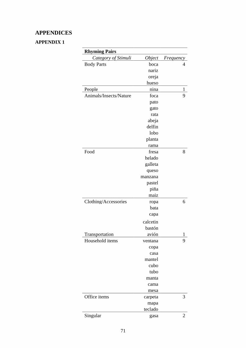
71
APPENDICES
APPENDIX 1
Rhyming Pairs
Category of Stimuli Object Frequency
Body Parts boca 4
nariz
oreja hueso
People nina 1
Animals/Insects/Nature foca 9
pato
gato
rata
abeja
delfin
lobo
planta
rama Food fresa 8
helado
galleta
queso
manzana
pastel
pina maiz
Clothing/Accessories ropa 6
bata
capa
calcetin
bastón Transportation avión 1
Household items ventana 9
copa
casa
mantel
cubo
tubo
manta
cama mesa
Office items carpeta 3
mapa teclado
Singular gasa 2

72
globo
Non-Rhyming Pairs
Category of Stimuli Object Frequency
Body Part pierna 3
esqueleto
mano People - 0
Animals/Insects/Nature pollo 7
cocodrilo
elefante
cerdo
vaca
perro rosa
Food cafe 6
vino
guisante
calabaza
lechuga tomate
Clothing/Accesories zapato 8
suéter
vaqueros
gorra
mochila
falda
chaqueta gorra
Transportation coche 1
Household Items reloj 7
bombilla
botella
cuna
vaso
clave
tenedor Office Items goma 4
lapiz
enchufe regla
Singular dado 3
cuchara
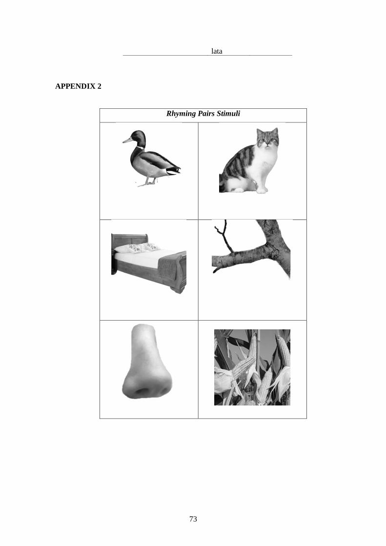
73
APPENDIX 2
Rhyming Pairs Stimuli
lata

74

75

76

77
Non-Rhyming Pairs

78

79

80

81
APPENDIX 3
Rhyming Pairs
Pair Name Stimulus 1 Frequency 1 Stimulus 2 Frequency 2
carpets- galleta carpeta 27 galleta 47
libreta 14
cuaderno 4
archivador 1
clasificador 1
ventana-manzana ventana 46 manzana 47
contras 1
foca-boca foca 46 boca 41
leopardo
marino 1 labios 6
manta-planta manta 38 planta 38
toalla 7 maceta 7
tela 2 sábana 1
palmera 1
casa-gasa casa 43 gasa 23
casita 3 venda 6
maqueta 1 toalla 5
manta 3
alfombra 2

82
plástico de
burbujas 2
rollo 2
paño de cocina 1
moqueta 1
lana 1
celofán 1
capa-mapa capa 46 mapa 46
mapa 1 cartografia 1
bata-rata bata 34 rata 38
albornoz 12 ratón 8
batin 1 rima 1
ropa-copa ropa 37 copa 47
vestidos 3
armario 2
ropero 1
vestidor 1
perchero 1
camisetas 1
rack 1
oreja-abeja oreja 47 abeja 46
avispa 1
niña-piña niña 45 piña 47
cara 2
fresa-mesa fresa 46 mesa 47
fresas 1
cubo-tubo cubo 45 tubo 45
cubeta 2 cono 1
cilindro 1
queso-hueso queso 47 hueso 46
fémur 1
cama-rama cama 47 rama 47
helado-teclado helado 42 teclado 47
cucurucho 5
pato-gato pato 47 gato 47
naríz-maíz naríz 47 maíz 46
mazorcas 1
globo-lobo globo 47 lobo 45
perro 2
mantel-pastel mantel 47 pastel 37
tarta 10
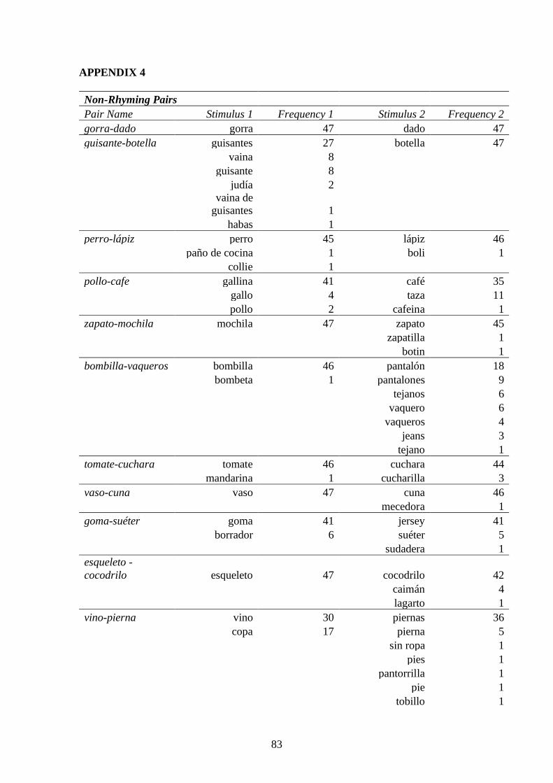
83
APPENDIX 4
Non-Rhyming Pairs
Pair Name Stimulus 1 Frequency 1 Stimulus 2 Frequency 2
gorra-dado gorra 47 dado 47
guisante-botella guisantes 27 botella 47
vaina 8
guisante 8
judía 2
vaina de
guisantes 1
habas 1
perro-lápiz perro 45 lápiz 46
paño de cocina 1 boli 1
collie 1
pollo-cafe gallina 41 café 35
gallo 4 taza 11
pollo 2 cafeina 1
zapato-mochila mochila 47 zapato 45
zapatilla 1
botin 1
bombilla-vaqueros bombilla 46 pantalón 18
bombeta 1 pantalones 9
tejanos 6
vaquero 6
vaqueros 4
jeans 3
tejano 1
tomate-cuchara tomate 46 cuchara 44
mandarina 1 cucharilla 3
vaso-cuna vaso 47 cuna 46
mecedora 1
goma-suéter goma 41 jersey 41
borrador 6 suéter 5
sudadera 1
esqueleto -
cocodrilo esqueleto 47 cocodrilo 42
caimán 4
lagarto 1
vino-pierna vino 30 piernas 36
copa 17 pierna 5
sin ropa 1
pies 1
pantorrilla 1
pie 1
tobillo 1
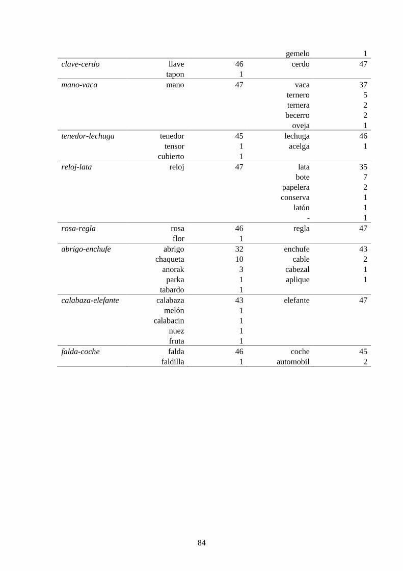
84
gemelo 1
clave-cerdo llave 46 cerdo 47
tapon 1
mano-vaca mano 47 vaca 37
ternero 5
ternera 2
becerro 2
oveja 1
tenedor-lechuga tenedor 45 lechuga 46
tensor 1 acelga 1
cubierto 1
reloj-lata reloj 47 lata 35
bote 7
papelera 2
conserva 1
latón 1
- 1
rosa-regla rosa 46 regla 47
flor 1
abrigo-enchufe abrigo 32 enchufe 43
chaqueta 10 cable 2
anorak 3 cabezal 1
parka 1 aplique 1
tabardo 1
calabaza-elefante calabaza 43 elefante 47
melón 1
calabacin 1
nuez 1
fruta 1
falda-coche falda 46 coche 45
faldilla 1 automobil 2
Copyright © 2022 FDOKUMEN