A CTP-Dependent Archaeal Riboflavin Kinase Forms a Bridge in the Evolution of Cradle-Loop Barrels
14
Structure Article A CTP-Dependent Archaeal Riboflavin Kinase Forms a Bridge in the Evolution of Cradle-Loop Barrels Moritz Ammelburg, 1,4 Marcus D. Hartmann, 1,4 Sergej Djuranovic, 1,5 Vikram Alva, 1 Kristin K. Koretke, 2 Jo ¨ rg Martin, 1 Guido Sauer, 3 Vincent Truffault, 1 Kornelius Zeth, 1 Andrei N. Lupas, 1, * and Murray Coles 1, * 1 Department of Protein Evolution, Max-Planck-Institute for Developmental Biology, 72076 Tu ¨ bingen, Germany 2 Computational Chemistry Group, GlaxoSmithKline, Collegeville, PA 19426, USA 3 Department of Biochemistry, Max-Planck-Institute for Developmental Biology, 72076 Tu ¨ bingen, Germany 4 These authors contributed equally to this work. 5 Present address: Howard Hughes Medical Institute, Department of Molecular Biology and Genetics, Johns Hopkins University School of Medicine, Baltimore, MD 21205, USA. *Correspondence: [email protected] (M.C.), [email protected] (A.N.L.) DOI 10.1016/j.str.2007.09.027 SUMMARY Proteins of the cradle-loop barrel metafold are formed by duplication of a conserved bab- element, suggesting a common evolutionary origin from an ancestral group of nucleic acid- binding proteins. The basal fold within this metafold, the RIFT barrel, is also found in a wide range of enzymes, whose homologous relationship with the nucleic acid-binding group is unclear. We have characterized a protein family that is intermediate in sequence and structure between the basal group of cradle- loop barrels and one family of RIFT-barrel enzymes, the riboflavin kinases. We report the structure, substrate-binding mode, and cata- lytic activity for one of these proteins, Methano- caldococcus jannaschii Mj0056, which is an archaeal riboflavin kinase. Mj0056 is unusual in utilizing CTP rather than ATP as the donor nucleotide, and sequence conservation in the relevant residues suggests that this is a general feature of archaeal riboflavin kinases. INTRODUCTION Riboflavin kinases (RFKs) from bacteria and eukaryotes catalyze the phosphorylation of riboflavin to form flavin mononucleotide (FMN). Systematically, they are classed as ATP:riboflavin 5 0 -phosphotransferases (EC 2.7.1.26). All examples known to date are closely related in se- quence, and the available structures of the enzyme (Homo sapiens, 1NB0, 1Q9S [Karthikeyan et al., 2003a, 2003b]; Schizosaccharomyces pombe, 1NO8,[Bauer et al., 2003]; and Thermotoga maritima, 1MRZ [Wang et al., 2003]) show a RIFT barrel fold, belonging to the cra- dle-loop barrel metafold of small b-barrels (Coles et al., 2006). In the course of a study into the evolution of this metafold, we identified a family of proteins exemplified by Methanocaldococcus jannaschii Mj0056, whose se- quence and gene environment suggested a RIFT barrel with a role in riboflavin biosynthesis. Here, we characterize Mj0056 as an archaeal riboflavin kinase, structurally simi- lar but topologically distinct from bacterial and eukaryotic examples. Surprisingly, Mj0056 utilizes CTP rather than ATP as the donor nucleotide, and therefore represents a rare CTP-dependent kinase. The cradle-loop barrel metafold comprises three dis- tinct topologies, all with (pseudo-) 2-fold symmetry: the double psi, the swapped hairpin, and the RIFT barrel (Coles et al., 1999, 2005, 2006). We have shown that a basal group of proteins spanning these three folds re- semble each other at a level indicative of homology and have proposed an evolutionary scenario (Figure 1) in which an ancestral homodimeric RIFT barrel gave rise to swapped hairpin barrels by strand invasion and to dou- ble-psi barrels by fusion and strand swapping (Coles et al., 2006). This scenario is underpinned by the hypoth- esis that folded proteins evolved from an ancestral pool of peptides, which had themselves evolved as cofactors of RNA-based catalysis and replication (the ‘‘RNA world’’) (Lupas et al., 2001; So ¨ ding and Lupas, 2003). In this case, the ancestral peptide consisted of a bab element that encloses an orthogonal turn with a conspicuous Gly-Asp motif (the GD box). Basal cradle-loop barrels retain the ability to bind nucleic acids, although in the case of the double-psi barrels found at the N terminus of AAA proteins, this activity is vestigial and superseded by poly- peptide binding. We proposed that the RIFT barrel is the ancestral form of cradle-loop barrels because of its simple topology and widespread occurrence in ancient proteins, such as Ef-Tu and related translation factors, ribosomal protein L3, the N-domain of the F 1 ATPase, and enzymes involved in riboflavin synthesis, including riboflavin kinases (Coles et al., 2006). This proposal remained inconclusive, due to the lack of evidence for the homologous origin of these proteins from the basal RIFT barrel we characterized, Structure 15, 1577–1590, December 2007 ª2007 Elsevier Ltd All rights reserved 1577
-
Upload
independent -
Category
Documents
-
view
0 -
download
0
Transcript of A CTP-Dependent Archaeal Riboflavin Kinase Forms a Bridge in the Evolution of Cradle-Loop Barrels

Structure
Article
A CTP-Dependent Archaeal RiboflavinKinase Forms a Bridge in theEvolution of Cradle-Loop BarrelsMoritz Ammelburg,1,4 Marcus D. Hartmann,1,4 Sergej Djuranovic,1,5 Vikram Alva,1 Kristin K. Koretke,2
Jorg Martin,1 Guido Sauer,3 Vincent Truffault,1 Kornelius Zeth,1 Andrei N. Lupas,1,* and Murray Coles1,*1Department of Protein Evolution, Max-Planck-Institute for Developmental Biology, 72076 Tubingen, Germany2Computational Chemistry Group, GlaxoSmithKline, Collegeville, PA 19426, USA3Department of Biochemistry, Max-Planck-Institute for Developmental Biology, 72076 Tubingen, Germany4These authors contributed equally to this work.5Present address: Howard Hughes Medical Institute, Department of Molecular Biology and Genetics, Johns Hopkins University
School of Medicine, Baltimore, MD 21205, USA.*Correspondence: [email protected] (M.C.), [email protected] (A.N.L.)
DOI 10.1016/j.str.2007.09.027
SUMMARY
Proteins of the cradle-loop barrel metafold areformed by duplication of a conserved bab-element, suggesting a common evolutionaryorigin from an ancestral group of nucleic acid-binding proteins. The basal fold within thismetafold, the RIFT barrel, is also found ina wide range of enzymes, whose homologousrelationship with the nucleic acid-binding groupis unclear. We have characterized a proteinfamily that is intermediate in sequence andstructure between the basal group of cradle-loop barrels and one family of RIFT-barrelenzymes, the riboflavin kinases. We report thestructure, substrate-binding mode, and cata-lytic activity for one of these proteins, Methano-caldococcus jannaschii Mj0056, which is anarchaeal riboflavin kinase. Mj0056 is unusualin utilizing CTP rather than ATP as the donornucleotide, and sequence conservation in therelevant residues suggests that this is a generalfeature of archaeal riboflavin kinases.
INTRODUCTION
Riboflavin kinases (RFKs) from bacteria and eukaryotes
catalyze the phosphorylation of riboflavin to form flavin
mononucleotide (FMN). Systematically, they are classed
as ATP:riboflavin 50-phosphotransferases (EC 2.7.1.26).
All examples known to date are closely related in se-
quence, and the available structures of the enzyme
(Homo sapiens, 1NB0, 1Q9S [Karthikeyan et al., 2003a,
2003b]; Schizosaccharomyces pombe, 1NO8, [Bauer
et al., 2003]; and Thermotoga maritima, 1MRZ [Wang
et al., 2003]) show a RIFT barrel fold, belonging to the cra-
dle-loop barrel metafold of small b-barrels (Coles et al.,
2006). In the course of a study into the evolution of this
Structure 15, 1577–159
metafold, we identified a family of proteins exemplified
by Methanocaldococcus jannaschii Mj0056, whose se-
quence and gene environment suggested a RIFT barrel
with a role in riboflavin biosynthesis. Here, we characterize
Mj0056 as an archaeal riboflavin kinase, structurally simi-
lar but topologically distinct from bacterial and eukaryotic
examples. Surprisingly, Mj0056 utilizes CTP rather than
ATP as the donor nucleotide, and therefore represents
a rare CTP-dependent kinase.
The cradle-loop barrel metafold comprises three dis-
tinct topologies, all with (pseudo-) 2-fold symmetry: the
double psi, the swapped hairpin, and the RIFT barrel
(Coles et al., 1999, 2005, 2006). We have shown that
a basal group of proteins spanning these three folds re-
semble each other at a level indicative of homology and
have proposed an evolutionary scenario (Figure 1) in
which an ancestral homodimeric RIFT barrel gave rise to
swapped hairpin barrels by strand invasion and to dou-
ble-psi barrels by fusion and strand swapping (Coles
et al., 2006). This scenario is underpinned by the hypoth-
esis that folded proteins evolved from an ancestral pool
of peptides, which had themselves evolved as cofactors
of RNA-based catalysis and replication (the ‘‘RNA world’’)
(Lupas et al., 2001; Soding and Lupas, 2003). In this case,
the ancestral peptide consisted of a bab element that
encloses an orthogonal turn with a conspicuous Gly-Asp
motif (the GD box). Basal cradle-loop barrels retain the
ability to bind nucleic acids, although in the case of the
double-psi barrels found at the N terminus of AAA
proteins, this activity is vestigial and superseded by poly-
peptide binding.
We proposed that the RIFT barrel is the ancestral form
of cradle-loop barrels because of its simple topology
and widespread occurrence in ancient proteins, such as
Ef-Tu and related translation factors, ribosomal protein
L3, the N-domain of the F1 ATPase, and enzymes involved
in riboflavin synthesis, including riboflavin kinases (Coles
et al., 2006). This proposal remained inconclusive, due
to the lack of evidence for the homologous origin of these
proteins from the basal RIFT barrel we characterized,
0, December 2007 ª2007 Elsevier Ltd All rights reserved 1577

Structure
Structure of the Archaeal Riboflavin Kinase Mj0056
Figure 1. A Scenario for Cradle-Loop Barrel EvolutionExamples are shown for the three folds for which homologous relationships have been established. For dimeric examples, the monomers are distin-
guished by light and dark colors. The invading strands of the swapped-hairpin barrel are shown in blue, while the swapped strands of the double-psi
barrel are in red. The example proteins are: VatN-N (double psi), PhS018 (RIFT), MraZ (monomeric swapped hairpin), and AbrB-N (dimeric swapped
hairpin). The structure of the presumed dimeric RIFT barrel ancestor is a homology model of Orf5 from the pME2200 plasmid of Methanothermobacter
thermautotrophicus (MTpME2200 Orf5) based on PhS018. For details of this scenario see (Coles et al., 2006).
PhS018. Here, we show that Mj0056 has sequence prop-
erties intermediate between basal cradle-loop barrels and
ATP-dependent riboflavin kinases. We propose that it
represents an evolutionary bridge between the two groups
of proteins.
RESULTS
BioinformaticsWe first noticed the sequence similarity between Mj0056
and cradle-loop barrels in our initial bioinformatic charac-
terization of these proteins (Coles et al., 1999) by using
a sequence search tool based on reciprocal PSI-Blast
searches (SENSER) (Koretke et al., 2002). At the time,
we considered Mj0056 to be an archaeal transcription
factor, due to its sequence similarity to the N-terminal
DNA-binding domains of AbrB-like transcription factors
and to the fact that most of its close homologs (but not
Mj0056 itself) carry a winged-helix HTH DNA-binding
domain at their N terminus. Indeed, in the COG database,
Mj0056 homologs are annotated as COG1339—tran-
scriptional regulator of a riboflavin/FAD biosynthetic
1578 Structure 15, 1577–1590, December 2007 ª2007 Elsevier
operon. However, subsequent experiments designed to
show an affinity of Mj0056 for DNA, including the se-
quence from the upstream region of its own gene, failed
completely.
Revisiting this analysis more recently with a new search
tool based on HMM-HMM comparisons (HHsenser)
(Soding et al., 2006), we found that the top matches for
Mj0056 not only included the expected basal cradle-
loop barrels, but also two riboflavin kinases (Table 1), pro-
viding a new functional hypothesis. We therefore decided
to analyze the sequence relationships between these
proteins with a clustering procedure based on the Fruch-
terman-Reingold algorithm (CLANS) (Frickey and Lupas,
2004). In the cluster map, Mj0056 appeared at an interme-
diate position between AbrB-like transcription factors and
riboflavin kinases (Figure 2). Although closer to the AbrB
N-domain (AbrB-N), a multiple alignment showed that
Mj0056 shared with riboflavin kinases three regions with
residues important for the catalytic activity of riboflavin
kinase (Karthikeyan et al., 2003a), distributed over its
entire length (Figure 3). In contrast, the similarity with
AbrB-N and other basal cradle-loop proteins only covered
Ltd All rights reserved
Structure
Structure of the Archaeal Riboflavin Kinase Mj0056
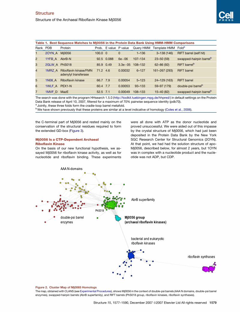
Table 1. Best Sequence Matches to Mj0056 in the Protein Data Bank Using HMM-HMM Comparisons
Rank PDB Protein Prob. E value P value Query HMM Template HMM Folda
1 2OYN_A Mj0056 100.0 0 0 1–136 3–138 (146) RIFT barrel (self hit)
2 1YFB_A AbrB-N 92.5 0.088 6e�06 107–134 23–50 (59) swapped-hairpin barrelb
3 2GLW_A PhS018 85.9 0.49 3.3e�05 108–132 62–86 (92) RIFT barrelb
4 1MRZ_A Riboflavin kinase/FMNadenylyl transferase
71.2 4.6 0.00032 6–127 161–267 (293) RIFT barrel
5 1N08_A Riboflavin kinase 66.7 7.9 0.00054 5–123 24–129 (163) RIFT barrel
6 1WLF_A PEX1-N 60.4 7.7 0.00053 93–133 59–97 (179) double-psi barrelb
7 1MVF_D MazE 52.5 7.1 0.00049 108–133 15–40 (82) swapped-hairpin barrelb
The search was done with the program HHsearch 1.5.0 (http://toolkit.tuebingen.mpg.de/hhpred/) in default settings on the Protein
Data Bank release of April 10, 2007, filtered for a maximum of 70% pairwise sequence identity (pdb70).a Jointly, these three folds form the cradle-loop barrel metafold.b We have shown previously that these proteins are similar at a level indicative of homology (Coles et al., 2006).
the C-terminal part of Mj0056 and rested mainly on the
conservation of the structural residues required to form
the extended GD-box (Figure 3).
Mj0056 Is a CTP-Dependent ArchaealRiboflavin KinaseOn the basis of our new functional hypothesis, we as-
sayed Mj0056 for riboflavin kinase activity, as well as for
nucleotide and riboflavin binding. These experiments
Structure 15, 1577–1590
were all done with ATP as the donor nucleotide and
proved unsuccessful. We were aided out of this impasse
by the crystal structure of Mj0056, which had just been
deposited in the Protein Data Bank by the New York
SGC Research Center for Structural Genomics (2OYN).
At that point, we had had the solution structure of apo-
Mj0056, described below, for almost 2 years, but 1OYN
was in complex with a nucleotide product and the nucle-
otide was not ADP, but CDP.
Figure 2. Cluster Map of Mj0065 Homologs
The map, obtained with CLANS (see Experimental Procedures), shows Mj0056 in the context of double-psi barrels (AAA N domains, double-psi barrel
enzymes), swapped-hairpin barrels (AbrB superfamily), and RIFT barrels (PhS018 group, riboflavin kinases, riboflavin synthases).
, December 2007 ª2007 Elsevier Ltd All rights reserved 1579

Structure
Structure of the Archaeal Riboflavin Kinase Mj0056
Figure 3. Multiple Alignment of Archaeal Riboflavin Kinases and Their Homologs
The N- and C-terminal halves of the cradle-loop barrel fold are shown in the upper and lower panel, respectively. The alignment shows a phylogenet-
ically representative selection of archaeal riboflavin kinases (RFKs), as well as the three bacterial and eukaryotic RFKs of known structure; sequence
motifs shared by sequences of all three kingdoms are colored magenta. Many archaeal RFKs contain an N-terminal winged helix-turn-helix
DNA-binding domain, indicated by the abbreviation: wHTH. Thermotoga RFK contains an N-terminal FMN adenylyltransferase domain, denoted
by FMNat, and both eukaryotic and bacterial RFKs contain a C-terminal extension, which is structurally equivalent to the two insert regions of archaeal
RFKs. The C-terminal half of archaeal RFKs is most similar in sequence to a group of cradle-loop barrels that we have described previously (Coles
et al., 2006); these are shown above the C-terminal half, and shared sequence motifs are colored red. In all sequences, residues buried in the core of
the structures, as judged by their relative solvent accessibility computed by the SARIG server (http://www.weizmann.ac.il/SARIG/), are colored blue.
The secondary structure is shown above the sequences (H, helix; S, strand; b, beta bulge; 3, 310 helix).
We therefore reanalyzed the ability of Mj0056 to con-
vert riboflavin to FMN at various temperatures by mass
spectrometry, this time with a range of donor nucleotides
(see Experimental Procedures). In electospray ionization
mass spectrometry, riboflavin (MW = 376 Da) exhibits
a strong response in positive ion mode at mass/charge
ratios of 377, 399, and 775 (Figure 4A), whereas FMN
(MW = 456 Da) shows an intense signal at 455 in negative
ion mode and characteristic signals at 911 and 933
(Figure 4B) (Susin et al., 1993). Under the chosen assay
conditions, we obtained riboflavin kinase activity with
both CTP and UTP as phosphate donors, with UTP being
at least one order of magnitude less efficient. At reaction
temperatures of up to 85�C—the temperature of the nat-
ural habitat of M. jannaschii—riboflavin was completely
converted to FMN (Figures 4C and 4D and see the
Supplemental Data available with this article online).
ATP and GTP did not support the production of FMN at
any reaction temperature (Figures 4E and 4F and Supple-
mental Data).
1580 Structure 15, 1577–1590, December 2007 ª2007 Elsevie
Solution Structure of Apo-Mj0056We determined the solution structure of apo-Mj0056 (136
residues, 15.7 kDa) in the earliest stages of this project.
High quality spectra led to largely complete resonance
assignments by using standard methods (see Experimen-
tal Procedures), with the notable exception of two larger
segments, G14-S23 and T99-S102, where the backbone
amide signals were not observed. Subsequent secondary
structure analysis showed that these regions correspond
to the two cradle loops expected in the RIFT barrel fold.
We obtained a detailed structure with a combination of
distance restraints derived from several 2D- and 3D-
NOESY spectra, chemical-shift-derived backbone torsion
angle restraints, and 3JHNHa and 3JNHai-1 coupling con-
stants (see Experimental Procedures). The final set of
experimental restraints is described in Table 2, and the en-
semble of 19 structures is shown in Figure 5. The ensem-
ble is well defined; the rmsd for superimposition over
structured residues is 0.25 A for backbone atoms and
0.61 A for all heavy atoms (Table 2). The restraint violations
r Ltd All rights reserved

Structure
Structure of the Archaeal Riboflavin Kinase Mj0056
Figure 4. MJ0056 Is a CTP-Specific Riboflavin Kinase
(A and B) MS spectra of the riboflavin and FMN controls measured in positive and negative ion mode, respectively. Adducts of riboflavin and FMN are
annotated in angular brackets next to the corresponding peaks.
(C and D) The positive and negative ion mode spectra of samples containing enzyme, riboflavin, and CTP. Complete turnover of riboflavin to FMN is
observed (60 min, 50�C).
(E and F) The positive and negative ion mode MS spectra of samples containing enzyme, riboflavin, and ATP.
are also very low, with no persistent distance restraint
violations over 0.1 A (4.0 violations > 0.05 A per structure)
and no dihedral restraint violation greater than 0.6�.
Mj0056 shows a six-stranded b-barrel fold (Figure 5)
with the expected RIFT barrel topology (for an explanation
of secondary structure notation, see the caption to Fig-
ure 5). In keeping with its sequence properties, the struc-
ture can be divided into a degenerate N-terminal and a
canonical C-terminal half. The latter is structurally similar
Structure 15, 1577–159
to the basal cradle-loop barrels. This similarity is centered
on a homologous bab element, the hallmark of which is the
GD box sequence motif (118-FnLkdGDvI-126 in Mj0056)
(Figure 3). This motif forms a very similar structure in all
examples known to date, as we have previously described
in detail (Coles et al., 2006). The b strands are orthogonal
and linked by the helix and GD-box, which cross over an
open end of the barrel. The GD box itself contains a type
II b turn positioned within the barrel architecture by
0, December 2007 ª2007 Elsevier Ltd All rights reserved 1581

Structure
Structure of the Archaeal Riboflavin Kinase Mj0056
Table 2. Apo-Mj0056 Solution Structure Statistics and Atomic Rmsds
Structural Statistics
Rmsd from distance restraints (A)a SA <SA>r
All (951) 0.012 ± 0.001 0.011
Intraresidue (145) 0.000 ± 0.001 0.000
Interresidue sequential (299) 0.009 ± 0.001 0.008
Medium range (80) 0.020 ± 0.003 0.017
Long range (315) 0.016 ± 0.001 0.015
H bond (112) 0.000 ± 0.000 0.000
Rmsd from dihedral restraints (399) 0.094 ± 0.002 0.092
Rmsd from J-coupling restraints (Hz) (180) 0.387 ± 0.004 0.386
H bond restraints; averages (A/deg.)b (58) 2.18 ± 0.12/11.9 ± 5.8 2.13 ± 0.13/11.9 ± 6.2
H bond restraints; min-max (A/deg.) 1.93–2.45/1.3–32.3 1.88–2.43/0.72–33.6
Deviations from Ideal Covalent Geometry
Bonds (A 3 10�3) 7.17 ± 0.01 7.14
Angles (deg.) 0.78 ± 0.01 0.77
Impropers (deg.) 1.46 ± 0.04 1.51
Structure Quality Indicatorsc
Ramachandran map regions (%) 98.0/100.0/0.0 98.5/100.0/0.0
Steric clashes >0.4 A per 1,000 atoms 0.0 0.0
Atomic Rmsd (A)d
SA versus <SA> SA versus <SA>r
Backbone All Backbone All
All residues 0.99 ± 0.32 1.44 ± 0.323 1.28 ± 0.32 1.80 ± 0.32
Secondary structuree 0.25 ± 0.10 0.61 ± 0.08 0.39 ± 0.08 0.73 ± 0.09
<SA> versus <SA>rf 0.30 0.48
Structures are labeled as follows: SA, the set of 19 final simulated annealing structures; <SA>, the mean structure calculated by
averaging the coordinates of SA structures after fitting over secondary structure elements; <SA>r, the structure obtained by reg-
ularizing the mean structure under experimental restraints.a Numbers in brackets indicate the number of restraints of each type.b Hydrogen bonds were restrained by treating them as pseudocovalent bonds (see Experimental Procedures section). Deviations
are expressed as the average distance/average deviation from linearity for restrained hydrogen bonds.c Determined with the program MolProbity (Lovell et al., 2003). Percentages are for residues in favored (98%), allowed (99.8%), anddisallowed regions of the Ramachandran map.d Based on heavy atoms superimpositions.e Defined as residues L4-S13, L24-G96, and E104-G132.f Rms difference for superimposition over ordered residues.
conserved hydrophobic and hydrogen bonding interac-
tions. One significant difference to the basal bab element
is in the entrance to the a helix, despite the conservation of
the PxxxR sequence motif (P111-R115) observed in
a wide range of cradle-loop barrels. In contrast to most
other examples, two residues in extended conformation
separate the proline from the helix, with the result that
the b strand is shifted outward. This conformation is stabi-
lized by a noncanonical hydrogen bond to Y40 on the
N-terminal half of the protein and creates a pocket behind
b20, later identified as the cytosine binding site.
In contrast to the C-terminal half, the degenerate
N-terminal half of Mj0056 deviates significantly from the
1582 Structure 15, 1577–1590, December 2007 ª2007 Elsevie
basal RIFT-barrel fold and lacks both helix a1 and the
GD box. The main differences are provided by two large
insertions (Figure 3). One insertion elongates the first
cradle loop (S13-T43), forming an a-helix (aI; P25-L35)
and contributing to a structured connector (G36-T43),
which leads into b2. The second insertion consists of
a short 310 helix and a b-hairpin (bI1-bI2), which continues
unbroken into strand b3.
Crystal Structures of Mj0056The first crystals we obtained for Mj0056 were cocrystals
with inorganic phosphate (Mj0056-PO4), and we solved
this structure by molecular replacement with a preliminary
r Ltd All rights reserved

Structure
Structure of the Archaeal Riboflavin Kinase Mj0056
Figure 5. The Solution Structure of Apo-Mj0056
(A) A secondary structure cartoon; b strands are in green, helices are yellow, and the two cradle-loops are blue. Secondary structure elements
corresponding to the basal RIFT barrel fold are given conventional notation, while inserted elements are denoted with I and shown in light colors.
The right view represents the left view rotated by 90� around the horizontal axis.
(B) A stereoview of the final set of 19 structures superimposed over ordered residues (defined in Table 2). Coloring is as in (A).
solution structure. After it became clear that Mj0056 is a
CTP-dependent riboflavin kinase, we focused on obtaining
cocrystals with substrates and products (Table 3). We
solved two structures, Mj0056-MgCDP and Mj0056-
MgCDP-FMN, in complex with natural reaction products
and a third, Mj0056-NaCDP-PO4, with inorganic phosphate
bound in a similar position as the FMN phosphate (Figure6).
Mj0056-MgCDP (Figure 7A) shows a nucleotide binding
site centered on a conserved motif at the N terminus of b2
(40-YegTLN-45). The cytosine ring packs between Y40
and L44, with further contacts to the pocket at the junction
Structure 15, 1577–1590
of b20 and a10, while the ribose interacts primarily with
R115 on a10. The a- and b-phosphates of CDP are coordi-
nated by T43 and N45 via the intermediate Mg2+ and inter-
act with a glycine-rich sequence motif (G14-G18) in the
first cradle loop. The induction of a transient helix begin-
ning at the last residue of this glycine-rich motif (a0, G18-
S23) represents the major difference between the nucleo-
tide-bound and apo forms of the protein (Figure 6). In the
solution structure, only a weak helical tendency or nascent
helix was detected for these residues. Our structure for
Mj0056-MgCDP resembles closely the Mj0056-NaCDP
, December 2007 ª2007 Elsevier Ltd All rights reserved 1583

Structure
Structure of the Archaeal Riboflavin Kinase Mj0056
Table 3. Summary of Mj0056 Structures
Name First Cradle Loopa Second Cradle Loop Oligomerb Space Group Resn. (A) PDBc
Apo-Mj0056 flexible flexible monomer solution - 2P3M
Mj0056-PO4 unstructured closed dimer P43212 3.0 2VBS
Mj0056-MgCDPd a0 helix open monomer I41 1.7 2VBU
‘‘ ‘‘ ‘‘ dimer P41212 2.6 -
Mj0056-NaCDP-PO4 a0 helix closed dimer P43212 2.7 2VBT
‘‘ ‘‘ ‘‘ ‘‘ P3221 3.3 -
Mj0056-MgCDP-FMN a0 helix closed dimer P212121 2.4 2VBV
Full structural statistics for solution and crystal structures are shown in Tables 2 and 4, respectively.a Indicates the state of first cradle loop in the region of the transient a0 helix.b All dimeric forms show a similar dimerization via pairing of the b1 strands.c Where structures were solved in two space groups, the higher resolution structure was chosen for deposition.d Similar to the New York SGC Research Center for Structural Genomics structure for Mj0056-NaCDP (2OYN).
structure of the New York SGC Research Center for Struc-
tural Genomics (2OYN).
The ternary complex, Mj0056-MgCDP-FMN, contains
the CDP in the same location as Mj0056-MgCDP. The
FMN is enclosed on three sides by the b-barrel, the tran-
sient helix a0, and the second cradle loop (K98-S103)
(Figure 7B). The latter is in a closed conformation, making
contacts to all moieties of FMN. This is in contrast to the
solution structure, where it is flexible, and to the Mj0056-
MgCDP structure, where it is in an open conformation
(Figure 6). The isoalloxazine ring forms p-stacking inter-
actions with the side chain of a conserved aromatic res-
1584 Structure 15, 1577–1590, December 2007 ª2007 Elsevie
idue (F21) on a0, and hydrogen bonds to the backbone
of F73 on bI2 and to the side chain of Y27 on aI via a
bridging water molecule. The 40 and 50 oxygens of FMN
contact the two carboxyl oxygens of the invariant gluta-
mate on b20 (E107) (Figure 3), suggesting that this residue
acts as a base in activating the 50 hydroxyl of riboflavin
for nucleophilic attack.
Two structures, Mj0056-PO4 and Mj0056-NaCDP-PO4,
contain a PO4 ion bound in the place of the FMN phos-
phate, albeit shifted away from the active site by approx-
imately 2 A. In both these structures, the second cradle
loop is in the closed conformation, while helix a0 is formed
Figure 6. The Proposed Sequential
Substrate Binding Cycle of Mj0056 and
Concerted Conformational Changes
The binding of the nucleotide to the apo struc-
ture (A and B) leads to the formation of the tran-
sient helix a0 in the first cradle loop; with the
subsequent binding of riboflavin/FMN (B and
C), the second cradle loop adopts a closed
conformation. As depicted in (D), this closure
can also be induced by binding of inorganic
phosphate, which is however not sufficient for
the formation of a0. In the structure Mj0056-
NaCDP-PO4 (not shown), the cradle loop con-
formations are similar to those shown in (C).
r Ltd All rights reserved

Structure
Structure of the Archaeal Riboflavin Kinase Mj0056
Figure 7. Ligand Binding in Mj0056
(A) The binding mode of MgCDP in the Mj0056-MgCDP structure (1.7 A). Three hydrogen bonds from the cytosine moiety to the backbone illustrate
the high specificity for this nucleotide. Superposed in green is the phosphate group of FMN from Mj0056-MgCDP-FMN, which has one of its oxygens
in the position of a water molecule in the first magnesium coordination sphere. The Fo-Fc omit map for CDP is contoured at 5s.
(B) Binding details of FMN in the Mj0056-MgCDP-FMN structure (2.4 A). The Fo-Fc omit map for FMN is contoured at 2.5s. Selected hydrogen bonds
are shown as thin black lines, water molecules as red spheres, Mg2+ is gray.
only in Mj0056-NaCDP-PO4. Thus, we observe two major
conformational changes upon ligand binding, both affect-
ing the FMN binding site: formation of helix a0 in the first
cradle loop, induced by CDP binding, and the closure of
the second cradle loop, induced by phosphate binding
(Figure 6 and Table 3).
With the exception of the Mj0056-MgCDP, which is
monomeric in one crystal form, all crystal structures pre-
sented here, plus Mj0056-NaCDP (2OYN), form dimers
via antiparallel pairing of their b1 strands. This dimerization
is independent of substrate binding (Table 3) and does not
affect functional regions of the protein. Analytical gel sizing
showed the solutions used for both crystallization and
enzyme assays to have some small dimeric component
(<5%). Thus, Mj0056 appears to have a weak tendency
to dimerize that is accentuated in crystallization but is irrel-
evant to primary riboflavin kinase activity. This may not be
the case for many of its close homologs that carry a winged
helix HTH DNA-binding domain at their N terminus and are
therefore likely to be active in a dimeric form.
Structure 15, 1577–1590
Structure-based searches on Mj0056-MgCDP-FMN us-
ing DALI (Holm and Sander, 1993) show the expected
similarity to bacterial and eukaryotic RFKs, with the en-
zyme from T. maritima being among the top three matches
(Z score/rmsd = 5.1/ 2.7 A). Also among the best matches
are other RIFT-barrel enzymes, such as siderophore-
interacting protein (2GPJ, 5.3/3.4 A), flavodoxin reductase
(1FDR, 4.6/2.7 A), and yeast riboflavin synthase (1KZL,
4.6/3.4 A). However, the next group of matches are dou-
ble-psi b barrels from AAA ATPase N domains, e.g., VAT
(1CZ4, 3.9/2.8 A) and NSF (1QCS, 3.8/3.4 A), underlining
the structural similarity of members of the cradle-loop
barrel metafold.
DISCUSSION
We have identified a family of proteins with properties
intermediate between basal cradle-loop barrels and ribo-
flavin kinases. Detailed structural and biochemical analy-
sis of one of these proteins, Mj0056, showed it to be an
, December 2007 ª2007 Elsevier Ltd All rights reserved 1585

Structure
Structure of the Archaeal Riboflavin Kinase Mj0056
archaeal riboflavin kinase, with a specificity for CTP as the
phosphate donor. This specificity is highly unusual; of the
25 families in the kinase classification of Grishin and
coworkers (Cheek et al., 2005), only one—dolichol ki-
nase—is CTP specific. As an all-helical integral membrane
protein, dolichol kinase clearly represents an analogous
development to Mj0056.
Mechanistic ImplicationsDespite its different nucleotide specificity, Mj0056 clearly
resembles bacterial and eukaryotic RFKs at several levels.
Both share the RIFT barrel fold and similar overall struc-
tures (Figure 8A). In the active site, both have the glycine
rich loop and the TxN motif, which coordinate the phos-
phates of the donor nucleotide, and the glutamate
residue, which is thought to activate the 50 hydroxyl of
riboflavin, initiating the phosphate transfer (Bauer et al.,
2003; Karthikeyan et al., 2003a).
Outside the phosphate transfer site, there are consider-
able differences in the nucleotide binding mode. For the
donor nucleotide, the two large hydrophobic residues
that sandwich the cytosine ring in Mj0056 are absent
from bacterial and eukaryotic RFKs, as is helix a10 and
the arginine coordinating the ribose hydroxyl groups. In-
stead, ATP-dependent RFKs use small side chains and
a wider loop in place of a10 to accommodate the larger ad-
enine moiety (Figure 8B). The flavin binding site also
shows considerable differences. The elaborations to the
RIFT barrel fold, which enclose the isoalloxazine ring,
have striking structural similarity but originate in entirely
different ways; in Mj0056, they are found in two insertions
into the N-terminal half of the barrel, while in bacterial/eu-
karyotic kinases, they form an extension to the C-terminal
half (Figure 3). Also, the transient helix a0 of the former is
present as a shorter 310 helix in the latter, where it lacks the
p-stacking aromatic residue. This leads to significant dif-
ferences in the position and mode of flavin binding in the
two groups (Figure 8C).
Comparisons between the different structures we have
determined allow us to make inferences regarding the
mechanism of archaeal RFKs (Figure 6). On one hand,
binding of CDP induces formation of the transient helix
a0 in all crystal forms containing the nucleotide (Table 3).
On the other, NMR binding studies with riboflavin and
FMN showed no measurable affinity in the absence of
the donor nucleotide (data not shown). We conclude that
substrate binding is sequential, with CTP binding first
and inducing the conformation required for flavin binding
in the first cradle loop. The interactions between the flavin
and the second cradle loop induce the closed conforma-
tion in the latter, initiating the transfer reaction. Our struc-
tural data do not indicate in which order the products
dissociate from the kinase.
Evolutionary ImplicationsThe structure of Mj0056 provides a number of clues in de-
vising a scenario for the evolution of riboflavin kinases
from the basal cradle-loop fold. The differences to a basal
RIFT barrel necessary for nucleotide binding are concen-
1586 Structure 15, 1577–1590, December 2007 ª2007 Elsevier
trated in the N-terminal half of the protein. Acquisition of
an aromatic residue in the aI-b2 loop allows this residue
to form the sides of the cytosine binding pocket in con-
junction with the hydrophobic anchoring residue of the
strand (YxGTLN motif). The buried orientation of these
two residues induces a g turn between them, resulting in
the formation of a backbone hydrogen bond between
the aromatic residue and the end of b20. This bond creates
a shift in the position of a10, thus providing the last adjust-
ment necessary to accommodate the cytosine moiety,
without requiring any but conformational changes in the
C-terminal half of the protein. Correspondingly, archaeal
RFKs have a divergent N-terminal half relative to basal
cradle-loop barrels, whereas their C-terminal half is indis-
tinguishable from these in its conservation patterns. From
these observations we conclude that CTP binding was an
ancestral property of riboflavin kinases.
Once nucleotide binding was established, the presence
of a glutamate in b20 would have allowed transfer of the
g-phosphate to a range of substrates, dependent on the
ability of the second cradle loop to assume a closed con-
formation. This cradle loop may also have conferred initial
specificity toward substrates such as riboflavin, as judged
by its interactions with flavins in present-day structures.
The lineage of archaeal RFKs would have diverged at
this point from that of bacteria and eukaryotes. Subse-
quently the latter altered their nucleotide specificity to
ATP, resulting in the divergence of the C-terminal half.
The wider space for the adenine moiety was obtained by
mutating the two large hydrophobic residues to smaller
residues and by a deletion in a10, which abolished this he-
lix and converted the region into an extended loop. Both
lineages evolved convergently toward higher specificity
for riboflavin. We conclude this from the considerable dif-
ferences in the geometry of flavin binding and from the fact
that the concomitant structural elaborations are superfi-
cially similar but show no sequence similarity and have
an entirely different topological origin.
In conclusion, archaeal RFKs show the properties we
would expect for an evolutionary bridge between basal
RIFT barrels and one of the ancient enzyme families with
this fold. This relationship also links basal RIFT barrels
that utilize a DNA-binding site between the two cradle
loops, with enzymes that additionally or exclusively use
a binding site between the first cradle loop and the cap-
ping a-helix. At present, there is no evidence for a homol-
ogous relationship to other flavin-dependent RIFT barrel
enzymes, such as riboflavin synthase. However, this
does not imply analogy but may simply reflect the absence
of supporting data. Our study emphasizes the importance
of evolutionary intermediates in tracing the origins of
structural and functional diversity in proteins.
EXPERIMENTAL PROCEDURES
Bioinformatics
We searched the nonredundant protein sequence database at NCBI,
nr, for homologs of Mj0056, AbrB-N (1YFB_A), PhS018 (2GLW), VatN
(1CZ5_A: 1–91), fission yeast riboflavin synthase (1KZL_A: 1–92),
and fission yeast riboflavin kinase (1N08_A) by using HHsenser
Ltd All rights reserved

Structure
Structure of the Archaeal Riboflavin Kinase Mj0056
Figure 8. Structural Comparison of Archaeal and Bacterial/Eukaryotic RFKs
Mj0056-MgCDP-FMN (A) is compared to HsRFK-MgADP-FMN (1Q9S) (B). A superposition of the nucleotide binding sites (C) illustrates how the
absence of a10 in HsRFK accommodates the larger adenosine moiety. The superimposition in (D) shows the FMN binding site, highlighting the struc-
tural equivalence of a1 in Mj0056 to the C-terminal extension in HsRFK.
(Soding et al., 2006), a tool for exhaustive transitive profile searches
with Hidden Markov Model (HMM) comparisons. The searches were
done in default settings. HHsenser returns two sets of alignments,
a strict and a permissive one. We pooled the strict sets to obtain 597
sequences, which we clustered in CLANS (Frickey and Lupas, 2004)
with BLAST 2.2.16 as a comparison tool on a 2 GHz 32-bit Intel
CPU. Clustering was done to equilibrium in 2D at a P-value cut off of
1.0e�04 with default settings, except for attract value = 20 and attract
Structure 15, 1577–1590
exponent = 2 (Figure 2). The multiple alignment in Figure 3 was gener-
ated interactively with MACAW (Schuler et al., 1991).
Sample Preparation
The DNA sequence encoding Mj0056 (gi:2128102) was amplified from
genomic DNA of M. janaschii by polymerase chain reaction (PCR) and
cloned into the pET-30b expression vector (Novagen) with Nde I and
Hind III restriction sites. The identity of the construct was confirmed
, December 2007 ª2007 Elsevier Ltd All rights reserved 1587

Structure
Structure of the Archaeal Riboflavin Kinase Mj0056
Table 4. Structural Statistics for Mj0056 Crystal Structures
Mj0056-MgCDP Mj0056-MgCDP-FMN Mj0056-NaCDP-PO4 Mj0056-PO4
Data Collection Statisticsa
Space group I41 P212121 P43212 P43212
Wavelength (A) 0.9762 1.2141 0.9762 1.0716
Resolution (A) 33.15�1.70
(1.80�1.70)
38.58�2.4
(2.55�2.4)
38.32�2.70
(2.87�2.70)
19.99�3.00
(3.20�3.00)
Unique reflections 20274 (3209) 12903 (2091) 9448 (1457) 6951 (1183)
Redundancy 7.82 (6.30) 5.50 (5.62) 5.50 (5.60) 4.28 (4.43)
Completeness (%) 99.8 (99.4) 98.6 (98.5) 99.5 (98.1) 99.3 (100)
Rsym 5.1 (46.8) 13.1 (68,7) 7.6 (91.8) 13.4 (32.6)
I/s(I) 23.1 (4.35) 8.67 (2.25) 16.57 (2.02) 8.43 (4.39)
Refinement Statistics
Space group I41 P212121 P43212 P43212
Resolution (A) 20�1.7 20�2.4 20�2.7 20�3.0
Rcryst (%) 16.1 22.7 20.9 23.0
Rfree (%) 21.2 32.3 26.6 28.2
Nonhydrogen atoms 1283 2336 1167 1078
Mean B value (A2) 22.2 42.4 60.5 49.9
Rmsd bond length (A) 0.012 0.015 0.014 0.014
Rmsd bond angle (deg.) 1.55 1.70 1.48 1.54
Crystallization Conditions
Protein preparationb Prep. C (uncolored) Prep. C (uncolored) Prep. B (yellow fraction) Prep. A
Protein concentration 10 mg/ml 10 mg/ml 20 mg/ml 10 mg/ml
Protein solution additives 10 mM MgCDP,10 mM MgADP,
saturated with riboflavin
10 mM MgCDP,10 mM FMN
nonec none
Reservoir solution 35% v/v MPD,
0.1 M imidazole
20% w/v PEG 8000,
0.2 M sodium iodide
40% v/v ethylene
glycol, 0.1 M phosphate-citrate(pH 4.2), 0.2 M NH4SO4
2 M NH4SO4,
10 mM Zn2+
a Figures in parenthesis refer to the highest resolution shell.b See Experimental Procedures.c The crystals from this preparation contained endogenous CDP. The yellow color could be tentatively attributed to a flavin by
UV/VIS spectroscopy (data not shown), but no flavin was observed in the resulting structure.
by DNA sequencing. The protein was expressed in E. coli C41 (DE3) or
C41 (DE3) RIL cells, which were grown at 37�C in LB medium
containing 75 mg/l Kanamycin, induced at an OD600 of 0.6 with
1 mM isopropylthiogalactoside (IPTG), and harvested after 4 hr. Uni-
formly 15N- and 15N, 13C-labeled Mj0056 was made by growing bacte-
ria in M9 minimal medium by using 15NH4Cl (0.7 g/l) or 13C6-glucose
(2 g/l) as the sole nitrogen and carbon sources, respectively. For
NMR studies, samples were purified with a combination of anion
(Mono-Q, Amersham) and cation exchange (SP Sepharose FF, Amer-
sham) chromatography with 20 mM Tris-HCl (pH 7.0) buffer and salt
gradient from 50 mM to 1 M NaCl. Fractions containing protein of inter-
est were pooled and applied to a gel-sizing chromatography (Sepha-
dex G-75, Amersham) equilibrated in buffer containing 30 mM sodium
phosphate, 150 mM NaCl (pH 7.4). Purified labeled protein was con-
centrated to 10 mg/ml by ultrafiltration with Vivaspin 10 kDa mem-
branes, and 0.02% (w/v) NaN3 was added to the sample. Monomeric
protein was confirmed by NMR diffusion measurements.
Three protein preparations were used for crystallization trials (Table
4). The first employed the purification strategy used to obtain NMR
1588 Structure 15, 1577–1590, December 2007 ª2007 Elsevier
samples, followed by dialysis against a buffer containing 20 mM
MOPS (pH 7.25), 120 mM NaCl, and 0.02% (w/v) NaN3 (Prep. A). A sec-
ond purification strategy was applied after the function of Mj0056
became clear, which met the requirement of low phosphate conditions
for enzyme assays. In this strategy, soluble fractions of cellular ex-
tracts were subjected to an anion exchange (Mono Q, Amersham)
and followed by a cation exchange chromatography (SP Sepharose
FF, Amersham). Bound protein was eluted by a linear sodium chloride
gradient from 50 mM to 1 M in Tris buffer (pH 6.8). Monitoring by SDS-
PAGE indicated the presence of the target protein in a yellow and an
uncolored fraction, which were pooled separately. Both pools were
heated to 80�C for 20 min to precipitate thermolabile E. coli proteins,
cooled to 4�C, and centrifuged. The yellow fraction was concentrated
by ultrafiltration by using Vivaspin 10 kDa membranes and used di-
rectly for crystallization trials without additives (Prep. B). The uncolored
fraction was applied to a Superdex G-75 preparative column that had
been equilibrated in 25 mM HEPES buffer (pH 7.4) containing 100 mM
NaCl. Eluted fractions were tested by SDS-PAGE, combined, and con-
centrated with Vivaspin 10 kDa concentrators (Prep. C). The resulting
Ltd All rights reserved

Structure
Structure of the Archaeal Riboflavin Kinase Mj0056
solution was used both for crystallizations trials with various additives
and for enzymatic assays. The oligomeric state of pure uncolored
Mj0056 obtained by Prep. C was analyzed on a calibrated analytical
gel-sizing column (Superose 12, Amersham).
Riboflavin Kinase Assays
Riboflavin kinase activity was assayed in reaction mixtures containing
40 mM Tris/HCl (pH 8) buffer, 50 mM NaCl, 5 mM MgCl2, 1 mM DTT,
50 mM riboflavin, 3 mM nucleotide (ATP, CTP, GTP, or UTP), and
1 mM Mj0056. Reaction mixtures were incubated at various tempera-
tures (25�, 37�, 50�, 70�, and 85�C) for 60 min and subsequently cooled
to 4�C. Controls were processed identically but in the absence of en-
zyme. FMN controls contained 50 mM FMN instead of riboflavin. Ribo-
flavin, FMN, ATP, CTP, and GTP were obtained from Sigma, UTP from
Roth. One hundred microliters of reaction mixtures were desalted prior
to MS analysis with C18 extraction tips (Rappsilber et al., 2003) and
eluted in 50 ml 50% acetonitrile/0.1% formic acid. MS data was ac-
quired on an HCT Ultra ion trap (Bruker-Daltonics, Bremen) by electro-
spray ionization in alternating positive and negative ion mode.
Solution Structure of Mj0056
All spectra were recorded at 300 K on Bruker DMX600, DMX750, and
DMX900 spectrometers. Backbone sequential assignments were
completed with standard triple resonance experiments implemented
by using selective proton flipback techniques for fast pulsing (Diercks
et al., 2005). Aliphatic side-chain assignments were completed by
a combination of HCCH-TOCSY and CCH-COSY experiments, while
aromatic assignments were made by linking aromatic spin systems
to the respective CbH2 protons in a 2D-NOESY spectrum. Stereospe-
cific assignments and the resulting c1 rotamer assignments were de-
termined for 54 of 93 prochiral CbH2 protons and for the CgH3 groups
of 7 of 8 valine residues. Assignments of c1 rotamers were also avail-
able all isoleucine residues and 3 of 4 threonine residues. Assignments
of c2 rotamers were made for all isoleucine and 9 of 13 leucine residues
by consideration of patterns of intraresidue NOE connectivities, lead-
ing to stereospecific assignment of the prochiral leucine CdH3 groups.
Distance data were derived from a set of five 3D-NOESY spectra, in-
cluding the heteronuclear edited NNH-, CCH-, and CNH-NOESY
spectra (Diercks et al., 1999) in addition to conventional 15N- and13C-HSQC-NOESY spectra and a 2D-NOESY spectrum recorded on
an unlabelled sample. NOESY crosspeaks were converted into dis-
tance ranges after rescaling of intensities in the 3D spectra according
to corresponding HSQC intensities. Crosspeaks were divided into four
classes: strong, medium, weak, and very weak, which resulted in re-
straints on upper distances of 2.7, 3.2, 4.0, and 5.0 A, respectively.
Lower distance restraints were also included for very weak or absent
sequential HN-HN crosspeaks with a minimum distance of 3.2 A and
medium intensity or weaker sequential and intraresidue HN-Ha cross-
peaks with a minimum distance of 2.7 A. Allowances for the use of
pseudoatoms (by using r�6 averaging) were added for methyl groups
and nonstereospecifically assigned methylene groups. Dihedral angle
restraints were derived for backbone f and c angles based on Ca, Cb,
C0, and Ha chemical shifts with the program TALOS (Cornilescu et al.,
1999). Restraints were applied for the 94 high-confidence predictions
found by the program with the calculated range ±5�. Dihedral restraints
were also applied for side-chain rotamers identified during stereospe-
cific assignment with a tolerance of 30�, with the exception of proline
residues where the c1 rotamer was restrained to ±30� with a tolerance
of 15�. Direct coupling constant restraints were included for the back-
bone f angles of 91 residues based on 3JHNHa coupling constants
measured from an HNHA experiment and for 90 backbone c angles
based on 3JNHa coupling constants measured from an HNHB
experiment (Wang and Bax, 1995). Hydrogen bond restraints were ap-
plied for 58 residues in secondary structure with low water exchange
rates, as judged by the strength of water exchange crosspeaks in
the 15N-HSQC-NOESY spectrum and where donor-acceptor pairs
were consistently identified in preliminary calculations. The restraints
Structure 15, 1577–159
were applied via inclusion of pseudocovalent bonds as described by
Truffault et al. (2001).
Structures were calculated with XPLOR (NIH version 2.9.3) by stan-
dard protocols. Structures calculated in an initial simulated annealing
protocol were refined in two further slow cooling stages, the first in-
cluding a conformational database potential and the second with the
force constant on peptide bond planarity relaxed to 50 kcal/mol/rad2.
For the final set, 50 structures were calculated and 21 chosen on the
basis of lowest restraint violations. An average structure was calcu-
lated and regularized to give a structure representative of the ensem-
ble (used here for all figures). Structures were validated with PRO-
CHECK (Laskowski et al., 1993), WHATCHECK (Hooft et al., 1996),
and MOLPROBITY (Lovell et al., 2003). Refinement was carried out
by comparison of experimental and back-calculated 15N-HSQC-
NOESY, CNH-, and NNH-NOESY spectra (in-house software). This
process resulted in adjustment of side-chain rotamers for several res-
idues. As crystals structures were available by this stage, back calcu-
lation was simultaneously used to justify any differences between the
solution and crystal structures.
X-Ray Crystallography
In all crystallization trials, 400 nl of protein solution were mixed with
400 nl of reservoir solution in 96-well Corning 3550 plates with 75 ml
reservoir volume by using the honeybee 961 crystallization robot (Ge-
nomic Solutions). Drop images were obtained with the RockImager 54
device (Formulatrix) and visually inspected. The crystals of the Mj0056-
MgCDP-FMN complex were cryoprotected in a solution containing
10% w/v PEG400 in addition to the reservoir solution; all other crystals
were loop mounted directly from the crystallization plates and shock
frozen in liquid nitrogen. All datasets were collected at beamline PXII
at the SLS (Swiss Light Source, Villigen, Switzerland) under cryo con-
ditions at 100 K on a MAR225 detector (mar research).
Diffraction images were integrated and scaled with the XDS program
package (Kabsch, 1993). A preliminary NMR model was used as an ini-
tial search model for structure solution of the Mj0056-PO4 complex by
molecular replacement with the program MOLREP (Vagin and Teplya-
kov, 2004). The structure Mj0056-NaCDP-PO4 was solved on the basis
of the refined coordinates of Mj0056-PO4 and subsequently used as
a search model for Mj0056-MgCDP and Mj0056-MgCDP-FMN. Struc-
tures were built and refined with the programs COOT (Emsley and
Cowtan, 2004) and REFMAC5 (Murshudov et al., 1999). Validation
with PROCHECK (Laskowski et al., 1993) and WHATCHECK (Hooft
et al., 1996) showed good geometries for all structures. Refinement
statistics are summarized in Table 4.
Supplemental Data
Supplemental Data show the positive and negative ion mode MS spec-
tra of the riboflavin kinase reaction in the presence of the donor nucle-
otides CTP, UTP, ATP, and GTP at 85�C and are available at http://
www.structure.org/cgi/content/full/15/12/1577/DC1/.
ACKNOWLEDGMENTS
The authors thank Prof. Horst Kessler and the staff of the Bavarian
Nuclear Magnetic Resonance Centre at the Technical University,
Munich, for access to spectrometers and technical support. We also
thank Johannes Soding for discussion of bioinformatics aspects. Pro-
tein purification and assays were performed by M.A., S.D., and J.M.;
mass spectrometry measurements by G.S. and M.A.; bioinformatic
analyses by V.A., K.K.K., and A.N.L.; NMR structure determination
by M.C. and V.T.; and crystallography by M.D.H. and K.Z. The authors
declare they have no competing financial interests.
Received: August 22, 2007
Revised: September 26, 2007
Accepted: September 26, 2007
Published: December 11, 2007
0, December 2007 ª2007 Elsevier Ltd All rights reserved 1589

Structure
Structure of the Archaeal Riboflavin Kinase Mj0056
REFERENCES
Bauer, S., Kemter, K., Bacher, A., Huber, R., Fischer, M., and
Steinbacher, S. (2003). Crystal structure of Schizosaccharomyces
pombe riboflavin kinase reveals a novel ATP and riboflavin-binding
fold. J. Mol. Biol. 326, 1463–1473.
Cheek, S., Ginalski, K., Zhang, H., and Grishin, N.V. (2005). A compre-
hensive update of the sequence and structure classification of kinases.
BMC Struct. Biol. 5, 6.
Coles, M., Diercks, T., Liermann, J., Groger, A., Rockel, B.,
Baumeister, W., Koretke, K.K., Lupas, A.N., Peters, J., and Kessler,
H. (1999). The solution structure of VAT-N reveals a ‘‘missing link’’ in
the evolution of complex enzymes from a simple babb-element.
Curr. Biol. 9, 1158–1168.
Coles, M., Djuranovic, S., Soding, J., Frickey, T., Koretke, K., Truffault,
V., Martin, J., and Lupas, A.N. (2005). AbrB-like transcription factors
assume a swapped hairpin fold that is evolutionarily related to
double-c b barrels. Structure 13, 919–928.
Coles, M., Hulko, M., Djuranovic, S., Truffault, V., Koretke, K., Martin,
J., and Lupas, A.N. (2006). Common evolutionary origin of swapped-
hairpin and double-c b barrels. Structure 14, 1489–1498.
Cornilescu, G., Delaglio, F., and Bax, A. (1999). Protein backbone
angle restraints from searching a database for chemical shift and
sequence homology. J. Biomol. NMR 13, 289–302.
Diercks, T., Coles, M., and Kessler, H. (1999). An efficient strategy for
assignment of cross-peaks in 3D heteronuclear NOESY experiments.
J. Biomol. NMR 15, 177–180.
Diercks, T., Daniels, M., and Kaptein, R. (2005). Extended flip-back
schemes for sensitivity enhancement in multidimensional HSQC-
type out-and-back experiments. J. Biomol. NMR 33, 243–259.
Emsley, P., and Cowtan, K. (2004). Coot: model-building tools for mo-
lecular graphics. Acta Crystallogr. D Biol. Crystallogr. 60, 2126–2132.
Frickey, T., and Lupas, A. (2004). CLANS: a Java application for
visualizing protein families based on pairwise similarity. Bioinformatics
20, 3702–3704.
Holm, L., and Sander, C. (1993). Protein structure comparison by
alignment of distance matrices. J. Mol. Biol. 233, 123–138.
Hooft, R.W., Vriend, G., Sander, C., and Abola, E.E. (1996). Errors in
protein structures. Nature 381, 272.
Kabsch, W. (1993). Automatic processing of rotation diffraction data
from crystals of initially unknown symmetry and cell constants.
J. Appl. Crystallogr. 26, 795–800.
Karthikeyan, S., Zhou, Q., Mseeh, F., Grishin, N.V., Osterman, A.L.,
and Zhang, H. (2003a). Crystal structure of human riboflavin kinase re-
veals a b barrel fold and a novel active site arch. Structure 11, 265–273.
Karthikeyan, S., Zhou, Q., Osterman, A.L., and Zhang, H. (2003b).
Ligand binding-induced conformational changes in riboflavin kinase:
structural basis for the ordered mechanism. Biochemistry 42,
12532–12538.
1590 Structure 15, 1577–1590, December 2007 ª2007 Elsevie
Koretke, K.K., Russell, R.B., and Lupas, A.N. (2002). Fold recognition
without folds. Protein Sci. 11, 1575–1579.
Laskowski, R.A., MacArthur, M.W., Moss, D.S., and Thornton, J.M.
(1993). PROCHECK: a program to check the stereochemical quality
of protein structures. J. Appl. Crystallogr. 26, 283–291.
Lovell, S.C., Davis, I.W., Arendall, W.B., de Bakker, P.I., Word, J.M., Pri-
sant, M.G., Richardson, J.S., and Richardson, D.C. (2003). Structure
validation by Ca geometry: f, c and Cb deviation. Proteins 50, 437–450.
Lupas, A.N., Ponting, C.P., and Russell, R.B. (2001). On the evolution
of protein folds: are similar motifs in different protein folds the result of
convergence, insertion, or relics of an ancient peptide world? J. Struct.
Biol. 134, 191–203.
Murshudov, G.N., Vagin, A.A., Lebedev, A., Wilson, K.S., and Dodson,
E.J. (1999). Efficient anisotropic refinement of macromolecular struc-
tures using FFT. Acta Crystallogr. D Biol. Crystallogr. 55, 247–255.
Rappsilber, J., Ishihama, Y., and Mann, M. (2003). Stop and go extrac-
tion tips for matrix-assisted laser desorption/ionization, nanoelectros-
pray, and LC/MS sample pretreatment in proteomics. Anal. Chem. 75,
663–670.
Schuler, G.D., Altschul, S.F., and Lipman, D.J. (1991). The cytoplasmic
helical linker domain of receptor histidine kinase and methyl-accepting
proteins is common to many prokaryotic signalling proteins. Proteins
9, 180–190.
Soding, J., and Lupas, A.N. (2003). More than the sum of their parts: on
the evolution of proteins from peptides. Bioessays 25, 837–846.
Soding, J., Remmert, M., Biegert, A., and Lupas, A.N. (2006).
HHsenser: exhaustive transitive profile search using HMM-HMM com-
parison. Nucleic Acids Res. 34, W374–W378.
Susin, S., Abian, J., Sanchez-Baeza, F., Peleato, M.L., Abadia, A.,
Gelpi, E., and Abadia, J. (1993). Riboflavin 30- and 50-sulfate, two novel
flavins accumulating in the roots of iron-deficient sugar beet (Beta
vulgaris). J. Biol. Chem. 268, 20958–20965.
Truffault, V., Coles, M., Diercks, T., Abelmann, K., Eberhardt, S., Lutt-
gen, H., Bacher, A., and Kessler, H. (2001). The solution structure of the
N-terminal domain of riboflavin synthase. J. Mol. Biol. 309, 949–960.
Vagin, A., and Teplyakov, A. (2004). An approach to multi-copy search
in molecular replacement. Acta Crystallogr. D Biol. Crystallogr. 56,
1622–1624.
Wang, A.C., and Bax, A. (1995). Reparametrization of the karplus
relation for 3J(Ha-N) and 3J(HN-C0 ) in peptides from uniformly 13C/15N-
enriched human ubiquitin. J. Am. Chem. Soc. 117, 1810–1813.
Wang, W., Kim, R., Jancarik, J., Yokota, H., and Kim, S.H. (2003).
Crystal structure of a flavin-binding protein from Thermotoga maritima.
Proteins 52, 633–635.
Accession Numbers
The coordinates for the NMR ensemble (accession codes 2P3M) and
the following crystal structures have been deposited in the Protein
Data Bank: Mj0056-PO4 (2VBS), Mj0056-MgCDP (2VBU), Mj0056-
NaCDP-PO4 (2VBT), and Mj0056-MgCDP-FMN (2VBV).
r Ltd All rights reserved