Dynamics of Male-on-Male Penetrative Sexual Assaults in the ...
Upload
khangminh22Category
view
0download
0
2021. Journal of Arachnology 49:108–121
A comparative study of the male genitalia of the Cacodemoniini (Pseudoscorpiones: Withiidae)
Catalina Romero-Ortiz & Carlos E. Sarmiento: Laboratorio de Sistematica y Biologıa Comparada de Insectos, Instituto
de Ciencias Naturales, Universidad Nacional de Colombia. Bogota, Colombia. E-mail: [email protected]
Abstract. Propositions of homology are fundamental in systematics, since they provide the basis for supporting clades.Consequently, such phylogenetic propositions rely on correct character and character state definitions. Although malegenital morphology is a key source of information for understanding the phylogeny and classification of the Withiidae(Pseudoscorpiones), they have only been subjected to examination in six of the 170 species of the family. The supragenericclassification of the Withiidae is unstable, as subfamilies and tribes are not well supported by morphological characters,and only the unranked group of genera Cacodemoniini is currently accepted. The aim of the present work is to characterizethe male genital armature of the Cacodemoniini and propose homology statements for these structures based upon theirmorphological correspondence. Through direct examination and literature review of 12 of the 13 genera of theCacodemoniini, we provide the first structural correspondence statements and descriptions of variation for the dorsalapodemes, the ejaculatory canal, the lateral apodemes, and the lateral rods; we also conclude that unlike otherpseudoscorpions, the Cacodemoniini have paired, independent lateral rods and a long ejaculatory canal formed not by thedorsal apodemes exclusively, but by a fusion of the dorsal and the lateral apodemes. The proposed interpretations lay thegroundwork for phylogenetic testing of homologies and may allow a better understanding of the formation of thespermatophore, given that it is molded by the genital armature.
Keywords: Chelonethi, male genital armature, homology.
https://doi.org/10.1636/JoA-S-19-068
In the phylogenomic era, large datasets of molecularinformation are being used to solve systematic problems.However, phylogenies need morphological data, since theyserve as independent sources of information for testinghypotheses (Pyron 2015; Reeder et al. 2015; Wipfler et al.2016) and improving estimations of topology and branchlengths (Donoghue et al. 1989; Wright & Hillis 2014; Pyron2015), as well as allowing the inclusion of fossil taxa (Wiens etal. 2010; Wright & Hillis 2014). Additionally, when thephylogeny is used in historical reconstructions of thephenotype, ecology, physiology and behavior of the taxaunder study, morphology becomes crucial (Giribet 2015).
Since homology propositions are fundamental to phyloge-netic systematics and provide the basis for supporting clades inan evolutionary framework (Patterson 1982; De Pinna 1991;Nixon & Carpenter 2012), authors have focused on theimportance of correct character and character state definitions(Sereno 2007; Vogt et al. 2010) standardizing terminology tofacilitate understandability, character statement constructionand information exchange (Vogt et al. 2013).
Pseudoscorpiones is a meso-diverse arachnid group withabout 3600 valid species (Harvey 2002, 2013). They are smallanimals (less than 1.2 cm long) with cryptic habits, and arepresent in all terrestrial biogeographical regions except for thepolar regions (Weygoldt 1969; Harvey 2013). The reproductivebehavior of pseudoscorpions show a wide array of patternsfrom simple deposition of the spermatophore without directinteraction between the sexes, to complex courtship danceswhere the male takes the female by the chelae and moves herto the place where he has deposited the spermatophore(Weygoldt 1969). These complex patterns are especially seen inthe superfamily Cheliferoidea (e.g., Weygoldt 1969; Harvey1992; Andrade & Gnaspini 2003), which in turn is the sister-group to (Sternophoroidea þ Cheiridioidea) (Harvey 1992;Benavides et al. 2019).
Withiidae is one of the four cheliferoid families,with about 171species which are arranged into two subfamilies: Paragoniocher-netinae, with five genera and 11 species, restricted to southernAfrica, and Withiinae, with four tribes (Cacodemoniini, Juxta-cheliferini, Protowithiini andWithiini), 32 genera and 159 speciesdistributed worldwide (Harvey 2015). Thirteen of the 37 generaof the family are monotypic and nine include only two species.WithiusKew, 1911 is an exceptionally large genuswith 50 species.TheWithiidae are cosmopolitanbut their highest diversity occursin tropical and subtropical regions (Harvey 2015).
Although the Withiidae appears to be a monophyletic group(Harvey 1992; Murienne et al. 2008; Harvey et al. 2016;Benavides et al. 2019) and is morphologically characterized bythe presence of patches of glandular setae in the abdomen(absent in Juxtachelifer Hoff, 1956, Termitowithius Muchmore,1990, and Protowithius Beier, 1955) (Fig. 1) and by theperpendicular division of the femur and patella of leg I(Weygoldt 1970; Harvey 2015), the classification of itssubfamilies and tribes was questioned by Harvey (2015), whostated that there are no synapomorphies for most of these taxa.
The Cacodemoniini is the only group of Withiidae that canbe morphologically characterized: the long lateral apodemes intheir male genital armature are developed into an extendedtriangle. Although this feature is not found in otherpseudoscorpions, its monophyletic status has not been testedphylogenetically (Harvey 2015). The name was proposed byChamberlin (1931a) based on the Neotropical genus Cacode-monius Chamberlin, 1931, and defined by several somaticcharacter states. However, Harvey (2015) broadened thedefinition to include those withiids with the long, triangularlateral apodemes, and included 12 additional genera from theAmericas, Australasia and Africa: Balanowithius Beier, 1959,Cystowithius Harvey, 2004, Dolichowithius Chamberlin, 1931,Metawithius Chamberlin, 1931, Microwithius Redikorzev,1938, Parawithius Chamberlin, 1931, Pycnowithius Beier,
108

1979, Rexwithius Heurtault, 1994, Rugowithius Harvey, 2015,Thaumatowithius Beier, 1940, Trichotowithius Beier, 1944, andVictorwithius Feio, 1944.
The morphology of the male reproductive system has beendescribed for a variety of pseudoscorpions (e.g., Vachon 1938;Legg 1971, 1974a, b, c, 1975a, b, c), and the most detailedcomparative study was presented by Klausen (2005) for thefamily Atemnidae. For the Withiidae, Mahnert (1975) andDashdamirov (1992) suggested that a thorough examinationof the male and female genitalia will be important forunderstanding the classification within the family. Neverthe-less, only 13 of the 170 species belonging to six genera, havebeen subjected to detailed studies (Heurtault 1971; Harvey1988, 2004, 2015; Mahnert 1988; Dashdamirov 1992), perhapsbecause it is difficult to examine the reproductive system in situ(Klausen 2005) and its extraction entails destruction of othertraditionally used characters.
Legg (1974c) provided a generalized account of the malegenitalia and associated glands for pseudoscorpions andestablished the correspondence between the terminologiesemployed by different authors such as Schtschelkanovzeff(1910), Kastner (1927), Chamberlin (1931b), Vachon (1938),andWeygoldt (1966, 1969). Legg (1974c) noted that the genitaliaconsist of the genital atrium, the genital armature, the accessoryglands, and the genital sacs. The genital atrium is constituted ofa series of apodemes which serve as muscle attachments andsupport the diverticula and the ejaculatory canal; the atrium also
receives the products of the testis and accessory glands.Although the genitalia of Withiidae fits this description ingeneral terms, their complexity and poor sclerotization make aproper understanding of its organization difficult.
In view of the above, the aims of this study are to (1)characterize the morphology of the male genital armature inseveral genera of the Cacodemoniini, and (2) proposehomology statements for these structures based on morpho-logical correspondence.
METHODS
We studied 19 cacodemoniine specimens, representing all sixNeotropical genera and thus about half of the global genera ofCacodemoniini (Table 1). This included: two specimens ofBalanowithius egregius Beier, 1959; for Cacodemonius Cham-berlin, 1931 and Cystowithius Harvey, 2004 two clearlydifferentiated specimens from different localities of each genuswere included. For Dolichowithius Chamberlin, 1931, fourspecimens belonging to three clearly differentiated morphospe-cies (msp) were examined. For Parawithius Chamberlin, 1931, asingle specimen was studied. For Victorwithius Feio, 1944 sevenspecimens belonging to five clearly differentiated morphospecies(msp) were examined. However, two of these morphospecies(msp. 4 and 5) exhibit significant differences in somatic andgenital morphology and thus are treated separately from thatgenus and named as nr. Victorwithius (see below).
Figure 1.—Habitus of a male pseudoscorpion of the family Withiidae (nr. Victorwithius): (A) Dorsal; (B) Ventral. Inset shows the patches ofglandular setae, one of the diagnostic characters of the family. Scale lines ¼ 500 lm.
ROMERO-ORTIZ & SARMIENTO—CACODEMONIINI GENITALIA 109

The specimens were identified based on published descrip-tions (e.g., Chamberlin 1931a; Feio 1944; Beier 1959; Harvey2004), comparison with specimens lodged in the WesternAustralian Museum, and the advice of Dr. Mark Harvey(Western Australian Museum). We also reviewed the availabledescriptions and illustrations of six of the seven genera of non-Neotropical Cacodemoniini (e.g., Harvey 1988, 2015; Mahnert1988; Dashdamirov 1992; Heurtault 1994; Johnson et al.2019). We had no access to specimens of ThaumatowithiusBeier, 1940 and no descriptions are available for the malegenitalia for either of the two described species. Thus, we haddata for 12 of the 13 cacodemoniine genera.
The Neotropical specimens came from the arachnologicalcollection of the Instituto de Ciencias Naturales-UniversidadNacional de Colombia (ICN-APs) and were preserved in 75%
or 100% ethanol.We cleared and dissected males using the following
protocol: (1) immerse the specimen in KOH 10% at roomtemperature for 4 hours or until the soft tissues disappeared;two lateral incisions in the abdomen of the individual were cutbeforehand to facilitate the penetration of the KOH; (2) rinsein distilled water for 3 minutes; (3) transfer to 5% acetic acidfor three minutes to neutralize the KOH; (4) rinse in distilledwater for 1 minute; (5) the genitalia were extracted as follows:with the specimen upside down, forceps were used to create a
gentle push over the proximal and distal regions of the genitalopercula and simultaneously, a minuten pin was used to pullthe opercula anteriorly such that the genital armature poppedout; once out, the minuten pin was used to sever theconnections between the opercula and the armature; (6) afterexamination in glycerin, the genitalia is stored in a PCR tubein glycerin that was placed inside the vial with the body inethanol. We took multifocal photographs of the genitalia witha Leica MC-170 HD digital camera attached to a LeicaM205A stereomicroscope, which were processed with LeicaApplication Suite version 4.6.0.
Morphological nomenclature mainly follows Legg (1974c).We followed Klausen (2005) in using the terms ‘‘anterior’’ forthe region of the genital armature that faces the prosoma, and‘‘posterior’’ for the region that faces the opisthosoma.However, we use ‘‘ventral’’ to refer to the region attached tothe genital opercula, and ‘‘dorsal’’ for the opposite region (Fig.2). As suggested by Mahnert (1975), we concentrated on thechitinized parts of the genitalia, because the shape andlocation of soft tissue such as the diverticula could varyaccording to the method of preparation. Structural equiva-lence between genitalia components of different genera wasbased on their relative position, without further considerationson the history of the character which would require athorough phylogenetic study.
Table 1.—Material examined for the study of the male genitalia in Cacodemoniini. All specimens were collected in Colombia (South America).Morphospecies codes are shown as ‘msp’.
Species Department Municipality LocalityCollectionnumber
No. ofspecimens
Balanowithius egregius Narino Barbacoas Vereda Altaquer, Reserva Natural Rıo Nambı ICN-APs-473 2Cacodemonius msp1 Cundinamarca Cachipay – ICN-APs-091 1Cacodemonius msp2 Atlantico Usiacurı Reserva La Montana ICN-APs-614 1Cystowithius msp1 Tolima Juntas Reserva Natural Ibanasca ICN-APs-077 1Cystowithius msp2 Cundinamarca San Antonio del
TequendamaParque Nacional Natural Chicaque ICN-APs-298 1
Dolichowithius msp1 Cundinamarca Cachipay – ICN-APs-027 2Dolichowithius msp2 Sucre San Marcos La Florida ICN-APs-145 1Dolichowithius msp3 Santander Suaita San Jose de Suaita. Fundacion San Cipriano ICN-APs-413 1Parawithius msp1 Cundinamarca Cogua Embalse del Neusa Teusa. Llano Grande ICN-APs-082 1Victorwithius msp1 Arauca Arauca Sede Universidad Nacional ICN-APs-384 1Victorwithius msp2 Meta Vista Hermosa Vereda La Reforma. Finca. Los Moriches ICN-APs-405 2Victorwithius msp3 Caqueta Florencia CIMAZ Macagual. Rıo Sarabando ICN-APs-573 1nr. Victorwithius msp1 Meta San Martın Vereda San Francisco. Hacienda La Marıa ICN-APs-076 1nr. Victorwithius msp2 Cesar Valledupar Ecoparque Los Besotes. Campamento base ICN-APs-597 1
Figure 2.—Schematic representation in lateral view showing the orientation of the male genitalia of Cacodemoniini. Abbreviations: A¼Anterior, P¼ Posterior, D¼Dorsal, V¼ Ventral face.
110 JOURNAL OF ARACHNOLOGY
Abbreviations used: da—dorsal apodemes, ejc—ejaculatorycanal, ejca—ejaculatory canal atrium, la—lateral apodemes,lr—lateral rods.
RESULTS AND DISCUSSION
As mentioned above, we identified two morphospecies thatare morphologically similar to Victorwithius. They shareimportant characters such as the presence of a patch of
glandular setae only on sternite VIII of the male, and the
presence of an inconspicuous tactile seta on leg IV. However,
they differ in other characteristics including the shape of the
patch of glandular setae, which is circular in Victorwithius but
oblong to elongated in the other taxa, and in the location of
the patch, which lies in the pleural membrane in Victorwithius
but lies within the sternite in the others. To signify these
differences, which are likely to be of generic significance, we
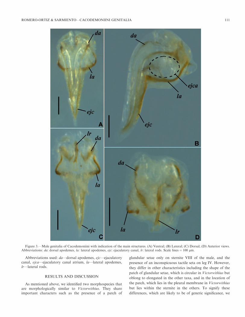
Figure 3.—Male genitalia of Cacodemoniini with indication of the main structures. (A) Ventral; (B) Lateral; (C) Dorsal; (D) Anterior views.Abbreviations: da: dorsal apodemes, la: lateral apodemes, ejc: ejaculatory canal, lr: lateral rods. Scale lines ¼ 100 lm.
ROMERO-ORTIZ & SARMIENTO—CACODEMONIINI GENITALIA 111

refer these two species as nr. Victorwithius. Further phyloge-netic analysis is needed to test their status and relationships.
Structural correspondence and variation.—Overall, the malegenital armature of Cacodemoniini comprises the dorsalapodemes (da) that extend posteriorly to form the ejaculatorycanal (ejc), which is elongated, giving a triangular shape to thegenitalia, and the lateral apodemes (la) which enclose theejaculatory canal atrium (ejca), which is supported in itsanterior region by the lateral rods (lr) (Fig. 3).
Dorsal apodemes (da): For Atemnidae, the closest familyto the Withiidae (Benavides et al. 2019), Klausen (2005)defines the dorsal apodemes as an elongated fusion of thelateral rods. In contrast, cacodemoniines, they are not fusedand appear as structures separate from the lateral rods (Figs.3C, D). The genital atrium and the ejaculatory canal areformed by the dorsal and the lateral apodemes. In the genitalatrium, the dorsal and lateral apodemes are separatedsurrounding it, while in the ejaculatory canal, the apodemesare fused (Fig. 3).
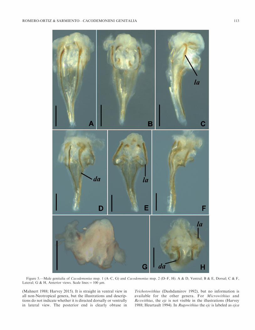
The degree and extent of sclerotization and the shape of thedorsal apodemes vary between genera. Sclerotization is very
marked in Cacodemonius (Figs. 5A, D) and nr. Victorwithius(Figs. 12A, D), while it is moderate in the genera Balanowi-thius (Fig. 4A), Cystowithius (Figs. 6A, D), Dolichowithius(Figs. 7A, D), and Victorwithius (Figs. 10A, D, G), and weakin Parawithius (Fig. 9A). The sclerotization covers the entiregenital atrium in Cacodemonius (Figs. 5B, E), Dolichowithius(Figs. 7C, F), Victorwithius (Figs. 10C, F), and nr. Victorwi-thius (Figs. 12B, C, E, F), while in the genera Balanowithius,Cystowithius and Parawithius it covers less than half of thegenital atrium (Figs. 4A, 6A, 9A). The first half of the da aresinuous in Dolichowithius (Fig. 7D) and straight in the othertaxa. The da are wider in the proximal third in Cystowithius(Fig. 6A), while they are uniform in the other genera.
The dorsal apodemes in Metawithius and Rugowithius werelabeled by Harvey (2015) and Johnson et al. (2019) as mediandiverticula; however, these structures appear in the corre-sponding position of the dorsal apodemes of the NeotropicalCacodemoniini. In addition, their illustrations suggest thatthese are sclerotized structures, whereas the diverticula are notsclerotized. The dorsal apodemes of Metawithius are short andstraight while these are long and straight in Rugowithius(Harvey 2015) and Trichotowithius (Dashdamirov 1992). Theillustration of the latter did not name this structure. InMicrowithius, the dorsal apodemes cannot be seen due to awide lateral apodeme covering the structures (Harvey 1988).Finally, in Pycnowithius and Rexwithius, these structures arenot visible in the published illustrations [Mahnert (1988) andHeurtault (1994) respectively].
Ejaculatory canal (ejc): Unlike that of other pseudoscorpi-ons (see e.g., Vachon 1938; Legg 1974c; Klausen 2005), theejaculatory canal of cacodemoniines extends posteriorly as aninverted triangle seen in either dorsal or ventral view. Theejaculatory canal is formed by the elongation of the dorsal andlateral apodemes. This is the diagnostic character for theCacodemoniini (Harvey 2015), and Klausen (2005) proposedthat this is a functional extension of the ejaculatory canalatrium. This ejaculatory canal atrium (ejca) is a capsulesurrounded by both the lateral apodemes and the dorsalapodemes (Fig. 3); as stated by Legg (1974c), it receivesproducts of the testis and the accessory glands.
We found differences for this structure in its length, shape,sclerotization of the posterior tip, and its direction in lateralview. The ejc is shorter than the width of the ejaculatory canalatrium in Cystowithius (Fig. 6) and Parawithius (Fig. 9), whileit is longer in the other genera. The posterior end of the ejc isacute in nr. Victorwithius (Figs. 12A, D), wide or evenlanceolate in Dolichowithius (Fig. 7D) and rounded in theother Neotropical taxa. The posterior tip of the ejc issclerotized in Cystowithius (Fig. 6A) and nr. Victorwithius(Fig. 12A) while it is not in the other genera (e.g. Figs. 7A, D,G, 10A). The ejc is straight in Cacodemonius (Figs. 5C, F),Victorwithius (Figs. 10E, H), and some species of Dolichowi-thius (Fig. 7E), while it is curved ventrally in Balanowithius(Fig. 4B), Cystowithius (Figs. 6B, E), and Parawithius (Fig.9B), and curved dorsally in nr. Victorwithius (Figs. 12B, E) andsome species of Dolichowithius (Figs. 7B, H).
The ejc is longer than the ejaculatory canal atrium inTrichotowithius and Metawithius (Dashdamirov 1992; Harvey2015) and is either as long as or shorter than the ejaculatorycanal atrium in Pycnowithius and Rugowithius respectively
Figure 4.—Male genitalia of Balanowithius egregius Beier, 1959.(A) Ventral; (B) Lateral; (C) Anterior views. Scale lines ¼ 100 lm.
112 JOURNAL OF ARACHNOLOGY
(Mahnert 1988; Harvey 2015). It is straight in ventral view inall non-Neotropical genera, but the illustrations and descrip-tions do not indicate whether it is directed dorsally or ventrallyin lateral view. The posterior end is clearly obtuse in
Trichotowithius (Dashdamirov 1992), but no information isavailable for the other genera. For Microwithius andRexwithius, the ejc is not visible in the illustrations (Harvey1988; Heurtault 1994). In Rugowithius the ejc is labeled as ejca
Figure 5.—Male genitalia of Cacodemonius msp. 1 (A–C, G) and Cacodemonius msp. 2 (D–F, H). A & D, Ventral; B & E, Dorsal; C & F,Lateral; G & H, Anterior views. Scale lines ¼ 100 lm.
ROMERO-ORTIZ & SARMIENTO—CACODEMONIINI GENITALIA 113

(Harvey 2015) but the structure does not correspond to ourdefinition.
Lateral apodemes (la): According to Klausen (2005) thelateral apodemes are formed by the anterolateral sides of thetwo dorsal and medial diverticula. Legg (1974b, c) describedthat the lateral apodemes support the dorsal and medialdiverticula and serve as attachment sites for the sterno-coxaland coxal muscles, specifically the segmental dorso-ventralmuscles, the longitudinal-ventral muscles, the appendicularmuscles, and the transverse muscle. We could not observewhether Neotropical specimens follow Klausen’s and Legg’sinterpretation as we did not study the diverticula due to theclearing process.
We observed that, in the Neotropical Cacodemoniini, thelateral apodemes (la) are paired and together with the dorsalapodeme form a bowl that protects the ejaculatory canalatrium. In lateral view, the apodemes exhibit different levels ofcurvature, and in ventral view they seem to merge with the
bifid dorsal apodeme. Together with the dorsal apodeme they
extend posteriorly to form the ejaculatory canal. This general
description of the lateral apodemes applies to the non-
Neotropical Cacodemoniini.
The la vary between genera in their extension, shape of the
basal region, and orientation of the concavity in lateral view.
The la cover almost the whole genital atrium in Balanowithius
(Fig. 4B), Cacodemonius (Figs. 5C, F), Victorwithius, and nr.
Victorwithius (Figs. 10B, E, H, 12B, E), but they do not go
beyond half of the genital atrium in Cystowithius (Fig. 6B, E),
Dolichowithius (Figs. 7B, E, H), and Parawithius (Figs. 9A, B).
The shape of the basal region is clearly angled in Cacodemo-
nius (Figs. 5D, E), Dolichowithius (Figs. 7A, D, G), and nr.
Victorwithius (Figs. 12A, D), but it is rounded in the other
genera. The curvature is more pronounced anteriorly in
Balanowithius (Fig. 4B) and Cacodemonius (Figs. 5C, F), but
it is more pronounced dorsally in the other genera.
Figure 6.—Male genitalia of Cystowithius msp. 1 (A–C, G) and Cystowithius msp. 2 (D–F, H). A & D, Ventral; B & E, Lateral, C & F, Dorsal;G & H, Anterior views. Scale lines ¼ 100 lm.
114 JOURNAL OF ARACHNOLOGY

Figure 7.—Male genitalia of Dolichowithius msp. 1 (A–C), Dolichowithius msp. 2 (D–F) and Dolichowithius msp. 3 (G–I). A, D & G, Ventral;B, E & H, Lateral; C, F & I, Dorsal views. Scale lines ¼ 100 lm.
ROMERO-ORTIZ & SARMIENTO—CACODEMONIINI GENITALIA 115

Heurtault (1971) named the lateral apodemes ‘‘arc chitinise’’for Withius hispanus (L. Koch, 1873), W. faunus (Simon, 1879)and W. neglectus (Simon, 1878). Using the English version,‘‘chitinized arch’’, Harvey (2015) and Johnson et al. (2019)follow this nomenclature for Metawithius. Since the chitinizedarch corresponds to the lateral apodemes of Legg (1974c), wefollow this original nomenclature. The la are wide in Micro-withius, Pycnowithius and Rugowithius (Harvey 1988, 2015;Mahnert 1988), while they are thin in Metawithius andTrichotowithius (Dashdamirov 1992; Harvey 2015; Johnsonet al. 2019). The only figure available for Rexwithius(Heurtault 1994) does not allow us to locate the la.
Lateral rods (lr): The lateral rods are only visible inanterior view. In the Cacodemoniini, they are placed betweenthe anterior ends of the dorsal and lateral apodemes, and varyin shape, alignment and length. They are sinuous only inBalanowithius (Fig. 4C). They diverge dorsally in Cystowithius(Figs. 6G, H) and Parawithius (Fig. 9D), while they areparallel in the other genera. The lateral rods do not extendbeyond the lateral apodemes in Balanowithius (Fig. 4C),whereas they are longer in the other genera. In many of thestudied specimens, they were not visible due to the highamount of tissue around this area.
Concerning the non-Neotropical genera, the lateral rods areonly visible in the illustrations of Metawithius andMicrowithiusprovided by Johnson et al. (2019) and Harvey (1988),
respectively. InMetawithius, they are sinuous and parallel, whilein Microwithius they are divergent dorsally. In Trichotowithius,Dashdamirov (1992) labels as lateral rods two longitudinalprojections located together with the ejaculatory canal; however,this location does not fit our observations in other Cacodemo-niini and we were unable to establish their correspondence toknown structures. Since lateral rods are present in all the othercheliferoid families (e.g., Legg 1974c; Klausen 2005), it is likelythat these structures are present in all Cacodemoniini.
Taxonomic characterization of the male genital armature for
each genus.—A description of the genital armature of each ofthe Neotropical genera of Cacodemoniini is provided below.
Balanowithius (Fig. 4)
Dorsal apodemes with sclerotization moderate to almostabsent, stronger in the first half of its length; straight and ofuniform width throughout its length; (Fig. 4A). Lateralapodemes covering most of the ejaculatory canal atrium;rounded basally; projecting anteriorly in lateral view (Fig. 4B).Ejaculatory canal long, two thirds length of genital armature;tip rounded, not sclerotized posteriorly; curved ventrally inlateral view (Fig. 4A). Lateral rods sinuous; parallel; notextending beyond lateral apodemes in dorsal view (Figs. 4B–C). This characterization is based on two specimens ofBalanowithius egregius.
Cacodemonius (Fig. 5)
Dorsal apodemes intensely and entirely sclerotized; straightand of uniform width throughout its length; (Figs. 5A, D).
Figure 8.—Male genitalia of Dolichowithius spp., anterior view: (A)Dolichowithius msp. 1; (B) Dolichowithius msp. 3; (C) Dolichowithiusmsp. 2. Scale lines ¼ 100 lm.
Figure 9.—Male genitalia of Parawithius msp1: (A) Ventral;(B)Lateral; (C) Dorsal; (D) Anterior views. Scale lines ¼ 100 lm.
116 JOURNAL OF ARACHNOLOGY

Lateral apodemes extending over most of ejaculatory canalatrium; angular basally; projecting anteriorly in lateral view(Figs. 5A, C, D). Ejaculatory canal long, two thirds length ofgenital armature; tip rounded, not sclerotized posteriorly;straight in lateral view (Figs. 5A, C, D, F). Lateral rodsextending beyond lateral apodemes in dorsal view (Figs. 5G,H). This characterization is based on two specimens of twomorphospecies of the genus Cacodemonius.
Cystowithius (Fig. 6)
Dorsal apodemes with sclerotization moderate to almost
absent, reaching half or less of its length; straight throughout
its length; the sclerotization of its border is wider at the
ejaculatory canal atrium (Figs. 6A, D). Lateral apodemes at
most half length of the ejaculatory canal atrium; rounded
basally; projecting dorsally (Figs. 6B, C, E, F). Ejaculatory
Figure 10.—Male genitalia of Victorwithius spp. Victorwithius msp. 1 (A–C), Victorwithius msp. 2 (D–F), and Victorwithius msp. 3 (G–I). A, D& G, Ventral; B, E & H, Lateral; C, F & I, Dorsal views. Scale lines ¼ 100 lm.
ROMERO-ORTIZ & SARMIENTO—CACODEMONIINI GENITALIA 117

canal short, one third of genital armature length; tip rounded,sclerotized posteriorly; curved dorsally in lateral view (Figs.6A, B, D, E). Lateral rods straight; divergent dorsally;extending beyond lateral apodemes in dorsal view (Figs.6F,G). This characterization is based on two specimens of twomorphospecies of the genus Cystowithius.
Dolichowithius (Figs. 7, 8)
Dorsal apodemes with sclerotization moderate to almostabsent, extending over half or less of its length; sinuous inproximal third and straight distally; of uniform widththroughout (Figs. 7A, D, G). Lateral apodemes extendingover at most half of ejaculatory canal atrium; angular basally;projecting dorsally (Fig. 7). Ejaculatory canal long, two thirdsof genital armature length; tip acute; not sclerotized posteri-orly; straight in lateral view, sometimes curved ventrally (Figs.7A, B, D, E, G, H). Lateral rods straight; parallel; extendingbeyond lateral apodemes in dorsal view (Fig. 8). Thischaracterization is based on four specimens belonging tothree morphospecies of the genus Dolichowithius.
Parawithius (Fig. 9)
Dorsal apodemes with sclerotization moderate to almostabsent, extending over half or less of its length; straightthroughout its length; of uniform width throughout (Fig. 9C).Lateral apodemes extending over at most half of ejaculatorycanal atrium; rounded basally, projecting dorsally (Figs. 9A–C). Ejaculatory canal short, one third of genital armaturelength; tip rounded, not sclerotized posteriorly; curveddorsally in lateral view (Figs. 9B, C). Lateral rods straight;divergent dorsally; extending beyond lateral apodemes in
dorsal view (Fig. 9D). This characterization is based on onespecimen of the genus Parawithius.
Victorwithius (Figs. 10, 11)
Dorsal apodemes entirely sclerotized but the sclerotization ismoderate to weak; straight and of uniform width throughoutits length (Figs. 10A, D, G). Lateral apodemes extending overmost of ejaculatory canal atrium; rounded basally; projectingdorsally (Fig. 10). Ejaculatory canal long, two thirds of genitalarmature length; tip rounded, not sclerotized posteriorly;straight in lateral view (Figs. 10A, B, D, E, G, H). Lateral rodsstraight, parallel; extending beyond lateral apodemes in dorsalview (Fig. 11). This characterization is based on five specimensbelonging to three morphospecies of the genus Victorwithius.
nr. Victorwithius (Fig. 12)
Dorsal apodemes intensely sclerotized; extending over itsentire length; straight and of uniform width throughout itslength (Figs. 12A, D). Lateral apodemes extending over mostof ejaculatory canal atrium; angular basally; projectingdorsally (Figs. 12A–F). Ejaculatory canal long, two thirds ofthe genital armature length; tip acute, sclerotized posteriorly;curved dorsally in lateral view (Figs. 12A, B, D, E). Lateralrods straight, parallel; extending beyond lateral apodemes indorsal view (Figs. 12G, H). This characterization is based ontwo specimens belonging to two morphospecies of thisundescribed taxon.
Final remarks.—Our analysis explored, for the first time, themale genitalia of the Cacodemoniini using a comparativeapproach and provides hypotheses of structural correspon-dences, covering six of the 13 cacodemoniine genera by directexamination of specimens, and six genera using publisheddescriptions and illustrations (Harvey 1988, 2015; Mahnert1988; Dashdamirov 1992; Heurtault 1994; Johnson et al.2019).
Structural correspondence conclusions: In agreement withHarvey (2015), our analyses allow us to characterize theCacodemoniini genitalia as follows: The dorsal apodemes andthe lateral apodemes are separated in the most anterior regionand surround the ejaculatory canal atrium. Posteriorly, theyare fused and extend to form the ejaculatory canal. The lateralrods are separated from the apodemes. Variation on thegenitalia is found in the shape of the lateral apodemes, thelength of the ejaculatory canal and its projection in lateralview.
Regarding the triangular projection of the ejaculatory canal,we disagree with Harvey (2015) who attributes this projectionto the lateral apodemes. Whenever we could inspect thegenitalia in sufficient detail, through direct examination ordrawings, we observed that such elongation includes thefusion of the dorsal apodemes with the lateral apodemes.
Taxonomic implications: Although we have not observedevery species of Neotropical Cacodemoniini, our data suggestthat the differences in the male genital armature of each genusfollow the current classification of the group, agreeing with theproposition of Mahnert (1975) that this is a helpful structurefor taxonomy at this taxonomic rank. This result contrast withthe findings of Klausen (2005) for the genera of Atemnidaewhere there is little taxonomic correspondence.
Figure 11.—Male genitalia of Victorwithius spp., anterior view: (A)Victorwithius msp. 2; (B) Victorwithius msp. 3; (C) Victorwithius msp.1. Scale lines ¼ 100 lm.
118 JOURNAL OF ARACHNOLOGY

Phylogenetic connotations: The taxonomic distribution of
the characters of the male genital armature across the
Cacodemoniini genera makes it difficult to even speculate on
their relationships using this character system. For example,
the dorsal apodeme sclerotization is moderate and straight
throughout its length in Balanowithius and Victorwithius, while
the lateral apodemes are projected anteriorly in Balanowithius
as opposed to dorsally in Cystowithius and Victorwithius.
Another example is the presence of a short ejaculatory canal in
Cystowithius and Parawithius, while Cystowithius and nr.
Victorwithius share a sclerotized tip of the ejaculatory canal
which is absent in Parawithius.
Figure 12.—Male genitalia of nr. Victorwithius msp. 1 (A–C, G); nr. Victorwithius msp. 2 (D–F, H). A & D, Ventral; B & E, Lateral; C & F,Dorsal; G & H, Anterior views. Scale lines ¼ 100 lm.
ROMERO-ORTIZ & SARMIENTO—CACODEMONIINI GENITALIA 119

Examining some species of Withius, Klausen (2005) foundthat, like in Atemnidae, the lateral rods are neither mergedproximally nor directed anteriorly and concluded that bothfamilies could be closely related. Our more extensive samplingof the Withiidae concur with his observation on Withius andwith the phylogenetic hypothesis of Benavides et al. (2019),who found strong molecular evidence to support a sister-group relationship between Atemnidae and Withiidae.
Despite the impressive advances using molecular data forphylogenetic and biodiversity studies, morphological analyses,such as the one provided here, are still required to understandthe history of the phenotypes as the substrate of evolution. Athorough understanding of the genitalia in pseudoscorpionsmay shed further light into the diversity and history of theircourtship strategies.
ACKNOWLEDGMENTS
We are very grateful to Dr. Gerald Legg and Dr. FinnKlausen for their contributions to our knowledge of thepseudoscorpion genitalia and their help answering ourquestions and giving us helpful comments on the developmentof this study. Also, we thank Andrea Carvajal for her time andwillingness to check the descriptions, as well as Dr. MarkHarvey and two anonymous reviewers who made severalcomments that greatly improved our manuscript. Weacknowledge the Universidad Nacional de Colombia andColciencias for support to CRO with the scholarshipDoctorados Nacionales: 727-2015.
LITERATURE CITED
Andrade R. de, Gnaspini P. 2003. Mating behavior and spermato-phore morphology of the cave pseudoscorpion Maxchernesiporangae (Arachnida Pseudoscorpiones Chernetidae). Journal ofInsect Behavior 16:37–48.
Beier M. 1940. Die Pseudoscorpionidenfauna der landfernen Inseln.Zoologische Jahrbucher, Abteilung fur Systematik, Okologie undGeographie der Tiere 74:161–192.
Beier M. 1944. Uber Pseudoscorpioniden aus Ostafrika. Eos 20:173–212.
Beier M. 1955. Pseudoscorpione von den Juan-Fernandez-Inseln(Arachnida Pseudoscorpionida). Revista Chilena de Entomologıa4:205–220.
Beier M. 1959. Zur Kenntnis der Pseudoscorpioniden-Fauna desAndengebietes. Beitrage zur Neotropischen Fauna 1:185–228.
Beier M. 1979. Neue afrikanische Pseudoskorpione aus dem MuseeRoyal de l’Afrique Central in Tervuren. Revue de ZoologieAfricaine 93:101–113.
Benavides LR, Cosgrove JG, Harvey MS, Giribet G. 2019.Phylogenomic interrogation resolves the backbone of the Pseudo-scorpiones tree of life. Molecular Phylogenetics and Evolution139:106509.
Chamberlin JC. 1931a. A synoptic revision of the generic classifica-tion of the chelonethid family Cheliferidae Simon (Arachnida).Canadian Entomologist 63:289–294.
Chamberlin JC. 1931b. The arachnid order Chelonethida. StanfordUniversity Publications, University Series, Biological Sciences7(1):1–284.
Dashdamirov S. 1992. Identity of Trichotowithius Beier 1944 with are-description of Trichotowithius abyssinicus Beier 1944 (ArachnidaPseudoscorpiones Withiidae). Tropical Zoology 5:293–298.
De Pinna MCC. 1991. Concepts and tests of homology in the cladisticparadigm. Cladistics 7:367–394.
Donoghue MJ, Doyle JA, Gauthier J, Kluge AG, Rowe T. 1989. Theimportance of fossils in phylogenetic reconstruction. AnnualReview of Ecology, Evolution and Systematics 20:431–460.
Feio JL de Araujo. 1944. Victorwithius monoplacophorus n. gen., n. sp.da subfamilia Withiinae Chamberlin, 1931 (Pseudoscorpiones:Cheliferidae). Boletim do Museu Nacional Rio de Janeiro, n.s.Zoologia 28:1–7.
Giribet G. 2015. Morphology should not be forgotten in the era ofgenomics—a phylogenetic perspective. Zoologischer Anzeiger256:96–103.
Harvey MS. 1988. Pseudoscorpions from the Krakatau Islands andadjacent regions, Indonesia (Chelicerata: Pseudoscorpionida).Memoirs of the Museum of Victoria 49:309–353.
Harvey MS. 1992. The phylogeny and classification of the Pseudo-scorpionida (Chelicerata: Arachnida). Invertebrate Taxonomy6:1373–1435.
Harvey MS. 2002. The neglected cousins: what do we know about thesmaller arachnid orders? Journal of Arachnology 30:357–372.
Harvey MS. 2004. Remarks on the New World pseudoscorpiongenera Parawithius and Victorwithius, with a new genus bearing aremarkable sternal modification (Pseudoscorpiones, Withiidae).Journal of Arachnology 32:436–456.
Harvey MS. 2013. Pseudoscorpions of the World, version 3.0.Western Australian Museum, Perth. Online at http://www.museum.wa.gov.au/catalogues/pseudoscorpions
Harvey MS. 2015. Revised diagnoses for the pseudoscorpion generaMetawithius and Microwithius, with the description of a newAustralian genus, and notes on Withius (Pseudoscorpiones, With-iidae). Journal of Arachnology 43:353–370.
Harvey MS, Huey JA, Hillyer MJ, McIntyre E, Giribet G. 2016. Thefirst troglobitic species of Gymnobisiidae (Pseudoscorpiones:Neobisioidea), from Table Mountain (Western Cape Province,South Africa) and its phylogenetic position. Invertebrate System-atics 30:75–85.
Heurtault J. 1971. Chambre genitale, armature genitale et caracteressexuels secondaires chez quelques especes de Pseudoscorpions(Arachnida) du genre Withius. Bulletin du Museum Nationald’Histoire Naturelle, Paris 42:1037–1053.
Heurtault J. 1994. Un cas indirect de phoresie: les pseudoscorpionsWithiidae des termitieres mortes de Macrotermes en Afriquetropicale. Bollettino dell’Accademia Gioenia di Scienze Naturali26:189–208.
Hoff CC. 1956. Pseudoscorpions of the family Cheliferidae from NewMexico. American Museum Novitates 1804:1–36.
Johnson J, Romero-Ortiz C, Mathew AV, Sebastian PA, Joseph MM,Harvey MS. 2019. A review of the pseudoscorpion genusMetawithius (Pseudoscorpiones: Withiidae) from the Indiansubcontinent. Journal of Arachnology 47:84–94.
Kastner A. 1927. Pseudoscorpiones. In Biologie der Tiere Deutsch-lands (Schulze P, ed.), 18:1–68. Gebruder Borntraeger, Berlin.
Kew HW. 1911. A synopsis of the false scorpions of Britain andIreland. Proceedings of the Royal Irish Academy (B) 29:38–64.
Klausen FE. 2005. The male genitalia of the family Atemnidae(Pseudoscorpiones). Journal of Arachnology 33:641–662.
Koch L. 1873. Uebersichtliche Dartstellung der EuropaischenChernetiden (Pseudoscorpione). Bauer & Raspe, Nurnberg.
Legg G. 1971. The comparative and functional morphology of thegenitalia of the British Pseudoscorpiones. PhD thesis, University ofManchester: Manchester.
Legg G. 1974a. The genitalia and accessory glands of thepseudoscorpion Cheiridium museorum (Cheiridiidae). Journal ofZoology, London 173:323–339.
Legg G. 1974b. An account of the genital musculature ofpseudoscorpions (Arachnida). Bulletin of the British ArachnologicalSociety 3:38–41.
Legg G. 1974c. A generalized account of the male genitalia and
120 JOURNAL OF ARACHNOLOGY
associated glands of pseudoscorpions (Arachnida). Bulletin of theBritish Arachnological Society 3:66–74.
Legg G. 1975a. The genitalia and associated glands of five Britishspecies belonging to the family Chthoniidae (Pseudoscorpiones:Arachnida). Journal of Zoology, London 177:99–121.
Legg G. 1975b. The genitalia and associated glands of five Britishspecies belonging to the family Neobisiidae (Pseudoscorpiones:Arachnida). Journal of Zoology, London 177:123–151.
Legg G. 1975c. Spermatophore formation in pseudoscorpions. Pp.141–144. In Proceedings of the 6th International ArachnologicalCongress Held at the Vrije Universiteit at Amsterdam, 22–26 April1974. Amsterdam, the Netherlands: Nederlandse EntomologischeVereniging.
Mahnert V. 1975. Pseudoskorpione der Insel Reunion und vonT.F.A.I. (Djibouti). Revue Suisse de Zoologie 82:539–561.
Mahnert V. 1988. Die Pseudoskorpione (Arachnida) Kenyas.Familien Withiidae und Cheliferidae. Tropical Zoology 1:39–89.
Muchmore WB. 1990. Termitowithius kistneri, a new genus andspecies of termitophilous pseudoscorpion from Tanzania (Pseudo-scorpionida: Withiidae). Bulletin of the British ArachnologicalSociety 8:125–127.
Murienne J, Harvey MS, Giribet G. 2008. First molecular phylogenyof the major clades of Pseudoscorpiones (Arthropoda: Chelicer-ata). Molecular Phylogenetics and Evolution 49:170–184.
Nixon KC, Carpenter JM. 2012. On homology. Cladistics 28:160–169.
Patterson C. 1982. Morphological characters and homology. Pp. 21–74. In Problems of Phylogenetic Reconstruction. (Joysey KA,Friday AE, eds). Academic Press, New York.
Pyron R.A. 2015. Post-molecular systematics and the future ofphylogenetics. Trends in Ecology and Evolution 30:384–389.
Redikorzev V. 1938. Les pseudoscorpions de l’Indochine francaiserecueillis par M. C. Dawydoff. Memoires du Museum Nationald’Histoire Naturelle, Paris 10:69–116.
Reeder TW, Townsend TM, Mulcahy DG, Noonan BP, Wood PLJr., Sites JW Jr., etal. 2015. Integrated analyses resolve conflictsover squamate reptile phylogeny and reveal unexpected placementsfor fossil taxa. PloS One 10(3):e0118199.
Schtschelkanovzeff JP. 1910. Der bau der Mannlichen Geschlecht-sorgane von Chelifer und Chernes. Zur Kenntnis der stellung derChelonethi im system. Festschrift 60. Geburtstag R. Hertwig 2:1–38.
Sereno PC. 2007. Logical basis for morphological characters in
phylogenetics. Cladistics 23:565–587.
Simon E. 1878. Liste des especes de la famille des Cheliferidae qui
habitant l’Algerie et le Maroc. Annales de la Societe Entomologique
de France (5)8:145–153.
Simon E. 1879. Les Ordres des Chernetes, Scorpiones et Opiliones;
Les Arachnides de France vol. 7. Librairie Encyclopedique de
Roret, Paris France.
Vachon M. 1938. Recherches anatomiques et biologiques sur la
reproduction et le developpement des pseudoscorpions. Annales
des Sciences Naturelles, Zoologie 11(1):1–207.
Vogt L, Bartolomaeus T, Giribet G. 2010. The linguistic problem of
morphology: structure versus homology and the standardization of
morphological data. Cladistics 26:301–325.
Vogt L, Nickel M, Jenner RA, Deans A. 2013. The need for data
standards in zoomorphology. Journal of Morphology 274:793–808.
Weygoldt P. 1966. Vergleichende Untersuchungen zur Fortpflan-
zungsbiologie der Pseudoscorpione: Beobachtungen uber das
Verhalten, die Samenubertragungsweisen und die Spermatophoren
einiger einheimischer Arten. Zeitschrift fur Morphologie und
Okologie der Tiere 56:39–92.
Weygoldt P. 1969. The Biology of Pseudoscorpions. Harvard
University Press, Cambridge MA.
Weygoldt P. 1970. Vergleichende Untersuchungen zur Fortpflan-
zungsbiologie der Pseudoscorpione II. Zeitschrift fur die Zoologi-
sche Systematik und Evolutionforschung 8:241–259.
Wiens JJ, Kuczynski CA, Townsend T, Reeder TW, Mulcahy DG,
Sites JW Jr. 2010. Combining phylogenomics and fossils in higher-
level squamate reptile phylogeny: molecular data change the
placement of fossil taxa. Systematic Biology 59:674–688.
Wipfler B, Pohl H, Yavorskaya MI, Beutel RG. 2016. A review of
methods for analysing insect structures — the role of morphology
in the age of phylogenomics. Current Opinion in Insect Science
18:60–68.
Wright AM, Hillis DM. 2014. Bayesian analysis using a simple
likelihood model outperforms parsimony for estimation of
phylogeny from discrete morphological data. PloS One9(10):e109210.
Manuscript received 14 November 2019, revised 28 September 2020.
ROMERO-ORTIZ & SARMIENTO—CACODEMONIINI GENITALIA 121
Copyright © 2022 FDOKUMEN