A comparative study of sporta perimedullaris musculosa in the renicule of six species of cetaceans
8
This article was downloaded by: [190.203.75.58] On: 31 March 2014, At: 10:53 Publisher: Taylor & Francis Informa Ltd Registered in England and Wales Registered Number: 1072954 Registered office: Mortimer House, 37-41 Mortimer Street, London W1T 3JH, UK Italian Journal of Zoology Publication details, including instructions for authors and subscription information: http://www.tandfonline.com/loi/tizo20 A comparative study of sporta perimedullaris musculosa in the renicule of six species of cetaceans Gianluca Tettamanti a , Annalisa Grimaldi b , Roberto Ferrarese b , Liliana Rinaldi b , Alessandro Bortolotto c , Giovanni di Guardo d & Magda de Eguileor b a Department of Structural and Functional Biology , University of Insubria , Via J. H. Dunant 3, I‐21100, Varese, Italy E-mail: b Department of Structural and Functional Biology , University of Insubria , Via J. H. Dunant 3, I‐21100, Varese, Italy c Zoonomia, via Don Carlo Gnocchi 5, 1–40128, Bologna, Italy d Department of Comparative Biomedical Sciences , University of Teramo , P.za A. Moro 45, I‐64100, Teramo, Italy Published online: 28 Jan 2009. To cite this article: Gianluca Tettamanti , Annalisa Grimaldi , Roberto Ferrarese , Liliana Rinaldi , Alessandro Bortolotto , Giovanni di Guardo & Magda de Eguileor (2004) A comparative study of sporta perimedullaris musculosa in the renicule of six species of cetaceans, Italian Journal of Zoology, 71:2, 115-121, DOI: 10.1080/11250000409356561 To link to this article: http://dx.doi.org/10.1080/11250000409356561 PLEASE SCROLL DOWN FOR ARTICLE Taylor & Francis makes every effort to ensure the accuracy of all the information (the “Content”) contained in the publications on our platform. However, Taylor & Francis, our agents, and our licensors make no representations or warranties whatsoever as to the accuracy, completeness, or suitability for any purpose of the Content. Any opinions and views expressed in this publication are the opinions and views of the authors, and are not the views of or endorsed by Taylor & Francis. The accuracy of the Content should not be relied upon and should be independently verified with primary sources of information. Taylor and Francis shall not be liable for any losses, actions, claims, proceedings, demands, costs, expenses, damages, and other liabilities whatsoever or howsoever caused arising directly or indirectly in connection with, in relation to or arising out of the use of the Content. This article may be used for research, teaching, and private study purposes. Any substantial or systematic reproduction, redistribution, reselling, loan, sub-licensing, systematic supply, or distribution in any form to anyone is expressly forbidden. Terms & Conditions of access and use can be found at http:// www.tandfonline.com/page/terms-and-conditions
-
Upload
independent -
Category
Documents
-
view
0 -
download
0
Transcript of A comparative study of sporta perimedullaris musculosa in the renicule of six species of cetaceans

This article was downloaded by: [190.203.75.58]On: 31 March 2014, At: 10:53Publisher: Taylor & FrancisInforma Ltd Registered in England and Wales Registered Number: 1072954 Registered office: MortimerHouse, 37-41 Mortimer Street, London W1T 3JH, UK
Italian Journal of ZoologyPublication details, including instructions for authors and subscription information:http://www.tandfonline.com/loi/tizo20
A comparative study of sporta perimedullarismusculosa in the renicule of six species ofcetaceansGianluca Tettamanti a , Annalisa Grimaldi b , Roberto Ferrarese b , Liliana Rinaldi b ,Alessandro Bortolotto c , Giovanni di Guardo d & Magda de Eguileor ba Department of Structural and Functional Biology , University of Insubria , Via J. H.Dunant 3, I‐21100, Varese, Italy E-mail:b Department of Structural and Functional Biology , University of Insubria , Via J. H.Dunant 3, I‐21100, Varese, Italyc Zoonomia, via Don Carlo Gnocchi 5, 1–40128, Bologna, Italyd Department of Comparative Biomedical Sciences , University of Teramo , P.za A.Moro 45, I‐64100, Teramo, ItalyPublished online: 28 Jan 2009.
To cite this article: Gianluca Tettamanti , Annalisa Grimaldi , Roberto Ferrarese , Liliana Rinaldi , AlessandroBortolotto , Giovanni di Guardo & Magda de Eguileor (2004) A comparative study of sporta perimedullaris musculosa inthe renicule of six species of cetaceans, Italian Journal of Zoology, 71:2, 115-121, DOI: 10.1080/11250000409356561
To link to this article: http://dx.doi.org/10.1080/11250000409356561
PLEASE SCROLL DOWN FOR ARTICLE
Taylor & Francis makes every effort to ensure the accuracy of all the information (the “Content”)contained in the publications on our platform. However, Taylor & Francis, our agents, and our licensorsmake no representations or warranties whatsoever as to the accuracy, completeness, or suitability for anypurpose of the Content. Any opinions and views expressed in this publication are the opinions and viewsof the authors, and are not the views of or endorsed by Taylor & Francis. The accuracy of the Contentshould not be relied upon and should be independently verified with primary sources of information.Taylor and Francis shall not be liable for any losses, actions, claims, proceedings, demands, costs,expenses, damages, and other liabilities whatsoever or howsoever caused arising directly or indirectly inconnection with, in relation to or arising out of the use of the Content.
This article may be used for research, teaching, and private study purposes. Any substantial or systematicreproduction, redistribution, reselling, loan, sub-licensing, systematic supply, or distribution in anyform to anyone is expressly forbidden. Terms & Conditions of access and use can be found at http://www.tandfonline.com/page/terms-and-conditions

Ital. J. Zool., 71: 115-121 (2004)
A comparative study of sportaperimedullaris musculosa in therenicule of six species of cetaceans
GIANLUCA TETTAMANTI*ANNALISA GRIMALDPROBERTO FERRARESELILIANA RINALDIDepartment of Structural and Functional Biology,University of Insubria, Via J. H. Dunant 3, I-21100 Varese (Italy)E-mail: [email protected]
ALESSANDRO BORTOLOTTOZoonomia, via Don Carlo Gnocchi 5, 1-40128 Bologna (Italy)
GIOVANNI DI GUARDODepartment of Comparative Biomedical Sciences,University of Teramo, P.za A. Moro 45, I-64100 Teramo (Italy)
MAGDA DE EGUILEORDepartment of Structural and Functional Biology,University of Insubria, Via J. H. Dunant 3, I-21100 Varese (Italy)
*, the two Authors contributed equally
ABSTRACT
Organs and physiological internal systems of diving mammalsdisplay diverse and peculiar features. The kidney of cetaceanshas been extensively studied and characterized. This organ is sub-divided in small functional units called "renicules" displaying apeculiar structure, the sporta perimedullaris musculosa. A com-parative study of renicules was made in six species belonging tomysticetes and odontocetes. In all six species, the sporta, a con-nective tissue basket, was located between the medullary and thecortical regions of renicules. The sporta is mainly composed of aconnective stroma and, in several species, the connective tissue(histochemically studied) was interposed among muscle fibres.Within the six studied species, the sporta varied in thickness,which appears to be related to the number, thickness and discon-tinuity of collagenous fibre bundles. The presence of smoothmuscle fibres, evidenced by immunostaining and by electron mi-croscopy analysis, was variable: whereas fibres were numerous inexamined toothed whales (Stenella, Delphinus, Grampus, Tur-siops), they were practically absent in baleen whales such as thefin whale Balaenoptera physalus. The data in the present studyshed light on the morpho-functional characterization of divingmammal anatomy and support the need to re-examine the gener-al structure of cetacean phylogeny, in line with the hypothesis ofMilinkovitch et al. (1994) and Hasegawa et al. (1997).
KEY WORDS: Cetacean - Renicule - Sporta perimidullaris mu-scolosa
ACKNOWLEDGMENTS
We are grateful to Dr Letizia Marsili (Dept of Environmental Sci-ence, Siena, Italy) for providing several kidney tissue samples aswell as to the Italian National Stranding Network (CSC); we thankDr. Perletti for critical reading of the manuscript.
(Received 7 April 2003 - Accepted 5 November 2003)
INTRODUCTION
Cetaceans are mammals displaying a number of ac-quired anatomical and physiological external and inter-nal adaptations. Fused body shape to reduce water fric-tion, the presence of dorsal and pectoral fins, a caudalpeduncle which generates the power for movement andthe localization of nostrils are external features linked toefficient underwater life and deepwater diving (morethan 900 m) (Andersen, 1969; Ridgway et al, 1969) forshort or long periods. These account for a number ofpeculiar characteristics related to several internal organs(cardiovascular apparatus, liver, lung, muscle system,kidney) which must sustain marked variations of pres-sure, low tissue oxygénation and decreased blood sup-ply (Slijper, 1962; Andersen, 1969; Ridgway et al, 1969;Kjekshus et al, 1982; Gaskin, 1986).
The morpho-functional modifications of cetacean kid-neys have been widely studied especially in relation toosmoregulation (McAlpine, 1985; Maluf & Gassman,1998; Ortiz, 2001). The kidney of marine mammals iscomposed of numerous, independently functioning,lobules called "renicules" (Munkacsi & Newstead, 1985;Cave & Aumonier, 1962, 1965, 1967). Each renicule unitis characterized by four major components: cortex,medullary region, papilla and calix, and can filtrate,concentrate, collect and transport urine. The subdivi-sion of the kidney into renicule units is an anatomicalfeature present in other mammals, both terrestrial andacquatic (rhinoceros, bear, elephant, and seal; Maluf &Gassman, 1998). Even though the specific function ofthis anatomical organization of the kidney has not beenfully investigated, according to Maluf & Gasmann(1998) the presence of a large number of renicules ex-pands the total surface of kidney cortex, thus increasingthe filtration activity performed by the glomerules con-tained in this region. In the absence of this anatomicalsubdivision of the kidney, this organ would be charac-terized by extremely long proximal and distal tubules toensure its optimal function.
Unlike other animals having reniculated kidneys, incetaceans this organ displays a peculiar structure local-ized at the interface between the cortical and themedullary region: the sporta perimedullaris, a discontin-uous, multi-layered structure mostly composed of con-nective tissue. In the past, several authors have focusedtheir attention on this structure but in many cetaceansits characterization is far from complete. The data arescarce due to the lack of sufficient biological materialand to its inadequacy for microscopical investigation,because all specimens used for research come frombeached cetaceans. Furthermore, a detailed compara-tive evaluation of the ratio between the fibrous compo-nent and the cellular elements (mainly muscle fibres) ofthe sporta is still lacking.
In the present work we give a morphological, histo-chemical and immunocytochemical description of thesporta perimedullaris musculosa of six different ceta-
Dow
nloa
ded
by [
190.
203.
75.5
8] a
t 10:
53 3
1 M
arch
201
4
116 G. TETTAMANTI, A. GRIMALDI, R. FERRARESE, L. RINALDI, A. BORTOLOTTO, G. DI GUARDO, M. DE EGUILEOR
céans species, focusing on the analysis of the cellularand extracellular components.
MATERIALS AND METHODS
The samples used in this study were obtained from cetaceansstranded along the Italian coasts and collected by the Italian "Cen-tro Studi Cetacei" (CSC) (work no. 93), by "Dipartimento di SanitàPubblica Veterinaria e Patologia Animale" (Bologna, Italy), orkindly provided by Dr. Giovanni Di Guardo ("Istituto Zooprofilat-tico Sperimentale delle Regioni Lazio e Toscana", Rome, Italy).
Animals were dissected and tissue samples were collected forhistological, microbiological and toxicological assays.
The following species were examined:- fin whale, Balaenoptera pbysalus (Linnaeus, 1758) (Ba-
laenopteridae) (one specimen);- common bottlenose dolphin, Tursiops truncatus (Montagu,
1821) (Delphinidae) (three specimens);- Risso's dolphin, Grampus griseus (G. Cuvier, 1812) (Del-
phinidae) (three specimens);- striped dolphin, Stenella coeruleoalba (Meyen, 1833) (Del-
phinidae) (six specimens);- short-beaked common dolphin, Delphinus delphis (Linnaeus,
1758) (Delphinidae) (two specimens);- Cuvier's beaked whale, Ziphius cavirostris (G. Cuvier, 1823)
(Ziphiidae) (one specimen).Cetacean renicules, preserved in 4% phosphate buffered
formaldehyde, were dissected into three regions: cortical region,medullary region, and the interposed layer, containing the sportaperimedullaris. Unfortunately post-mortem changes, which oc-curred before fixation of samples, sometimes rendered the studyproblematic but, nevertheless, the extreme rarity of the materialwas regarded as a sufficient justification for the histological prepa-ration and examination of these samples.
Light and electron microscopy (TEM)
Paraffin embedding
After four washes in phosphate buffered saline (PBS), pH 7.2,to remove formaldehyde, samples were dehydrated in an ethanolseries. A 1 h treatment in Bioclear (BioOptica, Milan, Italy) pre-ceded paraffin embedding. Sections (7 um) were cut with a JungMulticut 2045 microtome (Leica, Nussolch, Germany).
Epon embedding
After dissection of the renicule, small fragments (2 mm x 2 mmx 2 mm) of s/>ort«-containing tissue were fixed for 6 h in 1.5%glutaraldehyde in 0.1 M sodium-cacodylate buffer (CAC), pH 7.2.After several washes in CAC to remove the fixative solution, spec-imens were postfixed for 2 h with 1% osmic acid in CAC at roomtemperature. After a standard dehydration step in an ethanol se-ries, specimens were embedded in an epon-araldite 812 mixture,and cut with a Reichert Ultracut S ultratome (Leica). Semithin sec-tions (0.7 um) were stained by conventional methods (crystal vio-let and basic fuchsin) (Moore et al., I960) and observed at a lightmicroscope (Olympus, Tokyo, Japan). Thin sections (70 nm) werecollected on 300-mesh copper grids, stained with uranyl acetateand lead citrate, and observed with a Jeol 1010 EX electron mi-croscope (Jeol, Tokyo, Japan).
Histological and histochemical analysis
Paraffin sections were deparaffinized by a treatment with Bio-clear, rehydrated in an ethanol series, and then processed withspecific histological and histochemical stainings according tomethods and applications suggested by Bio-Optica histopatholog-ical kit (Bio-Optica) as follows:- Meyer hematoxylin (2% aqueous solution) and eosin (1% aque-
ous solution) were used to stain nuclei (blue-violet) and othercellular components (pale pink to red).
- Mallory trichrome. Standard procedure to characterize connec-tive tissue. Collagen fibrils were stained in deep blue, elastic fi-bres in pale pink-yellow or unstained, nuclei in red, myofibrilsin red.
- Picro-Mallory trichrome. Recommended to selectively stain theconnective tissue. Collagen fibrils were stained in dark blue,ground substance in varying shades of blue, elastic fibres inpale pink-yellow.
- Silver impregnation. A recommended method to show reticularfibres (black stained) in connective tissue (tobacco brownstained) and nervous fibres (in black); collagen (in yellow).
- DAPI (4'-6-diamidino-2-phenylindole) staining, specific for DNA(Russell et al, 1975).Samples were observed with a light microscope or with a con
focal laser microscope (see following section for details).
Immunocytochem istry
Paraffin sections were deparaffinized by a treatment with Bioclear,rehydrated in an ethanol series and then washed 4 times in PBS.
Slides were processed according to the method suggested by theProbes catalogue and sections were incubated for 1 h witha-Bungarotoxin eosin conjugate (Molecular Probes, CA, USA), usedto stain acetylcholine (ACh) receptors of neuromuscular junctions.
Other sections were incubated for 15 min with a Blue Evan's so-lution to minimize autofluorescence (De la Lande & Waterson,1968), washed with PBS, and incubated for 1 h with an anti-hu-man calmodulin polyclonal antibody (Santa Cruz Biotechnology,CA, USA), diluted 1:50 in 2% bovine serum albumin (BSA). Afterone hour of incubation with the primary antibody, specimenswere washed and incubated with a Cy3-conjugated anti-goat sec-ondary antibody (diluted 1:100) (Jackson> Immuno Research Labo-ratories, West Grove, PA, USA). Incubation was performed for 1 hin a dark moist chamber. Controls were obtained by omitting theprimary antibody and treating sections with BSA-containing PBS.
Coverslips were mounted in Vectashield Mounting Medium forfluorescence (Vector Laboratories, Burlingame, CA, USA) andslides were examined with a confocal laser microscope (laser 568nm for rhodamine) (MRC 1024, Bio-Rad Laboratories, HemelHempstead, UK) using x 40 and x 63 objectives (NA1.30, 1.25).Confocal images were superimposed using the Photoshop 5.0program: fluorescent images were then overlayed onto transmis-sion images showing the corresponding tissue sections.
RESULTS
The cetacean renicule is characterized by a corticaland a medullary region, separated by the sporta pe-rimedullaris muscolosa, made up of connective stroma,which, in some species, can also contain smooth mus-cle fibres.
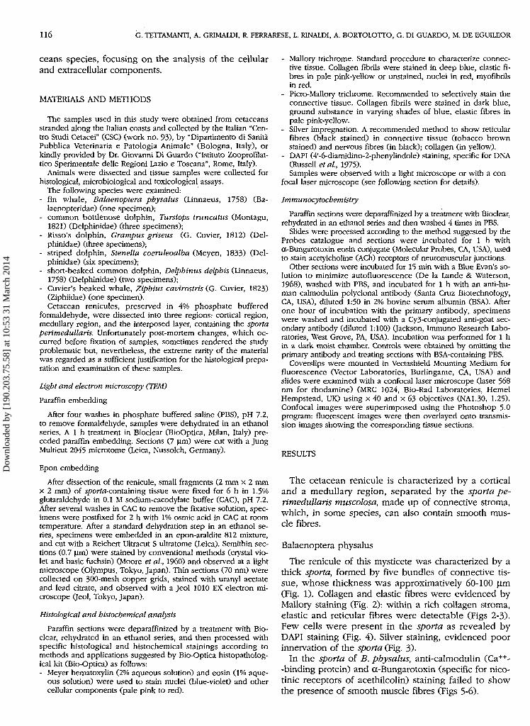
Balaenoptera physalus
The renicule of this mysticete was characterized by athick sporta, formed by five bundles of connective tis-sue, whose thickness was approximatively 60-100 u.m(Fig. 1). Collagen and elastic fibres were evidenced byMallory staining (Fig. 2): within a rich collagen stroma,elastic and reticular fibres were detectable (Figs 2-3).Few cells were present in the sporta as revealed byDAPI staining (Fig. 4). Silver staining, evidenced poorinnervation of the sporta (Fig. 3)-
In the sporta of B. pbysalus, anti-calmodulin (Ca++--binding protein) and a-Bungarotoxin (specific for nico-tinic receptors of acethilcolin) staining failed to showthe presence of smooth muscle fibres (Figs 5-6).
Dow
nloa
ded
by [
190.
203.
75.5
8] a
t 10:
53 3
1 M
arch
201
4

CETACEAN SPORTA 117
Figs 1-18 - Balaenoptera physalus. 1 - Hematoxylin/eosin: the sporta is a thick structure composed of five bundles of connective tissue(x80). 2 - Mallory trichrome: elastic fibres are detectable in the collagen stroma. 3 - Silver impregnation: reticular fibres and poor inner-vation of the sporta are evidenced. 4 - DAPI staining: weak positivity is detectable in the sporta. 5-6 - Anti-calmodulin and anti-a-Bun-garotoxin stainings: positivity is practically absent in the sporta region. Tursiops truncatus. 7 - Hematoxylin/eosin: the sporta is formedby four bundles of connective tissue (x40). 8 - Picro-Mallory trichrome: collagen fibres are abundant. 9 - Silver impregnation: good in-nervation is present. 10 - DAPI staining: some cells are present in the sporta. 11-12 - Anti-calmodulin and anti-oc-Bungarotoxin stain-ings: some cells are positive. Grampus griseus. 13 - Hematoxylin/eosin: the sporta is a thick structure (xl20). 14 - Mallory trichrome:sporta is mainly formed by collagen and elastic fibres. 15 - Silver impregnation: the staining shows the presence of reticular fibres andthe innervation of the sporta. 16 - DAPI staining: some cells are present in the sporta. 17-18 - Anti-calmodulin and anti-oc-Bungarotoxinstainings: the presence of muscle fibres is shown by these two stainings. Abbreviations: C, cortical region; v, vasa arcuata; arrows,sporta region.
Fig 19 - Grampus griseus. Muscle fibres (arrows) are small, with an elongated nucleus and scarce contractile material (TEM; X4000).
Dow
nloa
ded
by [
190.
203.
75.5
8] a
t 10:
53 3
1 M
arch
201
4

118 G. TETTAMANTI, A. GRIMALDI, R. FERRARESE, L. RINALDI, A. BORTOtOTTO, G. DI GUARDO, M. DE EGUILEOR
Tursiops truncatus
The sporta perimedullaris of this odontocete wasshown to be a structure formed by four collagenousbundles, different in thickness and length. The totalthickness of the connective laminae was quite relevant(100 u,m) and the connective tissue was also abundantaround the vasa arcuata (Figs 7-8). The fibromuscularbasket was mainly constituted by collagen fibres, as ev-idenced by picro-Mallory staining (Fig. 8). Silver stain-ing yielded a good innervation of this structure (Fig. 9),and DAPI staining (Fig. 10) showed that within thesporta the cellular component was relatively abundant.
The few cells present in the connective matrix weredetected by an anti-calmodulin antibody (Fig. 11) andby a-Bungarotoxin binding (Fig. 12), thus suggestingthe presence of muscle fibers likely responding tocholinergic signals.
Grampus griseus
The sporta of this odontocete was formed by bundlesof different length and thickness. With routine hema-toxylin-eosin staining it was possible to ascertain thatthe renicule was characterized by a sporta showing amaximum thickness of about 200 urn. Mallory and silverstainings showed the existence of a connective tissueformed by abundant collagen, as well as by elastic (Figs13-14) and reticular fibres (Fig. 15). The sporta showedthe presence of numerous cells, as demonstrated byDAPI staining (Fig. 16), whereas muscular innervationwas confirmed by silver staining (Fig. 15).
Immunoreactivity for anti-calmodulin antibodies (Fig.17) and a-Bungarotoxin binding (Fig. 18) confirmed thepresence of muscle fibres within the sporta of G.griseus. These fibres were better visible at the electronmicroscope: they were small in size (20 pm x 2 um)with a cytoplasm almost entirely occupied by a large,spindle-shaped heterochromatic nucleus, and by scarcecontractile material (Fig 19). Muscle fibres, surroundedby collagen bundles, were randomly organized.
Ziphius cavirostris
The sporta of this odontocete, formed by connectivelaminae of different lengths and thickness, was about90 urn in thickness (Fig. 20). Collagen was the mainconstituent of its structure (Fig. 21) which contained al-so a few elastic and reticular fibres (Figs 21-22). Thecells evidenced by the DAPI staining (Fig. 23) were few,as were muscle fibres, reacting to anti-calmodulin anda-Bungarotoxin stainings (Figs 24-25).
Delphinus delphis
In this odontocete, the sporta occupied a remarkablearea between the cortical and medullary regions ofrenicules and was characterized by the presence ofmultilayered, thick connective bundles (Fig. 26). Be-sides being composed of abundant collagen and elastic
fibres (Fig. 27), these bundles also contained reticular fi-bres (Fig. 28). Nuclei, stained by DAPI (Fig. 29), indicat-ed the existence of numerous cells in the sporta, andsilver staining evidenced a rich innervation of the ob-served region (Fig. 28). Positivity for anti-calmodulinantibody (Fig. 30) and for a-Bungarotoxin (Fig. 31) sug-gested that many of the nuclei in the sporta, evidencedby DAPI staining, belonged to muscle fibres.
Stenella coeruleoalba
The sporta of this odontocete showed numerous thinbundles (Fig. 32). Mallory staining demonstrated the pres-ence of abundant collagen fibers and a lesser amount ofelastic and reticular fibres (Figs 33-34). Cells in the fibro-muscular basket were numerous (Fig. 35) and the sportapresented a discrete innervation (Fig. 34). A remarkablenumber of cells was positive both for anti-calmodulin(Fig. 36) and for a-Bungarotoxin (Fig. 37), suggesting thepresence of an abundant muscular component.
Electron microscope observation revealed that fuse-shaped muscle fibres were small in size (20 pm x 2 pm),tightly packed and organized in bundles parallel to themain axis of fibres (Fig. 38).
DISCUSSION
The kidney of cetacean has been extensively studiedin the past (Cave & Aumonier, 1962, 1965, 1967; Maluf& Gassmann, 1998), because of its key role in osmoreg-ulation. This organ is subdivided into small functionalunits, "renicules", whose anatomical organization hasbeen well established (Maluf & Gassmann, 1998). How-ever, the functional utility of this subdivision is not yetclear. Our study was aimed at characterizing the mor-phology of a peculiar structure of cetacean renicules,the sporta perimedullaris musculosa, found in all thecetaceans examined. The fibromuscular basket is local-ized between the medullary and the cortical regions ofeach renicule, adjacent to the arcuata blood vessels inthe cortico-medullary region. It is important to empha-size that the presence of a sporta does not represent theinevitable concomitant feature of a kidney constructedupon the reniculate plan. In fact, a truly reniculate kid-ney occurs also in the Pinnipedia, but in none of theexamined species was present a real sporta (Simpson &Gardner, 1972; Munkacsi & Newstead, 1985; Newstead& Munkacsi, 1986). Consequently, even though somedata are available about cetacean sporta, further com-parative studies clarifying its morphology and physiolo-gy could be useful to shed light on the relationship be-tween the structure and function of this peculiaranatomical structure.
In agreement with previous studies (Cave & Aumonier,1961, 1962, 1966), all the species described in this paper,belonging to mysticetes and odontocetes, present a con-nective basket. The major difference between the sporta
Dow
nloa
ded
by [
190.
203.
75.5
8] a
t 10:
53 3
1 M
arch
201
4

CETACEAN SPORTA 119
Figs 20-37 - Ziphius cavirostris. 20 - Hematoxylin/eosin (x250). 21 - Mallory trichrome: the sporta is mainly composed by collagen andfew elastic fibres. 22 - Silver impregnation: reticular fibres are present in the sporta region. 23 - DAPI staining: few cells are stained. 24-25 - Anti-calmodulin and anti-a-Bungarotoxin stainings: few cells are positive. Delphinus delphis (x 120). 26 - Hematoxylin/eosin: thesporta is a thick multilayered structure. 27 - Picro-Mallory trichrome: collagen and elastic fibres are abundant. 28 - Silver impregnation:the staining demonstrates the existence of reticular fibres and rich innervation. 29 - DAPI staining: numerous cells are present in thesporta. 30-31 - Anti-calmodulin and anti-a-Bungarotoxin stainings: a strong signal is detectable. Stenella coeruleoalba (x60). 32 - Hema-toxylin/eosin: the sporta is characterized by thin bundles. 33 - Mallory trichrome: sporta is mainly formed by abundant collagen andfew elastic fibres. 34 - Silver impregnation: a discrete innervation characterizes the sporta. 35 - DAPI staining: numerous cells are pre-sent in the sporta. 36-37 - Anti-calmodulin and anti-a-Bungarotoxin stainings: a large amount of fibres are stained in the sporta. Abbre-viations: C, cortical region; arrows, sporta region.
Fig. 38 - Stenella coeruleoalba. Muscle fibres (arrows) are small and tightly packed, forming bundles (TEM; X6000).
Dow
nloa
ded
by [
190.
203.
75.5
8] a
t 10:
53 3
1 M
arch
201
4

120 G. TETTAMANTI, A. GRIMALDI, R. FERRARESE, L. RINALDI, A. BORTOLOTTO, G. DI GUARDO, M. DE EGUILEOR
within the six studied species is its thickness, a parame-ter closely related to the number of bundles of collagenfibres (four or five) and/or their thickness and disconti-nuity. The sporta is mainly composed of a connectivestroma (the number and the thickness of the collagenbundles can change in the different species) and, in sev-eral species, muscle fibres are scattered in the connec-tive tissue (Cave & Aumonier, 1961, 1962).
The comparison of specimens of different speciesshowed limited variability of the ratio of the connectivecomponent versus the elastic component: with the ex-ceptions of Z. cavirostris or D. delphis (showing onlytraces of elastic fibres) or S. coeruleoalba (displaying anabundant elastic component), all cases were character-ized by a collagen component more abundant than theelastic one. • •
The amount of cells in the sporta seemed to be de-pendent on the presence of muscle fibres and unrelatedto collagen/fibroblast abundance. In fact, a large num-ber of nuclei could be evidenced by DAPI staining insamples where muscle fibres were well represented;conversely, a reduced number of nuclei were detectedin samples of the fin whale's sporta, which displayedfew muscle fibres, even if the collagen component wasequal in both cases. The presence of smooth muscle fi-bres, highlighted byiimmunostaining and by electronmicroscopy, was variable: while muscle fibres wereabundant in the toothed whale genera examined(Stenella, Delphinus, Grampus, Tursiops), in the onlybaleen whale examined (B. physalus) the muscle com-ponent was virtually absent. While our data are in fullagreement with Cave & Aumonier's (Cave & Aumonier,1961) report on odontocetes and with Henk et al.'s ob-servations (1986) on Mysticetes, we are not able to ex-plain the discrepancy between our data on B. physalusand those of Cave & Aumonier (1964) on Minke whaleCB. acutorostrata, Lacépède, 1804), showing that thesporta contains muscle fibres. In addition, it needs to bepointed out that B. acutorostrata, the smallest of theBalenoperidae, is different with respect to other animalsbelonging to the same genera; this anatomical evidencemight support the taxonomic position of the species with-in the family Balaenopteridae presented by Milinkovitchand co-Authors (1995).
The amount of smooth muscle fibres in the sportaseems to be variable among the examined species. Arough quantification of fibres was possible by estimat-ing immunohistochemically the degree of muscle inner-vation (silver staining) as well as the presence ofsmooth muscle activity markers (calmodulin and a-Bun-garotoxin). Our data also suggest a possible functionalactivity of sporta, in contrast with the suggestions ofCave & Aumonier (1966), who stated that this structureplayed a merely accessory role, due to its poorly musclecomponent, incapable of affecting, by contraction, thepassage of the renal tubules into the renicular medulla.
The samples of odontocetes analyzed in our workpresented a more or less developed innervation system
(i.e., presence of muscle fibres), while B. physalusshowed very scarce innervation (i.e., presence of mus-cle fibres) in the sporta. The presence of innervation to-gether with muscle cells and the related positivity forcalmodulin and a-Bungarotoxin suggest the hypothesisthat the sporta perimedullaris musculosa, rich in colla-gen and elastic fibres, might have a mechanical func-tion, aimed at maintaining the integrity of the reniculestructure and avoiding the collapse of the blood vesselslocated between the cortical and the medullary regions.Thus, according to Cave & Aumonier (1962), this systemcould improve the renal function during immersions:while the presence of connective structure guaranteesresistance, muscle fibres could enable the sporta to"squeeze" the collecting ducts thus preventing hydrosta-tic pressure from inducing urine reflux. The presence ofnumerous smooth muscle fibres in the odontocetespecies relative to mysticetes could be related to the im-mersion behaviour of three cetacean taxa: D. delphis, S.coeruleoalba and T. truncatüs. These toothed whalesare able to perform frequent immersions at variabledeep (Goudappel & Slijper, 1958) and thus they mightneed a large number of muscle fibres in the sporta tosupport frequent kidney "stress". On the other hand, ac-cording to our analyses conducted on the renicule of B.physalus (see also Cave & Aumonier, 1964) and on themysticete bowhead whale (Balaena mysticetus, Lin-naeus, 1758) (Henk et ai, 1986), a member of the fami-ly Balaenidae whose behaviour is characterized by pro-longed immersions, very few smooth muscle fibres arepresent in their sporta.
Ziphius cavirostris diverges slightly from the structuralorganization of the sporta of other odontocetes, since itcontains a lesser number of muscle fibres. This fact fitswell with the hypothesis of a possible correlation be-tween the amount of muscle fibre in the sporta regionand the behaviour of the animal during immersion. Infact, 2. cavirostris performs infrequent but deep immer-sions, more similar to those of mysticetes (Leatherwood& Reeves, 1983). Further analysis of other species be-
•••-• longing to odontocetes, such as the northern bottlenosewhale, Hyperoodon ampullatus (Forster, 1770) and thesperm whale (Physeter macrocephalus, Linnaeus, 1758)whose diving behaviour is characterized by prolongedimmersions without frequent breaths, could be useful toconfirm this hypothesis.
Another interesting comparison can be made by con-sidering the perimedullary stromal basket of pinnipedkidney. This structure, similar to the cetacean sporta, isformed by a high number of muscle fibres embedded ina connective matrix. Interestingly, also in this case, pin-nipeds share the same functional needs as odontocetes,since they plunge frequently.
On the whole, our data could further confirm the hy-potheses (Cave & Aumonier, 1965; 1967) that sportacannot simply be a support to perimedullary blood-ves-sels, which are, in fact, equipped with their ownperivascular connective tissue coat, and are located in
Dow
nloa
ded
by [
190.
203.
75.5
8] a
t 10:
53 3
1 M
arch
201
4

CETACEAN SPORTA 121
districts far from the region of the sporta fundus (Cave& Aumonier, 1962).
The data collected in the present study on the struc-ture and composition of the fibromuscular basket of dif-ferent cetacean renicules, can be considered a "tessera"in the morpho-functional knowledge of diving mam-mals. Our data also support the statements of Milin-kovitch and co-workers (1994, 2001) and Hasegawa andco-workers (1997), who suggested the need to re-exam-ine the phylogenesis of cetaceans.
In fact, the Ziphiidae family presents some characterswhich are not typical of odontoceta, i.e. chromosomenumber and digestive apparatus; according to our data,also the sporta perimedullaris displays divergent modifi-cations, supporting the suggested monophyly of thebeaked whales (Milinkovitch et al, 1994).
REFERENCES
Andersen H. T., 1969 - The biology of marine mammals. Academ-ic Press, New York.
Cave A. J. E., Aumonier F. J., 1961 - The visceral histology of theprimitive cetacean Caperea (Neobalaena). J. R. Microsc. Soc,80: 25-33.
Cave A. J. E., Aumonier F. J., 1962 - Morphology of the cetaceanreniculus. Nature, 193: 799-800.
Cave A. J. E., Aumonier F. J., 1964 - The reniculus in certain ba-laenopterids. J. R. Microsc. Soc, 83: 255-264.
Cave A. J. E., Aumonier F. J., 1965 - Further observations on thecetacean reniculus. J. R. Microsc. Soc, 84: 539-548.
Cave A. J. E., Aumonier F. J., 1966 - The reniculus of Tursiopstruncatus, Stenella longirostris and other cetaceans. J. R. Mi-crosc. Soc, 86: 323-342.
Cave A. J. E., Aumonier F. J., 1967 - The reniculus of the seiwhale (Balaenoptera borealis). J. Nat. Hist., 4: 575-583.
De la Lande I. S., Waterson J. G., 1968 - Modification of autofluo-rescence in the formaldehyde-treated rabbit ear artery by Evansblue. J. Histochem. Cytochem., 16: 281-282.
Gaskin D. E., 1986 - Kidney and water metabolism. In: M. M. Bry-den & R. Harrison (eds.), Research on dolphins. ClarendonPress, Oxford, pp. 129-148.
Goudappel J. R., Slijper E., 1958 - Microscopic structure of thelungs of the bottlenose whale. Nature, 182: 479-483.
Hasegawa M., Adachi J., Milinkovitch M. C, 1997 - Novel phy-logeny of whales supported by total molecular evidence. J.Mol. Evol., 44: S117-S120.
Henk W. G., Abdelbaki Y. Z., Haldiman J. T., Albert T. F., 1986 -Microanatomy of the renicule of Balaena mysticetus. Anat.Rec, 214: 118-129.
Kjekshus J. K., Blix A. S., Eisner R., Hol R., Amundsen E., 1982 -Myocardial blood flow and metabolism in the diving seal. Am.J. Physiol., 242: R97-104.
Leatherwood S., Reeves R. R., 1983 - The Sierra Club handbook ofwhales and dolphins. Sierra Club Books, San Francisco, pp. 10-302.
Maluf N: S. R., Gassman J. J., 1998 - Kidney and significance ofreniculism. Anat. Rec, 250: 34-44.
McAlpine D. F., 1985 - Size and growth of heart, liver, and kid-neys in North Atlantic fin (Balaenoptera physalus), sei (B. bore-alis), and sperm (Physeter macrocephalus) whales. Can. J. Zo-ol., 63: 1402- 1409.
Milinkovitch M. C, Cassens I., 2001 - Deciphering river dolphinevolution. Science, 294: 787.
Milinkovitch M. C, Meyer A., Powell J. R., 1994 - Phytogeny of allmajor groups of cetaceans based on DNA sequences from threemitochndrial genes. Mol. Biol. Evol., 11: 939-948.
Milinkovitch M. C, Orti G., Meyer A., 1995 - Novel phytogeny ofwhales revisited but not revised. Mol. Biol. Evol., 12: 518-520.
Moore R. D., Mumaw V., Shonberg M. D., 1960 - Optical microscopyof ultrathin tissue sections. J. Ultrastruct. Res., 4: 113-116.
Munkacsi I. M., Newstead J. D., 1985 - The intrarenal and peri-capsular venous systems of kidneys of the ringed seal, Phocahispida:, J. Morphol., 184: 361-373.
Newstead J. D., Munkacsi I. M., 1986 - The fibromuscular basket(Sporta perimedullaris musculosa) in renculi of kidneys fromthe ringed seal, Phoca hispida (Pinnipedia). J. Morphol., 188:335-346.
Ortiz R. M., 2001 - Osmoregulation in marine mammals. J. Exp.Biol., 204: 1831-1844.
Ridgway S. H., Scronce B. L., Kanwisher J., 1969 - Respiration anddeep diving in the bottlenose porpoise. Science, 166: 1651-1654.
Russell W. C., Newman C, Williamson D. H., 1975 - A simple cy-tochemical technique for demonstration of DNA in cells infect-ed with mycoplasmas and viruses. Nature, 253: 461-462.
Simpson J. G., Gardner M. B., 1972 - Comparative microscopicanatomy of selected marine mammals. In: S. H. Ridgway (ed.),Mammals of the sea. Thomas, Springfield (IL), pp. 298-418.
Slijper E. J., 1962 - Heart circulation and blood. In: Ann Arbor(ed.), Whales, vol. 4. Hutchinson, London, pp. 117-153.D
ownl
oade
d by
[19
0.20
3.75
.58]
at 1
0:53
31
Mar
ch 2
014