Integrating Clinical and Translational Research Networks - MDPI
Upload
khangminh22Category
view
1download
0
Int. J. Mol. Sci. 2022, 23, 4233. https://doi.org/10.3390/ijms23084233 www.mdpi.com/journal/ijms
Article
Next-Generation Sequencing Screening of 43 Families with
Non-Syndromic Early-Onset High Myopia: A Clinical and
Genetic Study
Eva González-Iglesias 1,†, Ana López-Vázquez 2,†, Susana Noval 2, María Nieves-Moreno 2,
María Granados-Fernández 2, Natalia Arruti 2, Irene Rosa-Pérez 2, Marta Pacio-Míguez 3,4, Victoria E. F. Montaño 1,3,
Patricia Rodríguez-Solana 1, Angela del Pozo 3,5, Fernando Santos-Simarro 3,6 and Elena Vallespín 1,3,*
1 Section of Molecular Ophthalmology, Medical and Molecular Genetics Institute (INGEMM) IdiPaz,
La Paz University Hospital, 28046 Madrid, Spain; [email protected] (E.G.-I.);
[email protected] (V.E.F.M.); [email protected] (P.R.-S.);
[email protected] (E.V.) 2 Department of Ophthalmology, La Paz University Hospital, 28046 Madrid, Spain;
[email protected] (A.L.-V.); [email protected] (S.N.);
[email protected] (M.N.-M.); [email protected] (M.G.-F.);
[email protected] (N.A.); [email protected] (I.R.-P.) 3 Biomedical Research Center in the Rare Diseases Network (CIBERER), Carlos II Health Institute (ISCIII),
28029 Madrid, Spain; [email protected] (M.P.-M.); [email protected] (A.d.P.);
[email protected] (F.S.-S.) 4 Section of Neurodevelopmental Disorders, Medical and Molecular Genetics Institute (INGEMM) IdiPaz,
La Paz University Hospital, 28046 Madrid, Spain 5 Section of Clinical Bioinformatics, Medical and Molecular Genetics Institute (INGEMM) IdiPaz,
La Paz University Hospital, 28046 Madrid, Spain 6 Section of Clinical Genetics, Medical and Molecular Genetics Institute (INGEMM) IdiPaz, La Paz University
Hospital, 28046 Madrid, Spain
* Correspondence: [email protected]
† These authors contributed equally to this work.
Abstract: Early-onset high myopia (EoHM) is a disease that causes a spherical refraction error of
≥−6 diopters before 10 years of age, with potential multiple ocular complications. In this article, we
report a clinical and genetic study of 43 families with EoHM recruited in our center. A complete
ophthalmological evaluation was performed, and a sample of peripheral blood was obtained from
proband and family members. DNA was analyzed using a customized next-generation sequencing
panel that included 419 genes related to ophthalmological disorders with a suspected genetic cause,
and genes related to EoHM pathogenesis. We detected pathogenic and likely pathogenic variants
in 23.9% of the families and detected variants of unknown significance in 76.1%. Of these, 5.7% were
found in genes related to non-syndromic EoHM, 48.6% in genes associated with inherited retinal
dystrophies that can include a syndromic phenotype, and 45.7% in genes that are not directly related
to EoHM or retinal dystrophy. We found no candidate genes in 23% of the patients, which suggests
that further studies are needed. We propose a systematic genetic analysis for patients with EoHM
because it helps with follow-up, prognosis and genetic counseling.
Keywords: early-onset high myopia; next-generation sequencing; ophthalmogenetics
1. Introduction
High myopia (HM), the most challenging type, is defined as a spherical refraction error
of −6 spherical diopters (SDs) or more, or an axial length greater than 26 mm. From a phys-
iological point, myopia is a refractive error in which, in a state of relaxed accommodation,
light rays parallel to the eye with an origin greater than 6 m away focus anteriorly on the
Citation: González-Iglesias, E.;
López-Vázquez, A.; Noval, S.;
Nieves-Moreno, M.;
Granados-Fernández, M.; Arruti, N.;
Rosa-Pérez, I.; Pacio-Míguez, M.;
Montaño, V.E.F.; Rodríguez-Solana, P.;
et al. Next-Generation Sequencing
Screening of 43 Families with
Non-Syndromic Early-Onset High
Myopia: A Clinical and Genetic
Study. Int. J. Mol. Sci. 2022, 23, 4233.
https://doi.org/10.3390/ijms23084233
Academic Editor: Tomasz Żarnowski
Received: 22 March 2022
Accepted: 4 April 2022
Published: 11 April 2022
Publisher’s Note: MDPI stays neu-
tral with regard to jurisdictional
claims in published maps and institu-
tional affiliations.
Copyright: © 2022 by the authors. Li-
censee MDPI, Basel, Switzerland.
This article is an open access article
distributed under the terms and con-
ditions of the Creative Commons At-
tribution (CC BY) license (https://cre-
ativecommons.org/licenses/by/4.0/).

Int. J. Mol. Sci. 2022, 23, 4233 2 of 27
retina after passing through the eye’s refractive system [1,2]. This disease is a common cause
of vision loss, with uncorrected myopia the leading cause of vision impairment globally.
The condition is known to be associated with ocular complications such as cataracts, glau-
coma, neovascular membranes, retinal tears and retinal detachment.
The prevalence of myopia and HM worldwide is 30.6% and 2.7%, respectively [3]; how-
ever, recent studies have estimated that this prevalence will increase to 49.8% and 9.8%,
respectively, by 2050 [4]. The most recent study in a Spanish population showed that the
prevalence of myopia and HM in children aged 5–7 years is 20% and 3.6%, respectively [5].
The pathogenesis of myopia relies on genetic, environmental (external) and microenvi-
ronmental (internal) factors. Most children are born hyperopic, and, during the first 2 years
after birth, a process known as emmetropization occurs, which is mainly influenced by a
change in axial length [3]. From this moment on, environmental factors such as near work
(defined as activities with a short working distance) [6], exposure to artificial light [7–9] can
modify this condition. The onset of HM before 10 years of age, known as early-onset high
myopia (EoHM), is an important aspect for research, considering that environmental factors
lose relevance in children of this age, allowing an approach to the pathogenesis of high myopia
as a monogenic disease [10]. Familial genetics can also contribute to this condition [11].
Physiopathological factors of the microenvironment such as oxidative stress, inflam-
mation and angiogenesis can also induce EoHM or promote the exacerbation of the disease.
Oxidative stress is produced by the metabolism of reactive species of oxygen (ROS). Retinal
tissue has the highest oxygen consumption of the body and is directly exposed to natural
light. These two factors produce a higher concentration of ROS in the tissue. In addition to
this, EoHM patients are known to present an oxidative/antioxidative statue imbalance sug-
gesting that oxidative stress can induce EoHM onset directly [12]. Oxidative stress is also
responsible for causing an inflammatory state that may also lead to the development of the
disease [13]. In 2015, Zhu et al., and in 2021, Wei et al., determined that the microenviron-
ment of EoHM eyes is unique due to the increase of proinflammatory cytokines (IL6, IFN-
γ, IP-10, eotaxin, and MIP-1α) and angiogenic growth factors (VEGF, MCP1, and IL5) in the
aqueous and vitreous humor found in patients [12,14–16]. The presence of a higher concen-
tration of the vascular endothelial growth factor (VEGF) as an angiogenic factor could make
the EoHM patients’ eyes more susceptible to developing vascular diseases [14].
This manuscript focuses on the genetic factors that may be affecting the development of
EoHM, in which over 100 genes and 20 chromosomal loci have been identified in association
with myopia and the related quantitative traits via linkage analysis, candidate gene analysis,
genome-wide association study and next-generation sequencing (NGS). However, only a
small number of genes have been identified to predispose individuals to EoHM, suggesting a
complex mechanism [13,17,18]. Eleven genes have been identified as autosomal dominant
genes to produce non-syndromic EoHM (ZNF644, SCO2, SLC39A5, CCDC111, P4HA2, BSG,
CPSF1, NDUFAF7, TNFRSF21, XYLT and DZIP1); four as autosomal recessive (LRPAP1,
CTSH, LEPREL1 and LOXL3) and two linked to chromosome X (ARR3 and OPN1LW). To date,
26 loci have been discovered (23 in autosomal chromosomes and 3 in chromosome X) [7,19].
Other studies have linked EoHM with genes such as CTNND2, JOANA, CACNA1F and RPGR
[20,21]. Given that myopia is a refractive error disease, other studies have reported single nu-
cleotide polymorphisms (SNPs) related to refractive error in PRSS56, BMP3, KCNQ5, LAMA2,
TOX, TJP2, RDH5, ZIC2, RASGRF1, GJD2, RBFOX1, SHISA6, FAM150B-ACP1, LINC00340,
FBN1, DIS3L-MAP2K1, ARID2-SNAT1 and SLC14A2 [22–24].
The proper diagnosis of EoHM is essential because it can be the first sign of a syn-
dromic condition. For cases in which an NGS panel is performed with only non-syn-
dromic EoHM-related genes, a syndromic condition, such as Stickler syndrome, might be
overlooked. We therefore recommend a broader approach when faced with a child with
EoHM. Several ophthalmological diseases, such as childhood glaucoma, retinopathy of
prematurity, congenital stationary night blindness and cone-rod dystrophies, are known
to be involved in the development of EoHM [25]. It is also essential to consider the family
history in the early diagnosis, and treatment can prevent further complications.
Int. J. Mol. Sci. 2022, 23, 4233 3 of 27
This study is based on the hypothesis of the possible greater involvement of a genetic
cause in the development of EoHM when compared with late-onset HM (LoHM). To clar-
ify this, we analyzed 43 families with EoHM to describe not only the pathogenic genes
that have already been related to EoHM or related syndromes but also new affected genes
that have not been directly related and could be causing this condition.
2. Results
The study results were obtained from an ongoing project for identifying mutations
that cause EoHM in a sample of families at a tertiary hospital in Spain. The project also
aims to evaluate the implementation of NGS and its relevance as part of the approach for
patients with EoHM.
A total of 43 patients with EOHM from 43 unrelated families (51% male [22/43] and
49% female [21/43]) were recruited based on their phenotype and inclusion criteria (listed
in Section 4. Materials and Methods). A complete clinical evaluation and genetic analysis
were performed for the proband of each of the 43 families. After performing the genetic
analysis of all probands and their families, six genetically affected family members who
had not been considered as probands were detected and were added to the study group.
The mean visual acuity on the decimal scale was 0.59 for the right eye (OD) and 0.55
in the left eye (OS). Axial length (AL) was measured with the IOL master 500 (Carl Zeiss
Meditec, Jena, Germany) with a mean AL of 27.79 mm for the OD and 27.95 mm for the
OS. Only 2 patients had an AL shorter than 25.5 mm. The mean spherical refraction was
−10.8 SD for the OD and −10.44 SD for the OS, with a spherical equivalent of −11.22 for the
OD and −10.44 for the OS (Table 1).
Table 1. Refractive results.
Right Eye Left Eye
Best corrected visual acuity (decimal scale) 0.59 ± 0.33 0.55 ± 0.33
Axial length (mm) 27.79 ± 2.5 27.95 ± 2.59
Spherical refraction (diopters) −10.8 ± 6.1 −10.44 ± 5.38
Astigmatism (diopters) −1.71 ± 1.3 −1.92 ± 1.4
Spherical equivalent (diopters) −11.22 ± 5.45 −10.44 ± 4.66
Results are presented as mean ± standard deviation.
Strabismus was present in 12% of the patients (5/43), with esotropia being the most
common type (80%). Twelve percent (5/43) of the patients presented with nystagmus.
When we consider only the 37 patients younger than 12 years at the time of the ex-
amination, only two (5%) presented with a posterior staphyloma, four (11%) presented
with peripapillary atrophy, and 14 (38%) presented with diffuse chorioretinal atrophy.
One patient had a colobomatous papilla, and one patient had complete retrolental retinal
detachment in one eye (Table 2). We performed retinography with Optos (Marlborough,
MA, USA) and optical coherence tomography (OCT) with Heidelberg OCT (Heidelberg
Engineering GmbH, Heidelberg, Germany) (Figures 1–4).
Figure 1. Posterior staphyloma. Right and left eyes. Optos (Marlborough, MA, USA).
Int. J. Mol. Sci. 2022, 23, 4233 4 of 27
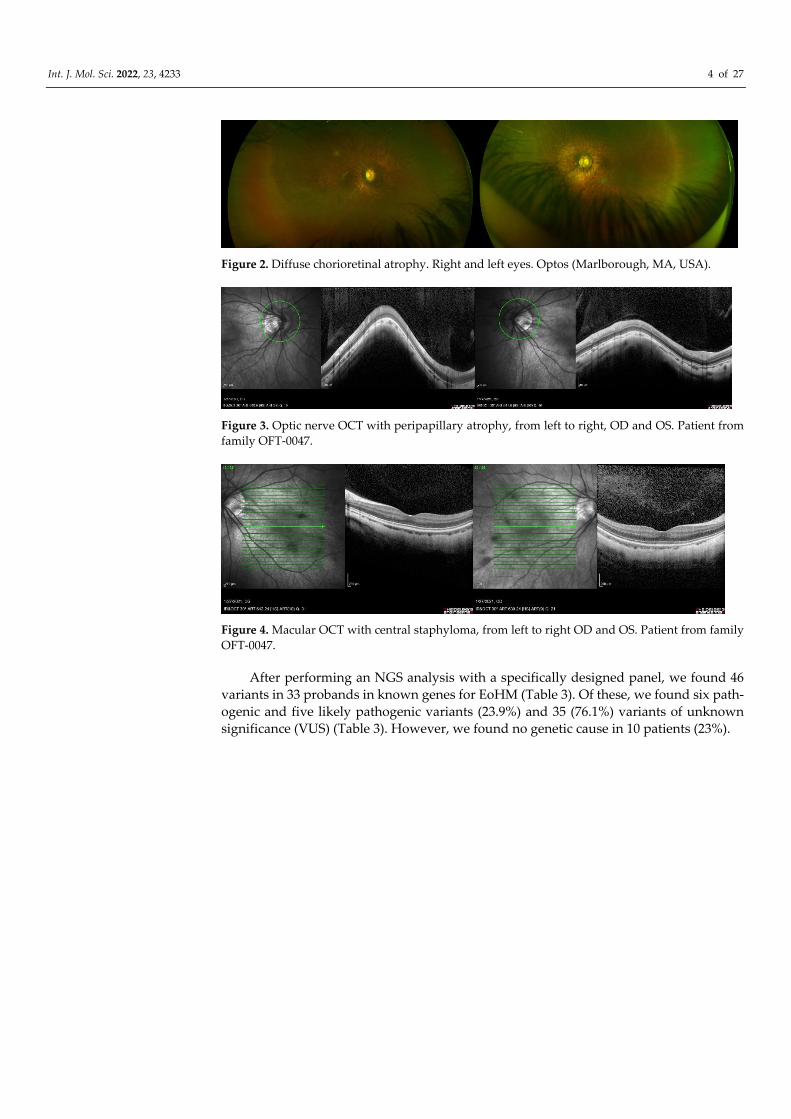
Figure 2. Diffuse chorioretinal atrophy. Right and left eyes. Optos (Marlborough, MA, USA).
Figure 3. Optic nerve OCT with peripapillary atrophy, from left to right, OD and OS. Patient from
family OFT-0047.
Figure 4. Macular OCT with central staphyloma, from left to right OD and OS. Patient from family
OFT-0047.
After performing an NGS analysis with a specifically designed panel, we found 46
variants in 33 probands in known genes for EoHM (Table 3). Of these, we found six path-
ogenic and five likely pathogenic variants (23.9%) and 35 (76.1%) variants of unknown
significance (VUS) (Table 3). However, we found no genetic cause in 10 patients (23%).

Int. J. Mol. Sci. 2022, 23, 4233 5 of 27
Table 2. Clinical Evaluation of Probands.
Family
Number Sex
BCVA
OD
BCVA
OS
AL
OD AL OS Funduscopic Examination OD Funduscopic Examination OS
SPcc
OD
Astig
OD
SE OD
cc
SPcc
OS
Astig
OS
SE OS
cc
OFT-00074 F 0.6 0.08 26.6 26.93 Diffuse chorioretinal atrophy, Central
staphyloma
Diffuse chorioretinal atrophy, Central
staphyloma −12 −0.5 −12.25 −12.75 −2.5 −14
OFT-00155 M 0.125 0.1 NA NA Healthy retina Healthy retina −10 −1.25 −10.6 −8.75 −2.75 −10.1
OFT-00209 M 0.6 0.7 NA NA Diffuse chorioretinal atrophy Diffuse chorioretinal atrophy −8.5 −3 −10 −7 −3 −8.5
OFT-00177 F NA NA NA NA Diffuse chorioretinal atrophy Diffuse chorioretinal atrophy −24 0 −24 −18 0 −18
OFT-00178 M 0.3 0.4 26.75 26.65 Healthy retina, Mild optic nerve pallor Healthy retina −6.75 −4 −8.75 −7.25 −3.25 −8.88
OFT-00181 M 0.9 0.9 26.6 26.7 Healthy retina Healthy retina −8.25 −1 −8.75 −8 −1 −8.5
OFT-00223 F 0.3 0.3 28.04 27.62 Atrophic optic nerve Atrophic optic nerve −13.5 −2.5 −14.75 −13 −0.5 −13.25
OFT-00092* M 0.1 0.05 NA NA Healthy retina Peripheral toxoplasma scar −0.5 −1.5 −1.25 −2.25 −0.75 −2.6
OFT-00097 M 0.4 0.2 26.84 26.47 Tessellated fundus, Healthy optic nerve Tessellated fundus, Healthy optic nerve −9.75 −5.25 −12.35 −10 −5.25 −12.6
OFT-00045 M 0.05 1 23.56 23.43 Hypopigmented fundus, Foveal
hypoplasia, Colobomatous optic nerve
Hypopigmented fundus, Foveal
hypoplasia −9.75 −2.5 −11 −10 −3 −11.5
OFT-00275 F 0.7 0.1 27.61 27.6 Diffuse chorioretinal atrophy Diffuse chorioretinal atrophy −11.5 −1 −12 −12 −3.25 −13.6
OFT-00332 M 0.25 0.3 29.41 29.02 Tessellated fundus, Epiretinal fibrosis Tessellated fundus, WWP on inferior and
temporal retina −15.25 −1 −15.75 −14.75 −0.5 −15
OFT-00343 F 0.8 0.8 NA NA Diffuse chorioretinal atrophy,
Peripapillary atrophy
Diffuse chorioretinal atrophy,
Peripapillary atrophy −15.75 −2.75 −17.1 −16 −1.25 −16.75
OFT-00191 M 0.5 0.5 26.05 26.15 Diffuse chorioretinal atrophy, Mild
optic nerve pallor
Diffuse chorioretinal atrophy, Mild optic
nerve pallor −9 −2 −10 −8.75 −3.25 −10.4
OFT-00391 M 0.9 NA NA NA Healthy retina, WWP inferotemporal Healthy retina −7.25 −2.25 −8.375 −7 −3 −8.5
OFT-00407 M 0.6 0.5 28.26 27.8 Diffuse chorioretinal atrophy, Mild
optic nerve pallor
Diffuse chorioretinal atrophy, Mild optic
nerve pallor −9.75 −3.5 −11.5 −9.5 −2.5 −10.75
OFT-00429 M 0.8 0.6 NA NA Diffuse chorioretinal atrophy,
Peripapillary atrophy, WWP inferiorly
Diffuse chorioretinal atrophy,
Peripapillary atrophy, WWP inferiorly −20 0 −20 −19 0 −19
OFT-00436 M 0.63 0.3 27.42 30.93 Diffuse chorioretinal atrophy Diffuse chorioretinal atrophy −7 −2.5 −8.25 −15 −3.75 −16.875
OFT-00453 F NFx Fx NA NA Complete retinal detachment Diffuse chorioretinal atrophy,
Peripapillary atrophy 2.25 −2.25 1.125 −9.5 −1.5 −10.25
OFT-00463 F 0.3 0.05 32.44 33.57 Severe peripapillary and macular
atrophy
Severe peripapillary and macular
atrophy NA NA NA NA NA NA
OFT-00474 M 0.1 0.7 27.43 25.99 Diffuse chorioretinal atrophy Diffuse chorioretinal atrophy −11.5 −1.25 −12.125 −10.25 −0.5 −10.5
OFT-00490 F NA NA NA NA NA NA NA NA NA NA NA NA
OFT-00506 F 0.7 0.7 NA NA Tessellated fundus Tessellated fundus −13.25 −2 −14.25 −12.5 −1.5 −13.25
OFT-00533 F NA NA NA NA NA NA NA NA NA NA NA NA
OFT-00546 M 1 1 24.45 24.12 Healthy retina Healthy retina −7.25 −0.75 −7.625 −5 −1 −5.5

Int. J. Mol. Sci. 2022, 23, 4233 6 of 27
OFT-00554 M 0.3 0.5 NA NA Healthy retina Healthy retina −9 −2.5 −10.25 −7.5 −1.25 −8.125
OFT-00559 M 0.4 0.3 NA NA
Diffuse increase in vascular
ramification, Avascular peripheral
retina
Avascular peripheral retina −7 −1.25 −7.625 −7.5 −1.75 −8.375
OFT-00568 F 0.16 0.8 34.09 33.89 Diffuse chorioretinal atrophy,
Peripapillary atrophy, Staphyloma
Diffuse chorioretinal atrophy,
Peripapillary atrophy, Staphyloma NA NA NA NA NA NA
OFT-00586 F 0.8 0.05 NA NA Diffuse chorioretinal atrophy Diffuse chorioretinal atrophy NA NA NA NA NA NA
OFT-00590 F 0.63 0.5 29.39 28.66 Diffuse chorioretinal atrophy Diffuse chorioretinal atrophy −18.25 −0.25 −18.37 −20.5 −1.25 −1.75
OFT-00601 M 1 0.9 27.92 27.68 Healthy retina Healthy retina −7 −1 −7.5 −6.5 −1 −7
OFT-00630 F 0.08 0.08 NA NA Diffuse chorioretinal atrophy, Central
staphyloma
Diffuse chorioretinal atrophy, Central
staphyloma −26 NA NA −26 NA NA
OFT-00493 F NA NA NA NA NA NA NA NA NA NA NA NA
OFT-00175 M 0.9 0.8 31.22 30.98 Diffuse chorioretinal atrophy,
Peripapillary atrophy
Diffuse chorioretinal atrophy,
Peripapillary atrophy −13.5 −4.75 −15.87 −13.25 −6 −16.25
OFT-00220 M 1 0.8 25.7 26.2 Healthy retina Healthy retina −5 −0.5 −5.25 −6 −2.5 −7.25
OFT-00253 F 0.9 0.9 29.59 29.1 Healthy retina Healthy retina −19.25 0 −19.25 −17.25 −0.5 −17.5
OFT-00268 M 0.5 0.6 27.08 27.18 Diffuse chorioretinal atrophy Diffuse chorioretinal atrophy −7.25 −0.75 −7.6 −7 −1 −7.5
OFT-00435 F 0.5 0.5 24.99 28.13 Healthy retina Healthy retina −21.25 −1.5 −22 −14 −3.25 −15.625
OFT-00443 M 1.2 1 NA NA RPE hypertrophy, WWP temporal and
superior Healthy retina −7.75 −0.5 −8 −8.5 −0.5 −8.75
OFT-00477 F 1.25 1.25 NA NA Healthy retina Healthy retina −7.5 −0.75 −7.875 −8 −1.5 −8.75
OFT-00517 F 1 0.8 NA NA Tessellated fundus Tessellated fundus −6 −1.25 −6.625 −5 −2 −6
OFT-00529* F NLP 0.63 NA NA Diffuse atrophy, Previous RD Diffuse NA NA NA −0.25 −1.75 −1.25
OFT-00623 F 0.5 0.67 NA NA Tessellated fundus Tessellated fundus −6 −2 −7 −2.75 −0.75 −3.5
M, male; F, female; BCVA, best-corrected visual acuity; NFx, non-fixation; Fx, fixation; NLP, no light perception; OD, right eye; OS, left eye; AL, axial
length; WWP, white without pressure; RD, retinal detachment; SPcc, sphere with cycloplegia; Astig, astigmatism; SE, spherical equivalent; NA, not
nvailable. * Patients from family OFT-00092 and OFT-00529 had already undertaken cataract surgery and therefore did not have a higher degree of
myopia in the last examination. However, the degree of preoperative myopia met the inclusion criteria.
Table 3. Genetic Results of Probands.
Famil
y ID
First
Diagnosi
s
Second
Diagnosis Gene Transcript Mutation
ACMG
Criteria *
ACM
G
Result
Variant Type Zygosity Inheritanc
e
Segregatio
n Analysis
Performed
De Novo
/Inherited Reported by
OFT-
00074 EoHM
NYSTAGMUS
AND
ESOTROPIA
TRPM1 NM_002420.5
Allele 1: c.3121C>T:
p.Arg1041Trp Allele
2: c.1023+1G>A
PM2/PVS1
, PM2,
PP3, PP5
VUS/P Allele 1: Missense
Allele 2: Splicing
Compoun
d Hetero AR Yes
Allele 1:
Maternal/Allel
e 2: Unknown
Allele 1:
Novel/Allele
2: Miraldi
Utz et al.,
2018 [26]

Int. J. Mol. Sci. 2022, 23, 4233 7 of 27
OFT-
00155 EoHM NYSTAGMUS
GPR143 NM_000273.2 c.1157G>A:
p.Ser386Asn
PM2, PP1,
PP2 VUS Missense Hemi X-linked Yes Maternal Novel **
CACNA1
F NM_005183.3
c.2924G>A:
p.Arg975Gln
PM2, PP1,
PP3 VUS Missense Hemi X-linked Yes Maternal Novel **
OFT-
00209 EoHM -
TIMP2 NM_003255.5 c.498C>G:
p.Ile166Met PM2, PP3 VUS Missense Hetero AD Yes Maternal Novel **
COL9A1 NM_001851.6 c.6G>T: p.Lys2Asn PM2 VUS Missense Hetero AD Yes Unknown Novel **
OFT-
00177 EoHM
CONE-ROD
DYSTROPHY
AND
SUBCAPSULAR
CATARACT
CEP290 NM_025114.4 c.5777G>C:
p.Arg1926Pro PM2, PP3 VUS Missense Hetero S No Unknown
Wiszniewski
et al., 2011
[27]; Sheck
et al., 2018
[28]; Sallum
et al., 2020
[29]
PCDH15 NM_001142763.
2
c.5308_5313del:
p.Ala1770_Pro1771de
l
PM2, PM4,
PP3 VUS Deletion Hetero S No Unknown Novel **
OFT-
00178 EoHM - LRP5 NM_002335.4
c.4610C>T:
p.Ala1537Val PM2 VUS Missense Hetero AD No Paternal Novel **
OFT-
00181 EoHM
RETINAL
DYSTROPHY COL2A1 NM_001844.5
c.2818C>T:
p.Arg940Ter
PVS1, PP5,
PM2, PP3 P Nonsense Hetero AD Yes Maternal
Kondo et al.,
2016 [30];
Maddirevul
a et al., 2018
[31]; Zhou et
al., 2018 [4]
OFT-
00223 EoHM -
PEX1 NM_000466.3 c.440T>C:
p.Val147Ala PM2, PP3 VUS Missense Hetero AD Yes Maternal Novel **
VDR NM_001017536.
2
c.1223G>A:
p.Arg408His PM1, PM2 VUS Missense Hetero AD No Maternal Novel **
MMP9 NM_004994.3 c.822G>C:
p.Glu274Asp PM2 VUS Missense Hetero AD Yes Paternal Novel **
OFT-
00092 EoHM
RETINAL
DYSTROPHY KCNV2 NM_133497.4
c.458G>A:
p.Arg153His PM2, PP3 VUS Missense Hetero S No Unknown Novel **
OFT-
00097 EoHM
NYSTAGMUS
AND
ASTIGMATISM
CFH NM_000186.4 c.907C>T:
p.Arg303Trp PM2, BP4 VUS Missense Hetero S No Unknown Novel **
CACNA1
F
NM_001256789.
3
c.4471C>T:
p.Arg1491Ter
PVS1, PP5,
PM2, PP3 P Nonsense Hemi AD No Maternal Novel **
OFT-
00045 EoHM
NYSTAGMUS
AND RETINAL
DYSTROPHY
PAX6 NM_001258462.
3 c.262A>G: p.Ser88Gly
PS2, PM1,
PM2, PP2,
PP3
P Missense Hetero AD Yes De novo Novel **
OFT-
00275 EoHM - COL2A1 NM_001844.5
c.1783delC:
p.Ala595LeufsTer34
PVS1, PS2,
PM2, PP3 P Frameshift Hetero AD Yes De novo Novel **

Int. J. Mol. Sci. 2022, 23, 4233 8 of 27
OFT-
00332 EoHM -
ZNF644 NM_201269.3 c.1366A>T:
p.Thr456Ser PM2 VUS Missense Hetero AD Yes Maternal Novel **
CRYBB3 NM_004076.5 c.547G>T: p.Glu183* PM2, PP3 VUS Nonsense Hetero AD Yes Maternal Novel **
LRP5 NM_002335.4 c.263A>G:
p.Lys88Arg PM2 VUS Missense Hetero AD Yes Maternal Novel **
OFT-
00343 EoHM - OPA1 NM_130837.3
c.1294G>A:
p.Val432Ile
PM1, PM2,
PP2, PP3,
PP5
LP Missense Hetero AD Yes Paternal
Stewart et
al., 2008 [32];
Yu-Wai-Man
et al., 2011
[33]
OFT-
00191 EoHM - COL11A1 NM_001854.4
c.2900G>T:
p.Gly967Val
PM2, PP1,
PP3 LP Missense Hetero AD Yes Paternal Novel **
OFT-
00391 EoHM ASTIGMATISM
CRYGC NM_020989.4 c.179G>A:
p.Arg60Gln PM2, PP2 VUS Missense Hetero S No Unknown Novel **
RDH5 NM_001199771.
2
c.683G>A:
p.Arg228Gln
PM2, PP2,
BP4 VUS Missense Hetero S No Unknown Novel **
OFT-
00407 EoHM
CONE-ROD
DYSTROPHY ARL6 NM_177976.3
c.362G>A:
p.Arg121His
PM2, PM3,
PP2, PP3,
PP5
LP Missense Homo AR Yes Maternal and
Paternal
Patel et al.,
2016 [34];
Abouelhoda
et al., 2016
[35]
OFT-
00429 EoHM -
MMP9 NM_004994.3 c.1270C>A:
p.Arg424Ser PM2, BP4 VUS Missense Hetero S No Unknown Novel **
IGF1R NM_000875.5 c.3784A>C:
p.Ile1262Leu PM2, PP3 VUS Missense Hetero S No Unknown Novel **
OFT-
00436 EoHM - MMP10 NM_002425.3 c.497-2A>G PP3, BS1 VUS Splicing Hetero AD Yes Maternal Novel **
OFT-
00453 EoHM
RETINAL
DYSTROPHY
AND
PERSISTENT
FETAL
VASCULATUR
E RIGHT EYE
COL2A1 NM_001844.5 c.157C>T: p.Arg53Trp PM2, PP2,
PP3 VUS Missense Hetero S No Unknown Novel **
TRPM1 NM_001252020.
1
c.3618C>G:
p.Phe1206Leu PM2 VUS Missense Hetero S No Unknown Novel **
OFT-
00463 EoHM - EPHA2 NM_004431.5
c.308G>A:
p.Arg103His PM2, PP3 VUS Missense Hetero S No Unknown Novel **
OFT-
00474 EoHM - MERTK NM_006343.3
c.2264G>A:
p.Arg755His PM2, PP3 VUS Missense Hetero AR Yes Maternal Novel **
OFT-
00490 EoHM -
COL11A1 NM_001854.4 c.1021G>C:
p.Glu341Gln PM2 VUS Missense Hetero S No Unknown Novel **
TRPM1 NM_001252020.
1
c.4550C>T:
p.Thr1517Met PM2, BP4 VUS Missense Hetero S No Unknown Novel **

Int. J. Mol. Sci. 2022, 23, 4233 9 of 27
OFT-
00493 EoHM - CRYGA NM_014617.4
c.287A>G:
p.Asp96Gly PM2, BP4 VUS Missense Hetero S No Unknown Novel **
OFT-
00506 EoHM - ZNRF3
NM_001206998.
2
c.2221G>A:
p.Glu741Lys PM2, BP4 VUS Missense Hetero AD Yes Unknown Novel **
OFT-
00533 EoHM - SCO2
NM_001169111.
1
c.334C>T:
p.Arg112Trp PM2, PP5 VUS Missense Hetero S No Unknown
Jiang et al.,
2015 [36]
OFT-
00546 EoHM - LAMA2 NM_000426.4
c.6880G>T:
p.Val2294Leu PM2, PP3 VUS Missense Hetero AR Yes Paternal Novel **
OFT-
00554 EoHM - SCO2
NM_001169111.
1
c.341G>A:
p.Arg114His
PS3, PM2,
PP3, PP5 LP Missense Hetero AD Yes
Maternal or
Paternal
Tran-Viet et
al., 2013 [37];
Pacheu-
Grau et al.,
2015 [38];
Kars et al.,
2021 [39]
OFT-
00559 EoHM NYSTAGMUS NDP NM_000266.4
c.313_314delGCinsTT
: p.Ala105Leu
PM1, PM2,
PM5, PS1,
PP2, PP3
P Deletion/Insertio
n Hemi S No Unknown Novel **
OFT-
00568 EoHM - PEX1 NM_000466.3
c.3250A>G:
p.Met1084Val PM2 VUS Missense Hetero S No Unknown Novel **
OFT-
00586 EoHM
RETINAL
DYSTROPHY
LEFT EYE
MMP1 NM_002421.4 c.1389G>A:
p.Trp463Ter PP3, BS1 VUS Nonsense Hetero S No Unknown Novel **
OFT-
00590 EoHM - COL11A1 NM_001854.4
c.1570C>T:
p.Arg524Trp PM2, PP3 LP Missense Hetero S No Unknown Novel **
OFT-
00601 EoHM - GPR143 NM_000273.3 c.47C>A: p.Ala16Glu
PM2, PP2,
PP3, BP6 VUS Missense Hemi X-linked Yes Maternal Novel **
OFT-
00630 EoHM - CRYBA1 NM_005208.5 c.190C>T: p.Arg64Trp PM2, PP3 VUS Missense Hetero S No Unknown Novel **
P, pathogenic; LP, likely pathogenic; VUS, variants of unknown significance; Hemi, hemizygous; Hetero, heterozygous; Homo, homozygous; AR,
autosomal recessive; R, recessive; AD, autosomal dominant; S, sporadic; ACMG, American College of Medical Genetics and Genomics. * ACMG
Criteria in Appendix B, Table A1/** Not previously reported in the literature.

Int. J. Mol. Sci. 2022, 23, 4233 10 of 27
Of the 11 variants classified as pathogenic and likely pathogenic, two families had an
affected variant in COL2A1 and another two had an affected variant in COL11A1, while the
rest had an affected variant in TRPM1, CACNA1F, PAX6, OPA1, ARL6, SCO2 and NDP (Fig-
ure 5). The rest of the variants were classified as VUS and were found in TIMP2, COL9A1,
CEP290, PCDH15, VDR, KCNV2, CFH, CACNA1F, COL2A1, ZNF644, CRYBB3, COL11A1,
CRYGC, RDH5, IGF1R, MMP10, EPHA2, MERTK, CRYGA, ZNRF3, LAMA2, SCO2, MMP1
and CRYBA1. A number of genes with variants classified as VUS were found in two families
(e.g., GPR143, LRP5, PEX1 and MMP9) and in three families (e.g., TRPM1) (Figure 6).
Figure 5. Representation of genes with pathogenic and likely pathogenic variants and their proportion.
Figure 6. Representation of genes with variants of unknown significance and their proportion.
Considering zygosity, 40 of the variants found were heterozygous (87%), whereas
only one variant was homozygous (2%) and five were hemizygous (11%).
3. Discussion
In this study, we performed a mutational analysis of Spanish patients diagnosed with
EoHM implementing a customized NGS panel containing 419 genes related to ophthal-
mological disorders with a suspected genetic cause. The use of an NGS panel strategy

Int. J. Mol. Sci. 2022, 23, 4233 11 of 27
rather than whole exome sequencing as a first approach for the genetic study is in re-
sponse to the need for a less expensive method that can provide efficient analysis with a
low rate of incidental findings.
3.1. Genes Related to Isolated EoHM
As previously mentioned, a few genes are related to non-syndromic EoHM, such as
SCO2 and ZNF644. Two patients (from families OFT-00533 and OFT-00554) in our cohort
showed previously reported variants in gene SCO2 (OMIM:604272) [36–39], which en-
codes a copper chaperone essential for the formation of cytochrome c oxidase (COX), the
last enzyme in the respiratory electron transport chain in the mitochondria [36]. The exact
mechanism of myopia development associated with COX deficiency specifically is cur-
rently unclear, but several studies, such as the 2013 study by Tran-Viet et al. [36,37,40,41],
have detected various mutations in gene SCO2 that have been considered pathogenic or
likely pathogenic for high or extreme myopia (>30 diopters). In contrast, Piekutowska-
Abramczuk et al. (2016) [42] indicated that heterozygous pathogenic variants in this gene
are not associated with high-grade myopia in either humans or mice. Considering the
contradictory published data, more studies on the involvement of SCO2 in the develop-
ment of EoHM are needed to determine whether the variants reported in the patients are
actually the cause of the phenotype or just a risk factor whose further development is
affected by the environment. In our cohort, we confirmed that the variants are just a risk
factor and not the cause, given that the proband of family OFT-00554 carried the same
heterozygous variants as their healthy parents.
Another gene related to non-syndromic autosomal dominant EoHM is ZNF644
(OMIM:614159), a zinc finger transcription factor expressed in the retina and the retinal
pigment epithelium. The protein’s biological function has not been identified. As a tran-
scription factor, however, it might regulate genes involved in eye development, triggering
a mutant protein able to modify the axial elongation of the eye globe and cause EoHM
[41,43]. Numerous studies have reported variants in this gene in non-syndromic EoHM
[4,37,40,43,44]. We found one previously unpublished variant classified as VUS according
to the American College of Medical Genetics and Genomics (ACMG) guidelines. The pro-
band of this family (OFT-00332) had inherited the variant from their mother, who appar-
ently did not have a pathogenic ocular phenotype, which suggests that this gene might
present incomplete penetrance.
3.2. Genes Related to Inherited Retinal Diseases
Inherited retinal dystrophies are sometimes associated with EoHM, which might be
the first isolated sign at presentation. Of the 46 variants found in our cohort, ten were
found in six genes associated with inherited retinal degeneration (based on the Retinal
Information Network [RetNet]) (21.7%, 10/46): TRPM1 (OMIM:603576), CACNA1F
(OMIM:300110), KCNV2 (OMIM:607604), MERTK (OMIM:604705), RDH5 (OMIM:601617)
and ARL6 (OMIM:608845).
We reported 4 variants in TRPM1, a gene associated with congenital stationary night
blindness (CSNB), a genetically and clinically heterogeneous disease that manifests as
non-progressive nyctalopia and with an electronegative full-field electroretinogram. Our
cohort had one patient (from family OFT-00074) with heterozygous compound variants
in this gene who presented with nystagmus, esotropia and EoHM. The patient underwent
an electroretinogram, which showed moderate-severe disease in the peripheral retina,
mild-moderate disease in the central retina and an electronegative result for the bilateral
scotopic response, which suggests a functional disorder of the bipolar cells. According to
Miraldi Utz et al. (2018), patients presenting EoHM, strabismus, nystagmus and a variant
in this gene should undergo full-field electroretinography, given that a considerable pro-
portion of these patients might not present nyctalopia at the start and that the procedure
could help diagnose CSNB [26]. In addition, two more VUS were reported, in families
OFT-00453 and OFT-00490.

Int. J. Mol. Sci. 2022, 23, 4233 12 of 27
Another gene associated with the same phenotype of CSNB is CACNA1F, a gene pre-
sent in the X chromosome and related to Aland Island disease and cone-rod dystrophy.
Wutz et al. (2002) mentioned the possibility of hemizygous variants in this gene causing a
phenotype of CSNB with EoHM as one of their most common features [21,45,46]. Our study
had two families (OFT-00155 and OFT-00097) with hemizygous variants and a phenotype
of EoHM and nystagmus. For family OFT-00155, the variants were maternally inherited,
and the proband had a brother with the same phenotype who also presented this variant.
One patient from family OFT-00391 had a variant in RDH5, a gene expressed in the
retinal pigmented epithelium and involved in the retinoid cycle, the metabolic pathway
that regenerates the visual chromophore following light exposure [22,47]. The phenotype
related to variants in this gene is fundus albipunctatus, a form of CSNB [48]. Although the
patient did not have retinopathy, this gene was established in 2013 as a susceptibility gene
for refractive error and myopia [18–50].
Patients from three families with a phenotype of EoHM and retinal dystrophy had
variants in KCNV2 (OFT-00092), MERTK (OFT-00474) and ARL6 (OFT-00407), genes with
a phenotype of retinal cone dystrophy in the case of KCNV2 and retinitis pigmentosa in
the other two cases. KCNV2 has EoHM as one of its ocular features. In the case of MERTK
and ARL6 where EoHM was not reported, however, this manifestation might be a second
consequence of the genetically determined retinal dystrophy.
3.3. Genes Related to Vitreoretinal Inherited Diseases
Regarding the association between EoHM and vitreoretinal inherited diseases, Marr
et al. (2001), showed an even higher association between HM and systemic and other oph-
thalmological conditions, taking into account that they did not consider an exclusion cri-
terion the presence of a syndromic phenotype as an exclusion criteria, as we did in our
study. Out Of the 112 probands, the authors found that only 8% had non-syndromic HM,
54% had an underlying systemic association, and the remaining 38% remaining had fur-
ther HM-related ocular problems associated with HM [17]. Other studies, such as that
performed by Logan et al. (2004) analyzed the genetic results of children with EoHM di-
agnosed before 10 years of age. Fifty-six percent of the children presented with simple
HM, 25% were diagnosed with inherited retinal dystrophies and amblyopia, and 19%
were diagnosed with an HM-related systemic disorder [51].
Based on these results, the RetNet genes associated with vitreoretinal inherited dis-
eases with syndromic manifestations should be included for patients with EoHM, such as
COL9A1 (OMIM:120210), COL2A1 (OMIM:120140), COL11A1 (OMIM:120280), CEP290
(OMIM:610142), PCDH15 (OMIM:605514), LRP5 (OMIM:603506), PEX1 (OMIM:602136),
CFH (OMIM:134370), OPA1 (OMIM:605290) and NDP (OMIM:300658).
Fifteen percent of the reported variants in our study were in three collagen genes
(COL2A, COL9A1 and COL11A1), divided into four pathogenic and likely pathogenic var-
iants and three VUS in OFT-00181(whose variant was inherited from the affected mother),
OFT-00191 (from the affected father), OFT-00209, OFT-00275, OFT-00453, OFT-00490 and
OFT-00590.
These genes are included in the collagen superfamily of proteins that have an essen-
tial role in the structural and mechanical properties of tissues and, more specifically, in
the connective tissue [52,53]. If we consider only the variants found in COL2A1 and
COL11A1, the percentage of mutations in these two genes in our cohort was 13%, which
is slightly higher than the 5% reported by Sun et al. in 2015 [54]. This result can be ex-
plained by the size of the authors’ cohort (298 probands), which can cause the percentage
of collagen genes to decrease when variants appear in other genes. The authors also used
whole exome sequencing analysis, which can result in more variants in different genes.
These three genes are considered to cause Stickler’s syndrome, a connective tissue
disorder caused by mutations in collagen genes, which can be inherited as an autosomal
dominant disorder if COL2A1, COL11A1 or COL11A2 (OMIM:120290) are affected, or as
autosomal recessive if COL9A1, COL9A2 (OMIM:120260) and COL9A3 (OMIM:120270) are

Int. J. Mol. Sci. 2022, 23, 4233 13 of 27
affected [55]. This syndrome can manifest as a systemic disease or as a mainly ocular condi-
tion. Despite the variability in phenotypic expression that can occur within and among fam-
ilies, the basic features of the disease include abnormal vitreous findings (which is a path-
ognomonic feature), high myopia in more than 90% of patients [54], orofacial abnormalities,
arthropathy and varying degrees of deafness. Stickler’s syndrome is the most common
cause of inherited rhegmatogenous retinal detachment, which can lead to severe vision loss.
Our study patients were recruited based on the fixed inclusion criteria; therefore, the
initial diagnosis was of EoHM, and the finding of the genetic alteration enabled the diag-
nosis of a possible syndromic disease that had not been previously suspected. A
funduscopic examination under sedation was performed on the patients with mutations
in one of these three collagen genes, where pathognomonic vitreous alterations for Stick-
ler’s syndrome were found, leading to the assessment of the patients by other specialists.
This was the case in our cohort with families OFT-00181 and OFT-00275, in which the
genetic diagnosis allowed for a preventive treatment for the existing retinal lesions that
could have led to further complications.
The importance of an early diagnosis of Stickler’s syndrome relies on closer follow-
up by various specialists to evaluate other possible phenotypic disorders, such as facial
alterations, cleft palate, elbow hypermobility, femoral head necrosis and valgus knee. Spe-
cifically, it allows for a more thorough evaluation by the ophthalmologist to anticipate
possible complications and perform preventive treatment to prevent vision loss in these
patients. Considering Stickler’s syndrome in patients with HM before 10 years of age is
therefore highly relevant, even if it appears to be the only manifestation, given that it
could be the presenting symptom [55,56].
Other syndromes with EoHM included in the ocular phenotype are Marfan syn-
drome and homocystinuria [57,58]. There are other cases in which EoHM is the secondary
feature, as is the case of the proband patient from family OFT-00559 who had a phenotype
of EoHM and nystagmus and a pathogenic variant in gene NDP that encodes the norrin
protein, a secretory growth factor that regulates retinal angiogenesis [59]. The reported
phenotypes related to this gene are Norrie disease, Coats disease and X-linked exudative
vitreoretinopathy. The overlap between the clinical features complicates the diagnosis. It
is therefore important to determine the age at onset, hearing difficulties, central nervous
system abnormalities and the family history when performing diagnosis and genetic
counseling [60]. In this case, the family history helped with the diagnosis, given that the
proband had two cousins already diagnosed with Norrie disease. The examination under
anesthesia after the genetic results showed avascular areas of the retina that required laser
treatment. Knowledge of the presence of this mutation enables a more thorough retinal
examination and preventive treatment that, having been missed, could have resulted in
retinal detachment or further visual loss. In the case of this family, EoHM was a conse-
quence of a genetically determined retinal disease.
In this study, one patient from family OFT-00177 with EoHM and retinal dystrophy
had two VUS, one in CEP290 (a gene related to Bardet-Biedl syndrome, Meckel syndrome,
Joubert syndrome and Senior-Løken syndrome) and the other in PCDH15 (a gene respon-
sible for Usher syndrome). Despite the fact that these two genes do not have a phenotype
of high myopia, Wan et al. (2018) reported CEP290 and PCDH15 as novel candidate genes
of myopia pathogenesis [61]. Young et al. (2006) reported that PCDH15 is included in a
novel locus for high-grade myopia [60].
Families OFT-00178 and OFT-00332 had variants in gene LRP5 that are related to syn-
dromes without ocular features and exudative vitreoretinopathy. Chen et al. (2018) ob-
served the presence of a synonymous variant in this gene as pathological and the cause of
EoHM presented by their patient. The authors suggested that although the variant does
not alter the amino acid chain, the altered nucleotide can modify the secondary structure
of the protein and can influence protein holding and gene expression [50]. PEX1 is a gene
whose variants are related to defects in peroxisome biogenesis. In our cohort, two families
(OFT-00223 and OFT-00568) had VUS in this gene. Poll-The et al. (2004) indicated that

Int. J. Mol. Sci. 2022, 23, 4233 14 of 27
variants in this gene can produce numerous phenotypes, EoHM being one common fea-
ture present in their patients [4,62].
Another gene of interest is CFH. Basal laminar drusen is the only ocular phenotype
reported with a mutation in CFH, but variants can also produce complement factor H
deficiency or hemolytic uremic syndrome. In the last few years, various researchers have
tried to link EoHM to changes in CFH. In 2021, García-Gen et al. established the key role
of CFH in the development of myopic disease [63]. In our study, there was a variant in
this gene in OFT-00097.
Previous studies have proven the relationship between alterations in OPA1 and high
myopia [17,64]. However, OPA1 is known for its relevance in autosomal dominant optic
atrophy (ADOA), which is the most common form of hereditary optic neuropathy. In fact,
OPA1 mutations are responsible for approximately 90% of cases [64]. The absence of a
normal OPA1 protein results in abnormal mitochondrial cristae, the release of cytochrome
c and apoptosis [65]. In our cohort, 1 family (OFT-00343) had a likely pathogenic mutation
in this gene, with two sisters affected and no reported phenotype for the parents. The
variant was inherited from the father. Variable expressivity and incomplete penetrance
were found in this gene, which could be why the variant was also found in the father, who
did not have a reported phenotype [66]. Chen S et al. (2007) observed a family with ADOA,
a novel OPA1 mutation, and a higher frequency of EoHM [50]. Nevertheless, the presence
of EoHM in one family member without ADOA and OPA1 challenged the assumption
that this mutation induced EoHM. ADOA is known to induce a thinning of the retinal
nerve fiber layer and of the ganglion cell layer, inducing a reduction in total macular thick-
ness [67]. Li C et al. (2005) observed the same results in a family with ADOA and hearing
loss who presented with myopia; however, it was not possible to demonstrate that EoHM
was a direct consequence of the mutation of this gene [65].
3.4. Other Genes
In contrast to the above, there were a few genes reported in our study that are not
clearly considered to cause EoHM or retinal dystrophy but could be related to the onset
of EoHM. Given that EoHM is characterized by scleral thinning, we studied another fam-
ily of proteins: matrix metalloproteinases (MMPs). In this case, we found VUS in the three
genes responsible for MMPs: MMP1 (OMIM:120353), MMP9 (OMIM:120361) and MMP10
(OMIM:185260) in families OFT-00223, OFT-00429, OFT-00436 and OFT-00586. In the case
of family OFT-00436, the variant was inherited from the affected mother. The relevance of
studying this family of proteins relies on their involvement in organizing the tissue, given
that the proteins are responsible for the degradation of collagen and other extracellular
matrix components. The structural organization and constant remodeling of the sclera is
highly dependent on the activity of the fibroblast, which is the major extracellular matrix-
producing cell [68]. Among these proteins, MMP1, MMP2 (OMIM:120360), MMP3
(OMIM:185250), MMP9 (gelatinase B) and MMP14 (OMIM:600754) have been shown to
be expressed in human sclera. These MMPs are therefore potentially responsible for scle-
ral remodeling [59]. MMP2, the most studied sclera metalloproteinase, causes scleral col-
lagen degradation when it increases its activity [69].
Given that the increase in myopia in highly myopic patients is almost always associ-
ated with an increase in AL, a thinning of the sclera occurs, mainly at the posterior pole.
The most relevant consequence is the potential formation of a posterior staphyloma, an
area in which the thin sclera becomes ectatic. Even without staphyloma, when the sclera
of a highly myopic patient is compared with that of a non-myopic patient, the highly my-
opic sclera is up to 50% thinner [68].
Due to this constant remodeling and thinning of the sclera, changes in the myopic
fundus increase as patients age [70]. When considering only those patients in our study
younger than 12 years at the time of examination, only 5% (2/37) presented with a poste-
rior staphyloma, 11% (4/37) presented with peripapillary atrophy and 38% (14/37) pre-
sented with diffuse chorioretinal atrophy. These findings are significantly different from

Int. J. Mol. Sci. 2022, 23, 4233 15 of 27
those of studies in older patients, such as that by Gozum et al., which showed posterior
staphylomas in 23.6% of cases and peripapillary crescents in 66.5% [71], results that agree
with those of Jagadeesh et al. (2020), who observed a higher prevalence of myopic
funduscopic changes in older patients [72]. These findings are consistent with the constant
corneal remodeling and thinning performed by these MMPs.
The variation in these MMP genes’ expression in the sclera can lead to greater sus-
ceptibility to increased axial elongation of the eye [73]. According to Yue et al. (2020), this
increase in AL is related to an excessive degradation and reduced synthesis of the scleral
extracellular matrix [74]. The authors found a positive correlation between MMP2 levels
in the aqueous humor and AL, which supports the hypothesis that misregulation of this
protein might be responsible for a higher degree of scleral remodeling and therefore in-
creased AL. However, the authors found no correlation between plasma MMP levels and
AL, supporting the hypothesis of a more local alteration of MMPs in the sclera.
The models used by David et al. (1997) indicated that the mechanical stress on the
retina and choroid during eye movements is significantly higher in larger eyes than
smaller eyes [75]. According to an in vitro study by Shelton et al. (2006), mechanical strain
stimulates MMP2 activation by scleral fibroblasts, which contributes to scleral extracellu-
lar matrix degeneration, scleral thinning and possible ocular ectasia [76,77].
Other studies, however, such as that by Schache et al. (2012) with an Australian co-
hort and the one by Nakanishi et al. (2010) with a Japanese cohort, found no association
between mutations in MMP1, MMP2, MMP3, MMP8 (OMIM:120355), MMP9, MMP10,
MMP11 (OMIM:185261),MMP13 and EOHM [73,78].
The development of myopia has been associated with a mean reduction in TIMP2
(OMIM:188825) mRNA expression, a variant of which was present in family OFT-00209.
The activity of MMP2 and TIMP2 is correlated to increased MMP2 activity over a critical
point, inducing inhibition of TIMP2 activity, thereby favoring collagen degradation [69].
A study by Leung et al. (2011) analyzed data from an adult Han Chinese population and
observed that MMP2, TIMP2 and TIMP3 (OMIM:188826) genes were not associated with
high myopia in their cohort [69]. However, in a study by Zhuang et al. (2014) measuring
MMP2, TIMP2 and TGFB2 (OMIM:190220) levels in vitreous samples from patients who
underwent vitrectomy, the MMP2/TIMP2 ratio was significantly higher in the vitreous
samples from the EoHM group than from those of the control group. MMP activity was
also significantly higher in the vitreous samples from the EoHM group than in those of
the control group [79,80]. From these studies, we can deduce that the elevated MMP/TIMP
ratio and MMP activity could play a role in the pathogenesis of human high myopia.
However, given that studies that have analyzed the relevance of MMP2, TIMP2 and
TIMP3 in blood samples have shown no differences between patients with EoHM and
controls, further studies are needed to evaluate whether the importance of MMPs and
TIMPs relies on changes on a more local level.
Patients with EoHM appear to have more lens changes [81,82]. The lens is a transpar-
ent ellipsoid organ located in the anterior segment of the eye. Due to the refractive me-
dium of the core, the lens is responsible for the full range of vision [82]. Although the
mechanism of myopia progression has been published [83,84], lens changes in highly my-
opic eyes and their molecular pathogenesis are still unknown [85,86]. In recent years, nu-
merous studies have been conducted to demonstrate that lens overgrowth is related to
myopia [87], resulting in a new hypothesis: the lens diameter of a highly myopic eye might
be larger than that of an emmetropic eye [87]. For the increased lens diameter, authors
have suggested the continuous production and accumulation of structural proteins,
mainly crystallins (which include three families: α, β and γ), given that they account for
90% of these proteins. Previous studies have reported a decrease in the soluble expression
of α-crystallins in lenses of patients with high myopia [88,89]. Zhu et al. (2021) [82] sug-
gested that changes in β and γ-crystallin should also be related to the development of
EoHM. The authors reported that the increase in these proteins might be due to an in-
crease in the expression of genes in these two families but might also be produced by the

Int. J. Mol. Sci. 2022, 23, 4233 16 of 27
activation of MAF transcription factor that activates crystallin proteins downstream. Consid-
ering the potential relationship between changes in β and γ-crystallin expression and EoHM,
we observed four VUS in four genes (CRYBB3 (OMIM:123630), CRYGC (OMIM:123680),
CRYGA (OMIM:123660) and CRYBA1 (OMIM:123610) in four families OFT-00332, OFT-00391,
OFT-00493 and OFT-00630, which have not been previously published.
Another of the EoHM-related genes is GPR143 (OMIM:300808), which is expressed
in the retinal pigment epithelium and is associated with the development of albinism and
nystagmus [90]. We found two VUS in these genes: one in a patient with EoHM (OFT-
00601) and the other in a patient with EoHM and nystagmus (OFT-00155). In these two
probands, the variant was inherited from the mother, who was also affected.
We found one variant in the VDR (OMIM:601769) gene in OFT-00223, which had al-
ready been reported as a phenotype of vitamin D resistance but is considered a risk factor
for developing EoHM [91–93].
LAMA2 (OMIM:156225) is considered to produce muscular dystrophy with very few
ocular features. Laminins are structural proteins that are the main components of the ex-
tracellular matrix, whose changes in its composition in the sclera are related to an AL
alteration. Specifically, laminin alpha 2 is expressed in the sclera and optic nerve, is pre-
sent in extraocular muscles during development and can act as a guide for retinal ganglion
cell growth. Kiefer et al. (2013) and Cheng et al. (2013) reported that this gene can be con-
sidered one of the multiple factors that act in the development of myopia [22,94,95]. In our
study, family OFT-00546 had a variant in LAMA2. Another gene related to AL abnormal-
ities is ZNRF3 (OMIM: 612062) [69], a protein involved in the Wnt signaling pathway;
family OFT-00506 had a variant in this gene.
A patient from family OFT-00463 had a variant in EPHA2 (OMIM:176946), a gene
related to age-related cortical cataracts. This phenotype was reported to have EoHM as an
ocular feature [4,96]. Considering that the patient was 57 years old, this gene could be
responsible for the EoHM, with no signs of cataract at present.
A patient from family OFT-00045 had a variant in PAX6 (OMIM:607108), the paired
box 6, which is considered a master gene for eye development. The gene’s best-known
phenotype is aniridia. According to a meta-analysis by Tang et al. (2014 and 2018), there
is a suggestive association between PAX6 and extreme/high myopia but not lower grade
myopia, although further studies are necessary for validation [97,98]. PAX6 plays an im-
portant role in controlling eye globe growth, according to Bilbao-Malavé et al. (2020), and
has been shown to have a suggestive association with EoHM, as demonstrated in a meta-
analysis that included studies mainly performed on Asian populations [99–101].
Lastly, a patient from family OFT-00429 presented a mutation in IGF1R, which en-
codes for IGF1R, a transmembrane receptor tyrosine kinase responsible for mediating pro-
liferation and protection from apoptosis. A study by Wang et al. observed no relationship
with mutations in IGF1R or IGF1 in the development of EoHM in a Chinese population
[102]. However, further studies are necessary to rule out IGFR1 mutations as a cause of
EoHM, given that a study performed on chickens by Penha et al. (2011) showed that over-
minus lens therapy, and therefore hyperopic defocus, influenced the expression of insulin
receptor and IGF1R receptor in the retinal pigment epithelium. The shift of the focal plane
behind the photoreceptor layer triggers substantially increased eye growth [102,103].
As mentioned above, high myopia may be related to the microenvironment as well
as genetics. In 2017, Fedor et al. demonstrated that serum zinc and copper levels in pa-
tients with EoHM were lower than those of the control group. As these two factors are
antioxidant elements, the existence of an oxidant/antioxidant imbalance in patients with
EoHM is suggested [104]. In 2020, Mérida et al., compared antioxidant-oxidant status in
aqueous humor samples of myopic and non myopic patients. They measured the total
antioxidant capacity (TAC) as an indicator of the overall capacity to neutralize ROS and
the total concentration of nitrite/nitrates. TAC levels were lower in the highly myopic
group which indicates that the level of ROS had increased and that the aqueous humor of
these patients was undergoing oxidative stress. They also demonstrated that the lower

Int. J. Mol. Sci. 2022, 23, 4233 17 of 27
TAC was directly related with the grade of myopia, which suggests that knowing the con-
centration of these factors could help preventing and treating these myopic patients before
severe complications appear [105].
Mérida et al. also found an increase in the total nitrite levels, the end product of nitric
oxide metabolite, in highly myopic patients. This parameter is considered as one of the ori-
gins of oxidative stress damage and reduced antioxidant capacity, but nowadays it has been
suggested to play a role as a nitric oxide reservoir that produces nitric oxide in hypoxia
conditions. Finally, it is known to act as an eye modulator in developing myopia, acting in
ocular growth, intraocular pressure regulation and retinal vascular development. Despite
this, it is not easy to understand the results because of the neurodegenerative/neuroprotec-
tive role of the nitric oxide. In our panel, we found variants in five genes that have been
linked to oxidative stress (ZNF644, TIMP2, MMP1, MMP9 and MMP10). Another four genes
of the panel, HGF, RBP3, MMP2 and MMP3, have been related to this factor even though
the patients did not show any variant in them [105–108]. Further studies are needed to clar-
ify the effect of these parameters, but oxidative stress may help explain the development of
myopia in some patients [105] as it is associated with other eye diseases such as cataract,
glaucoma, age-related macular degeneration and dry eye syndrome [108,109].
EoHM may be associated with more severe ocular complications such as choroidal
neovascularization (CNV) affecting 5–10% of these patients. Pathological myopia is the
main cause of onset of CNV in patients under 50 years of age [110]. The presence of a
higher concentration of vascular endothelial growth factor (VEGF) as an angiogenic factor
could make the patients’ eyes more likely to develop vascular diseases. This finding may
be the explanation of the increase of CNV cases in patients with EoHM. In addition, three
genes of our panel were related to vascular diseases such as ARSM2, HTRA1 and CFH, in
which a variant was found in family OFT-00097 [111]. In those patients where an aqueous
or vitreous humor sample is available, it would be interesting, in future studies, to meas-
ure the VEGF concentration to study this correlation [15].
As previously mentioned, the hypothesis of our study is based on is the potential
greater involvement of genetics in the development of EoHM than of LoHM. Zhou et al.
(2018) compared the mutation detection rate in a study of LoHM with one of their previ-
ous studies, considering the possible involvement of RetNet genes. The authors found a
genetic cause in only 7.2% of the patients with LoHM and in 23.8% of those with EoHM
[3,13], a result similar to the 21% (9/43) of our study patients with pathogenic and likely
pathogenic variants in RetNet genes. This provides further genetic evidence that EoHM
differs genetically from LoHM and that genetic testing of patients with EoHM is necessary
to find the cause of the disease.
4. Materials and Methods
The combined ophthalmological and genetic approach was performed by the Ophthal-
mogenetics Multidisciplinary Unit at La Paz University Hospital, Madrid, Spain, according
to the tenets of the Declaration of Helsinki and was approved by the ethics committee.
Participants: A total of 49 patients from 43 unrelated families aged 4–74 years were
recruited for the study, as well as both parents of the proband for those cases in which it
was possible. After informing and obtaining written consent from the proband and from
the patients’ parents or their guardians, a peripheral venous blood sample was collected
to isolate the genomic DNA from leukocytes.
The inclusion criteria were as follows: (1) bilateral myopia with a refraction error ≥−6
diopters in at least one eye with onset before 10 years of age; (2) absence of cataracts; (3)
absence of corneal disease or other ophthalmological diseases that produce secondary
high myopia; and (4) absence of syndromic phenotype. Patients were recruited according
to phenotype and inclusion/exclusion criteria and then underwent a genetic analysis.
Clinical evaluation: A complete ophthalmological evaluation of the proband was
performed, which included best corrected visual acuity, refraction before and after cyclo-
plegia, funduscopic examination, AL measurement, retinography images and OCT.

Int. J. Mol. Sci. 2022, 23, 4233 18 of 27
Genetic analysis: Genomic DNA was isolated from leukocytes in peripheral venous
blood samples in the preanalytical area of our institute using the commercial Chemagic
Magnetic Separation Module I (Chemagen, PerkinElmer, Waltham, MA, USA). DNA con-
centrations were measured by spectrofluorometer quantification using a NanoDrop ND-
1000 Spectrophotometer (ThermoFisher Scientific, Waltham, MA, USA). Paired-end li-
braries were created using 1 µg of genomic DNA with KAPA HyperPrep Kit (Roche, Nim-
bleGen, Inc., Pleasanton, CA, USA) and hybridization with a KAPA HyperCapture Rea-
gent Kit (Roche, NimbleGen, Inc., USA).
Custom panel design: The panel was captured using OFT-v3-1 design (Appendix A)
and sequencing was done on the Illumina HiSeq 4000 platform (Illumina, Inc., San Diego,
CA, USA). The data produced were aligned and mapped to the human genome reference
sequence (GRCh37/hg19). The strategy for screening mutations was based on the use of
NGS, implementing a customized panel (OFTv3.1) including 419 genes related to ophthal-
mological disorders with a suspected genetic cause (Appendix A), including 93 genes and
regions related to EoHM pathogenesis or within EoHM-related loci: ACAN, ACTC1,
ADORA2A, AP3B2, BLID, C3orf26, CD55, CFH, CHRM1, CHRM2, CHRM3, CHRM4,
CHRM5, COL18A1, COL1A1, COL2A1, CRISP3, CRYAB, CTNND2, CYP17A1, CYP19A1,
EGR1, FGF10, FGF2, FHIT, FMOD, FOS, FSCB, GC, GJD2, HGF, HSD17B1, HSD3B1, IGF1,
IGF1R, IGF2, IGF2R, IGFBP1, IGFBP3, IGFBP4, INS, INSR, IRS1, JUN, KERA, LAMA1,
LAMA2, LUM, LYPLAL1, MET, MIPEP, MMP1, MMP10, MMP2, MMP3, MMP9, NAP1L4,
PML, PPFIA2, PROM1, PTCHD2, PTPRR, RAD21, RARB, RASGRF1, RDH8, RSPO1, SCO2,
SHISA6, SLC30A10, SNTB1, SRD5A2, TCF4, TCTE1, TGFB1, TGFB2, TGIF1, TIMP1,
TIMP2, TIMP3, TOX, UHRF1BP1L, UMODL1, VCAN, VDR, VIP, VIPR2, WNT7B, ZEB2,
ZIC2, ZNF644, ZNRF3, ZWINT; chr 1: 219782981-219785408, chr 7: 130561506-130561569,
chr 8: 8905955-8906028, chr 8: 9760898-9760982 and chr 10: 59064239-59064319.
The OFTv3.1 panel was designed with NimbleDesign software Roche NimbleGen,
Inc., Pleasanton, CA, USA): HG19 NCBI Build 37.1/GRCh37, the target bases covered
99.5% and the size was 1,245,179 Kb. The mean horizontal coverage was 99.91%, and the
mean sequencing depth per nucleotide was 467.
Bioinformatic analysis: The first analysis was performed by the Institute of Medical
and Molecular Genetics (INGEMM) Clinical Bioinformatics team, who developed an an-
alytical algorithm that identifies point polymorphisms (SNP) and insertions and deletions
of small DNA fragments inside the capture regions that are included in the NGS panels.
The system comprises a sample pre-processing step, alignment of reads to a reference ge-
nome, identification and functional annotation of variants, and variant filtering. All these
steps employ open tools widely used in the scientific community, as well as proprietary
tools. Furthermore, all phases are designed in a robust manner and include statistical pa-
rameters that provide information on the status of the process and the convenience of
continuing with the analysis. This system allows for the monitoring of the process and the
appropriate quality controls to issue a reliable report on the aforementioned variants.
Lastly, the system backs up the raw and processed data, which are stored in a database
using encrypted and anonymized records to preserve patient confidentiality.
The bioinformatics analysis was performed by the Clinical Bioinformatics Unit of the
INGEMM center using the following software tools: trimmomatic-0.36, bowtie2-align ver-
sion 2.0.6, picard-tools 1.141, samtools version 1.3.1, bedtools v2.26 and GenomeAnaly-
sisTK version 3.3-0. The databases employed were dbNSFP version 3.5, dbSNP v151, Clin-
Var date 20180930, ExAC-1, SIFT ensembl 66, Polyphen-2 v2.2.2, MutationAssessor, re-
lease 3, FATHMM, v2.3, CADD, v1.4 and dbscSNV1.1.
Genotype-phenotype correlation: The second analysis consisted of evaluating the
pathogenic clinical significance of the variants found in the patients by employing multi-
ple databases. The first and most extended database is VarSome, which contains an algo-
rithm that uses the 2015 ACMG guidelines [112], based on a combination of previous re-
ports in the literature and computational, functional and population data as reference,
providing the classification of the variants as pathogenic, likely pathogenic, VUS, likely

Int. J. Mol. Sci. 2022, 23, 4233 19 of 27
benign or benign. Those variants in the proband and other family members classified as
pathogenic, likely pathogenic or VUS according to ACMG guidelines were validated us-
ing Sanger sequencing or other techniques, if possible.
Family studies: When the variant was fully validated in the proband and the family
members, the third part of the analysis consisted of studying the phenotypes related to
the gene, to determine whether the variant being studying was the causative variant (Fig-
ure 7). To address this, we used the online catalog of human genes and genetic disorders
known as the Online Mendelian Inheritance in Man [113], the available literature and spe-
cific databases such as RetNet. To check whether the variant had been previously re-
ported, we used the Human Gene Mutation Database Professional.
Figure 7. Diagnostic algorithm for early-onset high myopia. EoHM, early-onset high myopia; CNVs,
copy number variants; ACMG, American College of Medical Genetics; NGS, next-generation se-
quencing; WES, whole exome sequencing; WGS, whole genome sequencing.
5. Conclusions
Eleven families in our study had pathogenic and likely pathogenic variants, 1one family
had a variant in SCO2, a gene related to non-syndromic EoHM, three had variants in genes
associated with other retinal dystrophies (TRPM1, CACNA1F and ARL6), six had variants in
genes related to a syndromic disease that feature retinal dystrophy (COL2A1, COL11A1, OPA1
and NDP) and one had a variant in PAX6 that is not included in any of the groups mentioned
but was reported to have a suggestive association with EoHM development.
Furthermore, 24 families had VUS, two families with variants in genes related to non-
syndromic EoHM (SCO2 and ZNF644) and seven families with variants in genes associ-
ated with other retinal dystrophies (TRPM1, CACNA1F, KCNV2, RDH5 and MERTK).
These results support the hypothesis previously indicated by other researchers who found
25% and 38% of variants related to this type of ocular disease and who indicated that
EoHM could be a secondary consequence of a genetically determined retinal dystrophy.
Ten families had variants in genes related to a syndromic disease with retinal dystrophy
(COL9A1, CEP290, PCDH15, LRP5, PEX1, CFH, COL2A1 and COL11A1). Genetic analysis

Int. J. Mol. Sci. 2022, 23, 4233 20 of 27
of patients with EoHM is relevant to anticipate possible syndromic manifestations and other
ophthalmological conditions, especially in cases when the disease is present as a non-typical
phenotype. The detection of variants that indicate other than a simple EoHM allow these pa-
tients to be managed by multidisciplinary units and thereby perform more global patient care,
preventive treatment in those cases where it is possible and providing closer follow-up to pre-
vent vision loss. Lastly, 14 families had at least one variant in genes such as GPR143, TIMP2,
VDR, MMP1, MMP9, MMP10, PAX6, CRYBB3, CRYGC, CRYGA, GRYBA1, IGF1R, EPHA2,
ZNRF3 and LAMA2. Although these genes are not clearly considered to cause EoHM or retinal
dystrophy, the published information could relate them to the onset of EoHM. Further studies
are needed to confirm the hypothesis of the involvement of these genes.
The genetic alterations for most patients with EoHM have not yet been defined, with
the causative variant found in only a small number of patients. The genetic study of this
disease is currently focused on evaluating every single gene suspected of being a candi-
date of EoHM. We found no candidate genes in 23% of the patients, which suggests that
whole exome sequencing and whole genome sequencing studies, animal models and
other studies are needed to clarify the disease’s genetic cause.
Based on the above, we can state that EoHM is a complex disease based on a combination
of genetic, environmental and microenvironmental components. We recommend a systematic
genetic analysis of patients with EoHM and of their relatives, given that it promotes better
management of family members, helping with follow-up, prognosis and genetic counseling.
Author Contributions: Conceptualization, E.G.-I., A.L.-V., S.N. and E.V.; formal analysis, A.d.P.;
research, E.G.-I. and A.L.-V.; methodology, M.N.-M., M.G.-F. and V.E.F.M.; resources, F.S.-S.; su-
pervision, S.N. and E.V.; validation, N.A., M.P.-M. and P.R.-S.; visualization, E.G.-I., A.L.-V. and
I.R.-P.; writing—original draft, E.G.-I. and A.L.-V.; writing—review and editing, S.N., M.N.-M.,
N.A. and E.V. All authors have read and agreed to the published version of the manuscript.
Funding: This study was funded by Instituto de Salud Carlos III through the project “18/1234” (Co-
funded by the European Regional Development Fund “A way to make Europe”) and by a grant
from ONCE (grant number 2020/0197782).
Institutional Review Board Statement: The study was conducted according to the guidelines of the
Declaration of Helsinki and was approved by the Ethics Committee of La Paz University Hospital
of Madrid (protocol code PI-4016 approved on the 3 February 2020).
Informed Consent Statement: Informed consent was obtained from all participants involved in the
study.
Data Availability Statement: Not applicable.
Acknowledgments: We are grateful to the patients and their families.
Conflicts of Interest: The authors declare no conflicts of interest.
Appendix A
Genes included in the OFTv3.1 panel
ABCA4, ABCB6, ACAN, ACTC1, ADAM9, ADORA2A, AGBL1, AGK, AHI1, AIPL1,
ALDH1A3, ALMS1, AP3B1, AP3B2, APOE, ARL13B, ARL2BP, ARL6, ARMS2, ARNT2,
ASB10, B3GALTL, BBIP1, BBS1, BBS10, BBS12, BBS2, BBS4, BBS5, BBS7, BBS9, BCOR,
BDNF, BEST1, BFSP1, BFSP2, BLID, BLOC1S3, BLOC1S6, BMP4, C10ORF11, C1QTNF5,
C2, C2ORF71, C3, C3ORF26, C5ORF42, C8ORF37, CA4, CABP4, CACNA1F, CACNA2D4,
CAPN5, CC2D2A, CCDC28B, CD55, CDH23, CDH3, CDHR1, CEP164, CEP290, CEP41,
CERKL, CFB, CFH, CFHR1, CFHR3, CHM, CHMP4B, chr1:219782981, chr10:59064239,
chr7:130561506-130561569, chr8:8905955, chr8:9760898-9760982, CHRM1, CHRM2,
CHRM3, CHRM4, CHRM5, CHST6, CIB2, CLRN1, CNGA1, CNGA3, CNGB1, CNGB3,
CNNM4, COL11A1, COL11A2, COL17A1, COL18A1, COL1A1, COL2A1, COL4A1, COL8A2,
COL9A1, COL9A2, CRB1, CRISP3, CRX, CRYAA, CRYAB, CRYBA1, CRYBA4, CRYBB1,
CRYBB2, CRYBB3, CRYGA, CRYGC, CRYGD, CRYGS, CTNND2, CYP17A1, CYP19A1,
CYP1B1, DCN, DFNB31, DHDDS, DTNBP1, EFEMP1, EGR1, ELOVL4, EPHA2, EYA1, EYS,

Int. J. Mol. Sci. 2022, 23, 4233 21 of 27
FAM161A, FBLN5, FGF10, FGF2, FGFR1, FHIT, FLVCR1, FMOD, FOS, FOXC1, FOXE3,
FOXL2, FRAS1, FREM1, FREM2, FRMD7, FSCB, FSCN2, FTL, FYCO1, FZD4, G6PD,
GALK1, GC, GCNT2, GDF3, GDF6, GJA1, GJA3, GJA8, GJD2, GNAT1, GNAT2, GPR143,
GPR179, GPR98, GRIP1, GRK1, GRM6, GSN, GUCA1A, GUCA1B, GUCY2D, HARS, HCCS,
HESX1, HGF, HMCN1, HPS1, HPS3, HPS4, HPS5, HPS6, HSD17B1, HSD3B1, HSF4,
HTRA1, IDH3B, IFT172, IGF1, IGF1R, IGF2, IGF2R, IGFBP1, IGFBP3, IGFBP4, IMPDH1,
IMPG1, IMPG2, INPP5E, INS, INSR, INVS, IQCB1, IRS1, ITPR1, JUN, KCNJ13, KCNV2,
KERA, KIF7, KIZ, KLHL7, KRT12, KRT3, LAMA1, LAMA2, LCA5, LEPREL1, LIM2, LOXL1,
LRAT, LRIT3, LRP5, LTBP2, LUM, LYPLAL1, LYST, LZTFL1, MAF, MAK, MC1R, MERTK,
MET, MFN2, MFRP, MIP, MIPEP, MIR184, MKKS, MKS1, MMP1, MMP10, MMP2, MMP3,
MMP9, MYO5A, MYO7A, MYOC, NAP1L4, NDP, NEK2, NHS, NLRP1, NMNAT1, NPHP1,
NPHP3, NPHP4, NR2E3, NR2F1, NRL, NYX, OAT, OCA2, OFD1, OPA1, OPA3, OPTN,
OTX2, OVOL2, PAX6, PCDH15, PDE6A, PDE6B, PDE6C, PDE6D, PDE6G, PDE6H,
PDZD7, PEX1, PEX2, PEX26, PEX7, PHYH, PIKFYVE, PITPNM3, PITX2, PITX3, PLA2G5,
PLEKHA1, PML, POLA1, POLG, PPFIA2, PRCD, PROKR2, PROM1, PRPF3, PRPF31,
PRPF4, PRPF6, PRPF8, PRPH2, PRSS56, PTCHD2, PTPRR, PXDN, RAB27A, RAB28,
RAD21, RARB, RASGRF1, RAX, RAX2, RB1, RBP3, RBP4, RD3, RDH12, RDH5, RDH8,
RECQL4, RGR, RGS9, RGS9BP, RHO, RIMS1, RLBP1, ROM1, RP1, RP1L1, RP2, RP9,
RPE65, RPGR, RPGRIP1, RPGRIP1L, RS1, RSPO1, SAG, SCO2, SDCCAG8, SEMA4A, SHH,
SHISA6, SIL1, SIX6, SLC16A12, SLC24A1, SLC30A10, SLC45A2, SLC4A11, SLC7A14,
SMOC1, SNRNP200, SNTB1, SOX2, SOX3, SPATA7, SRD5A2, STRA6, STS, TACSTD2,
TCF4, TCTE1, TCTN1, TCTN2, TCTN3, TDRD7, TENM3, TGFB1, TGFB2, TGFBI, TGIF1,
TIMM8A, TIMP1, TIMP2, TIMP3, TMEM126A, TMEM138, TMEM216, TMEM231,
TMEM237, TMEM67, TMEM98, TOPORS, TOX, TRIM32, TRIM44, TRPM1, TSPAN12,
TTC8, TUB, TULP1, TYR, TYRP1, UBIAD1, UHRF1BP1L, UMODL1, UNC119, USH1C,
USH1G, USH2A, VAX1, VCAN, VDR, VHL, VIM, VIP, VIPR2, VPS13B, VSX1, VSX2,
WDPCP, WNT7B, WT1, ZEB1, ZEB2, ZIC2, ZNF423, ZNF513, ZNF644, ZNRF3 and ZWINT.
Appendix B
Table A1. American College of Medical Genetics and Genomics criteria.
CODE Description
PVS1 Null variant (nonsense, frameshift, canonical ±1 or 2 splice sites, initiation codon, single or
multiexon deletion) in a gene where LOF is a known mechanism of disease.
PS1 Same amino acid change as a previously established pathogenic variant regardless of nucleotide
change.
PS2 De novo (both maternity and paternity confirmed) in a patient with the disease and no family
history.
PS3 Well-established in vitro or in vivo functional studies supportive of a damaging effect on the gene or
gene product.
BS1 Allele frequency is greater than expected for disorder.
PM1 Located in a mutational hotspot and/or critical and well-established functional domain (e.g., active
site of an enzyme) without benign variation.
PM2 Absent from controls (or at extremely low frequency if recessive) in Exome Sequencing Project, 1000
Genomes Project, or Exome Aggregation Consortium.

Int. J. Mol. Sci. 2022, 23, 4233 22 of 27
PM3 For recessive disorders, detected in trans with a pathogenic variant
PM4 Protein length changes as a result of in-frame deletions/insertions in a non-repeat region or stop-loss
variants.
PM5 Novel missense change at an amino acid residue where a different missense change determined to
be pathogenic has been seen before.
PP1 Cosegregation with disease in multiple affected family members in a gene definitively known to
cause the disease.
PP2 Missense variant in a gene that has a low rate of benign missense variation and in which missense
variants are a common mechanism of disease.
PP3 Multiple lines of computational evidence support a deleterious effect on the gene or gene product
(conservation, evolutionary, splicing impact, etc.)
PP5 Reputable source recently reports variant as pathogenic, but the evidence is not available to the
laboratory to perform an independent evaluation.
BP4 Multiple lines of computational evidence suggest no impact on gene or gene product (conservation,
evolutionary, splicing impact, etc.)
BP6 Reputable source recently reports variant as benign, but the evidence is not available to the
laboratory to perform an independent evaluation.
References
1. Aránguez-Cortés, C.; Cortés-Orduña, I.; del-Río-Fernández, S.; Donate-López, J.; Franco-Iglesias, G.; García-Sáenz, S.; Gil-Ruiz,
R.; Isasi-Saseta, M.; Martín-Justicia, A.; Martín-Hernández, E.; et al. Manual de Refracción para Residentes de Oftalmología, 2nd ed.;
Essilor: Madrid, Spain, 2007.
2. Durajczyk, M.; Grudzińska, E.; Obszańska, A.; Modrzejewska, M. Myopia. Definition and classification according to the latest
knowledge. Ophtha Therapy 2021, 4, 226–231. https://doi.org/10.24292/01.OT.021221.
3. Holden, B.A.; Fricke, T.R.; Wilson, D.A.; Jong, M.; Naidoo, K.S.; Sankaridurg, P.; Wong, T.Y.; Naduvilath, T.; Resnikoff, S. Global
Prevalence of Myopia and High Myopia and Temporal Trends from 2000 through 2050. Ophthalmology 2016, 123, 1036–1042.
https://doi.org/10.1016/j.ophtha.2016.01.006.
4. Zhou, L.; Xiao, X.; Li, S.; Jia, X.; Zhang, Q. Frequent mutations of RetNet genes in eoHM: Further confirmation in 325 probands
and comparison with late-onset high myopia based on exome sequencing. Exp. Eye Res. 2018, 171, 76–91.
https://doi.org/10.1016/j.exer.2018.02.007.
5. Alvarez-Peregrina, C.C.; Sanchez-Tena, M.A.M.A.; Martinez-Perez, C.C.; Villa-Collar, C.C. Prevalence and Risk Factors of My-
opia in Spain. J. Ophthalmol. 2019, 2019, 1–7. https://doi.org/10.1155/2019/3419576.
6. Pärssinen, O.; Kauppinen, M. Risk factors for high myopia: A 22-year follow-up study from childhood to adulthood. Acta Oph-
thalmol. 2018, 97, 510–518. https://doi.org/10.1111/aos.13964.
7. Czepita, D.; Gosławski, W.; Mojsa, A.; Muszyńska-Lachota, I. Role of light emitted by incandescent or fluorescent lamps in the
development of myopia and astigmatism. Med Sci. Monit. 2004, 10, CR168–CR171.
8. Németh, J.; Tapasztó, B.; Aclimandos, W.A.; Kestelyn, P.; Jonas, J.B.; De Faber, J.T.H.; Januleviciene, I.; Grzybowski, A.; Nagy,
Z.Z.; Parssinen, O.; et al. Update and guidance on management of myopia. European Society of Ophthalmology in cooperation
with International Myopia Institute. Eur. J. Ophthalmol. 2021, 31, 853–883.
9. Chassine, T.; Villain, M.; Hamel, C.P.; Daien, V. How can we prevent myopia progression? Eur. J. Ophthalmol. 2015, 25, 280–285.
10. Liu, F.; Wang, J.; Xing, Y.; Li, T. Mutation screening of 17 candidate genes in a cohort of 67 probands with early-onset high
myopia. Ophthalmic Physiol. Opt. 2020, 40, 271–280. https://doi.org/10.1111/opo.12683.
11. Swierkowska, J.; Karolak, J.A.; Gambin, T.; Rydzanicz, M.; Frajdenberg, A.; Mrugacz, M.; Podfigurna-Musielak, M.; Stankie-
wicz, P.; Lupski, J.R.; Gajecka, M. Variants in FLRT3 and SLC35E2B identified using exome sequencing in seven high myopia
families from Central Europe. Adv. Med Sci. 2021, 66, 192–198. https://doi.org/10.1016/j.advms.2021.02.005.
12. Zhu, X.; Zhang, K.; He, W.; Yang, J.; Sun, X.; Jiang, C.; Dai, J.; Lu, Y. Proinflammatory status in the aqueous humor of high
myopic cataract eyes. Exp. Eye Res. 2016, 142, 13–18. https://doi.org/10.1016/j.exer.2015.03.017.

Int. J. Mol. Sci. 2022, 23, 4233 23 of 27
13. Coviltir, V.; Burcel, M.; Popa-Cherecheanu, A.; Ionescu, C.; Dascalescu, D.; Potop, V.; Burcea, M. Update on Myopia Risk Factors
and Microenvironmental Changes. J. Ophthalmol. 2019, 2019, 4960852–9. https://doi.org/10.1155/2019/4960852.
14. Wei, Q.; Zhuang, X.; Fan, J.; Jiang, R.; Chang, Q.; Xu, G.; Yu, Z. Proinflammatory and angiogenesis-related cytokines in vitreous
samples of highly myopic patients. Cytokine 2020, 137, 155308. https://doi.org/10.1016/j.cyto.2020.155308.
15. Yamamoto, Y.; Miyazaki, D.; Sasaki, S.-I.; Miyake, K.-I.; Kaneda, S.; Ikeda, Y.; Baba, T.; Yamasaki, A.; Noguchi, Y.; Inoue, Y.
ASSOCIATIONS OF INFLAMMATORY CYTOKINES WITH CHOROIDAL NEOVASCULARIZATION IN HIGHLY MYOPIC
EYES. Retina 2015, 35, 344–350. https://doi.org/10.1097/iae.0000000000000311.
16. Scimone, C.; Donato, L.; Alibrandi, S.; Vadalà, M.; Giglia, G.; Sidoti, A.; D'Angelo, R. N-retinylidene-N-retinylethanolamine
adduct induces expression of chronic inflammation cytokines in retinal pigment epithelium cells. Exp. Eye Res. 2021, 209, 108641.
https://doi.org/10.1016/j.exer.2021.108641.
17. Marr, J.E.; Halliwell-Ewen, J.; Fisher, B.; Soler, L.; Ainsworth, J.R. Associations of high myopia in childhood. Eye 2001, 15, 70–
74. https://doi.org/10.1038/eye.2001.17.
18. Verhoeven, V.J.; Hysi, P.G.; Wojciechowski, R.; Fan, Q.; Guggenheim, J.A.; Höhn, R.; MacGregor, S.; Hewitt, A.W.; Nag, A.;
Cheng, C.-Y.; et al. Genome-wide meta-analyses of multiancestry cohorts identify multiple new susceptibility loci for refractive
error and myopia. Nat. Genet. 2013, 45, 314–318. https://doi.org/10.1038/ng.2554.
19. Wang, J.; Liu, F.; Song, X.; Li, T. Association of 5p15.2 and 15q14 with high myopia in Tujia and Miao Chinese populations.
BMC Ophthalmol. 2020, 20, 1–10. https://doi.org/10.1186/s12886-020-01516-8.
20. Li, Y.-J.; Goh, L.; Khor, C.C.; Fan, Q.; Yu, M.; Han, S.; Sim, X.; Ong, R.T.-H.; Wong, T.-Y.; Vithana, E.N.; et al. Genome-Wide
Association Studies Reveal Genetic Variants in CTNND2 for High Myopia in Singapore Chinese. Ophthalmology 2011, 118, 368–
375. https://doi.org/10.1016/j.ophtha.2010.06.016.
21. Sun, W.; Huang, L.; Xu, Y.; Xiao, X.; Li, S.; Jia, X.; Gao, B.; Wang, P.; Guo, X.; Zhang, Q. Exome Sequencing on 298 Probands
with Early-Onset High Myopia: Approximately One-Fourth Show Potential Pathogenic Mutations in RetNet Genes. Investig.
Opthalmology Vis. Sci. 2015, 56, 8365–8372. https://doi.org/10.1167/iovs.15-17555.
22. Kiefer, A.K.; Tung, J.Y.; Do, C.B.; Hinds, D.A.; Mountain, J.L.; Francke, U.; Eriksson, N. Genome-Wide Analysis Points to Roles
for Extracellular Matrix Remodeling, the Visual Cycle, and Neuronal Development in Myopia. PLoS Genet. 2013, 9, e1003299.
https://doi.org/10.1371/journal.pgen.1003299.
23. Fan, Q.; Verhoeven, V.J.M.; Wojciechowski, R.; Barathi, V.A.; Hysi, P.G.; Guggenheim, J.A.; Höhn, R.; Vitart, V.; Khawaja, A.P.;
Yamashiro, K.; et al. Meta-analysis of gene–environment-wide association scans accounting for education level identifies addi-
tional loci for refractive error. Nat. Commun. 2016, 7, 11008.
24. Hysi, P.G.; Choquet, H.; Khawaja, A.P.; Wojciechowski, R.; Tedja, M.S.; Yin, J.; Simcoe, M.J.; Patasova, K.; Mahroo, O.A.; Thai,
K.K.; et al. Meta-analysis of 542,934 subjects of European ancestry identifies new genes and mechanisms predisposing to refrac-
tive error and myopia. Nat. Genet. 2020, 52, 401–407.
25. Hoyt, C.S.; Taylor, D. Pediatric Ophthalmology and Strabismus, 4th ed.; Elsevier Health Sciences: Nottingham, UK, 2012.
26. Utz, V.M.; Pfeifer, W.; Longmuir, S.Q.; Olson, R.; Wang, K.; Drack, A. Presentation of TRPM1-Associated Congenital Stationary
Night Blindness in Children. JAMA Ophthalmol. 2018, 136, 389–398. https://doi.org/10.1001/jamaophthalmol.2018.0185.
27. Wiszniewski, W.; Lewis, R.A.; Stockton, D.W.; Peng, J.; Mardon, G.; Chen, R.; Lupski, J.R. Potential involvement of more than
one locus in trait manifestation for individuals with Leber congenital amaurosis. Qual. Life Res. 2010, 129, 319–327.
https://doi.org/10.1007/s00439-010-0928-y.
28. Sheck, L.; Davies, W.I.L.; Moradi, P.; Robson, A.G.; Kumaran, N.; Liasis, A.C.; Webster, A.R.; Moore, A.T.; Michaelides, M. Leber
Congenital Amaurosis Associated with Mutations in CEP290, Clinical Phenotype, and Natural History in Preparation for Trials
of Novel Therapies. Ophthalmology 2018, 125, 894–903. https://doi.org/10.1016/j.ophtha.2017.12.013.
29. Sallum, J.M.F.; Motta, F.L.; Arno, G.; Porto, F.B.O.; Resende, R.G.; Belfort Jr, R. Clinical and molecular findings in a cohort of
152 Brazilian severe early onset inherited retinal dystrophy patients. Am. J. Med. Genet. 2020, 184, 728–752.
30. Kondo, H.; Matsushita, I.; Nagata, T.; Hayashi, T.; Kakinoki, M.; Uchio, E.; Kondo, M.; Ohji, M.; Kusaka, S. Novel mutations in
the COL2A1 gene in Japanese patients with Stickler syndrome. Hum. Genome Var. 2016, 3, 16018.
https://doi.org/10.1038/hgv.2016.18.
31. Maddirevula, S.; Alsahli, S.; Alhabeeb, L.; Patel, N.; Alzahrani, F.; Shamseldin, H.E.; Anazi, S.; Ewida, N.; Alsaif, H.S.; Mohamed,
J.Y.; et al. Expanding the phenome and variome of skeletal dysplasia. Genet. Med. 2018, 20, 1609–1616.
32. Stewart, J.D.; Hudson, G.; Yu-Wai-Man, P.; Blakeley, E.L.; He, L.; Horvath, R.; Maddison, P.; Wright, A.; Griffiths, P.G.; Turnbull,
D.M.; et al. OPA1 in multiple mitochondrial DNA deletion disorders. Neurology 2008, 71, 1829–1831.
https://doi.org/10.1212/01.wnl.0000335931.54095.0a.
33. Yu-Wai-Man, P.; Trenell, M.I.; Hollingsworth, K.G.; Griffiths, P.G.; Chinnery, P.F. OPA1 mutations impair mitochondrial func-
tion in both pure and complicated dominant optic atrophy. Brain 2010, 134, e164. https://doi.org/10.1093/brain/awq288.
34. Patel, N.; Aldahmesh, M.A.; Alkuraya, H.; Anazi, S.; Alsharif, H.; Khan, A.O.; Sunker, A.; Al-Mohsen, S.; Abboud, E.B.; Nowi-
laty, S.R.; et al. Expanding the clinical, allelic, and locus heterogeneity of retinal dystrophies. Genet. Med. 2016, 18, 554–562.
https://doi.org/10.1038/gim.2015.127.
35. Abouelhoda, M.; Sobahy, T.; El-Kalioby, M.; Patel, N.; Shamseldin, H.; Monies, D.; Al-Tassan, N.; Ramzan, K.; Imtiaz, F.; Sha-
heen, R.; et al. Clinical genomics can facilitate countrywide estimation of autosomal recessive disease burden. Genet. Med. 2016,
18, 1244–1249. https://doi.org/10.1038/gim.2016.37.

Int. J. Mol. Sci. 2022, 23, 4233 24 of 27
36. Jiang, D.; Li, J.; Xiao, X.; Li, S.; Jia, X.; Sun, W.; Guo, X.; Zhang, Q. Detection of Mutations in LRPAP1, CTSH, LEPREL1, ZNF644,
SLC39A5, and SCO2 in 298 Families with Early-Onset High Myopia by Exome Sequencing. Investig. Opthalmology Vis. Sci. 2014,
56, 339–345. https://doi.org/10.1167/iovs.14-14850.
37. Tran-Viet, K.-N.; Powell, C.; Barathi, V.A.; Klemm, T.; Maurer-Stroh, S.; Limviphuvadh, V.; Soler, V.; Ho, C.; Yanovitch, T.;
Schneider, G.; et al. Mutations in SCO2 Are Associated with Autosomal-Dominant High-Grade Myopia. Am. J. Hum. Genet.
2013, 92, 820–826. https://doi.org/10.1016/j.ajhg.2013.04.005.
38. Pacheu-Grau, D.; Bareth, B.; Dudek, J.; Juris, L.; Vögtle, F.-N.; Wissel, M.; Leary, S.C.; Dennerlein, S.; Rehling, P.; Deckers, M.
Cooperation between COA6 and SCO2 in COX2 Maturation during Cytochrome c Oxidase Assembly Links Two Mitochondrial
Cardiomyopathies. Cell Metab. 2015, 21, 823–833. https://doi.org/10.1016/j.cmet.2015.04.012.
39. Kars, M.E.; Başak, A.N.; Onat, O.E.; Bilguvar, K.; Choi, J.; Itan, Y.; Çağlar, C.; Palvadeau, R.; Casanova, J.-L.; Cooper, D.N.; et al.
The genetic structure of the Turkish population reveals high levels of variation and admixture. Proc. Natl. Acad. Sci. USA 2021,
118. https://doi.org/10.1073/pnas.2026076118.
40. Wakazono, T.; Miyake, M.; Yamashiro, K.; Yoshikawa, M.; Yoshimura, N. Association between SCO2 mutation and extreme
myopia in Japanese patients. Jpn. J. Ophthalmol. 2016, 60, 319–325. https://doi.org/10.1007/s10384-016-0442-4.
41. Zheng, Y.-H.; Cai, X.-B.; Xia, L.-Q.; Zhou, F.Y.; Wen, X.-R.; Chen, D.-F.; Han, F.; Zhou, K.J.; Jin, Z.-B.; Zhuang, W.-J.; et al. Mu-
tational screening of AGRN, SLC39A5, SCO2, P4HA2, BSG, ZNF644, and CPSF1 in a Chinese cohort of 103 patients with nonsyn-
dromic high myopia. Mol. Vis. 2021, 27, 706.
42. Piekutowska-Abramczuk, D.; Kocyła-Karczmarewicz, B.; Małkowska, M.; Łuczak, S.; Iwanicka-Pronicka, K.; Siegmund, S.; Yang,
H.; Wen, Q.; Hoang, Q.V.; Silverman, R.H.; et al. No Evidence for Association of SCO2 Heterozygosity with High-Grade Myopia
or Other Diseases with Possible Mitochondrial Dysfunction. In JIMD Reports; Springer: Berlin/Heidelberg, Germany, 2016.
43. Shi, Y.; Li, Y.; Zhang, D.; Zhang, H.; Li, Y.; Lu, F.; Liu, X.; He, F.; Gong, B.; Cai, L.; et al. Exome Sequencing Identifies ZNF644
Mutations in High Myopia. PLoS Genet. 2011, 7, e1002084. https://doi.org/10.1371/journal.pgen.1002084.
44. Wang, H.; Yang, M.; Su, S.; Kang, L.; Zhu, R.; Shi, J.; Guan, H. Association of ZNF644, GRM6 and CTNND2 genes polymor-
phisms with high myopia. Zhonghua Yi Xue Za Zhi 2014, 94, 1289–1293.
45. Wutz, K.; Sauer, C.; Zrenner, E.; Lorenz, B.; Alitalo, T.; Broghammer, M.; Hergersberg, M.; De La Chapelle, A.; Weber, B.H.;
Wissinger, B.; et al. Thirty distinct CACNA1F mutations in 33 families with incomplete type of XLCSNB and Cacna1f expression
profiling in mouse retina. Eur. J. Hum. Genet. 2002, 10, 449–456. https://doi.org/10.1038/sj.ejhg.5200828.
46. Mahmood, U.; Méjécase, C.; Ali, S.; Moosajee, M.; Kozak, I. A Novel Splice-Site Variant in CACNA1F Causes a Phenotype Syn-
onymous with Åland Island Eye Disease and Incomplete Congenital Stationary Night Blindness. Genes 2021, 12, 171.
https://doi.org/10.3390/genes12020171.
47. Yang, G.; Liu, Z.; Xie, S.; Li, C.; Lv, L.; Zhang, M.; Zhao, J. Genetic and phenotypic characteristics of four Chinese families with
fundus albipunctatus. Sci. Rep. 2017, 7, 46285. https://doi.org/10.1038/srep46285.
48. Donato, L.; Abdalla, E.; Scimone, C.; Alibrandi, S.; Rinaldi, C.; Nabil, K.; D'Angelo, R.; Sidoti, A. Impairments of Photoreceptor
Outer Segments Renewal and Phototransduction Due to a Peripherin Rare Haplotype Variant: Insights from Molecular Model-
ing. Int. J. Mol. Sci. 2021, 22, 3484. https://doi.org/10.3390/ijms22073484.
49. Kloss, B.A.; Tompson, S.W.; Whisenhunt, K.; Huang, S.J.; Rosenberg, T.; Young, T.L. Whole Exome Sequencing Identifies a Rare
Variant Co-segregating with High Myopia. IOVS 2016, 57, 3606–3606.
50. Chen, L.; Wei, Y.; Chi, W.; Fang, D.; Jiang, X.; Zhang, S. Potential Mutations in Chinese Pathologic Myopic Patients and Contri-
butions to Phenotype. Curr. Mol. Med. 2019, 18, 689–697. https://doi.org/10.2174/1566524019666190211120016.
51. Logan, N.S.; Gilmartin, B.; Marr, J.E.; Stevenson, M.R.; Ainsworth, J.R. Community-Based Study of the Association of High
Myopia in Children with Ocular and Systemic Disease. Optom. Vis. Sci. 2004, 81, 11–13. https://doi.org/10.1097/00006324-
200401000-00004.
52. Ricard-Blum, S. The collagen family. Cold Spring Harb. Perspect. Biol. 2011, 3, a004978.
53. Ruggiero, F. The Collagen Superfamily and Collagenopathies, 1st ed.; Springer: Berlin/Heidelberg, Germany, 2021.
54. Sun, W.; Xiao, X.; Li, S.; Jia, X.; Zhang, Q. A novel deep intronic COL2A1 mutation in a family with early-onset high myopia/oc-
ular-only Stickler syndrome. Ophthalmic Physiol. Opt. 2020, 40, 281–288. https://doi.org/10.1111/opo.12682.
55. Robin, N.H.; Moran, R.T.; Ala-Kokko, L. Stickler Syndrome. In GeneReviews®; Adam, M.P., Ardinger, H.H., Pagon, R.A., et al,
Eds.; University of Washington: Seattle, WA, USA, 2021.
56. Richards, A.J.; Martin, S.; Yates, J.R.W.; Scott, J.D.; Baguley, D.M.; Pope, F.M.; Snead, M.P. COL2A1 exon 2 mutations: Relevance
to the Stickler and Wagner syndromes. Br. J. Ophthalmol. 2000, 84, 364–371. https://doi.org/10.1136/bjo.84.4.364.
57. Rada, J.A. Shelton, S.; Norton, T.T. The sclera and myopia. Exp. Eye Res. 2006, 82, 185–200.
58. Salchow, D.J.; Gehle, P. Ocular manifestations of Marfan syndrome in children and adolescents. Eur. J. Ophthalmol. 2018, 29, 38–
43. https://doi.org/10.1177/1120672118761333.
59. De Silva, S.; Arno, G.; Robson, A.G.; Fakin, A.; Pontikos, N.; Mohamed, M.D.; Bird, A.C.; Moore, A.T.; Michaelides, M.; Webster,
A.R.; et al. The X-linked retinopathies: Physiological insights, pathogenic mechanisms, phenotypic features and novel therapies.
Prog. Retin. Eye Res. 2020, 82, 100898. https://doi.org/10.1016/j.preteyeres.2020.100898.
60. Young, T.L.; Nallasamy, S.; Paluru, P.; Devoto, M.; Wasserman, N.; Zhou, J. Novel Locus for High–Grade Myopia in a Hutterite
Population on Chromosome 10q. Invest. Ophthalmol. Vis. Sci. 2006, 47, 2297.
61. Wan, L.; Deng, B.; Wu, Z.; Chen, X. Exome sequencing study of 20 patients with high myopia. PeerJ 2018, 6, e5552.
https://doi.org/10.7717/peerj.5552.

Int. J. Mol. Sci. 2022, 23, 4233 25 of 27
62. Poll-The, B.T.; Gootjes, J.; Duran, M.; de Klerk, J.B.; Wenniger-Prick, L.J.M.D.B.; Admiraal, R.J.; Waterham, H.R.; Wanders, R.J.;
Barth, P.G. Peroxisome biogenesis disorders with prolonged survival: Phenotypic expression in a cohort of 31 patients. Am. J.
Med Genet. 2004, 126, 333–338. https://doi.org/10.1002/ajmg.a.20664.
63. García-Gen, E.; Penadés, M.; Mérida, S.; Desco, C.; Araujo-Miranda, R.; Navea, A.; Bosch-Morell, F. High Myopia and the Com-
plement System: Factor H in Myopic Maculopathy. J. Clin. Med. 2021, 10, 2600. https://doi.org/10.3390/jcm10122600.
64. Chen, S.; Zhang, Y.; Wang, Y.; Li, W.; Huang, S.; Chu, X.; Wang, L.; Zhang, M.; Liu, Z. A Novel OPA1 Mutation Responsible for
Autosomal Dominant Optic Atrophy with High Frequency Hearing Loss in a Chinese Family. Am. J. Ophthalmol. 2007, 143, 186–
188.e1. https://doi.org/10.1016/j.ajo.2006.06.049.
65. Li, C.; Kosmorsky, G.; Zhang, K.; Katz, B.J.; Ge, J.; Traboulsi, E.I. Optic atrophy and sensorineural hearing loss in a family caused
by an R445H OPA1 mutation. Am. J. Med. Genet. 2005, 138, 208–211.
66. Han, J.; Thompson-Lowrey, A.J.; Reiss, A.; Mayorov, V.; Jia, H.; Biousse, V.; Newman, N.J.; Brown, M.D. OPA1 mutations and
mitochondrial DNA haplotypes in autosomal dominant optic atrophy. Genet. Med. 2006, 8, 217–225.
https://doi.org/10.1097/01.gim.0000214299.61930.c0.
67. Amati-Bonneau, P.; Milea, D.; Bonneau, D.; Chevrollier, A.; Ferré, M.; Guillet, V.; Gueguen, N.; Loiseau, D.; de Crescenzo, M.-
A.P.; Verny, C.; et al. OPA1-associated disorders: Phenotypes and pathophysiology. Int. J. Biochem. Cell Biol. 2009, 41, 1855–1865.
https://doi.org/10.1016/j.biocel.2009.04.012.
68. McBrien, N.A.; Gentle, A. Role of the sclera in the development and pathological complications of myopia. Prog. Retin. Eye Res.
2003, 22, 307–338.
69. Leung, K.H.; Yiu, W.C.; Yap, M.K.; Ng, P.W.; Fung, W.Y.; Sham, P.C.; Yip, S.P. Systematic investigation of the relationship
between high myopia and polymorphisms of the MMP2, TIMP2, and TIMP3 genes by a DNA pooling approach. Investig. Oph-
thalmol. Vis. Sci. 2011, 52, 3893–3900.
70. Kobayashi, K.; Ohno-Matsui, K.; Kojima, A.; Shimada, N.; Yasuzumi, K.; Yoshida, T.; Futagami, S.; Tokoro, T.; Mochizuki, M. Fun-
dus Characteristics of High Myopia in Children. Jpn. J. Ophthalmol. 2005, 49, 306–311. https://doi.org/10.1007/s10384-004-0204-6.
71. Gözüm, N.; Çakir, M.; Gücukoglu, A.; Sezen, F. Relationship between Retinal Lesions and Axial Length, Age and Sex in High
Myopia. Eur. J. Ophthalmol. 1997, 7, 277–282. https://doi.org/10.1177/112067219700700313.
72. Jagadeesh, D.; Philip, K.; Fedtke, C.; Jong, M.; Ly, A.; Sankaridurg, P. Posterior segment conditions associated with myopia and
high myopia. Clin. Exp. Optom. 2020, 103, 756–765. https://doi.org/10.1111/cxo.13060.
73. Nakanishi, H.; Hayashi, H.; Yamada, R.; Yamashiro, K.; Nakata, I.; Shimada, N.; Ohno-Matsui, K.; Mochizuki, M.; Ozaki, M.;
Yoshitake, S.; et al. Single-Nucleotide Polymorphisms in the Promoter Region of Matrix Metalloproteinase-1, -2, and -3 in Jap-
anese with High Myopia. Investig. Opthalmology Vis. Sci. 2010, 51, 4432–4436. https://doi.org/10.1167/iovs.09-4871.
74. Yue, Y.; Hsiao, Y.-W.; Zhou, J.-B. Association between MMP/TIMP Levels in the Aqueous Humor and Plasma with Axial
Lengths in Myopia Patients. BioMed Res. Int. 2020, 2020, 1–11. https://doi.org/10.1155/2020/2961742.
75. David, T.; Smye, S.; James, T.; Dabbs, T. Time-dependent stress and displacement of the eye wall tissue of the human eye. Med.
Eng. Phys. 1997, 19, 131–139. https://doi.org/10.1016/s1350-4533(96)00052-5.
76. Shelton, L.; Rada, J.S. Effects of cyclic mechanical stretch on extracellular matrix synthesis by human scleral fibroblasts. Exp. Eye
Res. 2007, 84, 314–322. https://doi.org/10.1016/j.exer.2006.10.004.
77. She, M.; Li, B.; Li, T.; Hu, Q.; Zhou, X. Modulation of the ERK1/2-MMP-2 pathway in the sclera of guinea pigs following induc-
tion of myopia by flickering light. Exp. Ther. Med. 2021, 21, 1–9.
78. Schäche, M.; Baird, P.N. Assessment of the Association of Matrix Metalloproteinases with Myopia, Refractive Error and Ocular
Biometric Measures in an Australian Cohort. PLoS ONE 2012, 7, e47181. https://doi.org/10.1371/journal.pone.0047181.
79. Zhuang, H.; Zhang, R.; Shu, Q.; Jiang, R.; Chang, Q.; Huang, X.; Jiang, C.; Xu, G. Changes of TGF-β2, MMP-2, and TIMP-2 levels
in the vitreous of patients with high myopia. 2014, 252, 1763–1767. https://doi.org/10.1007/s00417-014-2768-2.
80. Jia, Y.; Hu, D.-N.; Sun, J.; Zhou, J. Correlations Between MMPs and TIMPs Levels in Aqueous Humor from High Myopia and
Cataract Patients. Curr. Eye Res. 2016, 42, 600–603. https://doi.org/10.1080/02713683.2016.1223317.
81. Pan, C.W.; Cheung, C.Y.; Aung, T.; Cheung, C.M.; Zheng, Y.F.; Wu, R.Y.; Mitchell, P.; Lavanya, R.; Baskaran, M.; Wang, J.J.; et
al. Differential associations of myopia with major age-related eye diseases: The Singapore Indian Eye Study. Ophthalmology
2013, 120, 284–291.
82. Zhu, X.; Du, Y.; Li, D.; Xu, J.; Wu, Q.; He, W.; Zhang, K.; Zhu, J.; Guo, L.; Qi, M.; et al. Aberrant TGF-β1 signaling activation by
MAF underlies pathological lens growth in high myopia. Nat. Commun. 2021, 12, 1–16. https://doi.org/10.1038/s41467-021-22041-2.
83. Shi, Y.; Qu, J.; Zhang, D.; Zhao, P.; Zhang, Q.; Tam, P.O.S.; Sun, L.; Zuo, X.; Zhou, X.; Xiao, X.; et al. Genetic Variants at 13q12.12
Are Associated with High Myopia in the Han Chinese Population. Am. J. Hum. Genet. 2011, 88, 805–813.
https://doi.org/10.1016/j.ajhg.2011.04.022.
84. Verkicharla, P.K.; Kammari, P.; Das, A.V. Myopia progression varies with age and severity of myopia. PLoS ONE 2020, 15,
e0241759. https://doi.org/10.1371/journal.pone.0241759.
85. Rozema, J.; Dankert, S.; Iribarren, R.; Lanca, C.; Saw, S.-M. Axial Growth and Lens Power Loss at Myopia Onset in Singaporean
Children. Investig. Opthalmology Vis. Sci. 2019, 60, 3091–3099. https://doi.org/10.1167/iovs.18-26247.
86. Mutti, D.O.; Mitchell, G.L.; Sinnott, L.T.; Jones-Jordan, L.A.; Moeschberger, M.L.; Cotter, S.A.; Kleinstein, R.N.; Manny, R.E.;
Twelker, J.D.; Zadnik, K. Corneal and Crystalline Lens Dimensions Before and After Myopia Onset. Optom. Vis. Sci. 2012, 89,
251–262. https://doi.org/10.1097/opx.0b013e3182418213.

Int. J. Mol. Sci. 2022, 23, 4233 26 of 27
87. Xie, R.; Zhou, X.-T.; Lu, F.; Chen, M.; Xue, A.; Chen, S.; Qu, J. Correlation Between Myopia and Major Biometric Parameters of
the Eye: A Retrospective Clinical Study. Optom. Vis. Sci. 2009, 86, E503–E508. https://doi.org/10.1097/opx.0b013e31819f9bc5.
88. Zhu, X.J.; Zhou, P.; Zhang, K.K.; Yang, J.; Luo, Y.; Lu, Y. Epigenetic regulation of αA-crystallin in high myopia-induced dark
nuclear cataract. PLoS ONE 2013, 8, e81900.
89. Yang, J.; Zhou, S.; Gu, J.; Guo, M.; Xia, H.; Liu, Y. UPR Activation and the Down–Regulation of α-Crystallin in Human High
Myopia-Related Cataract Lens Epithelium. PLoS ONE 2015, 10, e0137582. https://doi.org/10.1371/journal.pone.0137582.
90. Han, R.; Wang, X.; Wang, D.; Wang, L.; Yuan, Z.; Ying, M.; Li, N. GPR143 Gene Mutations in Five Chinese Families with X-
linked Congenital Nystagmus. Sci. Rep. 2015, 5, srep12031. https://doi.org/10.1038/srep12031.
91. Vishnupriya, S.; Bindu, C.H.; Annamaneni, S.; Reddy, K.P. Association of vitamin D receptor gene start codon (Fok1) polymor-
phism with high myopia. Oman J. Ophthalmol. 2011, 4, 57–62. https://doi.org/10.4103/0974-620X.83654.
92. Ramamurthy, D.; Chua, S.Y.L.; Saw, S. A review of environmental risk factors for myopia during early life, childhood and
adolescence. Clin. Exp. Optom. 2015, 98, 497–506. https://doi.org/10.1111/cxo.12346.
93. Lv, Y.; Yao, Q.; Ma, W.; Liu, H.; Ji, J.; Li, X. Associations of vitamin D deficiency and vitamin D receptor (Cdx-2, Fok I, Bsm I
and Taq I) polymorphisms with the risk of primary open-angle glaucoma. BMC Ophthalmol. 2016, 16, 116.
https://doi.org/10.1186/s12886-016-0289-y.
94. Cheng, C.Y.; Schache, M.; Ikram, M.K.; Young, T.L.; Guggenheim, J.A.; Vitart, V.; MacGregor, S.; Verhoeven, V.J.; Barathi, V.A.;
Liao, J.; et al. Nine loci for ocular axial length identified through genome-wide association studies, including shared loci with
refractive error. Am. J. Hum. Genet. 2013, 93, 264–277.
95. Tideman, J.W.L.; Fan, Q.; Polling, J.R.; Guo, X.; Yazar, S.; Khawaja, A.; Höhn, R.; Lu, Y.; Jaddoe, V.W.; Yamashiro, K.; et al. When
do myopia genes have their effect? Comparison of genetic risks between children and adults. Genet. Epidemiol. 2016, 40, 756–
766. https://doi.org/10.1002/gepi.21999.
96. Tan, W.; Hou, S.; Jiang, Z.; Hu, Z.; Yang, P.; Ye, J. Association of EPHA2 polymorphisms and age-related cortical cataract in a
Han Chinese population. Mol. Vis. 2011, 17, 1553–1558.
97. Tang, S.M.; Rong, S.S.; Young, A.L.; Tam, P.O.; Pang, C.P.; Chen, L.J. PAX6 gene associated with high myopia: A meta-analysis.
Optom. Vis. Sci. 2014, 91, 419–429.
98. Tang, S.M.; Ma, L.; Lu, S.Y.; Wang, Y.; Kam, K.W.; Tam, P.O.S.; Young, A.L.; Pang, C.P.; Yam, J.C.; Chen, L.J. Association of the
PAX6 gene with extreme myopia rather than lower grade myopias. Br. J. Ophthalmol. 2018, 102, 570–574.
https://doi.org/10.1136/bjophthalmol-2017-311327.
99. Bilbao-Malavé, V.; Recalde, S.; Bezunartea, J.; Hernandez-Sanchez, M.; González-Zamora, J.; Maestre-Rellan, L.; Ruiz-Moreno,
J.M.; Araiz-Iribarren, J.; Arias, L.; Ruiz-Medrano, J.; et al. Genetic and environmental factors related to the development of
myopic maculopathy in Spanish patients. PLoS ONE 2020, 15, e0236071. https://doi.org/10.1371/journal.pone.0236071.
100. Liang, C.-L.; Hsi, E.; Chen, K.-C.; Pan, Y.-R.; Wang, Y.-S.; Juo, S.-H.H. A Functional Polymorphism at 3′UTR of thePAX6Gene
May Confer Risk for Extreme Myopia in the Chinese. Investig. Opthalmology Vis. Sci. 2011, 52, 3500–3505.
https://doi.org/10.1167/iovs.10-5859.
101. Miyake, M.; Yamashiro, K.; Nakanishi, H.; Nakata, I.; Akagi-Kurashige, Y.; Tsujikawa, A.; Moriyama, M.; Ohno-Matsui, K.;
Mochizuki, M.; Yamada, R.; et al. Association of paired box 6 with high myopia in Japanese. Mol. Vis. 2012, 18, 2726–2735.
102. Wang, P.; Liu, X.; Ye, Z.; Gong, B.; Yang, Y.; Zhang, D.; Wu, X.; Zheng, H.; Li, Y.; Yang, Z.; et al. Association of IGF1 and IGF1R
gene polymorphisms with high myopia in a Han Chinese population. Ophthalmic Genet. 2016, 38, 122–126.
https://doi.org/10.3109/13816810.2016.1145699.
103. Penha, A.M.; Schaeffel, F.; Feldkaemper, M. Insulin, insulin-like growth factor-1, insulin receptor, and insulin-like growth fac-
tor-1 receptor expression in the chick eye and their regulation with imposed myopic or hyperopic defocus. Mol. Vis. 2011, 17,
1436–1448.
104. Fedor, M.; Socha, K.; Urban, B.; Soroczyńska, J.; Matyskiela, M.; Borawska, M.H.; Bakunowicz-Łazarczyk, A. Serum concentra-
tion of zinc, copper, selenium, manganese, and Cu/Zn ratio in children and adolescents with myopia. Biol. Trace Elem. Res. 2017,
176, 1–9.
105. Mérida, S.; Villar, V.M.; Navea, A.; Desco, C.; Sancho-Tello, M.; Peris, C.; Bosch-Morell, F. Imbalance Between Oxidative Stress
and Growth Factors in Human High Myopia. Front. Physiol. 2020, 11, 463. https://doi.org/10.3389/fphys.2020.00463.
106. Francisco, B.-M.; Salvador, M.; Amparo, N. Oxidative Stress in Myopia. Oxidative Med. Cell. Longev. 2015, 2015, 1–12.
https://doi.org/10.1155/2015/750637.
107. Yu, F.-J.; Lam, T.C.; Sze, A.Y.-H.; Li, K.-K.; Chun, R.K.-M.; Shan, S.; To, C.-H. Alteration of retinal metabolism and oxidative
stress may implicate myopic eye growth: Evidence from discovery and targeted proteomics in an animal model. J. Proteom.
2020, 221, 103684. https://doi.org/10.1016/j.jprot.2020.103684.
108. Kim, E.B.; Kim, H.K.; Hyon, J.Y.; Wee, W.R.; Shin, Y.J. Oxidative Stress Levels in Aqueous Humor from High Myopic Patients.
Korean J. Ophthalmol. 2016, 30, 172–179. https://doi.org/10.3341/kjo.2016.30.3.172.
109. Rinaldi, C.; Donato, L.; Alibrandi, S.; Scimone, C.; D’Angelo, R.; Sidoti, A. Oxidative Stress and the Neurovascular Unit. Life
2021, 11, 767. https://doi.org/10.3390/life11080767.
110. Weber, M.L.; Heier, J.S. Choroidal Neovascularization Secondary to Myopia, Infection and Inflammation. Retin. Pharmacother.
2015, 55, 167–175. https://doi.org/10.1159/000431194.
111. Guía de “Manejo de las Complicaciones Retinianas en la Alta Miopía’’. Guías de Práctica Clínica de la SERV”. Available online:
www.serv.es (accessed on 2 February 2022).

Int. J. Mol. Sci. 2022, 23, 4233 27 of 27
112. Richards, S.; Aziz, N.; Bale, S.; Bick, D.; Das, S.; Gastier-Foster, J.; Grody, W.W.; Hegde, M.; Lyon, E.; Spector, E.; et al. Standards
and guidelines for the interpretation of sequence variants: A joint consensus recommendation of the American College of Med-
ical Genetics and Genomics and the Association for Molecular Pathology. Genet. Med. 2015, 17, 405–423.
https://doi.org/10.1038/gim.2015.30.
113. Hamosh, A.; Scott, A.F.; Amberger, J.; Valle, D.; McKusick, V.A. Online Mendelian Inheritance in Man (OMIM). Hum. Mutat.
1999, 15, 57–61. https://doi.org/10.1002/(sici)1098-1004(200001)15:1<57::aid-humu12>3.0.co;2-g.
Copyright © 2022 FDOKUMEN