86. To Zafra chapter 2009 (2)
24
Research Signpost 37/661 (2), Fort P.O., Trivandrum-695 023, Kerala, India The Cephalic/Neural Phase in Nutrition, 2009: 93-116 ISBN: 978-81-308-0337-1 Editors: María A. Zafra, Filomena Molina and Amadeo Puerto 6 Clinical nutrition Stig Bengmark Emeritus professor Lund University, Lund, Sweden, Honorary Visiting Professor to University College London (UCL), London University affiliation to Departments of Hepatology and Surgery; Institute of Hepatology University College, London Medical School, 69-75 Chenies Mews, London WC1E 6HX, United Kingdom Introduction The gastrointestinal (GI) tract is sensitive to stress. One sign of this is the disproportionately large number of human diseases that occur in the GI tract or enter the body through the GI tract (infections). Furthermore, a large proportion of the body's immune cells are located in the GI wall in the so-called gut associated lymphoid tissue system. Mucosa cells are worn out very quickly: having an average life span of 3-4 days, 3-400 g of mucosa need to be replaced every Correspondence/Reprint request: Dr. Stig Bengmark, 185 Barrier Point Road, Royal Docks, London, E16 2SE, United Kingdom. E-mail: [email protected]
Transcript of 86. To Zafra chapter 2009 (2)

Research Signpost 37/661 (2), Fort P.O., Trivandrum-695 023, Kerala, India
The Cephalic/Neural Phase in Nutrition, 2009: 93-116 ISBN: 978-81-308-0337-1 Editors: María A. Zafra, Filomena Molina and Amadeo Puerto
6 Clinical nutrition
Stig Bengmark Emeritus professor Lund University, Lund, Sweden, Honorary Visiting Professor to University College London (UCL), London University affiliation to Departments of Hepatology and Surgery; Institute of Hepatology University College, London Medical School, 69-75 Chenies Mews, London WC1E 6HX, United Kingdom
Introduction The gastrointestinal (GI) tract is sensitive to stress. One sign of this is the disproportionately large number of human diseases that occur in the GI tract or enter the body through the GI tract (infections). Furthermore, a large proportion of the body's immune cells are located in the GI wall in the so-called gut associated lymphoid tissue system. Mucosa cells are worn out very quickly: having an average life span of 3-4 days, 3-400 g of mucosa need to be replaced every
Correspondence/Reprint request: Dr. Stig Bengmark, 185 Barrier Point Road, Royal Docks, London, E16 2SE, United Kingdom. E-mail: [email protected]

Stig Bengmark 94
day, or 55 million mucosa cells each minute [1]. This mucosal regeneration process is largely dependent on the availability of luminal nutrients including growth factors, such as polyamines, GI hormones, and epidermal growth factor (EGF). Some of these factors are delivered through external secretions within the GI tract. For example, saliva, together with secretions from the Brunner's glands of the duodenum is the main source of EGF [2]. Polyamines produced by the normal probiotic flora in the intestine are also important for growth [3]. Salivation and GI secretion in turn are dependent on an oral supply of nutrients. Additionally probiotic bacteria as well as the potentially pathogenic microorganisms (PPMs) are dependent on the availability of luminal nutrients [4]. Furthermore, the virulence of PPMs has been shown to be dramatically increased if the luminal food supply is restricted [5]. Intestinal cells are heavily influenced by supplied chemicals, especially synthetic chemicals such as pharmaceutical drugs, which often provide stress on the mucosa cells, especially so when the cells are in a starving or semi-starving condition. An indication of this is that 50% of the pharmaceuticals in use today have associated GI side effects varying from nausea and vomiting to constipation or diarrhoea [6]. Supply of vitamins and other antioxidants, normally through food, are important for the protection of the GI cells, but is often deficient in sick patients [7,8]. Enteral nutrition or parenteral nutrition to the sick? Currently, no data support the assumption that enteral nutrition (EN) and parenteral nutrition (PN) always should be used exclusively, instead these two feeding practices often complement one other. There are situations, when EN cannot meet a patient's nutritional needs and complementary PN will be necessary, especially during the first 1 or 2 days after major operations. But it is obvious; however, that PN has not fulfilled our high expectations of it: neither in providing nutritional support nor on preventing septic manifestations in postoperative and post trauma patients. Instead, much evidence supports the finding that the rate of postoperative and post trauma sepsis (POTS) today remains at the same level as before the era of antibiotics and PN [9]. Ten years have passed since Detsky et al published their extensive metaanalysis [10] evaluating the effects of perioperative PN based on 18 different controlled trials, but their conclusions "routine use of perioperative total parenteral nutrition in unselected patients having major surgery is not justified," seem in general not to have changed a worldwide clinical practice: PN is still routinely used.

Clinical nutrition 95
The groups of both Mulholland [11] and Rhoads [12] demonstrated 50 years ago based on controlled studies that oral or gastric feeding or feeding through needle jejunostomy has definite advantages over PN. Several subsequent experimental studies in animals as well as clinical studies in patients gave the same results. A metaanalysis, based on eight prospective randomized trials and published by Moore et al in 1991 [13], demonstrated an 18% sepsis rate in patients fed enterally compared to 35% in patients fed parenterally. A milestone study is no doubt the study by Kudsk et al published in 1992 [14]. In this study the rate of infection was 76% lower in abdominal trauma patients fed enterally than in those fed parenterally. Several other studies reached similar conclusions, all supporting that EN rather than PN should be the primary method for nutrition and control of sepsis in patients both after trauma and surgery. The failure of antibiotics (including selective decontamination) to solve infection problems in these patients, the increasing awareness of the dangers of using antibiotics, the failure of total parenteral nutrition (TPN) alone to solve the demands of nutrition and sepsis-prevention, plus the increasing evidence of ability of EN to meet these demands has made EN a promising alternative [15-18]. Reduced GI motility - An obstacle to successful enteral nutrition For many years surgical routine has been for patients to avoid eating for 12-18 hours before surgery and also during 1, 2 or more days after surgery or trauma. Fear of the patient vomiting at the introduction of inhalation anaesthesia plus the assumption that the GI tract is paralyzed during the first days after surgery led to this policy. There are, however, several reasons why even shorter periods of enteral starvation ought to be avoided. Short enteral starvation (>24 hours) leads to 1) reduced NO production and thereby inhibition of GI motility, splanchnic circulation, and bacteriostasis in the GI tract: 2) atrophy of the mucosa of the small intestine and colon: 3) reduced liver function: 4) inhibition of salivation and digestive tract secretions, known to be rich in anti-infection and growth factors: 5) reduction and inhibition in function of the probiotic flora: 6) overgrowth and increased virulence of the subflora of PPMs 7) increased microbial translocation and sepsis rate: 8) a reduced supply of cell-protecting antioxidants, flavonoids, phytoestrogens, and many other important food ingredients – only to mention a few [19,20]. A transitory suppression of GI motility is a common phenomenon in the early postoperative period, especially after laparotomy [21]. Although reduced motility affects mainly the stomach and to some extent the colon, alterations in electrical and mechanical activity also occur in the small

Stig Bengmark 96
intestine, in both experimental animals [21,22] and humans [23,24]. The reason for the pronounced problems of postoperative ileus in connection with laparotomy is that nociceptive stimulation of the peritoneum induces hyperactivity of the sympathetic nervous system, which seems to play a prominent role in the genesis of postoperative ileus [21,23,25,26]. Despite this, enteral feeding is almost always possible, especially if attempts are made to bypass the stomach through use of enteral feeding tubes. Not only the small intestine but also the large intestine maintains almost always its absorptive capabilities postoperatively [27,28]. Prophylactic nasogastric decompression - A relic? Postoperative ileus can lead to larger accumulation of fluids in the stomach and intestines. To prevent nausea, vomiting and abdominal distension: to decrease postoperative ileus and wound complications, and to protect enteric anastomoses, nasogastric tube decompression has for several decades been standard in perioperative care. The nasogastric decompression tube was once regarded as the amulet for success, Mayo himself is reported to have said once, that it is more important for a surgeon to carry a nasogastric tube than a stethoscope in his pocket [29]. Gerber [30,31] was the first to question the value of routine nasogastric decompression. He stated already in the 1950s, that nasogastric decompression was not only overused or abused but that it in fact led to increased rate of complications and to delay of return of adequate bowel function. Since then many studies have confirmed that nasogastric decompression can be omitted without any increase in mortality or morbidity [32-36]. Even if a slight gastric detention occurs, it will resolve spontaneously without serious sequalae. It was shown in a randomized trial performed in patients who had stomach operations that despite an increased incidence of gastric detention, no patient required insertion of a tube for treatment [37]. Another study based on 177 so-called usual GI operations compared immediate, early (within 48 hours), and late removal of the gastric tube after surgery [38]. No significant differences were noted in duration of hospital stay, time for return of adequate bowel function, or time before the start of an oral diet. The intubated patients, however, did suffer a significantly greater risk of respiratory complications. In another recently published out randomized trial [39], 40 patients with naso-gastric decompression kept in place until resumption of bowel function (group 1) were compared with 37 patients in whom the tube was removed in the recovery room (group 2). Despite decompression, 11/40 (28%) in group 1 compared with 13/37 (35%) in group 2 developed nausea and 5/40 (12%) and

Clinical nutrition 97
3/37 (8%) patients, respectively required re-intubation. One-third of the patients in each group suffered abdominal distension. Postoperative fever was noted in 23/40 (58%) in group 1 and 14/37 (38%) in group 2. Most important, the frequency of atelectasis was significantly lower in group 2, 14% compared with 38% in group 1 (P=0.03). A meta-analysis based on 26 trials (a total of 3964 patients) concludes that "routine nasogastric decompression is not supported in the literature by scientific evidence"[40]. For every patient requiring insertion of a naso-gastric tube in the postoperative period, at least 20 will not need it and be unnecessary treated. Although patients with or without a decompression tube will develop distension, this is not associated with an increase in complications or in length of stay. In fact fever, atelectasis and pneumonia are significantly less common and days to first oral intake significantly fewer in patients managed without nasogastric tubes [40]. The more than half a century old tradition of routine gastric decompression has delayed the acceptance among surgeons of GI feeding tubes. Many surgeons are still inclined to give priority to decompression over feeding. This is one of the reasons why leading tube producers have made attempts to develop special double lumen tubes [41], even if they are not supported by scientific evidence. Gastric distension under normal circumstances should not be regarded as a contraindication to jejunal feeding. Several reports have recommend [42,43] withholding feeding for >2 hours if the gastric residual volume is high. It is suggested that high residual volumes should not automatically lead to cessation of tube feeding, instead, if tube feeding is continued, the residual volume will soon decrease [44]. The feeding tube – An effective tool Although tube-feeding has been tried for 400 years, it is only during the past 25 years that it has been used more extensively, especially in connection with operations and trauma. Cappivocceus of Venice in 1598 [after 49], Aquapendente in the 1650s [after 50], Hunter in 1790 [after 51] and Busch in 1858 [52] are generally attributed as being the first to use a tube for feeding purpose. Hunter stretched an eel skin over a whale bone and devised the first smooth feeding tube [51]. Most attempts of gastric or intestinal intubation, even during the 20th century, have been made mainly for the purpose of decompressing the upper GI tract. A single lumen catheter for either decompression or feeding was introduced by Levin in 1921 [53] and a few years later, in 1931, Miller and Abbott introduced their long balloon-tipped tube, solely for decompression purposes [54]. The development of the double lumen, or Salem tube, was
Stig Bengmark 98
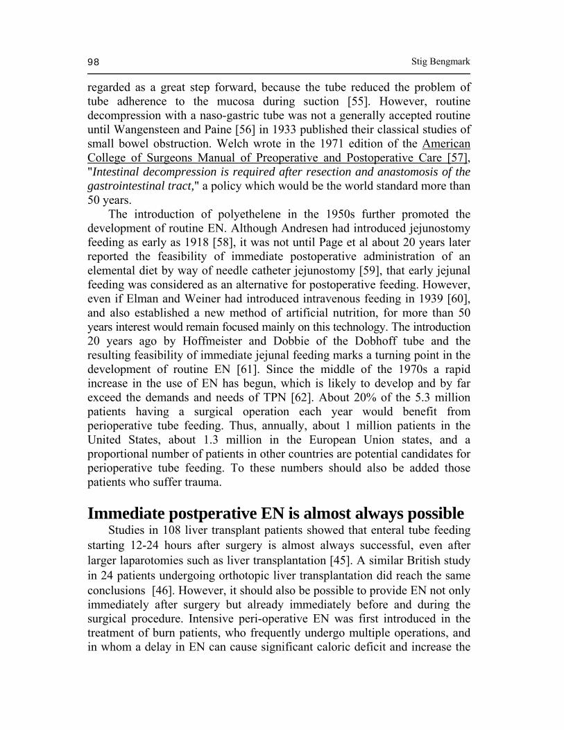
regarded as a great step forward, because the tube reduced the problem of tube adherence to the mucosa during suction [55]. However, routine decompression with a naso-gastric tube was not a generally accepted routine until Wangensteen and Paine [56] in 1933 published their classical studies of small bowel obstruction. Welch wrote in the 1971 edition of the American College of Surgeons Manual of Preoperative and Postoperative Care [57], "Intestinal decompression is required after resection and anastomosis of the gastrointestinal tract," a policy which would be the world standard more than 50 years. The introduction of polyethelene in the 1950s further promoted the development of routine EN. Although Andresen had introduced jejunostomy feeding as early as 1918 [58], it was not until Page et al about 20 years later reported the feasibility of immediate postoperative administration of an elemental diet by way of needle catheter jejunostomy [59], that early jejunal feeding was considered as an alternative for postoperative feeding. However, even if Elman and Weiner had introduced intravenous feeding in 1939 [60], and also established a new method of artificial nutrition, for more than 50 years interest would remain focused mainly on this technology. The introduction 20 years ago by Hoffmeister and Dobbie of the Dobhoff tube and the resulting feasibility of immediate jejunal feeding marks a turning point in the development of routine EN [61]. Since the middle of the 1970s a rapid increase in the use of EN has begun, which is likely to develop and by far exceed the demands and needs of TPN [62]. About 20% of the 5.3 million patients having a surgical operation each year would benefit from perioperative tube feeding. Thus, annually, about 1 million patients in the United States, about 1.3 million in the European Union states, and a proportional number of patients in other countries are potential candidates for perioperative tube feeding. To these numbers should also be added those patients who suffer trauma. Immediate postperative EN is almost always possible Studies in 108 liver transplant patients showed that enteral tube feeding starting 12-24 hours after surgery is almost always successful, even after larger laparotomies such as liver transplantation [45]. A similar British study in 24 patients undergoing orthotopic liver transplantation did reach the same conclusions [46]. However, it should also be possible to provide EN not only immediately after surgery but already immediately before and during the surgical procedure. Intensive peri-operative EN was first introduced in the treatment of burn patients, who frequently undergo multiple operations, and in whom a delay in EN can cause significant caloric deficit and increase the

Clinical nutrition 99
risk of infections [47,48]. It is therefore highly desirable to abandon the traditional praxis to withheld enteral nutritional support until bowel sounds return and to try to develop a routine of EN immediately before, during, and immediately after surgery. Peri-operative enteral feeding seems to have been introduced by the group of Pruitt [47] as late as 1990. Although in their short report no details are given regarding the time and extent of peri-operative nutrition, they conclude from a study in 47 patients that EN during the peri-operative period is safe as long as the tube is maintained in the small bowel. A more recent controlled study [48] confirmed that EN is not only safe but also efficient when given throughout the operative procedure. In this study 40 patients received EN during 161 surgical procedures and 40 had enteral support as traditionally practiced withheld during 129 procedures. The unfed group showed a significant caloric deficit (P<0.006), increased incidence of wound infections (P<0.02), and a significant necessity of higher albumin supplementation (P<0.04) compared with the fed group. Other studies have shown that early or immediate EN not only makes the use of stress ulcer prophylaxis unnecessary but also results in lower incidences of hospital infections and shorter hospital stays. Furthermore and today more important than ever, the savings in costs are considerable: 1 week of EN did in 1995 costs only one fourth: about $560 (US), compared with $2.100 (US) for treatment of TPN [45]. Intensive peri-operative EN will thus reduce costs not only through a reduction of nutrition costs but also through the decreased use of drugs such as H2-blockers, proton inhibitors, and antibiotics and through reduced morbidity and shorter hospital stays. Feeding tube technology The main obstacle to worldwide routine use of EN during the peri-operative period has so far been the lack of simple and reliable feeding tubes. A prerequisite for universal acceptance of intensive peri-operative EN as an aggressive peri-operative standard of feeding is the availability of feeding tubes that are easy to introduce into the jejunum, that will not obstruct the GI tract, and that not will be regurgitate prematurely. Under ideal circumstances, such tubes should within minutes or a few hours be able to propel themselves into the upper jejunum, without support of expensive techniques such as radiology or endoscopy. Whatley et al [63] were the first to prospectively study the rate and time course of spontaneous duodenal intubation after nasal insertion of three different weighted feeding tubes: Dobbhoff (Biosearch), Entriflex (Biosearch), and Duo-Tube (Argyle/Sherwood). After 4 hours, 8/20 (40%), 13/20 (65%),

Stig Bengmark 100
and 2/20 (10%), respectively had passed the pylorus. In total the tip of 32/60 tubes (53%) had passed the pylorus after 48 and 72 hours and 35/60 (58%) after 96 hours. Five years later, Levenson et al [64] in a similar study compared a weighted tube, Vitafeed (Health Tek/Kendall-McGaw), with a tube similarly constructed but without weights. After 4 hours the tip of 23/84 (27%) and 20/90 tubes (22%), respectively, had passed the pylorus. After 24 hours, 30/84 (36%) and 31/90 (34%) respectively, had passed the pylorus after 48 hours, 33/84 (39%), and 36/90 (40%), and after 72 hours, 35/84 (42%) and 37/90 (41%). Another similar study also published in 1988 compared three new naso-doudenal feeding tubes (Corpak): a bullet un-weighted, a bullet weighted, and a bolus weighted [65]. After 24 hours, the tip of 7/22 (32%), 7/26 (27%) and 9/21 tubes (43%), respectively had passed into the duodenum. Similar to the results of the previous studies, only one-third of the tubes spontaneously passed the pylorus. In addition this study showed no advantage to use of weighted tubes. Once through the pylorus, the un-weighted tubes stayed in position significantly longer than did the weighted ones (P<0.005). However, the spontaneous trans-pyloric passage (STP) is unacceptably low for all tubes currently on the market. This constitutes the main obstacle to the technique receiving acceptance as a standard feeding method during routine elective surgery. A generation of specialized feeding tubes Naso-enteric tube feeding is especially warranted in critically ill patients. However, in most of these patients a GI motility is no longer intact and the spontaneous transpyloric passage of feeding tubes, which is dependent on motility, can be expected to be low. In a recent study in Intensive Care Unit (ICU) patients requiring assisted ventilation, transpyloric placement was successful in only 10/68 patients (15%) [66]. The results of this study clearly shows that at present one cannot rely on STP in ICU patients. For these patients other solutions must be sought. The situation is different in the elective surgical patient, who has intact GI motility that can be used to transport the tip of the tube into the upper jejunum. It is likely that weights in the tip counteract this purpose, because the outlet of the stomach is not at its lowest part. Instead lightweight tips with the ability to maximally absorb and use the motility of the stomach have the best potential to be transported through the pylorus and down into the upper jejunum. One such tube is the "self-propelling" Flo-care tube (Nutricia), which was recently introduced on the European market. To maximally absorb motility, the tip of the tube includes a coil in its end. The purpose of this tube is to be introduced the evening before surgery so that it is in place after four
Clinical nutrition 101
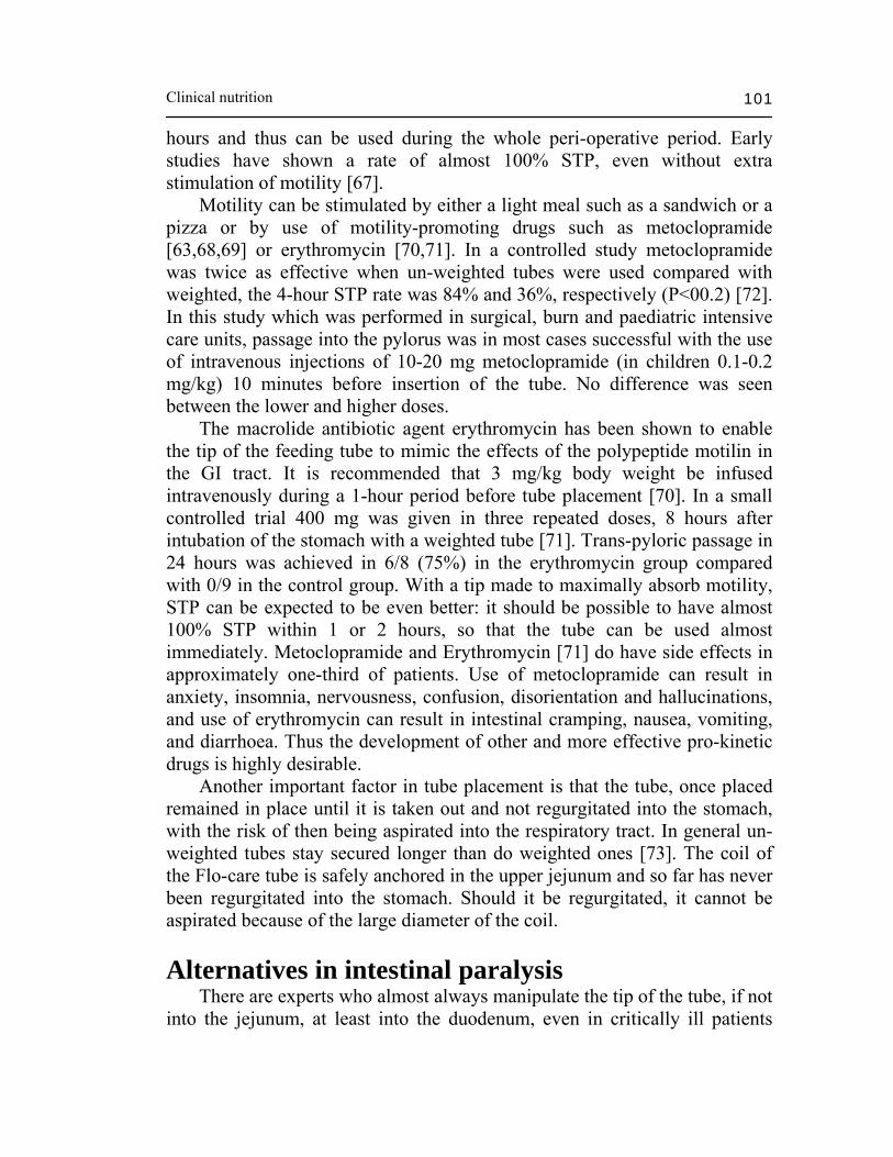
hours and thus can be used during the whole peri-operative period. Early studies have shown a rate of almost 100% STP, even without extra stimulation of motility [67]. Motility can be stimulated by either a light meal such as a sandwich or a pizza or by use of motility-promoting drugs such as metoclopramide [63,68,69] or erythromycin [70,71]. In a controlled study metoclopramide was twice as effective when un-weighted tubes were used compared with weighted, the 4-hour STP rate was 84% and 36%, respectively (P<00.2) [72]. In this study which was performed in surgical, burn and paediatric intensive care units, passage into the pylorus was in most cases successful with the use of intravenous injections of 10-20 mg metoclopramide (in children 0.1-0.2 mg/kg) 10 minutes before insertion of the tube. No difference was seen between the lower and higher doses. The macrolide antibiotic agent erythromycin has been shown to enable the tip of the feeding tube to mimic the effects of the polypeptide motilin in the GI tract. It is recommended that 3 mg/kg body weight be infused intravenously during a 1-hour period before tube placement [70]. In a small controlled trial 400 mg was given in three repeated doses, 8 hours after intubation of the stomach with a weighted tube [71]. Trans-pyloric passage in 24 hours was achieved in 6/8 (75%) in the erythromycin group compared with 0/9 in the control group. With a tip made to maximally absorb motility, STP can be expected to be even better: it should be possible to have almost 100% STP within 1 or 2 hours, so that the tube can be used almost immediately. Metoclopramide and Erythromycin [71] do have side effects in approximately one-third of patients. Use of metoclopramide can result in anxiety, insomnia, nervousness, confusion, disorientation and hallucinations, and use of erythromycin can result in intestinal cramping, nausea, vomiting, and diarrhoea. Thus the development of other and more effective pro-kinetic drugs is highly desirable. Another important factor in tube placement is that the tube, once placed remained in place until it is taken out and not regurgitated into the stomach, with the risk of then being aspirated into the respiratory tract. In general un-weighted tubes stay secured longer than do weighted ones [73]. The coil of the Flo-care tube is safely anchored in the upper jejunum and so far has never been regurgitated into the stomach. Should it be regurgitated, it cannot be aspirated because of the large diameter of the coil. Alternatives in intestinal paralysis There are experts who almost always manipulate the tip of the tube, if not into the jejunum, at least into the duodenum, even in critically ill patients
Stig Bengmark 102
without gastric motility. Ugo [74] could, using specific patient positioning, insertion techniques, and gastric distension with air, manipulate the tip behind the pylorus in 85/103 patients (83%), within an average time of 30 minutes. Schultz et al [75], using right lateral decubitus positioning plus gastric insufflation of air, reported an immediate success rate of placement in 37/42 patients (88%) and Zaloga [76] reported successful placement in 213/231 patients (92%) within an average time of 40 + 14 minutes, placement being confirmed by bile aspiration, determination of pH change from acidic to basic, and blue dye injection. A 30o bend is placed 3 cm from the distal tip (6.5 cm from the end of the tube) on either Entriflex (Biosearch) or Corsafe (Corpak) and a syringe was used to slowly inject air and rotate the tube down. With this method the tip of the tube will reach the proximal jejunum in 17% of cases: in the remaining 83%, the tip is placed somewhere in the duodenum, most often in the lower part. Although more expensive, fluoroscopic and endoscopic placements are generally faster: Ott et al [77] reported average time of 8.6 and 13.4 minutes respectively for the two techniques. In this study, fluoroscopic placement was successful in 94/104 cases (90%) and endoscopic placement was successful in all cases tried (100%), even in those for which fluoroscopic placement had failed. Mathus-Vliegen et al [78] showed that it is possible to place tubes endoscopically within an average of 13 minutes. Thus, all the above semi-invasive techniques (expert manipulation, fluoroscopy, and endoscopy) seem to give satisfactory results. It seems logical then that each medical centre use the technique that its staff perform best. Endoscopy may be the most favourable method in patients with inhibited GI motility, however, because expert manipulation can take long time, which might put the patient in unnecessary discomfort. Endoscopy, although expensive, is quick and almost always reliable. Today it can also be performed by through-the nose gastroscopy [78,79]. Using a paediatric gastroscope and local anaesthesia to the nose, the tube was successfully placed in 82/92 patients (89%), in 10/92 patients (11%) the turbinates were bilaterally too narrow [80]. For the introduction of PEGs and PEJs, alternative techniques such as ultrasound guidance [81] and laparoscopy[82] have been suggested as effective tools. Oral, gastric, or enteral feeding? Whenever possible, oral feeding is best. There are published reports of oral feeding, if not perioperatively or immediate after surgery, at least early after larger operations such as colonic resection [83]. The only indication for short term, for example peri-operative enteral tube feeding is lack of gastric motility and risk of vomiting and aspiration. Today, effective intensive
Clinical nutrition 103
peri-operative EN can be achieved only with enteral tube feeding. Indications for long-term enteral tube feeding are most often much different, for example, consciousness, paralysis of the swallowing mechanisms, or oesophageal obstructions. In most of these conditions in which the stomach can be expected to have normal motility, both gastric and enteral feeding exists as alternative. It is unfortunate that most of the reports on tube feeding in the literature are based on mixed use of short- and long-term, acute and chronic tube feeding. The advantages and disadvantages of gastric and jejunal tube feeding are summarized by Bochus [84]. Montecalvo et al [85] have compared gastric and jejunal tube feeding in a small, but well performed, controlled study in medical and surgical ICU patients. The mean duration of feeding was about 10 days for the two groups. In these patients, to a large extent as a result of either trauma or cardio-respiratory failures, jejunal feeding led to a significantly higher caloric intake (P=0.05), a greater increase in serum pre-albumin concentrations (P<0.005), and a lower rate of pneumonia compared with gastric tube feeding. The incidence of diarrhoea was low in both groups. Although the jejunal group had more days of diarrhoea (mean: 3.3 + 6.6 compared with 1.8 + 2.9 days), this difference was not significant. Furthermore, the ICU nurses reported that the patients fed with gastric tube were more likely to develop gastro-esophageal reflux (67.8%) and pulmonary aspiration (77%). Consequently, nurses in general favoured jejunal tube feeding compared with gastric (71.3% compared with 18.4%; 10.3% had no opinion on the subject). In a review of the literature [86] during the period from 1978 to 1989 only 2/45 papers compared the incidence of aspiration in gastric and jejunal feedings: both [87,88] clearly favoured jejunal feeding over gastric feeding in prevention of aspiration pneumonia. Salivation - Important for outcome Eating stimulates salivation and GI secretion. The 1-2 L of saliva normally secreted each day are extremely rich in infection-preventing and healing-inducing factors [89]. As Mandel [90] pointed out; "the saliva possesses a multiplicity of defence systems for antibacterial warfare that Pentagon can envy". The normal acidity of the stomach is also important to prevent colonization of the stomach with gram-negative bacilli, because pneumonia is likely to develop from a gastric reservoir of colonizing bacilli [90-93]. Even during short-term peri-operative enteral tube feeding, patients should intermittently chew something to stimulate salivation and the acidity of the stomach should be allowed to intermittently be low, thereby sterilizing the stomach[94]. Another reason for allowing an intermittently low stomach
Stig Bengmark 104
pH is that digestion and absorption are less interfered with if pepsin is inactivated and proteolysis not compromised [95]. This is why it is recommended that gastric feeding be given intermittently when indicated, in contrast with continuous jejunal tube feeding [94]. On the other hand, continuous naso-gastric feeding has been reported to effectively prevent stress ulceration [96]. It has also been observed that intermittent gastric tube feeding can lead to a significant deficit in caloric intake [97]. Another advantage of continuous naso-enteral feeding is that it counteracts the "hyperbilirubinemia" [98], known to occur in enteral fasting, especially in patients with Gilbert´s syndrome and Crigler-Najjar type 2 disease [99]. Our present knowledge is in many respects incomplete and further studies are needed. Complications associated with tube feeding Although the list of reported and potential complications to naso-enteric tube feeding is long, it does become shorter as surgeons gain experience. In one studied group of ICU patients [66], complications were observed in 10/68 patients (15%), of which 4 were tube dislodgements and only 1 was a more serious aspiration pneumonia. The complication rate, with peri-operative enteral tube feeding, should become much lower soon, especially if tube dislodgement problems can be minimized. It should also be remembered that the alternative, PN, is not only 10 fold more expensive but also fraught with its own potential complications. There are currently no clinical reports of the use of solely short-term peri-operative EN. In patients receiving this form of nutrition, unintentional removal of the tube and tube clogging are the most frequent complications interrupting feeding. Most of the other complications, such as severe diarrhoea, protracted vomiting, and gastric distension "may provide special challenges to tube feeding, but are not necessarily contraindications" [41]. The diarrhoea frequently observed is less often caused by infection and more often due to the content of sorbital in the formulas given[100], too high a content of fat [101] or the use of histamine H2-receptor antagonists [102-104], antacids (especially magnesium-containing preparations [95,105,106], and antibiotics [107-113]. For further review, see Gottschlich et al [101]. To prevent antibiotic-induced diarrhoea, supplementation with lactobacilli is increasingly recommended [15-19, 84]. The British Dietetic Association advises [after 78] that for hygienic reasons tubes be exchanged once every 2 weeks. The scientific evidence for this recommendation is not known. However, it is reasonable to request that enteral feeding tubes be made to stay in place and continue to function, if needed, for at least 2 weeks. This practice is unfortunately not followed today.
Clinical nutrition 105
Unintentional tube removal Unintentional removal, spontaneous migration of the tip of the tube back to the stomach, and clogging are all still frequent complications of the tube feeding. In the study conducted by Mathus-Vliegen [78], premature removal of the tube occurred in 29/46 patients (63%), patients being the cause in 16 (35%) and staff in 5 (11%): there was no explanation for tube removal in 7 cases. Similar experiences were reported by Benson et al [114], patients were the cause in 16/50 cases (32%), and staff in 9/50 (18%). In a study to be published by Silk et al [115] non-elective tube removal occurred in most patients in whom the following brands of tubes were used: Radius (Radius International, Grayslake, IL.), 80%, Flexiflo (Ross Products, Columbus, OH), 73.3%, and Freka (Fresenius Ltd, Runcorn Cheshire, UK), 73.3%. Mathus-Vliegen et al [78] observed that the unintentional removal rate was higher if shorter tubes (105 cm) were used compared with longer ones (145 cm), which could be positioned significantly deeper into the intestine. Because there are no negative consequences with use of longer feeding tubes, these tubes should probably be recommended. A slight anchoring of the tip of the tube in the duodenum, for example, as provided by the Flo-care tube, is highly desirable. The high rate of unintentional removal also calls for improved methods for taping the tube to the patient's nose. Burns et al [73] compared 3 securing methods available on the market: pink tape (Plastic Adhesive Tape, Surgical Product Corp), clear tape (Bioclusive, Johnson and Johnson) and a special tube-attachment device (Hollister Inc). The mean time until failure was 100, 56, and 30 hours respectively. As a consequence, these authors recommended that the tape be exchanged every day or every second day. It is cheap to replace tape, but costly, time-consuming, and unpleasant for the patient to replace a naso-gastric tube. Clogging - A remaining problem Clogging remains an obstacle to the routine use of enteral feeding tubes, especially when tubes are used for periods longer than 1 week [114], a length of time that fortunately is rarely needed after most surgical operations. The time that the three brands of enteral tubes studied by Silk et al [115] remained in situ was as follows: Radius, 6.0 + 1.0, Flexiflo, 5.3 + 0.8, and Freka, 4.4 + 0.7 days respectively. Thus, the tubes presently on the market are suitable mainly for short-term use. Mechanical tube problems or clogging occurred in 11% of the patients, no difference was observed among the three types of tubes tried. In a similar study by Mathus-Vliegen [78], in which tubes made of polyurethane with open-ended tips and side holes were used, the tubes
Stig Bengmark 106
remained in situ about 50% longer (about 10 days) but the clogging frequency was also almost twice as high; 11/46 (24%), clogging was transient in 7/46 cases(15%). Clogging could be corrected by stretching the tube (2 episodes) or by flushing or dissolving of the clot (5 episodes), but in 4/46 cases (9%), clogging necessitated exchange of the tube. Many factors contribute to early luminal occlusion; pump malfunction [116], high formula viscosity [116], medications [117], or failure to properly irrigate the tube whenever feeding is interrupted [116]. The type of tube, its construction, and the material used may influence the clogging rate, but solid data to support such an assumption are scarce. Gastric tubes have a greater tendency to become clogged compared with enteral tubes, because certain protein formulas clog instantly when mixed with gastric juice (pH 1.5) but not with jejunal aspirate (pH 7.6) [118]. The clotting occurs at a pH of 5, when intact protein formulas are acidified, but not with formulas containing only free amino acids and small peptides. Although prophylaxis by frequent irrigations of the feeding tube is the single most important measure to prevent clogging, attempts should also be made to develop effective methods to unclog occluded tubes. Knowledge about the role of clogged proteins made Marcuard et al [116] try to unclog tubes by using different proteolytic enzymes. In a controlled in vitro study performed with pre-clotted 8 F feeding tubes, the effects of papain (Adolf´s Meat Tenderizer): a mixture of trypsin, chymotrypsin, amylase and lipase (Viokase), Sprite, Pepsi, Coca Cola, and Mountain Dew were compared. The solution (5 ml) was injected into the tube, which was clamped for 5 min. Viokase adjusted by 1 mol NaOHl/L to pH 7.9 was significantly more effective than the other solutions, however, plain, non-adjusted Viokase (pH 5.9) was ineffective. In a small subsequent clinical study all clogged tubes could be cleared except those in which the clogging was due to knotted tubes, impacted tablets, or the duration of the clogging (>24 hours). A practical way to produce a Viokase pH 7.9 solution is to crush one tablet of Viokase and one tablet of sodium bicarbonate and then add 5 mL (one teaspoon) of water. A potential reservoir for pathogens Numerous studies have adressed the bacterial contamination of enteral feeding preparations but few have evaluated in vivo the microbial colonization of the feeding tube. (For review see Bussy et al. [119]). Colonization of the rinsing solutions, kept overnight in the tube, was studied in 31 consecutive patients. All proved to be contaminated and 102 strains were cultured with a median density of 106 colony-forming units/mL (range:

Clinical nutrition 107
10 to 1010). Of the strains detected 48/102 (47%) were Enterobacteriaceae, 20/102 (20%) were group D streptococci, 9/102 (9%) were Candida albicans, 9/102 (9%) were Pseudomonas aeruginosa, and 16 were other species [119]. Multiple antibiotic resistances were present in about 10% (12/102) of the strains and lower resistance in another 25%. Throat and faeces were frequently colonized by the same bacteria as the predominant bacteria cultured from the tube-rinsing solution. This study was performed in patients with naso-gastric feeding tubes only. Although there are no studies to support this theory, it is reasonable to assume that contamination may be lower with jejunal intubation, where there is no direct communication with the stomach, known to be a reservoir of pathogens in sick patients. If proven, this could eventually be an argument in favour of enteral feeding. It can also be assumed, without supporting data from the literature, that continuous day and night enteral feeding, in addition to frequent flushing of the tube, would reduce the rate of tube colonization. Use of sterile or filtered water instead of tap water for rinsing should be considered and it should also be remembered that mineral waters are not free from pathogens. Nosocomial legionellosis associated with naso-gastric feeding and tap water was recently reported [120]. Pseudomonad organisms were identified in 6/8 American [121] and 39/87 European [122] brands of bottled water. Although outbreaks of infections from mineral water are rare, the risk should be considered for very sick individuals. Hospital malnutrition needs further attention It is well known, that nutritional support can effectively address the problem of malnutrition, improve clinical outcome, reduce suffering, and reduce the costs of health care. Malnutrition occurs even today at the best hospitals of the world [123,124], 40-55% of patients are either malnourished or at risk of malnutrition: for review see article by Gallagher-Allred et al [124]. About 12% of patients are severely malnourished [10]. Surgical patients with a likelihood of malnutrition are three times more likely to have complications and excess mortality, they also have substantially longer hospital stays, and their hospital charges may be increase by 35-75% [124]. Effective programs to counteract malnutrition and reduce health expenditure are more important than ever. Governments, health maintenance organizations and insurance companies, doctors and nurses are increasingly aware of the importance of nutrition [125]. An important part in such programs is intensive peri-operative EN for which jejunal tube feeding, from the evidence available, is an important instrument. The old principles of preoperative abstinence from feeding and the "wait for flatus" principle for postoperative

Stig Bengmark 108
nutrition should give way to effective and continuous peri-operative nutrition. As emphasized by Baskin [126], the old adage "if the gut works, use it" should in light of present knowledge be updated to "use it, or lose it." Most of the formulas available on the market contain 1-2 kcal/mL and have an osmolarity of around 350 mosm/kg water. The amounts needed to satisfy a patient's daily energy daily needs is thus considerable, 50-100 mL/hour depending on the concentration of the solution and mode of supply (continuous or interrupted). Although it is probably not necessary even after larger operations, it is interesting that as much as 300 kcal/hour can be successfully administrered by postpyloric infusion, starting immediately after transfer of the patient to the recovery room [127]. Studies in healthy volunteers have shown a tolerance of starter flow rates up to 150 mL/hour and starter osmolarities up to 690 mosm/kg [128]. It is usually recommended that immediately after the operation 50 mL/hour be given and the volume carefully increased during the subsequent 24 hours. The necessary instrument for effective intensive peri-operative EN is available today although not always used. The reason is most likely lack of information and low interest among general practitioners. Enteral nutrition is becoming a speciality of its own and successful treatment is much dependent on the interest and skill of those responsible. Although universally recognized nutrition support teams are extremely effective, only a minority of hospitals, even in the Western world, have access to such specialists. As long as EN is left to the generalist and his or her own interest in and knowledge of the potential benefits of effective EN support, it will be available only at random to patients. Inflammation-induced impaired GI motility and secretion Alterations in motility, secretion and visceral sensation are of hallmarks increased systemic inflammation and closely associated of the acute phase response. Activation of immune cells may lead to changes in motor-sensory function in the gut. The stress-induced T helper 2 type immune response is critical in producing alterations in intestinal muscle function in both mental and physical stress and strongly associated with impaired host defense mechanisms. Recent data suggest that stress induced alterations in gastrointestinal inflammation may be mediated through changes in hypothalamic-pituitary-adrenal (HPA) axis function and alterations in bacterial-mucosal interactions, and via mucosal mast cells and mediators such as corticotrophin releasing factor (CRF) [129]. The systemic level of pre-morbid inflammation seems to increase with age both in humans and animals [130], resulting in an augmented sensitivity to inflammation-induced complications including

Clinical nutrition 109
infections and impairment of GI functions including impaired motility and secretion. Unfortunately, various treatments including modern enteral nutrition with industry-produced formulas do dramatically increase systemic inflammation, impair barrier function, suppress intestinal immunity and promote bacterial translocation [131-134]. The extent of inflammation is also significantly aggravated by mechanical therapeutic efforts such as handling of the bowels during operation [135], and ventilation of the lungs [136]. Both choice of type of anaesthesia and ventilation as well as surgery can influence outcome by control of inflammation. Laparoscopic surgery is generally regarded as associated with less systemic inflammation and impairment of motility and secretion than corresponding open procedure [137]. Manipulation of the intestines during surgery increases inflammation and dys-motility [138]. Early recovery of bowel function and low concentration of inflammatory cytokines suggest that the extra-peritoneal approach to the abdominal aorta in aortic surgery is less stressful than the trans-peritoneal approach [139]. Antecolic reconstruction for duodeno-jejunostomy during pylorus-preserving pancreat-doudenectomy is compared to retrocolic reconstruction reported to reduce the postoperative period of delayed gastric emptying, decrease postoperative morbidity and length of hospital stay [140]. Probiotics and antioxidants of importance Overgrowth of the potentially pathogenic enteral flora is a common consequence of impaired intestinal motility. But it is also so that disturbances in this flora play a role in the pathogenesis of mucosal inflammation, which in turn aggravates the intestinal dys-motility. Clearly, it is of great importance to explore the mechanism of alteration in motor function in normal and inflamed intestine. It was already in 1976 suggested that products derived from microbial fermentation in large intestine might promote colonic propulsion [141]. Subsequent animal studies, especially those performed in germ-free animals suggest different effects in different parts of the gastrointestinal tract, and especially different effects by different lactic acid bacteria. As an example, Lactobacillus acidophilus and Bifidobacterium bifidum are reported to reduce the migrating myoelectric complex (MMC) period and accelerated small intestinal transit (P < 0.05). while Micrococcus luteus and Escherichia coli are reported to have the opposite effects [142]. L. paracasei subsp. paracasei proved to be the most potent inducer of Th1 cytokines and potently repressed Th2 cytokines when many LAB organisms were compared with each other [143]. Lactobacillus paracasei, was also in contrast to Lactobacillus johnsonii,Bifidobacterium lactis, and Bifidobacterium longum, shown to attenuate smooth muscle contractility,
Stig Bengmark 110
and reduce T-helper 2 response as well as the levels of transforming growthfactor-beta1, cyclooxygenase-2, and prostaglandin E 2 levels in smooth muscle tissues [144]. Accelerated heme metabolism has been shown to be involved in anti-inflammatory and antioxidant processes in a number of models of acute and chronic inflammation including in the lungs [145] and the gastrointestinal tract [146]. New evidence suggests that the effects are mediated by simultaneous production of carbon monoxide [147]. Subsequent studies demonstrate that inhaling low dose of carbon monoxide [148-150] as well as infusion of a single intraperitoneal small dose carbon monoxide dissolved in Ringer´s lactate immediately after the surgical procedure [151] in experimental animals ameliorates the inflammatory response in the gut and the subsequent postoperative ileus induced by the surgical manipulation. Various COX-2 inhibitors [152] as well as antioxidants [153] have also been suggested to inhibit postoperative intestinal paralysis. A recent study demonstrates significant and dose-dependent inhibition of manipulation-induced intestinal paralysis and reduced gastrointestinal transit time by administration of Cox-2 inhibitors such as L-N(6)-(1-iminoethyl)lysine hydrochloride, (L-NIL), as well as N(omega)-nitro-L-arginine methyl ester (L-NAME) [152]. Various antioxidants can also be expected to promote GI motility as well as GI secretion. However, this far only a few antioxidants have been tried. When tried, the red wine and peanut antioxidant resveratrol does reverse the inhibitory effects on motility induced by laparotomy and manipulation of the intestines [154]. Among those COX-2 inhibitors and natural antioxidants worth trying should be substances such as the main active substance in green tea, epigallocatechin-3-gallate (EGCG) as well as in turmeric, curcumin, proven to not only be a strong and atoxic antioxidant but also a strong natural COX-2 inhibitor [155]. References 1. Takamizawa, K., Ivamori, M., Mutai, M. and Nagai, Y. 1986, Biochem. Biophys.
Acta 879,73. 2. Heitz, P.U., Kasper, M., Van Nordan, S., Polak, J.M., Gregory, H., Pearse, A.G.
1978, GUT 19,408. 3. Bardócz, S. 1995, Trends Food Sci. Trends Food Sci Technol 6,341 4. Edwards, C.A., Rowlands, I.R. 1992, Dietary Fiber - A Component of Food.
Schweizer, T.F., Edwards, C.A. (Eds.), Springer Verlag, Berlin, 119. -136,1992 5. Alverdy, J.C., Spitz J, Hecht, G., Ghandi, S. 1994, New Horizons 2,264. 6. Swedish Adverse Drug Reactions Advisory Committee. SEDRAC. 2006.
Clinical nutrition 111
7. Rice Evans, C.A., Diplock, A.T. 1993, Free Radic. Biol. Med. 15,77. 8. Schiller, H.J., Reilly, P.M., Bulkley, G.B. 1993, Crit. Care Med. 21,S92. 9. Felty, A.R., Keefer, C.S. 1924, JAMA 82,1430. 10. Detsky, A.S., Baker, J.P., O´Rourke, K. , Goel, V. 1987, Ann. Intern. Med. 107,195. 11. Mulholland, J.H., Tui, C., Wright, A.M., Vinci, V.J. 1943, Ann. Surg. 117,512. 12. Riegel, C., Koop, C.E., Drew, J., Stevens, L.W., Rhoads, J.E., Bullitt, L., Barrus,
D., Grigger, R.P., Barnes, M., Barnhart, A., Boger, J., Bowen, F., Goulding, E., McGinley, E. 1947, J Clin Invest 26,18.
13. Moore, F.A., Feliciano, D.F., Andrassy, J.R., McArdle, A.H., Booth, F.V., Morgenstein-Wagner, T.B., Kellum Jr, J.M., Welling, R.E., Moore, E.E. 1991, Ann Surg 216,172.
14. Kudsk ,K.A., Croce, M.A., Fabian, T.C., Minard, G., Tolley, E.A., Poret, H.A., Kuhl, M.R., Brown, R.O. 1992, Ann. Surg. 215,503.
15. Bengmark, S., Jeppsson, B. 1995, J. Parenter. Enteral Nutr. JPEN 19,410. 16. Bengmark, S., Larsson, K., Molin, G. 1995, Biotechnol. Therapeut. 5,171. 17. Bengmark, S. 1995, Anaesthesia, Pain, Intensive Care and Emergency Medicine -
A.P.I.C.E. Proc. 10th Postgraduate Course in Critical Care Medicine. A Gullo (Ed.) Trieste, 73.
18. Bengmark, S. 1996, Clin. Nutr. 15,1. 19. Bengmark, S.2005, Gastroenterol. Clin. North America 34,413. 20. Bengmark, S. 2006, Anaesthesiol. Clin. North America 24,299. 21. Sagrada, A., Fargeas, M.J., Bueno, L. 1987, Gut 28,955. 22. Appert, H.E., Howard, J.M. 1972, Fed. Proc. 31,828. 23. Dubois, A., Weise, V.K., Kopin, U. 1973, Ann. Surg. 178,781. 24. Dauchel, J., Schang, J.C., Kacheloffer, J., Eloy, R., Grenier, J.F. 1976, Digestion
14,293. 25. Sjöquist, A., Hallerbäck, B., Glise, H. 1985, 1985, Dig. Dis. Sci. 30,749. 26. Glise, H., Lindahl, B.-O., Abrahamsson, H. 1980, Scand. J. Gastroenterol.
15,673. 27. Gluckman, D.L., Halser, M.H., Warren, W.D. 1966, Surgery 60,1020. 28. Moore, E.E., Jones, T.N. 1983, J. Am. Coll. Nutr. 2,45. 29. Savassi-Rocha, P.R., Conceicao, S.A., Ferreira, J.T., Diniz, M.T., Campos, I.C.,
Fernandes, V.A., Garavini, D., Castro, L.P. 1992, Surg. Gynecol. Obstet. 174,317.
30. Gerber, A., Robert, F.A.Q., Smith, L.L. 1958 Surg. Gynecol. Obstet. 107,247. 31. Gerber, A. 1963, Surg. Gynecol. Obstet. 117,294. 32. Farris, J.M., Smith, K 1956, Ann. Surg. 144,475. 33. Mehnert, I.H., Brown, M.J., Woodward, B., Paetkeu, E., Donovan, P.B. 1959,
Surg. Gynecol. Obstet. 109,607. 34. Thomas, G.I., Metheny, D., Lundmark, V.O. 1961, Northwest. Med. 60,387. 35. Barnes, A.D., Williams, J.A. 1967, Am. J. Surg. 113,494. 36. Burg, R., Geigle, C.F., Faso, J.M., Theuerkauf Jr, F.J. 1978, Dis. Colon Rectum
21,98. 37. Sitges-Serra, A., Cabrol, J., Gubern, J.M., Simó, J. 1980, Surg. Gynecol. Obstet.
158,557.

Stig Bengmark 112
38. Schwartz, C.I., Heyman, A.S., Rao, A.C. 1995, Southern Med. J. 88,825. 39. Petrelli, N.J., Stulc, J.P., Rodrigues-Bigas, M., Blumenson, L. 1993, Am. Surg:
59,632. 40. Cheatham, M.L., Chapman, W.C., Key, S.P., Sawyers, J.L. 1995, Ann. Surg.
221,469. 41. Kirby, D.F., Delegge, M.H., Fleming, C.R. 1995, Gastroenterology 108,1282. 42. Rombeau, J.L., Jacobs, D.O. 1984, Enteral and tube feeding. Rombeau JL,
Caldwell MD (Eds.). Saunders, Philadelphia, 261. 43. Norton, J.A., Ott, L.G., McClain, C., Adams, L., Dempsey, R.J., Haack, D.,
Tibbs, P.A., Young, A.B. 1988, J. Neurosurg. 68,62. 44. McClave, S.A., Snider, H.L., Lowen, C.C., McLaughlin, A.J., Greene, L.M.,
McCombs, R.J., Rodgers, L., Wright, R.A., Roy, T.M., Schumer, M.P. 1992, J. Parenter. Enteral Nutr. JPEN 16,99.
45. Pescovitz, M.D., Metha, P.L., Leapman, S.B., Milgrom, M.L., Jindal, R.M., Filo, R.S. 1995, Surgery 117,642.
46. Wicks, C., Somasundaram, S., Bjarnason, I., Menzies, I.S., Routley, D., Potter, D., Tan, K.C., Williams, R. 1994, Lancet 344,837.
47. Buescher TM, Cioffi WG, Becker WK 1990, Abstract, Proc. Am Burn Assoc. 22,192.
48. Jenkins, M.E., Gottschlich, M.M., Warden, G.D. 1994, J. Burn Care Rehab. 15,199. 49. Hometer, J.C. 1985, N.Y. Med. J. 62,819. 50. Forlaw, L., Chernoff, R., Guenter, P. 1990, Clinical Nutrition. Enteral and tube
feeding. Rombeau JL, Caldwell MD (Eds). Saunders, Philadelphia. 174. 51. Palmer, J.F. 1935, Complete works of John Hunter. Haswell, Barrington, Haswell
Inc. Philadelphia, 622. 52. Busch, W. 1858, Virchows Arch. Cell Pathol. 14,140. 53. Levin, A.L. 1921, JAMA 76,1007. 54. Abbott, W.O., Rawson, A.J. 1937, JAMA 108,1873. 55. Argyle Division of Sherwood Medical. 1979, Clinical considerations in the use of
the Argyle Salem Sump Tube. St Louis MO,1. 56. Wangensteen, O.H., Paine, J.R. 1933, JAMA 101,1532. 57. Welch, C.E. American College of Surgeons Manuel of Preoperative and
Postoperative Care. Whinney, J.M. (Ed.) Saunders, Philadelphia. 58. Andresen, A.F.R. 1918, Ann. Surg. 67,565. 59. Page, C.P., Carlton, P.K., Andrassy, R.J., Feldtman, R.W., Shield, C.F. 1979,
Am. J. Surg. 138,934. 60. Elman, R., Weiner, D.O. 1939, JAMA 112,796. 61. Hoffmeister, J.A., Dobbie, R.P. 1977, Am. Surg. 43,6. 62. Shaffer, J.L.1989, Ann. Chir. Gynecol. 78,257. 63. Whatley, K., Turner, W.W., Dey, M., Leonard, J., Guthrie,M. 1984, J. Parenter.
Enteral Nutr. JPEN8, 679. 64. Levenson, R., Turner, W.W., Dyson, A., Zike, L., Reisch, J. 1988, J. Parenter.
Enteral Nutr. JPEN 12,135. 65. Rees, R.G.P., Payne-James, J.J., King, C., Silk, D.B. 1988, J. Parenter. Enteral
Nutr. JPEN 12,469.

Clinical nutrition 113
66. Marian, M., Rappaport, W., Cunningham, D., Thompson, C., Esser, M., William, F., Warneke, J., Hunter, G. 1993, Surg. Gynecol. Obstet. 176,475.
67. Jeppsson, B., Tranberg, K.-G., Bengmark, S. 1992, Clin. Nutr. 11,373. 68. Pirola, R.C. 1967, Am J Dig Dis 12:913-915,1967 69. Lobbezoo, M.W., Janszen, F.H., Tulp, M.T., Zwagemakers, J.M. 1985, Eur. J.
Pharmacol. 22,108. 70. Di Lorenzo, C., Lachman, R., Hyman, P.E. 1990, J. Pediatr. Gastroenterol. Nutr.
11,45. 71. Stern, M.A., Wolf, D.C.1994, Am. J. Gastroenterol. 89,2011. 72. Lord, L.M., Weiser-Maimone, A., Pulhamus, M., Sax, H.C. 1993, J. Parenter.
Enteral Nutr. JPEN 17,271. 73. Burns, S., Martin, M., Robbins, V., Friday, T., Coffindaffer, M., Burns, S.C.,
Burns, J.E. 1995, Am. J. Crit. Care 4,198. 74. Ugo, P.J., Mohler, P.A., Wilson, G.L. 1992, NCP 7:284-287,1992 75. Schultz, M.A., Santanello, S.A., Monk, J., Falcone, R.E. 1993, Int. Surg. 78,79. 76. Zaloga, G.P. 1991, Chest 100,1643. 77. Ott, D.J., Mattox, H.E, Gelfand, D.W., Chen, M.Y., Wu, W.C. 1991, Am. J.
Roentgenol. AJR 157,769. 78. Mathus-Vliegen, E.M.H., Tytgat, G.N., Merkus, M.P. 1993, Gastrointest.
Endosc. 39,537. 79. Fregonesi, D., Di Falco G. Through-the-nose gastroscopy. Am J Gastroenterol
86:381,1991. Letter to the Editor. 80. Fregonesi D, Di Falco, G. 1993, Endoscopy 25,539. 81. Pugash, R.A., Brady, A.P., Isaacson, S. 1995, Can. Assoc. Radiol. J. 46,196. 82. Albrink, M.H., Foster, J., Rosemurgy, A.S., Carey, L.C. 1992, Surg. Endosc.
6,259. 83. Moiniche, S., Bülow, S., Hesselfeldt, P., Hestbaek, A., Kehlet, H. 1995, Eur.J.
Surg. 161,283. 84. Bochus, S. 1993, Nursing J. 34. 85. Montecalvo, M.A., Steger, K.A., Farber, H.W. Smith, B.F., Dennis, R.C.,
Fitzpatrick, G.F., Pollack, S.D., Korsberg, T.Z. Birkett, D.H., Hirsch, E.F. 1992, Crit. Care Med. 20,1377.
86. Lazarus, B.A., Murphy, J.B., Culpepper, L. 1990, Arch. Phys. Med. Rehabil. 71,46.
87. Burth, G.D., Shatney, C.H. 1987, Am. Surg. 53,54. 88. Ho, C.S., Yee, A.C.N., McPherson, R. 1988, Gastroenterology 95,206. 89. Sreebny, L.M., Banoczy, J., Edgar, W.M. 1992, Int. Dent. 42,291. 90. Mandel, I.D.1987, J. Den.t Res. 66,623. 91. Atherton, S.T., White, D.J. 1978, Lancet 1,2968. 92. Heyland, D., Mandell, L.A. 1992, Chest 101,187. 93. Toung, T.J.K., Rosenfeld, B.A., Yoshiki, A., Grayson, R.F., Traystman, R.J.
1993, Crit. Care Med. 21,1359. 94. Bonten, M.J.M., Gaillard, C.A., van Thiel, F.H., van der Geest, S., Stobberingh,
E.E. 1994, Crit. Care Med. 22,939. 95. Morrissey, J.F., Barreras, R.F. 1974, N. Engl. J. Med. 290,550.
Stig Bengmark 114
96. Solem, L.D., Strate, R.G., Fischer, R.P. 1979, Surg. Gyn. Obstet. 148,267. 97. Ciocon, J.O., Galindo-Ciocon, D.J., Tiessen, C., Galindo, D. J. 1992, Parenter.
Enteral Nutr. JPEN 16,525. 98. Roongpisuthipong, C., Heymsfield, S.B., Casper, K., Hill, J.O. 1987, J. Parenter.
Enteral Nutr. JPEN 11,544. 99. Whitmer, D.I., Gollan, J.L. 1981, Semin. Liver Dis. 3,42. 100. Edes, T.E., Walk, B.E., Austin, J.L. 1990, Am. J.Med. 88,91. 101. Gottschlich, M.M., Warden, G.D., Michel, M.A., Havens, P., Kopcha, R.,
Jenkins, M., Alexander, J.W. 1988, J. Parenter. Enteral Nutr. JPEN 12,338. 102. Kirksey, T.D., Moncrief, J.A., Pruitt, B.A. Am. J. Surg. 116,627. 103. Ruddell, W.S.J., Losowsky, M.S. 1980, Br. Med. J. 281,273. 104. Kelly, T.W.J., Patrick, M.R., Hillman, K.M. 1983, Crit. Care Med. 11,7. 105. McAlhany, J.C., Czaja, A.J., Pruitt, P.A. 1976, J. Trauma 16,645. 106. Chernoff, R., Dean, J.A. 1980, J. Am. Diet Assoc. 76,161. 107. Jacobson, E.D., Faloon, W.W. 1961, JAMA 175,187. 108. Editorial. Br. Med. J. 1968, 4,402. 109. Wolfson, A.M., Saour, J.N., Ricketts, C.R., Pollard, B.J., Hardy, S.M., Allison,
S.P. 1976, Postgrad. Med. J. 52,678. 110. Broom, J., Jones. K. 1981, J. Hum. Nutr. 35,123. 111. Levine, H.G., Lamont, J.T. 1982, Compreh. Ther. 8,36. 112. Jones, B.M.J., Lees, R., Andrews, J., Frost, P., Silk, D.B. 1983, Gut 24,78. 113. Keohane, P.P., Attrill, H., Love,M., Frost, P., Silk, D.B. 1984, Br.Med. J. (Clin.
Res. Ed.). 288,778. 114. Benson, D.W., Griggs, B.A., Hamilton, F., Hiyama, D.T., Bower, R.H. 1990,
Nutr. Clin. Pract. 5,107. 115. Silk, D.B.A., Bray, M.J., Keele, A.M., Walters, E.R., Duncan. H.D. 1996, Gut
15,285. 116. Marcuard, S.P., Stegal, K.L., Trogdon, S. 1989, J. Parenter. Enteral Nutr. JPEN
13,81. 117. Gora, M.L., Tschampel, M.M., Visconti, J.A. 19898, Nutr. Clin. Pract. 4,105. 118. Marcuard, S.P., Perkins, A.M. 1988, J. Parenter. Enteral Nutr. JPEN 12,403. 119. Bussy, V., Marechal, F., Nasca. S. 1992, J. Parenter. Enteral Nutr. JPEN 16,
552. 120. Venezia, R.A., Agresto, M.D., Hanley, E.M., Urquhart, K., Schoonmaker. D.
1994, Infect. Control Hosp. Epidemiol.. 15,529. 121. Hernandez-Duquino, H., Rosenberg, F.A. 1987, Can. J. Microbiol. 33,286. 122. Rosenberg, F.A., Hernandez-Duquino, H. 1989, Toxic. Assess. 4,281. 123. McWhirter, J.P., Pennington, C.R. 1994, BMJ 308,945. 124. Gallagher-Allred, C.R., Voss, A.C., Finn, S.C., McCamish, M.A. 1996, J. Am.
Diet. Assoc. 96,361. 125. Coombs, J, Wellman, N. 1994, HMO Magazine 25:23-28,1994 126. Baskin, W.N. 1992, Am. J. Gastroenterol. 87,1547. 127. Moss, G. 1994, Am. J. Surg. 168,33. 128. Zarling, E.J., Parmar, J.R., Mobarhan, S., Clapper, M. 1986, J. Parenter. Enteral
Nutr. JPEN 10,588.

Clinical nutrition 115
129. Mawdsley, J.E., Rampton, D.S. 2005, Gut 2005,54. 130. Moore, B.A., Albers, K.M., Davis. B.M., Grandis, J.R., Tögel, S., Bauer, A.J.
2007, Am. J. Physiol. Gastrointest. Liver Physiol. 292,G1650. 131. Alverdy, J.C., Aoys, E., Moss, G.S. 1988, Surgery 104,185. 132. Spaeth, G., Berg, R.D., Specian, R.D., Deitch, E.A. 1995, Surgery 108,240. 133. Deitch. E.A., Xu, D., Naruhn, M.B., Deitch, D.C., Lu, Q., Marino, A.A. 1995,
Ann. Surg. 221,299. 134. Xu, D., Lu, Q., Deitch, E.A. 1998, J. Parenter. Enteral Nutr. JPEN 22,37. 135. Kalff, C., Carlos, T.M., Schraut, W.H., Billiar, T.R., Simmons, R.L., Bauer, A.J.
1999, Gastroenterology 117,378. 136. Wilson, M.R., Choudhury, S., Takata, M. 2005, Am. J. Physiol. Lung Cell Mol.
Physiol.288, L599. 137. Sido, B., Teklote, J.R., Hartel, M., Friess, H., Büchler, M.W. 2004, Best Pract.
Res. Clin. Anaesthesiol. 18,439. 138. Schwarz, N.T., Kalff, J.C., Türle,r A., Speidel, N., Grandis, J.R., Billiar, T.R.,
Bauer, A.J. 2004, 126,159. 139. Shindo, S., Kubota, K., Kojima, A., Matsumoto, M. 2005, Int. Angiol.
24,355. 140. Tani, M., Terasawa, H., Kawai, M., Ina, S., Hirono, S., Uchiyama, K., Yamaue,
H. 2006, Ann. Surg. 243,316. 141. Bennett, A., Eley, K.G. 1976, J. Pharm. Pharmacol. 28,192. 142. Husebye, E., Hellström, P.M., Sundler, F., Chen, J., Midtvedt, T. 2001, Am. J.
Physiol. Gastrointest. Liver Physiol. 280,G368. 143. Fujiwara, D., Inoue, S., Wakabayashi, H., Fujii, T. 2004, Int. Arch. Allergy
Immunol. 135,205. 144. Verdú, E.F., Bercík, P., Bergonzelli, G.E., Huang, X.X., Blennerhasset, P.,
Rochat, F., Fiaux, M., Mansourian, R., Corthésy-Theulaz, I., Collins, S.M. 2004, Gastroenterology. 127,826.
145. Minamino, T., Christou, H., Hsieh, C.M., Liu, Y., Dhawan, V., Abraham, N.G., Perrella, M.A.,Mitsialis, S.A., Kourembanas, S. 2001, Proc. Nat. Acad. Sci. U.S.A. 98,8798.
146. Wang, W.P., Guo, X., Koo, M.W., Wong, B.C., Lam, S.K., Ye, Y.N., Cho, C.H. 2001, Am. J. Physiol. Gastrointest. Liver Physiol. 281,G586.
147. Otterbein, L.E., Mantell, L.L., Choi, A.M. 1999, Am. J. Physiol. 276,L688. 148. Fujita, T., Toda, K., Karimova, A., Yan, S.F., Naka, Y., Yet, S.F., Pinsky, D.J.
2001, Nat. Med. 7,598. 149. Moore, B.A., Otterbein, L.E., Türler, A., Choi, A.M., Bauer, A.J. 2003,
Gastroenterology 124,377. 150. Moore, B.A., Overhaus, M., Whitcomb, J., Ifedigbo, E., Choi, A.M., Otterbein,
L.E., Bauer, A.J. 2005, Crit. Care Med. 33,1317. 151. Nakao, A., Schmidt, J., Harada, T., Tsung, A., Stoffels, B., Cruz Jr, R.J.,
Kohmoto, J., Peng, X., Tomiyama, K., Murase, N., Bauer, A.J., Fink ,M.P. 2006, J. Pharmacol. Exp. Ther. 319,1265.
152. Korolkiewicz, R.P., Ujda, M., Dabkowski, J., Ruczyński. J., Rekowski, P., Petrusewicz, J. 2003, J. Surg. Res. 109,161.

Stig Bengmark 116
153. Magomedov, M.A. 2004, [Article in Russian] Khirurgiia (Mosk).1,43. 154. Korolkiewicz, R.P., Sein-Anand, J., Ruczyński, J., Rekowski, P., Bieniaszewski,
L., Chodorowski, Z., Petrusewicz, J., Ujda, M., Dabkowski, J., Bitel, M., Kato, S., Takeuchi, K. 2004, J. Gastrointest. Surg. 8,346.
155. Bengmark, S. 2006, J. Parenter. Enteral. Nutr. JPEN 30,45.